LUP Lund University Publications Institutional Repository of Lund University This is an author produced version of a paper published in European Journal of Nuclear Medicine and Molecular Imaging. This paper has been peer-reviewed but does not include the final publisher proof-corrections or journal pagination. Citation for the published paper: Jonas Jögi, Marie Ekberg, Björn Jonson, Gracijela Bozovic, Marika Bajc "Ventilation/perfusion SPECT in chronic obstructive pulmonary disease: an evaluation by reference to symptoms, spirometric lung function and emphysema, as assessed with HRCT." European Journal of Nuclear Medicine and Molecular Imaging 2011 Mar 2 The original publication is available at www.springerlink.com http://dx.doi.org/10.1007/s00259-011-1757-5 Access to the published version may require journal subscription. Published with permission from: Springer
Transcript
LUPLund University Publications
Institutional Repository of Lund University
This is an author produced version of a paperpublished in European Journal of Nuclear Medicine
and Molecular Imaging. This paper has beenpeer-reviewed but does not include the final publisher
proof-corrections or journal pagination.
Citation for the published paper:Jonas Jögi, Marie Ekberg, Björn Jonson,
Gracijela Bozovic, Marika Bajc
"Ventilation/perfusion SPECT in chronic obstructivepulmonary disease: an evaluation by reference to
symptoms, spirometric lung function and emphysema,as assessed with HRCT."
European Journal of Nuclear Medicine and MolecularImaging
2011 Mar 2
The original publication is available atwww.springerlink.com
http://dx.doi.org/10.1007/s00259-011-1757-5
Access to the published version may require journalsubscription.
Published with permission from: Springer
1
TITLE:
Ventilation/Perfusion SPECT in chronic obstructive pulmonary disease: an
evaluation by reference to symptoms, spirometric lung function and
emphysema, as assessed with HRCT
AUTHORS:
Jonas Jögi1, Marie Ekberg2, Björn Jonson1, Gracijela Bozovic3, Marika Bajc1
(1) Department of Clinical Physiology, (2) Department of Respiratory Medicine and
Allergology and (3) Department of Radiology.
All at the Institution of Clinical Sciences, Lund University, Skåne University Hospital, Lund,
residual capacity (FRC) and diffusion capacity for carbon monoxide (DLCO) were measured
using a body plethysmograph (MasterScreen Body/Diffusion; Viasys Healthcare). Spirometry
was quality controlled according to the American Thoracic Society guidelines [26] and
performed in accordance with Swedish Board for Accreditation and Conformity Assessment
6
(SWEDAC) accreditation, fulfilling the requirements in ISO/IEC 17025. All measured values
were expressed as % of predicted (e.g. %FEV1). Absolute ratio of FEV1/VC was also
presented. The value of VC represents the best of forced VC (FVC) and slow VC.
HRCT
HRCT scanning, covering the whole lung, was performed with the patients in supine position,
using a multi detector CT scanner. Transaxial images, 1 mm thick, were reconstructed with
the lung algorithm. 28 out of 30 patients were examined with HRCT.
HRCT images were visually assessed with focus on emphysema type, its location and extent.
The emphysema extent was scored as a percentage of the total lung volume (EmphysemaHRCT).
Other findings such as bronchiectasis, thickening of bronchial wall and mucus plugs were also
identified but not further analyzed in this study. The review was performed by an experienced
chest radiologist, blinded to V/P SPECT results.
V/P SPECT
V/P SPECT was performed according to the guidelines of the European Association of
Nuclear Medicine (EANM) [17], as previously has been described [22, 27], fulfilling the
requirements in ISO/IEC 17025. In short, a large field-of-view dual-head gamma camera with
a low-energy, all-purpose collimator was used. Acquisition was performed in a 64 x 64
matrix, zoomed to a pixel size of 6.8 mm with 128 projections over 360°. Sixty-four steps,
each of 10s duration were used for the ventilation study, and of 5s duration for the perfusion
study. Total acquisition time was approximately 20 minutes which was well tolerated by all
patients. V/P SPECT was performed as one-day protocol. The examination started with the
inhalation of Technegas (Cyclomedica Ltd.) until 30 MBq had reached the lungs. Thereafter
ventilation tomography followed. After that, without patient movement and in carefully
maintained supine position, 100-120 MBq of 99mTc-labeled macroaggregates of human
albumin (Malinckrodt Medical BV) was slowly injected intravenously. Then, perfusion
tomography was performed. The effective dose for this protocol is 1.8 mSv [25]. After
reconstruction, V/P SPECT images were prepared for blinded review by an independent
technologist.
7
Evaluation of V/P SPECT images
All V/P SPECT images were independently reviewed by two experienced physicians in
accordance with a previously described scoring protocol [25]. A training session with the two
physicians was held to achieve consistency of scoring. The physicians were blinded to all
patient information. Ventilation images were visually reviewed first to evaluate three
qualitative parameters: The unevenness of regional ventilation, central hot-spots (i.e.,
deposition of aerosol in major and intermediate conductive airways), and peripheral hot-spots
(i.e., focal deposition of aerosol in distal airways). Each of these three parameter was graded
from 0 (none) to 10 (very high). Thereafter, ventilation and perfusion images were reviewed
together. The presence of regionally reduced ventilation and/or perfusion were assessed and
described as matched (reduction in V = reduction in P), mismatched (P < V) or reverse
mismatched (V < P) in accordance to the EANM guidelines [17]. The extent of V/P defects
was expressed as a percentage of the total lung volume [28]. The total sum of the V/P defects
was used to estimate the extent of total reduction in lung function (TotRedV/PSPECT) [28]. In
accordance with other COPD terminology, the physicians graded the degree of obstructive
disease (ObstrV/PSPECT), if present, as mild (approximately affecting < 20% of the lung
function), moderate (20-50% approx.) or severe (> 50%). The physicians were permitted to
use intermediate steps, e.g. mild-moderate. V/P SPECT images were finally reviewed
according to clinical routine, assessing the presence of PE, CHF or other cardiopulmonary
disease [19, 21, 22].
Statistics
The Spearman rank correlation test was used to calculate correlations between V/P SPECT,
MRC, CCQ, spitometry and EmphysemaHRCT. Two-tailed Mann-Whitney U test was used for
comparison of differences between groups. The null hypothesis was rejected when P<0.05.
8
RESULTS
Symptoms
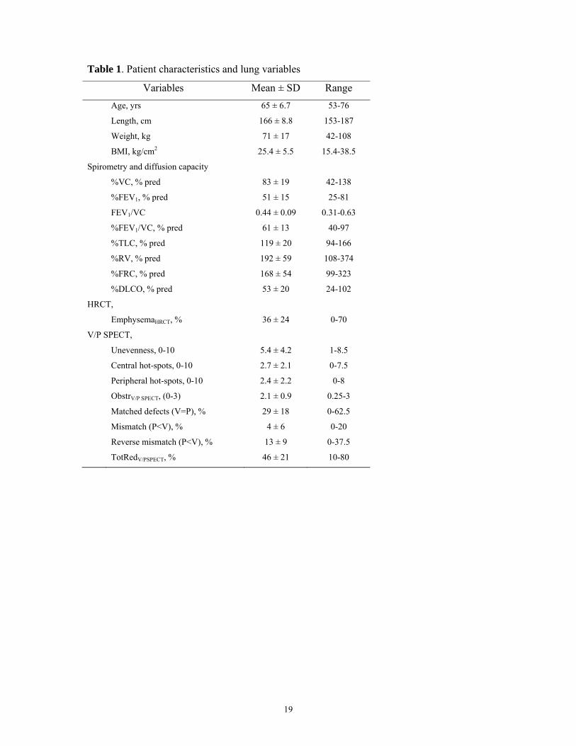
The distribution of MRC and CCQ scores is shown in Figure 1 and Table 1. Moderate
correlation was seen between the MRC dyspnea scale and the CCQ (r=0.62, P<0.001).
Otherwise, no correlations were found between the two clinical questionnaires and
spirometric parameters, V/P SPECT values or EmphysemaHRCT (Table 2).
HRCT
In 27 out of 28 patients, HRCT showed signs of emphysema with a mean extent of 36% of the
lung volume. Correlations between EmphysemaHRCT and other study parameters are shown in
Table 2. With increasing EmphysemaHRCT, a moderate correlating decrease in FEV1/VC (r=-
0.56, P=0.002) (Fig.2D), %FEV1/VC (r=-0.47, P=0.012) and %DLCO (r=-0.42, P=0.028)
was seen. A moderate correlating increase in %TLC was observed (r=0.50, P=0.007). No
significant correlation with %FEV1 was found.
V/P SPECT
Among V/P SPECT variables, strong to very strong correlation (r=0.79-0.99, P<0.0001) was
seen between unevenness of Technegas distribution (as seen in Fig.3), peripheral hot-spots (as
seen in Fig.4), TotRedV/PSPECT, matched defects and ObstrV/PSPECT (as seen in Fig.5). To avoid co-
linearity problems, further correlation analysis was restricted to TotRedV/PSPECT and ObstrV/PSPECT
(Table 2).
With increasing TotRedV/PSPECT, there was a correlating decrease in %FEV1 (r=-0.62,
P=0.0003), absolute FEV1/VC ratio (r=-0.74, P<0.0001) (Fig.2A, C) and %FEV1/VC (r=-
0.70, P<0.0001). A moderate to strong positive correlation was seen with EmphysemaHRCT
(r=0.69, P<0.0001) (Fig.2E, 6) as well as a weak to moderate correlating rise in %TLC and
%RV.
The higher the ObstrV/PSPECT, there was a correlating decrease in %FEV1 (r=-0.64, P =0.0001),
absolute FEV1/VC ratio (r=-0.71, P <0.0001) (Figure 2B) and %FEV1/VC (r=-0.67, P
<0.0001). There was a significant correlation between EmphysemaHRCT and ObstrV/PSPECT
(r=0.66, P=0.0001)(Fig.4-6).
ObstrV/PSPECT was in median 2.25 (range, 0.25-3) in the COPD patients compared to 0 (range,
0-1.25) in the 28 patients without any known obstructive disease (P<0.0001). Only 3 out of
the 28 patients without any previously known obstructive disease were scintigraphically
categorized as having mild obstructive disease.
9
Minor mismatched perfusion defects (≤ 20%) were found in 10 of the 30 patients. In 3 of
these cases, both physicians regarded the findings as consistent with PE, as they were of
segmental character.
In 4 patients, a pattern with redistribution of perfusion to nondependent lung zones, indicating
CHF was seen [19]. Three of these patients had a history of either multiple myocardial
infarctions with moderately decreased left ventricular function; known episodes of CHF; or
chronic atrial fibrillation.
10
DISCUSSION
In patients with COPD, reduction of total lung function (TotRedV/PSPECT) and grade of
obstructive disease (ObstrV/PSPECT), correlated well with both spirometric lung function and
emphysema extent (EmphysemaHRCT). The correlation between EmphysemaHRCT and
spirometric lung function was weaker.
The MRC dyspnea and the CCQ scales showed no significant correlations either to
spirometry, V/P SPECT or emphysema extent. MRC and CCQ can be used to predict
morbidity and mortality when applied to populations, but already the developers of these
scales found that the correlation to objective lung function tests, such as FEV1, was weak or
absent [11, 13]. This is also seen within this sample of COPD patients. The degree of
functional impairment, dyspnea and symptoms apparently do not relate to absolute measures
of lung function, but probably simply indicate the individual’s subjective experience of
inability with own habitual level as reference. The discrepancy between subjective symptoms
and lung function impairment is also one of the reasons why COPD diagnosis often is
delayed.
Increased resistance of small conducting airways and the emphysematous parenchyma
destruction seen in COPD affect both regional ventilation and perfusion. As seen in this study
and others, these V/P changes can be observed using lung scintigraphy (Fig3, 4, 5, 6). This
has previously been regarded mostly as a problem, obstructing interpretation of lung
scintigraphy with regards to PE [29]. This attitude is undergoing a shift since the introduction
of the 3-dimensional V/P SPECT technique [17]. V/P SPECT offers great advantages over
planar imaging [17, 20-24, 30, 31]. It has not only improved the diagnostic accuracy for PE
but may also facilitate diagnosis of other cardiopulmonary diseases, like COPD and CHF [19,
21, 25]. Despite this, there are few systematic studies of how ventilation and perfusion
patterns correlate to different phenotypes of COPD. It is clear that the potential of V/P
SPECT, to improve our understanding of the pathophysiology of COPD, needs to be explored
more fully [16]. This study is the first, to our knowledge, that systematically compares V/P
SPECT with clinical symptoms, functional state, extent of emphysema and the degree of
spirometric airway obstruction, as well as using V/P SPECT to identify comorbid disease.
COPD is today regarded as a treatable and preventable disease [1, 2]. Early detection is
essential as it encourages people to give up smoking, which is the most effective way to
prevent or delay airflow limitation in COPD [32]. FEV1 is a rather insensitive method in
11
detecting airway changes in COPD as these mainly occur in small airways. Ventilation lung
scintigraphy is a more sensitive indicator of obstructive lung disease than spirometric flow
rates and lung volumes [33]. In previous studies, V/P SPECT has also shown to be more
sensitive than HRCT in identifying small airway disease and emphysema [34, 35]. In the
present study, this was confirmed and reduced lung function, as a sign of small airway
disease, was identified with V/P SPECT even in regions with normal appearance on HRCT
(Fig.2E, Fig.3). The effective radiation dose from V/P SPECT is also considerably lower than
the radiation dose from HRCT [30].
Different V/P abnormalities observed with lung scintigraphy in COPD and emphysema have
been described in the literature, including general unevenness of radio-pharmaceutical
distribution, central and peripheral hot-spots, mismatch, reverse mismatch and matched
defects [33, 36]. These patterns are often simultaneously present in COPD, and the finding of
high to very high correlations between them in our study is therefore not surprising. Further
studies of the functional implications of these different V/P patterns may well increase our
understanding of different phenotypes of COPD.
Central hot-spots have been associated with increased resistance in major and intermediate
airways, disturbing the conductive flow and thus leading to impaction of radio-aerosol
particles (Fig.5) [36]. This is seen in asthma and in COPD. In severe cases, the presence of
central hot-spots may make it difficult to evaluate peripheral parts of the lung. The use of
Technegas has nearly eliminated this problem [25].
Peripheral hot-spot formation is seen in uneven ventilation and is associated with small
airway disease with obstruction of peripheral airways (Fig.4) [36]. In severe COPD, well
functioning regions may sometimes appear as relative “hot-spots” (Fig.5), but actually
represent areas with preserved and matched ventilation and perfusion.
Matched reduction of both ventilation and perfusion can be seen with parenchyma destruction
in emphysema (Fig.6), but can also be due to hypoxic vasoconstriction and diversion of blood
flow from poorly ventilated areas. In our study, the extent of matched defects increased with
increasing degree of emphysema. In this way both ventilation and perfusion is reduced and
the V/P imbalance is somewhat buffered. This could be one explanation as to why analysis
focusing on V/P ratio dispersion has not been able to separate different degrees of COPD
[37].
We found that reverse mismatch (V<P) already can be present in mild COPD (Fig.3). It can
be a sign of airway disease with incomplete hypoxic vasoconstriction that could lead to
12
shunting and hypoxemia (Fig.6) [38]. Reverse mismatch might also be seen in pneumonia
[18].
Segmental mismatch (P<V) is the fundamental criterion for PE diagnosis [17]. Because PE is
common among COPD patients, it is important to diagnose [6]. Three of the COPD patients
in our study had findings consistent with PE and another seven COPD patients had
mismatched defects of non-segmental character. The findings of mismatch in small areas
could represent PE but may also be caused by local vascular remodelling or vascular
obliteration seen in COPD.
CHF is another common comorbidity in COPD patients that is important to diagnose [5]. In
patients with COPD, the prevalence of CHF has been reported to be ≥20%, but the diagnosis
is often missed due to overlapping symptoms [39]. Four COPD patients (13%) showed
scintigraphic pattern of CHF, which has been found to have a high positive predictive value
[19]. According to patient records, three out of 4 of these patients had either CHF or other
known heart disease.
The sensitivity of V/P SPECT, as an indicator of airway obstruction, in combination with the
moderate to strong correlations of TotRedV/PSPECT to spirometry and EmphysemaHRCT indicate a
role for functional V/P SPECT imaging, as a bridge between spirometry and morphology in
COPD. The grading of obstructive disease (ObstrV/PSPECT) used in this study strongly correlates
to FEV1/VC and shows a substantial correlation to both %FEV1 and EmphysemaHRCT. In the
COPD patients, where reduction of lung function is primarily caused by obstructive changes,
ObstrV/PSPECT is very strongly correlated to TotRedV/PSPECT. However, the correlation between
TotRedV/PSPECT and ObstrV/PSPECT in the patients without known obstructive disease is
considerably lower (r=0.39, P=0.04). In these patients the reduced lung function, if present, is
primarily caused by other conditions. As expected, there was also a significant difference in
ObstrV/PSPECT between patients with COPD and those without obstructive disease. Worth
mentioning is that only 3 of the 28 patients without known obstructive disease were classified
as having mild obstructive disease by the blinded reviewers.
V/P SPECT has the unique possibility to image functional changes in regional ventilation and
perfusion and to quantify the proportion of functional loss that is caused by matched,
mismatched or reversed mismatched defects, respectively. This knowledge can be used to
characterize different phenotypes of COPD, which is important, as the degree of airway
13
obstruction and emphysema varies among patients. In this study, we also show that V/P
SPECT gives the possibility not only to diagnose COPD but also to grade its severity.
Furthermore, we show that V/P SPECT can be used to identify comorbid disease even in the
presence of severe COPD.
Although larger studies are needed, our findings indicate the possibility to implement V/P
SPECT in the diagnosis, staging and classification of COPD.
Conclusion: V/P SPECT is a sensitive method to detect early changes in COPD. V/P SPECT
also has the possibility to identify comorbid disease. Functional V/P SPECT findings show
moderate to strong correlations to morphological emphysema extent and spirometric lung
function. We therefore recommend that scintigraphic signs of COPD, whenever found, should
be reported. V/P SPECT can also be used to categorize the severity of functional changes in
COPD as mild, moderate or severe.
14
ACKNOWLEDGEMENTS
None of the authors have any financial disclosures or other conflicts of interest to reveal.
This study was partially financed by the Region of Scania (ALF).
The Clinical COPD questionnaire (CCQ) was used with kind permission from the author [13].
15
REFERENCES
1. Celli BR, MacNee W. Standards for the diagnosis and treatment of patients with COPD: a summary of the ATS/ERS position paper. Eur Respir J. 2004;23:932-46. 2. Rabe KF, Hurd S, Anzueto A, Barnes PJ, Buist SA, Calverley P, et al. Global strategy for the diagnosis, management, and prevention of chronic obstructive pulmonary disease: GOLD executive summary. Am J Respir Crit Care Med. 2007;176:532-55. 3. Mannino DM. COPD: epidemiology, prevalence, morbidity and mortality, and disease heterogeneity. Chest. 2002;121:121S-6S. 4. Hogg JC, Chu F, Utokaparch S, Woods R, Elliott WM, Buzatu L, et al. The nature of small-airway obstruction in chronic obstructive pulmonary disease. N Engl J Med. 2004;350:2645-53. doi:10.1056/NEJMoa032158 350/26/2645 [pii]. 5. Holguin F, Folch E, Redd SC, Mannino DM. Comorbidity and mortality in COPD-related hospitalizations in the United States, 1979 to 2001. Chest. 2005;128:2005-11. doi:128/4/2005 [pii] 10.1378/chest.128.4.2005. 6. Stein PD, Beemath A, Meyers FA, Olson RE. Pulmonary embolism and deep venous thrombosis in hospitalized adults with chronic obstructive pulmonary disease. J Cardiovasc Med (Hagerstown). 2007;8:253-7. 7. Voelkel NF, Cool CD. Pulmonary vascular involvement in chronic obstructive pulmonary disease. Eur Respir J Suppl. 2003;46:28s-32s. 8. Jones PW, Agusti AG. Outcomes and markers in the assessment of chronic obstructive pulmonary disease. Eur Respir J. 2006;27:822-32. 9. Cerveri I, Dore R, Corsico A, Zoia MC, Pellegrino R, Brusasco V, et al. Assessment of emphysema in COPD: a functional and radiologic study. Chest. 2004;125:1714-8. 10. Gelb AF, Hogg JC, Muller NL, Schein MJ, Kuei J, Tashkin DP, et al. Contribution of emphysema and small airways in COPD. Chest. 1996;109:353-9. 11. Bestall JC, Paul EA, Garrod R, Garnham R, Jones PW, Wedzicha JA. Usefulness of the Medical Research Council (MRC) dyspnoea scale as a measure of disability in patients with chronic obstructive pulmonary disease. Thorax. 1999;54:581-6. 12. Fletcher CM, Elmes PC, Fairbairn A, Wood CH. The significance of respiratory symptoms and the diagnosis of chronic bronchitis in a working population. British Medical Journal. 1959;2:257–66. 13. van der Molen T, Willemse BW, Schokker S, ten Hacken NH, Postma DS, Juniper EF. Development, validity and responsiveness of the Clinical COPD Questionnaire. Health Qual Life Outcomes. 2003;1:13. 14. Nishimura K, Izumi T, Tsukino M, Oga T. Dyspnea is a better predictor of 5-year survival than airway obstruction in patients with COPD. Chest. 2002;121:1434-40. 15. Wolkove N, Dajczman E, Colacone A, Kreisman H. The relationship between pulmonary function and dyspnea in obstructive lung disease. Chest. 1989;96:1247-51. 16. Mannino DM, Watt G, Hole D, Gillis C, Hart C, McConnachie A, et al. The natural history of chronic obstructive pulmonary disease. Eur Respir J. 2006;27:627-43. doi:27/3/627 [pii] 10.1183/09031936.06.00024605. 17. Bajc M, Neilly JB, Miniati M, Schuemichen C, Meignan M, Jonson B. EANM guidelines for ventilation/perfusion scintigraphy : Part 1. Pulmonary imaging with ventilation/perfusion single photon emission tomography. Eur J Nucl Med Mol Imaging. 2009;36:1356-70.
16
18. Freeman LM, Krynyckyi B, Zuckier LS. Enhanced lung scan diagnosis of pulmonary embolism with the use of ancillary scintigraphic findings and clinical correlation. Semin Nucl Med. 2001;31:143-57. 19. Jögi J, Palmer J, Jonson B, Bajc M. Heart failure diagnostics based on ventilation/perfusion single photon emission computed tomography pattern and quantitative perfusion gradients. Nucl Med Commun. 2008;29:666-73. 20. Bajc M, Bitzen U, Olsson B, Perez de Sa V, Palmer J, Jonson B. Lung ventilation/perfusion SPECT in the artificially embolized pig. J Nucl Med. 2002;43:640-7. 21. Bajc M, Olsson B, Palmer J, Jonson B. Ventilation/Perfusion SPECT for diagnostics of pulmonary embolism in clinical practice. Journal of internal medicine. 2008;264:379-87. 22. Bajc M, Olsson CG, Olsson B, Palmer J, Jonson B. Diagnostic evaluation of planar and tomographic ventilation/perfusion lung images in patients with suspected pulmonary emboli. Clin Physiol Funct Imaging. 2004;24:249-56. 23. Gutte H, Mortensen J, Jensen CV, von der Recke P, Petersen CL, Kristoffersen US, et al. Comparison of V/Q SPECT and planar V/Q lung scintigraphy in diagnosing acute pulmonary embolism. Nucl Med Commun. 2010;31:82-6. doi:10.1097/MNM.0b013e3283336747. 24. Reinartz P, Wildberger JE, Schaefer W, Nowak B, Mahnken AH, Buell U. Tomographic imaging in the diagnosis of pulmonary embolism: a comparison between V/Q lung scintigraphy in SPECT technique and multislice spiral CT. J Nucl Med. 2004;45:1501-8. 25. Jögi J, Jonson B, Ekberg M, Bajc M. Ventilation-perfusion SPECT with 99mTc-DTPA versus Technegas: a head-to-head study in obstructive and nonobstructive disease. J Nucl Med. 2010;51:735-41. doi:jnumed.109.073957 [pii] 10.2967/jnumed.109.073957. 26. Standardization of Spirometry, 1994 Update. American Thoracic Society. Am J Respir Crit Care Med. 1995;152:1107-36. 27. Palmer J, Bitzen U, Jonson B, Bajc M. Comprehensive ventilation/perfusion SPECT. J Nucl Med. 2001;42:1288-94. 28. Olsson CG, Bitzen U, Olsson B, Magnusson P, Carlsson MS, Jonson B, et al. Outpatient tinzaparin therapy in pulmonary embolism quantified with ventilation/perfusion scintigraphy. Med Sci Monit. 2006;12:PI9-13. 29. Value of the ventilation/perfusion scan in acute pulmonary embolism. Results of the prospective investigation of pulmonary embolism diagnosis (PIOPED). The PIOPED Investigators. Jama. 1990;263:2753-9. 30. Bajc M, Neilly JB, Miniati M, Schuemichen C, Meignan M, Jonson B. EANM guidelines for ventilation/perfusion scintigraphy : Part 2. Algorithms and clinical considerations for diagnosis of pulmonary emboli with V/P(SPECT) and MDCT. Eur J Nucl Med Mol Imaging. 2009;36:1528-38. 31. Leblanc M, Leveillee F, Turcotte E. Prospective evaluation of the negative predictive value of V/Q SPECT using 99mTc-Technegas. Nucl Med Commun. 2007;28:667-72. 32. Sundblad BM, Larsson K, Nathell L. Lung function testing influences the attitude toward smoking cessation. Nicotine Tob Res. 2010;12:37-42. doi:ntp170 [pii] 10.1093/ntr/ntp170. 33. Taplin GV, Tashkin DP, Chopra SK, Anselmi OE, Elam D, Calvarese B, et al. Early detection of chronic obstructive pulmonary disease using radionuclide lung-imaging procedures. Chest. 1977;71:567-75.
17
34. Satoh K, Nakano S, Tanabe M, Nishiyama Y, Takahashi K, Kobayashi T, et al. A clinical comparison between Technegas SPECT, CT, and pulmonary function tests in patients with emphysema. Radiat Med. 1997;15:277-82. 35. Yokoe K, Satoh K, Yamamoto Y, Nishiyama Y, Asakura H, Haba R, et al. Usefulness of 99mTc-Technegas and 133Xe dynamic SPECT in ventilatory impairment. Nucl Med Commun. 2006;27:887-92. 36. Santolicandro A, Ruschi S, Fornai E, Giuntini C. Imaging of ventilation in chronic obstructive pulmonary disease. J Thorac Imaging. 1986;1:36-53. 37. Rodriguez-Roisin R, Drakulovic M, Rodriguez DA, Roca J, Barbera JA, Wagner PD. Ventilation-perfusion imbalance and chronic obstructive pulmonary disease staging severity. J Appl Physiol. 2009;106:1902-8. doi:00085.2009 [pii] 10.1152/japplphysiol.00085.2009. 38. Palmaz JC, Barnett CA, Reich SB, Krumpe PE, Farrer PA. Reverse ventilation--perfusion mismatch. Clin Nucl Med. 1984;9:6-9. 39. Rutten FH, Cramer MJ, Lammers JW, Grobbee DE, Hoes AW. Heart failure and chronic obstructive pulmonary disease: An ignored combination? Eur J Heart Fail. 2006;8:706-11. doi:S1388-9842(06)00011-0 [pii] 10.1016/j.ejheart.2006.01.010.
18
Figure Legends
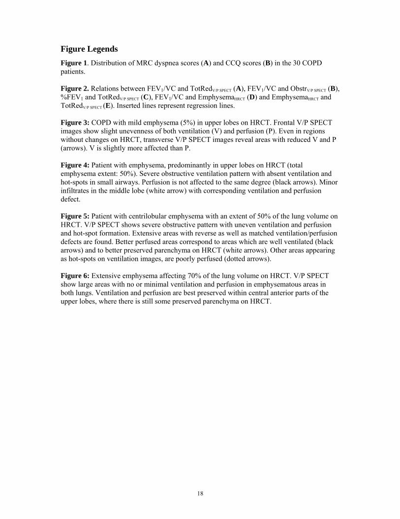
Figure 1. Distribution of MRC dyspnea scores (A) and CCQ scores (B) in the 30 COPD patients. Figure 2. Relations between FEV1/VC and TotRedV/P SPECT (A), FEV1/VC and ObstrV/P SPECT (B), %FEV1 and TotRedV/P SPECT (C), FEV1/VC and EmphysemaHRCT (D) and EmphysemaHRCT and TotRedV/P SPECT (E). Inserted lines represent regression lines. Figure 3: COPD with mild emphysema (5%) in upper lobes on HRCT. Frontal V/P SPECT images show slight unevenness of both ventilation (V) and perfusion (P). Even in regions without changes on HRCT, transverse V/P SPECT images reveal areas with reduced V and P (arrows). V is slightly more affected than P. Figure 4: Patient with emphysema, predominantly in upper lobes on HRCT (total emphysema extent: 50%). Severe obstructive ventilation pattern with absent ventilation and hot-spots in small airways. Perfusion is not affected to the same degree (black arrows). Minor infiltrates in the middle lobe (white arrow) with corresponding ventilation and perfusion defect. Figure 5: Patient with centrilobular emphysema with an extent of 50% of the lung volume on HRCT. V/P SPECT shows severe obstructive pattern with uneven ventilation and perfusion and hot-spot formation. Extensive areas with reverse as well as matched ventilation/perfusion defects are found. Better perfused areas correspond to areas which are well ventilated (black arrows) and to better preserved parenchyma on HRCT (white arrows). Other areas appearing as hot-spots on ventilation images, are poorly perfused (dotted arrows). Figure 6: Extensive emphysema affecting 70% of the lung volume on HRCT. V/P SPECT show large areas with no or minimal ventilation and perfusion in emphysematous areas in both lungs. Ventilation and perfusion are best preserved within central anterior parts of the upper lobes, where there is still some preserved parenchyma on HRCT.
19
Table 1. Patient characteristics and lung variables
Variables Mean ± SD Range
Age, yrs 65 ± 6.7 53-76
Length, cm 166 ± 8.8 153-187
Weight, kg 71 ± 17 42-108
BMI, kg/cm2 25.4 ± 5.5 15.4-38.5
Spirometry and diffusion capacity
%VC, % pred 83 ± 19 42-138
%FEV1, % pred 51 ± 15 25-81
FEV1/VC 0.44 ± 0.09 0.31-0.63
%FEV1/VC, % pred 61 ± 13 40-97
%TLC, % pred 119 ± 20 94-166
%RV, % pred 192 ± 59 108-374
%FRC, % pred 168 ± 54 99-323
%DLCO, % pred 53 ± 20 24-102
HRCT,
EmphysemaHRCT, % 36 ± 24 0-70
V/P SPECT,
Unevenness, 0-10 5.4 ± 4.2 1-8.5
Central hot-spots, 0-10 2.7 ± 2.1 0-7.5
Peripheral hot-spots, 0-10 2.4 ± 2.2 0-8
ObstrV/P SPECT, (0-3) 2.1 ± 0.9 0.25-3
Matched defects (V=P), % 29 ± 18 0-62.5
Mismatch (P<V), % 4 ± 6 0-20
Reverse mismatch (P<V), % 13 ± 9 0-37.5
TotRedV/PSPECT, % 46 ± 21 10-80
20
Table 2. Spearman correlation matrix between spirometry, symptoma questionnaires,
emphysema extent, V/P SPECT assessed reduction in lung function and obstructive disease