Eur ReaplrJ 1991,4,141-146 Lung CT density correlates with measurements of airflow limitation and the diffusing capacity G.A. Gould, AT. Redpath, M. Ryan, P.M. Warren, J.J.K. Best, D.C. Flenley, W. MacNee Lung CT density correlates with measurements of airflow limitation and the diffusing capacity. G.A. Gould, A.T. Redpath, M. Ryan, P.M. Warren, J.J.K. Best, D.C. Flenley, W. MacNee. ABSTRACT: We studied 80 subjects (63 M, 17 F; 23-82 yrs) and related lung computerized tomography (CT) density with age, height, spirom- etry, lung volumes, diffusing capacity and arterial blood gas tensions. These subjects demonstrated a wide range of physiological impairment (forced expiratory volume in one second (FEV 1 ) 8-116% predicted; dif- fusing capacity (Kco) 15-139% predicted; arterial oxygen tension (Pao 1 ) 38-91 mmHg). They ranged from normal subjects to patients with chrome respiratory failure. Lung density was derived from CT density histograms measured as both mean Emergency Medical Information (EMI) number (EMI scale: O=water, -500=air, EMI number of normal lung tissue range approximately -200 to -450) and the lowest 5th percentile EMI number, the latter value being more likely to represent the density of lung parenchyma. Lung CT density correlated most strongly with airflow obstruction (EMI 5th percentile versus FEY/forced vital capacity (FVC) % predicted, r=0.73, p<0.001) and diffusing capacity (EMI 5th percentile versus Kco, r=0.77, p<O.OOl). This suggests that reduction in lung density, which reflects loss of the surface area of the distal airspaces, is a major index of respiratory function in patients with smoking related chronic obstructive pulmonary disease (COPD). These data provide no indication of other factors such as small and large airways disease, and loss of elastic recoil, which may contribute to airflow limitation, or disruption of the pulmonary vascular bed which may also affect CT lung density. Eur Respir J., 1991, 4, 141-146. Dept of Respiratory Medicine, City Hospital, Edinburgh, Scotland. Correspondence: Dr W. MacNee, Dept of Respiratory Medicine, Rayne Laboratory, City Hospital, Greenbank Drive, Edinburgh EH10 5SB, Scotland, UK. Keywords: Airflow limitation; diffusing capacity; distal airspace size; emphysema; lung computerized tomography density. Received: October 3, 1989; accepted after revision May 8, 1990. This study was supported by The Norman Salvesen Emphysema Research Trust. Pulmonary emphysema, which is defined in pathologi- cal terms as a permanent abnormal increase in the size of distal airspaces with destruction of their walls [1] is a common condition, for which there is no preventative treatment. Detection of the disease during its early stages is difficult, if not impossible, using currently available methods. The results of the recent National Institute of Health intermittent positive pressure breathing (NIH IPPB) trial [2] showed poor correlations between clinical features and the severity of macroscopic emphysema observed at autopsy. Moreover, the large autopsy series of THURLBECK and SrMoN [3] demonstrated the inadequacy of conventional radiology in detecting even moderate degrees of emphysema. which, it must be stressed, was independent of the pres- ence or absence of macroscopic emphysema. This finding suggests that in emphysema a generalized increase in airspace size occurs with a concomitant fall in the alveolar surface area which is available for gas transfer. Numerous studies have attempted to identify physiological variables that could reliably detect and quantify emphysema, but the results of these studies have been disappointing [ 4-11]. However, the single measurement that most consistently relates to the sever- ity of emphysema is the diffusing capacity for carbon monoxide (DLco) [4-8, 10, 12]. In a previous study [13] we demonstrated a strong linear relationship between impairment of the DLco and the size of distal airspaces Visual assessment of computed tomographic (CT) images of the lungs, either subjectively, or using semi-quantitative numerical scoring systems can differentiate normal lungs from those with visible macroscopic emphysema [14-18]. Since each CT scan contains a large amount of numerical data, we have used these data to provide an objective measurement of lung density. We have previously demonstrated a strong correlation between lung CT density and morphometric measurements of the severity of emphysema, as measured by the alveolar wall per unit lung volume [13]. The purpose of this study was to use lung CT density, which we have shown to correlate with the size of distal airspaces, a defining characteristic of emphysema, and determine its relationship to measurements of respiratory function. The use of CT scanning enabled us to study a large number of both normal subjects and patients with
Transcript
Eur ReaplrJ 1991,4,141-146
Lung CT density correlates with measurements of airflow limitation and the diffusing capacity
G.A. Gould, AT. Redpath, M. Ryan, P.M. Warren, J.J.K. Best, D.C. Flenley, W. MacNee
Lung CT density correlates with measurements of airflow limitation and the diffusing capacity. G.A. Gould, A.T. Redpath, M. Ryan, P.M. Warren, J.J.K. Best, D.C. Flenley, W. MacNee. ABSTRACT: We studied 80 subjects (63 M, 17 F; 23-82 yrs) and related lung computerized tomography (CT) density with age, height, spirometry, lung volumes, diffusing capacity and arterial blood gas tensions. These subjects demonstrated a wide range of physiological impairment (forced expiratory volume in one second (FEV
38-91 mmHg). They ranged from normal subjects to patients with chrome respiratory failure. Lung density was derived from CT density histograms measured as both mean Emergency Medical Information (EMI) number (EMI scale: O=water, -500=air, EMI number of normal lung tissue range approximately -200 to -450) and the lowest 5th percentile EMI number, the latter value being more likely to represent the density of lung parenchyma. Lung CT density correlated most strongly with airflow obstruction (EMI 5th percentile versus FEY/forced vital capacity (FVC) % predicted, r=0.73, p<0.001) and diffusing capacity (EMI 5th percentile versus Kco, r=0.77, p<O.OOl). This suggests that reduction in lung density, which reflects loss of the surface area of the distal airspaces, is a major index of respiratory function in patients with smoking related chronic obstructive pulmonary disease (COPD). These data provide no indication of other factors such as small and large airways disease, and loss of elastic recoil, which may contribute to airflow limitation, or disruption of the pulmonary vascular bed which may also affect CT lung density. Eur Respir J., 1991, 4, 141-146.
Dept of Respiratory Medicine, City Hospital, Edinburgh, Scotland.
Correspondence: Dr W. MacNee, Dept of Respiratory Medicine, Rayne Laboratory, City Hospital, Greenbank Drive, Edinburgh EH10 5SB, Scotland, UK.
Received: October 3, 1989; accepted after revision May 8, 1990.
This study was supported by The Norman Salvesen Emphysema Research Trust.
Pulmonary emphysema, which is defined in pathological terms as a permanent abnormal increase in the size of distal airspaces with destruction of their walls [1] is a common condition, for which there is no preventative treatment. Detection of the disease during its early stages is difficult, if not impossible, using currently available methods. The results of the recent National Institute of Health intermittent positive pressure breathing (NIH IPPB) trial [2] showed poor correlations between clinical features and the severity of macroscopic emphysema observed at autopsy. Moreover, the large autopsy series of THURLBECK and SrMoN [3] demonstrated the inadequacy of conventional radiology in detecting even moderate degrees of emphysema.
which, it must be stressed, was independent of the presence or absence of macroscopic emphysema. This finding suggests that in emphysema a generalized increase in airspace size occurs with a concomitant fall in the alveolar surface area which is available for gas transfer.
Numerous studies have attempted to identify physiological variables that could reliably detect and quantify emphysema, but the results of these studies have been disappointing [ 4-11]. However, the single measurement that most consistently relates to the severity of emphysema is the diffusing capacity for carbon monoxide (DLco) [4-8, 10, 12]. In a previous study [13] we demonstrated a strong linear relationship between impairment of the DLco and the size of distal airspaces
Visual assessment of computed tomographic (CT) images of the lungs, either subjectively, or using semi-quantitative numerical scoring systems can differentiate normal lungs from those with visible macroscopic emphysema [14-18]. Since each CT scan contains a large amount of numerical data, we have used these data to provide an objective measurement of lung density. We have previously demonstrated a strong correlation between lung CT density and morphometric measurements of the severity of emphysema, as measured by the alveolar wall per unit lung volume [13].
The purpose of this study was to use lung CT density, which we have shown to correlate with the size of distal airspaces, a defining characteristic of emphysema, and determine its relationship to measurements of respiratory function. The use of CT scanning enabled us to study a large number of both normal subjects and patients with
142 G.A. GOULD ET AL.
COPD with a wide range of disability. By substituting er scan lung density for pathological measurements of emphysema, we therefore avoided the selection inherent in studies of patients undergoing lung resection.
Patients and methods
We studied 80 subjects (63 M, 17 F; 5 nonsmokers) who had no evidence of emphysematous bullae on the plain chest radiograph or er scan. All patients were clinically stable and had been free of infection for at least 6 wks prior to the study. All subjects gave their informed consent and the study was approved by our local Ethical Committee.
The physiological assessment was carried out 1-21 days before the CT scan. Clinical features and smoking history were recorded using the British Medical Research Council (MRC) questionnaire. Spirometry forced expiratory volume in one second as percentage of forced vital capacity (FEV/FVC) was measured using a 7 l dry spirometer (Vitalograph Ltd, UK) and lung volumes (total lung capacity (TLC), residual volume (RV)) by a body plethysmograph (Pulmorex, Fenyves and Gut, Basel, Switzerland). Single-breath diffusing capacity was measured using the method of 0GILVIE et al. [19] (Automatic Gas Transfer Test, Model A System, PK Morgan, UK), with breath-holding time calculated by a modified JoNES and MEADE technique [20]. Arterial blood gas tensions were measured when breathing air (613 blood gas analyser, Instrumentation Laboratories) and exercise capacity was assessed by a 12 min corridor walk. Normal predicted values for men were taken from CRAro and eo-workers [21] for FEV1 and FVC; CRAPo and eo-workers (22] for TLC and RV; CoTES [23] for diffusing capacity of the lung for carbon monoxide (DLco) and CoTES and HALL [24] for carbon monoxide transfer coefficient (Kco ). In women normal predicted values were taken from HALL et al. [25] for FEV
1, VC,
TLC and RV, BILLIET et al. [26] for DLco and CoTES and HALL [24] for Kco.
CT scanning
An Emergency Medical Information (EMI) er 5005 scanner with a 20 s scan time and a slice thickness of 13 mm was used. Patients were scanned during arrested deep inspiration close to total lung capacity. This scanner is a rotate-translate device. The 30 degree X-ray beam is seen by all detectors, and air and notional water reference readings are taken on each traverse. The system was calibrated daily with a water phantom, and in practice any scans containing artefacts were excluded from the analysis. The er density of tissue is expressed in EMI units (where water=O and air=-500). er density measured by the EMI CT 5005 scanner has a direct linear relationship to actual physical density between the range 0.001 (air) to 1.00 gm·cm·3 (water) [27, 28]. Thus, for lung tissue, which lies within the linear part of this relationship, this scanner provides a linear measurement
of physical density. Although this model of er scanner is no longer a "state-of-the-art" device, such early scanners were designed as densitometers and provide more accurate density measurements than some of the newer devices, where image quality is a more important property.
A 10.0
9.0
8.0
1 7.0
1 8.0
l f 5.0
:I I 4.0
J a. 3.0
2.0
1.0
EMI5th percentile -418
0 .___,~.....,......,
-1500 -480 -420 -380 -340 -300 -280 -220
EMI number
B
10.0
9.0
8.0
I 7.0
'ii 8.0
! l'; 5.0
! l 4.0
1 a. 3.0 EMI 5th percentile -479
2.0
1.0
0 -500 -480 -420 -380 -340 -300 -260 -220
EMI number
Fig. 1. - CT lung density histogram of (A) a subject with no emphysema, proven pathologically (case 14 from [13]). (B) a subject with gross emphysema proven pathologically (case S from [13]). EMI: Emergency Medical Information.
er DENSITY, AIRFLOW LIMITATION AND DIFFUSING CAPACITY 143
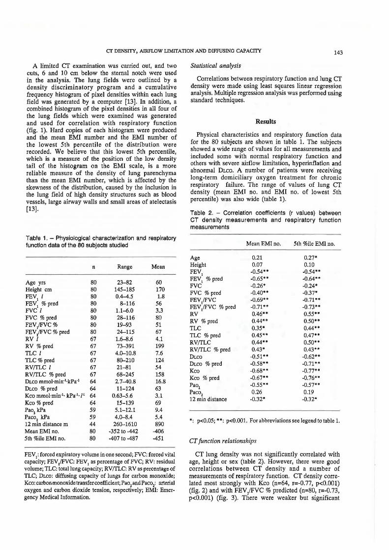
A limited er examination was carried out, and two cuts, 6 and 10 cm below the sterna} notch were used in the analysis. The lung fields were outlined by a density discriminatory program and a cumulative frequency histogram of pixel densities within each lung field was generated by a computer [13]. In addition, a combined histogram of the pixel densities in all four of the lung fields which were examined was generated and used for correlation with respiratory function (fig. 1). Hard copies of each histogram were produced and the mean EMI number and the EMI number of the lowest 5th percentile of the distribution were recorded. We believe that this lowest 5th percentile, which is a measure of the position of the low density tail of the histogram on the EMI scale, is a more reliable measure of the density of lung parenchyma than the mean EMI number, which is affected by the skewness of the distribution, caused by the inclusion in the lung field of high density structures such as blood vessels, large airway walls and small areas of atelectasis [13].
Table 1. - Physiological characterization and respiratory function data of the 80 subjects studied
n Range Mean
Age yrs 80 23-82 60 Height cm 80 145-185 170 FEV
1 l 80 0.4-4.5 1.8
FEV1
% pred 80 8-116 56 FVC l 80 1.1-6.0 3.3 FVC% pred 80 28-116 80 FEV/FVC% 80 19-93 51 FEV/FVC% pred 80 24-115 67 RV I 67 1.6-8.6 4.1 RV% pred 67 73-391 199 TLC I 67 4.0--10.8 7.6 TLC% pred 67 80--210 124 RV(TLC I 67 21-81 54 RV(I'LC % pred 67 68-245 158 DLco mmol·min·1·kPa·1 64 2.7-40.8 16.8 DLCO % pred 64 11-124 63 Kco mmol·min·1• kPa-1·1-1 64 0.63-5.6 3.1 Kco% pred 64 15-139 69 Pao
2 kPa 59 5.1-12.1 9.4
Paco2
kPa 59 4.0--8.4 5.4 12 min distance m 44 260--1610 890 MeanEMino. 80 -352 to -442 -406 5th %ile EMI no. 80 -407 to -487 -451
FEV1: forced expiratory volume in one second; FVC: forced vital
capacity; FEV/FVC: FEV1 as percentage of FVC; RV: residual volume; TLC: total lung capacity; RV(I'LC: RV as percentage of TLC; DLCo: diffusing capacity of lungs for carbon monoxide; Kco: carbon monoxide transfer coefficient; Pao
2 and Paco2: arterial
oxygen and carbon dioxide tension, respectively; EMI: Emer-gency Medical Information.
Statistical analysis
Correlations between respiratory function and lung er density were made using least squares linear regression analysis. Multiple regression analysis was performed using standard techniques.
Results
Physical characteristics and respiratory function data for the 80 subjects are shown in table 1. The subjects showed a wide range of values for all measurements and included some with normal respiratory function and others with severe airflow limitation, hyperinflation and abnormal DLco. A number of patients were receiving long-term domiciliary oxygen treatment for chronic respiratory failure. The range of values of lung CT density (mean EMI no. and EMI no. of lowest 5th percentile) was also wide (table 1).
Table 2. - Correlation coefficients (r values) between CT density measurements and respiratory function measurements
• : p<0.05; ••: p<O.OOl. For abbreviations see legend to table 1.
CT function relationships
er lung density was not significantly correlated with age, height or sex (table 2). However, there were good correlations between CT density and a number of measurements of respiratory function. er density correlated most strongly with Kco (n=64, r=-0.77, p<0.001) (fig. 2) and with FEV/FVC% predicted (n=80, r=-0.73, p<0.001) (fig. 3). There were weaker but significant
144 G.A. GOULD ET AL.
correlations with lung volumes (RV n=67, r=0.55, p<0.001), RVffLC (n=67, r=0.50, p<0.001); TLC (n=67, r=0.44, p<0.001), arterial oxygen tension (n=59, r=-0.57, p<0.001) and 12 min walking distance (n=44, r=-0.32, p<0.05).
1.8
1.6 ... ':- 1.4 ... I 11 a. 1.2 ¥ ... I c 1.0 ~ 0 0.8 E E 0 0.6 ~
Fig. 2. - Relationship between volume corrected diffusing capacity and lung er density in 64 subjects. Kco: carbon monoxide transfer coefficient; EMI: Emergency Medical Information.
-420 -440 -480 -480 EMI no. of loweat 5th percentile
In CT IC&n
Fig. 3. - Relationship between airways obstruction and lung er density in 80 subjects. FEV.tFVC: forced expiratory volume in one second as a percentage of forced vital capacity; EMI: Emergency Medical Information.
Discussion
A technique which can accurately quantify emphysema at an early stage could be used to monitor the progression of the disease and may increase our understanding of the pathophysiology of this condition. It would also allow us to assess the effects of interventions, such as the cessation of smoking, or more recent innovations, such as the effects of administration of a 1-antitrypsin replacement therapy in deficient patients [29].
Emphysematous bullae are easily located by CT (even those not visible on plain chest radiograph) but in addition, by use of the appropriate density window settings, it is possible to obtain a subjective estimate of the severity of emphysema in the remaining non-bullous parts of the lungs. Several workers have used methods for visually scoring CT scans, to assess the severity of bullous emphysema [30-33]. GoooARD et al. [14) demonstrated a correlation between CT visual emphysema score (high vs low) and respiratory function (FEV
1,
FEV/FVC, DLco). BERGIN et al. [15), using a similar CT visual scoring system, found a good correlation between CT and the pathological severity of emphysema, using a form of picture grading to score for macroscopic emphysema, in resected surgical specimens. However, even with state-of-the art, high resolution short scan time scanners, mild emphysema can easily be missed [17, 18].
In an attempt to exclude the errors inherent in any subjective scoring technique, we have developed a method of utilizing the numerical data present in every CT scan to provide an objective measurement of lung density. Using this technique we have previously demonstrated, in a group of 28 surgical patients, that the severity of "microscopic emphysema" (measured morphometrically as the alveolar wall per unit lung volume, a measurement of distal airspace size) correlates well with lung density measured by CT [13]. In the present study, using the assumption that CT density relates to distal airspace size, we have examined the relationship between respiratory function measurements and distal airspace size.
In our group of 80 subjects, mainly smokers, who had a very wide range of age and respiratory function, we have demonstrated a strong correlation between lung density, airflow limitation and impairment of the diffusing capacity. Both measurements are widely thought to reflect the severity of emphysema. We have also observed weak correlations between lung CT density and measurements of hyperinflation and hypoxaemia. We also demonstrated that age and height have no obvious effect on lung density, an observation previously made by others in normal subjects [34, 35]. This is consistent with our earlier observations in a group of surgical patients [13), where we demonstrated no correlation between airspace size (measured directly) and either age or height.
Thus, our method of CT analysis provides a measure of overall lung density which correlates well with direct measurements of distal airspace size and physiological measurement [13), but it is obvious from the correlation coefficients observed (r values up to 0.77) (table 2) that other factors may also be important. The diffusing
er DENSITY, AIRFLOW LIMITATION AND DIFFUSING CAPACITY 145
capacity, for example, will be affected not only by alveolar surface area, but also by disturbances of the pulmonary circulation and small and large airways function, both of which may contribute to ventilation/perfusion inequalities. However, preliminary unpublished observations from our group, using contrast CT scanning, do not suggest that blood present in the peripheral lung makes a major contribution to CT lung density. Similarly, airways obstruction, though correlating strongly with CT measurement of emphysema will also be affected by airways disease and by alterations in the elastic recoil of the whole lung. It is likely that all of these factors (airways disease, loss of alveolar surface area, changes in elastic recoil, abnormalities of pulmonary circulation) are interrelated in a complex way, and only further detailed studies correlating these variables will increase our understanding of the exact sequence of events which result in the development of smoking related chronic obstructive pulmonary disease. Furthermore, the analysis of CT lung density in this study was based on only two CT lung slices. Density measurements derived from whole lung CT scanning may improve the correlations between CT lung density and respiratory function .
The concept of "macroscopic" and "microscopic" emphysema requires further discussion. Our method of measuring CT lung density takes into account both the visible "holes" of macroscopic emphysema and the generalized increase in airspace size that is also present in the intervening lung. Measurement of alveolar surface area, such as the mean linear intercept used by THURLBECK and eo-workers [2, 3, 8], or our own method of directly measuring airspace size by a morphometric technique [13], also have th is lim itation although bo th groups, have observed strong correlations between these measurements and the DLCo [8, 13]. Until we can accurately define the normal range of the size of distal airspaces in a population study, we cannot define an absolute CT density value which defines the earliest lesions of emphysema. Furthermore, without such a study, the distinction between enlargement of distal airspaces due to destruction of their walls and chronic hyperinflation remains to be defined.
Further studies using CT scanning may help to determine the natural history of emphysema, in particular to assess when in the course of the disease macroscopic emphysema appears and what its functional effects are. Preliminary data indicate that the technique is reproducible in normal subjects and that it can be used to follow the progression of emphysema in patients with chronic obstructive pulmonary disease (COPD) [36]. Since it is now possible to identify centriacinar lesions of emphysema using new generation er scanners (37, 38], and analysis of lung er den ity histograms can quantify the decrease in alveolar surface area in the intervening lung tissue, it seems likely that this objective can be obtained.
Our findings confirm that lung density, as assessed by er scanning, and hence distal airspace size, correlates well with airflow limitation and impairment of the diffusing capacity for carbon monoxide.
Acknowledgements: The authors would like to thank the physicians in Lothian Health Board who referred patients for er scanning, and C. Hendrie who typed the manuscript.
References
1. Snider GL, Kleinerman J, Tburlbcck WM, Bengali ZH.The defini tion of emphysema. Report of a National Heart Lung and B.lood Institute, Division of Lung Diseases Workshop. Am Rev Respir Dis, 1985, 132, 182-185. 2. Nagai A, West WW, Thurlbeck WM. - The National Institute of Health intermittent positive pressure breathing trial: pathology studies. II. Correlation between morphologic findings, clinical findings and evidence of expiratory airflow obstruction. Am Rev Respir Dis, 1985, 132, 946-953. 3. Thurlbeck WM, Simon G. - Radiographic appearance of the chest in emphysema. Am J Roentgenol, 1978, 130, 429-440. 4. Jenkins DE, Greenberg SD, Boushy SF, Schweppe HI, O'Neal RM. - Correlation of morphologic emphysema with pulmonary function parameters. Trans Assoc Physician, 1965, 72, 218-230. 5. Burrows B, Fletcher CM, Heard BE, Jones NL, Wootliff JS. - The emphysematous and bronchial types of chronic airways obstruction: a clinicopathological study of patients in London and Chicago. Lancet, 1966, i, 830-834. 6. Berend N, Woolcock AJ, Marlin GE. - Correlation between the structure and function of the lungs in smokers. Am Rev Respir Dis, 1979, 119, 695-705. 7. Pare PD, Brooks LA, Bates J et al. - Exponential analysis of the lung pressure-volume curve as a predictor of pulmonary emphysema. Am Rev Respir Dis, 1982, 126, 54-{il. 8. Thurlbeck WM, Henderson JA, Fraser RG, Bates DV. -Chronic obstructive lung disease. A comparison between clinical, roentgenologic, functional and morphological criteria in chronic bronchitis, asthma and bronchiectasis. Medicine, 1970, 49, 81-145. 9. Watanabe S, Mitchell M, Renzetti AD. - Correlation of structure and function in chronic pulmonary emphysema. Am Rev Respir Dis, 1965, 92, 221-227. 10. Park SS, Janis M, Shim CS, Williams MH. -Relationship of bronchitis and emphysema to altered pulmonary function. Am Rev Respir Dis, 1970, 102, 927-936. 11. Sweet HC, Wyatt JP, Kinsella PW.- Correlation of lung macrosections with pulmonary function in emphysema. Am J Med, 1960, 24, 277-281. 12. Symonds G, Renzetti AD, Mitchell MM. - The diffusing capacity in pulmonary emphysema. Am Rev Respir Dis, 1974, 109, 391-394. 13. Gould GA, MacNee W, McLean A et al. - er measurements of lung density in life can quantitate distal airspace enlargement: an essential defining feature of human emphysema. Am Rev Respir Dis, 1988, 137, 380-392. 14. Goddard PR, Nicholson EM, Laszlo G, Watt I. -Computed tomography in pulmonary emphysema. Clin Radio/, 1982, 33, 379-387. 15. Bergin C, Muller N, Nichols DM et al. -The diagnosis of emphysema. A computed tomographic-pathologic correlation. Am Rev Respir Dis, 1986, 133, 541-546. 16. Coddington R, Mera SL, Goddard PR, Bradfield JWB. -Pathological evaluation of computed tomography images of lungs. J Clin Pathol, 1982, 35, 536-540. 17. Miller RA, Muller NL, Vidal S, Morrison NJ, Staples CA. - Limitations of computed tomography in the assessment of emphysema. Am Rev Respir Dis, 1989, 139, 980-983.
146 G.A. GOULD ET AL.
18. Muller NL, Staples CA, Miller RR, Abboud RT. -"Density mask": an objective method to quantitate emphysema using computed tomography. Chest, 1988, 94, 782-787. 19. Ogilvie CM, Forster RE, Blakemore WS, Morton JW. -A standardized breath-holding technique for the clinical measurement of the diffusing capacity of the lung for carbon monoxide. J Clin Invest, 1957, 36, 1-17. 20. Jones RS, Meade F. - A theoretical and experimental analysis of anomalies in the estimation of pulmonary diffusing capacity by the single-breath method. Q J Exp Physiol, 1961, 46, 131-143. 21. Crapo RO, Morris AH, Gardner RM. - Reference spirometric values using techniques and equipment that meet ATS recommendations. Am Rev Respir Dis, 1981, 123, 659-664. 22. Crapo RO, Morris AH, Clayton PD, Nixon CR. - Lung volumes in healthy non-smoking adults. Bull Eur Physiopathol Respir, 1982, 18, 419-425. 23. Cotes JE. - Lung function at different stages of life, including reference values. In: Lung function, 3rd edn. Blackwell Scientific Publications, Oxford, 1975, pp. 340-395. 24. Cotes JE, Hall AM. - The transfer factor for the lung: normal values in adults. In: Normal values for respiratory function in man. P. Arcangeli ed., Parminerva Medica, Torino, 1970, pp. 327-343. 25. Hall AM, Heywood C, Cotes JE. - Lung function in healthy British women. Thorax, 1979, 34, 359-365. 26. Billiet L, Baiser W, Naedts JP. - Effet de la taille du sexe et de !'age sur la capacite de diffusion pulmonaire de !'adult normal. J Physiol (Paris), 1963, 55, 199-200. 27. Gado M, Eichling J, Currie M. -The body scanner in neurological disease. In: Computerised axial tomography in clinical practice. G.H. due Boulay, H. Moseley eds, Springer Verlag, Berlin, 1977, pp. 312-321. 28. Parker RP, Hobday PR, Cassell KJ. - The direct use of CT numbers in radiotherapy dosage calculations for inhomogeneous media. Phys Med Bioi, 1979, 24, 802-809. 29. Crystal RG, Brantly ML, Hubbard RC, Curie! DT, States DJ, Holmes MD. - The alpha
1-antitrypsin gene and its
mutations. Clinical consequences and strategies for therapy. Chest, 1989, 95, 196-208. 30. Morgan MDL, Strickland B. - Computed tomography in the assessment of bullous lung disease. Br J Dis Chest, 1984, 78, 10-25. 31. Fiore D, Biondetti PR, Sartori F, Calabro F. -The role of computed tomography in the evaluation of bullous lung disease. J Comput Assist Tomogr, 1982, 6 (1), 105-108. 32. Carr DH, Pride NB. - Computed tomography in pre- operative assessment of bullous emphysema. Clin Radial, 1984, 35, 4~5. 33. Morgan MDL, Denison DM, Strickland B. - Value of computed tomography for selecting patients with bullous lung disease for surgery. Thorax, 1986, 41, 855-862. 34. Rosenblum U, Mauceri RA, Wellenstein DE et al. -Density patterns in the normal lung as determined by computed tomography. Radiology, 1980, 137, 409-416.
35. Wollmer P, Albrechtsson U, Brauer K, Eriksson L, Johnson B, Tylen U. - Measurement of pulmonary density by means of X-ray computerized tomography. Relation to pulmonary mechanics in normal subjects. Chest, 1986, 90 (3), 387-391. 36. Biemacki W, Ryan M, MacNee W, Flenley DC. - Can quantitative CT scanning detect the progression of emphysema? Am Rev Respir Dis, 1989, 139, A120. 37. Foster WL, Pratt PC, Roggli VI, Godwin JD, Halvorsen RA, Putman CE. - Centrilobular emphysema: eT-pathological correlation. Radiology, 1986, 159, 27-32. 38. Hruban RH, Meziane MA, Zerhouni EA et al. - High resolution computed tomography of inflammation fixed lungs. Pathologic-radiologic correlation of centrilobular emphysema. Am Rev Respir Dis, 1987, 136, 935-940.
La densite optique du CT scan pulmonaire est en correlation avec les mesures de limitation du debit aerien et avec la capacite de diffusion. GA. Gould, A.T. Redpath, M. Ryan, P.M. Warren, JJ.K. Best, D. Flenley, W. Macnee. RESUME: Nous avons etudie 80 sujets (63 hommes et 17 femmes entre 23 et 82 ans) et etabli une relation entre la densite optique pulmonaire (derivee d'une analyse quantitative des histogrammes de densite du CT scan) avec un certain nombre de variables physiologiques comportant !'age, la taille, la spirometrie, les volumes pulmonaires, la capacite de diffusion, et les tensions des gaz du sang arteriel. Ces sujets ont demontre une grande variete d'atteinte physiologique (VEMS entre 8 et 116% des valeurs predites, Kco entre 15 et 139% des valeurs predites, Pao2 entre 38 et 91 mmHg). 11 s'agissait de sujets allant de l'etat normal a la decompensation respiratoire chronique. La densite pulmonaire a ete derivee a partir des histogrammes de densite du CT scan, mesuree a la fois comme nombre moyen EMI (echelle EMI: O=eau, -500=air, eventail normal des nombres EMI pour le tissu pulmonaire de -200 a -450) ou encore comme le nombre EMI correspondant au percentile Se, cette demi~re valeur etant plus susceptible de representer la densite du parenchyme pulmonaire. La densite au CT scan pulmonaire est en relation lineaire etroite avec un certain nombre de variables physiologiques, et en particulier !'obstruction des debits aeriens (EMI au Se percentile VS VEMS/ CVF en% des valeurs predites, r=0.73, p<0.001), ainsi qu'avec la capacite de diffusion (EMI au 5e percentile vs Kco, r=0.77, p<0.001). Ceci sugg~re que la diminution de densite pulmonaire, qui refl~te une perte de la surface des espaces aeriens distaux, est un index majeur de fonction respiratoire chez les patients atteints de BPCO en rapport avec le tabagisme. Ces donnees ne donnent aucune indication concernant d'autres facteurs, comme la maladie des petites ou des grosses voies aeriennes, et la perte du recul elastique, qui peuvent contribuer, elles aussi, a la limitation des debits aeriens, ni sur les destructions du lit vasculaire pulmonaire qui peuvent egalement affecter la densite du CT scan pulmonaire. Eur Respir J., 1991, 4, 141-146.