Chicken lung graft experiments dating back to 1933demonstrated that mesenchyme is essential for airwaybranching in embryonic lung. Subsequent studies showed thatbronchial mesenchyme stimulates branching, while trachealmesenchyme inhibits branching (Hilfer, 1996). Recent studieshave identified fibroblast growth factor 10 (FGF10) as amesenchymal signal essential for airway branching (Min etal., 1998; Sekine et al., 1999). We present evidence thatan epithelial signal, FGF9, regulates airway branching bystimulating mesenchymal proliferation and modulating Fgf10expression.
Lung embryogenesis begins in mice at embryonic day(E)9.5 with the outpocketing of two epithelial buds from theventral foregut into the surrounding mesoderm. Later lungdevelopment is divided into several stages:
(1) During the early pseudoglandular stage (E9.5-E14.2), thelung epithelium branches to form the proximal conductingairways, which are lined by columnar epithelium. At this stage,the epithelial tubes are separated by abundant mesenchyme.
(2) During the late pseudoglandular and canalicular stages(E14.2-E16.6), further airway branching generates bronchialtubules with columnar epithelium, and, distally, acinar tubuleswith cuboidal epithelium. During this period, the epithelialcells progressively flatten, the proportion of mesenchyme toepithelial tissue decreases, and the terminal buds dilate.
(3) During the saccular stage (E16.6-P5 (postnatal day 5)),distal airspaces form. These airspaces are acinar tubulederivatives, including alveolar ducts, sacs and primitivealveolar structures.
(4) During the alveolar stage (beginning at P5 in mice),alveoli are formed by septation (Bellusci et al., 1997a; Hoganand Yingling, 1998; Quaggin et al., 1999; Ten Have-Opbroek,1981; Thurlebeck, 1995; Weinstein et al., 1998).
FGF signaling is essential at several stages of mammalianlung development. Fgf10 and its receptor, Fgfr2b,are requiredfor epithelial branching. Targeted deletion of either geneprevents branching, causing the trachea to terminate as a blindsac (De Moerlooze et al., 2000; Min et al., 1998; Sekine et al.,1999). Double Fgfr3/Fgfr4-null mice survive into adulthoodand exhibit normal embryonic lung development, but lackpostnatal formation of alveoli (Weinstein et al., 1998). Fgf7 isexpressed in developing lung mesenchyme; but Fgf7−/− miceexhibit normal lung morphology, indicating some functionalredundancy among Fgf genes in developing lung (Guo etal., 1996). To date, no member of the Fgf family has beendemonstrated to regulate lung mesenchyme development.
Fgf9 is one of at least 22 members of the Fgf family (Ornitzand Itoh, 2001). In situ hybridization on mouse embryosdetects Fgf9 expression in skeletal myoblasts, motoneurons,limb apical ectoderm, ventricular myocardium, the mesentericlining surrounding endoderm-derived organs, gut luminalepithelium and the inner ear (Colvin et al., 1999). At E10.5,
Mammalian lung develops as an evagination of ventral gutendoderm into the underlying mesenchyme. Iterativeepithelial branching, regulated by the surroundingmesenchyme, generates an elaborate network of airwaysfrom the initial lung bud. Fibroblast growth factors (FGFs)often mediate epithelial-mesenchymal interactions andmesenchymal Fgf10 is essential for epithelial branching inthe developing lung. However, no FGF has been shown toregulate lung mesenchyme. In embryonic lung, Fgf9 isdetected in airway epithelium and visceral pleura at E10.5,but is restricted to the pleura by E12.5. We report that micehomozygous for a targeted disruption of Fgf9 exhibit lunghypoplasia and early postnatal death. Fgf9−/− lungs exhibit
reduced mesenchyme and decreased branching of airways,but show significant distal airspace formation andpneumocyte differentiation. Our results suggest that Fgf9affects lung size by stimulating mesenchymal proliferation.The reduction in the amount of mesenchyme in Fgf9−/−
lungs limits expression of mesenchymal Fgf10. We suggesta model whereby FGF9 signaling from the epitheliumand reciprocal FGF10 signaling from the mesenchymecoordinately regulate epithelial airway branching andorgan size during lung embryogenesis.
Lung hypoplasia and neonatal death in Fgf9-null mice identify this gene as an
essential regulator of lung mesenchyme
Jennifer S. Colvin, Andrew C. White, Stephen J. Pratt and David M. Ornitz*
Department of Molecular Biology and Pharmacology, Washington University Medical School, Campus Box 8103, 660 S. EuclidAvenue, St Louis, MO 63110, USA*Author for correspondence (e-mail: [email protected])
Accepted 14 March 2001
2096
Fgf9 is expressed in the visceral pleura lining the outside ofthe lung bud and in the epithelium of the developing bronchi.At E12.5 and E14.5 (data not shown), Fgf9 expression persistsin the mesothelium of the visceral pleura, but is no longerdetected in airway epithelium (Colvin et al., 1999). This lungexpression pattern contrasts with expression of Fgf10and Fgf7,which are restricted to lung mesenchyme, and suggests novelroles for Fgf9in lung development.
To investigate the in vivo functions of Fgf9, we generatedmice homozygous for a targeted disruption of Fgf9 (Colvin etal., 2001). Here we report lung hypoplasia and early postnataldeath in Fgf9−/− mice. All Fgf9−/− lungs exhibit reducedmesenchyme and decreased airway branching. We furtherdemonstrate that both mesenchymal proliferation andmesenchymal Fgf10 expression are reduced in Fgf9−/− lung.These findings suggest a model in which epithelial FGF9 andmesenchymal FGF10 coordinately regulate epithelial airwaybranching and embryonic lung size.
MATERIALS AND METHODS
Gene targetingThe Fgf9 genomic locus was targeted in SM-1 embryonic stem (ES)cells using a targeting vector engineered to replace 500 bp of thegenomic locus with a neomycin resistance cassette. Homologousrecombination resulted in a deletion eliminating 224 bp of the 277 bpin the protein-coding region of exon 1 and extending into the firstintron (Colvin et al., 2001). Expression from the targeted allele wasassessed using RT-PCR of E18.5 brain RNA. The results indicatedthat no Fgf9transcript joining exon 1 to either of the 2 knowndownstream exons was produced from the mutant allele (Colvin et al.,2001). Fgf9−/− mice were maintained in a mixed 129/C57BL6background. Fgf9 genotype was determined by PCR or by Southernblot. Southern blots of EcoRI-digested DNA were probed with probe25.3. The PCR assay produced a 310 bp fragment from the genomiclocus and a 234 bp fragment from the targeted allele (Colvin et al.,2001).
Histology Tissues were fixed in 4% paraformaldehyde or 10% formalin,decalcified if necessary in Surgipath decalcifier II, dehydrated withgradient ethanol, placed in xylene and embedded in paraffin. Sections(5 µm) were cut and stained with Hematoxylin and Eosin. Computerimaging using Foster-Findley PC Image software was used tocalculate epithelial and mesenchymal areas on sections of E13.5lungs. Tissues for plastic sections and transmission electronmicroscopy were fixed in 2% gluteraldehyde and postfixed in osmiumtetroxide before embedding.
Proliferation analysisFgf9+/− females that had been mated with Fgf9+/− males were givenan intraperitoneal injection of BrdU (120 µg/gram body weight;Sigma) and FrdU (12 µg/gram body weight; Sigma) 1 hour beforesacrifice. BrdU antibody staining of lung sections from the resultingembryos was carried out as described (Naski et al., 1998). Sectionswere counterstained with Methyl Green. Proliferation ratios weredetermined as BrdU-labeled nuclei over total nuclei in fields viewedthrough a 20× objective. At E10.5, the 20× field generally includesthe entire lung bud. Results were determined to be significant atP<0.01.
Lung casts and scanning electron microscopyTracheas of E18.5 embryos (or of lungs that had been dissected out
of E18.5 embryos) were injected with a 50:1 mixture of Mercox resinand catalyst (Ladd Research Industries, #21245). After allowing theplastic to solidify at room temperature for 1 to 2 hours, lung tissuewas removed by immersion in 20% KOH for several days. For s.e.m.,plastic casts were dehydrated in graded alcohols, critical-point driedin liquid carbon dioxide, mounted on aluminum stubs and coated withgold (deMello et al., 1997).
ImmunohistochemistryParaffin sections were rehydrated, trypsin treated (for CCSP andvWF), microwaved (for SP-C and vWF) in antigen unmaskingsolution (Vector), incubated in methanol/hydrogen peroxide to blockendogenous peroxidases and blocked with diluted serum. The primaryantibodies used were: an anti-human antiserum against pro-surfactantC (SP-C) (rabbit; a gift from J. Whitsett;1:150 dilution) (Zhou et al.,1996), an anti-mouse CCSP antibody (rabbit; a gift from F.DeMayo;1:2500 dilution) (Ray et al., 1996) and an anti-human vWFantibody (rabbit; from DAKO; 1: 1000 dilution) (Schor et al., 1998).The secondary antibodies used were: a biotinylated goat anti-rabbitIgG (for SP-C and CCSP) and a Cy3-conjugated anti-rabbit IgG(for vWF). For CCSP, the Vectastain ABC-alkaline phosphatasereagent and AP substrate were used (Vector), followed by Eosincounterstaining. For SP-C, the Vectastain ABC-peroxidase reagentand DAB substrate were used (Vector).
Whole-mount in situ hybridization In situ hybridization was performed as described (Sasaki and Hogan,1993), with modifications. Control and Fgf9−/− tissues were processedtogether to ensure identical conditions. Lung tissue was dissected incold diethyl pyrocarbonate (DEPC) phosphate-buffered saline (PBS),fixed overnight in 4% paraformaldehyde (PFA)/DEPC PBS,dehydrated through graded methanols in DEPC PBT (PBS+0.1%Tween-20) (25%, 50%, 75%, 100%, 100%) and stored at −20°C. Foruse, tissue was rehydrated and washed with DEPC PBT, digested withproteinase K (10 µg/ml)/DEPC PBT (for 20-25 minutes), rinsed inPBT, fixed in 4% PFA/0.2% glutaraldehyde/DEPC PBT, and rinsedwith DEPC PBT. Three-step prehybridization included incubation in(1) 1:1 hybridization solution (50% formamide, 1.3×SSC, 5 mMEDTA, 50 µg/ml yeast tRNA, 0.2% Tween-20, 0.5% Chaps, 100µg/ml heparin) to PBT mix; (2) hybridization solution; and (3)hybridization solution at 65°C for 1 hour. Tissues were incubated inhybridization solution (~1 µg/ml) for 3 days at 65°C with digoxigenin(DIG)-labeled probes for Fgf10 (622 bp, provided by B. Hogan) orsonic hedgehog (Shh; 642 bp, provided by A. McMahon). Tissue wasthen washed with 2× SSC w/0.1% Chaps (three times for 20 minutesat 65°C), 0.2× SSC w/0.1% Chaps (three times for 20 minutes at65°C) and KTBT (50 mM Tris-HCl, 150 mM NaCl, 10 mM KCl, 1%Triton X-100) twice for 5 minutes. Tissue was preblocked withKTBT/20% sheep serum, incubated with anti-DIG Fab alkalinephosphatase conjugate (Boehringer Mannheim) in KTBT/20% sheepserum overnight at 4°C, washed with KTBT (four times for 1 hour),and left overnight in KTBT at 4°C. For the alkaline phosphatase colorreaction, tissue was washed with NTMT (40 mM Tris, 100 mM NaCl,40 mM MgCl, 0.2% Tween-20) twice for 15 minutes, then incubatedin the dark with NTMT/BCIP (4.5 µl/ml)/NBT(3 µl/ml). The reactionwas stopped by washing in KTBT and post fixing in 4% PFA/PBS for10 minutes. Stained tissues were dehydrated through gradedmethanols in PBT and stored at 4°C. For Fgf10in situ, n=3 at E12.5and n=3 at E13.5-E14.5; for Shhin situ, n=2.
RESULTS
Neonatal death of Fgf9−/− miceMice heterozygous for the targeted allele (Fgf9+/−) werephenotypically normal and were bred to produce homozygous
J. S. Colvin and others
2097Lung hypoplasia in Fgf9-null mice
null mice (Fgf9−/−). Among 138 heterozygous intercrossoffspring genotyped at 1 to 3 weeks postpartum, the genotypedistribution was: 36%, wild type; 64%, heterozygous; and 0%,null. Newborn pups from heterozygous intercrosses wereperiodically found dead. All nine of the dead pups yieldingusable DNA were Fgf9−/−. Among an additional 375 embryosharvested at E10.5-E18.5, the genotype distribution was: 24%,wild type; 51%, heterozygous; and 25%, null. These dataindicate that Fgf9−/− mice survive embryonic development butdie in the early postnatal period.
Definitive pathology in Fgf9−/− mice has been observed inthe lung and reproductive system. Fgf9−/− mice exhibitdramatic lung hypoplasia. Male-to-female sex reversal inFgf9−/− mice is described elsewhere (Colvin et al., 2001). Totalmean bodyweight of E18.5 Fgf9−/− mice was 84% of thatof controls (Table 1). The etiology of embryonic growthretardation is unclear. Variable dilation of cardiac atria andventricles was observed in E14.5-E18.5 Fgf9−/− embryos,suggesting possible cardiovascular insufficiency. Fgf9 isexpressed in E10.5-E12.5 heart in the region of the developinginterventricular septum (Colvin et al., 1999), suggesting apossible role for Fgf9in cardiac development. However,similar variable cardiac chamber dilation in Fgf10−/− embryos(J. S. C., unpublished), which lack lungs (Min et al., 1998;Sekine et al., 1999), suggests that cardiac dilation in Fgf9−/−
mice may be secondary to lung hypoplasia.Fgf9−/− mice die shortly after birth, apparently owing to
hypoxia. Among eight E18.5 Fgf9−/− pups observed afterCaesarian section, all pups moved, seven out of eight breathedand several became somewhat pink. By 30 minutespostpartum, all seven breathing pups exhibited agonalbreathing and cyanosis. One naturally delivered Fgf9−/− pupremained pink and active until sacrificed about 20 minutespostpartum. Thus, E18.5 Fgf9−/− pups can breathe, and(initially) some ventilate enough to become pink, indicatingoxygenation of systemic tissues; however, they becomeseverely cyanotic shortly thereafter. About 40% of Fgf9−/−
embryos exhibit cleft palate (A. C. W., unpublished). Cyanosisand neonatal death are associated with cleft palate in Tgfb3−/−
and Msx1−/− (Satokata and Maas, 1994) mice, suggesting thatcleft palate may contribute to the death of some Fgf9−/− mice.However, Tgfb3−/− mice die at 11-20 hours after birth(Kaartinen et al., 1995), a less precipitous decline thanobserved in Fgf9−/− mice. These findings suggest that hypoxiasecondary to lung hypoplasia causes the neonatal death ofFgf9−/− mice.
Lung hypoplasia in Fgf9−/− miceThe lung hypoplasia phenotype is 100% penetrant with littlevariation in severity. All Fgf9−/− E12.5-E18.5 lungs examinedwere hypoplastic (n=48). Mean wet weight of E18.5 Fgf9−/−
lungs was 29% that of controls, whereas mean Fgf9−/− totalbody weight was 84% that of controls (Table 1). Hence,generalized reduction in embryonic growth cannot account forthe decreased lung volume in Fgf9−/− mice. The number andorientation of lobes is preserved in Fgf9−/− lungs (Fig. 1A,B),indicating that very early epithelial branching occurs normally.Primary bronchi in E12.5-13.5 Fgf9-/- lungs were as long asthose of controls (data not shown), indicating that longitudinalgrowth of airways is at least initially preserved. However, incontrast to those of littermate controls, E14.5 Fgf9−/− lungsfailed to fill the thoracic cavity (Fig. 1C,D). The diaphragm inFgf9−/− mice was intact (Fig. 1D), and no abdominal organswere observed in the thoracic cavity of Fgf9−/− mice. Thesefindings suggest that a primary defect in lung growth, ratherthan compression of the developing lung, causes Fgf9−/− lunghypoplasia.
Mesenchymal hypoplasia in Fgf9−/− lungs To investigate the origin of the lung hypoplasia phenotype, weexamined Fgf9−/− lungs starting at E10.5, shortly after initialformation of the bronchial buds. E10.5 and most E11.5 Fgf9−/−
lungs were grossly and histologically indistinguishable fromcontrols (Fig. 1I,J). By E12.5, lung and mesenchymal tissuehypoplasia were apparent on gross examination of Fgf9−/−
lungs (Fig. 1E,F). Histology (Fig. 1K,L) and quantitativeanalysis of tissue areas (Table 2) revealed a reduced ratio ofmesenchyme to epithelial tissue in E13.5 Fgf9−/− lungs relativeto controls. All E12.5-E14.5 Fgf9−/− lungs grossly orhistologically examined showed lung hypoplasia and reducedlung mesenchyme (n=19). By E12.5, some peripheral epithelialbuds in Fgf9−/− lungs appeared to be dilating, a process thatnormally coincides with the thinning of mesenchyme and distalepithelial differentiation later in lung development (Fig. 1G,H;Bellusci et al., 1997a; Ten Have-Opbroek, 1981). Thisobservation suggests that the reduction in the amount ofmesenchyme in Fgf9−/− lungs may allow premature dilation ofepithelial buds.
Epithelial and mesenchymal proliferation in lung tissuewas assessed by BrdU immunohistochemistry after a 1 hourexposure to BrdU in utero (Table 3). Mesenchymalproliferation was significantly reduced in Fgf9−/− lungs relativeto controls at E10.5-E11.5. No difference in epithelial
Table 1. Fgf9−/− lung weight and bodyweight at E18.5Lung weight Bodyweight
*Mean lung weight or bodyweight for each group. ‡Two-tail P value from two-sample t-test assuming equal variances for
Fgf9−/− lung weight or bodyweight compared with controls. §n refers to the number of lungs weighed. The weight of each lung was
determined by averaging three weight measurements.
Table 2. Ratio of mesenchyme to total tissue in Fgf9−/−
lungs at E13.5Genotype n* % mesenchyme‡±s.d. P value§
+/+, +/− 18 73.2±4.9−/− 18 63.4±6.2 <0.001
*n refers to the number of 20×fields measured. Sections from threeembryos were analyzed for both Fgf9−/− and control groups.
‡Mean of mesenchyme tissue areas as percent of the sum of mesenchymeand epithelial tissue areas. Computer imaging using Foster-Findley PC imagesoftware was used to calculate tissue areas. Epithelial lumens were includedin epithelial areas.
§Two-tail Pvalue from two-sample t-test assuming equal variances for %mesenchyme in Fgf9−/− tissue compared with control tissue.
2098
proliferation between Fgf9−/− lungs and controls was observedat E10.5-E11.5. At E13.5, no difference in mesenchymal orepithelial proliferation between Fgf9−/− lungs and controls wasdetected. This suggests that FGF9 drives mesenchymalproliferation primarily early in lung development, when Fgf9is expressed in both the epithelium and the pleura. Althoughequal proportions of mesenchymal cells are proliferating incontrol and Fgf9−/− lungs at E13.5, there are fewer totalmesenchymal cells in E13.5 Fgf9−/− lungs than in controls.Hence, the normal decline in mesenchymal thickness aslung development progresses would be expected to occurprematurely in Fgf9−/− lungs.
Reduced airway branching in Fgf9−/− lungsDuring early embryonic development, extensive branching ofepithelial buds (about 16 generations in humans) generatesconducting airways (Fig. 2J). Conducting airways are linedwith columnar epithelium and function after birth, in part, totransport air in and out of the lung. Later iterations of epithelialbranching (generations 17-23 in humans) generate respiratoryairways, which function after birth to exchange oxygen andcarbon dioxide between air and blood (Hasleton, 1996a). Wallsof respiratory airways contain small alveolar sacs, which arefurther subdivided into alveoli by postnatal septation. Fgf9−/−
lung hypoplasia could be the result of (1) decreased branchingof airways and/or (2) failure of development of distal lungrespiratory structures, including the alveolar ducts and sacs.
Histological and morphological examination was used toassess airway branching in Fgf9−/− lungs. Preservation ofnumber and orientation of lobes in Fgf9−/− lungs suggests thatthe earliest stages of proximal branching are preserved (Fig.1A,B). By E12.5, however, epithelial branching was lessextensive in Fgf9−/− lungs than in controls (Fig. 1E,F). Theobservation that lung hypoplasia and reduced epithelialcomplexity in Fgf9−/− lungs is well established by E12.5,prior to the onset of distal airspace formation, suggests that
J. S. Colvin and others
Fig. 1.Morphological and histological analysis of Fgf9−/− lungs.(A,B) Anterior views of gross dissections of uninflated control (A)and Fgf9−/− (B) lungs at E18.5. The Fgf9−/− lung is smaller butcontains the same lobes as the control. Arrowhead in B indicates theaccessory lobe. (C,D) Sagittal sections of the right side of thethoracic cavity at similar levels from E14.5 control (C) and Fgf9−/−
(D) embryos. The control lung (lu) fills the thoracic cavity while theFgf9−/− lung does not. A dilated cardiac atrium (a) is observedadjacent to the Fgf9−/− lung (D). An intact diaphragm (arrowheads) isseen just superior to the liver (li) in both C,D. (E,F) High powerviews of the caudal part of the right lower lobe of grossly dissectedE12.5 control (E) and Fgf9−/− (F) lungs. Note the lack ofmesenchyme and the reduced branching complexity in the Fgf9−/−
lung relative to the control. (G,H) High-power views of the left lobeof grossly dissected E12.5 control (G) and Fgf9−/− (H) lungs.Epithelial buds in the Fgf9−/− lung (arrow in H) appear dilated andhave less adjacent mesenchyme than comparably located epithelialbuds in the control lung (arrow in G). In addition, a control lung bud(arrow in G) is branching into two buds, while the correspondingFgf9−/− bud (arrow in H) is not branching. (I,J) Sagittal sections ofE11.5 lungs showing similar appearance of control (I) and Fgf9−/− (J)samples. (K,L) E13.5 lung sections showing reduced mesenchyme inFgf9−/− (L) compared with control lung (K). Small diameter airwaysare more prevalent in control lung (arrow in K) than in Fgf9−/− lung.(I-L) are shown at the same magnification. m, mesenchyme; r, ribs;asterisks indicate future airspaces within epithelial branches.
Table 3. Cell proliferation in Fgf9−/− lungs% labeled‡
*n refers to number of 20×fields of view counted. At E10.5-E11.5,sections from five Fgf9−/− embryos and six control embryos were used. AtE13.5, sections from two Fgf9−/− embryos and two control embryos wereused.
‡Lung sections from BrdU-exposed embryos were stained using anti-BrdUantibody and counterstained with Methyl Green. Embryos were harvested 1hour after intraperitoneal BrdU injection of pregnant females. % labeledrefers to the number of BrdU labeled cells/total cells and represents the % ofproliferating cells within the epithelium or mesenchyme. The ratios of totalepithelial cells counted/total mesenchymal cells counted for each group are asfollows: (2904/9232) for E10.5-E11.5 control; (3588/8000) for E10.5-E11.5Fgf9−/−; (7495/18498) for E13.5 control; and (7136/13457) for E13.5 Fgf9−/−.
§Two-tail Pvalue from two-samplet-test assuming equal variances for %labeled cells in Fgf9−/− tissue compared with control tissue.
2099Lung hypoplasia in Fgf9-null mice
reduced proximal airway branching contributes significantlyto Fgf9−/− lung pathology. At E13.5, secondary branching ofthe accessory lobe had occurred in control lungs but wasabsent from Fgf9−/− lungs (data not shown). Althoughperipheral epithelial buds in E12.5-E13.5 control lungs werereadily observed in the process of budding into two epithelialbranches (arrow in Fig. 1G), peripheral buds in Fgf9−/− lungsgenerally appeared dilated and unbranched (Fig. 1H).Sections of Fgf9−/− lungs at E13.5 (Fig. 1L) and E14.5 (datanot shown) showed fewer small diameter airways thancontrols (arrow in Fig. 1K), also suggesting reduced airwaybranching. In sections of E18.5 lungs, fewer generations ofconducting airways were seen in Fgf9−/− lungs than incontrols (see Fig. 4A,B below). These results indicateimpaired airway branching in Fgf9−/− lungs.
To assess further whether decreased airway branching and/ordistal airspace formation contributed to lung hypoplasia inFgf9−/− mice, we studied airway branching using plasticepithelial casts of E18.5 lungs. All Fgf9−/− lung casts generated(n=15) were smaller than controls, but exhibited evidence ofboth proximal branching and distal airspace formation (Fig.2A-C). Proximal branching can be observed in underinjected
casts, generated by injecting less plastic than is required toexpand the lungs fully. Underinjected control casts exhibitedan extensive network of proximal branches (Fig. 2B). Thescaffolding of proximal branches in control casts was largerthan the total size of more fully injected Fgf9−/− casts (Fig. 2C).The main bronchus of the accessory lobe in control casts ishidden by an extensive network of airway branching (Fig. 2D).In contrast, the main bronchus of the accessory lobe is readilyvisible in Fgf9−/− lung casts. Even in overinjected Fgf9−/− casts,much of the main bronchus of the accessory lobes lackedsecondary branching (arrow in Fig. 2E). These findings furtherdemonstrate impaired airway branching in Fgf9−/− lungs.
Epithelial lung casts also allowed us to assess distal airspacedevelopment. Outer surfaces of control (Fig. 2A,F) and Fgf9−/−
(Fig. 2C,G) lung casts showed comparable density of alveolarsacs, indicating that alveolar sac formation can occur in theabsence of Fgf9. Scanning electron microscopy of lung castsshowed similar structure of distal alveolar sac clusters incontrol (Fig. 2H) and Fgf9−/− (Fig. 2I) casts. This indicates thatdevelopment of several generations of distal respiratoryairways can occur without Fgf9. Normal density of air sacs onthe lung surface and normal structure of distal airway clusters,
Fig. 2.Analysis of airway branching in E18.5 Fgf9−/−
lungs using epithelial casts. (A-C) Epithelial casts ofwhole control (A) and Fgf9−/− (C) lungs. The Fgf9−/−
lung cast is smaller than the control but exhibitsevidence of both proximal airway branching and distalairspace formation. Some proximal branches areobserved in the upper part of the Fgf9−/− cast, andalveolar sacs covering the surface indicate generationof distal airways. (B) An underinjected epithelial castof a control lung showing a network of proximalbranches that is larger than the total size of the Fgf9−/−
cast (C). (D,E) Accessory lobe casts dissected fromwhole lung epithelial casts of control (D) and Fgf9−/−
(E) lungs. (E is shown at 1.2×the magnification of D.)The lung cast shown in (E) was overinjected to ensurefilling of all airway branches. Note the absence ofbranches along much of the Fgf9−/− accessory lobebronchus (arrow), while further iterations of branchesobscure the main bronchus of the control accessorylobe. (F,G) High-power views of the dorsal side of theleft lobe of control (F) and Fgf9−/− (G) lung castsshowing a similar density of alveolar sacs on the lungsurface. (H,I) Scanning electron microscope images ofcontrol (H) and Fgf9−/− (I) lung casts, showing similarstructure of distal clusters of alveolar sacs. Thenormal structure of dense tufts of alveolar sacs in (I),and the normal density of surface alveolar sacs in (G),together suggest that significant distal airspaceformation occurs in Fgf9−/− lung. (J) Schematicdiagram of the progression of epithelial airways fromproximal conducting airways to distal respiratoryairways in embryonic lung. Respiratory airways aredistinguished from conducting airways by thepresence of alveolar sacs in the walls of respiratoryairways. (Respiratory bronchioles (rb), which serve asboth conducting and respiratory airways in humansand in many animals, are not found in rodents.)Alveoli are formed by septation of alveolar sacs, a process that occurs in mice only after birth. Scale bars: 50 µm in H,I. ad, alveolar duct; as,alveolar sac; br, bronchus; rb, respiratory bronchiole; tb, terminal bronchiole; tr, trachea.
2100
together indicate preservation of significant distal airwaydevelopment in Fgf9−/− lung. In contrast, Pod1−/− mice (Tcf21– Mouse Genome Informatics), which show moderatelyreduced proximal airway branching, exhibit nearly completeabsence of distal airspace formation (Quaggin et al., 1999).Fgf9−/− lungs clearly have fewer alveolar sacs overall thancontrols (Fig. 2A,C). This could result from either impairedbranching of proximal airways, or from generalizedimpairment of both proximal branching and distal airspaceformation.
Normal proximal and distal cellular morphology inFgf9−/− lungsTo carry out gas exchange after birth, the distal lung mustundergo extensive morphological changes late in fetaldevelopment. These changes include thinning of theepithelium, differentiation of pneumocytes, development ofcapillaries and close juxtaposition of capillaries to flattenedpneumocytes that line the walls of alveolar sacs. Gas
exchanged between blood in the capillaries and air in thealveolar sacs must diffuse across the three layers of the alveolarsac wall (i.e. the squamous Type I pneumocyte, the interveningbasement membrane and the capillary endothelial cell).Pneumocytes in alveolar sac walls must be thin and closelyassociated with thin capillary endothelial cells to allowefficient diffusion.
To assess distal differentiation, we analyzed E18.5 Fgf9−/−
lungs using plastic-embedded sections and transmissionelectron microscopy. Similar distal architecture was observedin Fgf9−/− and control lungs (Fig. 3A,B). Abundant capillariesand close apposition of capillaries to alveolar spaces wereseen in both Fgf9−/− and control lungs (Fig. 3A,B), as wasnormal air:blood diffusion barrier morphology (Fig. 3E-H).Transmission electron microscopy revealed lamellar bodieswithin Type II pneumocytes in control and Fgf9−/− lungs (Fig.3I,J). Type II cells in Fgf9−/− lungs also exhibited characteristicsurface microvilli (arrowhead in Fig. 3J). These results indicatethat distal lung differentiation is preserved in Fgf9−/− mice.
J. S. Colvin and others
Fig. 3.Normal distal and proximaldifferentiation in E18.5 Fgf9−/− lungs.(A-D) Toluidine Blue stained plastic sectionsof control (A,C) and Fgf9−/− (B,D) lungsshowing normal differentiation of distal (A,B)and proximal (C,D) epithelium.(A,B) Sections of control (A) and Fgf9−/− (B)distal lung showing similar epithelial structurebetween alveolar sac airspaces. Surfactant (s)is seen in airways in A,B. Note that capillaries(white arrows) are present and similarlylocated within surrounding tissue in A,B.Differentiating epithelial cells displaycharacteristic vacuolization due to loss ofglycogen during section processing (blackarrows in A,B). (C,D) Sections of proximalairway in control (C) and Fgf9−/− (D) lungsshow similar epithelial architecture. Normalappearing ciliated cells (arrowheads) andClara cells (arrows) are seen in control (C)and Fgf9−/− (D) sections. (E-L) Transmissionelectron micrographs of E18.5 control andFgf9−/− lungs. (E,F) Control (E) and Fgf9−/−
(F) lung capillaries showing the gas diffusionbarrier between airspace (a) and blood (b).(G,H) Higher power views of the diffusionbarrier in control (G) and Fgf9−/− (H) lungs,demonstrating normal close apposition ofendothelial cell processes (e) and Type Ipneumocyte processes (p), separated bybasement membrane (asterisks). (I) A Type IIpneumocyte from a control lung showinglamellar bodies (asterisk). (J) A Type IIpneumocyte from an Fgf9−/− lung showinglamellar bodies (asterisk) and surfacemicrovilli (arrowhead). (K) Transmissionelectron microscopy of the surface of anFgf9−/− lung showing the basement membrane(arrowheads) beneath and a long microvillus(arrow) on the surface of a pleural mesothelialcell. (L) Transmission electron micrograph ofthe surface of an Fgf9−/− lung showing a tightjunction (arrowheads) between two pleural mesothelial cells. (The tight junction is the extended s-shaped region of close apposition betweenthe plasma membranes of neighboring pleural cells.) Scale bars: 1.1 µm in E,F; 0.15 µm in G,H; 0.5 µm in I,J; 0.28 µm in K; 0.22µm in L.
2101Lung hypoplasia in Fgf9-null mice
Fgf9 is expressed in the developing visceral pleura (Colvin etal., 1999). However, pleural structure, including surfacemicrovilli on pleural mesothelial cells, tight junctions betweenpleural cells, and the underlying basement membrane, appearsto be preserved in Fgf9−/− lungs (Fig. 3K,L).
Finally, sections of proximal airways at E18.5 indicatenormal cellular differentiation and epithelial organization inFgf9−/− lungs (Fig. 3C,D). Proximal epithelium includes bothciliated cells and Clara cells. Clara cells are nonciliatedsecretory cells found mainly in terminal and respiratorybronchioles (Hasleton, 1996a). Both control and Fgf9−/−
lung sections exhibited histologically normal cilated cells(arrowheads in Fig. 3C,D) and Clara cells (arrows in Fig.3C,D).
Normal expression of proximal and distal markers inFgf9−/− lungsSections of E18.5 control (Fig. 4A) and Fgf9−/− lungs (Fig. 4B)both show large proximal epithelial branches, but control lungsections show more smaller diameter conducting airways(arrow in Fig. 4A) than do Fgf9−/− sections. Control andFgf9−/− lungs exhibit similar distal epithelial structures,including alveolar ducts and sacs, which are particularlyevident in the less expanded upper regions of the lung (leftmostregions of Fig. 4A,B). To evaluate epithelial cell differentiationin E18.5 lung, we analyzed expression of proximal and distal
lung epithelial cell markers using immunohistochemistry.Clara cell secretory protein (CCSP) is produced by Clara cellsin the proximal lung epithelium. Staining with CCSP antibodyrevealed CCSP expression in Clara cells in E18.5 control (Fig.4C) and Fgf9−/− (Fig. 4D) lungs. Surfactant C (SP-C) isexpressed in Type II pneumocytes in distal lung epithelium(Wert et al., 1993). Sections from E18.5 control (Fig. 4E) andFgf9−/− (Fig. 4F) lungs showed normal expression of SP-C inType II pneumocyte cytoplasm. Extensive expression of CCSPand SP-C indicates that normal differentiation of Clara cellsand Type II pneumocytes, respectively, can occur in theabsence of Fgf9.
The CCSP staining pattern indicates a lack of small diameterconducting airways in Fgf9−/− lung. The diameter of the mostdistal CCSP positive airways (arrowheads in Fig. 4C,D) islarger in Fgf9−/− sections than in controls. Because CCSP isexpressed in proximal airways, and airway diameter decreasesas branching progresses, this indicates that branching ofproximal airways is reduced in Fgf9−/− lung. As CCSP isexpressed primarily in the distalmost conducting airways (i.e.the terminal and respiratory bronchioles), this observationsuggests that branching of the smaller conducting airways isregulated by Fgf9.
To assess vascular differentiation in Fgf9−/− lungs, we usedimmunohistochemistry to analyze vonWillebrand Factor(vWF) expression in E18.5 lung. vWF antibody selectively
Fig. 4. Immunohistochemistry for epithelial andvascular markers in E18.5 Fgf9−/− lungs.(A,B) Hematoxylin and Eosin stained sectionsof the left lobe of control (A) and Fgf9−/− (B)lungs. Note that fewer generations ofconducting airways (i.e. airways withoutalveolar sacs in their walls) are seen in theFgf9−/− lungs compared with controls. (Thearrow in A indicates a conducting airway.)Distal aspects of both lungs have similaralveolar duct and alveolar sac structures, whichare particularly evident in the less expandedupper regions of the lung sections (leftmostregions of (A,B)). (C,D) Immunohistochemistryfor CCSP in lung sections from control (C) andFgf9−/− (D) embryos. Fgf9−/− lung (D) showsnormal expression of CCSP in Clara cells inproximal airways. Note the relatively largerdiameter of CCSP-positive airways(arrowheads) in Fgf9−/− versus control lung,indicating reduced branching of conductingairways. (E,F) Immunohistochemistry for SP-Cin lung sections from control (E) and Fgf9−/−
(F) embryos. Fgf9−/− lung (F) shows expressionof surfactant in Type II pneumocyte cytoplasmthat is comparable with that in the control (E).(G,H) Immunohistochemistry for vWF in lungsections from control (G) and Fgf9−/− (H)embryos, showing normal staining of largevessels in Fgf9−/− lung.
2102
stains large to medium sized arteries and veins (Kuzu et al.,1992). Expression of vWF in large blood vessels wascomparable in control and Fgf9−/− lungs (Fig. 4G,H). Plasticsections and transmission electron microscopy both showednormal capillary distribution and morphology in Fgf9−/− lung(Fig. 3B,F). Finally, Hematoxylin and Eosin stained sectionsof Fgf9−/− lungs exhibited normal histology and distribution ofblood vessels (data not shown). These findings suggest thatdevelopment of lung vasculature can occur in the absence ofFgf9. Hence, loss of blood supply secondary to reducedmesenchyme is unlikely to account for Fgf9−/− lunghypoplasia.
Shh and Fgf10 expression in Fgf9−/− lungsFgf10 and Shh are both essential for lung embryogenesis:Fgf10−/− mice lack lungs (Min et al., 1998; Sekine et al., 1999)and Shh−/− mice have dysmorphic lungs (Pepicelli et al., 1998).To determine whether aberrant expression of Shhand/or Fgf10contributes to the Fgf9−/− lung phenotype, we used whole-mount in situ hybridization to assess expression of Shh andFgf10 in embryonic Fgf9−/− lungs. Shh is expressed in the
epithelium of developing mouse lung from E11.5 through E18.5(Bellusci et al., 1997a). E14.5 control (Fig. 5A) and Fgf9−/−
(Fig. 5B) lungs showed comparable epithelial Shhexpression.During early lung embryogenesis, Fgf10 is detected in foci
of high expression in lung mesenchyme distal or adjacent tobudding airway epithelium. Fgf10 expression is observed inmesenchyme at the distal tip of the prospective lobes startingat about E10.0. By E12.5, Fgf10is also observed inmesenchyme immediately adjacent to lung buds (Bellusci etal., 1997b). At E12.5, control (Fig. 5C,E) and Fgf9−/− (Fig.5D,F) lungs exhibited comparable expression of Fgf10 inmesenchyme distal to budding airways.
At E13.5 (data not shown) and E14.5, Fgf10 expression inFgf9−/− (Fig. 5H,J) lungs was reduced relative to controls (Fig.5G,I). In control E13.5-E14.5 lungs, mesenchymal foci ofFgf10expression were observed between most distal lung buds(arrowheads in Fig. 5I). Interbud mesenchyme in E13.5-E14.5Fgf9−/− lungs often lacked Fgf10expression (arrowheads inFig. 5J). Lung organ culture experiments have indicated thatFgf10 expression in lateral interbud mesenchyme inducesbranching and outgrowth of new distal buds (Bellusci et al.,1997b). Hence, the lack of Fgf10 in interbud mesenchyme inFgf9−/− lungs may impair future branching of lung buds. InFgf9−/− lungs, some buds that lacked adjacent Fgf10expression(arrow in Fig. 5H) were dilated with little surroundingmesenchyme. Similar premature bud dilation andmesenchymal hypoplasia were observed in E12.5 Fgf9−/− lungs(Fig. 1H). This suggests that reduced Fgf10 expression inE13.5-E14.5 Fgf9−/− lungs may be secondary to a lack ofmesenchyme. Dilation of airway buds in E12.5-E14.5 Fgf9−/−
lungs and reduced Fgf10 expression in E13.5-E14.5 Fgf9−/−
lungs are both consistent with premature termination of airwaybranching in Fgf9−/− lungs.
DISCUSSION
In the embryonic lung, mesenchymally expressed Fgf10(Fig.6A, top panel) and Fgf7have been previously shown to regulateepithelial development (Bellusci et al., 1997b). Here we show
J. S. Colvin and others
Fig. 5.Whole-mount in situ hybridization for Shhand Fgf10inembryonic Fgf9−/− lungs. (A,B) In situ hybridization for Shhin E14.5control (A) and Fgf9−/− (B) lungs. Epithelial Shhexpression inFgf9−/− lung is comparable with that in control lungs. (C-F) Fgf10expression in E12.5 control (C,E) and Fgf9−/− (D,F) lungs.(E,F) Higher magnification images of the boxed regions in C,D,respectively. Note comparable expression of Fgf10 in mesenchymedistal to airway buds in control (C,E) and Fgf9−/− (D,F) lungs.(G-J)Fgf10expression in E14.5 control (G,I) and Fgf9−/− (H,J) lungs.Fgf9−/− lung (H) shows reduced expression of Fgf10 relative tocontrol lung (G). Note the absence of Fgf10expression in the regionof H indicated by the arrow and the presence of Fgf10expression inthe corresponding region of G. The indicated Fgf10 negative buds(arrow in H) are dilated with little surrounding mesenchyme (similarto buds in Fig. 2J); this suggests that absence of Fgf10 expressionmay be due to a lack of mesenchymal tissue. (H is shown at 1.4×themagnification of G.) (I,J) Higher magnification images of the boxedregions of G,H, respectively. Note control Fgf10 expression in themesenchyme between epithelial buds (arrowheads in I). No Fgf10expression is seen in the comparable regions of the Fgf9−/− lung(arrowheads in J). (I,J are at the same magnification.)
2103Lung hypoplasia in Fgf9-null mice
that an epithelial signal, FGF9, regulates lung mesenchymaldevelopment (Fig. 6A, top panel). While airway branching isreduced in Fgf9−/− lungs, it is completely absent in Fgf10−/−
lungs (Min et al., 1998; Sekine et al., 1999). This suggests thatFGF10 signaling initiates individual branch formation, whereasFGF9 signaling titrates the overall amount of branching indeveloping lung. FGF10, which is restricted to foci of highexpression in mesenchyme, stimulates localized proliferation ofadjacent epithelial cells (Bellusci et al., 1997b), and acts as achemotactic factor for lung epithelial cells in vitro (Park et al.,1998). New lung buds apparently grow toward localized foci ofhigh Fgf10 expression (Fig. 6B, top). In contrast, Fgf9 isexpressed throughout lung epithelium and pleura at E10.5, andstimulates mesenchymal proliferation (Fig. 6B, top).
Mesenchymal regulation of airway branchingThe reduced amount of mesenchyme in Fgf9−/− lungs suggeststhat Fgf9 regulates airway branching by controllingmesenchymal expansion. Regulation of epithelial branching bylung mesenchyme is well documented (Hilfer, 1996; Hogan andYingling, 1998). In fact, lung graft experiments have shown thatthe quantity of mesenchyme surrounding lung buds isproportional to the extent of branching observed (Masters,1976). Also, as developing lung leaves the pseudoglandularphase (at about E16), the amount of mesenchyme decreases;concurrently, airway lumens dilate and the epithelial lining thinsas differentiation of the epithelium progresses (Fig. 6B, bottom).
There are several mechanisms by which hypoplasia of lungmesenchyme could reduce airway branching. First, loss ofmesenchyme could reduce Fgf10 expression, thus removingthe signal that instructs individual epithelial buds to branch.Reduced Fgf10 expression in E13.5-E14.5 Fgf9−/− lungs isconsistent with this hypothesis. Second, loss of mesenchymein Fgf9−/− lungs could remove a signal that prevents prematureepithelial differentiation during the pseudoglandular period.Investigation of this possibility will require detailed temporalstudies of epithelial cell morphology and gene expression inFgf9−/− versus control lungs. Finally, thick lung mesenchymein the early embryo may mechanically inhibit dilation of distalepithelial buds. Thinning of mesenchyme in Fgf9−/− lungswould thus allow premature epithelial bud dilation bydecreasing a physical constraint on epithelial expansion.Indeed, some epithelial buds in Fgf9−/− lungs appear to bedilating at E12.5-E13.5, whereas control lung buds are stillbranching. Shh mutant mice support the hypothesis that theamount of mesenchyme around budding airways affects airwaydilation: lungs in which Shh is overexpressed using thesurfactant C promoter show increased mesenchyme andreduced airway dilation (Bellusci et al., 1997a).
Complementary roles for Fgf9 and Fgf10 in lungdevelopmentDynamic changes in foci of high Fgf10 expression in themesenchyme appear to stimulate epithelial branching early in
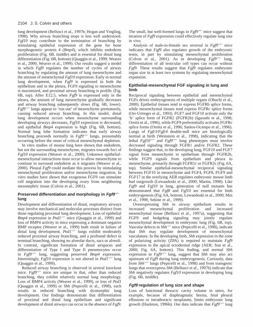
Fig. 6.Models for molecular signaling in developing lung.(A) Reciprocal FGF signaling between epithelium and mesenchymeduring lung (top) and limb (bottom) embryogenesis. (A, top) Wepropose that FGF9 from the lung epithelium and pleura activatesFGFR1c in the mesenchyme, while FGF10 from the mesenchymeactivates FGFR2b in the epithelium. (A, bottom) Similar essentialreciprocal epithelial-mesenchymal signaling between FGF10 in themesenchyme and FGF8 in the overlying apical ectodermal ridge (AER)regulates limb bud outgrowth in mouse embryos. (DM, distalmesenchyme; ZPA, zone of polarizing activity). (FGF8 and FGF9 mayalso signal through FGFR2c, but Fgfr1 is more highly expressed thanFgfr2 in embryonic lung and limb mesenchyme (Peters et al., 1992).)(B) Model for interactions between Fgf and non-Fgf signaling pathwaysin developing lung. (B, top) Signaling during proximal airway branchingin the early pseudoglandular period. (1) FGF9 (blue) in both airwayepithelium and pleura stimulates mesenchymal proliferation. (Greyshading indicates mesenchyme.) (2) FGF10 (red) in the mesenchymeinduces airway branching by stimulating endoderm proliferation andmigration. (3) SHH (hatched) in the airway epithelium promotesmesenchymal proliferation. (B, middle) Signaling during airwaybranching in the late pseudoglandular period. (1) FGF9, now limited tothe pleura, stimulates mesenchymal proliferation. (2) MesenchymalFGF10 stimulates endoderm proliferation and migration, and appears toinduce distal epithelial BMP4 expression (yellow). (3) BMP4 appears toinhibit airway branching by inhibiting endoderm proliferation, andperhaps by inhibiting endoderm migration. (4) SHH in the epitheliumstimulates mesenchymal proliferation. Vascular defects in Shh−/− lungsindicate that Shhregulates development of mesenchymal vasculature.SHH also appears to prevent more generalized mesenchymal expression of Fgf10(truncated symbol). (B, bottom) Signaling duringdevelopment of distal airspaces. Mesenchyme surrounding epithelial buds is sharply reduced; focally high Fgf10expression in the adjacentmesenchyme is lost; and epithelial differentiation begins with the narrowing of the epithelium. (1) Epithelial Bmp4expression is essential fordistal epithelial differentiation. (2) Continued Shhexpression in the epithelium may regulate vascular development. In this model, distal lungdevelopment proceeds when mesenchyme surrounding developing airways thins, Fgf10expression is reduced and Bmp4expression is high. InFgf9−/− lungs, loss of Fgf9-induced mesenchymal proliferation could result in premature reduction in mesenchyme surrounding buddingairways, leading to premature reduction in Fgf10expression. This would allow distal lung development to commence after fewer iterations ofairway branching.
2104
lung development (Bellusci et al., 1997b; Hogan and Yingling,1998). Why airway branching stops is less well understood.Fgf10 may contribute to the termination of branching bystimulating epithelial expression of the gene for bonemorphogenetic protein 4 (Bmp4), which inhibits endodermproliferation (Fig. 6B, middle) and is essential for distal lungdifferentiation (Fig. 6B, bottom) (Quaggin et al., 1999; Weaveret al., 2000; Weaver et al., 1999). Our results suggest a modelin which Fgf9 regulates the number of cycles of airwaybranching by regulating the amount of lung mesenchyme andthe amount of mesenchymal Fgf10expression. Early in normallung development, when Fgf9 is expressed in both theepithelium and in the pleura, FGF9 signaling to mesenchymeis maximized, and proximal airway branching is prolific (Fig.6B, top). After E12.5, when Fgf9 is expressed only in thepleura, the amount of lung mesenchyme gradually decreasesand airway branching subsequently slows (Fig. 6B, lower).Fgf9−/− lungs appear to ‘run out’ of mesenchyme prematurely,causing reduced airway branching. In this model, distallung development occurs when mesenchyme surroundingdeveloping airways decreases, Fgf10expression is decreased,and epithelial Bmp4expression is high (Fig. 6B, bottom).Normal lung lobe formation indicates that early airwaybranching proceeds normally in Fgf9−/− lungs, presumablyoccurring before the onset of critical mesenchymal depletion.
In vitro studies of mouse lung have shown that endoderm,but not the surrounding mesenchyme, migrates towards foci ofFgf10 expression (Weaver et al., 2000). Unknown epithelial-mesenchymal interactions must occur to allow mesenchyme tocontinue to surround endoderm as it migrates (Weaver et al.,2000). Pleural Fgf9could mediate this process by stimulatingmesenchymal proliferation and/or mesenchyme migration. Invitro studies have shown that exogenous FGF9 can stimulatecell migration into the developing ovary from neighboringmesonephric tissue (Colvin et al., 2001).
Preserved differentiation and morphology in Fgf9−/−
lungDevelopment and differentiation of distal, respiratory airwaysmay involve mechanical and molecular processes distinct fromthose regulating proximal lung development. Loss of epithelialBmp4expression in Pod1−/− mice (Quaggin et al., 1999) andloss of BMP4 activity in mice expressing a dominant negativeBMP receptor (Weaver et al., 1999) both result in failure ofdistal lung development. Pod1−/− lungs exhibit moderatelyreduced proximal airway branching, and a profound defect interminal branching, showing no alveolar ducts, sacs or alveoli.In contrast, significant formation of distal airspaces anddifferentiation of Type I and Type II pneumocytes occurin Fgf9−/− lung, suggesting preserved Bmp4 expression.Interestingly, Fgf10 expression is not altered in Pod1−/− lung(Quaggin et al., 1999).
Reduced airway branching is observed in several knockoutmice. Fgf9−/− mice are unique in that, other than reducedbranching, they exhibit relatively normal lung morphology.Loss of BMP4 activity (Weaver et al., 1999), or loss of Pod1(Quaggin et al., 1999) or Shh (Pepicelli et al., 1998), eachresults in reduced branching with dysmorphic lungdevelopment. Our findings demonstrate that differentiationof proximal and distal lung epithelium and significantdevelopment of distal airways can occur in the absence of Fgf9.
The small, but well-formed lungs in Fgf9−/− mice suggest thattitration of Fgf9expression could effectively regulate lung sizein vivo.
Analysis of male-to-female sex reversal in Fgf9−/− miceindicates that Fgf9also regulates growth of the embryonictestis, in part by stimulating mesenchymal proliferation(Colvin et al., 2001). As in developing Fgf9−/− lung,differentiation of all testicular cell types can occur withoutFgf9. These results suggest that Fgf9 regulates embryonicorgan size in at least two systems by regulating mesenchymalexpansion.
Epithelial-mesenchymal FGF signaling in lung andlimbReciprocal signaling between epithelial and mesenchymalFGFs drives embryogenesis of multiple organs (Ohuchi et al.,2000). Epithelial tissues tend to express FGFRb splice forms,while mesenchymal tissues tend express FGFRc splice forms(Orr-Urtreger et al., 1993). FGF7 and FGF10 activate only the‘b’ splice form of FGFR2 (FGFR2b) (Igarashi et al., 1998;Ornitz et al., 1996), while FGF9 preferentially activates FGFRcsplice forms (Ornitz et al., 1996; Santos-Ocampo et al., 1996).Lungs of Fgfr3/Fgfr4 double-null mice are histologicallynormal at birth (Weinstein et al., 1998), indicating that thelethal Fgf10−/− and Fgf9−/− lung phenotypes must involvedecreased signaling through FGFR1 and/or FGFR2. Thesefindings suggest that, in the developing lung, FGF10 and FGF7signal from mesenchyme to epithelium through FGFR2b,while FGF9 signals from epithelium and pleura tomesenchyme, primarily through FGFR1c or FGFR2c (Fig. 6A,top). Similar epithelial-mesenchymal reciprocal signalingbetween FGF10 in mesenchyme and FGF4, FGF8, FGF9 andFGF17 in the overlying AER regulates embryonic mouse limbbud outgrowth (Lewandoski et al., 2000; Martin, 1998). LikeFgf9 and Fgf10 in lung, generation of null mutants hasdemonstrated that Fgf8and Fgf10 are essential for limbembryogenesis (Fig. 6A, bottom; Lewandoski et al., 2000; Minet al., 1998; Sekine et al., 1999).
Overexpressing Shh in airway epithelium results inincreased mesenchymal proliferation and increasedmesenchymal tissue (Bellusci et al., 1997a), suggesting thatFGF9 and hedgehog signaling may jointly regulatemesenchymal development in embryonic lung (Fig. 6B, top).Vascular defects in Shh−/− mice (Pepicelli et al., 1998), indicatethat Shh may regulate development of mesenchymalvasculature. In the developing limb, Shhexpression in the zoneof polarizing activity (ZPA) is required to maintain Fgf9expression in the apical ectodermal ridge (AER; Sun et al.,2000; Fig. 6A, bottom). This finding, and normal Shhexpression in Fgf9−/− lung, suggest that Shh may also actupstream of Fgf9during lung embryogenesis. Curiously, datafrom Shh−/− lungs (Pepicelli et al., 1998) and from transgeniclungs that overexpress Shh(Bellusci et al., 1997b) indicate thatShhnegatively regulates Fgf10 expression in developing lung(Fig. 6B, middle).
Fgf9 regulation of lung size and shapeLoss of functional thoracic cavity volume in utero, forexample, because of diaphragmatic hernia, fetal pleuraleffusions or intrathoracic neoplasms, limits embryonic lunggrowth (Hasleton, 1996b). Our data indicate that Fgf9−/− lung
J. S. Colvin and others
2105Lung hypoplasia in Fgf9-null mice
hypoplasia represents a primary defect in lung growth. Noabdominal contents were ever found in the thoracic cavity ofFgf9−/− embryos, and Fgf9−/− mice could breathe and fill thelungs with air (data not shown), indicating that the diaphragmis intact and functional. Because Fgf9is expressed in thedeveloping pleura, it is possible that malformation of the pleuracould result in leakage of lung fluid into the thoracic cavity,constricting growth of thoracic organs. Selective loss of lungmesenchyme and the observation of cardiac dilation in someFgf9−/− mice suggest otherwise. In addition, preservation oftight junctions between pleural cells and of the pleuralbasement membrane suggests that the barrier function of thepleura is retained in Fgf9−/− lungs. Normal vascular histology,vWF expression and capillary formation in Fgf9−/− lungsindicate that lung hypoplasia in Fgf9−/− mice is not secondaryto gross vascular dysgenesis.
Fgf9−/− lungs fail to fill the thoracic cavity and lack the sharpcontours of normal lungs, suggesting that Fgf9 is an importantregulator of lung size and shape. Pleural expression of Fgf9could potentially be altered by physical contact with structuressurrounding the developing lung (such as the wall of thethoracic cavity in normal lung or intestine displaced into thethoracic cavity secondary to diaphragmatic hernia), allowingprecise fitting of the developing lung into the available space.In support of this model, rat visceral pleural cells in vitroexhibit altered gene expression when subjected to fluid shearstress simulating the rubbing of the lung pleura against thechest wall (Waters et al., 1997).
Possible roles for Fgf9 in carcinogenesisOur data indicate that Fgf9stimulates fetal lung cellproliferation and regulates fetal lung size. These results suggestthat exogenous FGF9 could potentially enhance human lunggrowth after surgical lung resection, a commonly usedtreatment for lung carcinoma. In addition, aberrant Fgf9upregulation could contribute to cancerous lung growth. Likeprostate cancer, most lung carcinoma is epithelial in origin.During normal lung embryogenesis, epithelial Fgf9 apparentlystimulates mesenchymal proliferation by activatingmesenchymal FGFRc splice forms. However, in vivo analysisof malignant transformation of prostate epithelial cells hasdemonstrated a switch from exclusive expression of FGFRb inepithelial cells to exclusive expression of FGFRc; upregulationof epithelial FGFs was also observed (Yan et al., 1993). In thelung, a similar upregulation of epithelial Fgf9, and aberrantexpression of FGFRc splice forms in the epithelium, couldcreate an autocrine loop driving pathologic epithelialproliferation.
In summary, we have demonstrated that Fgf9 is essential forlung embryogenesis and for postnatal survival in mice, and wehave identified a novel role for Fgf9 in regulating lungmesenchyme. We provide evidence that Fgf9 affects lung sizeby stimulating mesenchymal proliferation and mesenchymalFgf10 expression. Similar to Fgf function in otherdevelopmental systems, FGF9 signaling from the epitheliumand reciprocal FGF10 signaling from the mesenchyme appearcoordinately to regulate airway branching and lung growth inmouse embryos. As Fgf9−/− mice die at birth, study of thesemice could not address Fgf9 function in postnatal lung. Failureof alveogenesis in Fgfr3/Fgfr4double-null mice (Weinstein etal., 1998), and in vitro activation of FGFR3 and FGFR4 by
FGF9 (Ornitz et al., 1996), suggest a possible role for Fgf9 inalveogenesis, which occurs postnatally in mice.
We are grateful for technical assistance from E. Taylor, M. Scott,A. Johnson and B. Coleman (histology), E. Spinaio, X. Hua, L. Li,H. Walker and M. Wuerffel (animal husbandry), M. Veith (scanningelectron microscopy), L. LaRose and M. Levy (transmission electronmicroscopy) and J. Waggoner (figure preparation). We thank B.Hogan for the Fgf10 in situ probe, A. McMahon for the Shhin situprobe, J. Whitsett for the SP-C antibody and F. DeMayo for CCSPantibody. Our thanks to D. deMello and E. Crouch for helpfuldiscussions, and to R. Pierce, S. Brody, R. Mecham and R. Cagan forreview of the manuscript. This work was supported by grants fromMonsanto/Searle and the American Heart Association (# 974-0221N).
REFERENCES
Bellusci, S., Furuta, Y., Rush, M. G., Henderson, R., Winnier, G. andHogan, B. L. (1997a). Involvement of Sonic hedgehog (Shh) in mouseembryonic lung growth and morphogenesis. Development124, 53-63.
Bellusci, S., Grindley, J., Emoto, H., Itoh, N. and Hogan, B. L.(1997b).Fibroblast growth factor 10 (FGF10) and branching morphogenesis in theembryonic mouse lung. Development124, 4867-78.
Colvin, J. S., Feldman, B., Nadeau, J. H., Goldfarb, M. and Ornitz, D. M.(1999). Genomic organization and embryonic expression of the mousefibroblast growth factor 9 gene. Dev. Dyn.216, 72-88.
Colvin, J. S., Green, R. P., Schmahl, J., Capel, B. and Ornitz, D. M.(2001).Male-to-female sex reversal in mice lacking fibroblast growth factor 9. Cell104, 875-889.
De Moerlooze, L., Spencer-Dene, B., Revest, J., Hajihosseini, M., Rosewell,I. and Dickson, C. (2000). An important role for the IIIb isoform offibroblast growth factor receptor 2 (FGFR2) in mesenchymal-epithelialsignalling during mouse organogenesis. Development127, 483-492.
deMello, D. E., Sawyer, D., Galvin, N. and Reid, L. M.(1997). Early fetaldevelopment of lung vasculature. Am. J. Respir. Cell Mol. Biol.16, 568-81.
Guo, L., Degenstein, L. and Fuchs, E.(1996). Keratinocyte growth factor isrequired for hair development but not for wound healing. Genes Dev.10,165-175.
Hasleton, P. S. (1996a). Anatomy of the lung. In Spenser’s Pathology of theLung (ed. P. S. Hasleton), pp. 1-44. New York: McGraw-Hill.
Hasleton, P. S. (1996b). Embryology and development of the lung. InSpenser’s Pathology of the Lung(ed. P. S. Hasleton), pp. 45-55. New York:McGraw-Hill.
Hilfer, S. R. (1996). Morphogenesis of the lung: control of embryonic andfetal branching. Annu. Rev. Physiol.58, 93-113.
Hogan, B. L. M. and Yingling, J. M. (1998). Epithelial/mesenchymalinteractions and branching morphogenesis of the lung. Curr. Opin. Genet.Dev.8, 481-486.
Igarashi, M., Finch, P. W. and Aaronson, S. A.(1998). Characterization ofrecombinant human fibroblast growth factor (Fgf-10) reveals functionalsimilarities with keratinocyte growth factor (Fgf-7). J. Biol. Chem.273,13230-13235.
Kaartinen, V., Voncken, J. W., Shuler, C., Warburton, D., Bu, D.,Heisterkamp, N. and Groffen, J.(1995). Abnormal lung development andcleft palate in mice lacking TGF-beta 3 indicates defects of epithelial-mesenchymal interaction. Nat. Genet.11, 415-421.
Kuzu, I., Bicknell, R., Harris, A. L., Jones, M., Gatter, K. C. and Mason,D. Y. (1992). Heterogeneity of vascular endothelial cells with relevance todiagnosis of vascular tumours. J. Clin. Pathol.45, 143-148.
Lewandoski, M., Sun, X. and Martin, G. R. (2000). Fgf8 signalling fromthe AER is essential for normal limb development. Nat. Genet.26, 460-463.
Martin, G. R. (1998). The roles of FGFs in the early development ofvertebrate limbs. Genes Dev.12, 1571-1586.
Masters, J. R. (1976). Epithelial-mesenchymal interaction during lungdevelopment: the effect of mesenchymal mass. Dev. Biol.51, 98-108.
Min, H., Danilenko, D. M., Scully, S. A., Bolon, B., Ring, B. D., Tarpley,J. E., DeRose, M. and Simonet, W. S.(1998). Fgf-10 is required for bothlimb and lung development and exhibits striking functional similarity toDrosophilabranchless. Genes Dev.12, 3156-3161.
Naski, M. C., Colvin, J. S., Coffin, J. D. and Ornitz, D. M. (1998).Repression of hedgehog signaling and BMP4 expression in growth plate
2106
cartilage by fibroblast growth factor receptor 3. Development125, 4977-4988.
Ohuchi, H., Hori, Y., Yamasaki, M., Harada, H., Sekine, K., Kato, S. andItoh, N. (2000). FGF10 acts as a major ligand for FGF receptor 2 IIIb in mousemulti-organ development. Biochem. Biophys. Res. Commun.277, 643-649.
Ornitz, D. M., Xu, J., Colvin, J. S., McEwen, D. G., MacArthur, C. A.,Coulier, F., Gao, G. and Goldfarb, M.(1996). Receptor specificity of thefibroblast growth factor family. J. Biol. Chem.271, 15292-15297.
Ornitz, D. M. and Itoh, N. (2001). Fibroblast growth factors. Genome Biol.2, 3005.1-3005.12.
Orr-Urtreger, A., Bedford, M. T., Burakova, T., Arman, E., Zimmer, Y.,Yayon, A., Givol, D. and Lonai, P.(1993). Developmental localization ofthe splicing alternatives of fibroblast growth factor receptor-2 (FGFR2). Dev.Biol. 158, 475-486.
Park, W. Y., Miranda, B., Lebeche, D., Hashimoto, G. and Cardoso, W. V.(1998). FGF-10 is a chemotactic factor for distal epithelial buds during lungdevelopment. Dev. Biol.201, 125-34.
Pepicelli, C. V., Lewis, P. M. and McMahon, A. P.(1998). Sonic hedgehogregulates branching morphogenesis in the mammalian lung. Curr. Biol. 8,1083-1086.
Peters, K. G., Werner, S., Chen, G. and Williams, L. T.(1992). Two FGFreceptor genes are differentially expressed in epithelial and mesenchymaltissues during limb formation and organogenesis in the mouse. Development114, 233-243.
Quaggin, S. E., Schwartz, L., Cui, S., Igarashi, P., Deimling, J., Post, M.and Rossant, J.(1999). The basic-helix-loop-helix protein pod1 is criticallyimportant for kidney and lung organogenesis. Development126, 5771-5783.
Ray, M. K., Wang, G., Barrish, J., Finegold, M. J. and DeMayo, F. J.(1996). Immunohistochemical localization of mouse Clara cell 10-KDprotein using antibodies raised against the recombinant protein. J.Histochem. Cytochem.44, 919-927.
Santos-Ocampo, S., Colvin, J. S., Chellaiah, A. T. and Ornitz, D. M.(1996).Expression and biological activity of mouse fibroblast growth factor-9(FGF-9). J. Biol. Chem.271, 1726-1731.
Sasaki, H. and Hogan, B. L.(1993). Differential expression of multiple forkhead related genes during gastrulation and axial pattern formation in themouse embryo. Development118, 47-59.
Satokata, I. and Maas, R.(1994). Msx1 deficient mice exhibit cleft palateand abnormalities of craniofacial and tooth development. Nat. Genet.6, 348-356.
Schor, A. M., Pazouki, S., Morris, J., Smither, R. L., Chandrachud, L.M. and Pendleton, N.(1998). Heterogeneity in microvascular density inlung tumours: comparison with normal bronchus. Br. J. Cancer77, 946-951.
Sekine, K., Ohuchi, H., Fujiwara, M., Yamasaki, M., Yoshizawa, T., Sato,T., Yagishita, N., Matsui, D., Koga, Y., Itoh, N. et al. (1999). Fgf10 isessential for limb and lung formation. Nat. Genet.21, 138-141.
Sun, X., Lewandoski, M., Meyers, E. N., Liu, Y. H., Maxson, R. E., Jr andMartin, G. R. (2000). Conditional inactivation of Fgf4 reveals complexityof signalling during limb bud development. Nat. Genet.25, 83-86.
Ten Have-Opbroek, A. A.(1981). The development of the lung in mammals:an analysis of concepts and findings. Am. J. Anat.162, 201-219.
Thurlebeck, W. M. (1995). Lung growth and development. In Pathology ofthe Lung(ed. W. M. Thurlebeck and A. M. Churg), pp. 37-87. New York:Theime Medical Publishers.
Waters, C. M., Chang, J. Y., Glucksberg, M. R., DePaola, N. and Grotberg,J. B. (1997). Mechanical forces alter growth factor release by pleuralmesothelial cells. Am. J. Physiol.272, L552-L557.
Weaver, M., Yingling, J. M., Dunn, N. R., Bellusci, S. and Hogan, B. L.(1999). Bmp signaling regulates proximal-distal differentiation of endodermin mouse lung development. Development126, 4005-4015.
Weaver, M., Dunn, N. R. and Hogan, B. L.(2000). Bmp4 and Fgf10 playopposing roles during lung bud morphogenesis. Development127, 2695-2704.
Weinstein, M., Xu, X., Ohyama, K. and Deng, C. X.(1998). FGFR-3 andFGFR-4 function cooperatively to direct alveogenesis in the murine lung.Development125, 3615-3623.
Wert, S. E., Glasser, S. W., Korfhagen, T. R. and Whitsett, J. A.(1993).Transcriptional elements from the human SP-C gene direct expression in theprimordial respiratory epithelium of transgenic mice. Dev. Biol.156, 426-443.
Yan, G., Fukabori, Y., McBride, G., Nikolaropolous, S. and McKeehan,W. L. (1993). Exon switching and activation of stromal and embryonicfibroblast growth factor (FGF)-FGF receptor genes in prostate epithelialcells accompany stromal independence and malignancy. Mol. Cell. Biol.13,4513-4522.
Zhou, L., Lim, L., Costa, R. H. and Whitsett, J. A. (1996). Thyroidtranscription factor-1, hepatocyte nuclear factor-3beta, surfactant protein B,C, and Clara cell secretory protein in developing mouse lung. J. Histochem.Cytochem.44, 1183-1193.