The spread of tumour cells to sentinel, tumour-draining lymph nodes (TDLNs) has been documented for centuries. Most carcinomas first metastasize to the TDLNs, and the presence of cancer cells in TDLNs is a major prognostic indicator in skin, breast, colon and other cancers. Tumour cells are thought to gain access to a TDLN by recruiting and entering local lymphatic vessels, which provide a physical connection between the tumour and its TDLN. As such, lymphatic vessels in the tumour microenvi-ronment have an important role as a route for dissemination. However, the lymphatic vessels, and the interstitial and lymphatic flows that they enable, have other roles that may be equally or even more important to the survival and metastasis of the tumour.
Importantly, lymph nodes are major sites of immune regulation where antigen-specific immune responses are directed against both foreign or pathogenic antigens and self-antigens that are drained from the upstream peripheral tissue. This results in the establish-ment of productive immunity against patho-gens or the maintenance of immunological tolerance to self-antigens. The TDLN is
constantly bathed in cytokines and antigens deriving from the tumour, which are carried there by the tumour-associated lymphatic vessels, as well as antigen-presenting cells (APCs) from the tumour microenviron-ment. Lymph flow from tumours has been reported to be elevated compared with that from normal tissue1–3, and increased lymph drainage has been positively correlated with metastasis4. Thus, lymphatic drainage from the tumour along with tumour recruitment of surrounding lymphatic vessels are likely to have important roles in modulating the host anti-tumour immune responses.
Increased fluid drainage from the tumour microenvironment, which is enabled by the tumour-draining lymphatics, along with heightened pressure gradients (described below), implies that interstitial flow is increased at the tumour margin5,6. This heightened interstitial flow through the tumour stroma, in turn, induces mechanical stress on the extracellular matrix (ECM) and stromal cells7, which can increase transforming growth factor-β (TGFβ) expression and activation, myofibroblast differentiation and stromal stiffening. Such stromal changes might have
important implications for suppressing anti-tumour immune responses in the tumour stroma through multiple mechanisms.
We suggest that increased interstitial and lymphatic flow in the tumour microenviron-ment, and the resulting mechanical changes to the tumour stroma, may strongly alter host immunity to the tumour. We review the intersection between three traditionally unrelated topics: first, tumour and TDLN lymphangio genesis, lymphatic drainage and the changes in interstitial flow in the tumour microenvironment that they cause; second, interstitial flow and its mechanobiology on the tumour stroma; and third, the inflamma-tory microenvironment of the tumour and its draining lymph node, which suppress anti-tumour immunity. Therefore, we propose that lymphatic drainage links tumour mechano-biology with tumour immunology (FIG. 1).
Flow in the tumour microenvironmentPressure gradients in the tumour margin drive interstitial flow. Lymphatic flow is an impor-tant component of the circulation. In nearly all tissues, plasma leaks out of blood capillar-ies, flows through the interstitium and drains into lymphatic vessels, where it passes through lymph nodes before being returned to the venous blood. Interstitial fluid is a plasma filtrate, and fluid flow between the blood, interstitial and lymphatic compartments at the microcirculatory level is considered to be gov-erned by Starling forces (the net differences between hydrostatic pressure and osmotic pressure). Newly formed lymph has essentially the same composition as interstitial fluid, as there is very little filtration of interstitial fluid when traversing the lymphatic endothelium. This afferent lymph enters the lymph node where it is filtered owing to protein and par-ticulate uptake by the immune cells there; the efferent lymph exiting the lymph node thus has a different protein composition from that of the afferent lymph8,9.
Since the 1970s, it has been recognized that the balance of fluid between the blood, interstitium and lymph is altered in solid tumours10,11 (FIG. 1). The hypoxia that is gen-erated by a rapidly growing tumour mass drives aberrant tumour angiogenesis, which is one of the most well-studied hallmarks of cancer12. Tumour angiogenesis is rapid and
O P I N I O N
Lymphatic and interstitial flow in the tumour microenvironment: linking mechanobiology with immunityMelody A. Swartz and Amanda W. Lund
Abstract | Tumours often engage the lymphatic system in order to invade and metastasize. The tumour-draining lymph node may be an immune-privileged site that protects the tumour from host immunity, and lymph flow that drains tumours is often increased, enhancing communication between the tumour and the sentinel node. In addition to increasing the transport of tumour antigens and regulatory cytokines to the lymph node, increased lymph flow in the tumour margin causes mechanical stress-induced changes in stromal cells that stiffen the matrix and alter the immune microenvironment of the tumour. We propose that synergies between lymphatic drainage and flow-induced mechanotransduction in the stroma promote tumour immune escape by appropriating lymphatic mechanisms of peripheral tolerance.
PERSPECTIVES
210 | MARCH 2012 | VOLUME 12 www.nature.com/reviews/cancer
haphazard, leading to leaky tumour vessels and the accumulation of macromolecules, such as albumin, from the plasma into the tumour tissue. At the same time, ECM production and remodelling at the tumour margin generates mechanical stress13,14, which, together with the leaky vessels, leads to increased interstitial fluid pressure (IFP) within the tumour11,15. Whereas normal tis-sue pressures range from -2 to 0 mm Hg16,
tumour IFP can be as high as capillary pres-sure17, with values reported in humans in the range of 10–40 mm Hg11,15. Importantly, vessel normalization by anti-angiogenic agents can partially decrease tumour IFP15,18. The heightened IFP in the tumour leads to pressure gradients at the tumour margin5,6, which in turn may drive heightened intersti-tial flow in the tumour stroma and into surrounding lymphatic vessels.
Interstitial flow in the tumour microen-vironment is heterogeneous and difficult to measure directly. Butler et al.10 first meas-ured IFP in chambers implanted in mam-mary tumours in rats, and reported bulk fluid transfer out of the tumour by compar-ing red blood cell concentrations in the afferent and the efferent blood vasculature of the tumour10. In mice, magnetic resonance imaging has been used to demonstrate
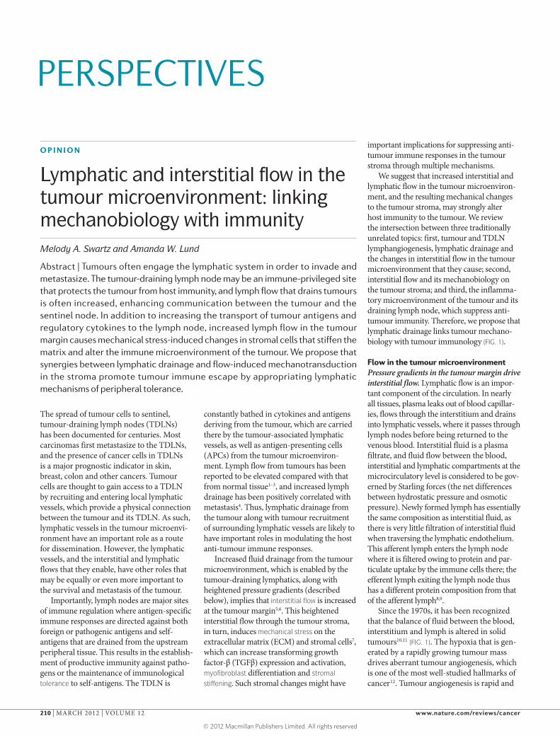
Figure 1 | Linking mechanobiology, lymphangiogenesis and anti-tumour immunity. Lymphangiogenesis, mechanobiology and immunity in the tumour microenvironment contain many interdependent features. a | Fluid pathways in the tumour microenvironment are shown. Angiogenic, immature tumour vessels (shown in red) are hyperpermea-ble, driving heightened interstitial fluid pressure (IFP) in the tumour. This creates steep pressure gradients at the tumour margin that drive inter-stitial flow through the stroma and into peritumoral lymphatic vessels (green), which are often expanded and hyperplastic. These lymphatic vessels carry tumour interstitial fluid to the sentinel or tumour-draining lymph node (TDLN), where lymphangiogenesis is also seen. b | The heightened interstitial and lymphatic flows (green) in the tumour micro-environment coincide with biomechanical (red) and immunological (pur-ple) changes in the tumour stroma. Biomechanical changes include
stiffening and alignment of the extracellular matrix (ECM) of the tumour stroma, owing to cancer-associated fibroblast (CAF) contraction and remodelling that is dependent on transforming growth factor-β (TGFβ). Matrix stiffening promotes tumour cell invasion through CAF-led collec-tive migration and activation of mechanically sensitive stromal compo-nents. Interestingly, stromal features of the tumour can mimic those of the TDLN. Immunological changes include biasing of the tumour-infiltrating lymphocyte populations by local factors. Tumour-promoting cytokines promote alternatively activated (M2) macrophages and myeloid-derived suppressor cells (MDSCs), hinder the maturation of immature dendritic cells (iDCs) and stimulate regulatory DCs (regDCs), which in turn promote regulatory T (T
Reg) cells and suppress local cytotoxic T cell activity. Similar
cell populations are biased in the TDLN. VEGF, vascular endothelial growth factor.
P E R S P E C T I V E S
NATURE REVIEWS | CANCER VOLUME 12 | MARCH 2012 | 211
increased fluid convection in the peritu-moral tissue1 or increased lymphatic drain-age to the sentinel lymph node2,3,19. Thus, although few direct measurements of inter-stitial flow in the tumour microenvironment have been reported, increased lymph flow to the TDLN along with high pressure gra-dients at the tumour margin should physi-cally reflect increased interstitial flow in the tumour stroma.
Tumour lymphangiogenesis and increased flow. In addition to inducing angiogenesis, tumours can also drive lymphangiogenesis or lymphatic hyperplasia in their microen-vironment; in fact, lymphangiogenesis and lymphatic expansion is seen in many types of chronic inflammation20–24. The molecular regulation of lymphangio genesis is well-described elsewhere25–27 and is not discussed here. Importantly, however, in many human and murine tumours, increased lymphatic density and expression of the lymphangi-ogenic vascular endothelial growth factor C (VEGFC) and VEGFD are correlated with poor prognosis, invasion and metasta-sis25,28,29. VEGFC secretion by tumours also increases lymph flow from the tumour to the TDLN4,30. Even when tumours do not explicitly express lymphangiogenic factors or induce lymphangiogenesis, their drain-ing lymph nodes undergo lymphangio-genesis before metastatic cells are seen in the TDLN2,3,25,29. Pathologists have described tumour-associated lymph node lymphangio-genesis as sinusoidal hyperplasia, which is a phenotype that correlates with metastasis31. Taken together, these observations suggest that lymph node lymphangiogenesis plays an important part in ‘preparing the soil’ for metastatic dissemination. The specific functional roles of this expanded lymphatic network and the mechanisms by which it might promote metastasis, however, remain unclear.
Lymphangiogenesis in the tumour mar-gin and TDLN may also be affected by the increased interstitial flow in the tumour stroma that drains to the TDLN. In dermal tissue regeneration, interstitial flow was found to be an important contributor to lymphangiogenesis32. Furthermore, intersti-tial flow helps to organize lymphatic capillar-ies in vitro33 partly by guiding local gradients of proteases and growth factors34, as well as by direct interactions between the ECM and lymphatic endothelium35. Fluid flow in the tumour margin may also cause channelling and may increase mechanical heterogeneity in the tumour microenvironment, altering the trafficking of macrophages that help to
guide lymphangiogenesis21,28,36. In support of this, Raju et al.37 reported a striking correla-tion between IFP and lymphatic vessel den-sity in rat squamous cell carcinoma, which also correlated with cancer progression37. However, direct studies of the effects of flow on tumour lymphangiogenesis are lacking, as it is not experimentally feasible to alter tumour interstitial flow in a selective way.
In summary, the strong clinical and experimental correlations observed among peritumoral and lymph node lymphangi-ogenesis or expansion, pressure gradients at the tumour margin and fluid flow to the draining lymphatics, and tumour progres-sion bolster the idea that lymphatic ves-sels and the TDLN support the tumour in multiple ways. Although one mechanism is to provide a physical transport route for tumour dissemination, we point out below how lymphatic drainage and tumour-associated lymphangiogenesis may alter the tumour microenvironment in other important ways.
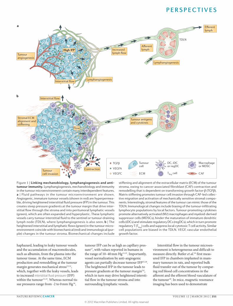
Flow effects on the tumour stromaAs described above, heightened interstitial flow is present in the stroma of most solid tumours. Several recent in vitro studies have provided clues about how flow might specifically affect tumour cells and cancer-associated fibroblasts (CAFs) in the tumour stroma using three-dimensional (3D) cell culture insert chambers with imposed flow38,39 or specially designed microfluidic chambers40,41(FIG. 2).
Flow activates TGFβ and fibroblasts to stiffen the ECM. Interstitial flow has impor-tant effects on fibroblasts and the ECM that are highly relevant to the tumour stroma (FIG. 3). In normal tissues, interstitial flow is thought to be in the order of 0.1–1 μm per second1,42, and interstitial and lymph flows from the tumour margin are substantially increased1–3,10,30. In experimental 3D culture models, it was found that interstitial flow of 3–10 μm per second can cause fibroblasts to align collagen fibres within 12–24 hours40.
Figure 2 | Modelling the biomechanical aspects of the tumour microenvironment in vitro. a | Normally (left), Starling forces are thought to drive low levels of interstitial flow between blood and lymphatic vessels. At the onset of inflammation, vascular permeability is rapidly upregulated, increas-ing interstitial flow by up to an order of magnitude. Lymphatic endothelial cells (LECs) respond to these changes by increasing permeability and active transport mechanisms, and by upregulating their expression of the chemokine CCL21 and intercellular adhesion molecule 1 (ICAM1) to enhance den-dritic cell (DC) and tumour cell trafficking. b | To recapitulate certain features of this three-dimensional (3D) interstitial flow microenvironment in vitro, a modified 3D Boyden chamber can be used. LECs are seeded on the underside of the porous insert, and an extracellular matrix (ECM) such as type I collagen, containing tumour cells or DCs, is added to the insert. Interstitial flow is controlled by maintaining a fixed pressure head (ΔP), and transmigrated cells can be counted. c | The radial flow chamber33,40,129, along with more recent microfluidic devices that capture similar features41, allow the direct observa-tion of cells in 3D matrices under interstitial flow. In the radial flow chamber, the cell-containing ECM is sandwiched between two glass coverslips and anchored with porous polyethylene boundaries; ECM remodelling can also be visualized in real time using confocal reflectance microscopy.
P E R S P E C T I V E S
212 | MARCH 2012 | VOLUME 12 www.nature.com/reviews/cancer
Matrix alignment is caused by fibroblast contraction, which can alter the matrix up to several cell lengths away, and such contraction-mediated alignment causes matrix stiffening both locally and globally43. One mechanism for flow-induced fibro-blast contraction and matrix remodelling is flow-induced upregulation of TGFβ by fibroblasts40, which causes myofibroblast differentiation, triggering further contrac-tion and matrix stiffening, as well as TGFβ production. When TGFβ signalling was blocked by TGFβ receptor-neutralizing antibodies, the interstitial flow-induced changes were abrogated. In addition to upregulating TGFβ production by fibro-blasts, flow-induced matrix contraction and shear stress can activate the stores of latent TGFβ in the ECM44,45. Therefore, flow acti-vates myofibroblast differentiation, as well as alignment and stiffening of the ECM via TGFβ-dependent mechanisms.
Stromal stiffening promotes tumour pro-gression and invasion. How can stiffening the stroma affect tumour progression? The work of several groups has demonstrated that stromal stiffening promotes tumour initiation, progression and invasion, at least in breast cancer, where it has been the most extensively studied46–49. Increased mam-mary tissue density correlates with the poor prognosis of patients with breast cancer, and collagen alignment at the invasive edge of breast tumours is negatively correlated with survival50. The expression of lysyl oxidase, which crosslinks and stiffens collagen, is
increased in malignant lesions in humans in response to intratumoral hypoxia and reactive oxygen species51,52, and lysyl oxidase expression is conversely downregulated by pro-inflammatory cytokines such as tumour necrosis factor (TNF)53. Additionally, trans-genic expression of lysyl oxidase in mice could induce the invasion of otherwise non-invasive cancer cells54. CAFs often exhibit myofibroblastic features55 and express caveolin 1, which drives RHOA and force-dependent contraction, matrix alignment and stromal stiffening; these collectively promote directional migration56. Migrating CAFs, in turn, use proteases and generate forces to tunnel through the matrix, thereby leading collective tumour cell invasion57,58 either by physical interactions or by the secretion of chemokines such as CXCL12 (REF. 59). Fibroblasts and tumour cells can also migrate along bundled collagen fibres that radiate outwards from the tumour60,61. Thus, stromal stiffening and remodelling is associated with tumour progression and invasion. As fluid flow upregulates TGFβ expression and activation from latent stores in the matrix, which in turn activates fibro-blasts to be more invasive and biosynthetic, fluid flow can promote tumour cell invasion via indirect actions on fibroblasts.
Cells can sense matrix stiffness either through endogenous force generation (contraction) or through exogenous forces such as interstitial flow. Stress is transmitted across the cell–matrix interface through focal adhesions, activating ERK and RHOA–Rho-associated protein kinase (ROCK), in turn
increasing myosin activity and actin assem-bly14,46,47,62. Softer matrices generate less ten-sion following cell contraction, and similarly, smaller exogenous forces generate less ten-sion on the focal adhesions. Thus, the effects of interstitial flow on the stromal matrix may be self-reinforcing: flow drives matrix tension and pulls on cell–matrix contacts, leading the cells to respond by aligning and stiffening the ECM, in turn generating more tension on the cells upon contraction, which makes them more sensitive to interstitial flow (FIG. 3).
Direct flow effects on cell migration. As for direct effects on tumour cells, flow has been shown to increase their invasiveness, at least in vitro. One suggested mechanism has been autologous chemotaxis39,41,63. For example, the secretion of the chemokine CCL21 by tumour cells that also express its receptor CCR7 leads to the formation of local (peri-cellular) gradients of CCL21 under fluid flow that drive chemotaxis and invasion in the flow direction. At the same time, fluid flow can also cause matrix tension behind the cell that promotes upstream migration; these two mechanisms may compete in a uniformly distributed tumour cell popula-tion in vitro41. Interstitial flow can also upregulate matrix metalloproteinases to further enhance tumour cell invasion64.
However, because tumour cells are often found following fibroblasts in invasive tumour margins57,58,61, flow effects on CAF migration may be more relevant than direct flow effects on tumour cells. First, interstitial flow can increase overall fibroblast motility65. Second,
Glossary
Cancer-associated fibroblasts(CAFs). Heterogeneous population of fibroblasts found in the tumour stroma that often exhibit myofibroblast features. They are responsible for stromal stiffening and can lead collective tumour cell invasion.
Dendritic cells(DCs). The most potent antigen-presenting cells that can activate T cells and thereby induce antigen-specific immune responses.
Fibroblastic reticular cells(FRCs). Lymph node stromal cells of the paracortical reticular meshwork, a specialized structure that directs the interactions between dendritic cells and T lymphocytes. FRCs express podoplanin (GP38), which is a key component of the reticular meshwork, and secrete cytokines such as CCL21 and CCL19 to attract lymphocytes and maintain their homeostasis. Importantly, FRC features can be exhibited by CAFs, and such lymphoid-like stromal components have been correlated with tumour invasion and metastasis.
Interstitial flowFluid flow within the interstitium, driven by pressure gradients between the blood, interstitial and lymphatic
compartments. Elevated tumour interstitial fluid pressure or increased lymphatic drainage can cause increased interstitial flow in the tumour stroma.
Interstitial fluid pressure(IFP). Hydrostatic pressure in the interstitium; it is usually subatmospheric, meaning that excised tissue imbibes water when placed in saline. IFP in solid tumours is often elevated owing to leaky tumour vessels.
Mechanical stressAn applied force per unit area. In tumours, stresses include IFP gradients, shear stresses owing to fluid flow, matrix tension caused by fluid flow or matrix contraction by CAFs, and compressive stresses from a growing tumour pushing on surrounding tissue. Mechanical stress in the extracellular matrix induces mechanical strain according to stiffness.
MyofibroblastA fibroblast subtype expressing α-smooth muscle actin that displays a contractile, synthetic and pro-fibrotic phenotype. Transforming growth factor-β (TGFβ) both activates, and is activated by, myofibroblasts.
Regulatory T (TReg) cellsFoxP3+ CD4+ T cells that suppress effector T cells and are important for maintaining peripheral tolerance to autoantigens, thereby preventing autoimmunity. Natural TReg cells are educated in the thymus, and inducible TReg cell activation in the periphery requires TGFβ and interleukin-10.
Stromal stiffeningA material property of the tumour stromal extracellular matrix that describes its resistance to deformation under mechanical stress. It can be altered by matrix protein synthesis, collagen crosslinking, matrix alignment and proteolysis.
ToleranceThe process that ensures that B and T cell repertoires are biased against self-reactivity, reducing the likelihood of autoimmunity.
Tumour-associated macrophages(TAMs). A heterogeneous population of generally immune suppressive, alternatively activated or M2-type macrophages derived from peripheral blood monocytes that are recruited into the tumour mass and that constitute a major component of the immune infiltrate.
P E R S P E C T I V E S
NATURE REVIEWS | CANCER VOLUME 12 | MARCH 2012 | 213
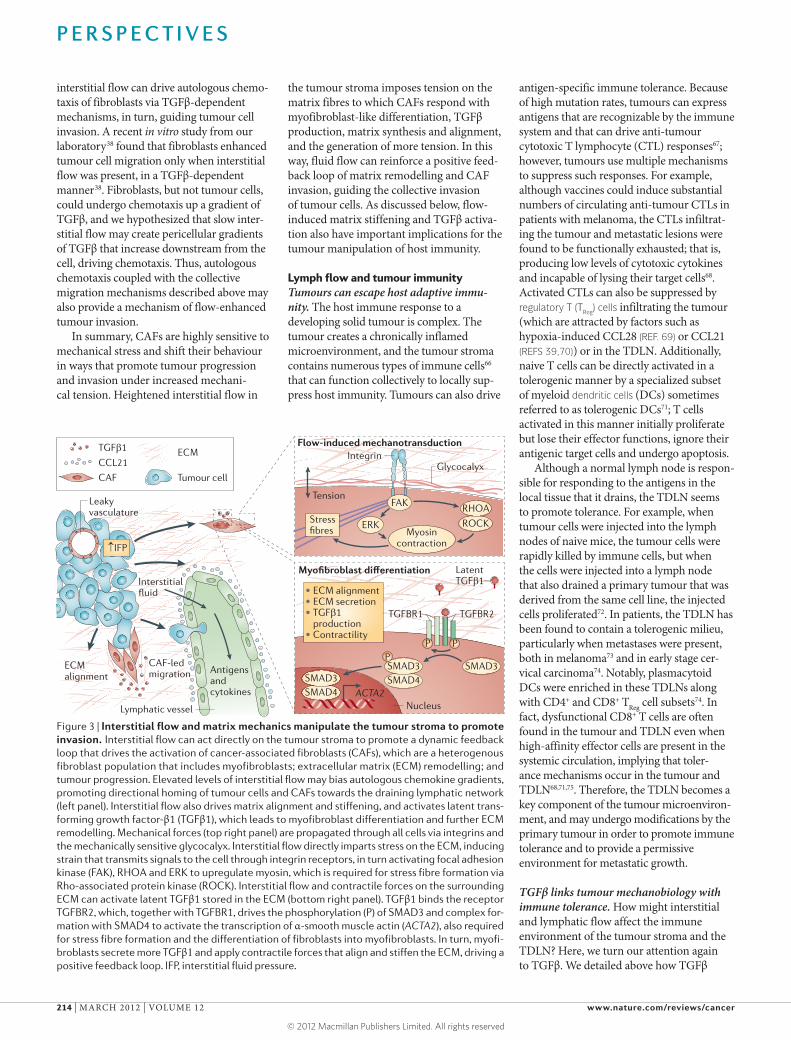
interstitial flow can drive autologous chemo-taxis of fibroblasts via TGFβ-dependent mechanisms, in turn, guiding tumour cell invasion. A recent in vitro study from our laboratory38 found that fibroblasts enhanced tumour cell migration only when interstitial flow was present, in a TGFβ-dependent manner38. Fibroblasts, but not tumour cells, could undergo chemotaxis up a gradient of TGFβ, and we hypothesized that slow inter-stitial flow may create pericellular gradients of TGFβ that increase downstream from the cell, driving chemotaxis. Thus, autologous chemotaxis coupled with the collective migration mechanisms described above may also provide a mechanism of flow-enhanced tumour invasion.
In summary, CAFs are highly sensitive to mechanical stress and shift their behaviour in ways that promote tumour progression and invasion under increased mechani-cal tension. Heightened interstitial flow in
the tumour stroma imposes tension on the matrix fibres to which CAFs respond with myofibroblast-like differentiation, TGFβ production, matrix synthesis and alignment, and the generation of more tension. In this way, fluid flow can reinforce a positive feed-back loop of matrix remodelling and CAF invasion, guiding the collective invasion of tumour cells. As discussed below, flow-induced matrix stiffening and TGFβ activa-tion also have important implications for the tumour manipulation of host immunity.
Lymph flow and tumour immunityTumours can escape host adaptive immu-nity. The host immune response to a developing solid tumour is complex. The tumour creates a chronically inflamed microenvironment, and the tumour stroma contains numerous types of immune cells66 that can function collectively to locally sup-press host immunity. Tumours can also drive
antigen-specific immune tolerance. Because of high mutation rates, tumours can express antigens that are recognizable by the immune system and that can drive anti-tumour cytotoxic T lymphocyte (CTL) responses67; however, tumours use multiple mechanisms to suppress such responses. For example, although vaccines could induce substantial numbers of circulating anti-tumour CTLs in patients with melanoma, the CTLs infiltrat-ing the tumour and metastatic lesions were found to be functionally exhausted; that is, producing low levels of cytotoxic cytokines and incapable of lysing their target cells68. Activated CTLs can also be suppressed by regulatory T (TReg) cells infiltrating the tumour (which are attracted by factors such as hypoxia-induced CCL28 (REF. 69) or CCL21 (REFS 39,70)) or in the TDLN. Additionally, naive T cells can be directly activated in a tolerogenic manner by a specialized subset of myeloid dendritic cells (DCs) sometimes referred to as tolerogenic DCs71; T cells activated in this manner initially proliferate but lose their effector functions, ignore their antigenic target cells and undergo apoptosis.
Although a normal lymph node is respon-sible for responding to the antigens in the local tissue that it drains, the TDLN seems to promote tolerance. For example, when tumour cells were injected into the lymph nodes of naive mice, the tumour cells were rapidly killed by immune cells, but when the cells were injected into a lymph node that also drained a primary tumour that was derived from the same cell line, the injected cells proliferated72. In patients, the TDLN has been found to contain a tolerogenic milieu, particularly when metastases were present, both in melanoma73 and in early stage cer-vical carcinoma74. Notably, plasmacytoid DCs were enriched in these TDLNs along with CD4+ and CD8+ TReg cell subsets74. In fact, dysfunctional CD8+ T cells are often found in the tumour and TDLN even when high-affinity effector cells are present in the systemic circulation, implying that toler-ance mechanisms occur in the tumour and TDLN68,71,75. Therefore, the TDLN becomes a key component of the tumour microenviron-ment, and may undergo modifications by the primary tumour in order to promote immune tolerance and to provide a permissive environment for metastatic growth.
TGFβ links tumour mechanobiology with immune tolerance. How might interstitial and lymphatic flow affect the immune environment of the tumour stroma and the TDLN? Here, we turn our attention again to TGFβ. We detailed above how TGFβ
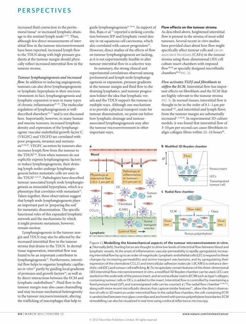
Figure 3 | Interstitial flow and matrix mechanics manipulate the tumour stroma to promote invasion. Interstitial flow can act directly on the tumour stroma to promote a dynamic feedback loop that drives the activation of cancer-associated fibroblasts (CAFs), which are a heterogenous fibroblast population that includes myofibroblasts; extracellular matrix (ECM) remodelling; and tumour progression. Elevated levels of interstitial flow may bias autologous chemokine gradients, promoting directional homing of tumour cells and CAFs towards the draining lymphatic network (left panel). Interstitial flow also drives matrix alignment and stiffening, and activates latent trans-forming growth factor-β1 (TGFβ1), which leads to myofibroblast differentiation and further ECM remodelling. Mechanical forces (top right panel) are propagated through all cells via integrins and the mechanically sensitive glycocalyx. Interstitial flow directly imparts stress on the ECM, inducing strain that transmits signals to the cell through integrin receptors, in turn activating focal adhesion kinase (FAK), RHOA and ERK to upregulate myosin, which is required for stress fibre formation via Rho-associated protein kinase (ROCK). Interstitial flow and contractile forces on the surrounding ECM can activate latent TGFβ1 stored in the ECM (bottom right panel). TGFβ1 binds the receptor TGFBR2, which, together with TGFBR1, drives the phosphorylation (P) of SMAD3 and complex for-mation with SMAD4 to activate the transcription of α-smooth muscle actin (ACTA2), also required for stress fibre formation and the differentiation of fibroblasts into myofibroblasts. In turn, myofi-broblasts secrete more TGFβ1 and apply contractile forces that align and stiffen the ECM, driving a positive feedback loop. IFP, interstitial fluid pressure.
P E R S P E C T I V E S
214 | MARCH 2012 | VOLUME 12 www.nature.com/reviews/cancer
can be activated in the tumour stroma by interstitial flow-activated CAFs38 and flow-induced mechanical stress on the ECM40,44, but TGFβ also has crucial and multiple roles in immune suppression. Although it should be emphasized that TGFβ stimulates both tumour-promoting and tumour-suppressing processes76,77, as well as epithelial-to-mes-enchymal transition78, it is among the most important regulators of effector T cell sup-pression and TReg cell activation. Importantly, TGFβ has a major role in defining the sup-pressive environment of the TDLN, where it functions synergistically with programmed cell death protein 1 (PD1) signalling for T cell suppression79. Thus, the overall effects of tumour TGFβ on host immunity are tolerogenic, and its activation in the tumour stroma by interstitial flow is a potentially important mechanism by which flow may promote immune tolerance (FIG. 4). Myeloid-derived suppressor cells (MDSCs), which are important for immune suppression, as well as for tumour angiogenesis and invasion80, produce TGFβ, although, paradoxically, their infiltration into tumours is increased when TGFβ signalling is ablated81. TGFβ attracts natural killer cells, neutrophils and macrophages to the tumour, but neutral-izes their anti-tumour effector functions76. TGFβ also strongly suppresses anti-tumour CTL responses in the tumour stroma, and blocking its signalling enhances anti-tumour immunity77.TGFβ is required for immature and tolerogenic DCs to selectively promote the differentiation and proliferation of TReg cells82, which are found localized to the tumour stroma83. In turn, activated TReg cells secrete more TGFβ. Therefore, abundant levels of TGFβ are required for inducing immune tolerance in the tumour and TDLN, and TGFβ activation is one mechanism by which interstitial flow could alter tumour immunity and promote tolerance.
Flow drives lymphoid features in the tumour stroma. In addition to regulating TGFβ, interstitial flow may promote lymphoid-like features to develop in the tumour stroma (FIG. 4). First, flow can modulate expres-sion of the cytokine CCL21 in the tumour microenvironment. Physiologically, CCL21 is expressed by lymphatic endothelial cells (LECs) and — along with CCL19 — lymph node fibroblastic reticular cells (FRCs); CCL21 normally functions as a lymphoid-homing chemokine, binding CCR7 on APCs and naive T cells to direct them to the lymph node and position them for efficient inter-actions84. CCL21 expression by LECs and FRCs is regulated by interstitial and lymph
flow85,86, as well as by VEGFC87. We and others have reported its expression by some tumours39,70,88,89, and we have recently cor-related its secretion with invasiveness and immune tolerance in murine melanoma70. Tumours that secreted CCL21 at physiologi-cally relevant levels (similar to levels in the lymph node) recruited CCR7+ lymphoid tissue-inducer cells. The recruitment of these cells correlated with the develop-ment of FRC-like features in the local CAF population — namely, expressing the reticu-lar matrix component ER-TR7, secreting more CCL19 and CCL21, and upregulating podoplanin, which is a type-1 transmem-brane sialomucin-like glycoprotein that is expressed by FRCs in the lymph node T cell zone. Podoplanin is also expressed by LECs, and has been found on some tumour cells and tumour-associated stromal cells, particu-larly in more aggressive tumours90. Although its direct role in tumour immunity is unclear, positive correlations between podoplanin expression and cancer stage, lymphatic invasion, lymph node metastasis, and peri-tumoral lymphatic vessel density have been
reported90–94. Therefore, from the perspective of circulating lymphocytes, the flow-activated tumour stroma mimics features of the lymph node T cell zone that are crucial for attract-ing, recruiting and educating T cells in the tumour microenvironment.
How might such tumour mimicry of lymph node stroma promote tolerance? In a normal lymph node, the cytokines and co-stimulatory molecules expressed by APCs determine how T cells are educated: danger signals such as Toll-like receptor activation promote effector responses, and signals from apoptotic cells or complement regula-tion after tissue injury, for example, can promote tolerance84. In contrast to a normal lymph node, the tumour stroma is steeped in suppressive cytokines, suppressor cells and factors that keep APCs in a functionally immature state. Furthermore, in the normal lymph node, CCL21 and CCL19 have key roles in modulating the balance between immunity and tolerance84,95. In addition to positioning DCs and naive T cells for adap-tive immune responses, these CCR7 ligands are required for TReg cell function96; they
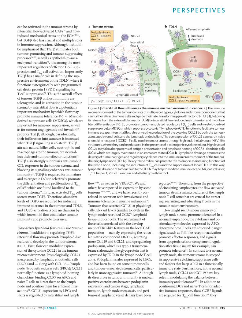
Figure 4 | Interstitial flow influences the immune microenvironment in cancer. a | The immune microenvironment of the tumour consists of multiple cell types, cytokines and stromal components that can further attract immune cells and guide their fate. Transforming growth factor-β1 (TGFβ1), following its release from the extracellular matrix (ECM) by interstitial flow-induced matrix tension and myofibro-blast differentiation (FIG. 3), promotes tumour-associated regulatory T (T
Reg) cells and myeloid-derived
suppressor cells (MDSCs), which suppress cytotoxic T lymphocyte (CTL) function to facilitate tumour immune escape. Interstitial flow also drives the production of the cytokine CCL21 by both the tumour-associated stromal cells and the lymphatic endothelium. The overexpression of CCL21 can recruit naive chemokine receptor 7 (CCR7)+ T cells into the tumour stroma through high endothelial venule (HEV)-like structures, where they can be educated in the presence of a tolerogenic cytokine milieu. High levels of CCL21 may also alter patterns of antigen presentation and lymphatic homing of CCR7+ dendritic cells (DCs), which are largely maintained in an immature state (iDCs). b | Lymphatic drainage promotes the delivery of tumour antigen and regulatory cytokines into the immune microenvironment of the tumour-draining lymph node (TDLN). This cytokine milieu can promote the tolerance-maintaining functions of the lymph node, including the induction of T
Reg cells and the suppression of local CTLs. In this way,
lymphatic drainage of tumour fluid to the TDLN may help to mediate immune escape. NK, natural killer; T
H1, T helper 1; VEGFC, vascular endothelial growth factor C.
P E R S P E C T I V E S
NATURE REVIEWS | CANCER VOLUME 12 | MARCH 2012 | 215
can also directly inhibit CTL proliferation97, and mice that lack CCR7 signalling cannot maintain tolerance to peripheral antigens98. By attracting naive and TReg cells to the tumour environment where they can inter-act with immature APCs and MDSCs while under the influence of regulatory cytokines, the tumour and its TDLN may encourage tolerogenic T cell education70. In this way, the lymphoid-like changes that may be initiated or enhanced by increased flow and VEGFC levels in the tumour microenvironment are likely to support immune tolerance.
Immunological roles of lymphangiogenesis. As mentioned above, lymphangiogenesis in the tumour and TDLN is correlated with invasion, metastasis and poor progno-sis25,29,99. In addition to cancer, lymphangio-genesis occurs during chronic inflammation and in the lymph nodes that drain the inflamed regions23,24,100–102. Currently, very little is known about how inflammatory lymphangiogenesis affects immunity, and recent reports have raised puzzling ques-tions. On the one hand, as detailed above, cancer progression is positively correlated with both lymphangiogenesis and immune suppression and tolerance. Furthermore, in mouse models of acute inflammation, lymphangiogenesis was important in resolv-ing the inflammation100. On the other hand, lymphangiogenesis is also frequently seen in autoimmunity-related chronic inflammatory disorders23,101 and in transplant rejection103, raising the possibility that lymphangio-genesis may contribute to immune rejec-tion. Blocking lymphangiogenesis before experimental corneal or islet transplantation could reduce rates of graft rejection102,104; however, in 1-year follow-ups of patients who had undergone renal transplant, trans-plant function was positively correlated with lymphangiogenesis105. Such findings raise questions regarding how inflamma-tory lymphangio genesis affects the immune response in different settings.
In considering the potential roles of lymphangiogenesis in inflammation, the context is likely to be important — that is, the cytokines, stromal changes and immune cells present — as this can be vastly dif-ferent in acute inflammation, chronic inflammatory diseases and cancer. Lymph node lymphangiogenesis following acute inflammatory stimulation may be driven by B cells106,107 and inhibited by CTLs and interferon-γ (IFNγ)108. Consistent with this idea, autoimmunity-associated tertiary lym-phoid structures are rich in B cell germinal centres that produce autoantibodies, and in
renal interstitial injury, for example, B cell recruitment promoted lymphangiogenesis around the B cell infiltrates109. T cells may be directly recruited to these newly formed structures to effectively bypass the local draining lymph node110. Additionally, local lymphatic vessels may carry fluid directly to the tertiary lymphoid structures rather than to the lymph node111; in other words, lymph-borne antigen may be constantly delivered to these ectopic B cell follicles to further promote autoantibody formation, and the tolerance-maintaining functions of the lymph node would not be present to temper these responses.
By contrast, lymphangiogenesis in the tumour microenvironment and the TDLN is clearly correlated with poor prognosis, as is tumour immune escape. VEGFC recruits VEGFR3+ tumour-associated macrophages (TAMs)28 that further promote immune suppression66 and that secrete VEGFC when stimulated by tumour-derived fac-tors such as TNF36. Ectopic germinal centres are not generally associated with cancer, and lymphangio genic tumours show increased, not decreased, lymph flow to the TDLN2,3,19,30. Furthermore, LECs can directly inhibit dendritic cell maturation and func-tion112, and can suppress autoreactive T cells in the lymph node for peripheral tolerance113. Thus, although the roles of lymphangio-genesis in modulating immunity remain to be clarified in different inflammatory condi-tions, lymphangiogenesis in the tumour and the TDLN does not seem to correlate with anti-tumour immunity. Instead, positive feedback loops exist in the tumour stroma between VEGFC expression, interstitial fluid flow, TGFβ upregulation, development of lymphoid-like stromal features that attract naive and regulatory T cells, infiltration of VEGFR3+ and VEGFC-secreting TAMs, and T cell tolerance in the tumour stroma (FIG. 4).
Lymph flow helps to regulate the immune functions of the lymph node. Lymph flow communicates information from the periph-ery to the local draining lymph node. It delivers both APCs carrying antigen cap-tured in the periphery, and soluble antigens, pathogens and cytokines for uptake by lymph node-resident DCs and macrophages. Lymph flow pathways through the various regions of the lymph node seem to be highly organized and act as a system of filters that direct differ-ent types of molecules or particles to different regions of the lymph node, in turn helping to regulate appropriate immune responses to pathogens, immunogenic antigens and auto-antigens8,114,115 (FIG. 5). Prenodal or afferent
lymph also contains self-peptides processed by enzymes during tissue catabolism and cell apoptosis, including caspases and matrix met-alloproteinases, that differ in epitopes from those processed by endosomal degradation in APCs116. In this way, the lymph-borne anti-gens carry different information to the lymph node than APC-delivered antigens.
Furthermore, the context in which antigen is taken up by APCs — that is, in the periph-ery versus in the lymph node — and the different kinetics of transport and presenta-tion to T cells can substantially influence the immunological outcome8,117,118. For example, on encountering pathogens, DCs that are activated in the periphery may be exposed to higher local concentrations of effector cytokines, and they often undergo matura-tion en route to the lymph node, upregulating co-stimulatory molecules and cytokines that are necessary for activating potent CTLs. By contrast, lymph node-resident DCs are main-tained in an immature state, and, although they can also be stimulated to undergo maturation and activate effector T cells, their interactions with T cells occur on a shorter timescale and in the presence of a different cytokine milieu, leading to faster but weaker T cell activation118.
Importantly, immature DCs residing in the lymph node have important roles in maintaining peripheral tolerance. In addi-tion to constitutively expressing endogenous autoantigens from the local draining tis-sue119, they constantly sample lymph-borne antigens while continuously interacting with circulating T cells. In 1972, studies using dermal-contact hypersensitivity raised the hypothesis that lymph flow is required for inducing new peripheral tolerance, as toler-ance to the applied chemicals could only be achieved when local lymphatic drainage from the skin was intact, but not when lym-phatic drainage was blocked in transplanted skin islands120.
In the context of tumours, as mentioned above, immature DCs resident in the TDLN are bathed in the suppressive cytokine milieu draining from the tumour, which prevents their maturation and thus biases them to tolerogenic antigen presentation. This cytokine milieu is facilitated at least in part by the expression of indoleamine 2,3-dioxygenase by DCs in the tumour and the TDLN, which can both specifi-cally and indirectly suppress anti-tumour immune responses and induce TReg cells121. Additionally, tumour-secreted exosomes, which are lipid vesicles containing cell mem-brane and cytoplasmic proteins, are found in abundance in tumour-draining lymphatic
P E R S P E C T I V E S
216 | MARCH 2012 | VOLUME 12 www.nature.com/reviews/cancer
vessels; these may also help to promote toler-ance in TDLNs122 and support the TDLN as a tumour stem cell niche123.
Inflammatory signals modulate lymph flow and lymphangiogenesis. As lymph flow seems to be so important in lymph node function, one might expect it to be regulated by inflam-matory signals. Unfortunately, very few stud-ies exist on the modulation of lymph flow by inflammatory cues (as opposed to inflamma-tion-induced lymphangiogenesis (discussed above)); however, emerging evidence suggests that the lymphatic endothelium may actively modulate flow in response to inflammatory cues. Fluid transport across LECs (or lymph formation) is typically considered passive, moving between loosely overlapping cell–cell junctions. However, vesicular-based trans-endothelial transport may constitute an important and even dominant mechanism of solute transport compared with passive
convective transport, at least as shown in recent in vitro studies85. Such active transport may be differentially regulated by pro-inflam-matory or anti-inflammatory cytokines124 and heightened interstitial flow85, both of which are altered in the tumour microenvironment. Furthermore, although nitric oxide (NO) mediates the contraction of collecting lym-phatic vessels that drive flow, inducible NO synthase (iNOS)-expressing MDSCs have been shown to inhibit lymphatic contraction but to increase lymphatic diameter125. Thus, although modulators of lymphatic flow are just beginning to be identified, lymph for-mation seems to be at least partially actively regulated by local inflammatory cues that limit or enhance the rate of antigen delivery to the lymph node.
In addition to fine-tuning antigen delivery to APCs, flow pathways in the lymph node are important for antigen delivery to the non-haematopoietic stromal cells that develop
and maintain the lymph node architecture. In fact, these stromal cells seem to be indispen-sible for maintaining peripheral tolerance to self-antigens126. Lymph node stromal cell subsets, including FRCs and LECs, present peripheral tissue-restricted antigens and can delete autoreactive T cells113,127. However, their roles in peripheral tolerance to exogenous lymph-borne antigen are only beginning to be elucidated.
The tolerance-maintaining functions of the lymph node stromal cells bring our discussion back full-circle to the changes in the tumour stroma that are accompanied by VEGFC, increased flow and increased drainage to the lymph node (FIG. 4). Indeed, the expansion of LECs in the tumour micro environment and the TDLN, together with the transforma-tion of CAFs to mimic lymphoid FRCs70,92,94, raises fascinating possibilities that tumours appropriate lymphoid stromal mechanisms of tolerance. As discussed above, the activated tumour stroma may express podoplanin and CCL21, and podoplanin-expressing CAFs display other features of FRCs including the secretion of CCL19, CCL21, CXCL12 and TRANCE94. If such tumour-associated FRC-like CAFs and LECs can in fact present tumour antigens to T cells, as they do in the lymph node with peripheral antigens, then they might directly modulate T cell education in addition to simply providing the chemo-attractive and structural cues necessary to promote DC–T cell interactions within the tumour microenvironment.
Indeed, recent work from our laboratory128 has shown that VEGFC promotes immune tol-erance in the tumour microenvironment and the TDLN in murine melanoma128. VEGFC expression increased flow to the TDLN and attracted more T cells. In tumours expressing a non-endogenous antigen (chicken ovalbu-min (OVA)), VEGFC could protect against even pre-existing anti-OVA immunity that was induced by vaccination before tumour implantation. Importantly, LECs in the TDLN could take up and cross-present OVA on major histocompatibility complex class I molecules, which caused the deletion of adoptively trans-ferred, OVA-specific CD8+ T cells. Together, these findings support the idea that tumour-associated lymphatic vessels and lymphatic drainage are key components of the immune-suppressive microenvironment of tumours.
ConclusionsIn conclusion, we have proposed arguments for how interstitial flow and lymphatic drain-age may affect tumour progression and inva-sion, both by direct mechanomodulation of the tumour stroma and also by altering the
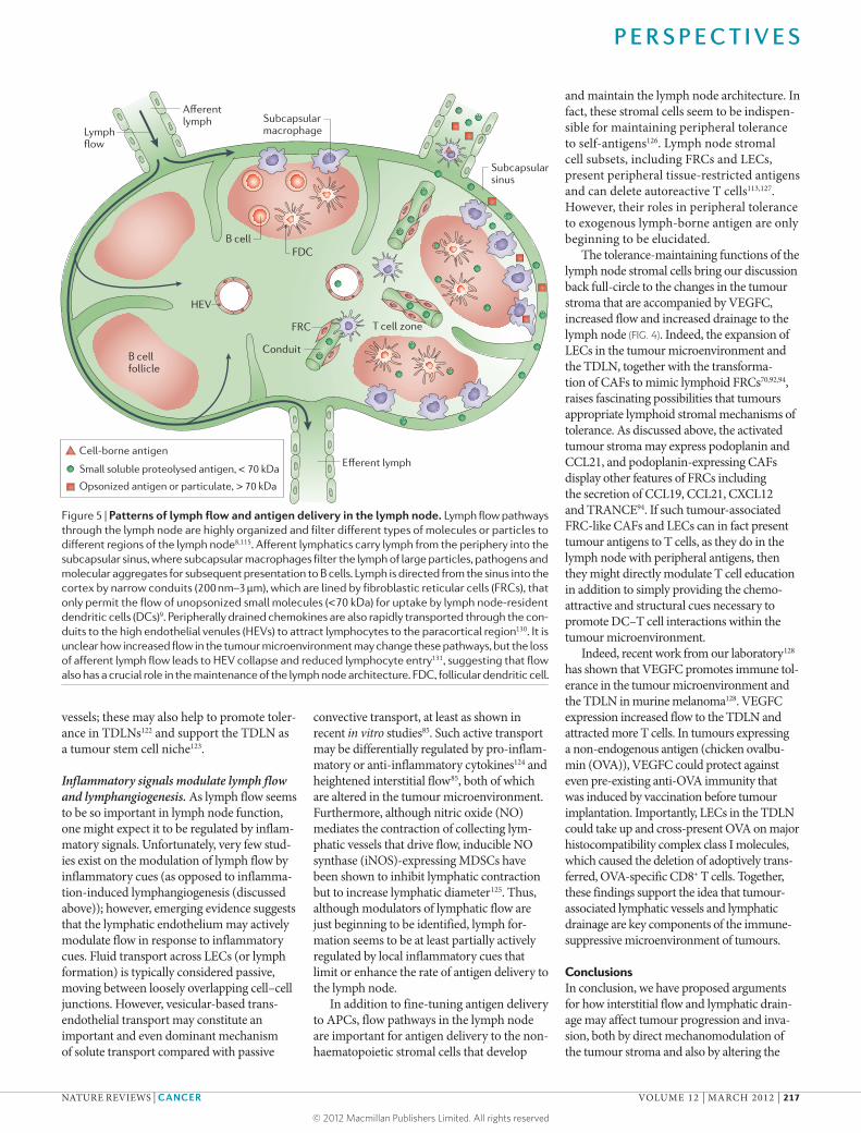
Figure 5 | Patterns of lymph flow and antigen delivery in the lymph node. Lymph flow pathways through the lymph node are highly organized and filter different types of molecules or particles to different regions of the lymph node8,115. Afferent lymphatics carry lymph from the periphery into the subcapsular sinus, where subcapsular macrophages filter the lymph of large particles, pathogens and molecular aggregates for subsequent presentation to B cells. Lymph is directed from the sinus into the cortex by narrow conduits (200 nm–3 μm), which are lined by fibroblastic reticular cells (FRCs), that only permit the flow of unopsonized small molecules (<70 kDa) for uptake by lymph node-resident dendritic cells (DCs)9. Peripherally drained chemokines are also rapidly transported through the con-duits to the high endothelial venules (HEVs) to attract lymphocytes to the paracortical region130. It is unclear how increased flow in the tumour microenvironment may change these pathways, but the loss of afferent lymph flow leads to HEV collapse and reduced lymphocyte entry131, suggesting that flow also has a crucial role in the maintenance of the lymph node architecture. FDC, follicular dendritic cell.
P E R S P E C T I V E S
NATURE REVIEWS | CANCER VOLUME 12 | MARCH 2012 | 217
host immune response. More research is needed to elucidate the direct roles of lym-phatic drainage in modulating immunity and in promoting immunological tolerance, and as tumours apparently appropriate lymphatics for progression, much can be learned from study-ing the effects of tumour lymphangiogenesis directly on the immune response. Future research should also better integrate tumour immunology with tumour mechanobiology, focusing on the similar features shared by tumour and lymph node stroma, as well as on the mechanisms of how T cells can be edu-cated or exhausted in the tumour microenvi-ronment and the contributions of stromal cells to this process. A deeper understanding of the relationships between lymph flow, stromal transformation and immunity should not only lead to new immunotherapeutic strategies to block the tolerance-promoting functions of lymphatic drainage in tumours, but may also lead to new tolerogenic therapies for auto immune diseases and transplantation.
Melody A. Swartz and Amanda W. Lund are at the Institute of Bioengineering and Swiss Institute of
Experimental Research (ISREC), SV-IBI-LLCB, Station 15, École Polytechnique Fédérale de Lausanne
(EPFL), Lausanne CH-1015, Switzerland. Correspondence to M.A.S.
1. Dafni, H., Israely, T., Bhujwalla, Z. M., Benjamin, L. E. & Neeman, M. Overexpression of vascular endothelial growth factor 165 drives peritumor interstitial convection and induces lymphatic drain: magnetic resonance imaging, confocal microscopy, and histological tracking of triple-labeled albumin. Cancer Res. 62, 6731–6739 (2002).
2. Harrell, M. I., Iritani, B. M. & Ruddell, A. Tumor-induced sentinel lymph node lymphangiogenesis and increased lymph flow precede melanoma metastasis. Am. J. Pathol. 170, 774–786 (2007).
3. Ruddell, A., Harrell, M. I. & Furuya, M. Sentinel lymph node lymphangiogenesis and increased lymph flow in murine tumor metastasis. Clin. Exp. Metast. 26, 883–884 (2009).
4. Pathak, A. P., Artemov, D., Neeman, M. & Bhujwalla, Z. M. Lymph node metastasis in breast cancer xenografts is associated with increased regions of extravascular drain, lymphatic vessel area, and invasive phenotype. Cancer Res. 66, 5151–5158 (2006).
5. Wiig, H., Tveit, E., Hultborn, R., Reed, R. K. & Weiss, L. Interstitial fluid pressure in DMBA-induced rat mammary tumours. Scand. J. Clin. Lab. Invest. 42, 159–164 (1982).
6. Flessner, M. F., Choi, J., Credit, K., Deverkadra, R. & Henderson, K. Resistance of tumor interstitial pressure to the penetration of intraperitoneally delivered antibodies into metastatic ovarian tumors. Clin. Cancer Res. 11, 3117–3125 (2005).
7. Pedersen, J. A., Boschetti, F. & Swartz, M. A. Effects of extracellular fiber architecture on cell membrane shear stress in a 3D fibrous matrix. J. Biomech. 40, 1484–1492 (2007).
8. Lammermann, T. & Sixt, M. The microanatomy of T-cell responses. Immunol. Rev. 221, 26–43 (2008).
9. Gretz, J. E., Norbury, C. C., Anderson, A. O., Proudfoot, A. E. & Shaw, S. Lymph-borne chemokines and other low molecular weight molecules reach high endothelial venules via specialized conduits while a functional barrier limits access to the lymphocyte microenvironments in lymph node cortex. J. Exp. Med. 192, 1425–1440 (2000).
10. Butler, T. P., Grantham, F. H. & Gullino, P. M. Bulk transfer of fluid in interstitial compartment of mammary tumors. Cancer Res. 35, 3084–3088 (1975).
11. Fukumura, D. & Jain, R. K. Tumor microenvironment abnormalities: causes, consequences, and strategies to normalize. J. Cell. Biochem. 101, 937–949 (2007).
12. Hanahan, D. & Weinberg, R. A. Hallmarks of cancer: the next generation. Cell 144, 646–674 (2011).
13. Dufort, C. C., Paszek, M. J. & Weaver, V. M. Balancing forces: architectural control of mechanotransduction. Nature Rev. Mol. Cell Biol. 12, 308–319 (2011).
14. Xu, R., Boudreau, A. & Bissell, M. Tissue architecture and function: dynamic reciprocity via extra- and intra-cellular matrices. Cancer Metastasis Rev. 28, 167–176 (2009).
15. Heldin, C. H., Rubin, K., Pietras, K. & Ostman, A. High interstitial fluid pressure - an obstacle in cancer therapy. Nature Rev. Cancer 4, 806–813 (2004).
16. Wiig, H. Evaluation of methodologies for measurement of interstitial fluid pressure (Pi): physiological implications of recent Pi data. Crit. Rev. Biomed. Eng. 18, 27–54 (1990).
17. Boucher, Y., Leunig, M. & Jain, R. K. Tumor angiogenesis and interstitial hypertension. Cancer Res. 56, 4264–4266 (1996).
18. Jain, R. K. Lessons from multidisciplinary translational trials on anti-angiogenic therapy of cancer. Nature Rev. Cancer 8, 309–316 (2008).
19. Proulx, S. T. et al. Quantitative imaging of lymphatic function with liposomal indocyanine green. Cancer Res. 70, 7053–7062 (2010).
20. Kim, K. E. et al. Role of CD11b+ macrophages in intraperitoneal lipopolysaccharide-induced aberrant lymphangiogenesis and lymphatic function in the diaphragm. Am. J. Pathol. 175, 1733–1745 (2009).
21. Kataru, R. P. et al. Critical role of CD11b+ macrophages and VEGF in inflammatory lymphangiogenesis, antigen clearance, and inflammation resolution. Blood 113, 5650–5659 (2009).
22. Flister, M. J. et al. Inflammation induces lymphangiogenesis through up-regulation of VEGFR-3 mediated by NF-κB and Prox1. Blood 115, 418–429 (2009).
23. Xing, L. P. & Ji, R. C. Lymphangiogenesis, myeloid cells and inflammation. Exp. Rev. Clin. Immunol. 4, 599–613 (2008).
24. Halin, C., Tobler, N. E., Vigl, B., Brown, L. F. & Detmar, M. VEGF-A produced by chronically inflamed tissue induces lymphangiogenesis in draining lymph nodes. Blood 110, 3158–3167 (2007).
25. Tammela, T. & Alitalo, K. Lymphangiogenesis: molecular mechanisms and future promise. Cell 140, 460–476 (2010).
26. Paupert, J., Sounni, N. E. & Noel, A. Lymphangiogenesis in post-natal tissue remodeling: lymphatic endothelial cell connection with its environment. Mol. Aspects Med. 32, 146–158 (2011).
27. Avraamides, C. J., Garmy-Susini, B. & Varner, J. A. Integrins in angiogenesis and lymphangiogenesis. Nature Rev. Cancer 8, 604–617 (2008).
28. Skobe, M. et al. Concurrent induction of lymphangiogenesis, angiogenesis, and macrophage recruitment by vascular endothelial growth factor-C in melanoma. Am. J. Pathol. 159, 893–903 (2001).
29. Mumprecht, V. & Detmar, M. Lymphangiogenesis and cancer metastasis. J. Cell. Mol. Med. 13, 1405–1416 (2009).
30. Ruddell, A. et al. Dynamic contrast-enhanced magnetic resonance imaging of tumor-induced lymph flow. Neoplasia 10, 706–713 (2008).
31. Kozlowski, H. & Hrabowska, M. Types of reaction in the regional lymph nodes in non-metastatic and minute-metastatic carcinoma of the uterine cervix. Arch. Geschwulstforsch. 45, 658–659 (1975).
32. Boardman, K. C. & Swartz, M. A. Interstitial flow as a guide for lymphangiogenesis. Circ. Res. 92, 801–808 (2003).
33. Ng, C. P., Helm, C. L. & Swartz, M. A. Interstitial flow differentially stimulates blood and lymphatic endothelial cell morphogenesis in vitro. Microvasc. Res. 68, 258–264 (2004).
34. Helm, C. E., Fleury, M. E., Zisch, A. H., Boschetti, F. & Swartz, M. A. Synergy between 3D flow and VEGF directs capillary morphogenesis in vitro: experiments and theoretical mechanisms. Proc. Natl Acad. Sci. USA 44, 15779–15784 (2005).
35. Wiig, H., Keskin, D. & Kalluri, R. Interaction between the extracellular matrix and lymphatics: consequences for lymphangiogenesis and lymphatic function. Matrix Biol. 29, 645–656 (2010).
36. Schoppmann, S. F. et al. Tumor-associated macrophages express lymphatic endothelial growth factors and are related to peritumoral lymphangiogenesis. Am. J. Pathol. 161, 947–956 (2002).
37. Raju, B., Haug, S. R., Ibrahim, S. O. & Heyeraas, K. J. High interstitial fluid pressure in rat tongue cancer is related to increased lymph vessel area, tumor size, invasiveness and decreased body weight. J. Oral Pathol. Med. 37, 137–144 (2008).
38. Shieh, A., Rozansky, H., Hinz, B. & Swartz, M. A. Tumor cell invasion is promoted by interstitial flow-induced matrix priming by stromal fibroblasts. Cancer Res. 71, 790–800 (2011).
39. Shields, J. D. et al. Autologous chemotaxis as a mechanism of tumor cell homing to lymphatics via interstitial flow and autocrine CCR7 signaling. Cancer Cell 11, 526–538 (2007).
40. Ng, C. P., Hinz, B. & Swartz, M. A. Interstitial fluid flow induces myofibroblast differentiation and collagen alignment in vitro. J. Cell Sci. 118, 4731–4739 (2005).
41. Polacheck, W. J., Charest, J. L. & Kamm, R. D. Interstitial flow influences direction of tumor cell migration through competing mechanisms. Proc. Natl Acad. Sci. USA 108, 11115–11120 (2011).
42. Chary, S. R. & Jain, R. K. Direct measurement of interstitial convection and diffusion of albumin in normal and neoplastic tissues by fluorescence photobleaching. Proc. Natl Acad. Sci. USA 86, 5385–5389 (1989).
43. Winer, J. P., Oake, S. & Janmey, P. A. Non-linear elasticity of extracellular matrices enables contractile cells to communicate local position and orientation. PLoS ONE 4, e6382 (2009).
44. Wipff, P. J., Rifkin, D. B., Meister, J. J. & Hinz, B. Myofibroblast contraction activates latent TGF-β1 from the extracellular matrix. J. Cell Biol. 179, 1311–1323 (2007).
45. Ahamed, J. et al. In vitro and in vivo evidence for shear-induced activation of latent transforming growth factor-β1. Blood 112, 3650–3660 (2008).
46. Yu, H. M., Mouw, J. K. & Weaver, V. M. Forcing form and function: biomechanical regulation of tumor evolution. Trends Cell Biol. 21, 47–56 (2011).
47. Paszek, M. J. et al. Tensional homeostasis and the malignant phenotype. Cancer Cell 8, 241–254 (2005).
48. Provenzano, P. P. et al. Collagen density promotes mammary tumor initiation and progression. BMC Med. 6, 11 (2008).
49. Ronnov-Jessen, L. & Bissell, M. J. Breast cancer by proxy: can the microenvironment be both the cause and consequence? Trends Mol. Med. 15, 5–13 (2009).
50. Conklin, M. W. et al. Aligned collagen is a prognostic signature for survival in human breast carcinoma. Am. J. Pathol. 178, 1221–1232 (2011).
51. Finger, E. C. & Giaccia, A. J. Hypoxia, inflammation, and the tumor microenvironment in metastatic disease. Cancer Met. Rev. 29, 285–293 (2010).
52. Guadall, A. et al. Hypoxia-induced ROS signaling is required for LOX up-regulation in endothelial cells. Frontiers Biosci. 3, 955–967 (2011).
53. Alcudia, J. F. et al. Lysyl oxidase and endothelial dysfunction: mechanisms of lysyl oxidase down-regulation by pro-inflammatory cytokines. Frontiers Biosci. 13, 2721–2727 (2008).
54. Levental, K. R. et al. Matrix crosslinking forces tumor progression by enhancing integrin signaling. Cell 139, 891–906 (2009).
55. Kalluri, R. & Zeisberg, M. Fibroblasts in cancer. Nature Rev. Cancer 6, 392–401 (2006).
56. Goetz, J. G. et al. Biomechanical remodeling of the microenvironment by stromal caveolin-1 favors tumor invasion and metastasis. Cell 146, 148–163 (2011).
57. Gaggioli, C. et al. Fibroblast-led collective invasion of carcinoma cells with differing roles for RhoGTPases in leading and following cells. Nature Cell Biol. 9, 1392–1400 (2007).
58. Khalil, A. A. & Friedl, P. Determinants of leader cells in collective cell migration. Integr. Biol. 2, 568–574 (2010).
59. Orimo, A. et al. Stromal fibroblasts present in invasive human breast carcinomas promote tumor growth and angiogenesis through elevated SDF-1/CXCL12 secretion. Cell 121, 335–348 (2005).
60. Provenzano, P. P. et al. Collagen reorganization at the tumor-stromal interface facilitates local invasion. BMC Med. 4, 38 (2006).
61. Friedl, P. & Gilmour, D. Collective cell migration in morphogenesis, regeneration and cancer. Nature Rev. Mol. Cell Biol. 10, 445–457 (2009).
62. Provenzano, P. P., Inman, D. R., Eliceiri, K. W., Trier, S. M. & Keely, P. J. Contact guidance mediated three-dimensional cell migration is regulated by Rho/ROCK-dependent matrix reorganization. Biophys. J. 95, 5374–5384 (2008).
P E R S P E C T I V E S
218 | MARCH 2012 | VOLUME 12 www.nature.com/reviews/cancer
63. Haessler, U., Teo, J. C., Foretay, D., Renaud, P. & Swartz, M. A. Migration dynamics of breast cancer cells in a tunable 3D interstitial flow chamber. Integr. Biol. 5 Dec 2011 (doi:10.1039/C1IB00128K).
64. Qazi, H., Shi, Z. D. & Tarbell, J. M. Fluid shear stress regulates the invasive potential of glioma cells via modulation of migratory activity and matrix metalloproteinase expression. PLoS ONE 6, e20348 (2011).
65. Shi, Z. D., Ji, X. Y., Qazi, H. & Tarbell, J. M. Interstitial flow promotes vascular fibroblast, myofibroblast, and smooth muscle cell motility in 3D collagen I via upregulation of MMP-1. Am. J. Physiol. Heart Circ. Physiol. 297, H1225–H1234 (2009).
66. Denardo, D., Andreu, P. & Coussens, L. M. Interactions between lymphocytes and myeloid cells regulate pro- versus anti-tumor immunity. Cancer Metast. Rev. 29, 309–316 (2010).
67. Munn, D. H. & Mellor, A. L. The tumor-draining lymph node as an immune-privileged site. Immunol. Rev. 213, 146–158 (2006).
68. Baitsch, L. et al. Exhaustion of tumor-specific CD8+ T cells in metastases from melanoma patients. J. Clin. Invest. 121, 2350–2360 (2011).
69. Facciabene, A. et al. Tumour hypoxia promotes tolerance and angiogenesis via CCL28 and Treg cells. Nature 475, 226–230 (2011).
70. Shields, J. D., Kourtis, I. C., Tomei, A. A., Roberts, J. M. & Swartz, M. A. Induction of lymphoidlike stroma and immune escape by tumors that express the chemokine CCL21. Science 328, 749–752 (2010).
71. Contassot, E., Preynat-Seauve, O., French, L. & Huard, B. Lymph node tumor metastases: more susceptible than primary tumors to CD8+ T-cell immune destruction. Trends Immunol. 30, 569–573 (2009).
72. Preynat-Seauve, O. et al. Extralymphatic tumors prepare draining lymph nodes to invasion via a T-cell cross-tolerance process. Cancer Res. 67, 5009–5016 (2007).
73. Gerlini, G. et al. Plasmacytoid dendritic cells represent a major dendritic cell subset in sentinel lymph nodes of melanoma patients and accumulate in metastatic nodes. Clin. Immunol. 125, 184–193 (2007).
74. Battaglia, A. et al. Metastatic tumour cells favour the generation of a tolerogenic milieu in tumour draining lymph node in patients with early cervical cancer. Cancer Immunol. Immunother. 58, 1363–1373 (2009).
75. Mantovani, A., Romero, P., Palucka, A. & Marincola, F. Tumour immunity: effector response to tumour and role of the microenvironment. Lancet 371, 771–783 (2008).
76. Bierie, B. & Moses, H. L. Transforming growth factor β (TGF-β) and inflammation in cancer. Cytokine Growth Factor Rev. 21, 49–59 (2010).
77. Flavell, R. A., Sanjabi, S., Wrzesinski, S. H. & Licona-Limon, P. The polarization of immune cells in the tumour environment by TGFβ. Nature Rev. Immunol. 10, 554–567 (2010).
78. Padua, D. & Massague, J. Roles of TGFβ in metastasis. Cell Res. 19, 89–102 (2009).
79. Wei, S. A. et al. Tumor-induced immune suppression of in vivo effector T-cell priming is mediated by the B7-H1/PD-1 axis and transforming growth factor β. Cancer Res. 68, 5432–5438 (2008).
80. Gabrilovich, D. I. & Nagaraj, S. Myeloid-derived suppressor cells as regulators of the immune system. Nature Rev. Immunol. 9, 162–174 (2009).
81. Yang, L. et al. Abrogation of TGF β signaling in mammary carcinomas recruits Gr-1+CD11b+ myeloid cells that promote metastasis. Cancer Cell 13, 23–35 (2008).
82. Ghiringhelli, F. et al. Tumor cells convert immature myeloid dendritic cells into TGF-β-secreting cells inducing CD4+CD25+ regulatory T cell proliferation. J. Exp. Med. 202, 919–929 (2005).
83. Tan, W. et al. Tumour-infiltrating regulatory T cells stimulate mammary cancer metastasis through RANKL-RANK signalling. Nature 470, 548–553 (2011).
84. Forster, R., Davalos-Misslitz, A. C. & Rot, A. CCR7 and its ligands: balancing immunity and tolerance. Nature Rev. Immunol. 8, 362–371 (2008).
85. Miteva, D. O. et al. Transmural flow modulates cell and fluid transport functions of lymphatic endothelium. Circ. Res. 106, 920–931 (2010).
86. Tomei, A. A., Siegert, S., Britschgi, M. R., Luther, S. A. & Swartz, M. A. Fluid flow regulates stromal cell organization and CCL21 expression in a tissue-engineered lymph node microenvironment. J. Immunol. 183, 4273–4283 (2009).
87. Issa, A., Le, T. X., Shoushtari, A. N., Shields, J. D. & Swartz, M. A. Vascular endothelial growth factor-C and C-C chemokine receptor 7 in tumor cell-lymphatic cross-talk promote invasive phenotype. Cancer Res. 69, 349–357 (2009).
88. Zhai, H. Y., Heppner, F. L. & Tsirka, S. E. Microglia/macrophages promote glioma progression. Glia 59, 472–485 (2011).
89. Machado, L. et al. Expression and function of T cell homing molecules in Hodgkin’s lymphoma. Cancer Immunol. Immunother. 58, 85–94 (2009).
90. Raica, M., Cimpean, A. M. & Ribatti, D. The role of podoplanin in tumor progression and metastasis. Anticancer Res. 28, 2997–3006 (2008).
91. Kitano, H. et al. Podoplanin expression in cancerous stroma induces lymphangiogenesis and predicts lymphatic spread and patient survival. Arch. Pathol. Lab. Med. 134, 1520–1527 (2010).
92. Cueni, L. N. et al. Tumor lymphangiogenesis and metastasis to lymph nodes induced by cancer cell expression of podoplanin. Am. J. Pathol. 177, 1004–1016 (2010).
93. Kawase, A. et al. Podoplanin expression by cancer associated fibroblasts predicts poor prognosis of lung adenocarcinoma. Int. J. Cancer 123, 1053–1059 (2008).
94. Peduto, L. et al. Inflammation recapitulates the ontogeny of lymphoid stromal cells. J. Immunol. 182, 5789–5799 (2009).
95. Ahrendt, M., Hammerschmidt, S. I., Pabst, O., Pabst, R. & Bode, U. Stromal cells confer lymph node-specific properties by shaping a unique microenvironment influencing local immune responses. J. Immunol. 181, 1898–1907 (2008).
96. Schneider, M., Meingassner, J., Lipp, M., Moore, H. & Rot, A. CCR7 is required for the in vivo function of CD4+ CD25+ regulatory T cells. J. Exp. Med. 204, 735–745 (2007).
97. Yasuda, T. et al. Chemokines CCL19 and CCL21 promote activation-induced cell death of antigen-responding T cells. Blood 109, 449–456 (2007).
98. Davalos-Misslitz, A. C. et al. Generalized multi-organ autoimmunity in CCR7-deficient mice. Eur. J. Immunol. 37, 613–622 (2007).
99. Hirakawa, S. et al. VEGF-C-induced lymphangiogenesis in sentinel lymph nodes promotes tumor metastasis to distant sites. Blood 109, 1010–1017 (2007).
100. Huggenberger, R. et al. An important role of lymphatic vessel activation in limiting acute inflammation. Blood 117, 4667–4678 (2011).
101. Von Der Weid, P. Y., Rehal, S. & Ferraz, J. G. P. Role of the lymphatic system in the pathogenesis of Crohn’s disease. Curr. Opin. Gastroenterol. 27, 335–341 (2011).
102. Yin, N. et al. Targeting lymphangiogenesis after islet transplantation prolongs islet allograft survival. Transplantation 92, 25–30 (2011).
103. Kerjaschki, D. et al. Lymphatic neoangiogenesis in human kidney transplants is associated with immunologically active lymphocytic infiltrates. J. Am. Soc. Nephrol. 15, 603–612 (2004).
104. Ling, S., Qi, C., Li, W., Xu, J. & Kuang, W. Crucial role of corneal lymphangiogenesis for allograft rejection in alkali-burned cornea bed. Clin. Exp. Ophthalmol. 37, 874–883 (2009).
105. Stuht, S. et al. Lymphatic neoangiogenesis in human renal allografts: results from sequential protocol biopsies. Am. J. Transplant. 7, 377–384 (2007).
106. Angeli, V. et al. B cell-driven lymphangiogenesis in inflamed lymph nodes enhances dendritic cell mobilization. Immunity 24, 203–215 (2006).
107. Shrestha, B. et al. B cell-derived vascular endothelial growth factor a promotes lymphangiogenesis and high endothelial venule expansion in lymph nodes. J. Immunol. 184, 4819–4826 (2010).
108. Kataru, R. P. et al. T lymphocytes negatively regulate lymph node lymphatic vessel formation. Immunity 34, 96–107 (2011).
109. Heller, F. et al. The contribution of B cells to renal interstitial inflammation. Am. J. Pathol. 170, 457–468 (2007).
110. Lee, Y. et al. Recruitment and activation of naive T cells in the islets by lymphotoxin β receptor-dependent tertiary lymphoid structure. Immunity 25, 499–509 (2006).
111. Thaunat, O., Kerjaschki, D. & Nicoletti, A. Is defective lymphatic drainage a trigger for lymphoid neogenesis? Trends Immunol. 27, 441–445 (2006).
112. Podgrabinska, S. et al. Inflamed lymphatic endothelium suppresses dendritic cell maturation and function via Mac-1/ICAM-1-dependent mechanism. J. Immunol. 183, 1767–1779 (2009).
113. Cohen, J. N. et al. Lymph node-resident lymphatic endothelial cells mediate peripheral tolerance via Aire-independent direct antigen presentation. J. Exp. Med. 207, 681–688 (2010).
114. Sixt, M. et al. The conduit system transports soluble antigens from the afferent lymph to resident dendritic cells in the T cell area of the lymph node. Immunity 22, 19–29 (2005).
115. Junt, T. et al. Subcapsular sinus macrophages in lymph nodes clear lymph-borne viruses and present them to antiviral B cells. Nature 450, 110–114 (2007).
116. Clement, C. C. et al. An expanded self-antigen peptidome is carried by the human lymph as compared to the plasma. PLoS ONE 5, e9863 (2010).
117. Batista, F. D. & Harwood, N. E. The who, how and where of antigen presentation to B cells. Nature Rev. Immunol. 9, 15–27 (2009).
118. Itano, A. A. & Jenkins, M. K. Antigen presentation to naive CD4 T cells in the lymph node. Nature Immunol. 4, 733–739 (2003).
119. Scheinecker, C., Mchugh, R., Shevach, E. M. & Germain, R. N. Constitutive presentation of a natural tissue autoantigen exclusively by dendritic cells in the draining lymph node. J. Exp. Med. 196, 1079–1090 (2002).
120. Friedlaender, M. H., Baer, H. Immunologic Tolerance: role of the regional lymph node. Science 176, 312–314 (1972).
121. Munn, D. & Mellor, A. Indoleamine 2,3-dioxygenase and tumor-induced tolerance. J. Clin. Invest. 117, 1147–1154 (2007).
122. Hood, J. L., San, R. S. & Wickline, S. A. Exosomes released by melanoma cells prepare sentinel lymph nodes for tumor metastasis. Cancer Res. 71, 3792–3801 (2011).
123. Peinado, H., Lavotshkin, S. & Lyden, D. The secreted factors responsible for pre-metastatic niche formation: old sayings and new thoughts. Semin. Cancer Biol. 21, 139–146 (2011).
124. Chaitanya, G. V. et al. Differential cytokine responses in human and mouse lymphatic endothelial cells to cytokines in vitro. Lymphat. Res. Biol. 8, 155–164 (2010).
125. Liao, S. et al. Impaired lymphatic contraction associated with immunosuppression. Proc. Natl Acad. Sci. USA 108, 18784–18789 (2011).
126. Fletcher, A. L., Malhotra, D. & Turley, S. J. Lymph node stroma broaden the peripheral tolerance paradigm. Trends Immunol. 32, 12–18 (2011).
127. Fletcher, A. L. et al. Lymph node fibroblastic reticular cells directly present peripheral tissue antigen under steady-state and inflammatory conditions. J. Exp. Med. 207, 689–697 (2010).
128. Lund, A. W. et al. VEGF-C promotes immune tolerance in B16 melanomas and cross-presentation of tumor antigen by lymph node lymphatics. Cell Rep. 23 Feb 2012 (doi:10.1016/j.celrep.2012.01.005).
129. Ng, C. P. & Swartz, M. A. Fibroblast alignment under interstitial fluid flow using a novel 3D tissue culture model. Am. J. Physiol. Heart Circ. Physiol. 284, H1771–H1777 (2003).
130. Roozendaal, R., Mebius, R. E. & Kraal, G. The conduit system of the lymph node. Int. Immunol. 20, 1483–1487 (2008).
131. Mebius, R. E., Streeter, P. R., Breve, J., Duijvestijn, A. M. & Kraal, G. The influence of afferent lymphatic vessel interruption on vascular addressin expression. J. Cell Biol. 115, 85–95 (1991).
AcknowledgementsThe authors apologize to the many researchers whose work could not be included owing to space constraints. They are grateful to the Swiss National Science Foundation, Swiss Cancer League, European Research Council and the Swiss NCCR in Molecular Oncology for funding.
Competing interests statementThe authors declare no competing financial interests.
FURTHER INFORMATIONMelody A. Swartz’s homepage: http://swartz-lab.epfl.ch/
ALL LINKS ARE ACTIVE IN THE ONLINE PDF
P E R S P E C T I V E S
NATURE REVIEWS | CANCER VOLUME 12 | MARCH 2012 | 219