57
LYMPHATIC DRAINAGE LYMPHATIC DRAINAGE OF HEAD AND NECK OF HEAD AND NECK Presented by: Dr.Ayesha Taha (JR I) Department of Pedodontics and Preventive Dentistry SPPGIDMS, Lucknow
| Date post: | 16-Jul-2015 |
| Category: |
Health & Medicine |
| Upload: | dr-ayesha-taha |
| View: | 151 times |
| Download: | 8 times |

LYMPHATIC DRAINAGE LYMPHATIC DRAINAGE OF HEAD AND NECKOF HEAD AND NECK
Presented by:Dr.Ayesha Taha (JR I)Department of Pedodontics and Preventive DentistrySPPGIDMS, Lucknow


Contents :-
IntroductionFunctions of Lymphatic SystemComponents of Lymphatic SystemLymph Nodes of Head & NeckLymphatic Drainage Conclusion

Introduction
Of all the body systems, the lymphatic system is perhaps the least familiar to most people. Yet without it, neither the circulatory system nor the immune system could function—circulation would shut down from fluid loss, and the body would be overrun by infection for lack of immunity.
The lymphatic system is an endothelium-lined network of blind-ended capillaries found in nearly all tissues, draining via collecting vessels into large vascular trunks that eventually empty via an evolutionarily conserved drainage point into the blood circulatory system.

Functions of Lymphatic System
The lymphatic system has three functions:

1. Fluid recovery Each day, we lose an excess of 2 to 4 L of water and
one-quarter to one-half of the plasma protein. The lymphatic system absorbs this excess fluid and returns it to the bloodstream by way of the lymphatic vessels.
2. Immunity As the lymphatic system recovers excess tissue fluid,
it also picks up foreign cells and chemicals from the tissues. On its way back to the bloodstream, the fluid passes through lymph nodes, where immune cells stand guard against foreign matter. When they detect it,
they activate a protective immune response.

3. Lipid absorption.
In the small intestine, special lymphatic vessels called lacteals absorb dietary lipids that are not absorbed by the blood capillaries.

Components of Lymphatic system

Lymph:
Lymph is usually a clear, colorless fluid, similar to blood plasma but low in protein. Its composition varies substantially from place to place.
Origin of Lymph :-Lymph originates in microscopic vessels called lymphatic capillaries. These vessels penetrate nearly every tissue of the body but are absent from the central nervous system, cartilage, bone, and bone marrow.
The gaps between lymphatic endothelial cells are so large that bacteria and other cells can enter along with the fluid.
Lymphatic Vessels:
They have a tunica interna with an endothelium and valve, a tunica media with elastic fibers and smooth muscle, and a thin outer tunica externa.
Their walls are thinner and their valves are more numerous than those of the veins.

Flow of Lymph :-Lymph takes the following route from the tissues back to the bloodstream:
Thus, there is a continual recycling of fluid from blood to tissue fluid to lymph and back to the blood.


Lymphatic Cells and Tissues:
T lymphocytes (T cells):
These are so-named because they develop for a time in the thymus and later depend on thymic hormones.
B lymphocytes (B cells):
These are named after an organ in birds (the bursa of Fabricius) in which they were first discovered. When activated, B cells differentiate into plasma cells, which produce circulating antibodies.

Macrophages:
These cells, derived from monocytes of the blood, phagocytize foreign matter (antigens) and “display” fragments of it to certain T cells, thus alerting the immune system to the presence of an enemy. Macrophages and other cells that do this are collectively called antigen-presenting cells (APCs).
Dendritic cells:
These are APCs found in the epidermis, mucous membranes, and lymphatic organs. (In the skin, they are often called Langerhans cells.)
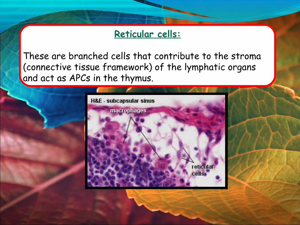
Reticular cells:
These are branched cells that contribute to the stroma (connective tissue framework) of the lymphatic organs and act as APCs in the thymus.

Mucosa-associated Lymphatic Tissue.
The simplest form of lymphatic tissue is diffuse lymphatic tissue—a sprinkling of lymphocytes in the mucous membranes and connective tissues of many organs.
It is particularly prevalent in body passages that are open to the exterior—the respiratory, digestive, urinary, and reproductive tracts—where it is called Mucosa-Associated Lymphatic Tissue (MALT).

Peyers patches:
In some places, lymphocytes and other cells congregate in dense masses called lymphatic nodules (follicles).
Lymphatic nodules are, however, a relatively constant feature of the lymph nodes and tonsils.
They also form clusters called Peyers patches in the ileum, the last segment of the small intestine.
Lymphatic Organs:
PRIMARY LYMPHATIC ORGANS :-
Lymphatic (lymphoid) organs contain large numbers of lymphocytes, a type of white blood cell that plays a pivotal role in immunity.
The primary lymphatic organs areRed bone marrow andThymus gland
Lymphocytes originate and/or mature in these organs.


Red Bone Marrow
It is the site of stem cells that are ever capable of dividing and producing blood cells.
Some of these cells become the various types of white blood cells: neutrophils, eosinophils, basophils, lymphocytes, and monocytes.
In a child, most of the bones have red bone marrow, but in an adult it is limited to the sternum, vertebrae, ribs, part of the pelvic girdle, and the proximal heads of the humerus and femur.

THYMUSThe thymus is a member of both the lymphatic and endocrine systems.
It houses developing lymphocytes and secretes hormones that regulate their activity.
It is located between the sternum and aortic arch in the superior mediastinum.
The thymus is very large in the fetus and grows slightly during childhood, when it is most active.
After age 14, however, it begins to undergo involution (shrinkage) so that it is quite small in adults.


THE SECONDARY LYMPHATIC ORGANS: arethe spleen,the lymph nodes and other organs, such as the tonsils.
All the secondary organs are the places where lymphocytes encounter and bind with antigens, after which they proliferate and become actively engaged cells.

SPLEENThe spleen is the body’s largest lymphatic organ. It is located in the left hypochondriac region, just inferior to the diaphragm and dorsolateral to the stomach.
Its parenchyma exhibits two types of tissue named for their appearance in fresh specimens (not in stained sections):
red pulp, which consists of sinuses gorged with concentrated erythrocytes, andwhite pulp, which consists of lymphocytes and macrophages aggregated like sleeves along small branches of the splenic artery.
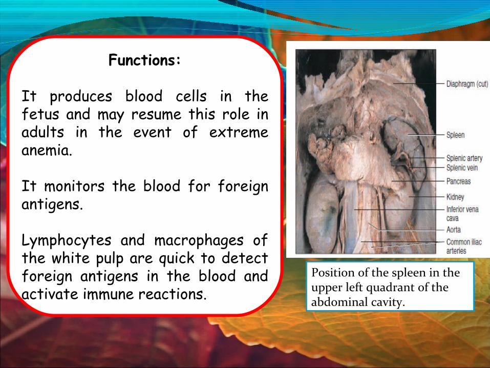
Functions:
It produces blood cells in the fetus and may resume this role in adults in the event of extreme anemia.
It monitors the blood for foreign antigens.
Lymphocytes and macrophages of the white pulp are quick to detect foreign antigens in the blood and activate immune reactions.
Position of the spleen in the upper left quadrant of the abdominal cavity.

The spleen is an “erythrocyte graveyard”—old, fragile RBCs rupture as they squeeze through the capillary walls into the sinuses. Splenic macrophages phagocytize their remains, just as they dispose of blood-borne bacteria and other cellular debris.
The spleen also compensates for excessive blood volume by transferring plasma from the bloodstream into the lymphatic system.
A person can live without a spleen, but is somewhat more vulnerable to infections.

LYMPH NODESIt serve two functions:
to cleanse the lymph and alert the immune system to pathogens.
There are hundreds of lymph nodes in the body.
They are especially concentrated in the cervical, axillary, and inguinal regions close to the body surface, and in thoracic, abdominal, and pelvic groups deep in the body cavities.
Most of them are embedded in fat.
A lymph node is an elongated or bean-shaped structure, usually less than 3 cm long, often with an indentation called the hilum on one side.
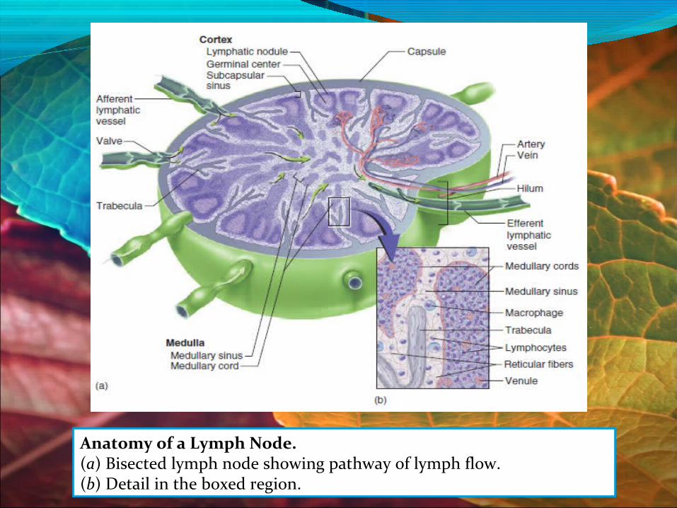
Anatomy of a Lymph Node. (a) Bisected lymph node showing pathway of lymph flow.(b) Detail in the boxed region.
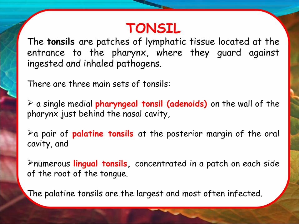
TONSILThe tonsils are patches of lymphatic tissue located at the entrance to the pharynx, where they guard against ingested and inhaled pathogens.
There are three main sets of tonsils:
a single medial pharyngeal tonsil (adenoids) on the wall of the pharynx just behind the nasal cavity,
a pair of palatine tonsils at the posterior margin of the oral cavity, and
numerous lingual tonsils, concentrated in a patch on each side of the root of the tongue.
The palatine tonsils are the largest and most often infected.

All lymph vessels of the head and neck drain into the deep cervical nodes, either directly from the tissues or indirectly via nodes in outlying groups.
Lymph is returned to the systemic venous circulation via either the right lymphatic duct or the thoracic duct.
All lymph vessels of the head and neck drain into the deep cervical nodes, either directly from the tissues or indirectly via nodes in outlying groups.
Lymph is returned to the systemic venous circulation via either the right lymphatic duct or the thoracic duct.
LYMPHATIC DRAINAGE OF HEAD & NECK
The lymph nodes in the head and neck region can be
grouped into:
• Superficial nodes
• Deep nodes
Classification of nodes in head and neck region

The superficial cervical lymph nodes lie above the above the investing layer of the deep fasciainvesting layer of the deep fascia.
They consist of a few small nodes that lie superficial to the external jugular and anterior jugular veins.
The superficial Lymph nodesThe superficial Lymph nodes

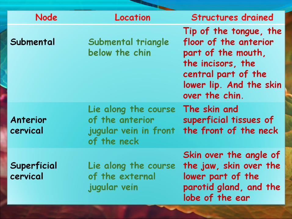
The superficial lymph nodes1. Occipital2. Mastoid (retro auricular/ post-auricular)3. Parotid (pre-auricular)4. Buccal5. Submandibular6. Submental 7. Anterior cervical8. Superficial cervical


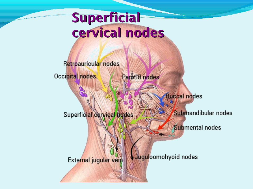
Superficial Superficial cervical nodescervical nodes

1. Upper deep cervical
2. Lower deep cervical
3. Waldyer’s ring
4. Nodes of midline
The Deep lymph nodes:The Deep lymph nodes:

1. The upper deep cervical:Jugulo-digastric group: lie along the upper part of internal jugular vein deep to the sternomastoid muscle.
Structures drained: Tonsil and Tongue.
2. The lower deep cervical:Jugulo-omohyoid group: arranged along the lower part internal jugular vein deep also to the sternomastoid muscle.
Structures drained: Tongue.

Deep cervical nodes
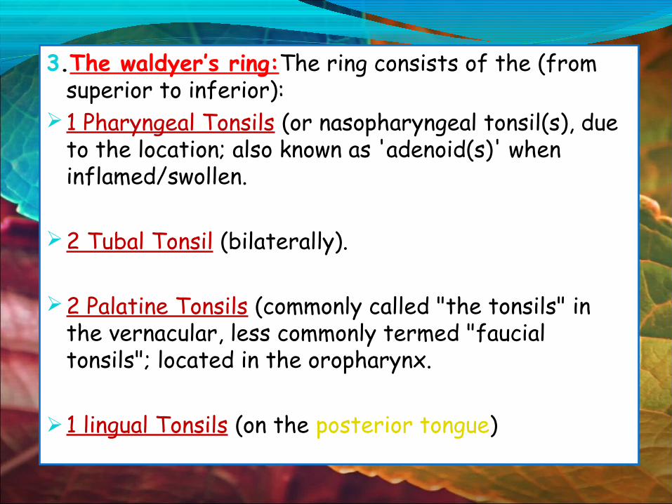
3.The waldyer’s ring:The ring consists of the (from superior to inferior):
1 Pharyngeal Tonsils (or nasopharyngeal tonsil(s), due to the location; also known as 'adenoid(s)' when inflamed/swollen.
2 Tubal Tonsil (bilaterally).
2 Palatine Tonsils (commonly called "the tonsils" in the vernacular, less commonly termed "faucial tonsils"; located in the oropharynx.
1 lingual Tonsils (on the posterior tongue)


4. Midline nodes are termed in correspondence to the anatomical area where they exist:
A. InfrahyoidB. PrelaryngealC. PretrachealD. Paratracheal

The skin of the head and neck drains :
• The scalp drains into the occipital, mastoid and parotid nodes.
• Lower eye lid and anterior cheek drains into buccal nodes.
• The cheeks drain into the parotid, buccal and submandibular nodes.
• The upper lips and sides of the lower lips drain into the submandibular nodes.
• While the middle third of the lower lip drains into the submental nodes.
• The skin of the neck drains into the cervical nodes.

The drainage of the oral structures
• The Gingivae drain into the submandibular, submental and upper deep cervical lymph nodes.
• The palate drains via lymph vessels that pass through the pharyngeal wall to the upper deep cervical nodes.
• Anterior part of mouth floor drain into submental and upper deep cervical
• while posterior part into submandibular and upper deep cervical.
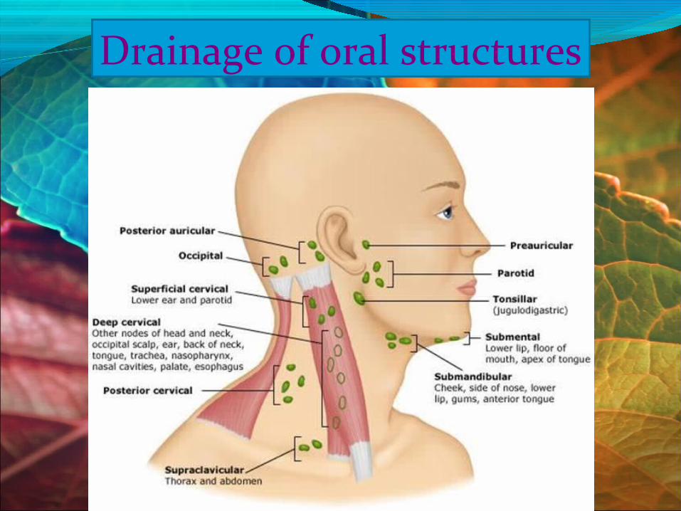
Drainage of oral structures
Lymph drainage of external noseLymph drainage of external nose
Lymph drainage of external nose is primarily to the submandibular group of nodes although lymph from the root of the nose drains to superficial parotid nodes.

Lymphatic drainage of nasal cavity
Lymph vessels from the anterior region of the nasal cavity pass superficially to join those draining the external nasal skin, and end in the submandibular nodes.
The rest of the nasal cavity, paranasal sinuses, nasopharynx and pharyngeal end of the pharyngotympanic tube, all drain to the upper deep cervical nodes either directly or through the retropharyngeal nodes.
The posterior nasal floor drains to the parotid nodes.

Lymphatic drainage of tongue:
The lymphatic drainage of the tongue can be divided into 3 main regions: Marginal, Central and Dorsal.
The anterior region of the tongue drains into marginal and central vessels, and the posterior part of the tongue behind the circumvallate papillae drains into the dorsal lymph vessels.
The more central regions drain bilaterally into sub-mental and sub-mandibular nodes.


Lymphatic drainage of teeth:
The lymph vessels from the teeth usually run directly into the ipsi-lateral submandibular lymph nodes.
Lymph from the mandibular incisors, however, drains into the submental lymph nodes.
Occasionally, lymph from the molars may pass directly into the jugulo-digastric group of nodes.

APPLIED ASPECT:
When a lymph node is under challenge from a foreign antigen, it may become swollen and painful to the touch— a condition called lymphadenitis.
Commonly palpated and accessible lymph nodes are - the cervical, axillary, and inguinal.
Lymph nodes are common sites of metastatic cancer because cancer cells from almost any organ can break loose, enter the lymphatic capillaries, and lodge in the nodes.
Lymphadenopathy is a collective term for all lymph node diseases.

Differential Diagnosis of cervical Lymphadenopathy based on palpatory findings. TEXTURE OF LYMPH NODES

Conclusion :-
In conclusion, the lymphatic system and its organs are widespread and scattered throughout the body.
Because the vessels of the lymphatic system span the entire body it becomes an easy portal for the spread of cancer and other diseases, which is why disorders and diseases of this system can be very devastating.
A sound knowledge of the regional lymph nodes of the head and neck is very important for dentists because it is a reliable guide towards the origin of problem, and because of the possible involvement of the lymphatic system in the spread of infection or the spread of malignant tumour cells (metastasis).

Textbook of Head and Neck Anatomy (Hiatt – Gartner) 4th Ed. 2010Grant's Atlas of Anatomy,13th Ed.Gray's Anatomy – 40th Ed.Anatomy of the Human Body - Henry GraySaladin: Anatomy & Physiology: The Unity of Form and Function, 3rd
EditionEmbryology Atlas , John F NeasLife Map – Embryonic development & stem cell compendiumButler M G, Isogai S, Weinstein B M. Lymphatic development, Birth
Defects Res C Embryo Today. 2009 September ; 87(3): 222–231.Albrecht I, Christofori G, Molecular mechanisms of
lymphangiogenesis in development and cancer; Int. J. Dev. Biol. 55: 483-494
References :-

Ferrer R, Lymphadenopathy: Differential Diagnosis and Evaluation; Am Fam Physician. 1998 Oct 15;58(6):1313-1320Som P M, Hugh D C, Mancuso A A, An imaging-based classification for the cervical nodes designed as an adjunct to recent clinically based nodal classification;Arch Otolaryngol Head Neck Surg. 1999;125:388-396.Oliver G, Detmar M, The rediscovery of the lymphatic system: old and new insights intothe development and biological function of the lymphatic vasculature; Genes Dev. 2002,16: 773-783Sambandan T, Mabel C R, Cervical lymphadenopathy- a review; JIADS,2011,2,1:31-33.Torabi M, Aquino S L, Harisinghani M G, Current concept in lymph node imaging; J Nucl Med, 2004; 45:1509-1518.Connor SEJ, Olliff JFC, Imaging of malignant cervical lymphadenopathy – a review; Dentomaxillofacial Radiology, 2000;29:133-143.

“The earliest evidence of ancient dentistry -an amazingly detailed dental work on a mummy from ancient Egypt that archaeologists have dated to 2000 BCE. The work shows intricate gold work around the teeth. This mummy was found with two donor teeth that had holes drilled into them. Wires were strung through the holes and then around the neighboring teeth.” Source: metalonmetal blog.


