56
Lymphatic System Vessels, Organs and Tissues
| Date post: | 21-Jan-2016 |
| Category: |
Documents |
| Upload: | thomasina-caldwell |
| View: | 233 times |
| Download: | 3 times |

Lymphatic System
Vessels, Organs and Tissues

Lymphatic System
Vessels: Connect to CV Lymphatics
Capillaries Collecting vessels Trunks
Transport fluid => lymph Organs: house immune cells
LN Spleen Thymus
Tissues Tonsils GALT MALT
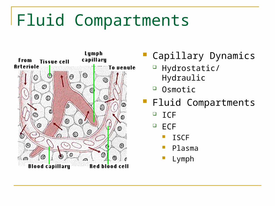
Fluid Compartments
Capillary Dynamics Hydrostatic/Hydraulic Osmotic
Fluid Compartments ICF ECF
ISCF Plasma Lymph
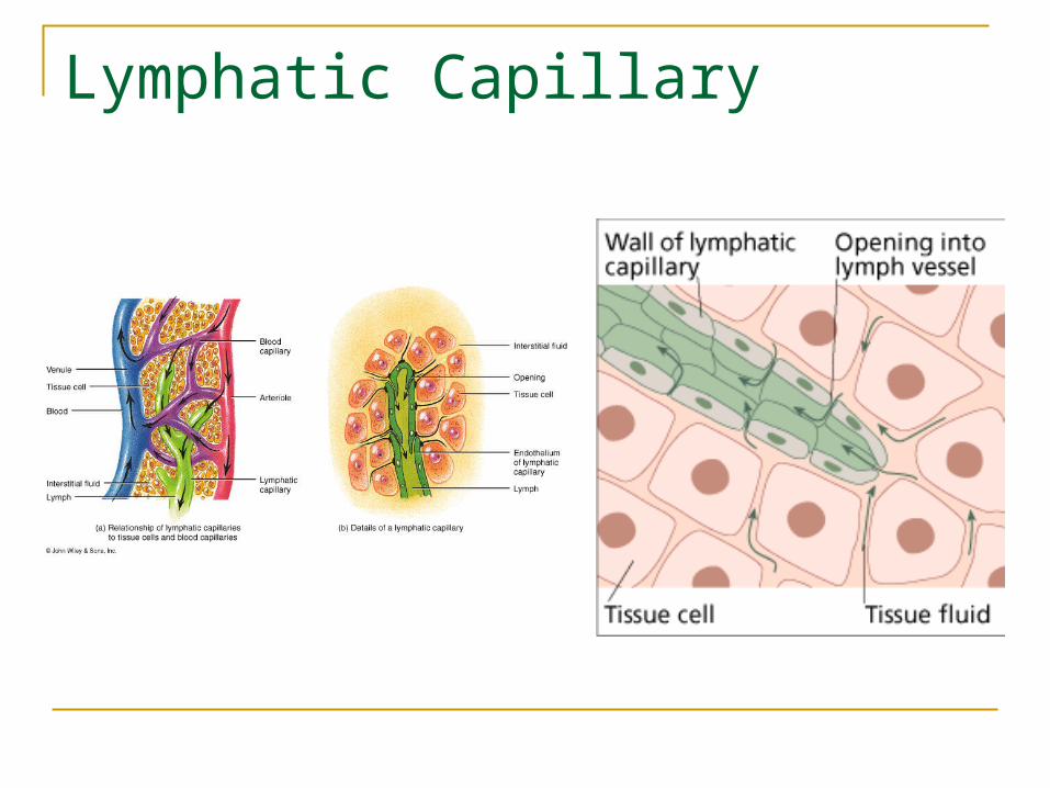
Lymphatic Capillary

Lymph Flow

Lymphatic Valve

Lymphatic Vessels and Ducts
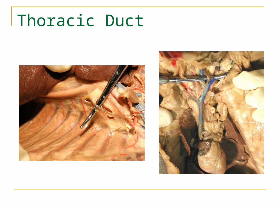
Thoracic Duct

Lymph Return

Problem Lymph Return : Lymphedema
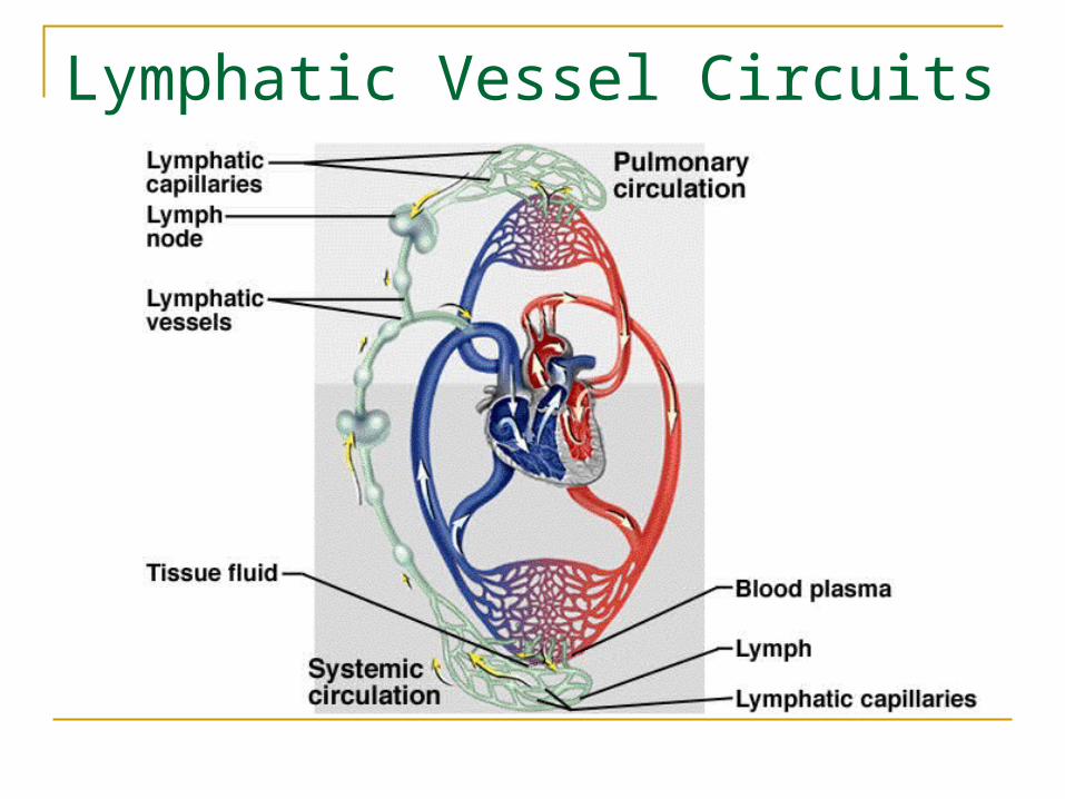
Lymphatic Vessel Circuits

Lymph Node: Location

L.N. : Specimen

Lymph Node: Gross Anatomy
1. Afferent Lymphatics 2. Cortex 3. Germinal Center 4. Capsule 5. Medullary cord 6. Valve 7. Efferent Lymphatics
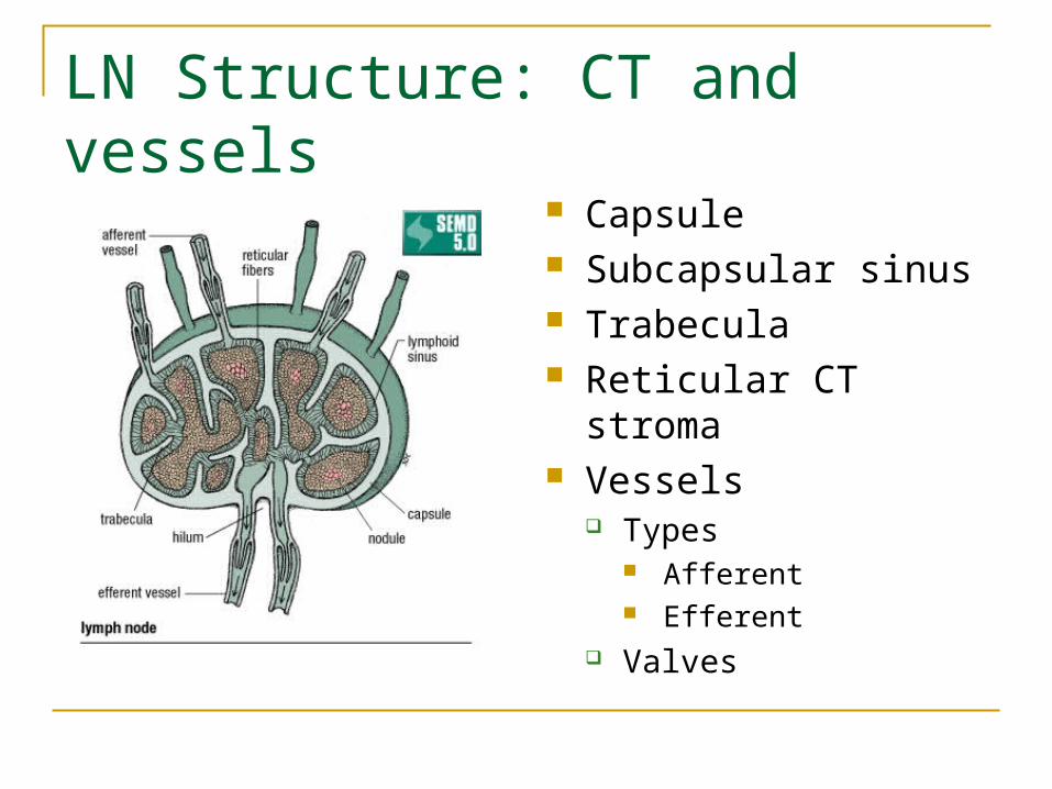
LN Structure: CT and vessels
Capsule Subcapsular sinus Trabecula Reticular CT stroma Vessels
Types Afferent Efferent
Valves

Lymph Node

Lymph Node: Histology
B cellsT cellsMO

Lymph Node: Capsule Region

Lymph Node: Cortex
Lymphatic NoduleGerminal Center

Lymph Node: Medulla

Lymph Node: Medullary Cords
Medullary CordsSinusoids
Macrophages

L.N. Medullary Cells
P = Plasma cells (daughter cells of Activated B cells M = Macrophages (mature monocytes in tissues)

LN Histology Summary

Diagnostics for Lymphatics and LN
Lymphangiogram
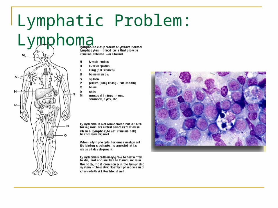
Lymphatic Problem: Lymphoma
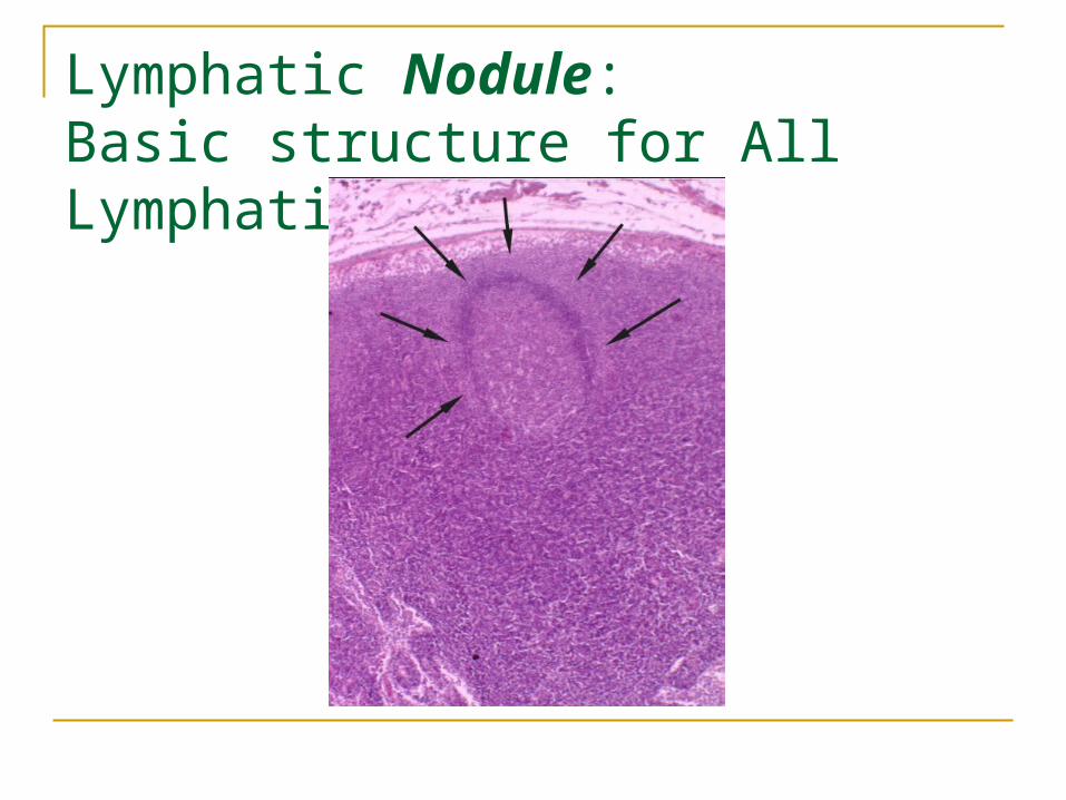
Lymphatic Nodule: Basic structure for All Lymphatic tissue
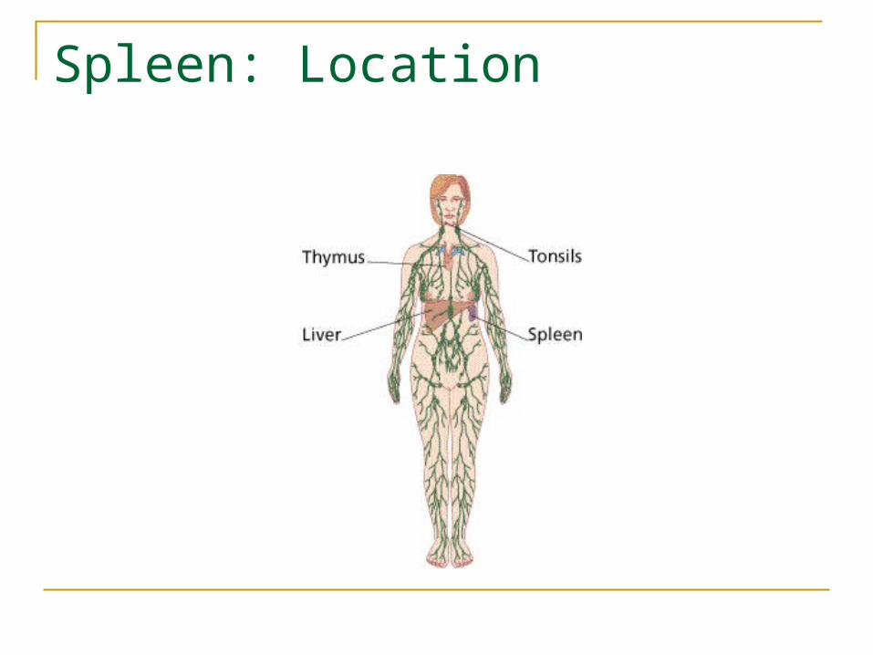
Spleen: Location

Spleen: Gross Anatomy

Spleen: Histology
SplenicSinusoid

Spleen Problems
Rupture Cancer Infection
Hydatid cyst

Thymus: Location

Thymus: Gross

Thymus Embryological Development 1. Mandibular arch
2. Hyoid arch 3. Cervical sinus entry 4. 3rd pharyngeal pouch 5. 4th pharyngeal pouch 6. Foramen cecum 7. Thyroid 8. Cervical sinus 9. Thymus (3rd pouch) 10. Thymus (4th pouch)

Thymic Cell Development 1. Thymic capsule 2. Thymic nurse cells 3. CT septa and BV 4. Subcapsular epithelium
(for the blood thymus barrier) 5. Cortical Epithelial cells 6. Medullary epithelial cells 7. Dendritic cells 8. Hassall’s Corpuscle 9. Macrophages 10. Cortex 11. Medulla
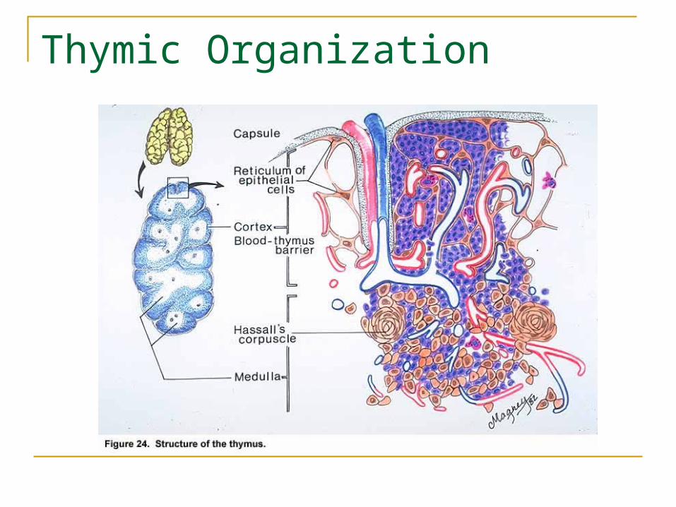
Thymic Organization

Thymic Capsule, Septa and Lobules

Thymus: Cortex

Thymic Medulla

Thymic Age Degeneration

Thymic Problem
Thymic Aplasia

Tonsil: Location
Pharyngeal [Adenoids] Palantine Lingual Tubal

Palatine Tonsil

Tonsil: Epithelial Histology

Tonsil Lymphatic Nodules
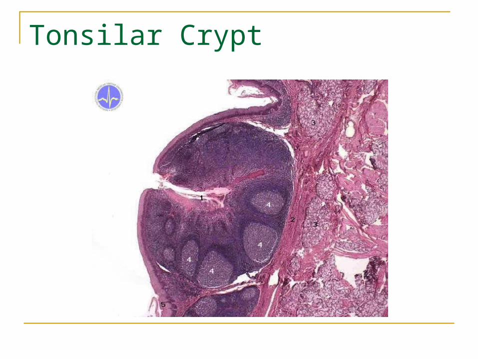
Tonsilar Crypt

Tonsil Problem: Tonsillitis

Gut Associated Lymphatic Tissue (GALT) Location
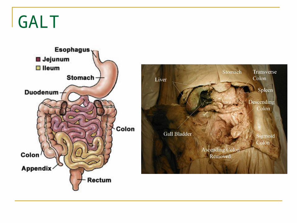
GALT

Peyer’s Patches

Ileum Problems
Inflammation Necrosis

Appendix: Gross
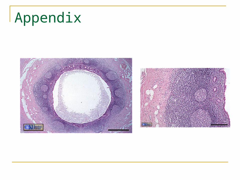
Appendix

Appendix Problems
Appendicitis
Appendix rupture and necrosis

MALT
bronchusepiglottis

Summary Overview

Questions?


