LYRIC/AEG-1Is Targeted to Different Subcellular Compartments byUbiquitinylation and Intrinsic Nuclear Localization SignalsHayleyJ. Thirkettle,1JoanneGirling,1AnneY. Warren,2 Ian G.Mills,1Kanagasabai Sahadevan,3 Hing Leung,4
Freddie Hamdy,5 Hayley C. Whitaker,1and David E. Neal1
Abstract Purpose: LYRIC/AEG-1has been reported to influence breast cancer survival and metastases,and its altered expression has been found in a number of cancers.The cellular function of LYRIC/AEG-1has previously been related to its subcellular distribution in cell lines. LYRIC/AEG-1containsthree uncharacterized nuclear localization signals (NLS), which may regulate its distribution and,ultimately, function in cells.Experimental Design: Immunohistochemistry of ahumanprostate tissuemicroarray composedof 179 prostate cancer and 24 benign samples was used to assess LYRIC/AEG-1distribution.Green fluorescent protein-NLS fusion proteins and deletion constructs were used to show theability of LYRIC/AEG-1NLS to target green fluorescent protein from the cytoplasm to the nucleus.Immunoprecipitation andWestern blotting were used to show posttranslational modification ofLYRIC/AEG-1NLS regions.Results: Using a prostate tissue microarray, significant changes in the distribution of LYRIC/AEG-1were observed in prostate cancer as an increased cytoplasmic distribution in tumorscompared with benign tissue.These differences were most marked in high grade and aggressiveprostate cancers and were associated with decreased survival. The COOH-terminal extendedNLS-3 (amino acids 546-582) is the predominant regulator of nuclear localization, whereasextended NLS-1 (amino acids 78-130) regulates its nucleolar localization.Within the extendedNLS-2 region (amino acids 415-486), LYRIC/AEG-1can be modified by ubiquitin almost exclu-sively within the cytoplasm.Conclusions: Changes in LYRIC/AEG-1subcellular distribution can predict Gleason grade andsurvival. Two lysine-rich regions (NLS-1 and NLS-3) can target LYRIC/AEG-1 to subcellularcompartments whereas NLS-2 is modified by ubiquitin in the cytoplasm.
Lysine-rich CEACAM-1–associated protein (LYRIC; ref. 1),also known as metastasis adhesion protein (metadherin;ref. 2), and astrocyte elevated gene-1 (AEG-1; ref. 3) wasoriginally identified as part of a serial analysis of gene
expression, investigating proteins up-regulated in the andro-gen-treated LNCaP prostate cancer cell line (4). LYRIC/AEG-1was subsequently found to be overexpressed in breast, brain,and prostate cancer (5–8) and localized to the cell membrane,endoplasmic reticulum, nucleolus, cytoplasm, and nucleus invarious cell lines (1, 5, 6).Various functions of LYRIC/AEG-1 have been suggested in a
number of subcellular compartments; however, its exactfunction remains poorly understood. At the membrane ofpolarized cells, LYRIC/AEG-1 has been linked to cell adhesionthrough colocalization with ZO-1 at tight junctions (5). Thismay be linked to the loss of cell polarity that is known to occurwith increased epithelial tumorigenicity (9). LYRIC/AEG-1 hasbeen shown to suppress the activation of FOXO3a, a tran-scription factor regulated by AKT phosphorylation (7). Theoncogenic potential of LYRIC/AEG-1 has been shown by itsability to transform melanocytes (3) and more recently shownby its ability to activate nuclear factor-nB through an interactionwith CREB-binding protein/p300 (6, 10). Furthermore, LYRIC/AEG-1 can be induced by oncogenic Ha-ras through phospha-tidylinositol 3-kinase/AKT (10, 11). Most recently, LYRIC/AEG-1 has recently been shown to interact with the cell cycleregulator BRCA2 and CDKN1A-interacting protein (BCCIPa)and down-regulate its expression by targeting it for degradation(12). Thus, LYRIC/AEG-1 has many functions in different
Human Cancer Biology
Authors’Affiliations: 1Uro-Oncology Research Group, Cancer Research UK,Cambridge Research Institute and 2Department of Pathology, AddenbrookesHospital, Cambridge, United Kingdom; 3Sunderland Royal Hospital, Kayll Road,Sunderland, United Kingdom; 4Beatson Institute for Cancer Research, GarscubeEstate, Switchback Road, Glasgow, United Kingdom; and 5Academic UrologyUnit, Division of Clinical Sciences South, University of Sheffield, Sheffield,United KingdomReceived8/5/08; revised1/15/09; accepted2/3/09;publishedOnlineFirst 4/21/09.Grant support: Cancer Research UK,The Medical Research Council,The BritishUrological Foundation, and The ShackmanTrust, with support from the PROMPTprostate cancer collaborative.The costs of publication of this article were defrayed in part by the payment of pagecharges.This article must therefore be hereby marked advertisement in accordancewith18 U.S.C. Section1734 solely to indicate this fact.Note: Supplementary data for this article are available at Clinical Cancer ResearchOnline (http://clincancerres.aacrjournals.org/).H.J.Thirkettle andJ. Girling, and H.C.Whitaker and D.E. Neal contributed equally tothe work.Requests for reprints: HayleyWhitaker, Uro-Oncology Research Group, CancerResearch UK Cambridge Research Institute, Li Ka Shing Centre, RobinsonWay,Cambridge CB2 0RE, United Kingdom. Phone: 44-01223-404450; Fax: 44-01223-404199; E-mail: [email protected].
F2009 American Association for Cancer Research.doi:10.1158/1078-0432.CCR-08-2046
www.aacrjournals.org Clin Cancer Res 2009;15(9) May1, 20093003
subcellular compartments and it is postulated that its localiza-tion may have a role in regulating its function.It has previously been noted that LYRIC/AEG-1 contains
many lysine residues (>12%) with the majority of these lysines(68%) clustered in three regions, which have been purported tobe nuclear localization signals (NLS; refs. 1, 6). NLS are well-characterized amino acid motifs that can be divided into twoclasses (monopartite and bipartite). Other lysine-rich motifscan act as nucleolar localization signals (NoLS) and targetproteins to the nucleolus. Although critical for nucleartargeting, lysine residues are also targets for posttranslationalmodification by ubiquitin and small-ubiquitin–like modifier(SUMO), which can either direct proteins to the proteasome fordegradation or target proteins to different regions of the cell,including the nucleus (13–15).In this present study, we have examined the localization
and expression of LYRIC/AEG-1 by immunohistochemistry ina number of tissues and in particular in a large cohort of pro-state cancer patients. We have seen a significant redistributionof LYRIC/AEG-1 from the nucleus in benign tissue to the cyto-plasm and membrane in tumorigenic and metastatic disease.To understand how LYRIC/AEG-1 localization is regulated, wehave characterized which of the lysine-rich regions of LYRIC/AEG-1 can act as NLS or NoLS. We show that lysine-rich regionsthat we have named exNLS-1 and exNLS-3 are required forredistribution of LYRIC/AEG-1 within the cell. We also showthat LYRIC/AEG-1 is posttranslationally modified by ubiquitinand, to a lesser degree, by SUMO, predominantly within theexNLS-2 lysine-rich region. Furthermore, we show that ubi-quitinylation is most likely monoubiquitinylation and itoccurs exclusively in the cytoplasm. Thus, LYRIC/AEG-1lysine-rich regions and their posttranslational modificationmay play an important role in regulating LYRIC/AEG-1 locali-zation and ultimately function in normal and tumorigenic cells.
Materials andMethods
LYRIC/AEG-1 antibodies and plasmids. LYRIC/AEG-1 was detectedusing two sheep anti-LYRIC/AEG-1 antibodies kindly supplied with
blocking peptides by Heidi Sutherland (MRC Human Genetics Unit,Edinburgh, United Kingdom) (1). The antibody that recognizesresidues 197SHREKRQQRKRDKV210 is called AK, whereas the COOH-terminal antibody recognizing residues 568SPKQIKKKKKARRET582 isreferred to as SS. Green fluorescent protein (GFP)-tagged NLSconstructs were created using pQBI25-fC3 and pQBI25-fN2 plasmids(Q-BIOgene) and by designing primers (Sigma Genosys) to includelysine-rich regions and/or flanking secondary structure, as predicted byPredictProtein. Site-directed mutagenesis was done using QuickChangeII Site-Directed Mutagenesis kit (Stratagene) following the manufac-turer’s instructions. All primer sequences and plasmids are detailed inSupplementary Table S1. HA-tagged ubiquitin was a gift from JochenRink (Max Planck Institute of Molecular Cell Biology and Genetics,Dresden, Germany) (16). The DSUMO-1 construct was a kind gift fromHelen Hurst (Barts and the London School of Medicine and Dentistry,London, United Kingdom) (17).
Patient population. The patient population was identical to the onepreviously described (18). The tumor grade was classified by a uro-pathologist (A.W.) using the Gleason grading system and classified intolow (Gleason 6 and below), moderate (Gleason 7), and high (Gleason8-10). Following immunostaining, tissue core loss reduced the cohort to179 cancer patients, 83 samples of coexisting benign prostatehyperplasia (BPH) and 24 control BPH patients. To confirm thatstaining in BPH samples from radical prostatectomy and TURP sampleswas identical, whole sections from two radical prostatectomy samplesobtained from Addenbrookes Hospital, Cambridge, were probed forLYRIC/AEG-1. Staining was also done on 11 patient samples from theRoyal Hallamshire Hospital, Sheffield, consisting of bone metastasisthat had received no hormone manipulation therapy and were under-going bilateral orchiectomy at the time of biopsy. Informed consent andethical approval was obtained for all procedures and studies.
Immunohistochemistry. Confirmation of tissue status (Gleasongrades and BPH) was conducted by an uropathologist, who assessedand marked the blocks appropriately. Duplicate 0.6-mm tissue coreswere cut and constructed according to predetermined tissue microarray(TMA) layout. Multiple 5-Am sections were cut from TMA forimmunohistochemistry. Full thickness bone biopsies (5-10 mm cores)taken from the iliac crest were fixed in formol saline (10%) for 48 h,decalcified for up to 3 wk in a solution containing EDTA before 4-Amsections cut, and mounted on slides as before (19). A normal bone TMA(US Biomax) was used as a control. A multinormal/tumor TMA (Zymed)was used to assess LYRIC/AEG-1 staining in a wide variety of tissues.
All staining was done under identical conditions to allow for
comparison over different TMA slides. Tissue was deparaffinized,
rehydrated, and antigen retrieval was done in citrate buffer (pH 6.0;
10 mmol/L trisodium citrate, 0.05% Tween) for 15 min at >90jC. Toreduce the possibility of epitope masking effects, LYRIC/AEG-1 was
detected using a 1:1 ratio of AK and SS antibodies (1:50) overnight at
4jC. Negative controls were done using sheep IgG (Upstate Cell
Bioclear) were used at 1:100, followed by ABC complex (Vectastain
ABC kit, Vector Laboratories, Inc.). Staining was visualized with
diaminobenzidine tetrahydrochloride (Vector Laboratories) and the
nucleus was counterstained lightly with hematoxylin (Vector Labora-
tories). Tissues were mounted and visualized as described (18).
Immunostaining assessment. In those patients where duplicate cores
remained intact, immunostaining was evaluated according to staining
intensity and localization, independently of each other. Scoring wasdone independently by two observers (one an independent specialist
uro-oncology pathologist) both blinded to the TMA plan. Staining was
classified into absent, low, moderate, and high intensity. Localization
was categorized according to the predominant staining region, i.e.,
nuclear, cytoplasmic, or membranous. If equal in two or more areas,
this was classed as mixed cytoplasmic/nuclear. If staining was equal in
all areas of the cell, it was classed as global. A consensus agreement was
reached on intensity and localization on each core. Statistical analysis
on immunohistochemistry data was done on the consensus score using
Translational Relevance
Previous studies noted the importance of LYRIC/AEG-1in tumorigenesis, linking it to survival in breast tumors, andsuggesting that it may function as a tumor suppressor.LYRIC/AEG-1localization is thought toplaya significant rolein regulating its function, resulting in speculation aboutthe importance and function of the lysine-rich regions asputative nuclear localization motifs.We have shown thatLYRIC/AEG-1 is lost from the nucleus with increasingtumorigenesis and decrease survival in prostate tumors.This is consistent with a potential loss of tumor suppressoractivity.Achangeindistributioncouldbeusedasabiomark-er of tumor progression. By understanding how LYRIC/AEG-1localization is regulated by its nuclear localizationmotifs, it may ultimately be possible to redistribute LYRIC/AEG-1 to the nucleus in tumors as a therapeutic tool tosuppress tumorigenesis.
Human Cancer Biology
www.aacrjournals.orgClin Cancer Res 2009;15(9)May1, 2009 3004
a using a two-tailed Fisher’s exact test. Results were considered
significant if the P value was <0.05.For a subset of 50 patients, survival data was collected for up to
120 mo. Patients were grouped according to their LYRIC/AEG-1 stainingpattern; those with any nuclear staining, including mixed cytoplasmic/nuclear, were placed in one group. Those patients with no nuclearstaining were placed in a separate group. Data were analyzed usingGraphPad Prism and statistical difference was determined usinga two-tailed t test.
Cell culture. All cell lines were purchased from the Cancer ResearchUK cell bank except the benign prostatic PNT1a cells, which were a kindgift from Norman Maitland (Yorkshire Cancer Research, Harrogate,
North Yorkshire, United Kingdom) and VCaP cells that were a kind giftfrom Marion Bussemakers (Radboud University Nijmegen MedicalCentre, Nijmegen, The Netherlands). PNT1a, LNCaP, VCaP, PC3, andNIH3T3 cells were routinely cultured in RPMI medium (Life Technol-ogies). COS-7 and MDBK cells were routinely cultured in DMEM (LifeTechnologies). All media were supplemented with 10% fetal bovineserum (Labtech). Androgen regulation experiments were done aspreviously described (20) and media were supplemented with eitherthe dihydrotestosterone analogue R1881 (10 nmol/L; Sigma) or anequal volume of vehicle (ethanol) for 24 h before being harvested. Fortransfection, cells were grown to 40% confluency. DNA (1 Ag per 6 wellsof a 24-well plate or 4 Ag per 14-cm dish) was transfected using FuGENE
Fig. 1. A, a schematic representation of LYRIC/AEG-1highlighting the leucine-rich region (black) and lysine-rich regions (light gray). Dotted lines, antibody epitopes forAK and SS. B, the specificity of the LYRIC/AEG-1antibodies (AK and SS) was tested by separating 20 Ag LNCaP (+/- R1881), COS, MDBK, and NIH3T3 whole-celllysate by SDS-PAGE, transferring to nitrocellulose, and probing with eitherAK or SS (1:2,000). Duplicate blots were incubated with antibody in the presence of 5 Agspecific blocking peptide but otherwise treated in an identical manner. Proteins were visualized with ECL-Plus. C, a multitumor/normalTMA and bone sections were stainedfor LYRIC/AEG-1 (1:50; brown) and the nuclei were counterstained with hematoxylin (blue): benign lung (i), benign thyroid (ii), benign prostate (iii), prostate tumor (iv),normal bone (v), prostate bone metastases (vi), and thyroid tumor (vii ; �60) and a zoomed image with arrows indicating the nucleolar staining (viii). All other imageswere captured using�40 magnification except bone images that were collected at �20 magnification.
Regulation of LYRIC/AEG-1Localization
www.aacrjournals.org Clin Cancer Res 2009;15(9) May1, 20093005
6 (Roche) following the manufacturer’s protocol. Cells were grown fora further 48 h before confocal microscopy or Western analysis.
Cellular fractionation. All procedures were done on ice or at 4jC.Cells were harvested at 95% confluency by scraping into 3 mL PBS andcentrifuging at 800 � g . Cells were resuspended in 500 AL modifiedRIPA buffer (as described in ref. 18) by ‘‘flicking’’ and incubated for5 min before centrifuging at 800 � g for 5 min. The supernatant wasretained as the cytoplasmic fraction. Nuclei were washed thrice in500 AL RIPA buffer and resuspended in 100 AL RIPA buffer, andsonicated thrice for 10 s. The sonicated nuclei were centrifuged at16,100 � g for 1 min and the supernatant was retained as the nuclearfraction. Protein concentrations were determined using the Bradfordreagent (Bio-Rad).
Cell lysates, Western blotting, and immunoprecipitation. All proce-dures were done as described in ref. (20). Membranes were incubatedwith primary antibody—anti-RNA polymerase II (1:2,000), anti-GFP(1:5,000), anti-fibrillarin (1:1,000), anti-histone H3 (1:5,000), andanti-actin (1:5,000; all from Abcam); anti–g-adaptin (1:2,000, BDTransduction Laboratories), anti-p65 (1:1,000, Abcam), or anti-LYRIC/AEG-1 SS or AK at 1:2,000. Secondary antibodies (1:1,000, DakoCy-
tomation) were used for all antibodies. Protein bands were detectedwith ECL-Plus (GE Healthcare), and where they exceeded the dynamicrange of film, diaminobenzidine (Vector Laboratories) was used. Forpeptide blocking experiments, duplicate Western blots were blocked in5% bovine serum albumin diluted in PBS. AK and SS antibodies werealso diluted in 5% bovine serum albumin-PBS either alone or with 5 Agof corresponding blocking peptide and incubated with membranesat 4jC overnight. Western blots were then completed as described.Densitometry was done using Image Scanner III and ImageQuant TLsoftware from Amersham Biosciences. SUMOylated and ubiquitinylatedLYRIC bands were normalized to GFP-LYRIC band intensity.For immunoprecipitation, all of the procedure was done at 4jC. For
each sample, 100 AL of protein G Sepharose beads (GE Healthcare) waswashed in modified RIPA buffer. After preclearing, 1 mg of total proteinwas incubated with antibody, LYRIC/AEG-1 SS (5 AL) or GFP (1 AL). Anequal concentration of sheep (Upstate Cell Signalling Solutions),mouse, or rabbit (Vector laboratories) immunoglobulin was used ascontrols. Following the addition of protein G, Sepharose beads werewashed in modified RIPA buffer containing 2% NP40 and boiled for10 min in 30 AL SDS-PAGE sample buffer before Western blot analysis.
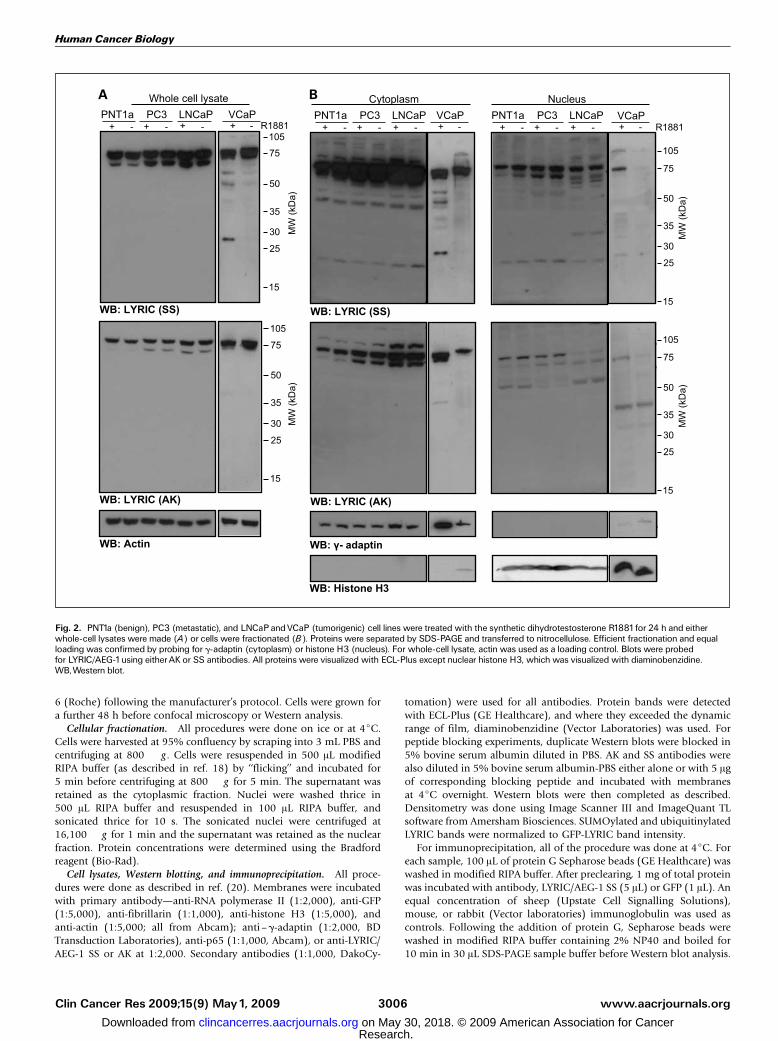
Fig. 2. PNT1a (benign), PC3 (metastatic), and LNCaPandVCaP (tumorigenic) cell lines were treated with the synthetic dihydrotestosterone R1881for 24 h and eitherwhole-cell lysates were made (A) or cells were fractionated (B). Proteins were separated by SDS-PAGE and transferred to nitrocellulose. Efficient fractionation and equalloading was confirmed by probing for g-adaptin (cytoplasm) or histone H3 (nucleus). For whole-cell lysate, actin was used as a loading control. Blots were probedfor LYRIC/AEG-1using eitherAK or SS antibodies. All proteins were visualized with ECL-Plus except nuclear histone H3, which was visualized with diaminobenzidine.WB,Western blot.
Human Cancer Biology
www.aacrjournals.orgClin Cancer Res 2009;15(9)May1, 2009 3006
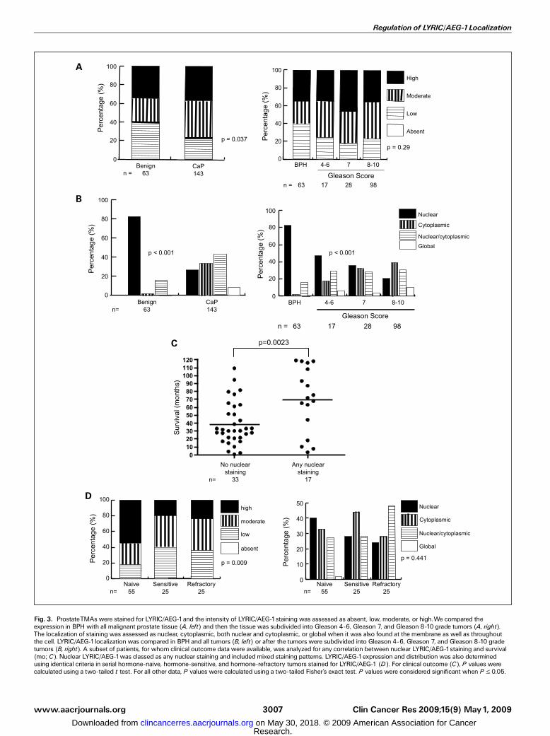
Fig. 3. ProstateTMAs were stained for LYRIC/AEG-1and the intensity of LYRIC/AEG-1staining was assessed as absent, low, moderate, or high.We compared theexpression in BPH with all malignant prostate tissue (A, left) and then the tissue was subdivided into Gleason 4-6, Gleason 7, and Gleason 8-10 grade tumors (A, right).The localization of staining was assessed as nuclear, cytoplasmic, both nuclear and cytoplasmic, or global when it was also found at the membrane as well as throughoutthe cell. LYRIC/AEG-1localization was compared in BPH and all tumors (B, left) or after the tumors were subdivided into Gleason 4-6, Gleason 7, and Gleason 8-10 gradetumors (B, right). A subset of patients, for whom clinical outcome data were available, was analyzed for any correlation between nuclear LYRIC/AEG-1staining and survival(mo; C). Nuclear LYRIC/AEG-1was classed as any nuclear staining and included mixed staining patterns. LYRIC/AEG-1expression and distribution was also determinedusing identical criteria in serial hormone-naive, hormone-sensitive, and hormone-refractory tumors stained for LYRIC/AEG-1 (D). For clinical outcome (C), P values werecalculated using a two-tailed t test. For all other data, P values were calculated using a two-tailed Fisher’s exact test. P values were considered significant when P V 0.05.
Regulation of LYRIC/AEG-1Localization
www.aacrjournals.org Clin Cancer Res 2009;15(9) May1, 20093007
Confocal microscopy. Cells were grown, fixed stained, and mountedas described (20) using an anti-fibrillarin antibody (1:400) and594-conjugated Alexafluor secondary antibody (1:10,000, MolecularProbes). All scale bars represent 10 Am.
Results
LYRIC/AEG-1 localization alters with tumorigenesis andpredicts longer survival. Anti-LYRIC/AEG-1 AK or SS anti-bodies, which recognize amino acids 197-210 (AK) and 568-582 (SS; Fig. 1A), have been characterized previously indifferent subnuclear compartments (1). As multiple bandswere seen by Western analysis, we examined their specificityusing blocking peptides corresponding to the antigenic aminoacids. We showed the loss of the majority of bands for bothantibodies, suggesting that staining is specific for LYRIC/AEG-1(Fig. 1B). We then used these antibodies to assess LYRIC/AEG-1expression in human tissues by staining a TMA consisting ofnormal and cancerous tissues from a variety of organs.All of the various tissues that we examined showed some
LYRIC/AEG-1 staining (Fig. 1C). Predominantly nuclear stainingwas only seen in benign tissues, including the prostate, thyroid,and lung. Tumorigenic tissue had comparatively low levels ofnuclear staining; however, nucleolar staining was noted (Fig. 1C,vii and viii), which was not seen in the benign tissues weexamined. As prostate cancer is known to frequently metastasizeto bone, we obtained 11 bone metastases and probed them forLYRIC/AEG-1 expression. When compared with normal bonecontrols, 81.8% (9 of 11) of prostate bonemetastases showed anincreased expression of LYRIC/AEG-1 (Fig. 1C, v and vi). LYRIC/AEG-1 was almost exclusively distributed in the cytoplasm andmembrane in the prostate bone metastases.To identify if LYRIC/AEG-1 distribution was altered by
tumorigenesis in the prostate, we examined its expression andlocalization in a range of cell lines: benign (PNT1a),tumorigenic LNCaP and VCaP, and highly metastatic (PC3)cells. All cells were treated with the dihydrotestosteroneanalogue R1881 for 24 hours to determine if androgens haveany effect on LYRIC/AEG-1 distribution or expression. The cellswere either lysed intact (Fig. 2A) or separated into nuclear andcytoplasmic fractions, which were confirmed by Westernblotting for g-adaptin (cytoplasm) and histone H3 (nucleus;Fig. 2B). In whole-cell lysates, we saw a singlet or doublet atf75 kDa, consistent with previous reports using these andother anti-LYRIC/AEG-1 antibodies. The lower molecularweight band of the doublet was weaker with the SS antibody(amino acids 568-582) or lost with the AK antibody (aminoacids 197-210) in the benign PNT1a cell line, suggesting thatthere are differences in LYRIC/AEG-1 distribution in benignand tumorigenic cells. Within the nuclear and cytoplasmic cellfractions, multiple LYRIC/AEG-1 bands were seen in all celllines (Fig. 2B). Some nuclear translocation of LYRIC/AEG-1 wasseen in response to R1881 treatment, particularly in VCaP cells.Loss of a 60-kDa nuclear LYRIC/AEG-1 band was particularlyevident in the nuclear fractions of LNCaP and VCaP cellsprobed with AK, compared with PC3 and PNT1a cells. Somebands, such as those at 28 kDa, were only seen with the AKantibody, whereas a cytoplasmic band at 50 kDa was only seenwith the SS antibody. Changes were also seen on lowermolecular weight bands, such as the appearance of a nuclear35 kDa band in LNCaP and VCaP cells, which suggests that
differences in nuclear and cytoplasmic LYRIC/AEG-1 occurbetween prostate cells at different stages of tumorigenesis.Our initial experiments (Fig. 2) and previously published
data (4, 5) suggest that LYRIC/AEG-1 may be up-regulated inprostate cancer; thus, we used immunohistochemistry to staina prostate TMA consisting of BPH (n = 63) and prostate cancer(n = 143) for LYRIC/AEG-1. The intensity of LYRIC/AEG-1staining increased significantly in malignancy (P = 0.037;Fig. 3A, left). We then determined if these changes in LYRIC/AEG-1 expression were associated with Gleason score bydividing the cancers into low, moderate, and high grade(Fig. 3A, right). Although LYRIC/AEG-1 expression increasesin low Gleason cancers before decreasing with higher Gleasongrade, these changes were not statistically significant (P = 0.29).As we have previously noted changes in LYRIC/AEG-1
distribution between benign and tumorigenic tissues (Fig. 1C)and prostate cell lines (Fig. 2B), we examined the localizationof LYRIC/AEG-1 in the same prostate cancer TMA. LYRIC/AEG-1was localized to the nucleus of luminal cells in 82.5% of benigncases (52 of 63; Fig. 3B, left). Some staining of basal cells wasseen in benign tissue, which was not seen as basal cells werelost with tumorigenesis. In contrast, nuclear LYRIC/AEG-1 wasonly seen in 26.6% (38 of 143) of tumors. As a result, there wasredistribution of LYRIC/AEG-1 to the cytoplasm alone (33.6%),the cytoplasm and nucleus (42.9%), or throughout the cell(8.8%). The decrease in nuclear LYRIC/AEG-1 was associatedwith increased Gleason grade (Fig. 3B, right ; P < 0.001) andmirrored by reciprocal increased changes in cytoplasmicstaining. Clinical follow-up was available for 50 of thesepatients and they were examined for any correlation betweensurvival and the presence or absence of any LYRIC/AEG-1 in thenuclei of cells (Fig. 3C). Patients with nuclear LYRIC/AEG-1had mean survival of 70 months compared with 39 months forpatients without any LYRIC/AEG-1 in the nucleus (P = 0.0023).This suggests that nuclear LYRIC/AEG-1 may have a function inthe nucleus of normal tissue that is lost in tumorigenesis.We also examined a small cohort of patients during hormone
treatment to determine any effects hormone therapy mayhave on LYRIC/AEG-1 expression and localization. Patients notreceiving hormone treatment (hormone naBve) showed greaterhigh-intensity LYRIC/AEG-1 staining than patients receivinghormone treatment regardless of whether the patient wasresponding to hormone treatment (hormone sensitive) or hadbecome hormone resistant (hormone insensitive; Fig. 3D, left ;P = 0.009). There was no significant change in LYRIC/AEG-1localization (P = 0.441; Fig. 3D, right) with hormone ablationtherapy although examination of patients with serial biopsiestaken throughout treatment revealed a more nuclear LYRIC/AEG-1 distribution in patients that remained hormone sensitive(Supplementary Table S2).
Characterization of LYRIC/AEG-1 nuclear localization signals.As LYRIC/AEG-1 is capable of localizing to both the cytoplasmand nucleus in cells, we have examined the regulation of LYRIC/AEG-1 localization by its NLS motifs. Putative LYRIC/AEG-1NLS motifs as suggested by Sutherland et al. (1) weredesignated NLS-1, NLS-2, and NLS-3 and tagged with GFP(Fig. 4A). Using PSORTII, we predicted that all of the NLSpeptides contained either an NLS or NoLS shown as bold (NLS)or underlined (NoLS; Fig. 4A). The localization of the GFP-NLSfusion constructs was determined by confocal microscopy using4¶,6-diamidino-2-phenylindole (DAPI) to colocalize with the
Human Cancer Biology
www.aacrjournals.orgClin Cancer Res 2009;15(9)May1, 2009 3008
Fig. 4. A schematic representation of LYRIC/AEG-1highlighting the NLS and exNLS peptides (A). Gray boxes, the predicted a-helical secondary structure. Predicted NLSand exNLS regions are indicated with amino acid numbering; NLS are indicated in bold and NoLS are underlined. COS-7 cells were transfected with GFP-tagged NLS orexNLS peptide fusion constructs (B ; green) in 24-well plates. Forty-eight hours posttransfection, cells were fixed and stained for the nucleolar marker fibrillarin (red) andDNAwas stained with DAPI (blue). Schematic representations of each construct consistent with A are given above each picture. Scale bars, 10 Am.
Regulation of LYRIC/AEG-1Localization
www.aacrjournals.org Clin Cancer Res 2009;15(9) May1, 20093009
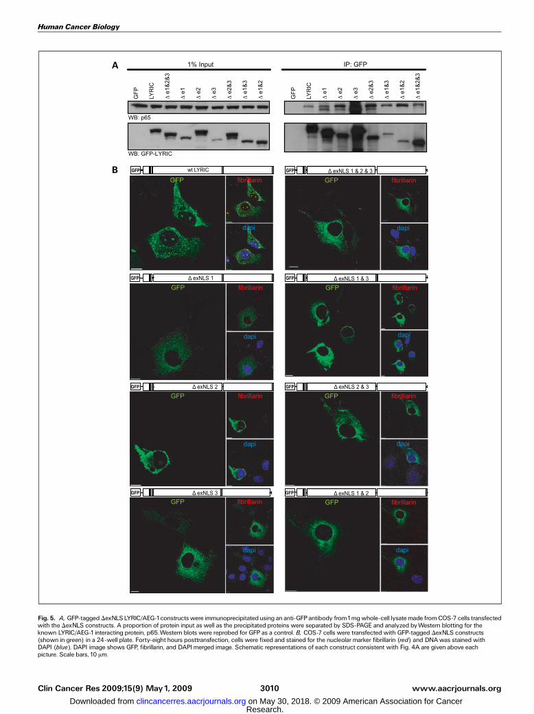
Fig. 5. A, GFP-tagged DexNLSLYRIC/AEG-1constructs were immunoprecipitatedusing an anti-GFPantibody from1mgwhole-cell lysatemade fromCOS-7 cells transfectedwith the DexNLS constructs. A proportion of protein input as well as the precipitated proteins were separated by SDS-PAGE and analyzed byWestern blotting for theknown LYRIC/AEG-1interacting protein, p65.Western blots were reprobed for GFP as a control. B, COS-7 cells were transfected with GFP-tagged DexNLS constructs(shown in green) in a 24-well plate. Forty-eight hours posttransfection, cells were fixed and stained for the nucleolar marker fibrillarin (red) and DNAwas stained withDAPI (blue). DAPI image shows GFP, fibrillarin, and DAPImerged image. Schematic representations of each construct consistent with Fig. 4A are given above eachpicture. Scale bars, 10 Am.
Human Cancer Biology
www.aacrjournals.orgClin Cancer Res 2009;15(9)May1, 2009 3010
nucleus and fibrillarin as a nucleolar marker. GFP alonelocalized throughout the cell, suggesting that any targeting ofthe fusion protein was a result of the associated LYRIC/AEG-1NLS peptide. All of the NLS constructs showed some degreeof nuclear or nucleolar distribution (Fig. 4B; SupplementaryTable S3); however, there were no distinctive differencesbetween the different lysine-rich regions and none of thepeptides were capable of completely translocating GFP to thenucleus. We then extended the NLS regions to include predictedflanking secondary structures (Fig. 4A). The resulting peptidescontained additional NLS and NoLS motifs compared with theshorter NLS peptides. These peptides were designated exNLS-1,exNLS-2, and exNLS-3 (Fig. 4A). The exNLS peptides weretagged with GFP to make a GFP fusion protein and examinedby confocal microscopy as before. The extended constructstargeted GFP to nuclear compartments much more effectively(Fig. 4B; Supplementary Table S3). The additional residues inexNLS-3 resulted in an almost exclusively nuclear and nucleolarlocalization. ExNLS-1, which contained an additional NoLSsequence compared with the shorter NLS fusion, was targetedmuch more strongly to the nucleolus. ExNLS-2 still retained alargely cytoplasmic localization.To confirm that the regions identified as exNLS-1 and exNLS-3
are required for the translocation of LYRIC/AEG-1 into thenucleus and nucleolus, key lysine residues (as defined by thesequence alignment shown in Supplementary Fig. S4) fromeach exNLS region were mutated to alanines in the wild-typeGFP-LYRIC/AEG-1 fusion protein using site-directed mutagen-esis. The resulting constructs still retained nuclear translocatingability, suggesting that the multiple lysines within the LYRIC/AEG-1 NLS regions are able to compensate for one another(Supplementary Fig. S4). To overcome this redundancy, all ofthe exNLS regions were deleted either alone or in combination(indicated by D) from the wild-type GFP-LYRIC/AEG-1 fusionprotein. To show that these constructs were all folded correctly,they were used to immunoprecipitate p65, a known LYRIC/EAG-1 interacting protein (Fig. 5A; ref. 6). All constructsretained p65 binding ability. All of the DexNLS deletionconstructs showed a cytoplasmic and perinuclear distributionand defective nuclear translocation (Fig. 5B; SupplementaryTable S3). The LYRIC/AEG-1 deletion mutant lacking all NLSregions (DexNLS-1/2/3) was completely cytoplasmic, indicatingthat the signal peptide mediating nuclear import resideswithin the extended lysine-rich regions. The DexNLS-2/3, whichcontains only exNLS-1, could not translocate LYRIC/AEG-1 intothe nucleus although the exNLS-1 peptide was capable oftargeting GFP to the nucleolus (Fig. 4B), suggesting that theNLS regions may work cooperatively in regulating LYRIC/AEG-1nuclear translocation. Nucleolar localization was retained incells transfected with mutants containing exNLS-3 but lostwhen exNLS-3 was deleted. This is consistent with exNLS-3being essential for LYRIC/AEG-1 nucleolar localization.
LYRIC/AEG-1 is posttranslationally modified within exNLS-2.Despite its predicted molecular weight of 64 kDa, LYRIC/AEG-1is detected by Western blotting as a 75-kDa band (Fig. 2) bymultiple anti–LYRIC/AEG-1 antibodies. As lysine residues arealso known to be modified by ubiquitin and/or SUMO, weinvestigated if the discrepancy in the molecular weight ofLYRIC/AEG-1 was due to its posttranslational modification.LYRIC/AEG-1 was immunoprecipitated from COS-7 cellsexpressing endogenous protein. Other cells were transiently
transfected with wtLYRIC/AEG-1 plus either SUMO-1 orubiquitin. Using endogenous protein, a single 75-kDa bandconsistent with LYRIC/AEG-1 was detected when Western blotswere probed for ubiquitin, but almost undetectable when blotswere probed for SUMO-1 (Fig. 6A, left). Ubiquitin modificationof LYRIC/AEG-1 was evenmore evident when LYRIC/AEG-1 andubiquitin were overexpressed together (Fig. 6A, central panel).Overexpression of SUMO-1 with wtLYRIC/AEG-1 also resultedin detection of SUMOylated LYRIC/AEG-1 (Fig. 6A, right). Onceagain, SUMOylated LYRIC/AEG-1 was present at a much lowerdegree than ubiquitinated LYRIC/AEG-1, suggesting it is amuch less abundant protein modification, especially underphysiological conditions. To identify if LYRIC/AEG-1 modifica-tion occurs in a specific subcellular compartment, cells werefractionated into nuclear and cytoplasmic fractions (Fig. 6B,left). Ubiquitinylated LYRIC/AEG-1 could only be detected inthe cytoplasm, suggesting that it may influence LYRIC/AEG-1localization (Fig. 6B, middle), possibly by helping retain LYRICin the cytoplasm. SUMOylated LYRIC/AEG-1 was barelydetectable in both subcellular compartments (Fig. 6B, right).To define which residues were modified by ubiquitin and
SUMO, we used the DexNLS-LYRIC/AEG-1 constructs thatlacked specific exNLS regions (Fig. 5). LYRIC/AEG-1 ubiquiti-nylation and any low-level SUMOylation were both lost whenexNLS-2 was deleted (DexNLS-2; Fig. 6C, left and middle).Modification was restored to almost wild-type levels when aconstruct containing only exNLS-2 was used (DexNLS-1/3). Thisconfirms that ubiquitinylation and possibly SUMOylation ofLYRIC/AEG-1 occurs in the exNLS-2 region. To determine ifubiquitinylated LYRIC/AEG-1 was targeted for degradation bypolyubiquitinylation, cells were treated with the proteasomeinhibitor MG132 for 16 hours before lysing. Probing forubiquitin resulted in a single band at 75 kDa and not a ladderof polyubiquitinated bands often seen with proteins destinedfor proteosomal degradation (Fig. 6D). We also noted noobvious stabilization of LYRIC/AEG-1 following MG132 treat-ment. These results suggest that ubiquitination may not targetLYRIC/AEG-1 for degradation and that the ubiquitin modifica-tion of LYRIC/AEG-1 is more likely to be monoubiquination.
Discussion
LYRIC/AEG-1 has previously been localized to numeroussubcellular compartments in a number of studies in cell linesand tissues (1, 2, 5, 7). In addition, studies on small cohorts ofbreast and prostate cancer patients have suggested it may beoverexpressed in tumors compared with benign tissue (2, 7).Using a large cohort of 206 patients, we have confirmed thatLYRIC/AEG-1 is overexpressed in tumorigenic prostate tissue(Fig. 3A). However, the most significant change is in LYRIC/AEG-1 distribution, which changes from nuclear in benigntissue to a predominantly cytoplasmic distribution in tumors, atrend that differs significantly across prostate increasing tumorgrades (Fig. 3B). Localization of LYRIC/AEG-1 to the nucleushas previously been shown in prostate (7) but not in breast (8).By examining patient survival, we have linked nuclear LYRIC/AEG-1 with an increase in mean survival of 31 monthsconsistent with an increase in survival seen in breast tumorswhen LYRIC/AEG-1 is overexpressed in breast cancer (8).Together, these data strongly suggest a tumor suppressorfunction for LYRIC/AEG-1 as has been previously suggested
Regulation of LYRIC/AEG-1Localization
www.aacrjournals.org Clin Cancer Res 2009;15(9) May1, 20093011
(3). Nuclear LYRIC/AEG-1 is also responsible for promotingFOXO-3a–induced apoptosis (7), a mechanism that is morelikely to be lost in tumorigenesis.Characterizing the regulation of LYRIC/AEG-1 distribution to
different subcellular compartments may help us to understand
how its function is regulated within the cell in both normal andtumorigenic tissues. LYRIC/AEG-1 has an unusually largeproportion of lysine residues, which has been suggested to actas NLS (1, 6). We termed the three clusters of lysine residuesNLS-1, NLS-2, and NLS-3 (Fig. 4A) and by tagging them to GFP
Fig. 6. LYRIC/AEG-1was immunoprecipitated (IP) from1mgwhole-cell lysate (WCL) from untransfected COS-7 cells (endogenous) or COS-7 cells transfected withLYRIC/AEG-1and either ubiquitin or SUMO-1 (A) using SS antibody and analyzed byWestern blotting for SUMO, ubiquitin, and LYRIC/AEG-1 (AK). COS-7 cells weretransfected with LYRIC/AEG-1and either ubiquitin or SUMO and 48 h later were fractionated into cytoplasmic or nuclear fractions (B). Efficient fractionation was confirmedbyWestern blotting for g-adaptin (cytoplasm) and RNA polymerase II (nucleus). After immunoprecipitating LYRIC/AEG-1from each fraction,Western blots were probedfor ubiquitin, SUMO-1, and LYRIC/AEG-1 (AK). Cells were also transfected with DexNLSLYRIC/AEG-1constructs and ubiquitin or SUMO-1and lysates were made.LYRIC/AEG-1was immunoprecipitated from1mgwhole-cell lysate using anti-GFP antibody and analyzed byWestern blotting for ubiquitin, SUMO, and GFP. ECL-pluswas used to detect all bands, except where they exceeded the dynamic range of film (RNA pol II and cytoplasmic LYRIC AK blots) where diaminobenzidine was used.SUMOylated/ubiquitinated LYRIC band intensities were compared after normalizing with GFP (C). COS-7 cells transfected with LYRIC/AEG-1and ubiquitin were alsotreated with the proteasome inhibitor MG132 or vehicle (DMSO) 16 h before harvest. Immunoprecipitated LYRIC/AEG-1fromwhole-cell lysate was analyzed usingWestern analysis for ubiquitin and LYRIC/AEG-1 (D).
Human Cancer Biology
www.aacrjournals.orgClin Cancer Res 2009;15(9)May1, 2009 3012
to create fusion proteins, we have shown that they possess onlya limited ability to target proteins to the nuclear compartment(Fig. 4B). When the NLS peptides were extended to includeflanking regions (exNLS; Fig. 4A), additional NLS and NoLSresidues were included and, as a result, their ability to targetGFP to the nucleus and nucleolus was dramatically increased.This shows that the short lysine-rich regions alone are notsufficient to confer nuclear trafficking ability and that otherlysine and charged residues found in the flanking regions areessential for this function, as reported for other proteins (21).Deleting all three exNLS regions seemingly does not alterLYRIC/AEG-1 folding as DexNLS-1/2/3 still retained p65binding capacity while resulting in a completely cytoplasmicform of LYRIC/AEG-1 (Fig. 5; ref. 6).To some extent, almost all NLS constructs targeted LYRIC/
AEG-1 to the nucleolus (Figs. 4 and 5; Supplementary TableS3), consistent with our observations of nucleolar LYRIC/AEG-1 in tumorigenic tissue (Fig. 1C, vii and viii) and existingreports that LYRIC/AEG-1 localizes to the dense fibrillarcompartment of the nucleolus in a mouse embryonic cell line(1). When exNLS-3 was deleted, the remaining NLS could nottarget LYRIC/AEG-1 to the nucleolus (Fig. 5), suggesting thatexNLS-3 is absolutely required for LYRIC/AEG-1 redistributionto the nucleolus. When the exNLS-1 region was deleted, allnuclear localization was abolished and LYRIC/AEG-1 wasdistributed throughout the cytoplasm. Completely nuclearLYRIC/AEG-1 was rarely seen in cultured cells, possibly dueto the leucine-rich putative nuclear export signal (amino acids61-68; Fig. 1A), which was retained in the LYRIC/AEG-1deletion mutants. The extension of NLS-3 resulted in theinclusion of a bipartite NLS (Fig. 4A), giving rise to a morenuclear rather than nucleolar localization (Fig. 4B). The mostpotent NLS is exNLS-1, which targeted GFP exclusively tosubnuclear compartments. We hypothesize that the differentLYRIC/AEG-1 NLS regions may work cooperatively to regulatenuclear and nucleolar localization, with exNLS-1 and exNLS-3being required for nuclear translocation and exNLS-3 for redis-tribution within the nucleus to the nucleolar subcompartment.
Several publications, using a variety of anti-LYRIC/AEG-1antibodies, have detected wtLYRIC/AEG-1 by Western blottingat 75 kDa. We have shown that this band represents LYRIC/AEG-1 modified by ubiquitin and, to a lesser extent, SUMO-1on lysines within the exNLS-2 region (Fig. 6). AlthoughSUMOylation of LYRIC/AEG-1 can be seen, it occurs atextremely low levels, particularly using endogenous proteins,indicating that ubiquitin modification, rather than SUMOyla-tion, is more likely to be significant in physiologic systems. TheexNLS-2 region that is modified by ubiquitin is the leasteffective as an NLS or NoLS. However, exNLS-2 modificationmay cooperate with exNLS-1 and exNLS-3 to act as a regulatorto control nuclear translocation. Treatment of cells with aproteasome inhibitor failed to stabilize LYRIC/AEG-1 orgenerate multiple ubiquitinylated bands, leading to thesuggestion that LYRIC/AEG-1 is modified by monoubiquitin,a known method of protein targeting (13, 14). Cell fraction-ation showed that ubiquitinylated LYRIC/AEG-1 is almostexclusively cytoplasmic (Fig. 6B). Such strict ubiquitination ofLYRIC/AEG-1 in the cytoplasmic compartment suggests thatregulating LYRIC/AEG-1 cellular distribution may be essentialfor cell maintenance or even survival within a tumorigenicmicroenvironment.
Disclosure of Potential Conflicts of Interest
No potential conflicts of interest were disclosed.
Acknowledgments
We thank Professor N.Maitland (Yorkshire Cancer Research) for the PNT1a cells,J. Lippitt (Sheffield) for the preparation of bone metastases, and J. Marioni (Cam-bridge) for his advice and assistance with statistical analysis; Cancer Research UKGenomics Core Facility, in particular Nik Mathews (Cambridge Research Institute),for help with DNA sequencing; all the members of the EU-FP6 PRIMA project(PRostate cancer Integral Management Approach) and The European Union forfinancial support (PRIMA: LSHC-CT-2004-504587);The University of Cambridge,Cancer Research UK, and HutchisonWhampoa Limited; and the National Institutefor Health Research, which funds the Cambridge Bio-medical Research Centre,Cambridge, United Kingdom.
References1. Sutherland HG, Lam YW, Briers S, Lamond AI,BickmoreWA. 3D3/lyric: a novel transmembranepro-tein of the endoplasmic reticulum and nuclear enve-lope, which is also present in the nucleolus. Exp CellRes 2004;294:94^105.
2. Brown DM, Ruoslahti E. Metadherin, a cell surfaceprotein in breast tumors that mediates lung metasta-sis. Cancer Cell 2004;5:365^74.
3. Kang DC, Su ZZ, Sarkar D, Emdad L, Volsky DJ,Fisher PB. Cloning and characterization of HIV-1-inducible astrocyte elevated gene-1, AEG-1. Gene2005;353:8^15.
4.WaghrayA, Feroze F, SchoberMS, et al. Identificationofandrogen-regulatedgenes intheprostate cancercellline LNCaP by serial analysis of gene expression andproteomic analysis. Proteomics2001;1:1327^38.
5. Britt DE,Yang DF,Yang DQ, et al. Identification of anovel protein, LYRIC, localized to tight junctions of po-larizedepithelial cells. ExpCell Res2004;300:134^48.
6. Emdad L, Sarkar D, Su ZZ, et al. Activation of the nu-clear factor nB pathway by astrocyte elevated gene-1:implications for tumor progression and metastasis.Cancer Res 2006;66:1509^16.
7. Kikuno N, Shiina H, Urakami S, et al. Knockdown ofastrocyte-elevated gene-1 inhibits prostate cancer
progression through upregulation of FOXO3a activity.Oncogene 2007;26:7647^55.
8. Li J, Zhang N, Song LB, et al. Astrocyte elevatedgene-1is a novel prognostic marker for breast cancerprogression and overall patient survival. Clin CancerRes 2008;14:3319^26.
9.Wodarz A, Nathke I. Cell polarity in development andcancer. Nat Cell Biol 2007;9:1016^24.
10. Lee SG, Su ZZ, Emdad L, Sarkar D, FrankeTF, FisherPB. Astrocyte elevated gene-1activates cell survivalpathways through PI3K-Akt signaling. Oncogene2008;27:1114^21.
11. Lee SG, Su ZZ, Emdad L, Sarkar D, Fisher PB.Astrocyte elevated gene-1 (AEG-1) is a target geneof oncogenic Ha-ras requiring phosphatidylinositol3-kinase and c-Myc. Proc Natl Acad Sci U S A2006;103:17390^5.
12. Ash SC,Yang DQ, Britt DE. LYRIC/AEG-1 overex-pression modulates BCCIPa protein levels in prostatetumor cells. Biochem Biophys Res Commun 2008;371:333^8.
13. FreimanRN,TjianR. Regulating the regulators: lysinemodificationsmake theirmark.Cell 2003;112:11^7.
14. Haglund K, Dikic I. Ubiquitylation and cell signaling.EMBO J 2005;24:3353^9.
15. Pichler A, Melchior F. Ubiquitin-related modifierSUMO1 and nucleocytoplasmic transport. Traffic2002;3:381^7.
16. Treier M, Staszewski LM, Bohmann D. Ubiquitin-dependent c-Jun degradation in vivo is mediated bythe y domain. Cell 1994;78:787^98.
17. Eloranta JJ, Hurst HC. Transcription factor AP-2interacts with the SUMO-conjugating enzyme UBC9and is sumolated in vivo. J Biol Chem 2002;277:30798^804.
18. Whitaker HC, Girling J, Warren AY, Leung H,Mills IG, Neal DE. Alterations in h-catenin expressionand localization in prostate cancer. Prostate 2008;68:1196^205.
19. Autzen P, Robson CN, Bjartell A, et al. Bone mor-phogenetic protein 6 in skeletalmetastases frompros-tate cancer and other common human malignancies.BrJCancer 1998;78:1219^23.
20.Whitaker HC, Stanbury DP, Brinham C, et al. Label-ing and identification of LNCaP cell surface proteins: apilot study. Prostate 2007;67:943^54.
21.Wagstaff KM, Jans DA. Intramolecular masking ofnuclear localization signals: analysis of importin bind-ing using a novel AlphaScreen-based method. AnalBiochem 2006;348:49^56.
Regulation of LYRIC/AEG-1Localization
www.aacrjournals.org Clin Cancer Res 2009;15(9) May1, 20093013
2009;15:3003-3013. Clin Cancer Res Hayley J. Thirkettle, Joanne Girling, Anne Y. Warren, et al. Localization SignalsCompartments by Ubiquitinylation and Intrinsic Nuclear LYRIC/AEG-1 Is Targeted to Different Subcellular


![ImprovingtheDeliveryofRadionuclidesforImagingand ...clincancerres.aacrjournals.org/content/clincanres/11/19/7109s.full.pdf · with the advent of positron emission tomography and [18F]deoxyglucose,](https://static.documents.pub/doc/80x56/5b1c26117f8b9a2d258f64bd/improvingthedeliveryofradionuclidesforimagingand-with-the-advent-of-positron.jpg)


