INFECTION AND IMMUNITY, June 1994, p. 2483-2489 0019-9567/94/$04.00+0 A Lytic Monoclonal Antibody to Trypanosoma cruzi Bloodstream Trypomastigotes Which Recognizes an Epitope Expressed in Tissues Affected in Chagas' Disease NORBERTO W. ZWIRNER, EMILIO L. MALCHIODI,* MONICA G. CHIARAMONTE, AND CARLOS A. FOSSATI Instituto de Estudios de la Inmunidad Humoral, Cdtedra de Inmunologia, Facultad de Farmacia y Bioquimica, Universidad de Buenos Aires, Buenos Aires, Argentina Received 13 September 1993/Returned for modification 2 November 1993/Accepted 4 March 1994 It has been suggested that molecular mimicry between the antigens of Trypanosoma cruzi and the host could have a role in the onset of the chronic stage of Chagas' disease. In this article, we report on a monoclonal antibody (MAb), CAK20.12 (immunoglobulin G2b), which reacts with a polypeptidic epitope of a 150-kDa antigen expressed on the surface of several strains of T. cruzi. This MAb also causes lysis of bloodstream trypomastigotes. Serum samples from 30 of 30 patients with chronic and 11 of 13 patients with acute Chagas' disease present specific antibodies to this antigen. MAb CAK20.12 reacts, by indirect immunofluorescence, with human and syngeneic murine striated muscle tissue, with the smooth muscle layer of cardiac arteries, with the lamina muscularis mucosae and the external striated muscle layer of the esophagus, and with the smooth muscle cells of the colon from normal syngeneic mice. Reactivity with the small intestine was very weak, and no reactivity with ventricle or atrium tissue was detected. Adsorption with an antigenic fraction from normal murine striated muscle or from T. cruzi epimastigotes confirmed that MAb CAK20.12 recognizes a common epitope present in parasites and host tissues. MAb CAK20.12, lytic for the infective form of T. cruzi, recognizes an epitope expressed in striated and smooth muscle cells of the host tissues affected in the chronic stage of Chagas' disease. American trypanosomiasis, or Chagas' disease, is caused by Trypanosoma cruzi. The disease has two clinically distinct stages: the acute stage is characterized by the presence of trypomastigotes in blood, and in the chronic stage, the para- sitemia is very low, detection being possible in only about 50% of patients by xenodiagnosis or hemoculture. Different types of cardiopathy may appear in this stage, such as mild arrhythmia, right or left branch block, and/or severe myocardiopathies which can cause death. Disorders of the esophagus and/or colon (megaviscera) may also be present in other patients. The humoral immune response to T. cruzi infection is complex. Some of the antibodies (Abs) which can be detected in the sera of naturally infected human beings or experimen- tally infected laboratory animals are lytic for the parasite. This type of Ab has not been found in the sera of animals immunized with different antigens (Ags) of the parasite (22). Lytic Abs have been thought to have a protective role against the disease in murine experimental models (18, 21, 22, 35), although it was not pointed out whether the immunoprotective effects were able to avoid or attenuate the chronic stage of the disease. The origin of the tissue injuries characteristic of the chronic stage has been a controversial issue. Different mechanisms have been proposed to explain the immunopathology of the chronic disease; e.g., that the Ags released by the parasite could be adsorbed on normal cells of the host, making them a target for the antiparasite response (42). Lysis of the host cell would release the cell's own Ags, leading to their contact with * Corresponding author. Mailing address: Instituto de Estudios de la Inmunidad Humoral (IDEHU-CONICET), Catedra de Inmunologia, FFyB, Junin 956, 4°P, 1113 Buenos Aires, Argentina. Phone: 54-1-961- 3021. Fax: 54-1-962-5341. Electronic mail address: [email protected]the immune system and giving rise to an autoimmune response (17). Other authors suggest that T. cruzi shares Ags with the host and that the effector elements produced as a response to infection recognize their own Ags (molecular mimicry) (5, 14, 30). Some homologous Ags between T. cruzi and mammals have been described, such as some ribosomal proteins (23, 32), laminin (34), tubulin (1, 26), a muscle sarcoplasmic Ag (28), and some Ags of the nervous system (33, 39, 43). Antibodies against these Ags have been found in the serum of individuals with Chagas' disease. Many monoclonal antibodies (MAbs) have been obtained, making possible the study of the Ags of T. cruzi which may be important for diagnosis (6, 15, 45) and the Ags which partici- pate in different processes of the host-parasite relationship (27, 29, 31, 37, 44). The production of an MAb which recognizes a 150-kDa Ag in T. cruzi and provokes lysis of trypomastigotes is described in this article. The MAb also recognizes an Ag present in the soluble sarcoplasmic fraction of normal murine striated muscle and in the smooth muscle of the cardiac arteries, esophagus, and colon of the host. MATERIALS AND METHODS Parasites. (i) Epimastigotes. T. cruzi epimastigotes from seven strains (Tulahuen, RA, CAI, AF, AWP, LP, and Mesa) and the k98 clone of the CA1 strain were cultured in a biphasic medium. The parasites were collected and processed to obtain live or Formalin-fixed epimastigotes as described previously (45). (ii) Trypomastigotes. Bloodstream trypomastigotes (RA strain) were isolated from the blood of acutely infected mice. To this end, Rockland mice, 23 ± 1 days of age, intraperito- neally infected 1 week earlier with 5 x 10⁵ blood forms were 2483 Vol. 62, No. 6 on June 8, 2019 by guest http://iai.asm.org/ Downloaded from
Transcript
INFECTION AND IMMUNITY, June 1994, p. 2483-24890019-9567/94/$04.00+0
A Lytic Monoclonal Antibody to Trypanosoma cruzi BloodstreamTrypomastigotes Which Recognizes an Epitope Expressed
in Tissues Affected in Chagas' DiseaseNORBERTO W. ZWIRNER, EMILIO L. MALCHIODI,* MONICA G. CHIARAMONTE,
AND CARLOS A. FOSSATI
Instituto de Estudios de la Inmunidad Humoral, Cdtedra de Inmunologia, Facultad de Farmacia y Bioquimica,Universidad de Buenos Aires, Buenos Aires, Argentina
Received 13 September 1993/Returned for modification 2 November 1993/Accepted 4 March 1994
It has been suggested that molecular mimicry between the antigens of Trypanosoma cruzi and the host couldhave a role in the onset of the chronic stage of Chagas' disease. In this article, we report on a monoclonalantibody (MAb), CAK20.12 (immunoglobulin G2b), which reacts with a polypeptidic epitope of a 150-kDaantigen expressed on the surface of several strains of T. cruzi. This MAb also causes lysis of bloodstreamtrypomastigotes. Serum samples from 30 of 30 patients with chronic and 11 of 13 patients with acute Chagas'disease present specific antibodies to this antigen. MAb CAK20.12 reacts, by indirect immunofluorescence,with human and syngeneic murine striated muscle tissue, with the smooth muscle layer of cardiac arteries, withthe lamina muscularis mucosae and the external striated muscle layer of the esophagus, and with the smoothmuscle cells of the colon from normal syngeneic mice. Reactivity with the small intestine was very weak, andno reactivity with ventricle or atrium tissue was detected. Adsorption with an antigenic fraction from normalmurine striated muscle or from T. cruzi epimastigotes confirmed that MAb CAK20.12 recognizes a commonepitope present in parasites and host tissues. MAb CAK20.12, lytic for the infective form of T. cruzi, recognizesan epitope expressed in striated and smooth muscle cells of the host tissues affected in the chronic stage ofChagas' disease.
American trypanosomiasis, or Chagas' disease, is caused byTrypanosoma cruzi. The disease has two clinically distinctstages: the acute stage is characterized by the presence oftrypomastigotes in blood, and in the chronic stage, the para-sitemia is very low, detection being possible in only about 50%of patients by xenodiagnosis or hemoculture. Different types ofcardiopathy may appear in this stage, such as mild arrhythmia,right or left branch block, and/or severe myocardiopathieswhich can cause death. Disorders of the esophagus and/orcolon (megaviscera) may also be present in other patients.The humoral immune response to T. cruzi infection is
complex. Some of the antibodies (Abs) which can be detectedin the sera of naturally infected human beings or experimen-tally infected laboratory animals are lytic for the parasite. Thistype of Ab has not been found in the sera of animalsimmunized with different antigens (Ags) of the parasite (22).Lytic Abs have been thought to have a protective role againstthe disease in murine experimental models (18, 21, 22, 35),although it was not pointed out whether the immunoprotectiveeffects were able to avoid or attenuate the chronic stage of thedisease.The origin of the tissue injuries characteristic of the chronic
stage has been a controversial issue. Different mechanismshave been proposed to explain the immunopathology of thechronic disease; e.g., that the Ags released by the parasitecould be adsorbed on normal cells of the host, making them atarget for the antiparasite response (42). Lysis of the host cellwould release the cell's own Ags, leading to their contact with
* Corresponding author. Mailing address: Instituto de Estudios de laInmunidad Humoral (IDEHU-CONICET), Catedra de Inmunologia,FFyB, Junin 956, 4°P, 1113 Buenos Aires, Argentina. Phone: 54-1-961-3021. Fax: 54-1-962-5341. Electronic mail address: [email protected]
the immune system and giving rise to an autoimmune response(17). Other authors suggest that T. cruzi shares Ags with thehost and that the effector elements produced as a response toinfection recognize their own Ags (molecular mimicry) (5, 14,30). Some homologous Ags between T. cruzi and mammalshave been described, such as some ribosomal proteins (23, 32),laminin (34), tubulin (1, 26), a muscle sarcoplasmic Ag (28),and some Ags of the nervous system (33, 39, 43). Antibodiesagainst these Ags have been found in the serum of individualswith Chagas' disease.Many monoclonal antibodies (MAbs) have been obtained,
making possible the study of the Ags of T. cruzi which may beimportant for diagnosis (6, 15, 45) and the Ags which partici-pate in different processes of the host-parasite relationship (27,29, 31, 37, 44).The production of an MAb which recognizes a 150-kDa Ag
in T. cruzi and provokes lysis of trypomastigotes is described inthis article. The MAb also recognizes an Ag present in thesoluble sarcoplasmic fraction of normal murine striated muscleand in the smooth muscle of the cardiac arteries, esophagus,and colon of the host.
MATERIALS AND METHODS
Parasites. (i) Epimastigotes. T. cruzi epimastigotes fromseven strains (Tulahuen, RA, CAI, AF, AWP, LP, and Mesa)and the k98 clone of the CA1 strain were cultured in a biphasicmedium. The parasites were collected and processed to obtainlive or Formalin-fixed epimastigotes as described previously(45).
(ii) Trypomastigotes. Bloodstream trypomastigotes (RAstrain) were isolated from the blood of acutely infected mice.To this end, Rockland mice, 23 ± 1 days of age, intraperito-neally infected 1 week earlier with 5 x 10⁵ blood forms were
bled, and the parasites were isolated by differential centrifu-gation. Blood was centrifuged for 1 min at 500 x g, and thetube was incubated at 37°C for 1 h. The supernatant containingthe parasites was separated, and the trypomastigotes werewashed with a solution containing 137 mM NaCl, 5 mM KCl,12 mM glucose, and 15 mM phosphate, pH 7.4 (GKN).Trypomastigotes were used in a complement-mediated lyticassay and in indirect immunofluorescence.For the isolation of trypomastigotes of the CA, strain of T.
cruzi, the same protocol was used but the infected mice werebled on day 25 postinfection (time of parasitemia peak).
Antigenic fractions of epimastigotes. One gram (wet weight)of washed epimastigotes was frozen and thawed three timesand then suspended in 2.65 ml of 0.25 M sucrose-5 mM KClwith protease inhibitors (2 mM phenylmethylsulfonyl fluoride,5 ,uM leupeptin, S FM E-64, and 5 ,uM pepstatin [Sigma]).After centrifugation for 10 min at 6,000 x g at 40C, thesupernatant (Si) was kept on ice. The pellet was suspended inthe same solution and centrifuged for 10 min at 17,000 x g at4°C. This supernatant (S2) was pooled with SI and centrifugedfor 30 min at 45,000 x g at 4°C. The resulting supernatant wascalled F45. Protein content was determined by the method ofBradford (4).
Infection of BALB/c mice. BALB/c mice were intraperitone-ally infected with 105 bloodstream trypomastigotes of the k98clone of the CA, strain of T. cruzi. Seventy-five days later, thespleen was removed and used for the production of hybrido-mas.MAbs to T. cruzi. MAb CAK20.12 (immunoglobulin G2b
[IgG2b]) was derived by somatic cell hybridization as describedby Kohler and Milstein (20), with polyethylene glycol as thefusogenic agent. NSO myeloma cells were fused with spleencells from infected mice, and the resulting hybridomas werecloned by limiting dilution. The screening assay was performedby an indirect immunoenzymatic method (enzyme-linked im-munosorbent assay [ELISA]) with F45 as the antigen. Theisotype of the MAb was determined by double diffusion ofculture supernatants with commercial anti-isotypic sera. TheAb concentration of ascitic fluid was determined by densito-metric scanning of an electrophoretic proteinogram.
Purification of MAb CAK20.12. MAb CAK20.12 was puri-fied from ascitic fluid by anion-exchange fast protein liquidchromatography (FPLC) with an XK16/40 column packed withS-Sepharose (LKB-Pharmacia). A discontinuous gradient over30 min of 50 mM MES (morpholineethanesulfonic acid)-20mM NaCl, pH 5.5 (buffer A) and 50 mM MES-1 M NaCl, pH5.5 (buffer B) at 10 ml/min was used. The collected fractionswere lyophilized. Purity was analyzed by sodium dodecylsulfate-polyacrylamide gel electrophoresis (SDS-PAGE), andAb activity was determined by an indirect ELISA with F45adsorbed onto microtiter plates.
Amplification ELISA. The experimental design of Voller etal. (41) was used for the amplification ELISA. Ten microgramsof F45 per milliliter was adsorbed onto Immulon 2 (Dynatech)microtiter plates. After being blocked with BS solution (phos-phate-buffered saline [PBS] containing 3% bovine serum albu-min [BSA; Sigma] and 0.1% gelatin [Merck]), the plates werewashed with PBS containing 0.05% Tween 20 (PBST). Hybrid-oma supernatants were assayed undiluted. After being washed,rabbit immunoglobulins to mouse immunoglobulins, coupledto horseradish peroxidase (DAKO), diluted 1:1,000 in 0.1%gelatin in PBST, were used as the second antibody. The firstand second antibodies were incubated for 1 h at 37°C. Afterthe wells were washed, the content of each well was developedwith a solution containing o-phenylenediamine (1 mg/ml;Merck) and 30% H202 (1 ,ul/ml) in 0.1 M citrate-phosphate
buffer, pH 5. The reaction was stopped with 2 M H2SO4 (30 ,Iper well). The resulting color was read at 490 nm in an ELISAreader (Metertech).
Capture ELISA. For the capture ELISA, FPLC-purifiedMAb CAK20.12 (2 jig per well) was adsorbed onto Immulon-2plates (Dynatech). After being blocked with BS solution, theplates were incubated with F45 (10 jig per well) in PBScontaining 1% BSA, 0.1% gelatin, and 0.05% Tween 20 for 1h at 37°C. After being washed, the human sera under study,diluted 1:400 in the same solution, were incubated for 30 minat room temperature. The plates were washed, and each wellwas incubated with 50 jl of anti-human IgG MAb-horseradishperoxidase conjugate diluted 1:8,000 for 45 min at roomtemperature. The content of each well was developed asdescribed above. Control experiments for nonspecific adsorp-tion of Ag with a control capture MAb (BI24 [10]) and withoutaddition of the capture MAb were performed throughout.Each serum was also tested in the absence of Ag to detectanti-murine IgG activity. The cutoff value was calculated as themean optical density plus 3 standard deviations of the serafrom the healthy controls.
IIF with parasites. Formalin-fixed trypomastigotes (RAstrain) and epimastigotes of several strains were used forindirect immunofluorescence (IIF) as described by Alvarez etal. (2). Ascitic fluid of MAb CAK20.12 diluted 1:30 wasassayed. As the second antibody, rabbit anti-mouse immuno-globulins coupled to fluorescein isothiocyanate was used. Theslides were observed in a Zeiss standard 14 IFD epifluores-cence microscope.
For IIF tests with live epimastigotes and trypomastigotes,106 parasites were incubated for 30 min at 4°C with 0.1 ml ofa 1:30 dilution of the ascitic fluid of MAb CAK20.12 in thepresence of 0.2% sodium azide. After being washed, theepimastigotes were air dried onto glass slides and fixed withcold acetone for 2 min. Incubation with the second antibodyand the rest of the assay were performed as described above.An isotype-matched anti-Brucella abortus MAb (BI24 [10])
was used as a negative control.IIF with mammalian tissues. Normal BALB/c mice (60 days
old, pathogen-free) were killed by cervical dislocation, and thethighs, heart, esophagus, small intestine, and colon wereexcised. The organs and tissues were mounted in 10.24%(wt/wt) polyvinyl alcohol, 4.26% Carbowax, and 85.5% (wt/wt)nonreactive material (OCT embedding compound; Histologi-cal Equipment Ltd.) and frozen in liquid nitrogen. Sections4-jim thick were cut in a cryostat (Minotome), mounted ontoglass slides, air dried, and fixed with cold acetone for 4 min.The slides were kept at -70°C until use. IIF tests wereperformed as described above, but the first and second anti-bodies were diluted in the presence of 3% heat-aggregatednormal rabbit immunoglobulins to minimize background stain-ing.
IIF tests of human striated muscle tissue were also per-formed on sections of a piece of the thigh of an amputated legfrom a healthy human who suffered an automobile accident.The assay was carried out as described above except that thefirst and second antibodies were diluted in the presence of 10%normal human serum instead of 3% heat-aggregated normalrabbit immunoglobulins.Adsorption of MAb CAK20.12 with antigenic fractions of T.
cruzi epimastigotes and striated muscle tissue. The F45 frac-tion of epimastigotes and an extract of striated muscle ob-tained by homogenization of thigh tissue with distilled waterwere lyophilized. Both fractions were used for the adsorptionof purified MAb CAK20.12 for 1 h at room temperature. Thesupernatants were used in the IIF tests as described above.
MAb TO T. CRUZI TRYPOMASTIGOTES AND HOST TISSUES 2485
Complement-mediated lytic assay of trypomastigotes. Asuspension of 2 x 10⁷ to 4 x 10⁷ bloodstream trypomastigotesper ml, obtained from infected mice, was incubated with asciticfluid or FPLC-purified MAbs for 30 min at 4°C. One volume offresh normal guinea pig serum was added, and the tubes wereincubated for 1 h at 37°C. Motile live parasites were counted ina Neubauer chamber, and the percent lysis was calculated withreference to the tube with no antibody (0% lysis). Intact butnonmotile trypomastigotes were not observed. All experimentswere performed in duplicate.SDS-PAGE. SDS-PAGE was performed by the method of
Hames (13) with 7.5% acrylamide gels. In some cases, thesamples were heated for 3 min at 100°C. Some gels werestained with Coomassie blue R-250.
Immunoblotting. Immunoblotting analysis was performed asdescribed by Tsang et al. (38). Nitrocellulose sheets wereblocked with 0.5% skim milk, and FPLC-purified MAbs wereassayed at 50 pLg/ml. As the secondary antibody, rabbit anti-mouse immunoglobulins coupled to horseradish peroxidasewere used. The reaction was developed with 4-chloro-1-naph-thol (0.37 mg/ml) and 30% H202 (0.5 ,ul/ml).
Fractions of striated muscle tissue. Soluble sarcoplasmicproteins and myofibrils were prepared as described by Goll andRobson (11). For this purpose, the thighs of normal BALB/cmice were homogenized in an Ultra Turrax tissue homogenizerwith 0.25 M sucrose (Mallinckrodt)-1 mM EDTA (Mallinck-rodt)-0.05 M Tris-HCl (BDH), pH 7.6. The extract was stirredfor 30 min, and the resulting suspension was centrifuged for 10min at 2,500 x g. The pellet was suspended in the abovesolution, stirred for 15 min, and centrifuged for 10 min at 2,500x g. Both supernatants were pooled and frozen. This fractioncontains all the soluble sarcoplasmic proteins. The pellet wassuspended in 1 mM EDTA-0.05 M Tris-HCl, pH 7.6. Theconnective tissue was removed, and the myofibrils were puri-fied by successive cycles of suspension-centrifugation in (i) 0.15M KCl-0.03 M Tris-HCl (pH 7.6), (ii) 1 mM EDTA (pH 7.6),(iii) distilled water, (iv) 0.15 M KCl-0.03 M Tris-HCl (pH 7.6),and (v) 0.15 M KCI-0.03 M Tris-HCl (pH 7.6). Purifiedmyofibrils were partially solubilized in 0.6 M KCl-0.03 MTris-HCl, pH 7.6. Protein content was determined by themethod of Bradford (4).
Periodate oxidation of antigenic fractions. The F45 fractionof epimastigotes was oxidized with sodium periodate as de-scribed by Vennegoor et al. (40). Briefly, 1 ml of the antigenicfraction, dialyzed against 0.1 M acetic acid-sodium acetate(pH 4.4), was incubated with 35 [l of a 2 M solution of sodiumm-periodate (Mallinckrodt) for 18 h at 4°C. Thirty-five micro-liters of a 1:5 solution of glycerin was added, and the solutionwas dialyzed against PBS and frozen until use.
Digestion of antigenic fractions with proteinase K. The F45fraction of epimastigotes was incubated overnight at 37°C withproteinase K (Sigma) and 0.2% sodium azide, at a ratio ofprotein to enzyme of 100:1. The digested fraction was frozenuntil use. The effect of the sodium azide on the proteolyticactivity of the proteinase K had been previously assayed.
Dot-blot. The F45 fraction of epimastigotes, the sarcoplas-mic fraction, and the myofibrils of striated muscle (wholefractions, periodate degraded or proteinase K digested) wereadsorbed onto nitrocellulose papers (20,ug per spot) with adot-blot apparatus (Bio-Rad). The blocking step, washingprocedure, and incubation with antibodies were performed asdescribed for immunoblotting.Human sera. Serum samples from 30 patients with chronic
Chagas' disease were studied. The cases were classified aschronic by the absence of parasitemia and positive conven-tional serological tests (direct agglutination of Formalin-fixed
100°hIysis
80
60
40
20 T
01 2 3 1 2 3 4 5 6 4 5 6
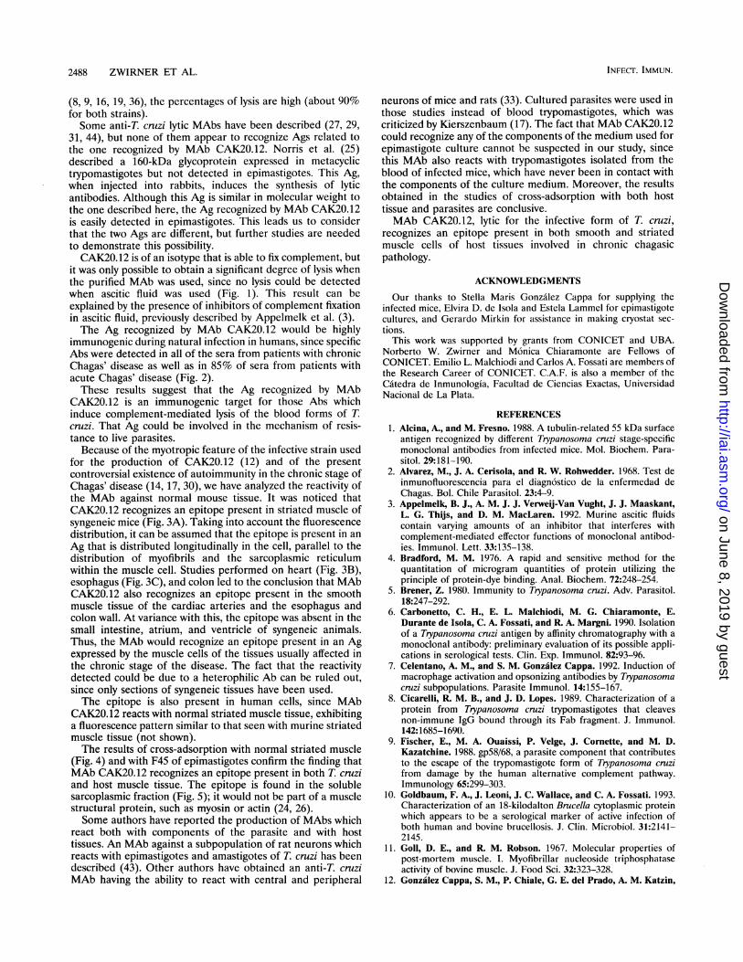
FIG. 1. Complement-mediated lysis of bloodstream trypomasti-gotes of T. cruzi (RA strain). FPLC-purified MAb CAK20.12 (solidbars) and MAb B124 (anti-B. aborttis negative control; hatched bars)and their respective ascitic fluids (open bars, MAb CAK20.12; stippledbars, MAb B124) were assayed. Purified MAbs were used at 2.5 mg/ml(bars 1), 1.25 mg/ml (bars 2), and 0.625 mg/ml (bars 3). Ascitic fluids(Ab concentration, 5 mg/ml) were used undiluted (bars 4) or diluted1:2 (bars 5) or 1:4 (bars 6). Error bars show the standard deviation.
epimastigotes with titers greater than 1:32, indirect hemagglu-tination with titers greater than 1:64, and/or IIF with Formalin-fixed epimastigotes with titers greater than 1:30). Some of thepatients had chronic chagasic pathologies, such as electrocar-diographic abnormalities, heart arrhythmia, severe myocardio-pathies, or megaviscera.
Moreover, 13 serum samples from acute chagasic patientswere studied. These patients had parasitemia as detected byxenodiagnosis, hemoculture, or microhematocrit. Some ofthem also had positive conventional serological tests.Serum samples from healthy volunteers negative for T. cruzi
Ags by serological tests were used as negative controls.
RESULTS
Reactivity of MAb CAK20.12 with T. cruzi strains. MAbCAK20.12 (IgG2b) was derived from the spleen of a BALB/cmouse chronically infected with the k98 clone of the CA, strain
OD1.2-1_
12D01S0
0.8I 0
80.6
o0.4 o0
O L_ h°
NHS AChS CChS
FIG. 2. Reactivity of human sera in a capture ELISA with MAbCAK20. 12. Each point represents the optical density (OD) resulting fromsubtracting the absorbance of the well without Ag from that of the wellwith Ag for sera from healthy volunteers (NHS) and patients with acutechagasic (AChS) and chronic chagasic (CChS) disease. The cutoff valuewas calculated as the mean absorbance of the negative-control sera plus 3standard deviations and is indicated by a horizontal dotted line.
FIG. 3. IIF on sections of normal BALB/c mouse tissues. (A) Longitudinal section of striated muscle tissue stained with MAb CAK20.12; threecells showing a pattern of cytoplasmic fluorescence distributed longitudinally, paralleling the myofibril array and the sarcoplasmic reticulum in themuscle cell, can be observed. Fluorescence of the cell membrane was also detected. Magnification, X400. (B) Section of heart tissue stained withMAb CAK20.12, showing fluorescence associated with the smooth muscle layer of the cardiac artheries. v, ventricle; a, atrium. Magnification,x250. (C) Section of esophagus stained with MAb CAK20.12; fluorescence is located in the smooth muscle layer of the mucous layer (laminamuscularis mucosae) and in the organ outer layer. m, mucous layer; mm, lamina muscularis mucosae; sm, submucous layer; em, external muscularlayer. Magnification, x250. (D) Longitudinal section of striated muscle tissue stained with MAb BI24 (negative control). Magnification, x400. (E)Section of heart tissue stained with MAb B124. v, ventricle; a, atrium. Magnification, x250. (F) Section of esophagus stained with MAb BI24. m,mucous layer; mm, lamina muscularis mucosae; sm, submucous layer; em, external muscular layer. Magnification, X250.
of T. cruzi. This MAb reacted by IIF with RA trypomastigotes,the fluorescence being detected mainly in the parasite mem-brane. Fluorescence was found to be uniformly distributedthroughout the parasite in epimastigotes of different T. cruzistrains (Tulahuen, RA, AWP, LP, Mesa, AF, CA,, and its k98clone). With live epimastigotes and trypomastigotes, fluores-cence was seen only on the cell membrane (results not shown).
Lytic activity on blood trypomastigotes. Since CAK20.12recognized a surface Ag of the parasite, its ability to lyse bloodforms in the presence of guinea pig complement was analyzed.The results obtained showed that MAb CAK20.12 lysed 90.2%± 5.4% and 88.5% ± 8% of trypomastigotes of the CA, andRA strains, respectively (Fig. 1). This effect depended on theAb dose and could only be detected when purified MAb wasused. No significant lysis was observed when ascitic fluid wasused in spite of equivalent Ab concentrations. No lysis was everfound in the negative controls.
Reactivity of human sera with the Ag recognized by MAb
CAK20.12. The reactivity of sera from patients infected with T.cruzi was studied with purified MAb CAK20.12 by means of acapture ELISA of the F45 fraction. It was shown that 100% (30of 30) of the samples from patients with chronic Chagas'disease and 85% (11 of 13) of those from patients with acuteChagas' disease exhibited a reactivity higher than the meanplus 3 standard deviations for sera from healthy controls (Fig.2).
IIF of mouse tissue sections. MAb CAK20.12 reacted withnormal syngeneic striated muscle tissue. Figure 3A shows apattern of cytoplasmic fluorescence distributed longitudinally,paralleling the myofibril array and the sarcoplasmic reticulumin the muscle cell. Fluorescence of the cell membrane was alsodetected. It was observed that CAK20.12 reacts with thesmooth muscle layer of the cardiac arteries when sections ofheart tissue of normal mice were used (Fig. 3B). Figure 3Cshows the results obtained with esophagus sections from anormal syngeneic mouse. Fluorescence is located in the
MAb TO T. CRUZI TRYPOMASTIGOTES AND HOST TISSUES 2487
FIG. 4. IIF of trypomastigotes of T. cruzi with MAb CAK20.12. (A)IIF of parasites stained with FPLC-purified MAb, showing a fluores-cence pattern along the parasite membrane. Magnification, X400. (B)IIF of trypomastigotes stained with FPLC-purified MAb preadsorbedwith an extract of normal mouse striated muscle tissue. Fluorescencewas abolished by adsorption of the MAb with an antigenic fraction ofstriated muscle from a normal mouse. Magnification, x400.
smooth muscle layer of the mucous layer (lamina muscularismucosae) and in the organ outer layer. The same studiesperformed on normal mouse colon (not shown) exhibitedfluorescence associated with the outer smooth muscle layer.On the other hand, very weak reactivity was detected in smallintestine sections. Sections of syngeneic ventricle or atriumwere negative (not shown). Significant fluorescence was neverdetected with the control MAb. Similar results were consis-tently obtained with tissues from three mice.MAb CAK20.12 also reacted with normal human striated
muscle tissue; a cytoplasmic fluorescence similar to that shownin Fig. 3A for murine striated muscle was observed (notshown).
Adsorption of MAb CAK20.12 with an antigenic fraction ofstriated muscle from a normal mouse inhibited fluorescence oneither epimastigotes (not shown) or trypomastigotes of T. cruzi(Fig. 4). Moreover, adsorption of the MAb with the F45fraction of epimastigotes inhibited the fluorescence on muscletissue (not shown).
Reactivity of MAb by dot-blot and Western blot. Figure SAshows that MAb CAK20.12 reacts by dot-blot with the F45fraction of the parasite. The same holds true for the solublesarcoplasmic fraction of normal murine striated muscle,whereas no reaction was observed with purified myofibrils. Noreactivity with any of the Ags studied was detected with thecontrol MAb.
Degradation with proteinase K of the F45 antigenic fractionsof T. cruzi epimastigotes caused loss of reactivity of MAbCAK20.12 by dot-blot. Conversely, chemical oxidation of thecarbohydrates of the same antigenic fraction caused no changein the recognition of the Ag by CAK20.12 (Fig. 5B).By Western blot (immunoblot) of the F45 fraction of T. cruzi
epimastigotes, MAb CAK20.12 recognized a 150-kDa compo-nent. Heating of the antigenic fraction at 100°C for 3 min inSDS-PAGE sample buffer abolished the reactivity of the MAb(Fig. SC).
DISCUSSION
MAb CAK20.12 (IgG2b) was derived from the spleen of amouse chronically infected with the k98 clone of the CA, strainof T. cruzi. This MAb recognizes an Ag that is widely distrib-uted among strains of T. cruzi, and from the IIF results withboth Formalin-treated trypomastigotes and live epimastigotesand trypomastigotes, it is located on the surface of T. cruzi. Ithas been demonstrated by Western blotting of the epimasti-
A
B
1 0
1 2 3 4 5 6
B124CAK20.12
1 2 3 4 5
C kDa
205 -
116-97.4-66-
45-29-
A B C DFIG. 5. Reactivity of MAb CAK20.12 by dot-blot and Western blot.
(A) F45 fraction of epimastigotes of T. cruzi (lanes 1 and 2), thesarcoplasmic fraction of normal mouse striated muscle cells (lanes 3and 4), and myofibrils (lanes 5 and 6) were assayed with MAbCAK20.12 (lanes 1, 3, and 5) and MAb B124 (negative control; lanes2, 4, and 6). (B) The untreated F45 fraction (lane 1), F45 digested withproteinase K (lane 3), and F45 oxidized with periodate (lane 5) wereassayed with the MAbs; mock-treated F45 fraction was also assayed(proteinase K treatment, lane 2; periodate treatment, lane 4). (C)Western blot of F45 fraction with MAb CAK20.12 (lanes A and B) andMAb B124 (lanes C and D). Nonboiled (lanes B and D) and boiled(lanes A and C) F45 fractions were also assayed.
gote lysate that MAb CAK20.12 recognizes an epitope presentin an Ag of 150 kDa (Fig. 5). This epitope is sensitive to theheating undergone by samples to be used in SDS-PAGE, beingmodified in such a way that it can no longer be recognized byits specific Ab. The epitope recognized might be a polypeptide,since it is damaged by proteinase K treatment but unaffectedby chemical oxidation with sodium periodate (Fig. 5).
Several authors have reported that either natural or exper-imental infection with T cruzi triggers the synthesis of lyticantibodies (18, 22). Particularly with clone k8 of the CA,strain, the sera of infected mice have a low lytic activity and aslight opsonization effect on trypomastigotes (7). This strain, aswell as its clone k98, is nonlethal for that host (12). By usingthat strain for infection, we have obtained an MAb with lyticactivity not only for the infective form of the homologous strain(CA,) but also for trypomastigotes of the RA strain, which ishighly lethal for mice. In spite of the different T. cruzitrypomastigote components described, which would providethem with the ability to avoid its lysis by Ab and complement
(8, 9, 16, 19, 36), the percentages of lysis are high (about 90%for both strains).Some anti-T. cruzi lytic MAbs have been described (27, 29,
31, 44), but none of them appear to recognize Ags related tothe one recognized by MAb CAK20.12. Norris et al. (25)described a 160-kDa glycoprotein expressed in metacyclictrypomastigotes but not detected in epimastigotes. This Ag,when injected into rabbits, induces the synthesis of lyticantibodies. Although this Ag is similar in molecular weight tothe one described here, the Ag recognized by MAb CAK20.12is easily detected in epimastigotes. This leads us to considerthat the two Ags are different, but further studies are neededto demonstrate this possibility.CAK20.12 is of an isotype that is able to fix complement, but
it was only possible to obtain a significant degree of lysis whenthe purified MAb was used, since no lysis could be detectedwhen ascitic fluid was used (Fig. 1). This result can beexplained by the presence of inhibitors of complement fixationin ascitic fluid, previously described by Appelmelk et al. (3).The Ag recognized by MAb CAK20.12 would be highly
immunogenic during natural infection in humans, since specificAbs were detected in all of the sera from patients with chronicChagas' disease as well as in 85% of sera from patients withacute Chagas' disease (Fig. 2).These results suggest that the Ag recognized by MAb
CAK20.12 is an immunogenic target for those Abs whichinduce complement-mediated lysis of the blood forms of T.cruzi. That Ag could be involved in the mechanism of resis-tance to live parasites.
Because of the myotropic feature of the infective strain usedfor the production of CAK20.12 (12) and of the present
controversial existence of autoimmunity in the chronic stage ofChagas' disease (14, 17, 30), we have analyzed the reactivity ofthe MAb against normal mouse tissue. It was noticed thatCAK20.12 recognizes an epitope present in striated muscle ofsyngeneic mice (Fig. 3A). Taking into account the fluorescencedistribution, it can be assumed that the epitope is present in an
Ag that is distributed longitudinally in the cell, parallel to thedistribution of myofibrils and the sarcoplasmic reticulumwithin the muscle cell. Studies performed on heart (Fig. 3B),esophagus (Fig. 3C), and colon led to the conclusion that MAbCAK20.12 also recognizes an epitope present in the smoothmuscle tissue of the cardiac arteries and the esophagus andcolon wall. At variance with this, the epitope was absent in thesmall intestine, atrium, and ventricle of syngeneic animals.Thus, the MAb would recognize an epitope present in an Agexpressed by the muscle cells of the tissues usually affected inthe chronic stage of the disease. The fact that the reactivitydetected could be due to a heterophilic Ab can be ruled out,
since only sections of syngeneic tissues have been used.The epitope is also present in human cells, since MAb
CAK20.12 reacts with normal striated muscle tissue, exhibitinga fluorescence pattern similar to that seen with murine striatedmuscle tissue (not shown).The results of cross-adsorption with normal striated muscle
(Fig. 4) and with F45 of epimastigotes confirm the finding thatMAb CAK20.12 recognizes an epitope present in both T. cruziand host muscle tissue. The epitope is found in the solublesarcoplasmic fraction (Fig. 5); it would not be part of a musclestructural protein, such as myosin or actin (24, 26).Some authors have reported the production of MAbs which
react both with components of the parasite and with hosttissues. An MAb against a subpopulation of rat neurons whichreacts with epimastigotes and amastigotes of T. cruzi has beendescribed (43). Other authors have obtained an anti-T. cruziMAb having the ability to react with central and peripheral
neurons of mice and rats (33). Cultured parasites were used inthose studies instead of blood trypomastigotes, which wascriticized by Kierszenbaum (17). The fact that MAb CAK20.12could recognize any of the components of the medium used forepimastigote culture cannot be suspected in our study, sincethis MAb also reacts with trypomastigotes isolated from theblood of infected mice, which have never been in contact withthe components of the culture medium. Moreover, the resultsobtained in the studies of cross-adsorption with both hosttissue and parasites are conclusive.MAb CAK20.12, lytic for the infective form of T. cruzi,
recognizes an epitope present in both smooth and striatedmuscle cells of host tissues involved in chronic chagasicpathology.
ACKNOWLEDGMENTSOur thanks to Stella Maris Gonzalez Cappa for supplying the
infected mice, Elvira D. de Isola and Estela Lammel for epimastigotecultures, and Gerardo Mirkin for assistance in making cryostat sec-tions.
This work was supported by grants from CONICET and UBA.Norberto W. Zwirner and M6nica Chiaramonte are Fellows ofCONICET. Emilio L. Malchiodi and Carlos A. Fossati are members ofthe Research Career of CONICET. C.A.F. is also a member of theCatedra de Inmunologia, Facultad de Ciencias Exactas, UniversidadNacional de La Plata.
REFERENCES1. Alcina, A., and M. Fresno. 1988. A tubulin-related 55 kDa surface
antigen recognized by different Trypanosoma cruzi stage-specificmonoclonal antibodies from infected mice. Mol. Biochem. Para-sitol. 29:181-190.
2. Alvarez, M., J. A. Cerisola, and R. W. Rohwedder. 1968. Test deinmunofluorescencia para el diagn6stico de la enfermedad deChagas. Bol. Chile Parasitol. 23:4-9.
3. Appelmelk, B. J., A. M. J. J. Verweij-Van Vught, J. J. Maaskant,L. G. Thijs, and D. M. MacLaren. 1992. Murine ascitic fluidscontain varying amounts of an inhibitor that interferes withcomplement-mediated effector functions of monoclonal antibod-ies. Immunol. Lett. 33:135-138.
4. Bradford, M. M. 1976. A rapid and sensitive method for thequantitation of microgram quantities of protein utilizing theprinciple of protein-dye binding. Anal. Biochem. 72:248-254.
5. Brener, Z. 1980. Immunity to Trypanosoma cruzi. Adv. Parasitol.18:247-292.
6. Carbonetto, C. H., E. L. Malchiodi, M. G. Chiaramonte, E.Durante de Isola, C. A. Fossati, and R. A. Margni. 1990. Isolationof a Trypanosoma cruzi antigen by affinity chromatography with amonoclonal antibody: preliminary evaluation of its possible appli-cations in serological tests. Clin. Exp. Immunol. 82:93-96.
7. Celentano, A. M., and S. M. Gonzalez Cappa. 1992. Induction ofmacrophage activation and opsonizing antibodies by Trypanosomacruzi subpopulations. Parasite Immunol. 14:155-167.
8. Cicarelli, R. M. B., and J. D. Lopes. 1989. Characterization of aprotein from Trypanosoma cruzi trypomastigotes that cleavesnon-immune IgG bound through its Fab fragment. J. Immunol.142:1685-1690.
9. Fischer, E., M. A. Ouaissi, P. Velge, J. Cornette, and M. D.Kazatchine. 1988. gp58/68, a parasite component that contributesto the escape of the trypomastigote form of Trypanosoma cruzifrom damage by the human alternative complement pathway.Immunology 65:299-303.
10. Goldbaum, F. A., J. Leoni, J. C. Wallace, and C. A. Fossati. 1993.Characterization of an 18-kilodalton Brucella cytoplasmic proteinwhich appears to be a serological marker of active infection ofboth human and bovine brucellosis. J. Clin. Microbiol. 31:2141-2145.
11. Goll, D. E., and R. M. Robson. 1967. Molecular properties ofpost-mortem muscle. I. Myofibrillar nucleoside triphosphataseactivity of bovine muscle. J. Food Sci. 32:323-328.
12. Gonzalez Cappa, S. M., P. Chiale, G. E. del Prado, A. M. Katzin,
MAb TO T CRUZI TRYPOMASTIGOTES AND HOST TISSUES 2489
G. W. Martini, E. Durante de Isola, L. Abramo Orrego, and E. L.Segura. 1980. Aislamiento de una cepa de Trypaniosonia cruzi deun paciente con miocardiopatia chagasica cr6nica y su caracter-izaci6n biol6gica. Medicina (Buenos Aires) 40:63-68.
13. Hames, B. D. 1981. An introduction to polyacrylamide gel elec-trophoresis, p. 1-86. In B. D. Hames and D. Rickwood (ed.), Gelelectrophoresis of proteins: a practical approach. IRL Press,London.
14. Hudson, L. 1985. Autoimmune phenomena in chronic chagasiccardiopathy. Parasitol. Today 1:6-9.
15. Hudson, L., F. Guhl, C. J. Marinkelle, and J. Rodriguez. 1987. Useof monoclonal antibodies for the differential detection of Trypano-somna cruzi and Trypanosoma rangeli in epidemiological studies andxenodiagnosis. Acta Trop. 44:387-394.
16. Joiner, K., W. Dias da Silva, M. T. Rimoldi, C. H. Hammer, A.Sher, and T. L. Kipnis. 1988. Biochemical characterization offactor by trypomastigotes of Trypanosoma cruzi which acceleratesthe decay of complement C3 convertases. J. Biol. Chem. 15:11327-11335.
17. Kierszenbaum, F. 1986. Autoimmunity in Chagas' disease. J.Parasitol. 72:201-211.
18. Kierszenbaum, F., and M. F. Lima. 1983. Susceptibility of insect-borne, metacyclic forms of Trypanosoma cruzi to antibody-medi-ated mechanisms of destruction. Am. J. Trop. Med. Hyg. 32:1236-1241.
19. Kipnis, T. L., and W. Dias da Silva. 1989. Evasion of Trypanosomacruzi from complement lysis. Braz. J. Med. Biol. Res. 22:1-16.
20. Kohler, G., and C. Milstein. 1975. Continuous culture of fusedcells secreting antibodies of predefined specificity. Nature (Lon-don) 256:495-497.
21. Krettli, A. U., and Z. Brener. 1976. Protective effects of specificantibodies in Tiypanosoma cruzi infections. J. Immunol. 116:755-760.
22. Krettli, A. U., and Z. Brener. 1982. Resistance against Trypano-soma cruzi associated to anti-living trypomastigote antibodies. J.Immunol. 128:2009-2012.
23. Levin, M. J., E. Mesri, R. Benarous, G. Levitus, A. Schijman, P.Levy-Yeyati, P. A. Chiale, A. M. Ruiz, A. Kahn, M. B. Rosenbaum,H. N. Torres, and E. L. Segura. 1989. Identification of majorTrypanosoina cruzi antigenic determinants in chronic Chagas'heart disease. Am. J. Trop. Med. Hyg. 41:530-538.
24. Mortara, R. A. 1989. Studies on trypanosomatid actin. I. Immu-nochemical and biochemical identification. J. Protozool. 36:8-13.
25. Norris, K. A., G. Harth, and M. So. 1989. Purification of aTiypaniosorna cnrzi glycoprotein which elicits lytic antibodies.Infect. Immun. 57:2372-2377.
26. Paulin, J. J., C. H. Keith, and R. L. Tarleton. 1988. A monoclonalantibody to alpha-tubulin recognizes host-cell and Trypanosomacruzi tubulins. J. Protozool. 35:123-129.
27. Prioli, R. P., J. S. Mejia, and M. E. A. Pereira. 1990. Monoclonalantibodies against Trypanosoma cruzi neuraminidase reveal en-zyme polymorphism, recognize a subset of trypomastigotes, andenhance infection in vitro. J. Immunol. 144:4384-4391.
28. Sadigursky, M., A. M. Acosta, and C. A. Santos Buch. 1982.Muscle sarcoplasmic reticulum antigen shared by a Trypanosomacruzi clone. Am. J. Trop. Med. Hyg. 31:934-941.
29. Schenkman, S., M. L. S. Giither, and N. Yoshida. 1986. Mecha-nism of resistance to lysis by the alternative complement pathwayin Th'panosoma cnizi trypomastigotes: effect of a specific mono-clonal antibody. J. Immunol. 137:1623-1628.
30. Schmufiis, G. 1987. Autoimmunity in Chagas' disease. Mem. Inst.Oswaldo Cruz Rio de J. 82(Suppl.):287-310.
31. Segura, E. L., J. Bua, A. Rosenstein de Campanini, E. Subias, M.Esteva, M. Moreno, and A. M. Ruiz. 1986. Monoclonal antibodiesagainst the flagellar fraction of epimastigotes of Trypanosoma cruzicomplement-mediated lytic activity against trypomastigotes andpassive immunoprotection in mice. Immunol. Lett. 13:165-171.
32. Skeiky, Y. A. W., D. R. Benson, M. Parsons, K. B. Elkon, and S.Reed. 1992. Cloning and expression of Trypanosoma cruzi ribo-somal protein PO and epitope analysis of anti-PO autoantibodiesin Chagas' disease patients. J. Exp. Med. 176:201-211.
33. Snary, D., J. E. Flint, J. N. Wood, M. T. Scott, M. D. Chapman, J.Dodd, T. M. Jessel, and M. A. Miles. 1983. A monoclonal antibodywith specificity for Trypanosoma cruzi, central and peripheralneurons and glia. Clin. Exp. Immunol. 54:617-624.
34. Szarfman, A., V. P. Terranova, S. I. Rennard, J. M. Foidart, M. F.Lima, J. I. Scheinman, and G. R. Martins. 1982. Antibodies tolaminin in Chagas' disease. J. Exp. Med. 155:1161-1171.
35. Takehara, H. A., A. Perini, M. H. da Silva, and I. Mota. 1981.Trypanosoma cruzi: role of different antibody classes in protectionagainst infection in the mouse. Exp. Parasitol. 52:137-146.
36. Tambourgi, D. V., T. L. Kipnis, W. Dias da Silva, K. A. Joiner, A.Sher, S. Heath, B. F. Hall, and G. B. Ogden. 1993. A partial cDNAclone of trypomastigote decay-accelerating factor (T-DAF), adevelopmentally regulated complement inhibitor of Trypanosomacruzi, has genetic and functional similarities to the human com-plement inhibitor DAF. Infect. Immun. 61:3656-3663.
37. Teixeira, M., and N. Yoshida. 1986. Stage-specific surface antigensof metacyclic trypomastigotes of Trypanosoma cruzi identified bymonoclonal antibodies. Mol. Biochem. Parasitol. 18:271-282.
38. Tsang, V. C. W., J. N. Peralta, and A. R. Simmons. 1983. Enzymelinked immunoelectrotransfer blot techniques (EITB) for studyingby gel electrophoresis. Methods Enzymol. 92:377-391.
39. Van Voorhis, W. C., and H. Eisen. 1989. Fl-160: a surface antigenof Trypanosoma cruzi that mimics mammalian nervous tissue. J.Exp. Med. 169:641-652.
40. Vennegoor, C., J. Calafat, and P. Hageman. 1985. Biochemicalcharacterization and cellular localization of a formalin-resistantmelanoma-associated antigen reacting with monoclonal antibodyNkl/C-3. Int. J. Cancer 35:287-295.
41. Voller, A., D. Bidwell, and A. Bartlett. 1988. Enzyme-linkedimmunosorbent assay, p. 359-371. In N. R. Rose and H. Friedman(ed.), Manual of clinical immunology, 2nd ed. American Societyfor Microbiology, Washington, D.C.
42. Williams, G. T., L. Fielder, H. Smith, and L. Hudson. 1985.Adsorption of Trypanosoma cruzi proteins to mammalian cells invitro. Acta Trop. 42:33-38.
43. Wood, J. N., L. Hudson, T. M. Jessel, and M. Yamamoto. 1982. Amonoclonal antibody defining antigenic determinants of mamma-lian neurons and Trypanosoma cruzi parasites. Nature (London)296:34-38.
44. Yoshida, N., R. A. Mortara, M. F. Araguth, J. C. Gonzalez, and M.Russo. 1989. Metacyclic neutralizing effect of monoclonal anti-body 10D8 directed to the 35- and 50-kilodalton surface glycocon-jugates of Trypanosoma cruzi. Infect. Immun. 57:1663-1667.
45. Zwirner, N. W., E. L. Malchiodi, M. G. Chiaramonte, R. A.Margni, M. Esteva, and C. A. Fossati. 1992. Caracterizaci6n de unantigeno de Trypanosorna cruzi purificado con anticuerpos mono-clonales. Utilidad diagn6stica y analisis de su immunogenicidad.Inmunologia 11:120-127.