34
MOLECULAR DETECTION TECHNIQUES IN FOOD QUALITY CONTROL: AN OVERVIEW By: Yakindra Prasad Timilsena ID- 111332
| Date post: | 22-Dec-2015 |
| Category: |
Documents |
| Upload: | nicholas-gregory |
| View: | 214 times |
| Download: | 0 times |

MOLECULAR DETECTION TECHNIQUES IN FOOD QUALITY
CONTROL: AN OVERVIEW
By: Yakindra Prasad TimilsenaID- 111332

BACKGROUND Food Quality control is the multidisciplinary
approaches of maintaining physical, chemical, microbiological, technological and sensory wholesomeness in foods
Method of detection of food adulteration is the core of food quality control program.
Traceability and quality assurance in the food and feed industry through detection technique at every step of the manufacturing chain 'from farm to fork’ are essential for regulatory agencies.

PROBLEM STATEMENT
Chemistry alone can’t solve all the problems of detection
Chemical methods of analysis are time consuming and costly. Need of rapid and reliable methods
Methods based on molecular biology and immunology approaches- better alternatives
Knowledge on molecular organization of the cell has led to the development of powerful new techniques that bring greater accuracy, rapid, cost effective
Molecular methods-more superior than immunological methods.

COMMON MOLECULAR METHODS
PCR (RT-PCR, Multiplex), RFLP, SSCP and sequencing
Plasmid profiling, ribotyping, macrorestriction analysis by pulsed-field gel electrophoresis (PFGE)
Newer techniques which use fluorescent dyes, DNA microarrays, protein chemistry and mass spectrometry.
DNA chip, the GeneChip,

COMMON MOLECULAR TECHNIQUES
Random Amplified Polymorphic DNA Analysis (RAPD)
Amplified Fragment Length Polymorphism (AFLP)Loop Mediated Isothermal Amplification (LAMP)
BiosensorsGold Nanoparticle-based BiosensorFiber Optic BiosensorElectrochemical Biosensor
Although there are many nucleic acid molecular detection methods, only DNA probe and PCR has been developed commercially for detection of food pathogens.

APPLICATIONS OF MOLECULAR METHOD
Detecting and identifying specific genes (GM foods)
Application to Food Authenticity and Legislation
Detection of microbial contamination of foods Species Identification Detection of Food Constituents (Ingredients or
Contaminants) Detection of antibiotics, pesticides residues etc.
Halal and Kosher certification

What is PCR?DNA replication in a tube (in vitro). Xeroxing (copying) of DNA.
The Components of PCRThe basic components of a PCR reaction are
- one or more molecules of target DNA- two oligonucleotide primers
- thermostable DNA polymerase- dNTPs
The Process of PCREach PCR cycle requires three temperature steps to complete a round of DNA synthesis:

MINIMUM CRITERIA FOR PCR
The sample must contain at least one intact DNA strand comprising the region to be amplified
impurities must be sufficiently diluted so as not to inhibit the polymerization step of the PCR reaction.
DNA samples for PCR, regardless of preparation method, are generally run in duplicate in order to provide a control for the relative quality and purity of the original sample.

PCR STEPS
isolation of DNA from the food (CTAB method is common)
amplification of the target sequences by PCR
separation of the amplification products by agarose gel electrophoresis
estimation of their fragment size by comparison with a DNA molecular mass marker after staining with ethidium bromide
verification of the PCR results by specific cleavage of the amplification products by restriction endonuclease, transfer of
separated amplification products onto membranes (Southern Blot) followed by hybridisation with a DNA probe specific for the target
sequence

Agarose Gels: • NuSieve agarose separates short products better
than the regular agarose. More expensive but use less for the same gel strength as regular agarose.
Real Time detection of PCR products • No gels required. Recent method. Relies on the
ability of a dye, SYBR Green, to interact with double stranded amplicons produced during PCR, to produce fluorescence which is detected in a flurometer.
Gel electrophoresis for detecting PCR products

MULTIPLEX PCR
Several primers pairs with similar annealing requirements can be added to a PCR mixture to simultaneously detect several target sequences
saves time and minimize the expense on detection of food borne pathogens
primers shoud have same melting temperature
must not interact with each other. the amplified fragments of same length
cannot be detected

MULTIPLEX PCR
Standard PCR- unable to differentiate viable and non-viable microorganisms
Ethidium monoazide can be used to separate dead and viable bacteria
Real-time PCR using RNA as template is more authentic since the RNA is present only in viable microbes.
RNA is first reverse transcribed to cDNA and then used for amplification.

POLYMERASE CHAIN REACTION – RESTRICTION
FRAGMENT LENGTH POLYMORPHISM(PCR-RFLP)
The method includes amplification of a known DNA sequence using two specific primers, subsequent digestion of an amplicon with restriction endonucleases and separation and comparison of DNA restriction fragments.
The disadvantage of RFLP analysis of PCR product is that incomplete digestion may occasionally occur and intra-specific variation could delete or create additional restriction sites (Lockley and Bardsley, 2000).

RAPD-PCR
Random amplified polymorphic DNA PCR uses a random primer (10-mer) to generate a DNA profile.
The primer anneals to several places on the DNA template and generate a DNA profile which is used for microbe identification.
RAPD has many advantages: Pure DNA is not neededLess labor intensive than RFLP.There is no need for prior DNA sequence
data.RAPD has been used to fingerprint the
outbreak of Listeria monocytogenes from milk.

RIBOTYPING
Ribotyping is a method that can identify and classify bacteria based upon differences in rRNA. It generates a highly reproducible and precise fingerprint that can be used to classify bacteria from the genus through and beyond the species level.
Databases for Listeria (80 pattern types), Salmonella (97 pattern types), Escherichia (65 pattern types) and Staphylococcus(252 pattern types) have been established.

PLASMID PROFILING
Plasmid profile analysis involves extraction of plasmid DNA and separation by electrophoresis. The plasmids are visualized under UV light and sized in relation to plasmids of known molecular mass carried in a reference strain of E. coli.
Plasmid analysis of over 120 strains of Cl. perfringens, isolated during food-poisoning incidents was carried out by Jones et al., 1989.
A high proportion (71%) of fresh and well-characterized food-poisoning strains possessed plasmids of 6.2 kb in size (compared with 19% of non-food-poisoning strains).

LAB-ON-A-CHIP TECHNOLOGY
An alternative approach for the visualization of the PCR products by the CE on a card-sized device.
Can be used to replace the gel-electrophoretic step in the PCR end-point detection,
DNA fragments were detected using laser-induced fluorescence, which enables accurate sizing and quantification of DNA fragments.
Higher speed, simplicity and safety. This approach allowed identification of 5%
fish species admixed into a product containing two fish species.

Direct method used for enumeration of microbe based on binding properties of flurochrome acridine orange dye.
Food samples are pretreated with detergents and proteolytic enzymes, filtered on to a polycarbonate membrane stained with acridine orange and examined under fluorescent microscope
Streptococcus and Staphylococcus can be detectedd by this method
Fig. Staphylococcus aureus - Acridine-orange leucocyte cytospin test
DIRECT EPIFLOURESCENT TECHNIQUE (DEFT)

ELECTROPHORETIC METHODS
Electrophoretic methods are based on the ability
of molecules to migrate according to their
molecular weight (Mw) in the electric field due to
the effect of electrostatic forces attracting them
to reversely charged electrode.
The migration is performed on agarose or
polyacrylamide gel. Various modifications of
electrophoretic methods are used depending on a
type of the analysed product:

ELECTROPHORETIC METHODS
isoelectric focusing (IEF)
urea isoelectric focusing (urea-IEF)
sodium dodecyl sulphate – polyacrylamide
gel electrophoresis (SDS-PAGE)
two dimensional electrophoresis (2DE)
capillary electrophoresis (CE)
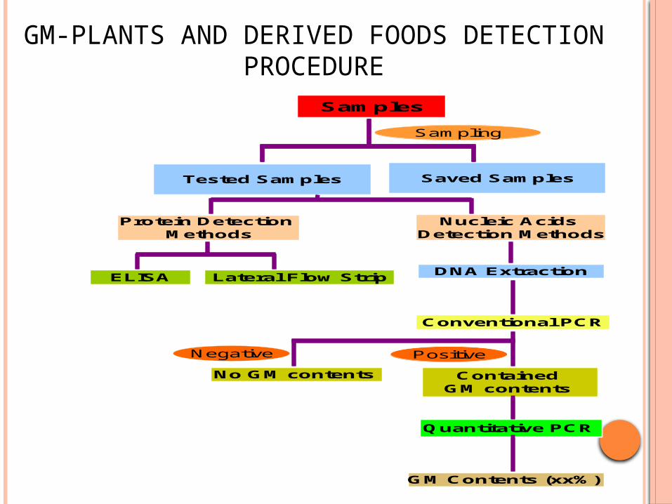
GM-PLANTS AND DERIVED FOODS DETECTION PROCEDURE
Samples
Tested Samples Saved Samples
Protein Detection Methods
Nucleic Acids Detection Methods
ELISA Lateral Flow StripDNA Extraction
Conventional PCR
Contained GM contents
No GM contents
Quantitative PCR
GM Contents (xx%)
Sampling
Negative Positive
Samples
Tested Samples Saved Samples
Protein Detection Methods
Nucleic Acids Detection Methods
ELISA Lateral Flow StripDNA Extraction
Conventional PCR
Contained GM contents
No GM contents
Quantitative PCR
GM Contents (xx%)
Sampling
Negative Positive

GM-PLANTS AND DERIVED FOODS DETECTION PROCEDURE
Commercial GMO contain the 35S promoter of Cauliflower Mosaic Virus and/or the NOS terminator of Agrobacterium, these genetic elements are used as target sequences for a general screening
Since primer selection has to be based on target sequences that are characteristic for the individual transgenic organism. Therefore, a prerequisite for designing specific primers for the identification of GMOs by PCR is the availability of detailed information on their molecular make-up.
Molecular make-up of non-authorized GMOs is generally not available and so impossible to detect the presence of non-authorized GMOs.

DETECTION OF FOOD-BORNE PATHOGENS A short cultural enrichment followed by
physical separation of the organisms from the culture medium is required for food samples prior to analysis. Enrichment prior to DNA extraction and PCR analysis results in a dilution of PCR inhibitors and an increased number of target cells and therefore in a higher sensitivity. Only viable cells are detected.
RNA based methods more preferred since mRNAs are short living molecules and can be amplified in the PCR system only in case of viable cells. Cultural enrichment step is not required.

DETECTION OF FOOD-BORNE PATHOGENS
In a study the development of a PCR-based technique for the rapid identification of the food-borne pathogens Salmonella and Escherichia coli was undertaken. Suitable primers were designed based on specific gene fimA of Salmonella and gene afa of pathogenic E. coli for amplification.
Agarose gel electrophoresis and subsequent staining with ethidium bromide were used for the identification of PCR products. The size of the amplified product was 120 bp as shown by comparison with marker DNA. These studies have established that fimA and afa primers were specific for detecting Salmonella and pathogenic E. coli, respectively, in the food samples (Naravaneni & Jamil, 2005)

DNA MICROARRAY DNA microarray (DNA chip) is rapid and provides
simultaneous DNA screening of hundreds of species at once.
The chip is a glass or nylon membrane with spots of probes oligonucleotides that are complementary to the specific target DNA sequence. The targets hybridize with the captured oligonucleotides on the chip and the fluorescent label, which is attached to the target during the PCR, is detected.
The oligonucleotide microarray analysis of the PCR product from the mt cyt b gene was applied to identify different animal species in food samples (Peter et al., 2004).

BIOSENSOR
Majority of the Biosensors are based on immunological methods, Ritcher 1993

IMPEDANCE-BASED BIOCHIP SENSOR
Based on the changes in conductance in a medium due to microbial breakdown of inert substances into electrically charged ionic compounds.
Allows the detection of only the viable cells
Very attractive and offers real time output, simplicity of use and cost effectiveness
Based on coating the surface of piezoelectric sensor with a selective binding substance e.g. antibodies, placing it in a solution containing bacteria, the bacteria/antigen will bind to the antibodies and the mass of the crystal increase while the resonance frequency will decrease
PIEZOELECTRIC BIOSENSOR

FOURIER TRANSFORM INFRARED (FT-IR) SPECTROSCOPY TECHNIQUES
FT-IR spectroscopy enables rapid and non-invasive characterization of molecular structures in a sample
Can be used to provide compositional and quantitative information.
Can be used for discriminating and classifying intact microbial cells down to the strain level in pure culture
With help of chemometric there has been improvement in the sensitivity of FT-IR to identify, discriminate, and quantify bacteria
Peaks in bacterial spectra are assigned to specific chemical bonds, which may be correlated to bacterial concentrations
Spectral libraries may be created for bacteria in foods and based on comparison between spectra of artificially contaminated samples with these libraries; the extent of contamination may be quantifiable.

DETECTION OF VIRUSES IN FOODS
Virus has been identified in food by Ligase
Chain Reaction (LCR), Nucleic Acid Sequence Based
Amplification (NASBA), Self sustaining sequence
replication (3SR), Strand Displacement Amplification
(SDA), situ hybridization (FISH),
development of gene probes and PCR
amplification techniques are used to
detect the virus in food samples

FLUORESCENT IN SITU HYBRIDIZATION (FISH)
A molecular technique often used to identify and enumerate specific microbial groups.
The FISH technique is dependent upon hybridizing a probe with a fluorescent tag, complementary in sequence, to a short section of DNA on a target gene.
The tag and probe are applied to a sample of interest under conditions that allow for the probe to attach itself to the complementary sequence in the specimen
After sample treatment, excess fluorophore is washed away and the sample can be visualized under a fluorescent microscope.

FLUORESCENT IN SITU HYBRIDIZATION (FISH)

REFERENCES
Mandal, P.K., A.K. Biswas, K. Choi and U.K. Pal, 2011. Methods of Rapid Detection of Foodborne Pathogens: An Overview. Am. J. Food Tech. 6(2): 87-102
http://www.fda.gov/food/scienceresearch/Laboratory
methods/bacteriologicalanalyticalmanualbam/ucm1
096 52.htm#ref4
http://www.worldfoodscience.org/cms/
Naravaneni R, Jamil K. J Med Microbiol. 2005
Jan;54(Pt 1):51-4

References: www.slideshare.net

THANK YOU for your kind attention!
The Karnali Bridge, Near My Home Town


