Proposition 65 Maximum Allowable Dose Level (MADL) for Reproductive Toxicity for Di(2-ethylhexyl)phthalate (DEHP) by Oral Exposure June 2005 Office of Environmental Health Hazard Assessment (OEHHA) Reproductive and Cancer Hazard Assessment Section SUMMARY The maximum allowable dose levels (MADL) for di(2-ethylhexyl)phthalate (DEHP) by the oral route of exposure are 410 micrograms/day (µg/day) for adults, 58 µg/day for infant boys and 20 µg/day for neonatal boys. These values are based on the male reproductive effects of DEHP observed in the study by David et al. (2000a). As specified in regulation, when the applicable reproductive effect is upon the male, the MADL is calculated based on a human body weight of 70 kg (Title 22, California Code of Regulations, Section12803(b)) 1 . In the case of DEHP, however, developing animals are sensitive to the testicular effects of DEHP (e.g., Sjoberg et al., 1985; 1986; Li et al., 2000; CERHR, 2000; U.S. FDA, 2001; Borch et al., 2004). Bodyweights of infants or neonates are markedly lower than that of an adult. Accordingly, age-specific MADLs have been calculated for infant and neonatal boys based on bodyweights of 10 and 3.5 kg, respectively (Sections 12801(a) and 12803(a)(6)). BACKGROUND This report describes the derivation of a maximum allowable dose level (MADL) for DEHP (CAS No. 117-81-7). DEHP is mainly used as a plasticizer of polyvinyl chloride (PVC) in the manufacture of a wide variety of consumer products for building construction, automobiles, clothing, toys and medical devices. (OEHHA, 1997; CERHR, 2000). DEHP was listed under the Safe Drinking Water and Toxic Enforcement Act of 1986 (commonly known as Proposition 65, codified at Health and Safety Code Section 25249.5 et seq.) as known to the State to cause reproductive toxicity (developmental and male reproductive toxicity), effective October 24, 2003. This listing was based on formal identification of DEHP as causing developmental and male reproductive toxicity, by the National Institute for Occupational Safety and Health (NIOSH, 1990) and the U.S. Food and Drug Administration (U.S. FDA, 2001). NIOSH and U.S. FDA are authoritative bodies under Proposition 65 for identification of chemicals as causing reproductive toxicity (Section12306(l)). 1 All further references to regulations are to Title 22, California Code of Regulations unless otherwise noted
Transcript
Proposition 65 Maximum Allowable Dose Level (MADL) for Reproductive Toxicity for
Di(2-ethylhexyl)phthalate (DEHP) by Oral Exposure
June 2005
Office of Environmental Health Hazard Assessment (OEHHA) Reproductive and Cancer Hazard Assessment Section
SUMMARY
The maximum allowable dose levels (MADL) for di(2-ethylhexyl)phthalate (DEHP) by the oral route of exposure are 410 microgramsday (microgday) for adults 58 microgday for infant boys and 20 microgday for neonatal boys These values are based on the male reproductive effects of DEHP observed in the study by David et al (2000a) As specified in regulation when the applicable reproductive effect is upon the male the MADL is calculated based on a human body weight of 70 kg (Title 22 California Code of Regulations Section12803(b))1 In the case of DEHP however developing animals are sensitive to the testicular effects of DEHP (eg Sjoberg et al 1985 1986 Li et al 2000 CERHR 2000 US FDA 2001 Borch et al 2004) Bodyweights of infants or neonates are markedly lower than that of an adult Accordingly age-specific MADLs have been calculated for infant and neonatal boys based on bodyweights of 10 and 35 kg respectively (Sections 12801(a) and 12803(a)(6))
BACKGROUND
This report describes the derivation of a maximum allowable dose level (MADL) for DEHP (CAS No 117-81-7)
DEHP is mainly used as a plasticizer of polyvinyl chloride (PVC) in the manufacture of a wide variety of consumer products for building construction automobiles clothing toys and medical devices (OEHHA 1997 CERHR 2000) DEHP was listed under the Safe Drinking Water and Toxic Enforcement Act of 1986 (commonly known as Proposition 65 codified at Health and Safety Code Section 252495 et seq) as known to the State to cause reproductive toxicity (developmental and male reproductive toxicity) effective October 24 2003 This listing was based on formal identification of DEHP as causing developmental and male reproductive toxicity by the National Institute for Occupational Safety and Health (NIOSH 1990) and the US Food and Drug Administration (US FDA 2001) NIOSH and US FDA are authoritative bodies under Proposition 65 for identification of chemicals as causing reproductive toxicity (Section12306(l))
1 All further references to regulations are to Title 22 California Code of Regulations unless otherwise noted
Procedures for the development of Proposition 65 MADLs are provided in Sections 12801 and 12803 Exposure at a level 1000 times greater than the MADL is expected to have no observable effect As defined in regulation a MADL is derived from a No Observable Effect Level (NOEL) based on the most sensitive study deemed to be of sufficient quality (Section 12803) This document addresses the oral route of exposure for DEHP to assist in the implementation of Proposition 65 relative to the widespread human exposures by this route
STUDY SELECTION
Relevant studies or reports that provide information on the developmental or male reproductive toxicity of DEHP have been identified through literature searches and through reviewing documents produced by authoritative bodies or other expert groups These documents include the two reports by the authoritative bodies that provided the primary support for the Proposition 65 listing of DEHP as a chemical known to cause reproductive toxicity ndash the US FDA (2001) document Safety Assessment of Di (2-ethylhexyl)phthalate (DEHP) Released from PVC Medical Devices and the NIOSH (1990) document NIOH and NIOSH basis for an Occupational Health Standard Di (2-ethylhexyl) phthalate (DEHP) In addition the detailed review by an expert panel convened by the National Toxicology Programrsquos Center for the Evaluation of Risks to Human Reproduction (2000) entitled NTP-CERHR Expert Panel Report on Di (2-ethylhexyl) Phthalate was consulted OEHHA staff have reviewed the relevant studies or reports cited in those documents OEHHA staff have also reviewed additional studies that are not cited in those documents Studies and documents reviewed by OEHHA and cited in the text of this report are listed in the References section Studies reviewed by OEHHA but not cited in the present document or in the documents by NIOSH (1990) CERHR (2000) or US FDA (2001) are listed in the Bibliography
There are numerous studies or review reports providing relevant information on the developmental or male reproductive toxicity of DEHP A majority of the studies published prior to 2000 were already included in the reviews by NIOSH (1990) CERHR (2000) and US FDA (2001) After briefly reviewing all studies or reports available to OEHHA staff OEHHA focused on studies that appear to be sensitive studies in order to identify ldquothe most sensitive study deemed to be of sufficient qualityrdquo
Developmental or Male Reproductive Toxicity in Humans
There are a few epidemiological studies investigating possible associations of exposure to DEHP and other phthalates with developmental or reproductive effects in humans (Modigh et al 2002 Latini et al 2003 Duty et al 2003a 2003b 2004 Rais-Bahrami et al 2004) One study used human sperm to study possible direct effects of DEHP and other phthalates on sperm motility in vitro (Fredricsson et al 1993)
Latini et al (2003) investigated the possible association of concentrations of DEHP and its main metabolite mono (2-ethylhexyl) phthalate (MEHP) in the cord blood of 84 newborns to birth outcomes including weight gestational age and other endpoints All
DEHP (oral) MADL -2- OEHHA June 2005
84 newborns were born at a general-practice hospital in Italy there were 82 singleton births one set of twins and 39 male and 45 female offspring Eleven were preterm and three of them had very low birth weight The maternal age range was from 18 to 24 years The authors found that DEHP or MEHP were present in 74 (881) of the 84 examined cord serum samples at mean concentrations of 119plusmn115 microgml and 052plusmn061 microgml (mean plusmnstandard deviation) respectively MEHP-positive newborns (65 or 774) had a significantly lower gestational age (3816plusmn234 weeks) compared with MEHP-negative infants (3935plusmn135 weeks plt005) Logistic regression analysis also indicated a positive correlation between absence of MEHP in cord blood and gestational age at delivery (odds ratio = 150 95 confidence interval = 1013-221) No other statistically significant relationships were observed between DEHP or MEHP concentrations and other birth outcomes (eg birth weight) The authors concluded that phthalate exposure is significantly associated with shortened pregnancy duration Altered gestation length associated with maternal exposure to a chemical can be an indicator of female reproductive toxicity (US EPA 1996) however female reproductive toxicity is not among the bases for the Proposition 65 listing of DEHP Therefore this study cannot be used as the basis for MADL development (Section 12803(a)(1))
Fredricsson et al (1993) studied effects of DEHP at concentrations of 0001-100 mM on human sperm motility in vitro The authors found that incubation of human sperm with DEHP caused a statistically significant decrease in sperm motility with a 25 reduction of motility at 1 mM The authors did not study the effect of DEHP metabolites (eg MEHP) on sperm motility
A series of recent studies by Duty et al (2003a 2003b 2004) investigated the relationship of sperm parameters to phthalate exposure among male partners of subfertile couples who presented to the Andrology Laboratory at the Massachusetts General Hospital in Boston for semen analysis as part of an infertility work-up The authors found sperm DNA damage decreased sperm motility andor reduced sperm concentration in semen samples to be associated with increased urinary levels of mono-ethyl phthalate or mono-butyl phthalate (metabolites of diethyl phthalate or dibutyl phthalate respectively) but not with urine levels of DEHP or MEHP
Modigh et al (2002) found no effect of DEHP at a mean exposure level of lt 05 mgm3
on time to pregnancy among partners of 193 men who were occupationally exposed to DEHP in air at three plants either producing DEHP or processing polyvinyl chloride (PVC) plastic A recent clinical study investigated testicular volume phallic length and the serum levels of luteinizing hormone (LH) follicle-stimulating hormone (FSH) and testosterone of 13 adolescent boys (14-16 years old) who were exposed to DEHP as neonates on extracorporeal membrane oxygenation (ECMO) support (Rais-Bahrami et al (2004) Mean values for these parameters were within the appropriate range for the degree of pubertal development Detailed information (eg time and duration on ECMO range of the values for sexual hormones or testicular volumes) was not reported no control group was included in the study The route of exposure in these two studies was either inhalation or intravenous injection not oral
DEHP (oral) MADL -3- OEHHA June 2005
All the studies in humans discussed above included relatively small numbers of subjects and have other important limitations (eg lack of appropriate control group phthalate levels based on a single spot urine samples selection of subjects from men who were part of subfertile couples visiting an andrology clinic in the studies by Duty et al) Although some findings in the human studies reported by Fredricsson et al (1993) or by Duty et al (2003a 2003b 2004) provide limited evidence on an association between exposure to phthalates and damaged sperm quality none of the human studies on the developmental or male reproductive effects of phthalates is ldquoof sufficient qualityrdquo for MADL development for the purposes of Proposition 65 Thus the MADL is necessarily based on animal studies
Male Reproductive Toxicity in Animals
The male reproductive toxicity of DEHP has been studied in many species including rats mice hamsters ferrets and non-human primates Findings from the majority of these studies have been well reviewed and summarized in many documents or review reports (eg CERHR 2000 US FDA 2001) Therefore detailed findings from each individual study are not discussed in this document Instead this document focuses on a number of studies that can be potentially identified as the most sensitive study of sufficient quality for the purpose of Proposition 65 and on relevant mechanistic data (eg metabolism cellular andor molecular targets and biochemical pathways) that are critical for determining the relevance of rodent data to humans
Studies in Rats The majority of studies on the male reproductive toxicity of DEHP were conducted in rats using oral administration (gavage feed or drinking water) Depending on the doses dosing duration age of animals and endpoints included it has been shown that oral treatment with DEHP causes reduced fertility decreased weights of male reproductive organs and histopathological changes in the testis of juvenile and adult rats (CERHR 2000 US FDA 2001) Characteristics of histopathological changes include vacuolation and rarefaction of the cytoplasm disruption of cytoskeletons destruction of intercellular specializations (eg ectoplasmic specialization occluding junctions) in Sertoli cells followed by degeneration of spermatocytes by apoptosis andor sloughing of germ cells into the lumen of seminiferous tubules (eg Saitoh et al 1997 Park et al 2002 Boekelheide 2004) Different groups of germ cells in the testis of rats are organized in an orderly manner along the length of seminiferous tubules A defined group of germ cells is called a stage Along the length of a seminiferous tubule there is a distinct ordering of stages namely from stage I to XIV Sertoli cells undergo morphological and functional fluctuation from stages I to XIV (Russell and Griswold 1993) In the testis of young rats (8-week-old) Sertoli cells and the spermatocytes associated with them in seminiferous tubules at stages IX-XIV and I are most sensitive to the testicular effects of DEHP (Saitoh et al 1997)
Oral administration of DEHP to rats during the perinatal period results in severe permanent abnormalities in the male reproductive system of male offspring (Tandon et al 1991 Arcadi et al 1998 Gray et al 1999 Schilling et al 1999 Moore et al 2001) Neonatal or young rats have been found to be more sensitive to the male reproductive
DEHP (oral) MADL -4- OEHHA June 2005
effects of DEHP than are adults (Gray and Butterworth 1980 Sjoberg et al 1985 1986 Dostal et al 1988 Li et al 2000 CERHR 2000 US FDA 2001 Cammack et al 2003 Akingbemi et al 2001 2004 Borch et al 2004) The testis at early developmental stages (late gestation and early days after birth in rats) is more sensitive to DEHP than that of juvenile or adult animals (Gray et al 1999 2000 Moore et al 2001 CERHR 2000) Thus the NOELs andor LOELs for the male reproductive effects of DEHP observed in studies that treated rats either perinatally or in the early weeks of the postnatal period are in general lower than those observed in young or adult animals Table 1 summarizes a list of studies that observed relatively low values of LOELs andor NOELs in rats The animals used in these studies received DEHP treatment either perinatally (Acardi et al 1998) or as juveniles (three-four weeks old Poon et al 1997 David et al 2000a Akingbemi et al 2001 2004) Manifestation of DEHP-caused testicular damage takes different forms depending on the age of animals dosing levels and dosing durations For example as stated in the document by CERHR (2000) ldquoduring the time of Sertoli cell divisions (before pnd [post natal day] 15 in rats) phthalate exposure apparently inhibits cell division In animals older than pnd 15 toxicity is manifest as vacuoles followed by germ cell sloughingrdquo Therefore when comparing different studies to identify ldquothe most sensitive studyrdquo OEHHA considered different endpoints used in different studies and attempted to compare different studies based on the same or similar endpoints In addition the clear difference in sensitivity to the testicular effects of DEHP between developing and adult rats suggests that a NOEL observed in adult animals should be compared to those observed in developing animals in order to determine if a NOEL in adult animals has no observable effects in developing animals
The study by Acardi et al (1998) observed the lowest LOEL (325 microlL in drinking water) in rats for the male reproductive effects of DEHP in male offspring exposed to DEHP from gestational day 1 to postnatal day 21 The authors stated this dose was roughly equivalent to 30-35 mgkg-day but assumptions of body weights and water consumption for their estimate were not reported This study has some limitations For example DEHP is essentially insoluble in water (3 microgL or approximately 0003 microlL CERHR 2000) The concentrations of DEHP used in the study were 325 and 325 microlL The authors stated that ldquothe suspension was prepared daily by adding DEHP to mineral water and then sonicating for 30 minrdquo However actual concentrations of DEHP in the drinking water were not verified Daily water consumption was not recorded Maternal body weights were not reported Therefore for purposes of MADL development this study is not ldquoof sufficient qualityrdquo for identification of a NOEL or LOEL although this study provided important evidence on the adverse effects of DEHP on rat testicular development during the perinatal period
Among other studies listed in Table 1 the studies by Akingbemi et al (2001 2004) observed an oral LOEL of 10 mgkg-day based on abnormal changes in testosterone production and altered Leydig cell proliferation in the testes of prepubertal rats This LOEL is markedly lower than those based on histopathological changes in adult animals following long-term treatment with DEHP (29 mgkg-day as observed by David et al (2000a) or 38 mgkg-day by Poon et al 1997) It should be noted that the NOELs
DEHP (oral) MADL -5- OEHHA June 2005
observed in adult animals by Poon et al or David et al respectively are lower than the LOEL of 10 mgkg-day observed in juvenile animals by Akingbemi et al (2001 2004) Therefore based on endpoints indicative of morphological or functional changes there is no observed effect of DEHP on the testis at doses lower than 10 mgkg-day following oral administration regardless of the age of rats used in the studies The highest dose below 10 mgkg-day used in the studies listed in Table 1 is the NOEL (58 mgkg-day) observed by David et al (2000a) Thus this NOEL (58 mgkg-day) has no observable testicular effects in rats of different ages The mgkg dose resulting from exposure to DEHP at a MADL based on this NOEL can therefore be expected to be protective against the testicular effects of DEHP for both developing and adult humans As noted above the apparently lower LOEL in the study by Acardi et al (1998) cannot be taken into account because the study is not of sufficient quality
DEHP (oral) MADL -6- OEHHA June 2005
Table 1 Oral studies that observed relatively low values of LOEL or NOEL for the male reproductive toxicity of DEHP in rats Study Reference
Animals Treatment General Toxicity
Male repro effects and LOEL
NOEL
Poon et al Sprague- Feed 0 5 50 Increased liver Sertoli cell vacuolation and 50 ppm 1997 Dawley rats 500 5000 ppm and kidney seminiferous tubular (37 mgkgshy
about 6 wks old at the beginning 10 rats per group
for 13 wks weights histopathological changes in the liver at 5000 ppm
atrophy at 5000 ppm Minimal Sertoli cell vacuolation in 710 rats at 500 ppm LOEL 500 ppm (38 mgkg-day)
day)
Arcadi et Long-Evans Drinking water No effects on Reduced testis weights and Not al 1998 rats 12
pregnant rats per group
0 325 325 microlL DEHP from gestational day 1 to postnatal day (PND) 21 Pups examined on PND 21 28 35 42 and 56
body weight gains of dams or pups Changed weights and pathology in the kidney and liver of pups at both doses
histopathological changes in the testes of male pups at both doses
LOEL 325 microlL (30-35 mgkg-day estimated by the study authors water consumption not reported)
observed
David et Fischer 344 Feed 0 100 Reduced survival Significantly increased 100 ppm al 2000a rats about 500 2500 or rates reduced incidence of (58 mgkgshy
six-wk-old 12500 ppm body weights aspermatogenesis at ge 500 day) at the start DEHP for 104 adverse effects in ppm at Week 105 55-80 rats wks the liver kidney per group and pituitary at LOEL 500 ppm (29
ge2500 ppm mgkg-day) Akingbemi Male Long- Gavage 0 1 10 No obvious Decreased testosterone (T) 1 mgkgshyet al 2001 Evans rats
21 35 or 62 days of age ten rats per group
100 or 200 mgkg-day PND 21-34 35-48 21shy48 or 62-89
general toxicity production by Leydig cells at ge10 mgkg-day at PND 21-34 increased T production when exposed at PND 21-48 LOEL 10 mgkg-day
day
Akingbemi Male Long- Gavage 0 10 No obvious Reduced T production in Not found et al 2004 Evans rats 100 mgkg-day general toxicity Leydig cells Increased
21 day of from postnatal numbers and proliferating age ten rats day (PND) 21 to activity of Leydig cells at ge per group PND 48 90 or 10 mgkg-day
120 LOEL 10 mgkg-day
Studies in Other Species The male reproductive effects of DEHP following oral administration have also been studied in mice guinea pigs hamster ferrets and nonshyhuman primates There is clear evidence indicating that oral administration of DEHP causes adverse effects in the male reproductive systems of mice guinea pigs hamsters and ferrets (eg Lake et al 1976 Gray et al 1982 Gangolli 1982 Lamb et al 1987 David et al 2000b CERHR 2000) The LOELs andor NOELs for the male reproductive effects of DEHP observed in mice are generally higher than those in rats Syrian hamsters are much less sensitive to the testicular effects of DEHP than rats (Gray et al 1982) The LOEL for the testicular effects of DEHP administered in diet for 14 months in mature albino ferrets were 1200 mgkg-day which again is much higher than
DEHP (oral) MADL -7- OEHHA June 2005
that in rats (eg David et al 2000a) The studies in mice hamsters and ferrets clearly demonstrated that male reproductive effects occur in these species However these species are less sensitive to the testicular effects of DEHP than is the rat based on similar endpoints indicative of testicular damage under similar treatment regimes Therefore for the purpose of Proposition 65 studies conducted in these species are not considered as ldquothe most sensitive studyrdquo for male reproductive effects of DEHP
In addition to rats mice hamsters and ferrets non-human primates have been used in several oral studies of the toxic effects of DEHP (Rhodes et al 1986 Kurata et al 1998 Pugh et al 2000 MCSI et al 2003) The studies by Rhodes et al (1986) Kurata et al (1998) and MCSI et al (2003) were conducted in common marmosets (Callithrix jacchus) a New World primate In the study by Pugh et al (2000) cynomolgus monkeys (Macaca fascicularis) an Old World primate were used Because results from these primate studies have been suggested as basis for determining the relevance of rodent data to humans (eg McKee et al 2004) details of these four primate studies are discussed below
In the study by Rhodes et al (1986) groups of five adult male marmosets (weighing 250shy400 g) were treated by gavage with 0 or 2000 mgkg-day for 14 days Body weight gain in the DEHP-treated group was significantly lower than that in the control (body weights in the DEHP-treated group are approximately 70 of those in the control group plt005) but no effect on testicular weights was observed The histopathological findings were not reported although the authors reported that they included testes for histopathological evaluation by light microscopy
In the study by Kurata et al (1998) groups of four adult male marmosets (body weights at the end of 13-week treatment averaged about 330 g) were treated orally with 0 100 500 and 2500 mgkg-day DEHP for 13 weeks Body weight gain was significantly reduced in males treated with 2500 mgkg-day but no significant effect on blood testosterone levels testis weights or morphology at light and electron microscopic levels was observed
The final report of a recent study in juvenile marmosets was submitted to OEHHA by the American Chemistry Council (ACC) In this study sponsored by the Japanese Plasticizer Industry Association conducted by Kurata et al at the Mitsubishi Chemical Safety Institute Ltd (MCSI 2003) groups of male marmosets (8-10 animals per group) aged from 90 to 110 days were treated by gavage for 65 weeks with 0 100 500 or 2500 mgkg-day DEHP The authors stated that there was no treatment-related effect on body weights or weights of reproductive organs including testes and epididymides No apparent histopathological changes in the testis were observed in DEHP-treated animals Epididymal sperm count in DEHP-treated animals was not different from that in the control animals There was no significant difference in mean levels of blood testosterone in blood samples collected at intervals during the treatment between DEHP-treated and control animals No treatment-related changes in histochemical and biochemical examinations for testicular functions were observed
DEHP (oral) MADL -8- OEHHA June 2005
The findings from three studies conducted in common marmosets indicate that DEHP even at very high dose levels does not cause testicular damage in this species Because the seminiferous epithelium in the testis of common marmoset is organized similarly to that in humans some have suggested the common marmoset to be a good model to predict the potential testicular effects of chemicals in humans (Millar et al 2000 Sharpe et al 2000 ACC 2004) while others have noted fundamental species differences and have concluded otherwise (Zuhkle and Weinbauer 2003) Based on relevant information regarding the male reproductive system of common marmosets that OEHHA staff has reviewed the testis of the common marmoset indeed has some unique characteristics that are dramatically different from other mammals including rats cynomolgus macaques and humans For example sperm production and androgen synthesis in humans macaque monkeys and rodents are regulated by hormones produced in the pituitary such as follicle-stimulating hormone (FSH) and luteinizing hormone (LH) However the pituitary of the common marmoset does not produce LH Instead it produces chorionic gonadotropin (CG) which is only produced in the placenta of humans or rodents (Muller et al 2004) Both CG and LH in mammals use the same receptor the LH receptor The gene for this receptor in common marmoset is lacking one segment called exon 10 Lack of exon 10 in the LH receptor causes androgen deficiency and hypogonadism in humans (Zhang et al 1998 Gromoll et al 2000) Recent studies using transplanting techniques have also shown that the conditions needed for initiation of spermatogenesis in the marmoset are remarkably different from those present in most other mammals (eg Wistuba et al 2004) Because of fundamental differences in the testis between common marmosets and humans it has been suggested that ldquothe use of this animal model cannot be recommended for reproductive toxicology assessmentrdquo (Zuhkle and Weinbauer 2003) In addition vitamins C and E are protective against the testicular effects of DEHP in rats or mice (Ishihara et al 2000 Ablake et al 2004) Common marmosets require high levels of dietary vitamin C so regular diets for this species usually contain high levels of vitamin C supplements (eg MCSI 2003) Serum levels of vitamin C in common marmosets are markedly higher (256 mg100ml in average) than most other mammals (063 mg100 ml in average in humans Flurer and Zucker 1987 1989 Hampl et al 2004) creating the possibility of reduced sensitivity to DEHP in this species Based on the facts discussed above OEHHA has determined that the data from studies in common marmosets should not be used as the basis for MADL development for DEHP
In addition to the three studies in common marmosets discussed above there is one study in cynomolgus monkeys reported by Pugh et al (2000) In this study male monkeys of about two years of age (weighing 1977-2921 g) four animals per group were treated by gavage with 0 500 mgkg-day DEHP 500 mgkg-day di-isononyl phthalate (DINP) or 250 mgkg-day clofibrate for 14 days The overall objective of this study was to assess the effects of DEHP DINP and clofibrate on peroxisome proliferation in the cynomolgus monkey The initial body weights for each group were not reported The final body weight of monkeys in the DEHP-treated group (2378plusmn194 g mean plusmn standard deviation) was lower than that of the control group (2590plusmn138 g) but the difference was not statistically significant (determined by ANOVA followed by a Dunnetrsquos test as reported by the authors) With regard to testicular effects of DEHP absolute testis or epididymis
DEHP (oral) MADL -9- OEHHA June 2005
weights were not reported Relative weight () of testesepididymides in the DEHP group (0069plusmn0005 mean plusmn standard deviation) was approximately 83 of that of the control animals (0083plusmn0018) indicating a 17 decrease but the difference is not statistically significant It is unclear whether the relative weight of testesepididymides as reported was a combined weight of testes and epididymides or testes only The authors stated that there was no treatment-related histopathological change in the testes but detailed information on histopathological observations was not reported No effect on liver or kidney weight hepatic peroxisomal beta-oxidation or replicative DNA synthesis and gap junctional intercellular communication in the liver was observed The authors concluded primates were unresponsive to the induction of DNA synthesis and peroxisomal beta-oxidation but did not make any conclusion regarding their observations on the possible testicular effects of DEHP
The study by Pugh et al (2000) used four monkeys per group The sample size is small and thus has limited statistical power to reveal treatment related effects among DEHP-treated animals Statistical power is the probability of detecting an effect if there really is one It is highly influenced by the size of a study (the number of subjects per group) A statistical power of 08 or higher is generally used (Schwetz et al 1980 Lenth 2001 Festing and Altman 2002) Based on reported means and standard deviations of relative testisepididymis weights the sample size only provides a statistical power of 02 ndash 03 Thus the study by Pugh et al (2000) has only approximately a 20-30 chance to detect a difference in testicular weights between the control and DEHP-treated monkeys if a real difference exists In order to detect a statistically significant difference (at a significance level of 005) in body weights or relative testisepididymis weight with a statistical power of 08 (ie an 80 likelihood of detecting the effect) at least 10-14 animals per group are required (Stata Corporation 2003) Thus the study by Pugh et al (2000) does not have sufficient power to detect a statistically significant difference in the relative weight of testisepididymis in cynomolgus monkeys between the control and treated group under the experimental designs used in the study
Cynomolgus monkeys used in the Pugh et al (2000) study were approximately two years of age weighing 1977-2921 g The testis in two-three year old cynomolgus monkeys is immature and relatively quiescent (eg Cho et al 1975 Kluin et al 1983 Liang et al 2001 Smedley et al 2002) Tightly-packed small-diameter seminiferous cords consist of Sertoli cells with few interspersed spermatogonia There are no spermatocytes or spermatids since meiosis does not occur until puberty around 35-4 years of age (Kluin et al 1983 Smedley et al 2002) Therefore degenerative changes in spermatocytes which are seen in young or adult rat testis following DEHP treatment may not be expected in the testis of cynomolgus monkeys two-three years of age Sertoli cell proliferation remains at very low levels with only approximately 03 of Sertoli cells in the S-phase of the cell cycle in cynomolgus monkeys two-three years of age as compared to approximately 10-20 in rats during the first two weeks after birth (Orth 1982 Kluin et al 1983 Liang et al 2001) This cellular event (ie Sertoli cell proliferation) is critical for establishing normal testis size in the adult (eg Orth et al 1988) and has been shown to be targeted by DEHP in developing testis (Li et al 1998 2000 Li and Kim 2003) Based on the physiological characteristics of the testis (eg slow growth in the testis low
DEHP (oral) MADL -10- OEHHA June 2005
proliferating activity in Sertoli cells low testosterone production in Leydig cells) in twoshyto-three years old cynomolgus monkeys it appears that the age of two-to-three years may represent a window of relatively low sensitivity to the testicular effects of DEHP Because proliferative activity of Sertoli cells is low any possible change in testis weight resulting from inhibition of Sertoli cell proliferation by DEHP treatment as seen in neonatal rat testis may not be dramatic in cynomolgus monkeys two-three years of age Nevertheless a decrease (by approximately 17) in relative weight of testesepdidymides (assuming combined weights) was observed in the DEHP-treated monkeys by Pugh et al (2000)
Based on considerations discussed above OEHHA concludes that testicular damages caused by DEHP in cynomolgus monkeys of 2-3 years of age cannot be ruled out Because of the low statistical power of the study and the use of only one dose level (500 mgkg-day) of DEHP this study does not provide a sufficient basis for establishing a NOEL for the testicular effects
Most Sensitive Study for the Male Reproductive Effects Based on the findings from all relevant studies reviewed and discussions presented above for the purpose of Proposition 65 OEHHA has determined that the study in rats by David et al (2000a) is ldquothe most sensitive study of sufficient qualityrdquo for the male reproductive toxicity of DEHP following oral treatment
Developmental Toxicity in Animals
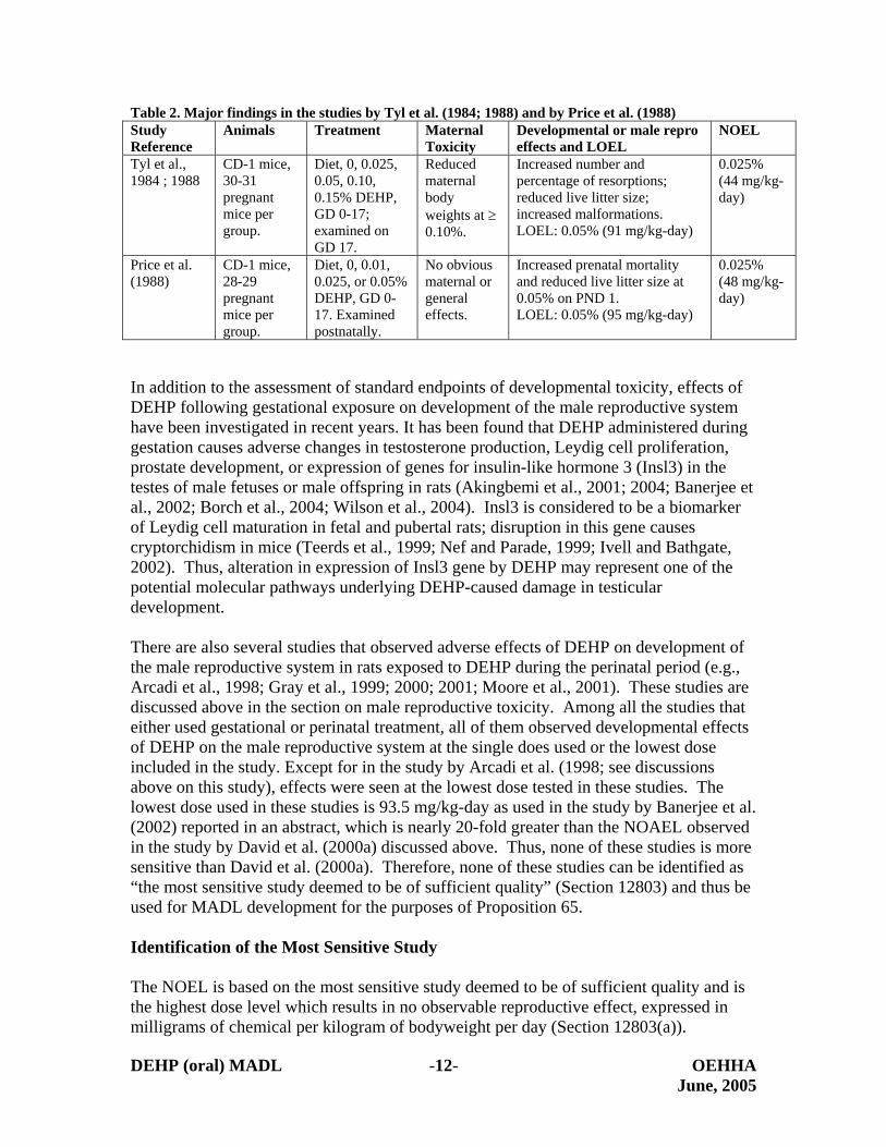
The developmental toxicity of DEHP in laboratory animals has been extensively studied In traditional developmental toxicity studies DEHP has been found to cause intrauterine death developmental delay and structural malformations and variations (CERHR 2000) Based on the relevant data available the CD-1 mouse appears to be the species most sensitive to the developmental effects of DEHP following oral treatment The lowest LOEL for the developmental toxicity of DEHP via the oral route of exposure was 005 in feed as observed in the studies reported by Tyl et al (1984 1988) and Price et al (1988) Major findings from these two studies were presented in Table 2 The estimated doses expressed as mgkg-day of DEHP used in the study by Price et al (1988) were slightly higher (95 mgkg-day for 005 48 mgkg-day for 0025) than those in the study by Tyl et al (1984 1988 91 mgkg-day for 005 44 mgkg-day for 0025) The NOEL (48 mgkg-day) for the developmental toxicity of DEHP observed in the study by Price et al (1988) is slightly higher than that (44 mgkg-day) in the study by Tyl et al (1984) and is lower than the LOEL from either study (91 or 95 mgkg-day) Therefore for the purpose of Proposition 65 the study by Price et al (1988) is identified by OEHHA as the most sensitive study for the developmental toxicity of DEHP following oral treatment
DEHP (oral) MADL -11- OEHHA June 2005
Table 2 Major findings in the studies by Tyl et al (1984 1988) and by Price et al (1988) Study Reference
Increased prenatal mortality and reduced live litter size at 005 on PND 1 LOEL 005 (95 mgkg-day)
0025 (48 mgkgshyday)
In addition to the assessment of standard endpoints of developmental toxicity effects of DEHP following gestational exposure on development of the male reproductive system have been investigated in recent years It has been found that DEHP administered during gestation causes adverse changes in testosterone production Leydig cell proliferation prostate development or expression of genes for insulin-like hormone 3 (Insl3) in the testes of male fetuses or male offspring in rats (Akingbemi et al 2001 2004 Banerjee et al 2002 Borch et al 2004 Wilson et al 2004) Insl3 is considered to be a biomarker of Leydig cell maturation in fetal and pubertal rats disruption in this gene causes cryptorchidism in mice (Teerds et al 1999 Nef and Parade 1999 Ivell and Bathgate 2002) Thus alteration in expression of Insl3 gene by DEHP may represent one of the potential molecular pathways underlying DEHP-caused damage in testicular development
There are also several studies that observed adverse effects of DEHP on development of the male reproductive system in rats exposed to DEHP during the perinatal period (eg Arcadi et al 1998 Gray et al 1999 2000 2001 Moore et al 2001) These studies are discussed above in the section on male reproductive toxicity Among all the studies that either used gestational or perinatal treatment all of them observed developmental effects of DEHP on the male reproductive system at the single does used or the lowest dose included in the study Except for in the study by Arcadi et al (1998 see discussions above on this study) effects were seen at the lowest dose tested in these studies The lowest dose used in these studies is 935 mgkg-day as used in the study by Banerjee et al (2002) reported in an abstract which is nearly 20-fold greater than the NOAEL observed in the study by David et al (2000a) discussed above Thus none of these studies is more sensitive than David et al (2000a) Therefore none of these studies can be identified as ldquothe most sensitive study deemed to be of sufficient qualityrdquo (Section 12803) and thus be used for MADL development for the purposes of Proposition 65
Identification of the Most Sensitive Study
The NOEL is based on the most sensitive study deemed to be of sufficient quality and is the highest dose level which results in no observable reproductive effect expressed in milligrams of chemical per kilogram of bodyweight per day (Section 12803(a))
DEHP (oral) MADL -12- OEHHA June 2005
The controlling regulation also specifies that ldquowhere multiple reproductive effects provide the basis for the determination that a chemical is known to the state to cause reproductive toxicity the reproductive effect for which studies produce the lowest NOEL shall be utilized for the determination of the NOELrdquo (Section 12803(a)(1)) The NOEL (58 mgkg-day) for the male reproductive toxicity as observed by David et al (2000a) is lower than the NOEL (48 mgkg-day) for the developmental toxicity of DEHP as observed by Price et al (1988) Therefore the oral study in rats reported by David et al (2000a) was used as basis for establishing the MADL for DEHP via the oral route of exposure
In the study reported by David et al (2000a) groups of Fischer 344 rats (55-80 animals per sex per group) were treated with 0 100 500 2500 or 12500 ppm DEHP in the diet for up to 104 weeks The animals were about six weeks old at the beginning of treatment The doses of DEHP were 0 58 289 1466 and 7890 mgkg-day for the five groups respectively as estimated by the study authors based on the average daily feed consumption Reduced mean body weights abnormal changes in serum chemistry hematology parameters increased liver and kidney weights and histopathological changes in the liver kidney pancreas and pituitary glands were observed in rats exposed to 12500 ppm DEHP Increased weights of liver and kidney and histopathological changes in the livers and kidneys were also observed in rats treated with 2500 ppm DEHP for 104 weeks Testis weights were significantly decreased in male rates treated with 12500 DEHP for 104 weeks Bilateral aspermatogenesis was observed in 10 out of 10 animals treated with 12500 ppm DEHP but not in any of 10 rats treated with 2500 ppm DEHP when examined at Week 78 After exposure for 104 weeks the incidence of bilateral aspermatogenesis was observed in 3764 (58) in the control group and 3450 (64) 4355 (78) 4865 (74) 6264 (97) in groups treated with 100 500 2500 or 12500 ppm DEHP respectively The increase in the incidence of bilateral aspermatogenesis was statistically significant in groups treated with ge 500 ppm DEHP Thus 500 ppm equivalent to 289 mgkg-day is identified as the LOEL The NOEL observed in this study 100 ppm (equivalent to 58 mgkg-day) is used as basis for establishing a MADL for DEHP by the oral route of exposure
Relevance of the Testicular Effects in Rats to Humans
For the purpose of Proposition 65 the study in rats reported by David et al (2000a) is identified as ldquothe most sensitive study deemed to be sufficient qualityrdquo and the NOEL for the testicular effects (indicative of the male reproductive toxicity) of DEHP observed in this study is used by OEHHA as the basis for establishing a MADL for DEHP by the oral route of exposure The relevance of testicular effects of DEHP in rats to humans was taken into account by OEHHA
It is generally accepted that ldquoan agent that produces an adverse reproductive effect in experimental animal studies is assumed to pose a potential reproductive threat to humansrdquo (US EPA 1996) However in the case of DEHP because DEHP does not cause obvious testicular damages in the common marmoset a non-human primate
DEHP (oral) MADL -13- OEHHA June 2005
(Rhodes et al 1986 Kurata et al 1998 MCSI 2003) and there are known inter-species differences in the testicular toxicity of DEHP (eg CERHR 2000 US FDA 2001) there have been questions raised regarding the relevance of testicular effects in rats to humans To determine if the testicular effects of DEHP observed in rats are relevant to humans OEHHA has reviewed relevant data on pharmacokinetics metabolism and mode(s) of action underlying the testicular effects of DEHP In particular OEHHA focused on similarities and differences in pharmacokinetics metabolism and mode(s) of action between rats and humans
Pharmacokinetics and Metabolism This subsection briefly summarize major pharmacokinetic characteristics of DEHP in rats non-human primates and humans There are numerous studies and comprehensive reviews on the absorption disposition metabolism and excretion of DEHP following oral administration in rats non-human primates and humans Discussions below are based on experimental data that have been repeatedly reviewed and summarized in published reviews (eg Albro 1986 Albro and Lavenhar 1989 Astill 1989) or regulatory or expert reports (eg CERHR 2000 US FDA 2001) References cited in the text are exemplary not comprehensive In addition several recent unpublished studies (Laignelet and Lhuguenot 2000a 2000b 2000c 2000d 2001) submitted to OEHHA by the ACC were also included for review Since exposure levels of DEHP in humans are generally low (CERHR 2000) special attention has been paid to data obtained from studies using relatively low doses of DEHP (eg below 500 mgkg-day)
Pharmacokinetic characteristics of DEHP are qualitatively similar among rats nonshyhuman primates and humans Briefly orally-administered DEHP is rapidly hydrolyzed to mono(2-ethylhexyl) phthalate (MEHP) and 2-ethylhexanol (2-EH) by ester hydrolases mainly in the gastrointestinal tract (GI) High levels of hydrolytic activity on DEHP have been found in the pancreatic juice intestinal contents or tissues and liver tissues of a wide variety of species including rats non-human primates and humans (Albro and Thomas 1973 Albro and Lavenhar 1989) Trace amounts or no intact DEHP have been found in the blood or liver tissues of rats or primates treated orally (either by gavage or in diet) with DEHP at levels below 500 mgkg (Albro et al 1982a Albro 1986 Astill 1989 MCSI 2003 Kessler et al 2004) In rats following oral administration in diet more than 90 or almost complete absorption as DEHP or its metabolites has been reported (Albro and Lavenhar 1989 Astill 1989) Rapid and near complete absorption of DEHP or its metabolites has also been observed in adult common marmosets treated with 100 mgkg-day DEHP in diet (MCSI 2003) However the exact extent of DEHP absorption in the GI tract in humans is not clear
Concentrations and kinetics of DEHP metabolites in the blood have been studied in rats and common marmosets In general blood concentrations of DEHP metabolites reach maximal levels within 4-8 hr after dosing in both species Maximal concentrations of DEHP metabolites in common marmosets are 13 to 10-fold lower than that in rats depending on the dose level (Rhodes et al 1986 Kessler et al 2004) However clearance of DEHP metabolites from the blood circulation appears to be slower in common marmosets than in rats (eg Albro and Lavenhar 1989 Rhodes et al 1986
DEHP (oral) MADL -14- OEHHA June 2005
MCSI 2003) Thus the difference in exposure based on blood concentration may be less than 13 to 10-fold
Following primary metabolism and absorption in the GI tract the primary metabolite of DEHP (ie MEHP) is further metabolized by one of three pathways hydrolysis to phthalic acid and 2-ethylhexonal conjugation to form glucuronide ester followed by rapid excretion and hydroxylation at various sites on the ethylhexyl chain by cytochrome P450 enzymes Hydroxylation of MEHP at ω- or ω1-position on the ethylhexyl chain is the major pathway and generates a variety of metabolites (Albro et al 1983 Lhuguenot et al 1985 Albro and Lavenhar 1989) Hydroxylation products are further metabolized by alcohol dehydrogenase and aldehyde dehydrogenase enzymes to yield ketoshymetabolites or dicarboxylic acids Dicarboxylic acids can then undergo α- or β-oxidation reaction In addition to MEHP itself more than 20 other metabolites of DEHP have been identified Major metabolites that have been found in the urine or fecal samples of rats have also been detected in the urine or fecal samples of non-human primates or humans There are a few metabolites that have been frequently analyzed in the urine or fecal samples from rodents non-human primates and humans These include metabolite V [mono(2-ethyl-5-carboxypentyl) phthalate product generated from hydroxylation at the ω- position on the hexyl branch] metabolite IX [mono(2-ethyl-5-hydroxy-hexyl) phthalate product of hydroxylation at the ω1- position on the hexyl branch] and metabolite VI [mono(2-ethyl-5-oxo-hexyl) phthalate keto-metabolite of metabolite IX]
Orally administered DEHP is quickly excreted from the body in urine as metabolites or in feces as either intact DEHP or metabolites with a near complete clearance from the body within two-four days in rats non-human primates and humans In addition to excretion in the urine and feces absorbed DEHP metabolites can be excreted in the bile and subsequently excreted in feces or re-absorbed into blood circulation via entero-hepatic circulation In rats and common marmosets approximately 40-50 of the metabolites of DEHP administered by intravenous injection can be excreted in the bile but only approximately 10-20 of the administered dose can be found in fecal samples suggesting that entero-hepatic circulation of DEHP metabolites is significant (Daniel and Bratt 1974 Chu et al 1978 Rhodes et al 1986 MCSI 2003)
MEHP and all of its major metabolites can be conjugated to glucuronide via glucuronyl transferase Glucuronide and MEHP or its metabolites in the conjugates can also be disassociated via β-glucuronidase In rats all metabolites excreted in the urine are in free (non-conjugated) form In non-human primates and humans glucuronide conjugates of DEHP metabolites account for 15-95 of the metabolites excreted in the urine depending on the chemical structure of metabolites route of exposure and individual primate or human subject (inter-individual variation) It has been shown that glucuronyl transferases from the rat are as active on MEHP as those from the mouse but high activity of β-glucuronidase activity in the rat results in absence of glucuronide-conjugates of MEHP or its metabolites in the urine of rats (Albro et al 1981 1982a Albro 1986 Albro and Lavenhar 1989) In addition predominant excretion of certain forms of glucuronide conjugates in the bile can also result in apparent absence of glucuronides in
DEHP (oral) MADL -15- OEHHA June 2005
the urine (Chiu and Huskey 1998) Therefore lack of glucuronide conjugates in the urine may not reflect the inability of an animal species to form such conjugates
Urinary excretion of DEHP metabolites accounts for about 30- 70 (approximately 50 on average) of DEHP orally administered in rats non-human primates and humans (Albro et al 1982a Astill 1989 CERHR 2000) Only approximately 30 of the administered dose is excreted in the urine as MEHP metabolite V metabolite VI and metabolite IX in rats These findings clearly indicate that the extent of oral absorption of DEHP andor its immediate metabolite MEHP at least 90 in rats is far higher than that excreted in the urine as the four metabolites discussed above Thus actual absorption rate or extent of DEHP in the GI tract is markedly higher than the extent of urinary excretion of major DEHP metabolites (approximately 30 of the dose) In humans urinary excretion of DEHP metabolites has been investigated in several studies (Peck and Albro 1982 Schmid amp Schlatter 1985 Dirven et al 1993 Anderson et al 2001 Koch et al 2004a Koch et al 2004b) Following oral administration up to approximately 70 of the administered dose of DEHP can be excreted in urine within the first 48 hours after administration (Koch et al 2003 2004) while Schmid and Schlatter (1985) reported that approximately 10-13 of the administered dose was excreted in the urine as MEHP metabolite XI IX and V None of the human studies determined levels of DEHP in feces or the possible extent of enterohepatic circulation The absolute or relative amount of DEHP metabolites excreted in urine samples is a clear indicator of human exposure to DEHP However based on the data observed in rats as discussed above the extent of absorption of DEHP or its metabolites in the GI tract in humans may well exceed the extent of urinary excretion of DEHP metabolites (ie more than up to 70 of orally administered DEHP can be expected to be absorbed in humans) In this regard the possible difference in the absorption rate or extent of DEHP or its metabolites between rats and humans may not be significant
Among the metabolites excreted in the urine in rats metabolite V accounts for approximately 10-25 of the dose administered MEHP metabolite VI and IX excretion accounts for approximately 8-10 of the dose administered (Albro et al 1981 1982a 1982b Astill 1989) Compared to the profile of DEHP metabolites in rats relatively less metabolite V (approximately 5 of the dose) and more metabolites of ω1-oxidation (metabolite IX and VI 14-40 of the dose) are excreted in the urine in non-human primates or humans (Rhodes et al 1986 Astill 1989 Schmid and Schlatter 1985 Koch et al 2003) Because of substantial biliary and fecal excretion of DEHP metabolites difference in the profile of DEHP metabolites in the urine may not reflect actual status of oxidative metabolism of MEHP For example Short et al (1987) compared DEHP metabolism and urinary excretion between rats and cynomolgus monkeys They reported that 84 and 22 of the dose administered to Fischer 344 rats was excreted as metabolite V in the urine and feces respectively In cynomolgus monkeys 57 and 53 of the dose administered was excreted as metabolite V in the urine and feces respectively suggesting that there was relatively less metabolite V excreted in the urine and more of it in the feces in cynomolgus monkeys than that in rats However if the relative amount of metabolite V excreted in the urine and feces is combined both species excreted about 11 of the dose as metabolites indicating that generation of metabolite V
DEHP (oral) MADL -16- OEHHA June 2005
in both species may be quantitatively similar even though the relative amount of this metabolite excreted in the urine is different This example clearly suggests that difference in the relative amount of metabolite V in the urine between rats and cynomolgus monkeys may not reflect actual status of oxidative metabolite of MEHP at organ levels It may also suggest that differences in the relative amount of metabolite V in urine samples between rats non-human primates and humans may not indicate actual differences in the oxidative metabolism of MEHP in the target organs of DEHP (eg testis or liver) between different species
From the data discussed above it is clear that pharmacokinetic characteristics and metabolism of DEHP in rats non-human primates and humans are both qualitatively similar in many aspects and quantitatively similar to a large extent at relatively low exposure levels (eg below 500 mgkg-day) There are some quantitative differences in the blood concentration of DEHP or its metabolites and in the profiles of DEHP metabolites in the urine among rats non-human primates and humans but these differences may not reflect actual extent of absorption of DEHP in the GI tract and actual status of oxidative metabolism of MEHP they may also play little role in the dramatic difference in testicular response to DEHP between rats and common marmosets As stated in the study report by MCSI (2003) ldquoit can no longer be assumed that this is due to poor absorption This difference is thought to arise from a difference in target organs physiology between the two animal species rather than from any significant differences in metabolic kineticsrdquo Similarly Kessler et al (2004) found that toxicokinetics alone could not account for the observed differences in toxicity suggesting that toxicodynamic factors (possibly interactions of MEHP with receptor-mediated processes) may also contribute to this pronounced difference between the rodent and the marmoset Physiological features of the testis in common marmosets that are fundamentally different from those in rats cynomolgus monkeys and humans may explain at least in part the lack of testicular effects of DEHP in the common marmoset
In conclusion similarities in pharmacokinetics and metabolism of DEHP between rats and humans strongly suggest that the testicular effects of DEHP observed in rats are relevant to humans Quantitative not qualitative difference in blood burdens of DEHP metabolites and in the profiles of DEHP metabolites in the urine between rats and nonshyhuman primates or humans do not explain the lack of testicular effects in common marmosets
Active Metabolite(s) Responsible for the Testicular Effects of DEHP The active metabolite(s) responsible for the testicular effects of DEHP has been studied in rats using both in vivo and in vitro approaches MEHP mimics the testicular effects of DEHP both in vivo and in vitro in juvenile rats but not 2-ethylhexanol or any of three major oxidative metabolites including metabolite V VI and IX (Gangolli 1982 Gray and Ganagolli 1986 Sjoberg et al 1986 Albro et al 1989 Grasso et al 1993 Jones et al 1993) DEHP but not 2-ethylhexanol causes reduction in Sertoli cell proliferation in neonatal rats (Li et al 2000) MEHP but not DEHP itself also causes decreased proliferation of cultured Sertoli cells isolated from neonatal rats (Li et al 1998 Li and
DEHP (oral) MADL -17- OEHHA June 2005
Kim 2003) These data clearly indicate that MEHP is the proximal metabolite for DEHP-induced testicular damage in rats
Distribution of DEHP metabolites to the testis has been reported in rats and common marmosets (Williams and Blanchfield 1974 Tanaka et al 1975 Rhodes et al 1986 MCSI 2003 Ono et al 2004) In rats radioactivity of DEHP 3H-labeled at the phthalic acid moiety was found in the basal area of seminiferous tubules at the stages IX-XIV and I of the spermatogenic cycle within six hours after a single oral dose As discussed in the subsection of ldquoMale Reproductive Toxicity in Animalsrdquo seminiferous tubules at the stages IX to I of the spermatogenic cycle have been shown to be more sensitive to the testicular effects of DEHP than those at other stages (eg Saitoh et al 1997 CERHR 2000) Within the seminiferous epithelium high levels of DEHP metabolites were mainly found in Sertoli cells and in the cytoplasm of spermatocytes The Sertoli cell has been shown to be the initial target testicular cell of DEHP in juvenile and adult rats (see discussions below) By 24 hours after dosing DEHP metabolites in the testis decreased to approximately 50 of the level observed 6 hours after dosing suggesting rapid clearance of DEHP metabolites from the testis in rats (Ono et al 2004) These data suggest not only that MEHP andor MEHP metabolites reach the testis after oral administration they are also distributed to the seminiferous tubules that have been shown to be targeted by DEHP in young or adult animals In this regard it should be noted that the Sertoli cells in common marmosets are morphologically uniform ie there is no morphological variation along the eight stages of seminiferous tubules in this species (Rune et al 1992) This feature of Sertoli cells in marmosets is different from these cells in most other mammals including humans indicating another difference in the physiological features of the testis between common marmosets and humans
Potential Modes of Actions Using both in vivo and in vitro approaches it has been repeatedly shown that the Sertoli cell and the Leydig cell appear to be the initial target cells of MEHP in the testis (eg Gray and Beamand 1984 Heindel and Powell 1992 Li et al 1998 Akingbemi et al 2001 CERHR 2000) The Sertoli cell is the somatic cell that provides a supportive role in spermatogenesis in adult animals and whose population established during proliferating periods determines the size of the testis and the volume of daily sperm production in the adult Maintenance and development of germ cells into functionally normal spermatozoa depend on a permissive milieu provided by the Sertoli cells (Russell and Griswold 1993 Boekelheide 2000) The Leydig cell produces androgen that regulates development of the male reproductive system and plays a critical role in spermatogenesis in the adult (Payne et al 1996) In the adult the effect of DEHPMEHP on the Leydig cells at high doses probably plays a minimal role in the overall testicular toxicity of DEHP even though there is clear evidence that DEHP in vivo and MEHP in vitro damages the Leydig cells in rats (eg Jones et al 1993 CERHR 2000) On the other hand both DEHP in vivo and MEHP in vitro damage both Sertoli cells and Leydig cells in fetal or neonatal testes from rats at doses that have no obvious effects on the testis in young or adult animals (eg Dostal et al 1988 Li et al 2000 Akingbemi et al 2004 Boekelheide 2004)
DEHP (oral) MADL -18- OEHHA June 2005
The exact biochemical or molecular mechanism(s) underlying the testicular effects of DEHP remains unclear Several hypotheses have been proposed including (1) alterations in testicular zinc levels or zinc-dependent enzymatic activities (2) oxidative stress in the testis (3) FSH receptor-dependent pathways (4) estrogenic activity or interactions with estrogen receptors (5) peroxisome proliferator-activated receptor (PPAR)-dependent pathways and (6) other cellular or molecular events or pathways (eg alterations in Sertoli-germ cell interactions or changed expression of genes critical for germ cell survival or functions of Sertoli cells or Leydig cells) OEHHA has reviewed a large amount of the relevant mechanistic data that are available For the purposes of this document the discussion below focuses on the role of PPAR in the male reproductive effects of DEHP This issue is critical for determining the relevance of the rodent data to humans since it has been proposed that induction of liver tumors by DEHP via PPARαshymediated mechanism(s) as observed in rodents is not relevant to humans (Klaunig et al 2003) It has been suggested that PPARα may also play an important role in the testicular effects of DEHP and thus PPARα-mediated testicular effects in rats are also not relevant to humans (ACC 2004 McKee et al 2004)
Two lines of evidence have been cited to support an active role of PPAR-α in the testicular effects of DEHP (ACC 2004 McKee et al 2004) One is the finding from the study by Ward et al (1998) that compared the toxicity including testicular lesions caused by oral administration of DEHP at 12000 ppm in diet for up to 24 weeks between wild-type (normal) mice and those lacking PPARα receptors (knock-out mice) The authors found that DEHP-induced testicular lesions in knock-out mice were less severe and required longer treatment than in the wild-type animals The authors suggested that both PPARα-dependent and ndashindependent pathways are involved in the testicular effects of DEHP In discussing presence of DEHP-induced kidney toxicity in PPARα knock-out mice the authors stated that ldquoit is possible that other receptor subtypes (PPARδ or γ) may play a role in the observed delayed kidney toxicity or the high dose of DEHP may modify the pharmacokinetics of DEHP in these micerdquo
The other line of evidence cited to support an active role of PPAR in DEHP-caused testicular damage primarily comes from studies that investigated the roles of PPARα in induction of Leydig cell tumors (LCTs) by peroxisome proliferators (PPs) in rodents (eg Cook et al 1992 Klaunig et al 2003) DEHP has been shown to cause Leydig cell hyperplasia and tumors in rats (eg Akingbemi et al 2004 Voss et al 2005) Cook and his co-workers have found that ammonium perfluorooctonate (C8) a peroxisome proliferating agent causing Leydig cell tumors causes imbalance between testosterone and estradiol by directly inhibiting testosterone production in Leydig cells andor by inducing synthesis of aromatase (which converts testosterone to estradiol) in the liver (Cook et al 1992 Biegel et al 1995 2001 Liu et al 1996a 1996b)
In addition to the studies on C8 the study by Gazouli et al (2002) investigated the effects of several PPs (including DEHP bezafibrate WY-14643) on steroid synthesis in Leydig cells and the mechanism underlying these effects The authors found that the anti-androgenic effects of some PPs are mediated by suppression of PPARα-mediated transcription of peripheral-type benzodiazepine receptor (PBR) gene This gene encodes
DEHP (oral) MADL -19- OEHHA June 2005
a high-affinity mitochondrial cholesterol-binding protein which plays an important role in transportation of cholesterol into mitochondria a hormone-induced rate-determining step in steroid synthesis The authors also reported several other important findings For example the circulating testosterone levels in PPARα knock out mice were significantly lower than that in the wild-type mice suggesting that PPARα may play a positive role in maintaining the balance of circulating testosterone levels When the animals were treated with 1 gkgday DEHP or 50 mgkgday WY-14643 for eight days circulating testosterone levels were significantly decreased in the wild-type mice However circulating testosterone levels were markedly increased to a level significantly higher than the knock-out controls and even slightly higher than that in the wild-type control animals indicating some PPs like DEHP may act through PPARβ or other unknown mechanisms to disrupt the balance of circulating testosterone levels In addition the authors found that bezafibrate acts mainly on the step of cholesterol transportation in steroid formation while MEHP acts on many steps of steroidogenesis Other than cholesterol transportation the role of PPARs in many steps in the biochemical cascades of steroidogenesis in Leydig cells remains unclear
In spite of the arguments discussed above there are numerous data suggesting that PPARα plays a minimal role if any in the testicular effects of DEHP First of all the testicular toxicity of DEHP is characterized by disruption in Sertoli cell function or proliferation followed by apoptosis in spermatocytes and alterations in Leydig cell function andor proliferation with subsequent disruption in androgen-dependent development of the male reproductive system There is no evidence indicating that C8 causes similar testicular damage (eg Kennedy et al 2004) The findings by Gazouli et al (2002) also clearly indicate that the disruptive effects of DEHP andor MEHP may be mediated by both PPARα-dependent and ndashindependent mechanism(s) Therefore there are differences in the mechanism(s) underlying the disruptive effects on testosterone synthesis or balance among different PPs Moreover even if DEHP and other PPs (eg C8) cause LCTs via similar mechanism(s) a mode of action for the non-cancer testicular toxicity of DEHP based on evidence from studies on a chemical that does not cause similar non-cancer testicular damage is not a valid comparison
Secondly two modes of actions (MOAs) have been postulated by Klaunig et al (2003) to describe the etiology of Leydig cell tumors in PPARα agonist-treated rats The authors have concluded that ldquothe weight of evidence available to date to support virtually all of the postulated key events is weak overall and moderate at best for only two or three of the postulated eventsrdquo Furthermore Klaunig et al (2003) concluded that ldquothe proposed animal MOAs - induction of aromatase secondary to liver induction (Pathway 1) and the direct inhibition of testosterone biosynthesis (Pathway 2) - are plausible mechanisms and could occur in humans If PPARα is mediating the induction of aromatase this mechanism could occur in humans due to the expression of PPARα in human liver The inhibition of testosterone biosynthesis by PPAR agonists is better established than the induction of aromatase and is also plausible as PPARα is present in human Leydig cells The pathways for the regulation of the HPT [hypothalamic-pituitary-testicular] axis of rats and humans also are similar in that compounds that decrease testosterone will
DEHP (oral) MADL -20- OEHHA June 2005
increase LH levels Hence compounds that induce LCTs in rats by disruption of the HPT axis pose a potential risk to human healthrdquo
Thirdly as stated by the Phthalate Expert Panel of CERHR (2000) ldquoin contrast to hepatic toxicity testicular toxicity is noted in PPAR-alpha knockout mice exposed to DEHP albeit that appearance of the testicular effects was delayed compared to wild-type mice In addition the guinea pig a non-responding species to the peroxisomalshyproliferating effects of DEHP is susceptible to the testicular effects of this agentrdquo The Phthalate Expert Panel of CERHR concluded that ldquoOverall the Panel believes that the reproductive toxicity of DEHP appears independent of PPAR-alpha However other members of the PPAR family (beta or delta and gamma) have not been extensively studied with regard to activation by phthalates PPAR-gamma has been found in human testis ovary placenta and embryo MEHP (but not DEHP 2-EH or 2-EHA) has been shown to activate PPARgamma receptor in a transcription reporter assay [Maloney and Waxman 1999]rdquo
Therefore the weight of evidence does not indicate that the non-cancer testicular effects of DEHP are mainly mediated by PPARα Even if PPARs including PPARα β and γ play any important role in DEHP-induced damage in testicular development and functions as suggested by evidence summarized in a recent comprehensive review by Corton and Lapinskas (2004) PPARs are expressed in human male reproductive organs (eg Elbrecht et al 1996 Schultz et al 1999 Collett et al 2000 Hase et al 2002) Therefore PPAR-mediated testicular effects of DEHP in rats are relevant to humans Possible modes of actions underlying the induction of Leydig cell tumors in rodents including those involving PPARs are also plausible in humans
With regard to the other hypotheses proposed for the testicular effects of DEHP the male reproductive system in humans has capabilities to carry out all of them There is no evidence to indicate otherwise
Conclusion on Relevance to Humans Based on the data that are available to OEHHA orally administered DEHP at doses relatively low (lt500 mgkg-day) but still markedly higher than the LOEL for testicular effects in rats (10 ndash 40 mgkg-day) is absorbed and metabolized in humans in ways in general qualitatively and quantitatively similar to those in rats and non-human primates Lack of testicular effects in common marmosets may be due to fundamental differences in testicular physiology between this species and other mammals including cynomolgus monkeys and humans Potential testicular effects of DEHP at a relatively low dose observed by Pugh et al (2000) in late-infantile cynomolgus monkey and similarities in the testicular physiology between cynomolgus monkeys and humans indicate that DEHP may cause testicular damages in humans All potential modes of actions or mechanisms underlying the testicular effects of DEHP in rats are also plausible in humans Therefore OEHHA concludes that the weight of the evidence supports a finding that the testicular effects of DEHP observed in rodents are relevant to humans
DEHP (oral) MADL -21- OEHHA June 2005
MADL Calculation
The NOEL is the highest dose level that results in no observable reproductive effect expressed in milligrams of chemical per kilogram of bodyweight per day The NOEL is converted to a milligram per day dose level by multiplying the assumed human body weight by the NOEL (Section 12803(b)) When the applicable reproductive effect is upon the male the MADL is generally calculated based on a human body weight of 70 kg (Section 12803(b)) As already noted however developing animals are sensitive to the testicular effects of DEHP (eg Sjoberg et al 1985 1986 Li et al 2000 CERHR 2000 US FDA 2001 Borch et al 2004)The bodyweights of infants and neonates are approximately 7-20 fold lower than that of an adult (National Center for Health Statistics 2005) Thus exposure of an infant or neonate to DEHP at a MADL calculated on the basis of an adult body weight of 70 kg would result in a dose up to 20-fold higher than the corresponding dose in adults Accordingly age-specific MADLs have been calculated for infant and neonatal boys based on bodyweights of 10 and 35 kg respectively as also allowed by regulation (Sections 12801(a) and 12803(a)(6))
The following calculations were performed to derive the MADLs for DEHP via the oral route of exposure based on a NOEL of 58 mgkg-day found in rats by David et al (2000a)
For Adults
When the applicable reproductive effect is upon the male human body weight of 70 kilograms shall be assumed (Section 12803(b))
Calculation of the NOEL for a 70 kg man 58 mgkg-day times 70 kg = 4060 mgday
The MADL is derived by dividing the NOEL by one thousand (Section 12801(b)(1)) Thus the adjusted NOEL was divided by 1000 to obtain the MADL
MADLadult oral = 406 mgday divide 1000 = 406 microgday or 410 microgday after rounding
For Neonates and Infants
Assuming a body weight of 10 kg for a one-year-old infant (National Center for Health Statistics 2000) an exposure of an infant to DEHP at the level of the MADLadult (410 microgday) is equivalent to 41 microgkg-day In order to derive a MADL for infants of 410 microgday it would require a NOEL of 41 mgkg-day (410 microgday divide 10 kg times 1000 = 41 mgkg-day) This estimated infant NOEL would be seven-fold higher than the NOEL for the adult (58 mgkg-day) indicating that application of the adult-derived MADL would result in a 7-fold higher dose in infants and a higher dose in neonates It is even higher than the LOELs observed by Poon et al (1997) (38 mgkg-day) in rats treated for 13 weeks beginning 6 weeks postnatal and by Akingbemi et al (2001 2004) (10 mgkgshyday)in rats treated for various periods of time beginning 21 days postnatal Therefore a
DEHP (oral) MADL -22- OEHHA June 2005
MADL based on the body weight of an adult human may not be protective against male reproductive effects in a neonatal or infant boy
Section 12801(a) specifies that ldquonothing in this article shall preclude a person from using evidence standards assessment methodologies principles assumptions or levels not described in this article to establish that a level of exposure has no observable effect at one thousand (1000) times the level in questionrdquo while Section 12803(a)(6) specifies that ldquowhen available data are of such quality that anatomic physiologic pharmacokinetic and metabolic considerations can be taken into account with confidence they may be used in the assessmentrdquo In this case the anatomic and physiologic differences between an infant boy and an adult man can be taken into account with much confidence Therefore MADLs specific to infants and neonates are developed as follows
For infants 0-2 years of age the average body weight of 10 kg over this developmental period is used (Section 12703(a)(8) OEHHA 2000 National Center for Health Statistics 2005)
Calculation of the NOEL for a 10 kg infant 58 mgkg-day times 10 kg = 58 mgday
All the MADLs derived above (410 microgday for adults 58 microgday for infant boys and 20 microgday for neonatal boys) apply to exposure to DEHP by the oral route
References
Ablake M Itoh M Terayama H Hayashi S Shoji S Naito M Takahashi K Suna S Jitsunari F (2004) Di-(2-ethylhexyl) phthalate induces severe aspermatogenesis in mice however subsequent antioxidant vitamins supplementation accelerates regeneration of the seminiferous epithelium Int J Androl 27 274-81
Akingbemi BT Ge R Klinefelter GR Zirkin BR Hardy MP (2004) Phthalate-induced Leydig cell hyperplasia is associated with multiple endocrine disturbances Proc Natl Acad Sci U S A 101 775-80
Akingbemi BT Youker RT Sottas CM Ge R Katz E Klinefelter GR Zirkin BR Hardy MP (2001) Modulation of rat Leydig cell steroidogenic function by di(2shy
DEHP (oral) MADL -23- OEHHA June 2005
ethylhexyl)phthalate Biol Reprod 65 1252-9
Albro PW (1986) Absorption metabolism and excretion of di(2-ethylhexyl) phthalate by rats and mice Environ Health Perspect 65 293-8
Albro PW Chapin RE Corbett JT Schroeder J Phelps JL (1989) Mono-2-ethylhexyl phthalate a metabolite of di-(2-ethylhexyl) phthalate causally linked to testicular atrophy in rats Toxicol Appl Pharmacol 100 193-200
Albro PW Corbett JT Schroeder JL Jordan S Matthews HB (1982a) Pharmacokinetics interactions with macromolecules and species differences in metabolism of DEHP Environ Health Perspect 45 19-25
Albro PW Hass JR Peck CC Jordan ST Corbett JT Schroeder J (1982b) Applications of isotope differentiation for metabolic studies with di-(2-ethylhexyl) phthalate J Environ Sci Health B 17 701-14
Albro PW Hass JR Peck CC Odam DG Corbett JT Bailey FJ Blatt HE Barrett BB (1981) Identification of the metabolites of di-(2-ethylhexyl) phthalate in urine from the African green monkey Drug Metab Dispos 9 223-5
Albro PW Lavenhar SR (1989) Metabolism of di(2-ethylhexyl)phthalate Drug Metab Rev 21 13-34
Albro PW Thomas RO (1973) Enzymatic hydrolysis of di-(2-ethylhexyl) phthalate by lipases Biochim Biophys Acta 306 380-90
Albro PW Tondeur I Marbury D Jordan S Schroeder J Corbett JT (1983) Polar metabolites of di-(2-ethylhexyl)phthalate in the rat Biochim Biophys Acta 760 283-92
American Chemistry Council (ACC 2004) Information pertaining to development of a maximum allowable dose level for di(2-ethylhexyl) phthalate Submitted to the Office of Environmental Health Hazard Assessment California Environmental Protection Agency Sacramento on May 12 2004
Anderson WA Castle L Scotter MJ Massey RC Springall C (2001) A biomarker approach to measuring human dietary exposure to certain phthalate diesters Food Addit Contam 18 1068-74
Arcadi FA Costa C Imperatore C Marchese A Rapisarda A Salemi M Trimarchi GR Costa G (1998) Oral toxicity of bis(2-ethylhexyl) phthalate during pregnancy and suckling in the Long-Evans rat Food Chem Toxicol 36 963-70
Aslan AR Kogan BA and Gondos B (2003) Testicular development In Polin RA Fox WW and Abman SH eds Fetal and Neonatal Physiology Sunders Philadelphia PA 3rd
edi Volume 2 Chapter 191 pp1950-1960
DEHP (oral) MADL -24- OEHHA June 2005
Astill BD (1989) Metabolism of DEHP effects of prefeeding and dose variation and comparative studies in rodents and the cynomolgus monkey (CMA studies) Drug Metab Rev 21 35-53
Banerjee S Thuillier R Culty M Papadopoulos V Brown TR Banerjee PP (2002) In utero exposure to di(2-ethylhexyl) phthalate alters growth tissue organization and the expression of androgen receptor protein of rat prostate Biol Reprod 66(Suppl 1) 200
Biegel LB Hurtt ME Frame SR OConnor JC Cook JC (2001) Mechanisms of extrahepatic tumor induction by peroxisome proliferators in male CD rats Toxicol Sci 60 44-55
Biegel LB Liu RC Hurtt ME Cook JC (1995) Effects of ammonium perfluorooctanoate on Leydig cell function in vitro in vivo and ex vivo studies Toxicol Appl Pharmacol 134 18-25
Boekelheide K (2004) Cracking the nut Toxicol Sci 81 1-2
Boekelheide K Fleming SL Johnson KJ Patel SR Schoenfeld HA (2000) Role of Sertoli cells in injury-associated testicular germ cell apoptosis Proc Soc Exp Biol Med 225 105-15
Borch J Ladefoged O Hass U Vinggaard AM (2004) Steroidogenesis in fetal male rats is reduced by DEHP and DINP but endocrine effects of DEHP are not modulated by DEHA in fetal prepubertal and adult male rats Reprod Toxicol 18 53-61
Cammack JN White RD Gordon D Gass J Hecker L Conine D Bruen US Friedman M Echols C Yeh TY Wilson DM (2003 ) Evaluation of reproductive development following intravenous and oral exposure to DEHP in male neonatal rats Int J Toxicol 22 159-74
Center for The Evaluation of Risks to Human Reproduction (CERHR 2000) NTPshyCERHR Expert Panel Report on Di (2-ethylhexyl) Phthalate National Toxicology Program US Department of Health and Human Services Research Triangle Park NC October
Chiu SH Huskey SW (1998) Species differences in N-glucuronidation Drug Metab Dispos 26 838-47
Cho F Yabe M Honjo S (1975) The weight of the reproductive organs hypophysis and thyroid of male cynomolgus monkeys (Macaca fascicularis) Jikken Dobutsu 24 173-5
Chu I Villeneuve DC Secours V Franklin C Rock G Viau A (1978) Metabolism and tissue distribution of mono-2-ethylhexyl phthalate in the rat Drug Metab Dispos 6 146shy9
DEHP (oral) MADL -25- OEHHA June 2005
Collett GP Betts AM Johnson MI Pulimood AB Cook S Neal DE Robson CN (2000) Peroxisome proliferator-activated receptor alpha is an androgen-responsive gene in human prostate and is highly expressed in prostatic adenocarcinoma Clin Cancer Res 6 3241-8
Cook JC Murray SM Frame SR Hurtt ME (1992) Induction of Leydig cell adenomas by ammonium perfluorooctanoate a possible endocrine-related mechanism Toxicol Appl Pharmacol 113 209-17
Corton JC Lapinskas PJ (2004) Peroxisome Proliferator-Activated Receptors Mediators of Phthalate Ester-Induced Effects in the Male Reproductive Tract Toxicol Sci
Daniel JW Bratt H (1974) The absorption metabolism and tissue distribution of di(2shyethylhexyl)phthalate in rats Toxicology 2 51-65
David RM Moore MR Finney DC Guest D (2000a) Chronic toxicity of di(2shyethylhexyl)phthalate in rats Toxicol Sci 55 433-43
David RM Moore MR Finney DC Guest D (2000b) Chronic toxicity of di(2shyethylhexyl)phthalate in mice Toxicol Sci 58 377-85
Dirven HA van den Broek PH Arends AM Nordkamp HH de Lepper AJ Henderson PT Jongeneelen FJ (1993) Metabolites of the plasticizer di(2-ethylhexyl)phthalate in urine samples of workers in polyvinylchloride processing industries Int Arch Occup Environ Health 64 549-54
Dostal LA Chapin RE Stefanski SA Harris MW Schwetz BA (1988) Testicular toxicity and reduced Sertoli cell numbers in neonatal rats by di(2-ethylhexyl)phthalate and the recovery of fertility as adults Toxicol Appl Pharmacol 95 104-21
Duty SM Calafat AM Silva MJ Brock JW Ryan L Chen Z Overstreet J Hauser R (2004) The relationship between environmental exposure to phthalates and computer-aided sperm analysis motion parameters J Androl 25 293-302
Duty SM Silva MJ Barr DB Brock JW Ryan L Chen Z Herrick RF Christiani DC Hauser R (2003a) Phthalate exposure and human semen parameters Epidemiology 14 269-77
Duty SM Singh NP Silva MJ Barr DB Brock JW Ryan L Herrick RF Christiani DC Hauser R (2003b) The relationship between environmental exposures to phthalates and DNA damage in human sperm using the neutral comet assay Environ Health Perspect 111 1164-9
Elbrecht A Chen Y Cullinan CA Hayes N Leibowitz M Moller DE Berger J (1996) Molecular cloning expression and characterization of human peroxisome proliferator
DEHP (oral) MADL -26- OEHHA June 2005
activated receptors gamma 1 and gamma 2 Biochem Biophys Res Commun 224 431-7 Festing MF Altman DG (2002) Guidelines for the design and statistical analysis of experiments using laboratory animals ILAR J 43 244-58
Flurer CI Zucker H (1987) Difference in serum ascorbate in two species of Callithricidae Int J Vitam Nutr Res 57 297-8
Flurer CI Zucker H (1989) Ascorbic acid in a New World monkey family species difference and influence of stressors on ascorbic acid metabolism Z Ernahrungswiss 28 49-55
Fredricsson B Moller L Pousette A Westerholm R (1993) Human sperm motility is affected by plasticizers and diesel particle extracts Pharmacol Toxicol 72 128-33
Gangolli SD (1982) Testicular effects of phthalate esters Environ Health Perspect 45 77-84
Gazouli M Yao ZX Boujrad N Corton JC Culty M Papadopoulos V (2002) Effect of peroxisome proliferators on Leydig cell peripheral-type benzodiazepine receptor gene expression hormone-stimulated cholesterol transport and steroidogenesis role of the peroxisome proliferator-activator receptor alpha Endocrinology 143 2571-83
Grasso P Heindel JJ Powell CJ Reichert LE Jr (1993) Effects of mono(2-ethylhexyl) phthalate a testicular toxicant on follicle-stimulating hormone binding to membranes from cultured rat Sertoli cells Biol Reprod 48 454-9
Gray LE Hotchkiss AK Price M Wolf CJ Furr J Ostby J Lambright C Parks L Wilson V Bobseine K and others (2001) Adverse effects of antiandrogenic pesticides and toxic substances on reproductive development in the male Biol Reprod 64(Suppl 1) 87-8
Gray LE Jr Ostby J Furr J Price M Veeramachaneni DN Parks L (2000) Perinatal exposure to the phthalates DEHP BBP and DINP but not DEP DMP or DOTP alters sexual differentiation of the male rat Toxicol Sci 58 350-65
Gray LE Jr Wolf C Lambright C Mann P Price M Cooper RL Ostby J (1999) Administration of potentially antiandrogenic pesticides (procymidone linuron iprodione chlozolinate pp-DDE and ketoconazole) and toxic substances (dibutyl- and diethylhexyl phthalate PCB 169 and ethane dimethane sulphonate) during sexual differentiation produces diverse profiles of reproductive malformations in the male rat Toxicol Ind Health 15 94-118
Gray TJ Beamand JA (1984) Effect of some phthalate esters and other testicular toxins on primary cultures of testicular cells Food Chem Toxicol 22 123-31
Gray TJ Butterworth KR (1980) Testicular atrophy produced by phthalate esters Arch
DEHP (oral) MADL -27- OEHHA June 2005
Toxicol Suppl 4 452-5
Gray TJ Gangolli SD (1986) Aspects of the testicular toxicity of phthalate esters Environ Health Perspect 65 229-35
Gray TJ Rowland IR Foster PM Gangolli SD (1982) Species differences in the testicular toxicity of phthalate esters Toxicol Lett 11 141-7
Gromoll J Eiholzer U Nieschlag E Simoni M (2000) Male hypogonadism caused by homozygous deletion of exon 10 of the luteinizing hormone (LH) receptor differential action of human chorionic gonadotropin and LH J Clin Endocrinol Metab 85 2281-6
Hampl JS Taylor CA Johnston CS (2004) Vitamin C deficiency and depletion in the United States the Third National Health and Nutrition Examination Survey 1988 to 1994 Am J Public Health 94 870-5
Hase T Yoshimura R Mitsuhashi M Segawa Y Kawahito Y Wada S Nakatani T Sano H (2002) Expression of peroxisome proliferator-activated receptors in human testicular cancer and growth inhibition by its agonists Urology 60 542-7
Heindel JJ Powell CJ (1992) Phthalate ester effects on rat Sertoli cell function in vitro effects of phthalate side chain and age of animal Toxicol Appl Pharmacol 115 116-23
Hilscher B Engemann A (1992) Histological and morphometric studies on the kinetics of germ cells and immature Sertoli cells during human prespermatogenesis Andrologia 24 7-10
Ishihara M Itoh M Miyamoto K Suna S Takeuchi Y Takenaka I Jitsunari F (2000) Spermatogenic disturbance induced by di-(2-ethylhexyl) phthalate is significantly prevented by treatment with antioxidant vitamins in the rat Int J Androl 23 85-94
Ivell R Bathgate RA (2002) Reproductive biology of the relaxin-like factor (RLFINSL3) Biol Reprod 67 699-705
Jones HB Garside DA Liu R Roberts JC (1993) The influence of phthalate esters on Leydig cell structure and function in vitro and in vivo Exp Mol Pathol 58 179-93
Kennedy GL Jr Butenhoff JL Olsen GW OConnor JC Seacat AM Perkins RG Biegel LB Murphy SR Farrar DG (2004 ) The toxicology of perfluorooctanoate Crit Rev Toxicol 34 351-84
Kessler W Numtip W Grote K Csanady GA Chahoud I Filser JG (2004) Blood burden of di(2-ethylhexyl) phthalate and its primary metabolite mono(2-ethylhexyl) phthalate in pregnant and nonpregnant rats and marmosets Toxicol Appl Pharmacol 195 142-53
DEHP (oral) MADL -28- OEHHA June 2005
Klaunig JE Babich MA Baetcke KP Cook JC Corton JC David RM DeLuca JG Lai DY McKee RH Peters JM Roberts RA Fenner-Crisp PA (2003) PPARalpha agonistshyinduced rodent tumors modes of action and human relevance Crit Rev Toxicol 33 655shy780
Kluin PM Kramer MF de Rooij DG (1983) Testicular development in Macaca irus after birth Int J Androl 6 25-43
Koch HM Bolt HM Angerer J (2004a) Di(2-ethylhexyl)phthalate (DEHP) metabolites in human urine and serum after a single oral dose of deuterium-labelled DEHP Arch Toxicol 78 123-30
Koch HM Preuss R Drexler H Angerer J Bolt HM Biological monitoring of (DEHP) exposure the relevance of the oxidative metabolites of Di(2-ethylhexyl)phthalate (DEHP) compared to the classical parameter MEHP (2004b) Annual Meeting of the International Society of Exposure Analysis Philadelphia Pennsylvania USA October 17-21 2004 Abstract M1A-06
Kurata Y Kidachi F Yokoyama M Toyota N Tsuchitani M Katoh M (1998) Subchronic toxicity of Di(2-ethylhexyl)phthalate in common marmosets lack of hepatic peroxisome proliferation testicular atrophy or pancreatic acinar cell hyperplasia Toxicol Sci 42 49-56
Laignelet L Lhuguenot JC (2000a) Di-(2-ethylhexyl)phthalate (DEHP) absorption excretion metabolism and pharmacokinetic profile in Wistar female rats Report No 199 Submitted by the American Chemistry Council to OEHHA California EPA on May 12 2004
Laignelet L Lhuguenot JC (2000b) Di-(2-ethylhexyl)phthalate (DEHP) absorption excretion metabolism and pharmacokinetic profile in pregnant Wistar rats Report No 299 Submitted by the American Chemistry Council to OEHHA California EPA on May 12 2004
Laignelet L Lhuguenot JC (2000c) I-(2-ethylhexyl)phthalate (DEHP) absorption excretion metabolism and pharmacokinetic profile in pregnant CD1 mice Report No 399 Submitted by the American Chemistry Council to OEHHA California EPA on May 12 2004
Laignelet L Lhuguenot JC (2000d) Di-(2-ethylhexyl)phthalate (DEHP) absorption excretion metabolism and pharmacokinetic profile in female CD1 mice Report No 499 Submitted by the American Chemistry Council to OEHHA California EPA on May 12 2004
Laignelet L Lhuguenot JC (2001) Di-(2-ethylhexyl)phthalate (DEHP) absorption excretion metabolism and pharmacokinetic profile in pregnant and non-pregnant rats and
DEHP (oral) MADL -29- OEHHA June 2005
mice Synthesis of Reports No 199 to 499 Submitted by the American Chemistry Council to OEHHA California EPA on May 12 2004
Lake BG Brantom PG Gangolli SD Butterworth KR Grasso P (1976) Studies on the effects of orally administered Di-(2-ethylhexyl) phthalate in the ferret Toxicology 6 341shy56
Lamb JC 4th Chapin RE Teague J Lawton AD Reel JR (1987) Reproductive effects of four phthalic acid esters in the mouse Toxicol Appl Pharmacol 88 255-69
Latini G De Felice C Presta G Del Vecchio A Paris I Ruggieri F Mazzeo P (2003) In utero exposure to di-(2-ethylhexyl)phthalate and duration of human pregnancy Environ Health Perspect 111 1783-5
Lenth RV (2001) Some practical guidelines for effective sample size determination Am Statist 55 187-193
Lhuguenot JC Mitchell AM Milner G Lock EA Elcombe CR (1985) The metabolism of di(2-ethylhexyl) phthalate (DEHP) and mono-(2-ethylhexyl) phthalate (MEHP) in rats in vivo and in vitro dose and time dependency of metabolism Toxicol Appl Pharmacol 80 11-22
Li LH Donald J Golub M (2004) Testicular development structure function and regulation in common marmosets Southern CaliforniaNorthern California Chapters of Society of Toxicology 2004 Annual Meeting September 30-October 1 2004 San Diego CA
Li H Kim KH (2003) Effects of mono-(2-ethylhexyl) phthalate on fetal and neonatal rat testis organ cultures Biol Reprod 69 1964-72
Li LH Jester WF Jr Laslett AL Orth JM (2000) A single dose of Di-(2-ethylhexyl) phthalate in neonatal rats alters gonocytes reduces sertoli cell proliferation and decreases cyclin D2 expression Toxicol Appl Pharmacol 166 222-9
Li LH Jester WF Jr Orth JM (1998) Effects of relatively low levels of mono-(2shyethylhexyl) phthalate on cocultured Sertoli cells and gonocytes from neonatal rats Toxicol Appl Pharmacol 153 258-65
Liang JH Sankai T Yoshida T Yoshikawa Y (2001) Immunolocalization of proliferating cell nuclear antigen (PCNA) in cynomolgus monkey (Macaca fascicularis) testes during postnatal development J Med Primatol 30 107-11
Liu RC Hahn C Hurtt ME (1996a) The direct effect of hepatic peroxisome proliferators on rat Leydig cell function in vitro Fundam Appl Toxicol 30 102-8
Liu RC Hurtt ME Cook JC Biegel LB (1996b) Effect of the peroxisome proliferator
DEHP (oral) MADL -30- OEHHA June 2005
ammonium perfluorooctanoate (C8) on hepatic aromatase activity in adult male CrlCD BR (CD) rats Fundam Appl Toxicol 30 220-8
Maloney EK Waxman DJ (1999) Trans-activation of PPARα and PPARγ by structurally diverse environmental chemicals Toxicol Appl Pharmacol 161 209-218
McKee RH Butala JH David RM Gans G (2004) NTP center for the evaluation of risks to human reproduction reports on phthalates addressing the data gaps Reprod Toxicol 18 1-22
Millar MR Sharpe RM Weinbauer GF Fraser HM Saunders PT (2000) Marmoset spermatogenesis organizational similarities to the human Int J Androl 23 266-77
Mitsubishi Chemical Safety Institute Ltd (MCSI 2003) Sixty-five week repeated oral dose toxicity study of di(2-ethylhexyl) phthalate (DEHP) in juvenile common marmosets Mitsubishi Chemical Safety Institute Ltd Study No B000496 [A copy of the final study report was provided to OEHHA by the American Chemistry Council]
Modigh CM Bodin SL Lillienberg L Dahlman-Hoglund A Akesson B Axelsson G (2002) Time to pregnancy among partners of men exposed to di(2-ethylhexyl)phthalate Scand J Work Environ Health 28 418-28
Moore RW Rudy TA Lin TM Ko K Peterson RE (2001) Abnormalities of sexual development in male rats with in utero and lactational exposure to the antiandrogenic plasticizer Di(2-ethylhexyl) phthalate Environ Health Perspect 109 229-37
Muller T Simoni M Pekel E Luetjens CM Chandolia R Amato F Norman RJ Gromoll J (2004) Chorionic gonadotrophin beta subunit mRNA but not luteinising hormone beta subunit mRNA is expressed in the pituitary of the common marmoset (Callithrix jacchus) J Mol Endocrinol 32 115-28
National Center for Health Statistics (2005) Clinical growth charts for infants US Department of Health and Human Services Centers for Disease Control and Prevention National Center for Health Statistics Hyattsville MD Available at httpwwwcdcgovgrowthcharts
National Institute for Occupational Safety and Health (NIOSH 1990) NIOH and NIOSH basis for an Occupational Health Standard Di (2-ethylhexyl) phthalate (DEHP) US Department of Health and Human Services Public Health Service Centers for Disease Control NIOSH
Nef S Parada LF (1999) Cryptorchidism in mice mutant for Insl3 Nat Genet 22 295-9
Office of Environmental Health Hazard Assessment (OEHHA 1997) Public Health Goal for Di(2-ethylhexyl) Phthalate (DEHP) in Drinking Water OEHHA California Environmental Protection Agency Sacramento California February
DEHP (oral) MADL -31- OEHHA June 2005
Office of Environmental Health Hazard Assessment (OEHHA 2000) Air Toxics Hot Spots Program Risk Assessment Guidelines Part IV Technical Support Document for Exposure Assessment and Stochastic Analysis OEHHA California Environmental Protection Agency Sacramento California September
Ono H Saito Y Imai K Kato M (2004) Subcellular distribution of di-(2shyethylhexyl)phthalate in rat testis J Toxicol Sci 29 113-24
Orth JM (1982) Proliferation of Sertoli cells in fetal and postnatal rats a quantitative autoradiographic study Anat Rec 203 485-92
Orth JM Gunsalus GL Lamperti AA (1988) Evidence from Sertoli cell-depleted rats indicates that spermatid number in adults depends on numbers of Sertoli cells produced during perinatal development Endocrinology 122 787-94
Park JD Habeebu SS Klaassen CD (2002) Testicular toxicity of di-(2shyethylhexyl)phthalate in young Sprague-Dawley rats Toxicology 171 105-15
Payne AH Hardy MP and Russell LD (1996) The Leydig Cell Cache River Press Clearwater FL
Peck CC Albro PW (1982) Toxic potential of the plasticizer Di(2-ethylhexyl) phthalate in the context of its disposition and metabolism in primates and man Environ Health Perspect 45 11-7
Poon R Lecavalier P Mueller R Valli VE Procter BG Chu I (1997) Subchronic oral toxicity of di-n-octyl phthalate and di(2-Ethylhexyl) phthalate in the rat Food Chem Toxicol 35 225-39
Price CJ Tyl RW Marr MC Myers CB Sadler BM Kimmel CA (1988) Reproduction and fertility evaluation of diethylhexy phthalate (CAS No 117-81-7) in CD-1 mice exposed during gestation Report No NTP 88-092
Pugh G Jr Isenberg JS Kamendulis LM Ackley DC Clare LJ Brown R Lington AW Smith JH Klaunig JE (2000) Effects of di-isononyl phthalate di-2-ethylhexyl phthalate and clofibrate in cynomolgus monkeys Toxicol Sci 56 181-8
Rais-Bahrami K Nunez S Revenis ME Short BL Luban NL (2004) Follow-up study of adolescents exposed to di-2-ethylhexyl phthalate (DEHP) as neonates on extracorporeal membrane oxygenation (ECMO) support Environ Health Persp 112 1339-40
Rhodes C Orton TC Pratt IS Batten PL Bratt H Jackson SJ Elcombe CR (1986) Comparative pharmacokinetics and subacute toxicity of di(2-ethylhexyl) phthalate (DEHP) in rats and marmosets extrapolation of effects in rodents to man Environ Health
DEHP (oral) MADL -32- OEHHA June 2005
Perspect 65 299-307
Rune GM Pretzer D De Souza P Bollmann U Merker HJ (1992) Ultrastructure of adult and juvenile marmoset (Callithrix jacchus) Sertoli cells in vivo and in vitro J Androl 13 560-70
Russell LD and Griswold MD (1993) The Sertoli Cell Cache River Press Clearwater FL
Saitoh Y Usumi K Nagata T Marumo H Imai K Katoh M (1997) Early changes in the rat testis induced by di-(2-Ethylhexyl) phthalate and 25-hexanedione ultrastructure and lanthanum trace study J Toxicol Pathol 10(1) 51-7
Schilling K Gembardt C Hellwig J (1999) Reproduction toxicity of di-2-ethylhexyl phthalate (DEHP) Toxicologist 48(1-S)147-8
Schmid P Schlatter C (1985) Excretion and metabolism of di(2-ethylhexyl)phthalate in man Xenobiotica 15 251-6
Schultz R Yan W Toppari J Volkl A Gustafsson JA Pelto-Huikko M (1999) Expression of peroxisome proliferator-activated receptor alpha messenger ribonucleic acid and protein in human and rat testis Endocrinology 140 2968-75
Schwetz BA Rao KS Park CN (1980) Insensitivity of tests for reproductive problems J Environ Pathol Toxicol 3 81-98
Sharpe RM Walker M Millar MR Atanassova N Morris K McKinnell C Saunders PT Fraser HM (2000) Effect of neonatal gonadotropin-releasing hormone antagonist administration on Sertoli cell number and testicular development in the marmoset comparison with the rat Biol Reprod 62 1685-93
Short RD Robinson EC Lington AW Chin AE (1987) Metabolic and peroxisome proliferation studies with di(2-ethylhexyl)phthalate in rats and monkeys Toxicol Ind Health 3 185-95
Sjoberg P Bondesson U Gray TJ Ploen L (1986) Effects of di-(2-ethylhexyl) phthalate and five of its metabolites on rat testis in vivo and in in vitro Acta Pharmacol Toxicol (Copenh) 58 225-33
Sjoberg P Bondesson U Kjellen L Lindquist NG Montin G Ploen L (1985) Kinetics of di-(2-ethylhexyl) phthalate in immature and mature rats and effect on testis Acta Pharmacol Toxicol (Copenh) 56 30-7
Smedley JV Bailey SA Perry RW O Rourke CM (2002) Methods for predicting sexual maturity in male cynomolgus macaques on the basis of age body weight and histologic evaluation of the testes Contemp Top Lab Anim Sci 41 18-20
DEHP (oral) MADL -33- OEHHA June 2005
Stata Corporation (2003) Small Stata 80 for Windows Stata Corporation College Station TX
Tanaka A Adachi T Takahashi T Yamaha T (1975) Biochemical studies on phthalic esters I Elimination distribution and metabolism of di-(2-ethylhexyl)phthalate in rats Toxicology 4 253-64
Tandon R Seth PK Srivastava SP (1991) Effect of in utero exposure to di(2shyethylhexyl)phthalate on rat testes Indian J Exp Biol 29 1044-6
Teerds KJ de Boer-Brouwer M Dorrington JH Balvers M Ivell R (1999) Identification of markers for precursor and leydig cell differentiation in the adult rat testis following ethane dimethyl sulphonate administration Biol Reprod 60 1437-45
Tyl RW and Jones-Price C (1984) Teratological evaluation of diethylhexylphthalate (CAS No 117-81-7) in Fischer 344 rats Jefferson AR National Center for Toxicological Research
Tyl RW Price CJ Marr MC Kimmel CA (1988) Developmental toxicity evaluation of dietary di(2-ethylhexyl)phthalate in Fischer 344 rats and CD-1 mice Fundam Appl Toxicol 10 395-412
US Environmental Protection Agency (US EPA 1996) Guidelines for reproductive toxicity risk assessment EPA630R-96009 FRL-5630-6
US Food and Drug Administration (US FDA 2001) Safety Assessment of Di (2shyethylhexyl)phthalate (DEHP) Released from PVC Medical Devices Centers for Devices and Rediological Health US Food and Drug Administration Rockville MD
Voss C Zerban H Bannasch P Berger MR (2005) Lifelong exposure to di-(2shyethylhexyl)-phthalate induces tumors in liver and testes of Sprague-Dawley rats Toxicology 206 359-71
Ward JM Peters JM Perella CM Gonzalez FJ (1998) Receptor and nonreceptorshymediated organ-specific toxicity of di(2-ethylhexyl)phthalate (DEHP) in peroxisome proliferator-activated receptor alpha-null mice Toxicol Pathol 26 240-6
Williams DT Blanchfield BJ (1974) Retention excretion and metabolism of di-(2shyethylhexyl) phthalate administered orally to the rat Bull Environ Contam Toxicol 11 371-8
Wilson VS Lambright C Furr J Ostby J Wood C Held G Gray LE Jr (2004) Phthalate ester-induced gubernacular lesions are associated with reduced insl3 gene expression in the fetal rat testis Toxicol Lett 146 207-15
DEHP (oral) MADL -34- OEHHA June 2005
Wistuba J Mundry M Luetjens CM Schlatt S (2004) Co-grafting of hamster (Phodopus sungorus) and marmoset (Callithrix jacchus) testicular tissues into nude mice does not overcome blockade of early spermatogenic differentiation in primate grafts Biol Reprod 71 2087-91
Zhang FP Kero J Huhtaniemi I (1998) The unique exon 10 of the human luteinizing hormone receptor is necessary for expression of the receptor protein at the plasma membrane in the human luteinizing hormone receptor but deleterious when inserted into the human follicle-stimulating hormone receptor Mol Cell Endocrinol 142 165-74
Zuhlke U Weinbauer G (2003) The common marmoset (Callithrix jacchus) as a model in toxicology Toxicol Pathol 31 Suppl 123-7
DEHP (oral) MADL -35- OEHHA June 2005
Bibliography
Additional Studies or Reports Reviewed by OEHHA
Abbott DH Hearn JP (1978) Physical hormonal and behavioural aspects of sexual development in the marmoset monkey Callithrix jacchus J Reprod Fertil 53 155-66
Agarwal DK Eustis S Lamb JC 4th Jameson CW Kluwe WM (1986a) Influence of dietary zinc on di(2-ethylhexyl)phthalate-induced testicular atrophy and zinc depletion in adult rats Toxicol Appl Pharmacol 84 12-24
Anderson WA Barnes KA Castle L Damant AP Scotter MJ (2002) Determination of isotopically labelled monoesterphthalates in urine by high performance liquid chromatography-mass spectrometry Analyst 127 1193-7
Anderson WA Castle L Scotter MJ Massey RC Springall C (2001) A biomarker approach to measuring human dietary exposure to certain phthalate diesters Food Addit Contam 18 1068-74
Arslan M Weinbauer GF Schlatt S Shahab M Nieschlag E (1993) FSH and testosterone alone or in combination initiate testicular growth and increase the number of spermatogonia and Sertoli cells in a juvenile non-human primate (Macaca mulatta) J Endocrinol 136 235-43
Asaoka K Hagihara K Kabaya H Sakamoto Y Katayama H Yano K (2000) Uptake of phthalate esters di(n-butyl)phthalate and di(2-ethylhexyl)phthalate as environmental chemicals in monkeys in Japan Bull Environ Contam Toxicol 64 679-85
Barr AB (1973) Timing of spermatogenesis in four nonhuman primate species Fertil Steril 24 381-9
Behr R Hunt N Ivell R Wessels J Weinbauer GF (2000) Cloning and expression analysis of testis-specific cyclic 3 5-adenosine monophosphate-responsive element modulator activators in the nonhuman primate (Macaca fascicularis) comparison with other primate and rodent species Biol Reprod 62 1344-51
Berensztein EB Sciara MI Rivarola MA Belgorosky A (2002) Apoptosis and proliferation of human testicular somatic and germ cells during prepuberty high rate of testicular growth in newborns mediated by decreased apoptosis J Clin Endocrinol Metab 87 5113-8
Blair RM Fang H Branham WS Hass BS Dial SL Moland CL Tong W Shi L Perkins R Sheehan DM (2000) The estrogen receptor relative binding affinities of 188 natural and xenochemicals structural diversity of ligands Toxicol Sci 54 138-53
Borch J Vinggaard AM Ladefoged O (2002) The effect of combined prenatal
DEHP (oral) MADL -36- OEHHA June 2005
exposure to di(2-ethylhexyl)phthalate and di(2-ethylhexyl)adipate on testosterone production in rats Reprod Toxicol 16(4) 406
Borch J Vinggaard AM Ladefoged O (2003) The effect of combined exposure to di(2shyethylhexyl)phthalate and diisononylphthalate on testosterone production in rats Reprod Toxicol 17(4) 487-8
Brock JW Caudill SP Silva MJ Needham LL Hilborn ED (2002) Phthalate monoesters levels in the urine of young children Bull Environ Contam Toxicol 68 309-14
Chapin RE Gray TJ Phelps JL Dutton SL (1988) The effects of mono-(2-ethylhexyl)shyphthalate on rat Sertoli cell-enriched primary cultures Toxicol Appl Pharmacol 92 467shy79
Chemes HE (2001) Infancy is not a quiescent period of testicular development Int J Androl 24 2-7
Clermont Y (1972) Kinetics of spermatogenesis in mammals seminiferous epithelium cycle and spermatogonial renewal Physiol Rev 52 198-236
Colon I Caro D Bourdony CJ Rosario O (2000) Identification of phthalate esters in the serum of young Puerto Rican girls with premature breast development Environ Health Perspect 108 895-900
Cortes D Muller J Skakkebaek NE (1987) Proliferation of Sertoli cells during development of the human testis assessed by stereological methods Int J Androl 10 589shy96
Dalgaard M Nellemann C Lam HR Sorensen IK Ladefoged O (2001) The acute effects of mono(2-ethylhexyl)phthalate (MEHP) on testes of prepubertal Wistar rats Toxicol Lett 122 69-79
Dalgaard M Ostergaard G Lam HR Hansen EV Ladefoged O (2000) Toxicity study of di(2-ethylhexyl)phthalate (DEHP) in combination with acetone in rats Pharmacol Toxicol 86 92-100
David RM (2004) Commentary regarding the article by Koch et al an estimation of the daily intake of di(2-ethylhexyl)phthalate (DEHP) and other phthalates in the general population Int J Hyg Environ Health 206 77-83 (2003) Int J Hyg Environ Health 207 75-6 author reply 77-8
Dhanya CR Gayathri NS Mithra K Nair KV Kurup PA (2004) Vitamin E prevents deleterious effects of di (2-ethyl hexyl) phthalate a plasticizer used in PVC blood storage bags Indian J Exp Biol 42 871-5
Dhanya CR Indu AR Deepadevi KV Kurup PA (2003) Inhibition of membrane Na(+)shy
DEHP (oral) MADL -37- OEHHA June 2005
K+ Atpase of the brain liver and RBC in rats administered di(2-ethyl hexyl) phthalate (DEHP) a plasticizer used in polyvinyl chloride (PVC) blood storage bags Indian J Exp Biol 41 814-20
Dirven HA van den Broek PH Arends AM Nordkamp HH de Lepper AJ Henderson PT Jongeneelen FJ (1993) Metabolites of the plasticizer di(2-ethylhexyl)phthalate in urine samples of workers in polyvinylchloride processing industries Int Arch Occup Environ Health 64 549-54
Eisler JA Tannenbaum PL Mann DR Wallen K (1993) Neonatal testicular suppression with a GnRH agonist in rhesus monkeys effects on adult endocrine function and behavior Horm Behav 27 551-67
Elcombe CR Mitchell AM (1986) Peroxisome proliferation due to di(2-ethylhexyl) phthalate (DEHP) species differences and possible mechanisms Environ Health Perspect 70 211-9
Fan LQ You L Brown-Borg H Brown S Edwards RJ Corton JC (2004) Regulation of phase I and phase II steroid metabolism enzymes by PPARalpha activators Toxicology 204 109-21
Fisher JS Millar MR Majdic G Saunders PT Fraser HM Sharpe RM (1997 ) Immunolocalisation of oestrogen receptor-alpha within the testis and excurrent ducts of the rat and marmoset monkey from perinatal life to adulthood J Endocrinol 153 485-95
Fisher JS Turner KJ Fraser HM Saunders PT Brown D Sharpe RM (1998) Immunoexpression of aquaporin-1 in the efferent ducts of the rat and marmoset monkey during development its modulation by estrogens and its possible role in fluid resorption Endocrinology 139 3935-45
Fisher JS (2004) Environmental anti-androgens and male reproductive health focus on phthalates and testicular dysgenesis syndrome Reproduction 127 305-15
Foster PM Mylchreest E Gaido KW Sar M (2001) Effects of phthalate esters on the developing reproductive tract of male rats Hum Reprod Update 7 231-5
Fouquet JP Dang DC (1980) A comparative study of the development of the fetal testis and ovary in the monkey (Macaca fascicularis) Reprod Nutr Dev 20 1439-59
Fritz IB (1994) Somatic cell-germ cell relationships in mammalian testes during development and spermatogenesis Ciba Found Symp 182 271-4 discussion 274-81
Fukuwatari T Suzuki Y Sugimoto E Shibata K (2002) Elucidation of the toxic mechanism of the plasticizers phthalic acid esters putative endocrine disrupters effects of dietary di(2-ethylhexyl)phthalate on the metabolism of tryptophan to niacin in rats Biosci Biotechnol Biochem 66 705-10
DEHP (oral) MADL -38- OEHHA June 2005
Garde SV Sheth AR Kulkarni SA (1991a) Cellular distribution of inhibin in marmoset testes during development Anat Rec 229 334-8
Garde SV Sheth AR Kulkarni SA (1991b) FSH in testes of marmosets during development immunocytochemical localization and de novo biosynthesis Anat Rec 231 119-24
Giammona CJ Sawhney P Chandrasekaran Y Richburg JH (2002) Death receptor response in rodent testis after mono-(2-ethylhexyl) phthalate exposure Toxicol Appl Pharmacol 185 119-27
Gray LE Ostby J Furr J Wolf CJ Lambright C Parks L Veeramachaneni DN Wilson V Price M Hotchkiss A Orlando E Guillette L (2001) Effects of environmental antiandrogens on reproductive development in experimental animals Hum Reprod Update 7 248-64
Gromoll J Weinbauer GF Skaletsky H Schlatt S Rocchietti-March M Page DC Nieschlag E (1999) The Old World monkey DAZ (Deleted in AZoospermia) gene yields insights into the evolution of the DAZ gene cluster on the human Y chromosome Hum Mol Genet 8 2017-24
Gromoll J Wistuba J Terwort N Godmann M Muller T Simoni M (2003) A new subclass of the luteinizing hormonechorionic gonadotropin receptor lacking exon 10 messenger RNA in the New World monkey (Platyrrhini) lineage Biol Reprod 69 75-80
Haider SG Passia D Treiber A Milhorst S (1989) Description of eight phases of spermiogenesis in the marmoset testis Acta Anat (Basel) 135 180-4
Haishima Y Matsuda R Hayashi Y Hasegawa C Yagami T Tsuchiya T (2004) Risk assessment of di(2-ethylhexyl)phthalate released from PVC blood circuits during hemodialysis and pump-oxygenation therapy Int J Pharm 274 119-29
Hasmall SC James NH Macdonald N Soames AR Roberts RA (2000) Species differences in response to diethylhexylphthalate suppression of apoptosis induction of DNA synthesis and peroxisome proliferator activated receptor alpha-mediated gene expression Arch Toxicol 74 85-91
Heyn R Makabe S Motta PM (1998) Ultrastructural dynamics of human testicular cords from 6 to 16 weeks of embryonic development Study by transmission and high resolution scanning electron microscopy Ital J Anat Embryol 103 17-29
Heyn R Makabe S Motta PM (2001) Ultrastructural morphodynamics of human Sertoli cells during testicular differentiation Ital J Anat Embryol 106 163-71
Hodges JK Hearn JP (1977) Effects of immunisation against luteinising hormone
DEHP (oral) MADL -39- OEHHA June 2005
releasing hormone on reproduction of the marmoset monkey Callithrixjacchus Nature 265 746-8
Holt WV Moore HD (1984) Ultrastructural aspects of spermatogenesis in the common marmoset (Callithrix jacchus) J Anat 138 ( Pt 1) 175-88
Hoppin JA (2003) Male reproductive effects of phthalates an emerging picture Epidemiology 14 259-60
Howarth JA Price SC Dobrota M Kentish PA Hinton RH (2001) Effects on male rats of di-(2-ethylhexyl) phthalate and di-n-hexylphthalate administered alone or in combination Toxicol Lett 121 35-43
Hurst CH Waxman DJ (2003) Activation of PPARalpha and PPARgamma by environmental phthalate monoesters Toxicol Sci 74 297-308
Husen B Giebel J Rune G (1999) Expression of the integrin subunits alpha 5 alpha 6 and beta 1 in the testes of the common marmoset Int J Androl 22 374-84
Jackh R Rhodes C Grasso P Carter JT (1984) Genotoxicity studies on di-(2-ethylhexyl) phthalate and adipate and toxicity studies on di-(2-ethylhexyl) phthalate in the rat and marmoset Food Chem Toxicol 22 151-5
Jackson MR Edmunds JG (1984) Morphological assessment of testicular maturity in marmosets (Callithrix jacchus) Lab Anim 18 173-8
Johnson L Chaturvedi PK Williams JD (1992) Missing generations of spermatocytes and spermatids in seminiferous epithelium contribute to low efficiency of spermatogenesis in humans Biol Reprod 47 1091-8
Johnson L Mckenzie KS Snell JR (1996) Partial wave in human seminiferous tubules appears to be a random occurrence Tissue Cell 28 127-36
Kang KS Lee YS Kim HS Kim SH (2002) DI-(2-ethylhexyl) phthalate-induced cell proliferation is involved in the inhibition of gap junctional intercellular communication and blockage of apoptosis in mouse Sertoli cells J Toxicol Environ Health A 65 447-59
Kasahara E Sato EF Miyoshi M Konaka R Hiramoto K Sasaki J Tokuda M Nakano Y Inoue M (2002) Role of oxidative stress in germ cell apoptosis induced by di(2shyethylhexyl)phthalate Biochem J 365 849-56
Kavlock R Boekelheide K Chapin R Cunningham M Faustman E Foster P Golub M Henderson R Hinberg I Little R Seed J Shea K Tabacova S Tyl R Williams P Zacharewski T (2002) NTP Center for the Evaluation of Risks to Human Reproduction phthalates expert panel report on the reproductive and developmental toxicity of di(2shyethylhexyl) phthalate Reprod Toxicol 16 529-653
DEHP (oral) MADL -40- OEHHA June 2005
Kelnar CJ McKinnell C Walker M Morris KD Wallace WH Saunders PT Fraser HM Sharpe RM (2002) Testicular changes during infantile quiescence in the marmoset and their gonadotrophin dependence a model for investigating susceptibility of the prepubertal human testis to cancer therapy Hum Reprod 17 1367-78
Keys DA Wallace DG Kepler TB Conolly RB (2000) Quantitative evaluation of alternative mechanisms of blood disposition of di(n-butyl) phthalate and mono(n-butyl) phthalate in rats Toxicol Sci 53 173-84
Kholkute SD Aitken RJ Lunn SF (1983) Plasma testosterone response to hCG stimulation in the male marmoset monkey (Callithrix jacchus jacchus) J Reprod Fertil 67 457-63
Kijima K Toyosawa K Yasuba M Matsuoka N Adachi T Komiyama M Mori C (2004) Gene expression analysis of the rat testis after treatment with di(2-ethylhexyl) phthalate using cDNA microarray and real-time RT-PCR Toxicol Appl Pharmacol 200 103-10
Kim HS Ishizuka M Kazusaka A Fujita S (2004) Alterations of activities of cytosolic phospholipase a(2) and arachidonic Acid-metabolizing enzymes in di-(2shyethylhexyl)phthalate-induced testicular atrophy J Vet Med Sci 66 1119-24
Kim HS Saito K Ishizuka M Kazusaka A Fujita S (2003) Short period exposure to dishy(2-ethylhexyl) phthalate regulates testosterone metabolism in testis of prepubertal rats Arch Toxicol 77 446-51
Kluin PM Kramer MF de Rooij DG (1983) Testicular development in Macaca irus after birth Int J Androl 6 25-43
Kluwe WM (1982) Overview of phthalate ester pharmacokinetics in mammalian species Environ Health Perspect 45 3-9
Kubota Y Nef S Farmer PJ Temelcos C Parada LF Hutson JM (2001 ) Leydig insulin-like hormone gubernacular development and testicular descent J Urol 165 1673-5
Kulkarni SA Garde SV Sheth AR (1992) Immunocytochemical localization of bioregulatory peptides in marmoset testes Arch Androl 29 87-102
Kumar RA Phillips DM (1991) Spermiation and sperm maturation in the marmoset Anat Rec 229 315-20
Kuwada M Kawashima R Nakamura K Kojima H Hasumi H Maki J Sugano S (2002) Neonatal exposure to endocrine disruptors suppresses juvenile testis weight and steroidogenesis but spermatogenesis is considerably restored during puberty Biochem Biophys Res Commun 295 193-7
DEHP (oral) MADL -41- OEHHA June 2005
Laignelet L Lhuguenot JC (2001) Di-(2-ethylhexyl)phthalate (DEHP) absorption excretion metabolism and pharmacokinetic profile in pregnant and non-pregnant rats and mice Synthesis of Reports No 199 to 499 Submitted by the American Chemistry Council to OEHHA California EPA on May 12 2004
Lampen A Zimnik S Nau H (2002) Teratogenic phthalates and metabolites activate the nuclear receptors PPARs and induce differentiation of F9 cells Reprod Toxicol 16(4) 430
Latini G Gallo F De Felice C (2004) Birth characteristics and hepatoblastoma risk in young children Cancer 101 210
Lee BC Pineda JL Spiliotis BE Brown TJ Bercu BB (1983) Male sexual development in the nonhuman primate III Sertoli cell culture and age-related differences Biol Reprod 28 1207-15
Lee J Park J Jang B Knudsen TB (2004) Altered expression of genes related to zinc homeostasis in early mouse embryos exposed to di-2-ethylhexyl phthalate Toxicol Lett 152 1-10
Ljungvall K Tienpont B David F Magnusson U Torneke K (2004) Kinetics of orally administered di(2-ethylhexyl) phthalate and its metabolite mono(2-ethylhexyl) phthalate in male pigs Arch Toxicol
Lovekamp-Swan T Davis BJ (2003) Mechanisms of phthalate ester toxicity in the female reproductive system Environ Health Perspect 111 139-45
Lovekamp TN Davis BJ (2001) Mono-(2-ethylhexyl) phthalate suppresses aromatase transcript levels and estradiol production in cultured rat granulosa cells Toxicol Appl Pharmacol 172 217-24
Lunn SF Cowen GM Morris KD Fraser HM (1992) Influence of the gonad on the degree of suppression induced by an LHRH agonist implant in the marmoset monkey J Endocrinol 132 217-24
Mann DR Akinbami MA Gould KG Paul K Wallen K (1998) Sexual maturation in male rhesus monkeys importance of neonatal testosterone exposure and social rank J Endocrinol 156 493-501
Mann DR Lunn SF Akinbami MA Samuel K Waterfall M Fraser HM (1999) Effect of neonatal treatment with a GnRH antagonist on development of the cell-mediated immune response in marmosets Am J Reprod Immunol 42 175-86
Manojkumar V Padmakumaran Nair KG Santhosh A Deepadevi KV Arun P Lakshmi LR Kurup PA (1998) Decrease in the concentration of vitamin E in blood and tissues
DEHP (oral) MADL -42- OEHHA June 2005
caused by di(2-ethylhexyl) phthalate a commonly used plasticizer in blood storage bags and medical tubing Vox Sang 75 139-44
McKee RH (2004) Phthalate exposure and early thelarche Environ Health Perspect 112 A541-3
McKinnell C Saunders PT Fraser HM Kelnar CJ Kivlin C Morris KD Sharpe RM (2001) Comparison of androgen receptor and oestrogen receptor beta immunoexpression in the testes of the common marmoset (Callithrix jacchus) from birth to adulthood low androgen receptor immunoexpression in Sertoli cells during the neonatal increase in testosterone concentrations Reproduction 122 419-29
Merkle J Klimisch HJ Jackh R (1988) Developmental toxicity in rats after inhalation exposure of di-2-ethylhexylphthalate (DEHP) Toxicol Lett 42 215-23
Miraglia T Telles Filho M Branco AL (1970) The male reproductive system of the common marmoset (Callithrix jacchus) Acta Anat (Basel) 76 594-611
Moore NP (2000) The oestrogenic potential of the phthalate esters Reprod Toxicol 14 183-92
Morrissey RE Harris MW Schwetz BA (1989) Developmental toxicity screen results of rat studies with diethylhexyl phthalate and ethylene glycol monomethyl ether Teratog Carcinog Mutagen 9 119-29
Muller T Simoni M Pekel E Luetjens CM Chandolia R Amato F Norman RJ Gromoll J (2004) Chorionic gonadotrophin beta subunit mRNA but not luteinising hormone beta subunit mRNA is expressed in the pituitary of the common marmoset (Callithrix jacchus) J Mol Endocrinol 32 115-28
Murature DA Tang SY Steinhardt G Dougherty RC (1987) Phthalate esters and semen quality parameters Biomed Environ Mass Spectrom 14 473-7
Nagano M McCarrey JR Brinster RL (2001) Primate spermatogonial stem cells colonize mouse testes Biol Reprod 64 1409-16
Narotsky MG Hamby BT Mitchell DS Weller E Chinchilli VM Kavlock RJ (1992) Non-additive developmental toxicity in mixtures of trichloroethylene (TCE) di(2shyethylhexyl)phthalate (DEHP) and heptachlor (HEPT) Teratology 45(5) 489-90
Nunes S Brown C French JA (2002) Variation in circulating and excreted estradiol associated with testicular activity in male marmosets Am J Primatol 56 27-42
Ohlson CG Hardell L (2000) Testicular cancer and occupational exposures with a focus on xenoestrogens in polyvinyl chloride plastics Chemosphere 40 1277-82
DEHP (oral) MADL -43- OEHHA June 2005
Oishi S (1989) Effects of co-administration of di(2-ethylhexyl)phthalate and testosterone on several parameters in the testis and pharmacokinetics of its mono-de-esterified metabolite Arch Toxicol 63 289-95
Oishi S (1990) Effects of phthalic acid esters on testicular mitochondrial functions in the rat Arch Toxicol 64 143-7
Oishi S (1993) Strain differences in susceptibility to di-2-ethylhexyl phthalate-induced testicular atrophy in mice Toxicol Lett 66 47-52
Oishi S Hiraga K (1980a) Testicular atrophy induced by phthalic acid monoesters effects of zinc and testosterone concentrations Toxicology 15 197-202
Oishi S Hiraga K (1980b) Effect of phthalic acid esters on mouse testes Toxicol Lett 5 413-6
Oishi S Hiraga K (1983) Testicular atrophy induced by di-2-ethylhexyl phthalate effect of zinc supplement Toxicol Appl Pharmacol 70 43-8
Preslock JP Steinberger E (1976) Pathway of testosterone biosynthesis in the testis of the marmoset Saguinus oedipus Steroids 28 775-84
Preslock JP Steinberger E (1977a) Androgen biosynthesis by marmoset testes in vitro Gen Comp Endocrinol 31 101-5
Preslock JP Steinberger E (1977b) Testicular steroidogenesis in the common marmoset Callithrix jacchus Biol Reprod 17 289-93
Preslock JP Steinberger E (1979) Metabolism of pregnenolone and progesterone by testicular microsomes of the baboon Papio anubis and the marmoset Saguinus oedipus J Steroid Biochem 10 75-80
Pretzer D Ghaida JA Rune GM (1994) Growth factors (EGF IGF-I) modulate the morphological differentiation of adult marmoset (Callithrix jacchus) Sertoli cells in vitro J Androl 15 398-409
Prince FP Mann DR Fraser HM (1998) Blockade of the hypothalamic-pituitaryshytesticular axis with a GnRH antagonist in the neonatal marmoset monkey changes in Leydig cell ultrastructure Tissue Cell 30 651-61
Rhodes C Elcombe CR Batten PL Bratt H Jackson SJ Pratt IS Orton TC (1983) The disposition of 14C-di-2-ethylhexylphthalate (DEHP) in the marmoset Dev Toxicol Environ Sci 11 579-81
Richburg JH (2000) The relevance of spontaneous- and chemically-induced alterations in testicular germ cell apoptosis to toxicology Toxicol Lett 112-113 79-86
DEHP (oral) MADL -44- OEHHA June 2005
Richburg JH Boekelheide K (1996) Mono-(2-ethylhexyl) phthalate rapidly alters both Sertoli cell vimentin filaments and germ cell apoptosis in young rat testes Toxicol Appl Pharmacol 137 42-50
Richburg JH Johnson KJ Schoenfeld HA Meistrich ML Dix DJ (2002) Defining the cellular and molecular mechanisms of toxicant action in the testis Toxicol Lett 135 167shy83
Richburg JH Nanez A Gao H (1999) Participation of the Fas-signaling system in the initiation of germ cell apoptosis in young rat testes after exposure to mono-(2-ethylhexyl) phthalate Toxicol Appl Pharmacol 160 271-8
Richburg JH Nanez A Williams LR Embree ME Boekelheide K (2000) Sensitivity of testicular germ cells to toxicant-induced apoptosis in gld mice that express a nonfunctional form of Fas ligand Endocrinology 141 787-93
Roberts RA Chevalier S Hasmall SC James NH Cosulich SC Macdonald N (2002) PPAR alpha and the regulation of cell division and apoptosis Toxicology 181-182 167shy70
Rozati R Reddy PP Reddanna P Mujtaba R (2002) Role of environmental estrogens in the deterioration of male factor fertility Fertil Steril 78 1187-94
Rune GM de Souza P Merker HJ (1991) Ultrastructural and histochemical characterization of marmoset (Callithrix jacchus) Leydig cells during postnatal development Anat Embryol (Berl) 183 179-91
Schlatt S Kim SS Gosden R (2002) Spermatogenesis and steroidogenesis in mouse hamster and monkey testicular tissue after cryopreservation and heterotopic grafting to castrated hosts Reproduction 124 339-46
Schmezer P Pool BL Klein RG Komitowski D Schmahl D (1988) Various short-term assays and two long-term studies with the plasticizer di(2-ethylhexyl)phthalate in the Syrian golden hamster Carcinogenesis 9 37-43
Scientific Committee on Toxicity Ecotoxicity and the Environment (CSTEE 2004) Opinion on the results of a second risk assessment of bis(2ethylhexyl) phthalate [DEHP] human health part Health and Consumer Protection Directorate-general European Commission Brussels
Sharpe RM (2001) Hormones and testis development and the possible adverse effects of environmental chemicals Toxicol Lett 120 221-32
Sharpe RM Martin B Morris K Greig I McKinnell C McNeilly AS Walker M (2002) Infant feeding with soy formula milk effects on the testis and on blood testosterone
DEHP (oral) MADL -45- OEHHA June 2005
levels in marmoset monkeys during the period of neonatal testicular activity Hum Reprod 17 1692-703
Sharpe RM McKinnell C Kivlin C Fisher JS (2003) Proliferation and functional maturation of Sertoli cells and their relevance to disorders of testis function in adulthood Reproduction 125 769-84
Sharpe RM Fraser HM Brougham MF McKinnell C Morris KD Kelnar CJ Wallace WH Walker M (2003) Role of the neonatal period of pituitary-testicular activity in germ cell proliferation and differentiation in the primate testis Hum Reprod 18 2110-7
Silva MJ Barr DB Reidy JA Malek NA Hodge CC Caudill SP Brock JW Needham LL Calafat AM (2004) Urinary levels of seven phthalate metabolites in the US population from the National Health and Nutrition Examination Survey (NHANES) 1999-2000 Environ Health Perspect 112 331-8
Smith D Trennery P Farningham D Klapwijk J (2001) The selection of marmoset monkeys (Callithrix jacchus) in pharmaceutical toxicology Lab Anim 35 117-30
Swan SH (2003) Do environmental agents affect semen quality Epidemiology 14 261shy2
Tanaka T (2002) Reproductive and neurobehavioural toxicity study of bis(2-ethylhexyl) phthalate (DEHP) administered to mice in the diet Food Chem Toxicol 40 1499-506
Tanaka T (2003) Effects of bis(2-ethylhexyl) phthalate (DEHP) on secondary sex ratio of mice in a cross-mating study Food Chem Toxicol 41 1429-32
Tickner JA Schettler T Guidotti T McCally M Rossi M (2001) Health risks posed by use of Di-2-ethylhexyl phthalate (DEHP) in PVC medical devices a critical review Am J Ind Med 39 100-11
Toda C Okamoto Y Ueda K Hashizume K Itoh K Kojima N (2004) Unequivocal estrogen receptor-binding affinity of phthalate esters featured with ring hydroxylation and proper alkyl chain size Arch Biochem Biophys 431 16-21
van Wezel AP van Vlaardingen P Posthumus R Crommentuijn GH Sijm DT (2000) Environmental risk limits for two phthalates with special emphasis on endocrine disruptive properties Ecotoxicol Environ Saf 46 305-21
Veeramachaneni DN (2000) Deteriorating trends in male reproduction idiopathic or environmental Anim Reprod Sci 60-61 121-30
Waliszewski M Szymczynski GA (1990) Determination of phthalate esters in human semen Andrologia 22 69-73
DEHP (oral) MADL -46- OEHHA June 2005
Weinbauer GF Schubert J Yeung CH Rosiepen G Nieschlag E (1998) Gonadotrophinshyreleasing hormone antagonist arrests premeiotic germ cell proliferation but does not inhibit meiosis in the male monkey a quantitative analysis using 5-bromodeoxyuridine and dual parameter flow cytometry J Endocrinol 156 23-34
Weinbauer GF Aslam H Krishnamurthy H Brinkworth MH Einspanier A Hodges JK (2001) Quantitative analysis of spermatogenesis and apoptosis in the common marmoset (Callithrix jacchus) reveals high rates of spermatogonial turnover and high spermatogenic efficiency Biol Reprod 64 120-6
Wolfe GW Layton K Nehrebeckyj L Wang Y Chapin R Rousselle SD Bishop J (2002) Reproductive effects of diethylhexylphthalate (DEHP) in Sprague-Dawley rats when assessed by the continuous breeding protocol Toxicologist 66(1-S) 234
Wong JS Ye X Muhlenkamp CR Gill SS (2002) Effect of a peroxisome proliferator on 3 beta-hydroxysteroid dehydrogenase Biochem Biophys Res Commun 293 549-53
Worrell NR Cook WM Thompson CA Gray TJB (1989) Effect of mono-(2shyethlyhexyl) phthalate on the metabolism of energy-yielding substrates in rat Sertoli cell-enriched cultures Toxi in Vitro 3 77-81
DEHP (oral) MADL -47- OEHHA June 2005
Structure Bookmarks
SUMMARY
BACKGROUND
STUDY SELECTION
Developmental or Male Reproductive Toxicity in Humans
Male Reproductive Toxicity in Animals
Developmental Toxicity in Animals
Identification of the Most Sensitive Study
Relevance of the Testicular Effects in Rats to Humans
MADL Calculation
For Adults
For Neonates and Infants
References
Bibliography
Additional Studies or Reports Reviewed by OEHHA
Procedures for the development of Proposition 65 MADLs are provided in Sections 12801 and 12803 Exposure at a level 1000 times greater than the MADL is expected to have no observable effect As defined in regulation a MADL is derived from a No Observable Effect Level (NOEL) based on the most sensitive study deemed to be of sufficient quality (Section 12803) This document addresses the oral route of exposure for DEHP to assist in the implementation of Proposition 65 relative to the widespread human exposures by this route
STUDY SELECTION
Relevant studies or reports that provide information on the developmental or male reproductive toxicity of DEHP have been identified through literature searches and through reviewing documents produced by authoritative bodies or other expert groups These documents include the two reports by the authoritative bodies that provided the primary support for the Proposition 65 listing of DEHP as a chemical known to cause reproductive toxicity ndash the US FDA (2001) document Safety Assessment of Di (2-ethylhexyl)phthalate (DEHP) Released from PVC Medical Devices and the NIOSH (1990) document NIOH and NIOSH basis for an Occupational Health Standard Di (2-ethylhexyl) phthalate (DEHP) In addition the detailed review by an expert panel convened by the National Toxicology Programrsquos Center for the Evaluation of Risks to Human Reproduction (2000) entitled NTP-CERHR Expert Panel Report on Di (2-ethylhexyl) Phthalate was consulted OEHHA staff have reviewed the relevant studies or reports cited in those documents OEHHA staff have also reviewed additional studies that are not cited in those documents Studies and documents reviewed by OEHHA and cited in the text of this report are listed in the References section Studies reviewed by OEHHA but not cited in the present document or in the documents by NIOSH (1990) CERHR (2000) or US FDA (2001) are listed in the Bibliography
There are numerous studies or review reports providing relevant information on the developmental or male reproductive toxicity of DEHP A majority of the studies published prior to 2000 were already included in the reviews by NIOSH (1990) CERHR (2000) and US FDA (2001) After briefly reviewing all studies or reports available to OEHHA staff OEHHA focused on studies that appear to be sensitive studies in order to identify ldquothe most sensitive study deemed to be of sufficient qualityrdquo
Developmental or Male Reproductive Toxicity in Humans
There are a few epidemiological studies investigating possible associations of exposure to DEHP and other phthalates with developmental or reproductive effects in humans (Modigh et al 2002 Latini et al 2003 Duty et al 2003a 2003b 2004 Rais-Bahrami et al 2004) One study used human sperm to study possible direct effects of DEHP and other phthalates on sperm motility in vitro (Fredricsson et al 1993)
Latini et al (2003) investigated the possible association of concentrations of DEHP and its main metabolite mono (2-ethylhexyl) phthalate (MEHP) in the cord blood of 84 newborns to birth outcomes including weight gestational age and other endpoints All
DEHP (oral) MADL -2- OEHHA June 2005
84 newborns were born at a general-practice hospital in Italy there were 82 singleton births one set of twins and 39 male and 45 female offspring Eleven were preterm and three of them had very low birth weight The maternal age range was from 18 to 24 years The authors found that DEHP or MEHP were present in 74 (881) of the 84 examined cord serum samples at mean concentrations of 119plusmn115 microgml and 052plusmn061 microgml (mean plusmnstandard deviation) respectively MEHP-positive newborns (65 or 774) had a significantly lower gestational age (3816plusmn234 weeks) compared with MEHP-negative infants (3935plusmn135 weeks plt005) Logistic regression analysis also indicated a positive correlation between absence of MEHP in cord blood and gestational age at delivery (odds ratio = 150 95 confidence interval = 1013-221) No other statistically significant relationships were observed between DEHP or MEHP concentrations and other birth outcomes (eg birth weight) The authors concluded that phthalate exposure is significantly associated with shortened pregnancy duration Altered gestation length associated with maternal exposure to a chemical can be an indicator of female reproductive toxicity (US EPA 1996) however female reproductive toxicity is not among the bases for the Proposition 65 listing of DEHP Therefore this study cannot be used as the basis for MADL development (Section 12803(a)(1))
Fredricsson et al (1993) studied effects of DEHP at concentrations of 0001-100 mM on human sperm motility in vitro The authors found that incubation of human sperm with DEHP caused a statistically significant decrease in sperm motility with a 25 reduction of motility at 1 mM The authors did not study the effect of DEHP metabolites (eg MEHP) on sperm motility
A series of recent studies by Duty et al (2003a 2003b 2004) investigated the relationship of sperm parameters to phthalate exposure among male partners of subfertile couples who presented to the Andrology Laboratory at the Massachusetts General Hospital in Boston for semen analysis as part of an infertility work-up The authors found sperm DNA damage decreased sperm motility andor reduced sperm concentration in semen samples to be associated with increased urinary levels of mono-ethyl phthalate or mono-butyl phthalate (metabolites of diethyl phthalate or dibutyl phthalate respectively) but not with urine levels of DEHP or MEHP
Modigh et al (2002) found no effect of DEHP at a mean exposure level of lt 05 mgm3
on time to pregnancy among partners of 193 men who were occupationally exposed to DEHP in air at three plants either producing DEHP or processing polyvinyl chloride (PVC) plastic A recent clinical study investigated testicular volume phallic length and the serum levels of luteinizing hormone (LH) follicle-stimulating hormone (FSH) and testosterone of 13 adolescent boys (14-16 years old) who were exposed to DEHP as neonates on extracorporeal membrane oxygenation (ECMO) support (Rais-Bahrami et al (2004) Mean values for these parameters were within the appropriate range for the degree of pubertal development Detailed information (eg time and duration on ECMO range of the values for sexual hormones or testicular volumes) was not reported no control group was included in the study The route of exposure in these two studies was either inhalation or intravenous injection not oral
DEHP (oral) MADL -3- OEHHA June 2005
All the studies in humans discussed above included relatively small numbers of subjects and have other important limitations (eg lack of appropriate control group phthalate levels based on a single spot urine samples selection of subjects from men who were part of subfertile couples visiting an andrology clinic in the studies by Duty et al) Although some findings in the human studies reported by Fredricsson et al (1993) or by Duty et al (2003a 2003b 2004) provide limited evidence on an association between exposure to phthalates and damaged sperm quality none of the human studies on the developmental or male reproductive effects of phthalates is ldquoof sufficient qualityrdquo for MADL development for the purposes of Proposition 65 Thus the MADL is necessarily based on animal studies
Male Reproductive Toxicity in Animals
The male reproductive toxicity of DEHP has been studied in many species including rats mice hamsters ferrets and non-human primates Findings from the majority of these studies have been well reviewed and summarized in many documents or review reports (eg CERHR 2000 US FDA 2001) Therefore detailed findings from each individual study are not discussed in this document Instead this document focuses on a number of studies that can be potentially identified as the most sensitive study of sufficient quality for the purpose of Proposition 65 and on relevant mechanistic data (eg metabolism cellular andor molecular targets and biochemical pathways) that are critical for determining the relevance of rodent data to humans
Studies in Rats The majority of studies on the male reproductive toxicity of DEHP were conducted in rats using oral administration (gavage feed or drinking water) Depending on the doses dosing duration age of animals and endpoints included it has been shown that oral treatment with DEHP causes reduced fertility decreased weights of male reproductive organs and histopathological changes in the testis of juvenile and adult rats (CERHR 2000 US FDA 2001) Characteristics of histopathological changes include vacuolation and rarefaction of the cytoplasm disruption of cytoskeletons destruction of intercellular specializations (eg ectoplasmic specialization occluding junctions) in Sertoli cells followed by degeneration of spermatocytes by apoptosis andor sloughing of germ cells into the lumen of seminiferous tubules (eg Saitoh et al 1997 Park et al 2002 Boekelheide 2004) Different groups of germ cells in the testis of rats are organized in an orderly manner along the length of seminiferous tubules A defined group of germ cells is called a stage Along the length of a seminiferous tubule there is a distinct ordering of stages namely from stage I to XIV Sertoli cells undergo morphological and functional fluctuation from stages I to XIV (Russell and Griswold 1993) In the testis of young rats (8-week-old) Sertoli cells and the spermatocytes associated with them in seminiferous tubules at stages IX-XIV and I are most sensitive to the testicular effects of DEHP (Saitoh et al 1997)
Oral administration of DEHP to rats during the perinatal period results in severe permanent abnormalities in the male reproductive system of male offspring (Tandon et al 1991 Arcadi et al 1998 Gray et al 1999 Schilling et al 1999 Moore et al 2001) Neonatal or young rats have been found to be more sensitive to the male reproductive
DEHP (oral) MADL -4- OEHHA June 2005
effects of DEHP than are adults (Gray and Butterworth 1980 Sjoberg et al 1985 1986 Dostal et al 1988 Li et al 2000 CERHR 2000 US FDA 2001 Cammack et al 2003 Akingbemi et al 2001 2004 Borch et al 2004) The testis at early developmental stages (late gestation and early days after birth in rats) is more sensitive to DEHP than that of juvenile or adult animals (Gray et al 1999 2000 Moore et al 2001 CERHR 2000) Thus the NOELs andor LOELs for the male reproductive effects of DEHP observed in studies that treated rats either perinatally or in the early weeks of the postnatal period are in general lower than those observed in young or adult animals Table 1 summarizes a list of studies that observed relatively low values of LOELs andor NOELs in rats The animals used in these studies received DEHP treatment either perinatally (Acardi et al 1998) or as juveniles (three-four weeks old Poon et al 1997 David et al 2000a Akingbemi et al 2001 2004) Manifestation of DEHP-caused testicular damage takes different forms depending on the age of animals dosing levels and dosing durations For example as stated in the document by CERHR (2000) ldquoduring the time of Sertoli cell divisions (before pnd [post natal day] 15 in rats) phthalate exposure apparently inhibits cell division In animals older than pnd 15 toxicity is manifest as vacuoles followed by germ cell sloughingrdquo Therefore when comparing different studies to identify ldquothe most sensitive studyrdquo OEHHA considered different endpoints used in different studies and attempted to compare different studies based on the same or similar endpoints In addition the clear difference in sensitivity to the testicular effects of DEHP between developing and adult rats suggests that a NOEL observed in adult animals should be compared to those observed in developing animals in order to determine if a NOEL in adult animals has no observable effects in developing animals
The study by Acardi et al (1998) observed the lowest LOEL (325 microlL in drinking water) in rats for the male reproductive effects of DEHP in male offspring exposed to DEHP from gestational day 1 to postnatal day 21 The authors stated this dose was roughly equivalent to 30-35 mgkg-day but assumptions of body weights and water consumption for their estimate were not reported This study has some limitations For example DEHP is essentially insoluble in water (3 microgL or approximately 0003 microlL CERHR 2000) The concentrations of DEHP used in the study were 325 and 325 microlL The authors stated that ldquothe suspension was prepared daily by adding DEHP to mineral water and then sonicating for 30 minrdquo However actual concentrations of DEHP in the drinking water were not verified Daily water consumption was not recorded Maternal body weights were not reported Therefore for purposes of MADL development this study is not ldquoof sufficient qualityrdquo for identification of a NOEL or LOEL although this study provided important evidence on the adverse effects of DEHP on rat testicular development during the perinatal period
Among other studies listed in Table 1 the studies by Akingbemi et al (2001 2004) observed an oral LOEL of 10 mgkg-day based on abnormal changes in testosterone production and altered Leydig cell proliferation in the testes of prepubertal rats This LOEL is markedly lower than those based on histopathological changes in adult animals following long-term treatment with DEHP (29 mgkg-day as observed by David et al (2000a) or 38 mgkg-day by Poon et al 1997) It should be noted that the NOELs
DEHP (oral) MADL -5- OEHHA June 2005
observed in adult animals by Poon et al or David et al respectively are lower than the LOEL of 10 mgkg-day observed in juvenile animals by Akingbemi et al (2001 2004) Therefore based on endpoints indicative of morphological or functional changes there is no observed effect of DEHP on the testis at doses lower than 10 mgkg-day following oral administration regardless of the age of rats used in the studies The highest dose below 10 mgkg-day used in the studies listed in Table 1 is the NOEL (58 mgkg-day) observed by David et al (2000a) Thus this NOEL (58 mgkg-day) has no observable testicular effects in rats of different ages The mgkg dose resulting from exposure to DEHP at a MADL based on this NOEL can therefore be expected to be protective against the testicular effects of DEHP for both developing and adult humans As noted above the apparently lower LOEL in the study by Acardi et al (1998) cannot be taken into account because the study is not of sufficient quality
DEHP (oral) MADL -6- OEHHA June 2005
Table 1 Oral studies that observed relatively low values of LOEL or NOEL for the male reproductive toxicity of DEHP in rats Study Reference
Animals Treatment General Toxicity
Male repro effects and LOEL
NOEL
Poon et al Sprague- Feed 0 5 50 Increased liver Sertoli cell vacuolation and 50 ppm 1997 Dawley rats 500 5000 ppm and kidney seminiferous tubular (37 mgkgshy
about 6 wks old at the beginning 10 rats per group
for 13 wks weights histopathological changes in the liver at 5000 ppm
atrophy at 5000 ppm Minimal Sertoli cell vacuolation in 710 rats at 500 ppm LOEL 500 ppm (38 mgkg-day)
day)
Arcadi et Long-Evans Drinking water No effects on Reduced testis weights and Not al 1998 rats 12
pregnant rats per group
0 325 325 microlL DEHP from gestational day 1 to postnatal day (PND) 21 Pups examined on PND 21 28 35 42 and 56
body weight gains of dams or pups Changed weights and pathology in the kidney and liver of pups at both doses
histopathological changes in the testes of male pups at both doses
LOEL 325 microlL (30-35 mgkg-day estimated by the study authors water consumption not reported)
observed
David et Fischer 344 Feed 0 100 Reduced survival Significantly increased 100 ppm al 2000a rats about 500 2500 or rates reduced incidence of (58 mgkgshy
six-wk-old 12500 ppm body weights aspermatogenesis at ge 500 day) at the start DEHP for 104 adverse effects in ppm at Week 105 55-80 rats wks the liver kidney per group and pituitary at LOEL 500 ppm (29
ge2500 ppm mgkg-day) Akingbemi Male Long- Gavage 0 1 10 No obvious Decreased testosterone (T) 1 mgkgshyet al 2001 Evans rats
21 35 or 62 days of age ten rats per group
100 or 200 mgkg-day PND 21-34 35-48 21shy48 or 62-89
general toxicity production by Leydig cells at ge10 mgkg-day at PND 21-34 increased T production when exposed at PND 21-48 LOEL 10 mgkg-day
day
Akingbemi Male Long- Gavage 0 10 No obvious Reduced T production in Not found et al 2004 Evans rats 100 mgkg-day general toxicity Leydig cells Increased
21 day of from postnatal numbers and proliferating age ten rats day (PND) 21 to activity of Leydig cells at ge per group PND 48 90 or 10 mgkg-day
120 LOEL 10 mgkg-day
Studies in Other Species The male reproductive effects of DEHP following oral administration have also been studied in mice guinea pigs hamster ferrets and nonshyhuman primates There is clear evidence indicating that oral administration of DEHP causes adverse effects in the male reproductive systems of mice guinea pigs hamsters and ferrets (eg Lake et al 1976 Gray et al 1982 Gangolli 1982 Lamb et al 1987 David et al 2000b CERHR 2000) The LOELs andor NOELs for the male reproductive effects of DEHP observed in mice are generally higher than those in rats Syrian hamsters are much less sensitive to the testicular effects of DEHP than rats (Gray et al 1982) The LOEL for the testicular effects of DEHP administered in diet for 14 months in mature albino ferrets were 1200 mgkg-day which again is much higher than
DEHP (oral) MADL -7- OEHHA June 2005
that in rats (eg David et al 2000a) The studies in mice hamsters and ferrets clearly demonstrated that male reproductive effects occur in these species However these species are less sensitive to the testicular effects of DEHP than is the rat based on similar endpoints indicative of testicular damage under similar treatment regimes Therefore for the purpose of Proposition 65 studies conducted in these species are not considered as ldquothe most sensitive studyrdquo for male reproductive effects of DEHP
In addition to rats mice hamsters and ferrets non-human primates have been used in several oral studies of the toxic effects of DEHP (Rhodes et al 1986 Kurata et al 1998 Pugh et al 2000 MCSI et al 2003) The studies by Rhodes et al (1986) Kurata et al (1998) and MCSI et al (2003) were conducted in common marmosets (Callithrix jacchus) a New World primate In the study by Pugh et al (2000) cynomolgus monkeys (Macaca fascicularis) an Old World primate were used Because results from these primate studies have been suggested as basis for determining the relevance of rodent data to humans (eg McKee et al 2004) details of these four primate studies are discussed below
In the study by Rhodes et al (1986) groups of five adult male marmosets (weighing 250shy400 g) were treated by gavage with 0 or 2000 mgkg-day for 14 days Body weight gain in the DEHP-treated group was significantly lower than that in the control (body weights in the DEHP-treated group are approximately 70 of those in the control group plt005) but no effect on testicular weights was observed The histopathological findings were not reported although the authors reported that they included testes for histopathological evaluation by light microscopy
In the study by Kurata et al (1998) groups of four adult male marmosets (body weights at the end of 13-week treatment averaged about 330 g) were treated orally with 0 100 500 and 2500 mgkg-day DEHP for 13 weeks Body weight gain was significantly reduced in males treated with 2500 mgkg-day but no significant effect on blood testosterone levels testis weights or morphology at light and electron microscopic levels was observed
The final report of a recent study in juvenile marmosets was submitted to OEHHA by the American Chemistry Council (ACC) In this study sponsored by the Japanese Plasticizer Industry Association conducted by Kurata et al at the Mitsubishi Chemical Safety Institute Ltd (MCSI 2003) groups of male marmosets (8-10 animals per group) aged from 90 to 110 days were treated by gavage for 65 weeks with 0 100 500 or 2500 mgkg-day DEHP The authors stated that there was no treatment-related effect on body weights or weights of reproductive organs including testes and epididymides No apparent histopathological changes in the testis were observed in DEHP-treated animals Epididymal sperm count in DEHP-treated animals was not different from that in the control animals There was no significant difference in mean levels of blood testosterone in blood samples collected at intervals during the treatment between DEHP-treated and control animals No treatment-related changes in histochemical and biochemical examinations for testicular functions were observed
DEHP (oral) MADL -8- OEHHA June 2005
The findings from three studies conducted in common marmosets indicate that DEHP even at very high dose levels does not cause testicular damage in this species Because the seminiferous epithelium in the testis of common marmoset is organized similarly to that in humans some have suggested the common marmoset to be a good model to predict the potential testicular effects of chemicals in humans (Millar et al 2000 Sharpe et al 2000 ACC 2004) while others have noted fundamental species differences and have concluded otherwise (Zuhkle and Weinbauer 2003) Based on relevant information regarding the male reproductive system of common marmosets that OEHHA staff has reviewed the testis of the common marmoset indeed has some unique characteristics that are dramatically different from other mammals including rats cynomolgus macaques and humans For example sperm production and androgen synthesis in humans macaque monkeys and rodents are regulated by hormones produced in the pituitary such as follicle-stimulating hormone (FSH) and luteinizing hormone (LH) However the pituitary of the common marmoset does not produce LH Instead it produces chorionic gonadotropin (CG) which is only produced in the placenta of humans or rodents (Muller et al 2004) Both CG and LH in mammals use the same receptor the LH receptor The gene for this receptor in common marmoset is lacking one segment called exon 10 Lack of exon 10 in the LH receptor causes androgen deficiency and hypogonadism in humans (Zhang et al 1998 Gromoll et al 2000) Recent studies using transplanting techniques have also shown that the conditions needed for initiation of spermatogenesis in the marmoset are remarkably different from those present in most other mammals (eg Wistuba et al 2004) Because of fundamental differences in the testis between common marmosets and humans it has been suggested that ldquothe use of this animal model cannot be recommended for reproductive toxicology assessmentrdquo (Zuhkle and Weinbauer 2003) In addition vitamins C and E are protective against the testicular effects of DEHP in rats or mice (Ishihara et al 2000 Ablake et al 2004) Common marmosets require high levels of dietary vitamin C so regular diets for this species usually contain high levels of vitamin C supplements (eg MCSI 2003) Serum levels of vitamin C in common marmosets are markedly higher (256 mg100ml in average) than most other mammals (063 mg100 ml in average in humans Flurer and Zucker 1987 1989 Hampl et al 2004) creating the possibility of reduced sensitivity to DEHP in this species Based on the facts discussed above OEHHA has determined that the data from studies in common marmosets should not be used as the basis for MADL development for DEHP
In addition to the three studies in common marmosets discussed above there is one study in cynomolgus monkeys reported by Pugh et al (2000) In this study male monkeys of about two years of age (weighing 1977-2921 g) four animals per group were treated by gavage with 0 500 mgkg-day DEHP 500 mgkg-day di-isononyl phthalate (DINP) or 250 mgkg-day clofibrate for 14 days The overall objective of this study was to assess the effects of DEHP DINP and clofibrate on peroxisome proliferation in the cynomolgus monkey The initial body weights for each group were not reported The final body weight of monkeys in the DEHP-treated group (2378plusmn194 g mean plusmn standard deviation) was lower than that of the control group (2590plusmn138 g) but the difference was not statistically significant (determined by ANOVA followed by a Dunnetrsquos test as reported by the authors) With regard to testicular effects of DEHP absolute testis or epididymis
DEHP (oral) MADL -9- OEHHA June 2005
weights were not reported Relative weight () of testesepididymides in the DEHP group (0069plusmn0005 mean plusmn standard deviation) was approximately 83 of that of the control animals (0083plusmn0018) indicating a 17 decrease but the difference is not statistically significant It is unclear whether the relative weight of testesepididymides as reported was a combined weight of testes and epididymides or testes only The authors stated that there was no treatment-related histopathological change in the testes but detailed information on histopathological observations was not reported No effect on liver or kidney weight hepatic peroxisomal beta-oxidation or replicative DNA synthesis and gap junctional intercellular communication in the liver was observed The authors concluded primates were unresponsive to the induction of DNA synthesis and peroxisomal beta-oxidation but did not make any conclusion regarding their observations on the possible testicular effects of DEHP
The study by Pugh et al (2000) used four monkeys per group The sample size is small and thus has limited statistical power to reveal treatment related effects among DEHP-treated animals Statistical power is the probability of detecting an effect if there really is one It is highly influenced by the size of a study (the number of subjects per group) A statistical power of 08 or higher is generally used (Schwetz et al 1980 Lenth 2001 Festing and Altman 2002) Based on reported means and standard deviations of relative testisepididymis weights the sample size only provides a statistical power of 02 ndash 03 Thus the study by Pugh et al (2000) has only approximately a 20-30 chance to detect a difference in testicular weights between the control and DEHP-treated monkeys if a real difference exists In order to detect a statistically significant difference (at a significance level of 005) in body weights or relative testisepididymis weight with a statistical power of 08 (ie an 80 likelihood of detecting the effect) at least 10-14 animals per group are required (Stata Corporation 2003) Thus the study by Pugh et al (2000) does not have sufficient power to detect a statistically significant difference in the relative weight of testisepididymis in cynomolgus monkeys between the control and treated group under the experimental designs used in the study
Cynomolgus monkeys used in the Pugh et al (2000) study were approximately two years of age weighing 1977-2921 g The testis in two-three year old cynomolgus monkeys is immature and relatively quiescent (eg Cho et al 1975 Kluin et al 1983 Liang et al 2001 Smedley et al 2002) Tightly-packed small-diameter seminiferous cords consist of Sertoli cells with few interspersed spermatogonia There are no spermatocytes or spermatids since meiosis does not occur until puberty around 35-4 years of age (Kluin et al 1983 Smedley et al 2002) Therefore degenerative changes in spermatocytes which are seen in young or adult rat testis following DEHP treatment may not be expected in the testis of cynomolgus monkeys two-three years of age Sertoli cell proliferation remains at very low levels with only approximately 03 of Sertoli cells in the S-phase of the cell cycle in cynomolgus monkeys two-three years of age as compared to approximately 10-20 in rats during the first two weeks after birth (Orth 1982 Kluin et al 1983 Liang et al 2001) This cellular event (ie Sertoli cell proliferation) is critical for establishing normal testis size in the adult (eg Orth et al 1988) and has been shown to be targeted by DEHP in developing testis (Li et al 1998 2000 Li and Kim 2003) Based on the physiological characteristics of the testis (eg slow growth in the testis low
DEHP (oral) MADL -10- OEHHA June 2005
proliferating activity in Sertoli cells low testosterone production in Leydig cells) in twoshyto-three years old cynomolgus monkeys it appears that the age of two-to-three years may represent a window of relatively low sensitivity to the testicular effects of DEHP Because proliferative activity of Sertoli cells is low any possible change in testis weight resulting from inhibition of Sertoli cell proliferation by DEHP treatment as seen in neonatal rat testis may not be dramatic in cynomolgus monkeys two-three years of age Nevertheless a decrease (by approximately 17) in relative weight of testesepdidymides (assuming combined weights) was observed in the DEHP-treated monkeys by Pugh et al (2000)
Based on considerations discussed above OEHHA concludes that testicular damages caused by DEHP in cynomolgus monkeys of 2-3 years of age cannot be ruled out Because of the low statistical power of the study and the use of only one dose level (500 mgkg-day) of DEHP this study does not provide a sufficient basis for establishing a NOEL for the testicular effects
Most Sensitive Study for the Male Reproductive Effects Based on the findings from all relevant studies reviewed and discussions presented above for the purpose of Proposition 65 OEHHA has determined that the study in rats by David et al (2000a) is ldquothe most sensitive study of sufficient qualityrdquo for the male reproductive toxicity of DEHP following oral treatment
Developmental Toxicity in Animals
The developmental toxicity of DEHP in laboratory animals has been extensively studied In traditional developmental toxicity studies DEHP has been found to cause intrauterine death developmental delay and structural malformations and variations (CERHR 2000) Based on the relevant data available the CD-1 mouse appears to be the species most sensitive to the developmental effects of DEHP following oral treatment The lowest LOEL for the developmental toxicity of DEHP via the oral route of exposure was 005 in feed as observed in the studies reported by Tyl et al (1984 1988) and Price et al (1988) Major findings from these two studies were presented in Table 2 The estimated doses expressed as mgkg-day of DEHP used in the study by Price et al (1988) were slightly higher (95 mgkg-day for 005 48 mgkg-day for 0025) than those in the study by Tyl et al (1984 1988 91 mgkg-day for 005 44 mgkg-day for 0025) The NOEL (48 mgkg-day) for the developmental toxicity of DEHP observed in the study by Price et al (1988) is slightly higher than that (44 mgkg-day) in the study by Tyl et al (1984) and is lower than the LOEL from either study (91 or 95 mgkg-day) Therefore for the purpose of Proposition 65 the study by Price et al (1988) is identified by OEHHA as the most sensitive study for the developmental toxicity of DEHP following oral treatment
DEHP (oral) MADL -11- OEHHA June 2005
Table 2 Major findings in the studies by Tyl et al (1984 1988) and by Price et al (1988) Study Reference
Increased prenatal mortality and reduced live litter size at 005 on PND 1 LOEL 005 (95 mgkg-day)
0025 (48 mgkgshyday)
In addition to the assessment of standard endpoints of developmental toxicity effects of DEHP following gestational exposure on development of the male reproductive system have been investigated in recent years It has been found that DEHP administered during gestation causes adverse changes in testosterone production Leydig cell proliferation prostate development or expression of genes for insulin-like hormone 3 (Insl3) in the testes of male fetuses or male offspring in rats (Akingbemi et al 2001 2004 Banerjee et al 2002 Borch et al 2004 Wilson et al 2004) Insl3 is considered to be a biomarker of Leydig cell maturation in fetal and pubertal rats disruption in this gene causes cryptorchidism in mice (Teerds et al 1999 Nef and Parade 1999 Ivell and Bathgate 2002) Thus alteration in expression of Insl3 gene by DEHP may represent one of the potential molecular pathways underlying DEHP-caused damage in testicular development
There are also several studies that observed adverse effects of DEHP on development of the male reproductive system in rats exposed to DEHP during the perinatal period (eg Arcadi et al 1998 Gray et al 1999 2000 2001 Moore et al 2001) These studies are discussed above in the section on male reproductive toxicity Among all the studies that either used gestational or perinatal treatment all of them observed developmental effects of DEHP on the male reproductive system at the single does used or the lowest dose included in the study Except for in the study by Arcadi et al (1998 see discussions above on this study) effects were seen at the lowest dose tested in these studies The lowest dose used in these studies is 935 mgkg-day as used in the study by Banerjee et al (2002) reported in an abstract which is nearly 20-fold greater than the NOAEL observed in the study by David et al (2000a) discussed above Thus none of these studies is more sensitive than David et al (2000a) Therefore none of these studies can be identified as ldquothe most sensitive study deemed to be of sufficient qualityrdquo (Section 12803) and thus be used for MADL development for the purposes of Proposition 65
Identification of the Most Sensitive Study
The NOEL is based on the most sensitive study deemed to be of sufficient quality and is the highest dose level which results in no observable reproductive effect expressed in milligrams of chemical per kilogram of bodyweight per day (Section 12803(a))
DEHP (oral) MADL -12- OEHHA June 2005
The controlling regulation also specifies that ldquowhere multiple reproductive effects provide the basis for the determination that a chemical is known to the state to cause reproductive toxicity the reproductive effect for which studies produce the lowest NOEL shall be utilized for the determination of the NOELrdquo (Section 12803(a)(1)) The NOEL (58 mgkg-day) for the male reproductive toxicity as observed by David et al (2000a) is lower than the NOEL (48 mgkg-day) for the developmental toxicity of DEHP as observed by Price et al (1988) Therefore the oral study in rats reported by David et al (2000a) was used as basis for establishing the MADL for DEHP via the oral route of exposure
In the study reported by David et al (2000a) groups of Fischer 344 rats (55-80 animals per sex per group) were treated with 0 100 500 2500 or 12500 ppm DEHP in the diet for up to 104 weeks The animals were about six weeks old at the beginning of treatment The doses of DEHP were 0 58 289 1466 and 7890 mgkg-day for the five groups respectively as estimated by the study authors based on the average daily feed consumption Reduced mean body weights abnormal changes in serum chemistry hematology parameters increased liver and kidney weights and histopathological changes in the liver kidney pancreas and pituitary glands were observed in rats exposed to 12500 ppm DEHP Increased weights of liver and kidney and histopathological changes in the livers and kidneys were also observed in rats treated with 2500 ppm DEHP for 104 weeks Testis weights were significantly decreased in male rates treated with 12500 DEHP for 104 weeks Bilateral aspermatogenesis was observed in 10 out of 10 animals treated with 12500 ppm DEHP but not in any of 10 rats treated with 2500 ppm DEHP when examined at Week 78 After exposure for 104 weeks the incidence of bilateral aspermatogenesis was observed in 3764 (58) in the control group and 3450 (64) 4355 (78) 4865 (74) 6264 (97) in groups treated with 100 500 2500 or 12500 ppm DEHP respectively The increase in the incidence of bilateral aspermatogenesis was statistically significant in groups treated with ge 500 ppm DEHP Thus 500 ppm equivalent to 289 mgkg-day is identified as the LOEL The NOEL observed in this study 100 ppm (equivalent to 58 mgkg-day) is used as basis for establishing a MADL for DEHP by the oral route of exposure
Relevance of the Testicular Effects in Rats to Humans
For the purpose of Proposition 65 the study in rats reported by David et al (2000a) is identified as ldquothe most sensitive study deemed to be sufficient qualityrdquo and the NOEL for the testicular effects (indicative of the male reproductive toxicity) of DEHP observed in this study is used by OEHHA as the basis for establishing a MADL for DEHP by the oral route of exposure The relevance of testicular effects of DEHP in rats to humans was taken into account by OEHHA
It is generally accepted that ldquoan agent that produces an adverse reproductive effect in experimental animal studies is assumed to pose a potential reproductive threat to humansrdquo (US EPA 1996) However in the case of DEHP because DEHP does not cause obvious testicular damages in the common marmoset a non-human primate
DEHP (oral) MADL -13- OEHHA June 2005
(Rhodes et al 1986 Kurata et al 1998 MCSI 2003) and there are known inter-species differences in the testicular toxicity of DEHP (eg CERHR 2000 US FDA 2001) there have been questions raised regarding the relevance of testicular effects in rats to humans To determine if the testicular effects of DEHP observed in rats are relevant to humans OEHHA has reviewed relevant data on pharmacokinetics metabolism and mode(s) of action underlying the testicular effects of DEHP In particular OEHHA focused on similarities and differences in pharmacokinetics metabolism and mode(s) of action between rats and humans
Pharmacokinetics and Metabolism This subsection briefly summarize major pharmacokinetic characteristics of DEHP in rats non-human primates and humans There are numerous studies and comprehensive reviews on the absorption disposition metabolism and excretion of DEHP following oral administration in rats non-human primates and humans Discussions below are based on experimental data that have been repeatedly reviewed and summarized in published reviews (eg Albro 1986 Albro and Lavenhar 1989 Astill 1989) or regulatory or expert reports (eg CERHR 2000 US FDA 2001) References cited in the text are exemplary not comprehensive In addition several recent unpublished studies (Laignelet and Lhuguenot 2000a 2000b 2000c 2000d 2001) submitted to OEHHA by the ACC were also included for review Since exposure levels of DEHP in humans are generally low (CERHR 2000) special attention has been paid to data obtained from studies using relatively low doses of DEHP (eg below 500 mgkg-day)
Pharmacokinetic characteristics of DEHP are qualitatively similar among rats nonshyhuman primates and humans Briefly orally-administered DEHP is rapidly hydrolyzed to mono(2-ethylhexyl) phthalate (MEHP) and 2-ethylhexanol (2-EH) by ester hydrolases mainly in the gastrointestinal tract (GI) High levels of hydrolytic activity on DEHP have been found in the pancreatic juice intestinal contents or tissues and liver tissues of a wide variety of species including rats non-human primates and humans (Albro and Thomas 1973 Albro and Lavenhar 1989) Trace amounts or no intact DEHP have been found in the blood or liver tissues of rats or primates treated orally (either by gavage or in diet) with DEHP at levels below 500 mgkg (Albro et al 1982a Albro 1986 Astill 1989 MCSI 2003 Kessler et al 2004) In rats following oral administration in diet more than 90 or almost complete absorption as DEHP or its metabolites has been reported (Albro and Lavenhar 1989 Astill 1989) Rapid and near complete absorption of DEHP or its metabolites has also been observed in adult common marmosets treated with 100 mgkg-day DEHP in diet (MCSI 2003) However the exact extent of DEHP absorption in the GI tract in humans is not clear
Concentrations and kinetics of DEHP metabolites in the blood have been studied in rats and common marmosets In general blood concentrations of DEHP metabolites reach maximal levels within 4-8 hr after dosing in both species Maximal concentrations of DEHP metabolites in common marmosets are 13 to 10-fold lower than that in rats depending on the dose level (Rhodes et al 1986 Kessler et al 2004) However clearance of DEHP metabolites from the blood circulation appears to be slower in common marmosets than in rats (eg Albro and Lavenhar 1989 Rhodes et al 1986
DEHP (oral) MADL -14- OEHHA June 2005
MCSI 2003) Thus the difference in exposure based on blood concentration may be less than 13 to 10-fold
Following primary metabolism and absorption in the GI tract the primary metabolite of DEHP (ie MEHP) is further metabolized by one of three pathways hydrolysis to phthalic acid and 2-ethylhexonal conjugation to form glucuronide ester followed by rapid excretion and hydroxylation at various sites on the ethylhexyl chain by cytochrome P450 enzymes Hydroxylation of MEHP at ω- or ω1-position on the ethylhexyl chain is the major pathway and generates a variety of metabolites (Albro et al 1983 Lhuguenot et al 1985 Albro and Lavenhar 1989) Hydroxylation products are further metabolized by alcohol dehydrogenase and aldehyde dehydrogenase enzymes to yield ketoshymetabolites or dicarboxylic acids Dicarboxylic acids can then undergo α- or β-oxidation reaction In addition to MEHP itself more than 20 other metabolites of DEHP have been identified Major metabolites that have been found in the urine or fecal samples of rats have also been detected in the urine or fecal samples of non-human primates or humans There are a few metabolites that have been frequently analyzed in the urine or fecal samples from rodents non-human primates and humans These include metabolite V [mono(2-ethyl-5-carboxypentyl) phthalate product generated from hydroxylation at the ω- position on the hexyl branch] metabolite IX [mono(2-ethyl-5-hydroxy-hexyl) phthalate product of hydroxylation at the ω1- position on the hexyl branch] and metabolite VI [mono(2-ethyl-5-oxo-hexyl) phthalate keto-metabolite of metabolite IX]
Orally administered DEHP is quickly excreted from the body in urine as metabolites or in feces as either intact DEHP or metabolites with a near complete clearance from the body within two-four days in rats non-human primates and humans In addition to excretion in the urine and feces absorbed DEHP metabolites can be excreted in the bile and subsequently excreted in feces or re-absorbed into blood circulation via entero-hepatic circulation In rats and common marmosets approximately 40-50 of the metabolites of DEHP administered by intravenous injection can be excreted in the bile but only approximately 10-20 of the administered dose can be found in fecal samples suggesting that entero-hepatic circulation of DEHP metabolites is significant (Daniel and Bratt 1974 Chu et al 1978 Rhodes et al 1986 MCSI 2003)
MEHP and all of its major metabolites can be conjugated to glucuronide via glucuronyl transferase Glucuronide and MEHP or its metabolites in the conjugates can also be disassociated via β-glucuronidase In rats all metabolites excreted in the urine are in free (non-conjugated) form In non-human primates and humans glucuronide conjugates of DEHP metabolites account for 15-95 of the metabolites excreted in the urine depending on the chemical structure of metabolites route of exposure and individual primate or human subject (inter-individual variation) It has been shown that glucuronyl transferases from the rat are as active on MEHP as those from the mouse but high activity of β-glucuronidase activity in the rat results in absence of glucuronide-conjugates of MEHP or its metabolites in the urine of rats (Albro et al 1981 1982a Albro 1986 Albro and Lavenhar 1989) In addition predominant excretion of certain forms of glucuronide conjugates in the bile can also result in apparent absence of glucuronides in
DEHP (oral) MADL -15- OEHHA June 2005
the urine (Chiu and Huskey 1998) Therefore lack of glucuronide conjugates in the urine may not reflect the inability of an animal species to form such conjugates
Urinary excretion of DEHP metabolites accounts for about 30- 70 (approximately 50 on average) of DEHP orally administered in rats non-human primates and humans (Albro et al 1982a Astill 1989 CERHR 2000) Only approximately 30 of the administered dose is excreted in the urine as MEHP metabolite V metabolite VI and metabolite IX in rats These findings clearly indicate that the extent of oral absorption of DEHP andor its immediate metabolite MEHP at least 90 in rats is far higher than that excreted in the urine as the four metabolites discussed above Thus actual absorption rate or extent of DEHP in the GI tract is markedly higher than the extent of urinary excretion of major DEHP metabolites (approximately 30 of the dose) In humans urinary excretion of DEHP metabolites has been investigated in several studies (Peck and Albro 1982 Schmid amp Schlatter 1985 Dirven et al 1993 Anderson et al 2001 Koch et al 2004a Koch et al 2004b) Following oral administration up to approximately 70 of the administered dose of DEHP can be excreted in urine within the first 48 hours after administration (Koch et al 2003 2004) while Schmid and Schlatter (1985) reported that approximately 10-13 of the administered dose was excreted in the urine as MEHP metabolite XI IX and V None of the human studies determined levels of DEHP in feces or the possible extent of enterohepatic circulation The absolute or relative amount of DEHP metabolites excreted in urine samples is a clear indicator of human exposure to DEHP However based on the data observed in rats as discussed above the extent of absorption of DEHP or its metabolites in the GI tract in humans may well exceed the extent of urinary excretion of DEHP metabolites (ie more than up to 70 of orally administered DEHP can be expected to be absorbed in humans) In this regard the possible difference in the absorption rate or extent of DEHP or its metabolites between rats and humans may not be significant
Among the metabolites excreted in the urine in rats metabolite V accounts for approximately 10-25 of the dose administered MEHP metabolite VI and IX excretion accounts for approximately 8-10 of the dose administered (Albro et al 1981 1982a 1982b Astill 1989) Compared to the profile of DEHP metabolites in rats relatively less metabolite V (approximately 5 of the dose) and more metabolites of ω1-oxidation (metabolite IX and VI 14-40 of the dose) are excreted in the urine in non-human primates or humans (Rhodes et al 1986 Astill 1989 Schmid and Schlatter 1985 Koch et al 2003) Because of substantial biliary and fecal excretion of DEHP metabolites difference in the profile of DEHP metabolites in the urine may not reflect actual status of oxidative metabolism of MEHP For example Short et al (1987) compared DEHP metabolism and urinary excretion between rats and cynomolgus monkeys They reported that 84 and 22 of the dose administered to Fischer 344 rats was excreted as metabolite V in the urine and feces respectively In cynomolgus monkeys 57 and 53 of the dose administered was excreted as metabolite V in the urine and feces respectively suggesting that there was relatively less metabolite V excreted in the urine and more of it in the feces in cynomolgus monkeys than that in rats However if the relative amount of metabolite V excreted in the urine and feces is combined both species excreted about 11 of the dose as metabolites indicating that generation of metabolite V
DEHP (oral) MADL -16- OEHHA June 2005
in both species may be quantitatively similar even though the relative amount of this metabolite excreted in the urine is different This example clearly suggests that difference in the relative amount of metabolite V in the urine between rats and cynomolgus monkeys may not reflect actual status of oxidative metabolite of MEHP at organ levels It may also suggest that differences in the relative amount of metabolite V in urine samples between rats non-human primates and humans may not indicate actual differences in the oxidative metabolism of MEHP in the target organs of DEHP (eg testis or liver) between different species
From the data discussed above it is clear that pharmacokinetic characteristics and metabolism of DEHP in rats non-human primates and humans are both qualitatively similar in many aspects and quantitatively similar to a large extent at relatively low exposure levels (eg below 500 mgkg-day) There are some quantitative differences in the blood concentration of DEHP or its metabolites and in the profiles of DEHP metabolites in the urine among rats non-human primates and humans but these differences may not reflect actual extent of absorption of DEHP in the GI tract and actual status of oxidative metabolism of MEHP they may also play little role in the dramatic difference in testicular response to DEHP between rats and common marmosets As stated in the study report by MCSI (2003) ldquoit can no longer be assumed that this is due to poor absorption This difference is thought to arise from a difference in target organs physiology between the two animal species rather than from any significant differences in metabolic kineticsrdquo Similarly Kessler et al (2004) found that toxicokinetics alone could not account for the observed differences in toxicity suggesting that toxicodynamic factors (possibly interactions of MEHP with receptor-mediated processes) may also contribute to this pronounced difference between the rodent and the marmoset Physiological features of the testis in common marmosets that are fundamentally different from those in rats cynomolgus monkeys and humans may explain at least in part the lack of testicular effects of DEHP in the common marmoset
In conclusion similarities in pharmacokinetics and metabolism of DEHP between rats and humans strongly suggest that the testicular effects of DEHP observed in rats are relevant to humans Quantitative not qualitative difference in blood burdens of DEHP metabolites and in the profiles of DEHP metabolites in the urine between rats and nonshyhuman primates or humans do not explain the lack of testicular effects in common marmosets
Active Metabolite(s) Responsible for the Testicular Effects of DEHP The active metabolite(s) responsible for the testicular effects of DEHP has been studied in rats using both in vivo and in vitro approaches MEHP mimics the testicular effects of DEHP both in vivo and in vitro in juvenile rats but not 2-ethylhexanol or any of three major oxidative metabolites including metabolite V VI and IX (Gangolli 1982 Gray and Ganagolli 1986 Sjoberg et al 1986 Albro et al 1989 Grasso et al 1993 Jones et al 1993) DEHP but not 2-ethylhexanol causes reduction in Sertoli cell proliferation in neonatal rats (Li et al 2000) MEHP but not DEHP itself also causes decreased proliferation of cultured Sertoli cells isolated from neonatal rats (Li et al 1998 Li and
DEHP (oral) MADL -17- OEHHA June 2005
Kim 2003) These data clearly indicate that MEHP is the proximal metabolite for DEHP-induced testicular damage in rats
Distribution of DEHP metabolites to the testis has been reported in rats and common marmosets (Williams and Blanchfield 1974 Tanaka et al 1975 Rhodes et al 1986 MCSI 2003 Ono et al 2004) In rats radioactivity of DEHP 3H-labeled at the phthalic acid moiety was found in the basal area of seminiferous tubules at the stages IX-XIV and I of the spermatogenic cycle within six hours after a single oral dose As discussed in the subsection of ldquoMale Reproductive Toxicity in Animalsrdquo seminiferous tubules at the stages IX to I of the spermatogenic cycle have been shown to be more sensitive to the testicular effects of DEHP than those at other stages (eg Saitoh et al 1997 CERHR 2000) Within the seminiferous epithelium high levels of DEHP metabolites were mainly found in Sertoli cells and in the cytoplasm of spermatocytes The Sertoli cell has been shown to be the initial target testicular cell of DEHP in juvenile and adult rats (see discussions below) By 24 hours after dosing DEHP metabolites in the testis decreased to approximately 50 of the level observed 6 hours after dosing suggesting rapid clearance of DEHP metabolites from the testis in rats (Ono et al 2004) These data suggest not only that MEHP andor MEHP metabolites reach the testis after oral administration they are also distributed to the seminiferous tubules that have been shown to be targeted by DEHP in young or adult animals In this regard it should be noted that the Sertoli cells in common marmosets are morphologically uniform ie there is no morphological variation along the eight stages of seminiferous tubules in this species (Rune et al 1992) This feature of Sertoli cells in marmosets is different from these cells in most other mammals including humans indicating another difference in the physiological features of the testis between common marmosets and humans
Potential Modes of Actions Using both in vivo and in vitro approaches it has been repeatedly shown that the Sertoli cell and the Leydig cell appear to be the initial target cells of MEHP in the testis (eg Gray and Beamand 1984 Heindel and Powell 1992 Li et al 1998 Akingbemi et al 2001 CERHR 2000) The Sertoli cell is the somatic cell that provides a supportive role in spermatogenesis in adult animals and whose population established during proliferating periods determines the size of the testis and the volume of daily sperm production in the adult Maintenance and development of germ cells into functionally normal spermatozoa depend on a permissive milieu provided by the Sertoli cells (Russell and Griswold 1993 Boekelheide 2000) The Leydig cell produces androgen that regulates development of the male reproductive system and plays a critical role in spermatogenesis in the adult (Payne et al 1996) In the adult the effect of DEHPMEHP on the Leydig cells at high doses probably plays a minimal role in the overall testicular toxicity of DEHP even though there is clear evidence that DEHP in vivo and MEHP in vitro damages the Leydig cells in rats (eg Jones et al 1993 CERHR 2000) On the other hand both DEHP in vivo and MEHP in vitro damage both Sertoli cells and Leydig cells in fetal or neonatal testes from rats at doses that have no obvious effects on the testis in young or adult animals (eg Dostal et al 1988 Li et al 2000 Akingbemi et al 2004 Boekelheide 2004)
DEHP (oral) MADL -18- OEHHA June 2005
The exact biochemical or molecular mechanism(s) underlying the testicular effects of DEHP remains unclear Several hypotheses have been proposed including (1) alterations in testicular zinc levels or zinc-dependent enzymatic activities (2) oxidative stress in the testis (3) FSH receptor-dependent pathways (4) estrogenic activity or interactions with estrogen receptors (5) peroxisome proliferator-activated receptor (PPAR)-dependent pathways and (6) other cellular or molecular events or pathways (eg alterations in Sertoli-germ cell interactions or changed expression of genes critical for germ cell survival or functions of Sertoli cells or Leydig cells) OEHHA has reviewed a large amount of the relevant mechanistic data that are available For the purposes of this document the discussion below focuses on the role of PPAR in the male reproductive effects of DEHP This issue is critical for determining the relevance of the rodent data to humans since it has been proposed that induction of liver tumors by DEHP via PPARαshymediated mechanism(s) as observed in rodents is not relevant to humans (Klaunig et al 2003) It has been suggested that PPARα may also play an important role in the testicular effects of DEHP and thus PPARα-mediated testicular effects in rats are also not relevant to humans (ACC 2004 McKee et al 2004)
Two lines of evidence have been cited to support an active role of PPAR-α in the testicular effects of DEHP (ACC 2004 McKee et al 2004) One is the finding from the study by Ward et al (1998) that compared the toxicity including testicular lesions caused by oral administration of DEHP at 12000 ppm in diet for up to 24 weeks between wild-type (normal) mice and those lacking PPARα receptors (knock-out mice) The authors found that DEHP-induced testicular lesions in knock-out mice were less severe and required longer treatment than in the wild-type animals The authors suggested that both PPARα-dependent and ndashindependent pathways are involved in the testicular effects of DEHP In discussing presence of DEHP-induced kidney toxicity in PPARα knock-out mice the authors stated that ldquoit is possible that other receptor subtypes (PPARδ or γ) may play a role in the observed delayed kidney toxicity or the high dose of DEHP may modify the pharmacokinetics of DEHP in these micerdquo
The other line of evidence cited to support an active role of PPAR in DEHP-caused testicular damage primarily comes from studies that investigated the roles of PPARα in induction of Leydig cell tumors (LCTs) by peroxisome proliferators (PPs) in rodents (eg Cook et al 1992 Klaunig et al 2003) DEHP has been shown to cause Leydig cell hyperplasia and tumors in rats (eg Akingbemi et al 2004 Voss et al 2005) Cook and his co-workers have found that ammonium perfluorooctonate (C8) a peroxisome proliferating agent causing Leydig cell tumors causes imbalance between testosterone and estradiol by directly inhibiting testosterone production in Leydig cells andor by inducing synthesis of aromatase (which converts testosterone to estradiol) in the liver (Cook et al 1992 Biegel et al 1995 2001 Liu et al 1996a 1996b)
In addition to the studies on C8 the study by Gazouli et al (2002) investigated the effects of several PPs (including DEHP bezafibrate WY-14643) on steroid synthesis in Leydig cells and the mechanism underlying these effects The authors found that the anti-androgenic effects of some PPs are mediated by suppression of PPARα-mediated transcription of peripheral-type benzodiazepine receptor (PBR) gene This gene encodes
DEHP (oral) MADL -19- OEHHA June 2005
a high-affinity mitochondrial cholesterol-binding protein which plays an important role in transportation of cholesterol into mitochondria a hormone-induced rate-determining step in steroid synthesis The authors also reported several other important findings For example the circulating testosterone levels in PPARα knock out mice were significantly lower than that in the wild-type mice suggesting that PPARα may play a positive role in maintaining the balance of circulating testosterone levels When the animals were treated with 1 gkgday DEHP or 50 mgkgday WY-14643 for eight days circulating testosterone levels were significantly decreased in the wild-type mice However circulating testosterone levels were markedly increased to a level significantly higher than the knock-out controls and even slightly higher than that in the wild-type control animals indicating some PPs like DEHP may act through PPARβ or other unknown mechanisms to disrupt the balance of circulating testosterone levels In addition the authors found that bezafibrate acts mainly on the step of cholesterol transportation in steroid formation while MEHP acts on many steps of steroidogenesis Other than cholesterol transportation the role of PPARs in many steps in the biochemical cascades of steroidogenesis in Leydig cells remains unclear
In spite of the arguments discussed above there are numerous data suggesting that PPARα plays a minimal role if any in the testicular effects of DEHP First of all the testicular toxicity of DEHP is characterized by disruption in Sertoli cell function or proliferation followed by apoptosis in spermatocytes and alterations in Leydig cell function andor proliferation with subsequent disruption in androgen-dependent development of the male reproductive system There is no evidence indicating that C8 causes similar testicular damage (eg Kennedy et al 2004) The findings by Gazouli et al (2002) also clearly indicate that the disruptive effects of DEHP andor MEHP may be mediated by both PPARα-dependent and ndashindependent mechanism(s) Therefore there are differences in the mechanism(s) underlying the disruptive effects on testosterone synthesis or balance among different PPs Moreover even if DEHP and other PPs (eg C8) cause LCTs via similar mechanism(s) a mode of action for the non-cancer testicular toxicity of DEHP based on evidence from studies on a chemical that does not cause similar non-cancer testicular damage is not a valid comparison
Secondly two modes of actions (MOAs) have been postulated by Klaunig et al (2003) to describe the etiology of Leydig cell tumors in PPARα agonist-treated rats The authors have concluded that ldquothe weight of evidence available to date to support virtually all of the postulated key events is weak overall and moderate at best for only two or three of the postulated eventsrdquo Furthermore Klaunig et al (2003) concluded that ldquothe proposed animal MOAs - induction of aromatase secondary to liver induction (Pathway 1) and the direct inhibition of testosterone biosynthesis (Pathway 2) - are plausible mechanisms and could occur in humans If PPARα is mediating the induction of aromatase this mechanism could occur in humans due to the expression of PPARα in human liver The inhibition of testosterone biosynthesis by PPAR agonists is better established than the induction of aromatase and is also plausible as PPARα is present in human Leydig cells The pathways for the regulation of the HPT [hypothalamic-pituitary-testicular] axis of rats and humans also are similar in that compounds that decrease testosterone will
DEHP (oral) MADL -20- OEHHA June 2005
increase LH levels Hence compounds that induce LCTs in rats by disruption of the HPT axis pose a potential risk to human healthrdquo
Thirdly as stated by the Phthalate Expert Panel of CERHR (2000) ldquoin contrast to hepatic toxicity testicular toxicity is noted in PPAR-alpha knockout mice exposed to DEHP albeit that appearance of the testicular effects was delayed compared to wild-type mice In addition the guinea pig a non-responding species to the peroxisomalshyproliferating effects of DEHP is susceptible to the testicular effects of this agentrdquo The Phthalate Expert Panel of CERHR concluded that ldquoOverall the Panel believes that the reproductive toxicity of DEHP appears independent of PPAR-alpha However other members of the PPAR family (beta or delta and gamma) have not been extensively studied with regard to activation by phthalates PPAR-gamma has been found in human testis ovary placenta and embryo MEHP (but not DEHP 2-EH or 2-EHA) has been shown to activate PPARgamma receptor in a transcription reporter assay [Maloney and Waxman 1999]rdquo
Therefore the weight of evidence does not indicate that the non-cancer testicular effects of DEHP are mainly mediated by PPARα Even if PPARs including PPARα β and γ play any important role in DEHP-induced damage in testicular development and functions as suggested by evidence summarized in a recent comprehensive review by Corton and Lapinskas (2004) PPARs are expressed in human male reproductive organs (eg Elbrecht et al 1996 Schultz et al 1999 Collett et al 2000 Hase et al 2002) Therefore PPAR-mediated testicular effects of DEHP in rats are relevant to humans Possible modes of actions underlying the induction of Leydig cell tumors in rodents including those involving PPARs are also plausible in humans
With regard to the other hypotheses proposed for the testicular effects of DEHP the male reproductive system in humans has capabilities to carry out all of them There is no evidence to indicate otherwise
Conclusion on Relevance to Humans Based on the data that are available to OEHHA orally administered DEHP at doses relatively low (lt500 mgkg-day) but still markedly higher than the LOEL for testicular effects in rats (10 ndash 40 mgkg-day) is absorbed and metabolized in humans in ways in general qualitatively and quantitatively similar to those in rats and non-human primates Lack of testicular effects in common marmosets may be due to fundamental differences in testicular physiology between this species and other mammals including cynomolgus monkeys and humans Potential testicular effects of DEHP at a relatively low dose observed by Pugh et al (2000) in late-infantile cynomolgus monkey and similarities in the testicular physiology between cynomolgus monkeys and humans indicate that DEHP may cause testicular damages in humans All potential modes of actions or mechanisms underlying the testicular effects of DEHP in rats are also plausible in humans Therefore OEHHA concludes that the weight of the evidence supports a finding that the testicular effects of DEHP observed in rodents are relevant to humans
DEHP (oral) MADL -21- OEHHA June 2005
MADL Calculation
The NOEL is the highest dose level that results in no observable reproductive effect expressed in milligrams of chemical per kilogram of bodyweight per day The NOEL is converted to a milligram per day dose level by multiplying the assumed human body weight by the NOEL (Section 12803(b)) When the applicable reproductive effect is upon the male the MADL is generally calculated based on a human body weight of 70 kg (Section 12803(b)) As already noted however developing animals are sensitive to the testicular effects of DEHP (eg Sjoberg et al 1985 1986 Li et al 2000 CERHR 2000 US FDA 2001 Borch et al 2004)The bodyweights of infants and neonates are approximately 7-20 fold lower than that of an adult (National Center for Health Statistics 2005) Thus exposure of an infant or neonate to DEHP at a MADL calculated on the basis of an adult body weight of 70 kg would result in a dose up to 20-fold higher than the corresponding dose in adults Accordingly age-specific MADLs have been calculated for infant and neonatal boys based on bodyweights of 10 and 35 kg respectively as also allowed by regulation (Sections 12801(a) and 12803(a)(6))
The following calculations were performed to derive the MADLs for DEHP via the oral route of exposure based on a NOEL of 58 mgkg-day found in rats by David et al (2000a)
For Adults
When the applicable reproductive effect is upon the male human body weight of 70 kilograms shall be assumed (Section 12803(b))
Calculation of the NOEL for a 70 kg man 58 mgkg-day times 70 kg = 4060 mgday
The MADL is derived by dividing the NOEL by one thousand (Section 12801(b)(1)) Thus the adjusted NOEL was divided by 1000 to obtain the MADL
MADLadult oral = 406 mgday divide 1000 = 406 microgday or 410 microgday after rounding
For Neonates and Infants
Assuming a body weight of 10 kg for a one-year-old infant (National Center for Health Statistics 2000) an exposure of an infant to DEHP at the level of the MADLadult (410 microgday) is equivalent to 41 microgkg-day In order to derive a MADL for infants of 410 microgday it would require a NOEL of 41 mgkg-day (410 microgday divide 10 kg times 1000 = 41 mgkg-day) This estimated infant NOEL would be seven-fold higher than the NOEL for the adult (58 mgkg-day) indicating that application of the adult-derived MADL would result in a 7-fold higher dose in infants and a higher dose in neonates It is even higher than the LOELs observed by Poon et al (1997) (38 mgkg-day) in rats treated for 13 weeks beginning 6 weeks postnatal and by Akingbemi et al (2001 2004) (10 mgkgshyday)in rats treated for various periods of time beginning 21 days postnatal Therefore a
DEHP (oral) MADL -22- OEHHA June 2005
MADL based on the body weight of an adult human may not be protective against male reproductive effects in a neonatal or infant boy
Section 12801(a) specifies that ldquonothing in this article shall preclude a person from using evidence standards assessment methodologies principles assumptions or levels not described in this article to establish that a level of exposure has no observable effect at one thousand (1000) times the level in questionrdquo while Section 12803(a)(6) specifies that ldquowhen available data are of such quality that anatomic physiologic pharmacokinetic and metabolic considerations can be taken into account with confidence they may be used in the assessmentrdquo In this case the anatomic and physiologic differences between an infant boy and an adult man can be taken into account with much confidence Therefore MADLs specific to infants and neonates are developed as follows
For infants 0-2 years of age the average body weight of 10 kg over this developmental period is used (Section 12703(a)(8) OEHHA 2000 National Center for Health Statistics 2005)
Calculation of the NOEL for a 10 kg infant 58 mgkg-day times 10 kg = 58 mgday
All the MADLs derived above (410 microgday for adults 58 microgday for infant boys and 20 microgday for neonatal boys) apply to exposure to DEHP by the oral route
References
Ablake M Itoh M Terayama H Hayashi S Shoji S Naito M Takahashi K Suna S Jitsunari F (2004) Di-(2-ethylhexyl) phthalate induces severe aspermatogenesis in mice however subsequent antioxidant vitamins supplementation accelerates regeneration of the seminiferous epithelium Int J Androl 27 274-81
Akingbemi BT Ge R Klinefelter GR Zirkin BR Hardy MP (2004) Phthalate-induced Leydig cell hyperplasia is associated with multiple endocrine disturbances Proc Natl Acad Sci U S A 101 775-80
Akingbemi BT Youker RT Sottas CM Ge R Katz E Klinefelter GR Zirkin BR Hardy MP (2001) Modulation of rat Leydig cell steroidogenic function by di(2shy
DEHP (oral) MADL -23- OEHHA June 2005
ethylhexyl)phthalate Biol Reprod 65 1252-9
Albro PW (1986) Absorption metabolism and excretion of di(2-ethylhexyl) phthalate by rats and mice Environ Health Perspect 65 293-8
Albro PW Chapin RE Corbett JT Schroeder J Phelps JL (1989) Mono-2-ethylhexyl phthalate a metabolite of di-(2-ethylhexyl) phthalate causally linked to testicular atrophy in rats Toxicol Appl Pharmacol 100 193-200
Albro PW Corbett JT Schroeder JL Jordan S Matthews HB (1982a) Pharmacokinetics interactions with macromolecules and species differences in metabolism of DEHP Environ Health Perspect 45 19-25
Albro PW Hass JR Peck CC Jordan ST Corbett JT Schroeder J (1982b) Applications of isotope differentiation for metabolic studies with di-(2-ethylhexyl) phthalate J Environ Sci Health B 17 701-14
Albro PW Hass JR Peck CC Odam DG Corbett JT Bailey FJ Blatt HE Barrett BB (1981) Identification of the metabolites of di-(2-ethylhexyl) phthalate in urine from the African green monkey Drug Metab Dispos 9 223-5
Albro PW Lavenhar SR (1989) Metabolism of di(2-ethylhexyl)phthalate Drug Metab Rev 21 13-34
Albro PW Thomas RO (1973) Enzymatic hydrolysis of di-(2-ethylhexyl) phthalate by lipases Biochim Biophys Acta 306 380-90
Albro PW Tondeur I Marbury D Jordan S Schroeder J Corbett JT (1983) Polar metabolites of di-(2-ethylhexyl)phthalate in the rat Biochim Biophys Acta 760 283-92
American Chemistry Council (ACC 2004) Information pertaining to development of a maximum allowable dose level for di(2-ethylhexyl) phthalate Submitted to the Office of Environmental Health Hazard Assessment California Environmental Protection Agency Sacramento on May 12 2004
Anderson WA Castle L Scotter MJ Massey RC Springall C (2001) A biomarker approach to measuring human dietary exposure to certain phthalate diesters Food Addit Contam 18 1068-74
Arcadi FA Costa C Imperatore C Marchese A Rapisarda A Salemi M Trimarchi GR Costa G (1998) Oral toxicity of bis(2-ethylhexyl) phthalate during pregnancy and suckling in the Long-Evans rat Food Chem Toxicol 36 963-70
Aslan AR Kogan BA and Gondos B (2003) Testicular development In Polin RA Fox WW and Abman SH eds Fetal and Neonatal Physiology Sunders Philadelphia PA 3rd
edi Volume 2 Chapter 191 pp1950-1960
DEHP (oral) MADL -24- OEHHA June 2005
Astill BD (1989) Metabolism of DEHP effects of prefeeding and dose variation and comparative studies in rodents and the cynomolgus monkey (CMA studies) Drug Metab Rev 21 35-53
Banerjee S Thuillier R Culty M Papadopoulos V Brown TR Banerjee PP (2002) In utero exposure to di(2-ethylhexyl) phthalate alters growth tissue organization and the expression of androgen receptor protein of rat prostate Biol Reprod 66(Suppl 1) 200
Biegel LB Hurtt ME Frame SR OConnor JC Cook JC (2001) Mechanisms of extrahepatic tumor induction by peroxisome proliferators in male CD rats Toxicol Sci 60 44-55
Biegel LB Liu RC Hurtt ME Cook JC (1995) Effects of ammonium perfluorooctanoate on Leydig cell function in vitro in vivo and ex vivo studies Toxicol Appl Pharmacol 134 18-25
Boekelheide K (2004) Cracking the nut Toxicol Sci 81 1-2
Boekelheide K Fleming SL Johnson KJ Patel SR Schoenfeld HA (2000) Role of Sertoli cells in injury-associated testicular germ cell apoptosis Proc Soc Exp Biol Med 225 105-15
Borch J Ladefoged O Hass U Vinggaard AM (2004) Steroidogenesis in fetal male rats is reduced by DEHP and DINP but endocrine effects of DEHP are not modulated by DEHA in fetal prepubertal and adult male rats Reprod Toxicol 18 53-61
Cammack JN White RD Gordon D Gass J Hecker L Conine D Bruen US Friedman M Echols C Yeh TY Wilson DM (2003 ) Evaluation of reproductive development following intravenous and oral exposure to DEHP in male neonatal rats Int J Toxicol 22 159-74
Center for The Evaluation of Risks to Human Reproduction (CERHR 2000) NTPshyCERHR Expert Panel Report on Di (2-ethylhexyl) Phthalate National Toxicology Program US Department of Health and Human Services Research Triangle Park NC October
Chiu SH Huskey SW (1998) Species differences in N-glucuronidation Drug Metab Dispos 26 838-47
Cho F Yabe M Honjo S (1975) The weight of the reproductive organs hypophysis and thyroid of male cynomolgus monkeys (Macaca fascicularis) Jikken Dobutsu 24 173-5
Chu I Villeneuve DC Secours V Franklin C Rock G Viau A (1978) Metabolism and tissue distribution of mono-2-ethylhexyl phthalate in the rat Drug Metab Dispos 6 146shy9
DEHP (oral) MADL -25- OEHHA June 2005
Collett GP Betts AM Johnson MI Pulimood AB Cook S Neal DE Robson CN (2000) Peroxisome proliferator-activated receptor alpha is an androgen-responsive gene in human prostate and is highly expressed in prostatic adenocarcinoma Clin Cancer Res 6 3241-8
Cook JC Murray SM Frame SR Hurtt ME (1992) Induction of Leydig cell adenomas by ammonium perfluorooctanoate a possible endocrine-related mechanism Toxicol Appl Pharmacol 113 209-17
Corton JC Lapinskas PJ (2004) Peroxisome Proliferator-Activated Receptors Mediators of Phthalate Ester-Induced Effects in the Male Reproductive Tract Toxicol Sci
Daniel JW Bratt H (1974) The absorption metabolism and tissue distribution of di(2shyethylhexyl)phthalate in rats Toxicology 2 51-65
David RM Moore MR Finney DC Guest D (2000a) Chronic toxicity of di(2shyethylhexyl)phthalate in rats Toxicol Sci 55 433-43
David RM Moore MR Finney DC Guest D (2000b) Chronic toxicity of di(2shyethylhexyl)phthalate in mice Toxicol Sci 58 377-85
Dirven HA van den Broek PH Arends AM Nordkamp HH de Lepper AJ Henderson PT Jongeneelen FJ (1993) Metabolites of the plasticizer di(2-ethylhexyl)phthalate in urine samples of workers in polyvinylchloride processing industries Int Arch Occup Environ Health 64 549-54
Dostal LA Chapin RE Stefanski SA Harris MW Schwetz BA (1988) Testicular toxicity and reduced Sertoli cell numbers in neonatal rats by di(2-ethylhexyl)phthalate and the recovery of fertility as adults Toxicol Appl Pharmacol 95 104-21
Duty SM Calafat AM Silva MJ Brock JW Ryan L Chen Z Overstreet J Hauser R (2004) The relationship between environmental exposure to phthalates and computer-aided sperm analysis motion parameters J Androl 25 293-302
Duty SM Silva MJ Barr DB Brock JW Ryan L Chen Z Herrick RF Christiani DC Hauser R (2003a) Phthalate exposure and human semen parameters Epidemiology 14 269-77
Duty SM Singh NP Silva MJ Barr DB Brock JW Ryan L Herrick RF Christiani DC Hauser R (2003b) The relationship between environmental exposures to phthalates and DNA damage in human sperm using the neutral comet assay Environ Health Perspect 111 1164-9
Elbrecht A Chen Y Cullinan CA Hayes N Leibowitz M Moller DE Berger J (1996) Molecular cloning expression and characterization of human peroxisome proliferator
DEHP (oral) MADL -26- OEHHA June 2005
activated receptors gamma 1 and gamma 2 Biochem Biophys Res Commun 224 431-7 Festing MF Altman DG (2002) Guidelines for the design and statistical analysis of experiments using laboratory animals ILAR J 43 244-58
Flurer CI Zucker H (1987) Difference in serum ascorbate in two species of Callithricidae Int J Vitam Nutr Res 57 297-8
Flurer CI Zucker H (1989) Ascorbic acid in a New World monkey family species difference and influence of stressors on ascorbic acid metabolism Z Ernahrungswiss 28 49-55
Fredricsson B Moller L Pousette A Westerholm R (1993) Human sperm motility is affected by plasticizers and diesel particle extracts Pharmacol Toxicol 72 128-33
Gangolli SD (1982) Testicular effects of phthalate esters Environ Health Perspect 45 77-84
Gazouli M Yao ZX Boujrad N Corton JC Culty M Papadopoulos V (2002) Effect of peroxisome proliferators on Leydig cell peripheral-type benzodiazepine receptor gene expression hormone-stimulated cholesterol transport and steroidogenesis role of the peroxisome proliferator-activator receptor alpha Endocrinology 143 2571-83
Grasso P Heindel JJ Powell CJ Reichert LE Jr (1993) Effects of mono(2-ethylhexyl) phthalate a testicular toxicant on follicle-stimulating hormone binding to membranes from cultured rat Sertoli cells Biol Reprod 48 454-9
Gray LE Hotchkiss AK Price M Wolf CJ Furr J Ostby J Lambright C Parks L Wilson V Bobseine K and others (2001) Adverse effects of antiandrogenic pesticides and toxic substances on reproductive development in the male Biol Reprod 64(Suppl 1) 87-8
Gray LE Jr Ostby J Furr J Price M Veeramachaneni DN Parks L (2000) Perinatal exposure to the phthalates DEHP BBP and DINP but not DEP DMP or DOTP alters sexual differentiation of the male rat Toxicol Sci 58 350-65
Gray LE Jr Wolf C Lambright C Mann P Price M Cooper RL Ostby J (1999) Administration of potentially antiandrogenic pesticides (procymidone linuron iprodione chlozolinate pp-DDE and ketoconazole) and toxic substances (dibutyl- and diethylhexyl phthalate PCB 169 and ethane dimethane sulphonate) during sexual differentiation produces diverse profiles of reproductive malformations in the male rat Toxicol Ind Health 15 94-118
Gray TJ Beamand JA (1984) Effect of some phthalate esters and other testicular toxins on primary cultures of testicular cells Food Chem Toxicol 22 123-31
Gray TJ Butterworth KR (1980) Testicular atrophy produced by phthalate esters Arch
DEHP (oral) MADL -27- OEHHA June 2005
Toxicol Suppl 4 452-5
Gray TJ Gangolli SD (1986) Aspects of the testicular toxicity of phthalate esters Environ Health Perspect 65 229-35
Gray TJ Rowland IR Foster PM Gangolli SD (1982) Species differences in the testicular toxicity of phthalate esters Toxicol Lett 11 141-7
Gromoll J Eiholzer U Nieschlag E Simoni M (2000) Male hypogonadism caused by homozygous deletion of exon 10 of the luteinizing hormone (LH) receptor differential action of human chorionic gonadotropin and LH J Clin Endocrinol Metab 85 2281-6
Hampl JS Taylor CA Johnston CS (2004) Vitamin C deficiency and depletion in the United States the Third National Health and Nutrition Examination Survey 1988 to 1994 Am J Public Health 94 870-5
Hase T Yoshimura R Mitsuhashi M Segawa Y Kawahito Y Wada S Nakatani T Sano H (2002) Expression of peroxisome proliferator-activated receptors in human testicular cancer and growth inhibition by its agonists Urology 60 542-7
Heindel JJ Powell CJ (1992) Phthalate ester effects on rat Sertoli cell function in vitro effects of phthalate side chain and age of animal Toxicol Appl Pharmacol 115 116-23
Hilscher B Engemann A (1992) Histological and morphometric studies on the kinetics of germ cells and immature Sertoli cells during human prespermatogenesis Andrologia 24 7-10
Ishihara M Itoh M Miyamoto K Suna S Takeuchi Y Takenaka I Jitsunari F (2000) Spermatogenic disturbance induced by di-(2-ethylhexyl) phthalate is significantly prevented by treatment with antioxidant vitamins in the rat Int J Androl 23 85-94
Ivell R Bathgate RA (2002) Reproductive biology of the relaxin-like factor (RLFINSL3) Biol Reprod 67 699-705
Jones HB Garside DA Liu R Roberts JC (1993) The influence of phthalate esters on Leydig cell structure and function in vitro and in vivo Exp Mol Pathol 58 179-93
Kennedy GL Jr Butenhoff JL Olsen GW OConnor JC Seacat AM Perkins RG Biegel LB Murphy SR Farrar DG (2004 ) The toxicology of perfluorooctanoate Crit Rev Toxicol 34 351-84
Kessler W Numtip W Grote K Csanady GA Chahoud I Filser JG (2004) Blood burden of di(2-ethylhexyl) phthalate and its primary metabolite mono(2-ethylhexyl) phthalate in pregnant and nonpregnant rats and marmosets Toxicol Appl Pharmacol 195 142-53
DEHP (oral) MADL -28- OEHHA June 2005
Klaunig JE Babich MA Baetcke KP Cook JC Corton JC David RM DeLuca JG Lai DY McKee RH Peters JM Roberts RA Fenner-Crisp PA (2003) PPARalpha agonistshyinduced rodent tumors modes of action and human relevance Crit Rev Toxicol 33 655shy780
Kluin PM Kramer MF de Rooij DG (1983) Testicular development in Macaca irus after birth Int J Androl 6 25-43
Koch HM Bolt HM Angerer J (2004a) Di(2-ethylhexyl)phthalate (DEHP) metabolites in human urine and serum after a single oral dose of deuterium-labelled DEHP Arch Toxicol 78 123-30
Koch HM Preuss R Drexler H Angerer J Bolt HM Biological monitoring of (DEHP) exposure the relevance of the oxidative metabolites of Di(2-ethylhexyl)phthalate (DEHP) compared to the classical parameter MEHP (2004b) Annual Meeting of the International Society of Exposure Analysis Philadelphia Pennsylvania USA October 17-21 2004 Abstract M1A-06
Kurata Y Kidachi F Yokoyama M Toyota N Tsuchitani M Katoh M (1998) Subchronic toxicity of Di(2-ethylhexyl)phthalate in common marmosets lack of hepatic peroxisome proliferation testicular atrophy or pancreatic acinar cell hyperplasia Toxicol Sci 42 49-56
Laignelet L Lhuguenot JC (2000a) Di-(2-ethylhexyl)phthalate (DEHP) absorption excretion metabolism and pharmacokinetic profile in Wistar female rats Report No 199 Submitted by the American Chemistry Council to OEHHA California EPA on May 12 2004
Laignelet L Lhuguenot JC (2000b) Di-(2-ethylhexyl)phthalate (DEHP) absorption excretion metabolism and pharmacokinetic profile in pregnant Wistar rats Report No 299 Submitted by the American Chemistry Council to OEHHA California EPA on May 12 2004
Laignelet L Lhuguenot JC (2000c) I-(2-ethylhexyl)phthalate (DEHP) absorption excretion metabolism and pharmacokinetic profile in pregnant CD1 mice Report No 399 Submitted by the American Chemistry Council to OEHHA California EPA on May 12 2004
Laignelet L Lhuguenot JC (2000d) Di-(2-ethylhexyl)phthalate (DEHP) absorption excretion metabolism and pharmacokinetic profile in female CD1 mice Report No 499 Submitted by the American Chemistry Council to OEHHA California EPA on May 12 2004
Laignelet L Lhuguenot JC (2001) Di-(2-ethylhexyl)phthalate (DEHP) absorption excretion metabolism and pharmacokinetic profile in pregnant and non-pregnant rats and
DEHP (oral) MADL -29- OEHHA June 2005
mice Synthesis of Reports No 199 to 499 Submitted by the American Chemistry Council to OEHHA California EPA on May 12 2004
Lake BG Brantom PG Gangolli SD Butterworth KR Grasso P (1976) Studies on the effects of orally administered Di-(2-ethylhexyl) phthalate in the ferret Toxicology 6 341shy56
Lamb JC 4th Chapin RE Teague J Lawton AD Reel JR (1987) Reproductive effects of four phthalic acid esters in the mouse Toxicol Appl Pharmacol 88 255-69
Latini G De Felice C Presta G Del Vecchio A Paris I Ruggieri F Mazzeo P (2003) In utero exposure to di-(2-ethylhexyl)phthalate and duration of human pregnancy Environ Health Perspect 111 1783-5
Lenth RV (2001) Some practical guidelines for effective sample size determination Am Statist 55 187-193
Lhuguenot JC Mitchell AM Milner G Lock EA Elcombe CR (1985) The metabolism of di(2-ethylhexyl) phthalate (DEHP) and mono-(2-ethylhexyl) phthalate (MEHP) in rats in vivo and in vitro dose and time dependency of metabolism Toxicol Appl Pharmacol 80 11-22
Li LH Donald J Golub M (2004) Testicular development structure function and regulation in common marmosets Southern CaliforniaNorthern California Chapters of Society of Toxicology 2004 Annual Meeting September 30-October 1 2004 San Diego CA
Li H Kim KH (2003) Effects of mono-(2-ethylhexyl) phthalate on fetal and neonatal rat testis organ cultures Biol Reprod 69 1964-72
Li LH Jester WF Jr Laslett AL Orth JM (2000) A single dose of Di-(2-ethylhexyl) phthalate in neonatal rats alters gonocytes reduces sertoli cell proliferation and decreases cyclin D2 expression Toxicol Appl Pharmacol 166 222-9
Li LH Jester WF Jr Orth JM (1998) Effects of relatively low levels of mono-(2shyethylhexyl) phthalate on cocultured Sertoli cells and gonocytes from neonatal rats Toxicol Appl Pharmacol 153 258-65
Liang JH Sankai T Yoshida T Yoshikawa Y (2001) Immunolocalization of proliferating cell nuclear antigen (PCNA) in cynomolgus monkey (Macaca fascicularis) testes during postnatal development J Med Primatol 30 107-11
Liu RC Hahn C Hurtt ME (1996a) The direct effect of hepatic peroxisome proliferators on rat Leydig cell function in vitro Fundam Appl Toxicol 30 102-8
Liu RC Hurtt ME Cook JC Biegel LB (1996b) Effect of the peroxisome proliferator
DEHP (oral) MADL -30- OEHHA June 2005
ammonium perfluorooctanoate (C8) on hepatic aromatase activity in adult male CrlCD BR (CD) rats Fundam Appl Toxicol 30 220-8
Maloney EK Waxman DJ (1999) Trans-activation of PPARα and PPARγ by structurally diverse environmental chemicals Toxicol Appl Pharmacol 161 209-218
McKee RH Butala JH David RM Gans G (2004) NTP center for the evaluation of risks to human reproduction reports on phthalates addressing the data gaps Reprod Toxicol 18 1-22
Millar MR Sharpe RM Weinbauer GF Fraser HM Saunders PT (2000) Marmoset spermatogenesis organizational similarities to the human Int J Androl 23 266-77
Mitsubishi Chemical Safety Institute Ltd (MCSI 2003) Sixty-five week repeated oral dose toxicity study of di(2-ethylhexyl) phthalate (DEHP) in juvenile common marmosets Mitsubishi Chemical Safety Institute Ltd Study No B000496 [A copy of the final study report was provided to OEHHA by the American Chemistry Council]
Modigh CM Bodin SL Lillienberg L Dahlman-Hoglund A Akesson B Axelsson G (2002) Time to pregnancy among partners of men exposed to di(2-ethylhexyl)phthalate Scand J Work Environ Health 28 418-28
Moore RW Rudy TA Lin TM Ko K Peterson RE (2001) Abnormalities of sexual development in male rats with in utero and lactational exposure to the antiandrogenic plasticizer Di(2-ethylhexyl) phthalate Environ Health Perspect 109 229-37
Muller T Simoni M Pekel E Luetjens CM Chandolia R Amato F Norman RJ Gromoll J (2004) Chorionic gonadotrophin beta subunit mRNA but not luteinising hormone beta subunit mRNA is expressed in the pituitary of the common marmoset (Callithrix jacchus) J Mol Endocrinol 32 115-28
National Center for Health Statistics (2005) Clinical growth charts for infants US Department of Health and Human Services Centers for Disease Control and Prevention National Center for Health Statistics Hyattsville MD Available at httpwwwcdcgovgrowthcharts
National Institute for Occupational Safety and Health (NIOSH 1990) NIOH and NIOSH basis for an Occupational Health Standard Di (2-ethylhexyl) phthalate (DEHP) US Department of Health and Human Services Public Health Service Centers for Disease Control NIOSH
Nef S Parada LF (1999) Cryptorchidism in mice mutant for Insl3 Nat Genet 22 295-9
Office of Environmental Health Hazard Assessment (OEHHA 1997) Public Health Goal for Di(2-ethylhexyl) Phthalate (DEHP) in Drinking Water OEHHA California Environmental Protection Agency Sacramento California February
DEHP (oral) MADL -31- OEHHA June 2005
Office of Environmental Health Hazard Assessment (OEHHA 2000) Air Toxics Hot Spots Program Risk Assessment Guidelines Part IV Technical Support Document for Exposure Assessment and Stochastic Analysis OEHHA California Environmental Protection Agency Sacramento California September
Ono H Saito Y Imai K Kato M (2004) Subcellular distribution of di-(2shyethylhexyl)phthalate in rat testis J Toxicol Sci 29 113-24
Orth JM (1982) Proliferation of Sertoli cells in fetal and postnatal rats a quantitative autoradiographic study Anat Rec 203 485-92
Orth JM Gunsalus GL Lamperti AA (1988) Evidence from Sertoli cell-depleted rats indicates that spermatid number in adults depends on numbers of Sertoli cells produced during perinatal development Endocrinology 122 787-94
Park JD Habeebu SS Klaassen CD (2002) Testicular toxicity of di-(2shyethylhexyl)phthalate in young Sprague-Dawley rats Toxicology 171 105-15
Payne AH Hardy MP and Russell LD (1996) The Leydig Cell Cache River Press Clearwater FL
Peck CC Albro PW (1982) Toxic potential of the plasticizer Di(2-ethylhexyl) phthalate in the context of its disposition and metabolism in primates and man Environ Health Perspect 45 11-7
Poon R Lecavalier P Mueller R Valli VE Procter BG Chu I (1997) Subchronic oral toxicity of di-n-octyl phthalate and di(2-Ethylhexyl) phthalate in the rat Food Chem Toxicol 35 225-39
Price CJ Tyl RW Marr MC Myers CB Sadler BM Kimmel CA (1988) Reproduction and fertility evaluation of diethylhexy phthalate (CAS No 117-81-7) in CD-1 mice exposed during gestation Report No NTP 88-092
Pugh G Jr Isenberg JS Kamendulis LM Ackley DC Clare LJ Brown R Lington AW Smith JH Klaunig JE (2000) Effects of di-isononyl phthalate di-2-ethylhexyl phthalate and clofibrate in cynomolgus monkeys Toxicol Sci 56 181-8
Rais-Bahrami K Nunez S Revenis ME Short BL Luban NL (2004) Follow-up study of adolescents exposed to di-2-ethylhexyl phthalate (DEHP) as neonates on extracorporeal membrane oxygenation (ECMO) support Environ Health Persp 112 1339-40
Rhodes C Orton TC Pratt IS Batten PL Bratt H Jackson SJ Elcombe CR (1986) Comparative pharmacokinetics and subacute toxicity of di(2-ethylhexyl) phthalate (DEHP) in rats and marmosets extrapolation of effects in rodents to man Environ Health
DEHP (oral) MADL -32- OEHHA June 2005
Perspect 65 299-307
Rune GM Pretzer D De Souza P Bollmann U Merker HJ (1992) Ultrastructure of adult and juvenile marmoset (Callithrix jacchus) Sertoli cells in vivo and in vitro J Androl 13 560-70
Russell LD and Griswold MD (1993) The Sertoli Cell Cache River Press Clearwater FL
Saitoh Y Usumi K Nagata T Marumo H Imai K Katoh M (1997) Early changes in the rat testis induced by di-(2-Ethylhexyl) phthalate and 25-hexanedione ultrastructure and lanthanum trace study J Toxicol Pathol 10(1) 51-7
Schilling K Gembardt C Hellwig J (1999) Reproduction toxicity of di-2-ethylhexyl phthalate (DEHP) Toxicologist 48(1-S)147-8
Schmid P Schlatter C (1985) Excretion and metabolism of di(2-ethylhexyl)phthalate in man Xenobiotica 15 251-6
Schultz R Yan W Toppari J Volkl A Gustafsson JA Pelto-Huikko M (1999) Expression of peroxisome proliferator-activated receptor alpha messenger ribonucleic acid and protein in human and rat testis Endocrinology 140 2968-75
Schwetz BA Rao KS Park CN (1980) Insensitivity of tests for reproductive problems J Environ Pathol Toxicol 3 81-98
Sharpe RM Walker M Millar MR Atanassova N Morris K McKinnell C Saunders PT Fraser HM (2000) Effect of neonatal gonadotropin-releasing hormone antagonist administration on Sertoli cell number and testicular development in the marmoset comparison with the rat Biol Reprod 62 1685-93
Short RD Robinson EC Lington AW Chin AE (1987) Metabolic and peroxisome proliferation studies with di(2-ethylhexyl)phthalate in rats and monkeys Toxicol Ind Health 3 185-95
Sjoberg P Bondesson U Gray TJ Ploen L (1986) Effects of di-(2-ethylhexyl) phthalate and five of its metabolites on rat testis in vivo and in in vitro Acta Pharmacol Toxicol (Copenh) 58 225-33
Sjoberg P Bondesson U Kjellen L Lindquist NG Montin G Ploen L (1985) Kinetics of di-(2-ethylhexyl) phthalate in immature and mature rats and effect on testis Acta Pharmacol Toxicol (Copenh) 56 30-7
Smedley JV Bailey SA Perry RW O Rourke CM (2002) Methods for predicting sexual maturity in male cynomolgus macaques on the basis of age body weight and histologic evaluation of the testes Contemp Top Lab Anim Sci 41 18-20
DEHP (oral) MADL -33- OEHHA June 2005
Stata Corporation (2003) Small Stata 80 for Windows Stata Corporation College Station TX
Tanaka A Adachi T Takahashi T Yamaha T (1975) Biochemical studies on phthalic esters I Elimination distribution and metabolism of di-(2-ethylhexyl)phthalate in rats Toxicology 4 253-64
Tandon R Seth PK Srivastava SP (1991) Effect of in utero exposure to di(2shyethylhexyl)phthalate on rat testes Indian J Exp Biol 29 1044-6
Teerds KJ de Boer-Brouwer M Dorrington JH Balvers M Ivell R (1999) Identification of markers for precursor and leydig cell differentiation in the adult rat testis following ethane dimethyl sulphonate administration Biol Reprod 60 1437-45
Tyl RW and Jones-Price C (1984) Teratological evaluation of diethylhexylphthalate (CAS No 117-81-7) in Fischer 344 rats Jefferson AR National Center for Toxicological Research
Tyl RW Price CJ Marr MC Kimmel CA (1988) Developmental toxicity evaluation of dietary di(2-ethylhexyl)phthalate in Fischer 344 rats and CD-1 mice Fundam Appl Toxicol 10 395-412
US Environmental Protection Agency (US EPA 1996) Guidelines for reproductive toxicity risk assessment EPA630R-96009 FRL-5630-6
US Food and Drug Administration (US FDA 2001) Safety Assessment of Di (2shyethylhexyl)phthalate (DEHP) Released from PVC Medical Devices Centers for Devices and Rediological Health US Food and Drug Administration Rockville MD
Voss C Zerban H Bannasch P Berger MR (2005) Lifelong exposure to di-(2shyethylhexyl)-phthalate induces tumors in liver and testes of Sprague-Dawley rats Toxicology 206 359-71
Ward JM Peters JM Perella CM Gonzalez FJ (1998) Receptor and nonreceptorshymediated organ-specific toxicity of di(2-ethylhexyl)phthalate (DEHP) in peroxisome proliferator-activated receptor alpha-null mice Toxicol Pathol 26 240-6
Williams DT Blanchfield BJ (1974) Retention excretion and metabolism of di-(2shyethylhexyl) phthalate administered orally to the rat Bull Environ Contam Toxicol 11 371-8
Wilson VS Lambright C Furr J Ostby J Wood C Held G Gray LE Jr (2004) Phthalate ester-induced gubernacular lesions are associated with reduced insl3 gene expression in the fetal rat testis Toxicol Lett 146 207-15
DEHP (oral) MADL -34- OEHHA June 2005
Wistuba J Mundry M Luetjens CM Schlatt S (2004) Co-grafting of hamster (Phodopus sungorus) and marmoset (Callithrix jacchus) testicular tissues into nude mice does not overcome blockade of early spermatogenic differentiation in primate grafts Biol Reprod 71 2087-91
Zhang FP Kero J Huhtaniemi I (1998) The unique exon 10 of the human luteinizing hormone receptor is necessary for expression of the receptor protein at the plasma membrane in the human luteinizing hormone receptor but deleterious when inserted into the human follicle-stimulating hormone receptor Mol Cell Endocrinol 142 165-74
Zuhlke U Weinbauer G (2003) The common marmoset (Callithrix jacchus) as a model in toxicology Toxicol Pathol 31 Suppl 123-7
DEHP (oral) MADL -35- OEHHA June 2005
Bibliography
Additional Studies or Reports Reviewed by OEHHA
Abbott DH Hearn JP (1978) Physical hormonal and behavioural aspects of sexual development in the marmoset monkey Callithrix jacchus J Reprod Fertil 53 155-66
Agarwal DK Eustis S Lamb JC 4th Jameson CW Kluwe WM (1986a) Influence of dietary zinc on di(2-ethylhexyl)phthalate-induced testicular atrophy and zinc depletion in adult rats Toxicol Appl Pharmacol 84 12-24
Anderson WA Barnes KA Castle L Damant AP Scotter MJ (2002) Determination of isotopically labelled monoesterphthalates in urine by high performance liquid chromatography-mass spectrometry Analyst 127 1193-7
Anderson WA Castle L Scotter MJ Massey RC Springall C (2001) A biomarker approach to measuring human dietary exposure to certain phthalate diesters Food Addit Contam 18 1068-74
Arslan M Weinbauer GF Schlatt S Shahab M Nieschlag E (1993) FSH and testosterone alone or in combination initiate testicular growth and increase the number of spermatogonia and Sertoli cells in a juvenile non-human primate (Macaca mulatta) J Endocrinol 136 235-43
Asaoka K Hagihara K Kabaya H Sakamoto Y Katayama H Yano K (2000) Uptake of phthalate esters di(n-butyl)phthalate and di(2-ethylhexyl)phthalate as environmental chemicals in monkeys in Japan Bull Environ Contam Toxicol 64 679-85
Barr AB (1973) Timing of spermatogenesis in four nonhuman primate species Fertil Steril 24 381-9
Behr R Hunt N Ivell R Wessels J Weinbauer GF (2000) Cloning and expression analysis of testis-specific cyclic 3 5-adenosine monophosphate-responsive element modulator activators in the nonhuman primate (Macaca fascicularis) comparison with other primate and rodent species Biol Reprod 62 1344-51
Berensztein EB Sciara MI Rivarola MA Belgorosky A (2002) Apoptosis and proliferation of human testicular somatic and germ cells during prepuberty high rate of testicular growth in newborns mediated by decreased apoptosis J Clin Endocrinol Metab 87 5113-8
Blair RM Fang H Branham WS Hass BS Dial SL Moland CL Tong W Shi L Perkins R Sheehan DM (2000) The estrogen receptor relative binding affinities of 188 natural and xenochemicals structural diversity of ligands Toxicol Sci 54 138-53
Borch J Vinggaard AM Ladefoged O (2002) The effect of combined prenatal
DEHP (oral) MADL -36- OEHHA June 2005
exposure to di(2-ethylhexyl)phthalate and di(2-ethylhexyl)adipate on testosterone production in rats Reprod Toxicol 16(4) 406
Borch J Vinggaard AM Ladefoged O (2003) The effect of combined exposure to di(2shyethylhexyl)phthalate and diisononylphthalate on testosterone production in rats Reprod Toxicol 17(4) 487-8
Brock JW Caudill SP Silva MJ Needham LL Hilborn ED (2002) Phthalate monoesters levels in the urine of young children Bull Environ Contam Toxicol 68 309-14
Chapin RE Gray TJ Phelps JL Dutton SL (1988) The effects of mono-(2-ethylhexyl)shyphthalate on rat Sertoli cell-enriched primary cultures Toxicol Appl Pharmacol 92 467shy79
Chemes HE (2001) Infancy is not a quiescent period of testicular development Int J Androl 24 2-7
Clermont Y (1972) Kinetics of spermatogenesis in mammals seminiferous epithelium cycle and spermatogonial renewal Physiol Rev 52 198-236
Colon I Caro D Bourdony CJ Rosario O (2000) Identification of phthalate esters in the serum of young Puerto Rican girls with premature breast development Environ Health Perspect 108 895-900
Cortes D Muller J Skakkebaek NE (1987) Proliferation of Sertoli cells during development of the human testis assessed by stereological methods Int J Androl 10 589shy96
Dalgaard M Nellemann C Lam HR Sorensen IK Ladefoged O (2001) The acute effects of mono(2-ethylhexyl)phthalate (MEHP) on testes of prepubertal Wistar rats Toxicol Lett 122 69-79
Dalgaard M Ostergaard G Lam HR Hansen EV Ladefoged O (2000) Toxicity study of di(2-ethylhexyl)phthalate (DEHP) in combination with acetone in rats Pharmacol Toxicol 86 92-100
David RM (2004) Commentary regarding the article by Koch et al an estimation of the daily intake of di(2-ethylhexyl)phthalate (DEHP) and other phthalates in the general population Int J Hyg Environ Health 206 77-83 (2003) Int J Hyg Environ Health 207 75-6 author reply 77-8
Dhanya CR Gayathri NS Mithra K Nair KV Kurup PA (2004) Vitamin E prevents deleterious effects of di (2-ethyl hexyl) phthalate a plasticizer used in PVC blood storage bags Indian J Exp Biol 42 871-5
Dhanya CR Indu AR Deepadevi KV Kurup PA (2003) Inhibition of membrane Na(+)shy
DEHP (oral) MADL -37- OEHHA June 2005
K+ Atpase of the brain liver and RBC in rats administered di(2-ethyl hexyl) phthalate (DEHP) a plasticizer used in polyvinyl chloride (PVC) blood storage bags Indian J Exp Biol 41 814-20
Dirven HA van den Broek PH Arends AM Nordkamp HH de Lepper AJ Henderson PT Jongeneelen FJ (1993) Metabolites of the plasticizer di(2-ethylhexyl)phthalate in urine samples of workers in polyvinylchloride processing industries Int Arch Occup Environ Health 64 549-54
Eisler JA Tannenbaum PL Mann DR Wallen K (1993) Neonatal testicular suppression with a GnRH agonist in rhesus monkeys effects on adult endocrine function and behavior Horm Behav 27 551-67
Elcombe CR Mitchell AM (1986) Peroxisome proliferation due to di(2-ethylhexyl) phthalate (DEHP) species differences and possible mechanisms Environ Health Perspect 70 211-9
Fan LQ You L Brown-Borg H Brown S Edwards RJ Corton JC (2004) Regulation of phase I and phase II steroid metabolism enzymes by PPARalpha activators Toxicology 204 109-21
Fisher JS Millar MR Majdic G Saunders PT Fraser HM Sharpe RM (1997 ) Immunolocalisation of oestrogen receptor-alpha within the testis and excurrent ducts of the rat and marmoset monkey from perinatal life to adulthood J Endocrinol 153 485-95
Fisher JS Turner KJ Fraser HM Saunders PT Brown D Sharpe RM (1998) Immunoexpression of aquaporin-1 in the efferent ducts of the rat and marmoset monkey during development its modulation by estrogens and its possible role in fluid resorption Endocrinology 139 3935-45
Fisher JS (2004) Environmental anti-androgens and male reproductive health focus on phthalates and testicular dysgenesis syndrome Reproduction 127 305-15
Foster PM Mylchreest E Gaido KW Sar M (2001) Effects of phthalate esters on the developing reproductive tract of male rats Hum Reprod Update 7 231-5
Fouquet JP Dang DC (1980) A comparative study of the development of the fetal testis and ovary in the monkey (Macaca fascicularis) Reprod Nutr Dev 20 1439-59
Fritz IB (1994) Somatic cell-germ cell relationships in mammalian testes during development and spermatogenesis Ciba Found Symp 182 271-4 discussion 274-81
Fukuwatari T Suzuki Y Sugimoto E Shibata K (2002) Elucidation of the toxic mechanism of the plasticizers phthalic acid esters putative endocrine disrupters effects of dietary di(2-ethylhexyl)phthalate on the metabolism of tryptophan to niacin in rats Biosci Biotechnol Biochem 66 705-10
DEHP (oral) MADL -38- OEHHA June 2005
Garde SV Sheth AR Kulkarni SA (1991a) Cellular distribution of inhibin in marmoset testes during development Anat Rec 229 334-8
Garde SV Sheth AR Kulkarni SA (1991b) FSH in testes of marmosets during development immunocytochemical localization and de novo biosynthesis Anat Rec 231 119-24
Giammona CJ Sawhney P Chandrasekaran Y Richburg JH (2002) Death receptor response in rodent testis after mono-(2-ethylhexyl) phthalate exposure Toxicol Appl Pharmacol 185 119-27
Gray LE Ostby J Furr J Wolf CJ Lambright C Parks L Veeramachaneni DN Wilson V Price M Hotchkiss A Orlando E Guillette L (2001) Effects of environmental antiandrogens on reproductive development in experimental animals Hum Reprod Update 7 248-64
Gromoll J Weinbauer GF Skaletsky H Schlatt S Rocchietti-March M Page DC Nieschlag E (1999) The Old World monkey DAZ (Deleted in AZoospermia) gene yields insights into the evolution of the DAZ gene cluster on the human Y chromosome Hum Mol Genet 8 2017-24
Gromoll J Wistuba J Terwort N Godmann M Muller T Simoni M (2003) A new subclass of the luteinizing hormonechorionic gonadotropin receptor lacking exon 10 messenger RNA in the New World monkey (Platyrrhini) lineage Biol Reprod 69 75-80
Haider SG Passia D Treiber A Milhorst S (1989) Description of eight phases of spermiogenesis in the marmoset testis Acta Anat (Basel) 135 180-4
Haishima Y Matsuda R Hayashi Y Hasegawa C Yagami T Tsuchiya T (2004) Risk assessment of di(2-ethylhexyl)phthalate released from PVC blood circuits during hemodialysis and pump-oxygenation therapy Int J Pharm 274 119-29
Hasmall SC James NH Macdonald N Soames AR Roberts RA (2000) Species differences in response to diethylhexylphthalate suppression of apoptosis induction of DNA synthesis and peroxisome proliferator activated receptor alpha-mediated gene expression Arch Toxicol 74 85-91
Heyn R Makabe S Motta PM (1998) Ultrastructural dynamics of human testicular cords from 6 to 16 weeks of embryonic development Study by transmission and high resolution scanning electron microscopy Ital J Anat Embryol 103 17-29
Heyn R Makabe S Motta PM (2001) Ultrastructural morphodynamics of human Sertoli cells during testicular differentiation Ital J Anat Embryol 106 163-71
Hodges JK Hearn JP (1977) Effects of immunisation against luteinising hormone
DEHP (oral) MADL -39- OEHHA June 2005
releasing hormone on reproduction of the marmoset monkey Callithrixjacchus Nature 265 746-8
Holt WV Moore HD (1984) Ultrastructural aspects of spermatogenesis in the common marmoset (Callithrix jacchus) J Anat 138 ( Pt 1) 175-88
Hoppin JA (2003) Male reproductive effects of phthalates an emerging picture Epidemiology 14 259-60
Howarth JA Price SC Dobrota M Kentish PA Hinton RH (2001) Effects on male rats of di-(2-ethylhexyl) phthalate and di-n-hexylphthalate administered alone or in combination Toxicol Lett 121 35-43
Hurst CH Waxman DJ (2003) Activation of PPARalpha and PPARgamma by environmental phthalate monoesters Toxicol Sci 74 297-308
Husen B Giebel J Rune G (1999) Expression of the integrin subunits alpha 5 alpha 6 and beta 1 in the testes of the common marmoset Int J Androl 22 374-84
Jackh R Rhodes C Grasso P Carter JT (1984) Genotoxicity studies on di-(2-ethylhexyl) phthalate and adipate and toxicity studies on di-(2-ethylhexyl) phthalate in the rat and marmoset Food Chem Toxicol 22 151-5
Jackson MR Edmunds JG (1984) Morphological assessment of testicular maturity in marmosets (Callithrix jacchus) Lab Anim 18 173-8
Johnson L Chaturvedi PK Williams JD (1992) Missing generations of spermatocytes and spermatids in seminiferous epithelium contribute to low efficiency of spermatogenesis in humans Biol Reprod 47 1091-8
Johnson L Mckenzie KS Snell JR (1996) Partial wave in human seminiferous tubules appears to be a random occurrence Tissue Cell 28 127-36
Kang KS Lee YS Kim HS Kim SH (2002) DI-(2-ethylhexyl) phthalate-induced cell proliferation is involved in the inhibition of gap junctional intercellular communication and blockage of apoptosis in mouse Sertoli cells J Toxicol Environ Health A 65 447-59
Kasahara E Sato EF Miyoshi M Konaka R Hiramoto K Sasaki J Tokuda M Nakano Y Inoue M (2002) Role of oxidative stress in germ cell apoptosis induced by di(2shyethylhexyl)phthalate Biochem J 365 849-56
Kavlock R Boekelheide K Chapin R Cunningham M Faustman E Foster P Golub M Henderson R Hinberg I Little R Seed J Shea K Tabacova S Tyl R Williams P Zacharewski T (2002) NTP Center for the Evaluation of Risks to Human Reproduction phthalates expert panel report on the reproductive and developmental toxicity of di(2shyethylhexyl) phthalate Reprod Toxicol 16 529-653
DEHP (oral) MADL -40- OEHHA June 2005
Kelnar CJ McKinnell C Walker M Morris KD Wallace WH Saunders PT Fraser HM Sharpe RM (2002) Testicular changes during infantile quiescence in the marmoset and their gonadotrophin dependence a model for investigating susceptibility of the prepubertal human testis to cancer therapy Hum Reprod 17 1367-78
Keys DA Wallace DG Kepler TB Conolly RB (2000) Quantitative evaluation of alternative mechanisms of blood disposition of di(n-butyl) phthalate and mono(n-butyl) phthalate in rats Toxicol Sci 53 173-84
Kholkute SD Aitken RJ Lunn SF (1983) Plasma testosterone response to hCG stimulation in the male marmoset monkey (Callithrix jacchus jacchus) J Reprod Fertil 67 457-63
Kijima K Toyosawa K Yasuba M Matsuoka N Adachi T Komiyama M Mori C (2004) Gene expression analysis of the rat testis after treatment with di(2-ethylhexyl) phthalate using cDNA microarray and real-time RT-PCR Toxicol Appl Pharmacol 200 103-10
Kim HS Ishizuka M Kazusaka A Fujita S (2004) Alterations of activities of cytosolic phospholipase a(2) and arachidonic Acid-metabolizing enzymes in di-(2shyethylhexyl)phthalate-induced testicular atrophy J Vet Med Sci 66 1119-24
Kim HS Saito K Ishizuka M Kazusaka A Fujita S (2003) Short period exposure to dishy(2-ethylhexyl) phthalate regulates testosterone metabolism in testis of prepubertal rats Arch Toxicol 77 446-51
Kluin PM Kramer MF de Rooij DG (1983) Testicular development in Macaca irus after birth Int J Androl 6 25-43
Kluwe WM (1982) Overview of phthalate ester pharmacokinetics in mammalian species Environ Health Perspect 45 3-9
Kubota Y Nef S Farmer PJ Temelcos C Parada LF Hutson JM (2001 ) Leydig insulin-like hormone gubernacular development and testicular descent J Urol 165 1673-5
Kulkarni SA Garde SV Sheth AR (1992) Immunocytochemical localization of bioregulatory peptides in marmoset testes Arch Androl 29 87-102
Kumar RA Phillips DM (1991) Spermiation and sperm maturation in the marmoset Anat Rec 229 315-20
Kuwada M Kawashima R Nakamura K Kojima H Hasumi H Maki J Sugano S (2002) Neonatal exposure to endocrine disruptors suppresses juvenile testis weight and steroidogenesis but spermatogenesis is considerably restored during puberty Biochem Biophys Res Commun 295 193-7
DEHP (oral) MADL -41- OEHHA June 2005
Laignelet L Lhuguenot JC (2001) Di-(2-ethylhexyl)phthalate (DEHP) absorption excretion metabolism and pharmacokinetic profile in pregnant and non-pregnant rats and mice Synthesis of Reports No 199 to 499 Submitted by the American Chemistry Council to OEHHA California EPA on May 12 2004
Lampen A Zimnik S Nau H (2002) Teratogenic phthalates and metabolites activate the nuclear receptors PPARs and induce differentiation of F9 cells Reprod Toxicol 16(4) 430
Latini G Gallo F De Felice C (2004) Birth characteristics and hepatoblastoma risk in young children Cancer 101 210
Lee BC Pineda JL Spiliotis BE Brown TJ Bercu BB (1983) Male sexual development in the nonhuman primate III Sertoli cell culture and age-related differences Biol Reprod 28 1207-15
Lee J Park J Jang B Knudsen TB (2004) Altered expression of genes related to zinc homeostasis in early mouse embryos exposed to di-2-ethylhexyl phthalate Toxicol Lett 152 1-10
Ljungvall K Tienpont B David F Magnusson U Torneke K (2004) Kinetics of orally administered di(2-ethylhexyl) phthalate and its metabolite mono(2-ethylhexyl) phthalate in male pigs Arch Toxicol
Lovekamp-Swan T Davis BJ (2003) Mechanisms of phthalate ester toxicity in the female reproductive system Environ Health Perspect 111 139-45
Lovekamp TN Davis BJ (2001) Mono-(2-ethylhexyl) phthalate suppresses aromatase transcript levels and estradiol production in cultured rat granulosa cells Toxicol Appl Pharmacol 172 217-24
Lunn SF Cowen GM Morris KD Fraser HM (1992) Influence of the gonad on the degree of suppression induced by an LHRH agonist implant in the marmoset monkey J Endocrinol 132 217-24
Mann DR Akinbami MA Gould KG Paul K Wallen K (1998) Sexual maturation in male rhesus monkeys importance of neonatal testosterone exposure and social rank J Endocrinol 156 493-501
Mann DR Lunn SF Akinbami MA Samuel K Waterfall M Fraser HM (1999) Effect of neonatal treatment with a GnRH antagonist on development of the cell-mediated immune response in marmosets Am J Reprod Immunol 42 175-86
Manojkumar V Padmakumaran Nair KG Santhosh A Deepadevi KV Arun P Lakshmi LR Kurup PA (1998) Decrease in the concentration of vitamin E in blood and tissues
DEHP (oral) MADL -42- OEHHA June 2005
caused by di(2-ethylhexyl) phthalate a commonly used plasticizer in blood storage bags and medical tubing Vox Sang 75 139-44
McKee RH (2004) Phthalate exposure and early thelarche Environ Health Perspect 112 A541-3
McKinnell C Saunders PT Fraser HM Kelnar CJ Kivlin C Morris KD Sharpe RM (2001) Comparison of androgen receptor and oestrogen receptor beta immunoexpression in the testes of the common marmoset (Callithrix jacchus) from birth to adulthood low androgen receptor immunoexpression in Sertoli cells during the neonatal increase in testosterone concentrations Reproduction 122 419-29
Merkle J Klimisch HJ Jackh R (1988) Developmental toxicity in rats after inhalation exposure of di-2-ethylhexylphthalate (DEHP) Toxicol Lett 42 215-23
Miraglia T Telles Filho M Branco AL (1970) The male reproductive system of the common marmoset (Callithrix jacchus) Acta Anat (Basel) 76 594-611
Moore NP (2000) The oestrogenic potential of the phthalate esters Reprod Toxicol 14 183-92
Morrissey RE Harris MW Schwetz BA (1989) Developmental toxicity screen results of rat studies with diethylhexyl phthalate and ethylene glycol monomethyl ether Teratog Carcinog Mutagen 9 119-29
Muller T Simoni M Pekel E Luetjens CM Chandolia R Amato F Norman RJ Gromoll J (2004) Chorionic gonadotrophin beta subunit mRNA but not luteinising hormone beta subunit mRNA is expressed in the pituitary of the common marmoset (Callithrix jacchus) J Mol Endocrinol 32 115-28
Murature DA Tang SY Steinhardt G Dougherty RC (1987) Phthalate esters and semen quality parameters Biomed Environ Mass Spectrom 14 473-7
Nagano M McCarrey JR Brinster RL (2001) Primate spermatogonial stem cells colonize mouse testes Biol Reprod 64 1409-16
Narotsky MG Hamby BT Mitchell DS Weller E Chinchilli VM Kavlock RJ (1992) Non-additive developmental toxicity in mixtures of trichloroethylene (TCE) di(2shyethylhexyl)phthalate (DEHP) and heptachlor (HEPT) Teratology 45(5) 489-90
Nunes S Brown C French JA (2002) Variation in circulating and excreted estradiol associated with testicular activity in male marmosets Am J Primatol 56 27-42
Ohlson CG Hardell L (2000) Testicular cancer and occupational exposures with a focus on xenoestrogens in polyvinyl chloride plastics Chemosphere 40 1277-82
DEHP (oral) MADL -43- OEHHA June 2005
Oishi S (1989) Effects of co-administration of di(2-ethylhexyl)phthalate and testosterone on several parameters in the testis and pharmacokinetics of its mono-de-esterified metabolite Arch Toxicol 63 289-95
Oishi S (1990) Effects of phthalic acid esters on testicular mitochondrial functions in the rat Arch Toxicol 64 143-7
Oishi S (1993) Strain differences in susceptibility to di-2-ethylhexyl phthalate-induced testicular atrophy in mice Toxicol Lett 66 47-52
Oishi S Hiraga K (1980a) Testicular atrophy induced by phthalic acid monoesters effects of zinc and testosterone concentrations Toxicology 15 197-202
Oishi S Hiraga K (1980b) Effect of phthalic acid esters on mouse testes Toxicol Lett 5 413-6
Oishi S Hiraga K (1983) Testicular atrophy induced by di-2-ethylhexyl phthalate effect of zinc supplement Toxicol Appl Pharmacol 70 43-8
Preslock JP Steinberger E (1976) Pathway of testosterone biosynthesis in the testis of the marmoset Saguinus oedipus Steroids 28 775-84
Preslock JP Steinberger E (1977a) Androgen biosynthesis by marmoset testes in vitro Gen Comp Endocrinol 31 101-5
Preslock JP Steinberger E (1977b) Testicular steroidogenesis in the common marmoset Callithrix jacchus Biol Reprod 17 289-93
Preslock JP Steinberger E (1979) Metabolism of pregnenolone and progesterone by testicular microsomes of the baboon Papio anubis and the marmoset Saguinus oedipus J Steroid Biochem 10 75-80
Pretzer D Ghaida JA Rune GM (1994) Growth factors (EGF IGF-I) modulate the morphological differentiation of adult marmoset (Callithrix jacchus) Sertoli cells in vitro J Androl 15 398-409
Prince FP Mann DR Fraser HM (1998) Blockade of the hypothalamic-pituitaryshytesticular axis with a GnRH antagonist in the neonatal marmoset monkey changes in Leydig cell ultrastructure Tissue Cell 30 651-61
Rhodes C Elcombe CR Batten PL Bratt H Jackson SJ Pratt IS Orton TC (1983) The disposition of 14C-di-2-ethylhexylphthalate (DEHP) in the marmoset Dev Toxicol Environ Sci 11 579-81
Richburg JH (2000) The relevance of spontaneous- and chemically-induced alterations in testicular germ cell apoptosis to toxicology Toxicol Lett 112-113 79-86
DEHP (oral) MADL -44- OEHHA June 2005
Richburg JH Boekelheide K (1996) Mono-(2-ethylhexyl) phthalate rapidly alters both Sertoli cell vimentin filaments and germ cell apoptosis in young rat testes Toxicol Appl Pharmacol 137 42-50
Richburg JH Johnson KJ Schoenfeld HA Meistrich ML Dix DJ (2002) Defining the cellular and molecular mechanisms of toxicant action in the testis Toxicol Lett 135 167shy83
Richburg JH Nanez A Gao H (1999) Participation of the Fas-signaling system in the initiation of germ cell apoptosis in young rat testes after exposure to mono-(2-ethylhexyl) phthalate Toxicol Appl Pharmacol 160 271-8
Richburg JH Nanez A Williams LR Embree ME Boekelheide K (2000) Sensitivity of testicular germ cells to toxicant-induced apoptosis in gld mice that express a nonfunctional form of Fas ligand Endocrinology 141 787-93
Roberts RA Chevalier S Hasmall SC James NH Cosulich SC Macdonald N (2002) PPAR alpha and the regulation of cell division and apoptosis Toxicology 181-182 167shy70
Rozati R Reddy PP Reddanna P Mujtaba R (2002) Role of environmental estrogens in the deterioration of male factor fertility Fertil Steril 78 1187-94
Rune GM de Souza P Merker HJ (1991) Ultrastructural and histochemical characterization of marmoset (Callithrix jacchus) Leydig cells during postnatal development Anat Embryol (Berl) 183 179-91
Schlatt S Kim SS Gosden R (2002) Spermatogenesis and steroidogenesis in mouse hamster and monkey testicular tissue after cryopreservation and heterotopic grafting to castrated hosts Reproduction 124 339-46
Schmezer P Pool BL Klein RG Komitowski D Schmahl D (1988) Various short-term assays and two long-term studies with the plasticizer di(2-ethylhexyl)phthalate in the Syrian golden hamster Carcinogenesis 9 37-43
Scientific Committee on Toxicity Ecotoxicity and the Environment (CSTEE 2004) Opinion on the results of a second risk assessment of bis(2ethylhexyl) phthalate [DEHP] human health part Health and Consumer Protection Directorate-general European Commission Brussels
Sharpe RM (2001) Hormones and testis development and the possible adverse effects of environmental chemicals Toxicol Lett 120 221-32
Sharpe RM Martin B Morris K Greig I McKinnell C McNeilly AS Walker M (2002) Infant feeding with soy formula milk effects on the testis and on blood testosterone
DEHP (oral) MADL -45- OEHHA June 2005
levels in marmoset monkeys during the period of neonatal testicular activity Hum Reprod 17 1692-703
Sharpe RM McKinnell C Kivlin C Fisher JS (2003) Proliferation and functional maturation of Sertoli cells and their relevance to disorders of testis function in adulthood Reproduction 125 769-84
Sharpe RM Fraser HM Brougham MF McKinnell C Morris KD Kelnar CJ Wallace WH Walker M (2003) Role of the neonatal period of pituitary-testicular activity in germ cell proliferation and differentiation in the primate testis Hum Reprod 18 2110-7
Silva MJ Barr DB Reidy JA Malek NA Hodge CC Caudill SP Brock JW Needham LL Calafat AM (2004) Urinary levels of seven phthalate metabolites in the US population from the National Health and Nutrition Examination Survey (NHANES) 1999-2000 Environ Health Perspect 112 331-8
Smith D Trennery P Farningham D Klapwijk J (2001) The selection of marmoset monkeys (Callithrix jacchus) in pharmaceutical toxicology Lab Anim 35 117-30
Swan SH (2003) Do environmental agents affect semen quality Epidemiology 14 261shy2
Tanaka T (2002) Reproductive and neurobehavioural toxicity study of bis(2-ethylhexyl) phthalate (DEHP) administered to mice in the diet Food Chem Toxicol 40 1499-506
Tanaka T (2003) Effects of bis(2-ethylhexyl) phthalate (DEHP) on secondary sex ratio of mice in a cross-mating study Food Chem Toxicol 41 1429-32
Tickner JA Schettler T Guidotti T McCally M Rossi M (2001) Health risks posed by use of Di-2-ethylhexyl phthalate (DEHP) in PVC medical devices a critical review Am J Ind Med 39 100-11
Toda C Okamoto Y Ueda K Hashizume K Itoh K Kojima N (2004) Unequivocal estrogen receptor-binding affinity of phthalate esters featured with ring hydroxylation and proper alkyl chain size Arch Biochem Biophys 431 16-21
van Wezel AP van Vlaardingen P Posthumus R Crommentuijn GH Sijm DT (2000) Environmental risk limits for two phthalates with special emphasis on endocrine disruptive properties Ecotoxicol Environ Saf 46 305-21
Veeramachaneni DN (2000) Deteriorating trends in male reproduction idiopathic or environmental Anim Reprod Sci 60-61 121-30
Waliszewski M Szymczynski GA (1990) Determination of phthalate esters in human semen Andrologia 22 69-73
DEHP (oral) MADL -46- OEHHA June 2005
Weinbauer GF Schubert J Yeung CH Rosiepen G Nieschlag E (1998) Gonadotrophinshyreleasing hormone antagonist arrests premeiotic germ cell proliferation but does not inhibit meiosis in the male monkey a quantitative analysis using 5-bromodeoxyuridine and dual parameter flow cytometry J Endocrinol 156 23-34
Weinbauer GF Aslam H Krishnamurthy H Brinkworth MH Einspanier A Hodges JK (2001) Quantitative analysis of spermatogenesis and apoptosis in the common marmoset (Callithrix jacchus) reveals high rates of spermatogonial turnover and high spermatogenic efficiency Biol Reprod 64 120-6
Wolfe GW Layton K Nehrebeckyj L Wang Y Chapin R Rousselle SD Bishop J (2002) Reproductive effects of diethylhexylphthalate (DEHP) in Sprague-Dawley rats when assessed by the continuous breeding protocol Toxicologist 66(1-S) 234
Wong JS Ye X Muhlenkamp CR Gill SS (2002) Effect of a peroxisome proliferator on 3 beta-hydroxysteroid dehydrogenase Biochem Biophys Res Commun 293 549-53
Worrell NR Cook WM Thompson CA Gray TJB (1989) Effect of mono-(2shyethlyhexyl) phthalate on the metabolism of energy-yielding substrates in rat Sertoli cell-enriched cultures Toxi in Vitro 3 77-81
DEHP (oral) MADL -47- OEHHA June 2005
Structure Bookmarks
SUMMARY
BACKGROUND
STUDY SELECTION
Developmental or Male Reproductive Toxicity in Humans
Male Reproductive Toxicity in Animals
Developmental Toxicity in Animals
Identification of the Most Sensitive Study
Relevance of the Testicular Effects in Rats to Humans
MADL Calculation
For Adults
For Neonates and Infants
References
Bibliography
Additional Studies or Reports Reviewed by OEHHA
MADL Calculation
The NOEL is the highest dose level that results in no observable reproductive effect expressed in milligrams of chemical per kilogram of bodyweight per day The NOEL is converted to a milligram per day dose level by multiplying the assumed human body weight by the NOEL (Section 12803(b)) When the applicable reproductive effect is upon the male the MADL is generally calculated based on a human body weight of 70 kg (Section 12803(b)) As already noted however developing animals are sensitive to the testicular effects of DEHP (eg Sjoberg et al 1985 1986 Li et al 2000 CERHR 2000 US FDA 2001 Borch et al 2004)The bodyweights of infants and neonates are approximately 7-20 fold lower than that of an adult (National Center for Health Statistics 2005) Thus exposure of an infant or neonate to DEHP at a MADL calculated on the basis of an adult body weight of 70 kg would result in a dose up to 20-fold higher than the corresponding dose in adults Accordingly age-specific MADLs have been calculated for infant and neonatal boys based on bodyweights of 10 and 35 kg respectively as also allowed by regulation (Sections 12801(a) and 12803(a)(6))
The following calculations were performed to derive the MADLs for DEHP via the oral route of exposure based on a NOEL of 58 mgkg-day found in rats by David et al (2000a)
For Adults
When the applicable reproductive effect is upon the male human body weight of 70 kilograms shall be assumed (Section 12803(b))
Calculation of the NOEL for a 70 kg man 58 mgkg-day times 70 kg = 4060 mgday
The MADL is derived by dividing the NOEL by one thousand (Section 12801(b)(1)) Thus the adjusted NOEL was divided by 1000 to obtain the MADL
MADLadult oral = 406 mgday divide 1000 = 406 microgday or 410 microgday after rounding
For Neonates and Infants
Assuming a body weight of 10 kg for a one-year-old infant (National Center for Health Statistics 2000) an exposure of an infant to DEHP at the level of the MADLadult (410 microgday) is equivalent to 41 microgkg-day In order to derive a MADL for infants of 410 microgday it would require a NOEL of 41 mgkg-day (410 microgday divide 10 kg times 1000 = 41 mgkg-day) This estimated infant NOEL would be seven-fold higher than the NOEL for the adult (58 mgkg-day) indicating that application of the adult-derived MADL would result in a 7-fold higher dose in infants and a higher dose in neonates It is even higher than the LOELs observed by Poon et al (1997) (38 mgkg-day) in rats treated for 13 weeks beginning 6 weeks postnatal and by Akingbemi et al (2001 2004) (10 mgkgshyday)in rats treated for various periods of time beginning 21 days postnatal Therefore a
DEHP (oral) MADL -22- OEHHA June 2005
MADL based on the body weight of an adult human may not be protective against male reproductive effects in a neonatal or infant boy
Section 12801(a) specifies that ldquonothing in this article shall preclude a person from using evidence standards assessment methodologies principles assumptions or levels not described in this article to establish that a level of exposure has no observable effect at one thousand (1000) times the level in questionrdquo while Section 12803(a)(6) specifies that ldquowhen available data are of such quality that anatomic physiologic pharmacokinetic and metabolic considerations can be taken into account with confidence they may be used in the assessmentrdquo In this case the anatomic and physiologic differences between an infant boy and an adult man can be taken into account with much confidence Therefore MADLs specific to infants and neonates are developed as follows
For infants 0-2 years of age the average body weight of 10 kg over this developmental period is used (Section 12703(a)(8) OEHHA 2000 National Center for Health Statistics 2005)
Calculation of the NOEL for a 10 kg infant 58 mgkg-day times 10 kg = 58 mgday
All the MADLs derived above (410 microgday for adults 58 microgday for infant boys and 20 microgday for neonatal boys) apply to exposure to DEHP by the oral route
References
Ablake M Itoh M Terayama H Hayashi S Shoji S Naito M Takahashi K Suna S Jitsunari F (2004) Di-(2-ethylhexyl) phthalate induces severe aspermatogenesis in mice however subsequent antioxidant vitamins supplementation accelerates regeneration of the seminiferous epithelium Int J Androl 27 274-81
Akingbemi BT Ge R Klinefelter GR Zirkin BR Hardy MP (2004) Phthalate-induced Leydig cell hyperplasia is associated with multiple endocrine disturbances Proc Natl Acad Sci U S A 101 775-80
Akingbemi BT Youker RT Sottas CM Ge R Katz E Klinefelter GR Zirkin BR Hardy MP (2001) Modulation of rat Leydig cell steroidogenic function by di(2shy
DEHP (oral) MADL -23- OEHHA June 2005
ethylhexyl)phthalate Biol Reprod 65 1252-9
Albro PW (1986) Absorption metabolism and excretion of di(2-ethylhexyl) phthalate by rats and mice Environ Health Perspect 65 293-8
Albro PW Chapin RE Corbett JT Schroeder J Phelps JL (1989) Mono-2-ethylhexyl phthalate a metabolite of di-(2-ethylhexyl) phthalate causally linked to testicular atrophy in rats Toxicol Appl Pharmacol 100 193-200
Albro PW Corbett JT Schroeder JL Jordan S Matthews HB (1982a) Pharmacokinetics interactions with macromolecules and species differences in metabolism of DEHP Environ Health Perspect 45 19-25
Albro PW Hass JR Peck CC Jordan ST Corbett JT Schroeder J (1982b) Applications of isotope differentiation for metabolic studies with di-(2-ethylhexyl) phthalate J Environ Sci Health B 17 701-14
Albro PW Hass JR Peck CC Odam DG Corbett JT Bailey FJ Blatt HE Barrett BB (1981) Identification of the metabolites of di-(2-ethylhexyl) phthalate in urine from the African green monkey Drug Metab Dispos 9 223-5
Albro PW Lavenhar SR (1989) Metabolism of di(2-ethylhexyl)phthalate Drug Metab Rev 21 13-34
Albro PW Thomas RO (1973) Enzymatic hydrolysis of di-(2-ethylhexyl) phthalate by lipases Biochim Biophys Acta 306 380-90
Albro PW Tondeur I Marbury D Jordan S Schroeder J Corbett JT (1983) Polar metabolites of di-(2-ethylhexyl)phthalate in the rat Biochim Biophys Acta 760 283-92
American Chemistry Council (ACC 2004) Information pertaining to development of a maximum allowable dose level for di(2-ethylhexyl) phthalate Submitted to the Office of Environmental Health Hazard Assessment California Environmental Protection Agency Sacramento on May 12 2004
Anderson WA Castle L Scotter MJ Massey RC Springall C (2001) A biomarker approach to measuring human dietary exposure to certain phthalate diesters Food Addit Contam 18 1068-74
Arcadi FA Costa C Imperatore C Marchese A Rapisarda A Salemi M Trimarchi GR Costa G (1998) Oral toxicity of bis(2-ethylhexyl) phthalate during pregnancy and suckling in the Long-Evans rat Food Chem Toxicol 36 963-70
Aslan AR Kogan BA and Gondos B (2003) Testicular development In Polin RA Fox WW and Abman SH eds Fetal and Neonatal Physiology Sunders Philadelphia PA 3rd
edi Volume 2 Chapter 191 pp1950-1960
DEHP (oral) MADL -24- OEHHA June 2005
Astill BD (1989) Metabolism of DEHP effects of prefeeding and dose variation and comparative studies in rodents and the cynomolgus monkey (CMA studies) Drug Metab Rev 21 35-53
Banerjee S Thuillier R Culty M Papadopoulos V Brown TR Banerjee PP (2002) In utero exposure to di(2-ethylhexyl) phthalate alters growth tissue organization and the expression of androgen receptor protein of rat prostate Biol Reprod 66(Suppl 1) 200
Biegel LB Hurtt ME Frame SR OConnor JC Cook JC (2001) Mechanisms of extrahepatic tumor induction by peroxisome proliferators in male CD rats Toxicol Sci 60 44-55
Biegel LB Liu RC Hurtt ME Cook JC (1995) Effects of ammonium perfluorooctanoate on Leydig cell function in vitro in vivo and ex vivo studies Toxicol Appl Pharmacol 134 18-25
Boekelheide K (2004) Cracking the nut Toxicol Sci 81 1-2
Boekelheide K Fleming SL Johnson KJ Patel SR Schoenfeld HA (2000) Role of Sertoli cells in injury-associated testicular germ cell apoptosis Proc Soc Exp Biol Med 225 105-15
Borch J Ladefoged O Hass U Vinggaard AM (2004) Steroidogenesis in fetal male rats is reduced by DEHP and DINP but endocrine effects of DEHP are not modulated by DEHA in fetal prepubertal and adult male rats Reprod Toxicol 18 53-61
Cammack JN White RD Gordon D Gass J Hecker L Conine D Bruen US Friedman M Echols C Yeh TY Wilson DM (2003 ) Evaluation of reproductive development following intravenous and oral exposure to DEHP in male neonatal rats Int J Toxicol 22 159-74
Center for The Evaluation of Risks to Human Reproduction (CERHR 2000) NTPshyCERHR Expert Panel Report on Di (2-ethylhexyl) Phthalate National Toxicology Program US Department of Health and Human Services Research Triangle Park NC October
Chiu SH Huskey SW (1998) Species differences in N-glucuronidation Drug Metab Dispos 26 838-47
Cho F Yabe M Honjo S (1975) The weight of the reproductive organs hypophysis and thyroid of male cynomolgus monkeys (Macaca fascicularis) Jikken Dobutsu 24 173-5
Chu I Villeneuve DC Secours V Franklin C Rock G Viau A (1978) Metabolism and tissue distribution of mono-2-ethylhexyl phthalate in the rat Drug Metab Dispos 6 146shy9
DEHP (oral) MADL -25- OEHHA June 2005
Collett GP Betts AM Johnson MI Pulimood AB Cook S Neal DE Robson CN (2000) Peroxisome proliferator-activated receptor alpha is an androgen-responsive gene in human prostate and is highly expressed in prostatic adenocarcinoma Clin Cancer Res 6 3241-8
Cook JC Murray SM Frame SR Hurtt ME (1992) Induction of Leydig cell adenomas by ammonium perfluorooctanoate a possible endocrine-related mechanism Toxicol Appl Pharmacol 113 209-17
Corton JC Lapinskas PJ (2004) Peroxisome Proliferator-Activated Receptors Mediators of Phthalate Ester-Induced Effects in the Male Reproductive Tract Toxicol Sci
Daniel JW Bratt H (1974) The absorption metabolism and tissue distribution of di(2shyethylhexyl)phthalate in rats Toxicology 2 51-65
David RM Moore MR Finney DC Guest D (2000a) Chronic toxicity of di(2shyethylhexyl)phthalate in rats Toxicol Sci 55 433-43
David RM Moore MR Finney DC Guest D (2000b) Chronic toxicity of di(2shyethylhexyl)phthalate in mice Toxicol Sci 58 377-85
Dirven HA van den Broek PH Arends AM Nordkamp HH de Lepper AJ Henderson PT Jongeneelen FJ (1993) Metabolites of the plasticizer di(2-ethylhexyl)phthalate in urine samples of workers in polyvinylchloride processing industries Int Arch Occup Environ Health 64 549-54
Dostal LA Chapin RE Stefanski SA Harris MW Schwetz BA (1988) Testicular toxicity and reduced Sertoli cell numbers in neonatal rats by di(2-ethylhexyl)phthalate and the recovery of fertility as adults Toxicol Appl Pharmacol 95 104-21
Duty SM Calafat AM Silva MJ Brock JW Ryan L Chen Z Overstreet J Hauser R (2004) The relationship between environmental exposure to phthalates and computer-aided sperm analysis motion parameters J Androl 25 293-302
Duty SM Silva MJ Barr DB Brock JW Ryan L Chen Z Herrick RF Christiani DC Hauser R (2003a) Phthalate exposure and human semen parameters Epidemiology 14 269-77
Duty SM Singh NP Silva MJ Barr DB Brock JW Ryan L Herrick RF Christiani DC Hauser R (2003b) The relationship between environmental exposures to phthalates and DNA damage in human sperm using the neutral comet assay Environ Health Perspect 111 1164-9
Elbrecht A Chen Y Cullinan CA Hayes N Leibowitz M Moller DE Berger J (1996) Molecular cloning expression and characterization of human peroxisome proliferator
DEHP (oral) MADL -26- OEHHA June 2005
activated receptors gamma 1 and gamma 2 Biochem Biophys Res Commun 224 431-7 Festing MF Altman DG (2002) Guidelines for the design and statistical analysis of experiments using laboratory animals ILAR J 43 244-58
Flurer CI Zucker H (1987) Difference in serum ascorbate in two species of Callithricidae Int J Vitam Nutr Res 57 297-8
Flurer CI Zucker H (1989) Ascorbic acid in a New World monkey family species difference and influence of stressors on ascorbic acid metabolism Z Ernahrungswiss 28 49-55
Fredricsson B Moller L Pousette A Westerholm R (1993) Human sperm motility is affected by plasticizers and diesel particle extracts Pharmacol Toxicol 72 128-33
Gangolli SD (1982) Testicular effects of phthalate esters Environ Health Perspect 45 77-84
Gazouli M Yao ZX Boujrad N Corton JC Culty M Papadopoulos V (2002) Effect of peroxisome proliferators on Leydig cell peripheral-type benzodiazepine receptor gene expression hormone-stimulated cholesterol transport and steroidogenesis role of the peroxisome proliferator-activator receptor alpha Endocrinology 143 2571-83
Grasso P Heindel JJ Powell CJ Reichert LE Jr (1993) Effects of mono(2-ethylhexyl) phthalate a testicular toxicant on follicle-stimulating hormone binding to membranes from cultured rat Sertoli cells Biol Reprod 48 454-9
Gray LE Hotchkiss AK Price M Wolf CJ Furr J Ostby J Lambright C Parks L Wilson V Bobseine K and others (2001) Adverse effects of antiandrogenic pesticides and toxic substances on reproductive development in the male Biol Reprod 64(Suppl 1) 87-8
Gray LE Jr Ostby J Furr J Price M Veeramachaneni DN Parks L (2000) Perinatal exposure to the phthalates DEHP BBP and DINP but not DEP DMP or DOTP alters sexual differentiation of the male rat Toxicol Sci 58 350-65
Gray LE Jr Wolf C Lambright C Mann P Price M Cooper RL Ostby J (1999) Administration of potentially antiandrogenic pesticides (procymidone linuron iprodione chlozolinate pp-DDE and ketoconazole) and toxic substances (dibutyl- and diethylhexyl phthalate PCB 169 and ethane dimethane sulphonate) during sexual differentiation produces diverse profiles of reproductive malformations in the male rat Toxicol Ind Health 15 94-118
Gray TJ Beamand JA (1984) Effect of some phthalate esters and other testicular toxins on primary cultures of testicular cells Food Chem Toxicol 22 123-31
Gray TJ Butterworth KR (1980) Testicular atrophy produced by phthalate esters Arch
DEHP (oral) MADL -27- OEHHA June 2005
Toxicol Suppl 4 452-5
Gray TJ Gangolli SD (1986) Aspects of the testicular toxicity of phthalate esters Environ Health Perspect 65 229-35
Gray TJ Rowland IR Foster PM Gangolli SD (1982) Species differences in the testicular toxicity of phthalate esters Toxicol Lett 11 141-7
Gromoll J Eiholzer U Nieschlag E Simoni M (2000) Male hypogonadism caused by homozygous deletion of exon 10 of the luteinizing hormone (LH) receptor differential action of human chorionic gonadotropin and LH J Clin Endocrinol Metab 85 2281-6
Hampl JS Taylor CA Johnston CS (2004) Vitamin C deficiency and depletion in the United States the Third National Health and Nutrition Examination Survey 1988 to 1994 Am J Public Health 94 870-5
Hase T Yoshimura R Mitsuhashi M Segawa Y Kawahito Y Wada S Nakatani T Sano H (2002) Expression of peroxisome proliferator-activated receptors in human testicular cancer and growth inhibition by its agonists Urology 60 542-7
Heindel JJ Powell CJ (1992) Phthalate ester effects on rat Sertoli cell function in vitro effects of phthalate side chain and age of animal Toxicol Appl Pharmacol 115 116-23
Hilscher B Engemann A (1992) Histological and morphometric studies on the kinetics of germ cells and immature Sertoli cells during human prespermatogenesis Andrologia 24 7-10
Ishihara M Itoh M Miyamoto K Suna S Takeuchi Y Takenaka I Jitsunari F (2000) Spermatogenic disturbance induced by di-(2-ethylhexyl) phthalate is significantly prevented by treatment with antioxidant vitamins in the rat Int J Androl 23 85-94
Ivell R Bathgate RA (2002) Reproductive biology of the relaxin-like factor (RLFINSL3) Biol Reprod 67 699-705
Jones HB Garside DA Liu R Roberts JC (1993) The influence of phthalate esters on Leydig cell structure and function in vitro and in vivo Exp Mol Pathol 58 179-93
Kennedy GL Jr Butenhoff JL Olsen GW OConnor JC Seacat AM Perkins RG Biegel LB Murphy SR Farrar DG (2004 ) The toxicology of perfluorooctanoate Crit Rev Toxicol 34 351-84
Kessler W Numtip W Grote K Csanady GA Chahoud I Filser JG (2004) Blood burden of di(2-ethylhexyl) phthalate and its primary metabolite mono(2-ethylhexyl) phthalate in pregnant and nonpregnant rats and marmosets Toxicol Appl Pharmacol 195 142-53
DEHP (oral) MADL -28- OEHHA June 2005
Klaunig JE Babich MA Baetcke KP Cook JC Corton JC David RM DeLuca JG Lai DY McKee RH Peters JM Roberts RA Fenner-Crisp PA (2003) PPARalpha agonistshyinduced rodent tumors modes of action and human relevance Crit Rev Toxicol 33 655shy780
Kluin PM Kramer MF de Rooij DG (1983) Testicular development in Macaca irus after birth Int J Androl 6 25-43
Koch HM Bolt HM Angerer J (2004a) Di(2-ethylhexyl)phthalate (DEHP) metabolites in human urine and serum after a single oral dose of deuterium-labelled DEHP Arch Toxicol 78 123-30
Koch HM Preuss R Drexler H Angerer J Bolt HM Biological monitoring of (DEHP) exposure the relevance of the oxidative metabolites of Di(2-ethylhexyl)phthalate (DEHP) compared to the classical parameter MEHP (2004b) Annual Meeting of the International Society of Exposure Analysis Philadelphia Pennsylvania USA October 17-21 2004 Abstract M1A-06
Kurata Y Kidachi F Yokoyama M Toyota N Tsuchitani M Katoh M (1998) Subchronic toxicity of Di(2-ethylhexyl)phthalate in common marmosets lack of hepatic peroxisome proliferation testicular atrophy or pancreatic acinar cell hyperplasia Toxicol Sci 42 49-56
Laignelet L Lhuguenot JC (2000a) Di-(2-ethylhexyl)phthalate (DEHP) absorption excretion metabolism and pharmacokinetic profile in Wistar female rats Report No 199 Submitted by the American Chemistry Council to OEHHA California EPA on May 12 2004
Laignelet L Lhuguenot JC (2000b) Di-(2-ethylhexyl)phthalate (DEHP) absorption excretion metabolism and pharmacokinetic profile in pregnant Wistar rats Report No 299 Submitted by the American Chemistry Council to OEHHA California EPA on May 12 2004
Laignelet L Lhuguenot JC (2000c) I-(2-ethylhexyl)phthalate (DEHP) absorption excretion metabolism and pharmacokinetic profile in pregnant CD1 mice Report No 399 Submitted by the American Chemistry Council to OEHHA California EPA on May 12 2004
Laignelet L Lhuguenot JC (2000d) Di-(2-ethylhexyl)phthalate (DEHP) absorption excretion metabolism and pharmacokinetic profile in female CD1 mice Report No 499 Submitted by the American Chemistry Council to OEHHA California EPA on May 12 2004
Laignelet L Lhuguenot JC (2001) Di-(2-ethylhexyl)phthalate (DEHP) absorption excretion metabolism and pharmacokinetic profile in pregnant and non-pregnant rats and
DEHP (oral) MADL -29- OEHHA June 2005
mice Synthesis of Reports No 199 to 499 Submitted by the American Chemistry Council to OEHHA California EPA on May 12 2004
Lake BG Brantom PG Gangolli SD Butterworth KR Grasso P (1976) Studies on the effects of orally administered Di-(2-ethylhexyl) phthalate in the ferret Toxicology 6 341shy56
Lamb JC 4th Chapin RE Teague J Lawton AD Reel JR (1987) Reproductive effects of four phthalic acid esters in the mouse Toxicol Appl Pharmacol 88 255-69
Latini G De Felice C Presta G Del Vecchio A Paris I Ruggieri F Mazzeo P (2003) In utero exposure to di-(2-ethylhexyl)phthalate and duration of human pregnancy Environ Health Perspect 111 1783-5
Lenth RV (2001) Some practical guidelines for effective sample size determination Am Statist 55 187-193
Lhuguenot JC Mitchell AM Milner G Lock EA Elcombe CR (1985) The metabolism of di(2-ethylhexyl) phthalate (DEHP) and mono-(2-ethylhexyl) phthalate (MEHP) in rats in vivo and in vitro dose and time dependency of metabolism Toxicol Appl Pharmacol 80 11-22
Li LH Donald J Golub M (2004) Testicular development structure function and regulation in common marmosets Southern CaliforniaNorthern California Chapters of Society of Toxicology 2004 Annual Meeting September 30-October 1 2004 San Diego CA
Li H Kim KH (2003) Effects of mono-(2-ethylhexyl) phthalate on fetal and neonatal rat testis organ cultures Biol Reprod 69 1964-72
Li LH Jester WF Jr Laslett AL Orth JM (2000) A single dose of Di-(2-ethylhexyl) phthalate in neonatal rats alters gonocytes reduces sertoli cell proliferation and decreases cyclin D2 expression Toxicol Appl Pharmacol 166 222-9
Li LH Jester WF Jr Orth JM (1998) Effects of relatively low levels of mono-(2shyethylhexyl) phthalate on cocultured Sertoli cells and gonocytes from neonatal rats Toxicol Appl Pharmacol 153 258-65
Liang JH Sankai T Yoshida T Yoshikawa Y (2001) Immunolocalization of proliferating cell nuclear antigen (PCNA) in cynomolgus monkey (Macaca fascicularis) testes during postnatal development J Med Primatol 30 107-11
Liu RC Hahn C Hurtt ME (1996a) The direct effect of hepatic peroxisome proliferators on rat Leydig cell function in vitro Fundam Appl Toxicol 30 102-8
Liu RC Hurtt ME Cook JC Biegel LB (1996b) Effect of the peroxisome proliferator
DEHP (oral) MADL -30- OEHHA June 2005
ammonium perfluorooctanoate (C8) on hepatic aromatase activity in adult male CrlCD BR (CD) rats Fundam Appl Toxicol 30 220-8
Maloney EK Waxman DJ (1999) Trans-activation of PPARα and PPARγ by structurally diverse environmental chemicals Toxicol Appl Pharmacol 161 209-218
McKee RH Butala JH David RM Gans G (2004) NTP center for the evaluation of risks to human reproduction reports on phthalates addressing the data gaps Reprod Toxicol 18 1-22
Millar MR Sharpe RM Weinbauer GF Fraser HM Saunders PT (2000) Marmoset spermatogenesis organizational similarities to the human Int J Androl 23 266-77
Mitsubishi Chemical Safety Institute Ltd (MCSI 2003) Sixty-five week repeated oral dose toxicity study of di(2-ethylhexyl) phthalate (DEHP) in juvenile common marmosets Mitsubishi Chemical Safety Institute Ltd Study No B000496 [A copy of the final study report was provided to OEHHA by the American Chemistry Council]
Modigh CM Bodin SL Lillienberg L Dahlman-Hoglund A Akesson B Axelsson G (2002) Time to pregnancy among partners of men exposed to di(2-ethylhexyl)phthalate Scand J Work Environ Health 28 418-28
Moore RW Rudy TA Lin TM Ko K Peterson RE (2001) Abnormalities of sexual development in male rats with in utero and lactational exposure to the antiandrogenic plasticizer Di(2-ethylhexyl) phthalate Environ Health Perspect 109 229-37
Muller T Simoni M Pekel E Luetjens CM Chandolia R Amato F Norman RJ Gromoll J (2004) Chorionic gonadotrophin beta subunit mRNA but not luteinising hormone beta subunit mRNA is expressed in the pituitary of the common marmoset (Callithrix jacchus) J Mol Endocrinol 32 115-28
National Center for Health Statistics (2005) Clinical growth charts for infants US Department of Health and Human Services Centers for Disease Control and Prevention National Center for Health Statistics Hyattsville MD Available at httpwwwcdcgovgrowthcharts
National Institute for Occupational Safety and Health (NIOSH 1990) NIOH and NIOSH basis for an Occupational Health Standard Di (2-ethylhexyl) phthalate (DEHP) US Department of Health and Human Services Public Health Service Centers for Disease Control NIOSH
Nef S Parada LF (1999) Cryptorchidism in mice mutant for Insl3 Nat Genet 22 295-9
Office of Environmental Health Hazard Assessment (OEHHA 1997) Public Health Goal for Di(2-ethylhexyl) Phthalate (DEHP) in Drinking Water OEHHA California Environmental Protection Agency Sacramento California February
DEHP (oral) MADL -31- OEHHA June 2005
Office of Environmental Health Hazard Assessment (OEHHA 2000) Air Toxics Hot Spots Program Risk Assessment Guidelines Part IV Technical Support Document for Exposure Assessment and Stochastic Analysis OEHHA California Environmental Protection Agency Sacramento California September
Ono H Saito Y Imai K Kato M (2004) Subcellular distribution of di-(2shyethylhexyl)phthalate in rat testis J Toxicol Sci 29 113-24
Orth JM (1982) Proliferation of Sertoli cells in fetal and postnatal rats a quantitative autoradiographic study Anat Rec 203 485-92
Orth JM Gunsalus GL Lamperti AA (1988) Evidence from Sertoli cell-depleted rats indicates that spermatid number in adults depends on numbers of Sertoli cells produced during perinatal development Endocrinology 122 787-94
Park JD Habeebu SS Klaassen CD (2002) Testicular toxicity of di-(2shyethylhexyl)phthalate in young Sprague-Dawley rats Toxicology 171 105-15
Payne AH Hardy MP and Russell LD (1996) The Leydig Cell Cache River Press Clearwater FL
Peck CC Albro PW (1982) Toxic potential of the plasticizer Di(2-ethylhexyl) phthalate in the context of its disposition and metabolism in primates and man Environ Health Perspect 45 11-7
Poon R Lecavalier P Mueller R Valli VE Procter BG Chu I (1997) Subchronic oral toxicity of di-n-octyl phthalate and di(2-Ethylhexyl) phthalate in the rat Food Chem Toxicol 35 225-39
Price CJ Tyl RW Marr MC Myers CB Sadler BM Kimmel CA (1988) Reproduction and fertility evaluation of diethylhexy phthalate (CAS No 117-81-7) in CD-1 mice exposed during gestation Report No NTP 88-092
Pugh G Jr Isenberg JS Kamendulis LM Ackley DC Clare LJ Brown R Lington AW Smith JH Klaunig JE (2000) Effects of di-isononyl phthalate di-2-ethylhexyl phthalate and clofibrate in cynomolgus monkeys Toxicol Sci 56 181-8
Rais-Bahrami K Nunez S Revenis ME Short BL Luban NL (2004) Follow-up study of adolescents exposed to di-2-ethylhexyl phthalate (DEHP) as neonates on extracorporeal membrane oxygenation (ECMO) support Environ Health Persp 112 1339-40
Rhodes C Orton TC Pratt IS Batten PL Bratt H Jackson SJ Elcombe CR (1986) Comparative pharmacokinetics and subacute toxicity of di(2-ethylhexyl) phthalate (DEHP) in rats and marmosets extrapolation of effects in rodents to man Environ Health
DEHP (oral) MADL -32- OEHHA June 2005
Perspect 65 299-307
Rune GM Pretzer D De Souza P Bollmann U Merker HJ (1992) Ultrastructure of adult and juvenile marmoset (Callithrix jacchus) Sertoli cells in vivo and in vitro J Androl 13 560-70
Russell LD and Griswold MD (1993) The Sertoli Cell Cache River Press Clearwater FL
Saitoh Y Usumi K Nagata T Marumo H Imai K Katoh M (1997) Early changes in the rat testis induced by di-(2-Ethylhexyl) phthalate and 25-hexanedione ultrastructure and lanthanum trace study J Toxicol Pathol 10(1) 51-7
Schilling K Gembardt C Hellwig J (1999) Reproduction toxicity of di-2-ethylhexyl phthalate (DEHP) Toxicologist 48(1-S)147-8
Schmid P Schlatter C (1985) Excretion and metabolism of di(2-ethylhexyl)phthalate in man Xenobiotica 15 251-6
Schultz R Yan W Toppari J Volkl A Gustafsson JA Pelto-Huikko M (1999) Expression of peroxisome proliferator-activated receptor alpha messenger ribonucleic acid and protein in human and rat testis Endocrinology 140 2968-75
Schwetz BA Rao KS Park CN (1980) Insensitivity of tests for reproductive problems J Environ Pathol Toxicol 3 81-98
Sharpe RM Walker M Millar MR Atanassova N Morris K McKinnell C Saunders PT Fraser HM (2000) Effect of neonatal gonadotropin-releasing hormone antagonist administration on Sertoli cell number and testicular development in the marmoset comparison with the rat Biol Reprod 62 1685-93
Short RD Robinson EC Lington AW Chin AE (1987) Metabolic and peroxisome proliferation studies with di(2-ethylhexyl)phthalate in rats and monkeys Toxicol Ind Health 3 185-95
Sjoberg P Bondesson U Gray TJ Ploen L (1986) Effects of di-(2-ethylhexyl) phthalate and five of its metabolites on rat testis in vivo and in in vitro Acta Pharmacol Toxicol (Copenh) 58 225-33
Sjoberg P Bondesson U Kjellen L Lindquist NG Montin G Ploen L (1985) Kinetics of di-(2-ethylhexyl) phthalate in immature and mature rats and effect on testis Acta Pharmacol Toxicol (Copenh) 56 30-7
Smedley JV Bailey SA Perry RW O Rourke CM (2002) Methods for predicting sexual maturity in male cynomolgus macaques on the basis of age body weight and histologic evaluation of the testes Contemp Top Lab Anim Sci 41 18-20
DEHP (oral) MADL -33- OEHHA June 2005
Stata Corporation (2003) Small Stata 80 for Windows Stata Corporation College Station TX
Tanaka A Adachi T Takahashi T Yamaha T (1975) Biochemical studies on phthalic esters I Elimination distribution and metabolism of di-(2-ethylhexyl)phthalate in rats Toxicology 4 253-64
Tandon R Seth PK Srivastava SP (1991) Effect of in utero exposure to di(2shyethylhexyl)phthalate on rat testes Indian J Exp Biol 29 1044-6
Teerds KJ de Boer-Brouwer M Dorrington JH Balvers M Ivell R (1999) Identification of markers for precursor and leydig cell differentiation in the adult rat testis following ethane dimethyl sulphonate administration Biol Reprod 60 1437-45
Tyl RW and Jones-Price C (1984) Teratological evaluation of diethylhexylphthalate (CAS No 117-81-7) in Fischer 344 rats Jefferson AR National Center for Toxicological Research
Tyl RW Price CJ Marr MC Kimmel CA (1988) Developmental toxicity evaluation of dietary di(2-ethylhexyl)phthalate in Fischer 344 rats and CD-1 mice Fundam Appl Toxicol 10 395-412
US Environmental Protection Agency (US EPA 1996) Guidelines for reproductive toxicity risk assessment EPA630R-96009 FRL-5630-6
US Food and Drug Administration (US FDA 2001) Safety Assessment of Di (2shyethylhexyl)phthalate (DEHP) Released from PVC Medical Devices Centers for Devices and Rediological Health US Food and Drug Administration Rockville MD
Voss C Zerban H Bannasch P Berger MR (2005) Lifelong exposure to di-(2shyethylhexyl)-phthalate induces tumors in liver and testes of Sprague-Dawley rats Toxicology 206 359-71
Ward JM Peters JM Perella CM Gonzalez FJ (1998) Receptor and nonreceptorshymediated organ-specific toxicity of di(2-ethylhexyl)phthalate (DEHP) in peroxisome proliferator-activated receptor alpha-null mice Toxicol Pathol 26 240-6
Williams DT Blanchfield BJ (1974) Retention excretion and metabolism of di-(2shyethylhexyl) phthalate administered orally to the rat Bull Environ Contam Toxicol 11 371-8
Wilson VS Lambright C Furr J Ostby J Wood C Held G Gray LE Jr (2004) Phthalate ester-induced gubernacular lesions are associated with reduced insl3 gene expression in the fetal rat testis Toxicol Lett 146 207-15
DEHP (oral) MADL -34- OEHHA June 2005
Wistuba J Mundry M Luetjens CM Schlatt S (2004) Co-grafting of hamster (Phodopus sungorus) and marmoset (Callithrix jacchus) testicular tissues into nude mice does not overcome blockade of early spermatogenic differentiation in primate grafts Biol Reprod 71 2087-91
Zhang FP Kero J Huhtaniemi I (1998) The unique exon 10 of the human luteinizing hormone receptor is necessary for expression of the receptor protein at the plasma membrane in the human luteinizing hormone receptor but deleterious when inserted into the human follicle-stimulating hormone receptor Mol Cell Endocrinol 142 165-74
Zuhlke U Weinbauer G (2003) The common marmoset (Callithrix jacchus) as a model in toxicology Toxicol Pathol 31 Suppl 123-7
DEHP (oral) MADL -35- OEHHA June 2005
Bibliography
Additional Studies or Reports Reviewed by OEHHA
Abbott DH Hearn JP (1978) Physical hormonal and behavioural aspects of sexual development in the marmoset monkey Callithrix jacchus J Reprod Fertil 53 155-66
Agarwal DK Eustis S Lamb JC 4th Jameson CW Kluwe WM (1986a) Influence of dietary zinc on di(2-ethylhexyl)phthalate-induced testicular atrophy and zinc depletion in adult rats Toxicol Appl Pharmacol 84 12-24
Anderson WA Barnes KA Castle L Damant AP Scotter MJ (2002) Determination of isotopically labelled monoesterphthalates in urine by high performance liquid chromatography-mass spectrometry Analyst 127 1193-7
Anderson WA Castle L Scotter MJ Massey RC Springall C (2001) A biomarker approach to measuring human dietary exposure to certain phthalate diesters Food Addit Contam 18 1068-74
Arslan M Weinbauer GF Schlatt S Shahab M Nieschlag E (1993) FSH and testosterone alone or in combination initiate testicular growth and increase the number of spermatogonia and Sertoli cells in a juvenile non-human primate (Macaca mulatta) J Endocrinol 136 235-43
Asaoka K Hagihara K Kabaya H Sakamoto Y Katayama H Yano K (2000) Uptake of phthalate esters di(n-butyl)phthalate and di(2-ethylhexyl)phthalate as environmental chemicals in monkeys in Japan Bull Environ Contam Toxicol 64 679-85
Barr AB (1973) Timing of spermatogenesis in four nonhuman primate species Fertil Steril 24 381-9
Behr R Hunt N Ivell R Wessels J Weinbauer GF (2000) Cloning and expression analysis of testis-specific cyclic 3 5-adenosine monophosphate-responsive element modulator activators in the nonhuman primate (Macaca fascicularis) comparison with other primate and rodent species Biol Reprod 62 1344-51
Berensztein EB Sciara MI Rivarola MA Belgorosky A (2002) Apoptosis and proliferation of human testicular somatic and germ cells during prepuberty high rate of testicular growth in newborns mediated by decreased apoptosis J Clin Endocrinol Metab 87 5113-8
Blair RM Fang H Branham WS Hass BS Dial SL Moland CL Tong W Shi L Perkins R Sheehan DM (2000) The estrogen receptor relative binding affinities of 188 natural and xenochemicals structural diversity of ligands Toxicol Sci 54 138-53
Borch J Vinggaard AM Ladefoged O (2002) The effect of combined prenatal
DEHP (oral) MADL -36- OEHHA June 2005
exposure to di(2-ethylhexyl)phthalate and di(2-ethylhexyl)adipate on testosterone production in rats Reprod Toxicol 16(4) 406
Borch J Vinggaard AM Ladefoged O (2003) The effect of combined exposure to di(2shyethylhexyl)phthalate and diisononylphthalate on testosterone production in rats Reprod Toxicol 17(4) 487-8
Brock JW Caudill SP Silva MJ Needham LL Hilborn ED (2002) Phthalate monoesters levels in the urine of young children Bull Environ Contam Toxicol 68 309-14
Chapin RE Gray TJ Phelps JL Dutton SL (1988) The effects of mono-(2-ethylhexyl)shyphthalate on rat Sertoli cell-enriched primary cultures Toxicol Appl Pharmacol 92 467shy79
Chemes HE (2001) Infancy is not a quiescent period of testicular development Int J Androl 24 2-7
Clermont Y (1972) Kinetics of spermatogenesis in mammals seminiferous epithelium cycle and spermatogonial renewal Physiol Rev 52 198-236
Colon I Caro D Bourdony CJ Rosario O (2000) Identification of phthalate esters in the serum of young Puerto Rican girls with premature breast development Environ Health Perspect 108 895-900
Cortes D Muller J Skakkebaek NE (1987) Proliferation of Sertoli cells during development of the human testis assessed by stereological methods Int J Androl 10 589shy96
Dalgaard M Nellemann C Lam HR Sorensen IK Ladefoged O (2001) The acute effects of mono(2-ethylhexyl)phthalate (MEHP) on testes of prepubertal Wistar rats Toxicol Lett 122 69-79
Dalgaard M Ostergaard G Lam HR Hansen EV Ladefoged O (2000) Toxicity study of di(2-ethylhexyl)phthalate (DEHP) in combination with acetone in rats Pharmacol Toxicol 86 92-100
David RM (2004) Commentary regarding the article by Koch et al an estimation of the daily intake of di(2-ethylhexyl)phthalate (DEHP) and other phthalates in the general population Int J Hyg Environ Health 206 77-83 (2003) Int J Hyg Environ Health 207 75-6 author reply 77-8
Dhanya CR Gayathri NS Mithra K Nair KV Kurup PA (2004) Vitamin E prevents deleterious effects of di (2-ethyl hexyl) phthalate a plasticizer used in PVC blood storage bags Indian J Exp Biol 42 871-5
Dhanya CR Indu AR Deepadevi KV Kurup PA (2003) Inhibition of membrane Na(+)shy
DEHP (oral) MADL -37- OEHHA June 2005
K+ Atpase of the brain liver and RBC in rats administered di(2-ethyl hexyl) phthalate (DEHP) a plasticizer used in polyvinyl chloride (PVC) blood storage bags Indian J Exp Biol 41 814-20
Dirven HA van den Broek PH Arends AM Nordkamp HH de Lepper AJ Henderson PT Jongeneelen FJ (1993) Metabolites of the plasticizer di(2-ethylhexyl)phthalate in urine samples of workers in polyvinylchloride processing industries Int Arch Occup Environ Health 64 549-54
Eisler JA Tannenbaum PL Mann DR Wallen K (1993) Neonatal testicular suppression with a GnRH agonist in rhesus monkeys effects on adult endocrine function and behavior Horm Behav 27 551-67
Elcombe CR Mitchell AM (1986) Peroxisome proliferation due to di(2-ethylhexyl) phthalate (DEHP) species differences and possible mechanisms Environ Health Perspect 70 211-9
Fan LQ You L Brown-Borg H Brown S Edwards RJ Corton JC (2004) Regulation of phase I and phase II steroid metabolism enzymes by PPARalpha activators Toxicology 204 109-21
Fisher JS Millar MR Majdic G Saunders PT Fraser HM Sharpe RM (1997 ) Immunolocalisation of oestrogen receptor-alpha within the testis and excurrent ducts of the rat and marmoset monkey from perinatal life to adulthood J Endocrinol 153 485-95
Fisher JS Turner KJ Fraser HM Saunders PT Brown D Sharpe RM (1998) Immunoexpression of aquaporin-1 in the efferent ducts of the rat and marmoset monkey during development its modulation by estrogens and its possible role in fluid resorption Endocrinology 139 3935-45
Fisher JS (2004) Environmental anti-androgens and male reproductive health focus on phthalates and testicular dysgenesis syndrome Reproduction 127 305-15
Foster PM Mylchreest E Gaido KW Sar M (2001) Effects of phthalate esters on the developing reproductive tract of male rats Hum Reprod Update 7 231-5
Fouquet JP Dang DC (1980) A comparative study of the development of the fetal testis and ovary in the monkey (Macaca fascicularis) Reprod Nutr Dev 20 1439-59
Fritz IB (1994) Somatic cell-germ cell relationships in mammalian testes during development and spermatogenesis Ciba Found Symp 182 271-4 discussion 274-81
Fukuwatari T Suzuki Y Sugimoto E Shibata K (2002) Elucidation of the toxic mechanism of the plasticizers phthalic acid esters putative endocrine disrupters effects of dietary di(2-ethylhexyl)phthalate on the metabolism of tryptophan to niacin in rats Biosci Biotechnol Biochem 66 705-10
DEHP (oral) MADL -38- OEHHA June 2005
Garde SV Sheth AR Kulkarni SA (1991a) Cellular distribution of inhibin in marmoset testes during development Anat Rec 229 334-8
Garde SV Sheth AR Kulkarni SA (1991b) FSH in testes of marmosets during development immunocytochemical localization and de novo biosynthesis Anat Rec 231 119-24
Giammona CJ Sawhney P Chandrasekaran Y Richburg JH (2002) Death receptor response in rodent testis after mono-(2-ethylhexyl) phthalate exposure Toxicol Appl Pharmacol 185 119-27
Gray LE Ostby J Furr J Wolf CJ Lambright C Parks L Veeramachaneni DN Wilson V Price M Hotchkiss A Orlando E Guillette L (2001) Effects of environmental antiandrogens on reproductive development in experimental animals Hum Reprod Update 7 248-64
Gromoll J Weinbauer GF Skaletsky H Schlatt S Rocchietti-March M Page DC Nieschlag E (1999) The Old World monkey DAZ (Deleted in AZoospermia) gene yields insights into the evolution of the DAZ gene cluster on the human Y chromosome Hum Mol Genet 8 2017-24
Gromoll J Wistuba J Terwort N Godmann M Muller T Simoni M (2003) A new subclass of the luteinizing hormonechorionic gonadotropin receptor lacking exon 10 messenger RNA in the New World monkey (Platyrrhini) lineage Biol Reprod 69 75-80
Haider SG Passia D Treiber A Milhorst S (1989) Description of eight phases of spermiogenesis in the marmoset testis Acta Anat (Basel) 135 180-4
Haishima Y Matsuda R Hayashi Y Hasegawa C Yagami T Tsuchiya T (2004) Risk assessment of di(2-ethylhexyl)phthalate released from PVC blood circuits during hemodialysis and pump-oxygenation therapy Int J Pharm 274 119-29
Hasmall SC James NH Macdonald N Soames AR Roberts RA (2000) Species differences in response to diethylhexylphthalate suppression of apoptosis induction of DNA synthesis and peroxisome proliferator activated receptor alpha-mediated gene expression Arch Toxicol 74 85-91
Heyn R Makabe S Motta PM (1998) Ultrastructural dynamics of human testicular cords from 6 to 16 weeks of embryonic development Study by transmission and high resolution scanning electron microscopy Ital J Anat Embryol 103 17-29
Heyn R Makabe S Motta PM (2001) Ultrastructural morphodynamics of human Sertoli cells during testicular differentiation Ital J Anat Embryol 106 163-71
Hodges JK Hearn JP (1977) Effects of immunisation against luteinising hormone
DEHP (oral) MADL -39- OEHHA June 2005
releasing hormone on reproduction of the marmoset monkey Callithrixjacchus Nature 265 746-8
Holt WV Moore HD (1984) Ultrastructural aspects of spermatogenesis in the common marmoset (Callithrix jacchus) J Anat 138 ( Pt 1) 175-88
Hoppin JA (2003) Male reproductive effects of phthalates an emerging picture Epidemiology 14 259-60
Howarth JA Price SC Dobrota M Kentish PA Hinton RH (2001) Effects on male rats of di-(2-ethylhexyl) phthalate and di-n-hexylphthalate administered alone or in combination Toxicol Lett 121 35-43
Hurst CH Waxman DJ (2003) Activation of PPARalpha and PPARgamma by environmental phthalate monoesters Toxicol Sci 74 297-308
Husen B Giebel J Rune G (1999) Expression of the integrin subunits alpha 5 alpha 6 and beta 1 in the testes of the common marmoset Int J Androl 22 374-84
Jackh R Rhodes C Grasso P Carter JT (1984) Genotoxicity studies on di-(2-ethylhexyl) phthalate and adipate and toxicity studies on di-(2-ethylhexyl) phthalate in the rat and marmoset Food Chem Toxicol 22 151-5
Jackson MR Edmunds JG (1984) Morphological assessment of testicular maturity in marmosets (Callithrix jacchus) Lab Anim 18 173-8
Johnson L Chaturvedi PK Williams JD (1992) Missing generations of spermatocytes and spermatids in seminiferous epithelium contribute to low efficiency of spermatogenesis in humans Biol Reprod 47 1091-8
Johnson L Mckenzie KS Snell JR (1996) Partial wave in human seminiferous tubules appears to be a random occurrence Tissue Cell 28 127-36
Kang KS Lee YS Kim HS Kim SH (2002) DI-(2-ethylhexyl) phthalate-induced cell proliferation is involved in the inhibition of gap junctional intercellular communication and blockage of apoptosis in mouse Sertoli cells J Toxicol Environ Health A 65 447-59
Kasahara E Sato EF Miyoshi M Konaka R Hiramoto K Sasaki J Tokuda M Nakano Y Inoue M (2002) Role of oxidative stress in germ cell apoptosis induced by di(2shyethylhexyl)phthalate Biochem J 365 849-56
Kavlock R Boekelheide K Chapin R Cunningham M Faustman E Foster P Golub M Henderson R Hinberg I Little R Seed J Shea K Tabacova S Tyl R Williams P Zacharewski T (2002) NTP Center for the Evaluation of Risks to Human Reproduction phthalates expert panel report on the reproductive and developmental toxicity of di(2shyethylhexyl) phthalate Reprod Toxicol 16 529-653
DEHP (oral) MADL -40- OEHHA June 2005
Kelnar CJ McKinnell C Walker M Morris KD Wallace WH Saunders PT Fraser HM Sharpe RM (2002) Testicular changes during infantile quiescence in the marmoset and their gonadotrophin dependence a model for investigating susceptibility of the prepubertal human testis to cancer therapy Hum Reprod 17 1367-78
Keys DA Wallace DG Kepler TB Conolly RB (2000) Quantitative evaluation of alternative mechanisms of blood disposition of di(n-butyl) phthalate and mono(n-butyl) phthalate in rats Toxicol Sci 53 173-84
Kholkute SD Aitken RJ Lunn SF (1983) Plasma testosterone response to hCG stimulation in the male marmoset monkey (Callithrix jacchus jacchus) J Reprod Fertil 67 457-63
Kijima K Toyosawa K Yasuba M Matsuoka N Adachi T Komiyama M Mori C (2004) Gene expression analysis of the rat testis after treatment with di(2-ethylhexyl) phthalate using cDNA microarray and real-time RT-PCR Toxicol Appl Pharmacol 200 103-10
Kim HS Ishizuka M Kazusaka A Fujita S (2004) Alterations of activities of cytosolic phospholipase a(2) and arachidonic Acid-metabolizing enzymes in di-(2shyethylhexyl)phthalate-induced testicular atrophy J Vet Med Sci 66 1119-24
Kim HS Saito K Ishizuka M Kazusaka A Fujita S (2003) Short period exposure to dishy(2-ethylhexyl) phthalate regulates testosterone metabolism in testis of prepubertal rats Arch Toxicol 77 446-51
Kluin PM Kramer MF de Rooij DG (1983) Testicular development in Macaca irus after birth Int J Androl 6 25-43
Kluwe WM (1982) Overview of phthalate ester pharmacokinetics in mammalian species Environ Health Perspect 45 3-9
Kubota Y Nef S Farmer PJ Temelcos C Parada LF Hutson JM (2001 ) Leydig insulin-like hormone gubernacular development and testicular descent J Urol 165 1673-5
Kulkarni SA Garde SV Sheth AR (1992) Immunocytochemical localization of bioregulatory peptides in marmoset testes Arch Androl 29 87-102
Kumar RA Phillips DM (1991) Spermiation and sperm maturation in the marmoset Anat Rec 229 315-20
Kuwada M Kawashima R Nakamura K Kojima H Hasumi H Maki J Sugano S (2002) Neonatal exposure to endocrine disruptors suppresses juvenile testis weight and steroidogenesis but spermatogenesis is considerably restored during puberty Biochem Biophys Res Commun 295 193-7
DEHP (oral) MADL -41- OEHHA June 2005
Laignelet L Lhuguenot JC (2001) Di-(2-ethylhexyl)phthalate (DEHP) absorption excretion metabolism and pharmacokinetic profile in pregnant and non-pregnant rats and mice Synthesis of Reports No 199 to 499 Submitted by the American Chemistry Council to OEHHA California EPA on May 12 2004
Lampen A Zimnik S Nau H (2002) Teratogenic phthalates and metabolites activate the nuclear receptors PPARs and induce differentiation of F9 cells Reprod Toxicol 16(4) 430
Latini G Gallo F De Felice C (2004) Birth characteristics and hepatoblastoma risk in young children Cancer 101 210
Lee BC Pineda JL Spiliotis BE Brown TJ Bercu BB (1983) Male sexual development in the nonhuman primate III Sertoli cell culture and age-related differences Biol Reprod 28 1207-15
Lee J Park J Jang B Knudsen TB (2004) Altered expression of genes related to zinc homeostasis in early mouse embryos exposed to di-2-ethylhexyl phthalate Toxicol Lett 152 1-10
Ljungvall K Tienpont B David F Magnusson U Torneke K (2004) Kinetics of orally administered di(2-ethylhexyl) phthalate and its metabolite mono(2-ethylhexyl) phthalate in male pigs Arch Toxicol
Lovekamp-Swan T Davis BJ (2003) Mechanisms of phthalate ester toxicity in the female reproductive system Environ Health Perspect 111 139-45
Lovekamp TN Davis BJ (2001) Mono-(2-ethylhexyl) phthalate suppresses aromatase transcript levels and estradiol production in cultured rat granulosa cells Toxicol Appl Pharmacol 172 217-24
Lunn SF Cowen GM Morris KD Fraser HM (1992) Influence of the gonad on the degree of suppression induced by an LHRH agonist implant in the marmoset monkey J Endocrinol 132 217-24
Mann DR Akinbami MA Gould KG Paul K Wallen K (1998) Sexual maturation in male rhesus monkeys importance of neonatal testosterone exposure and social rank J Endocrinol 156 493-501
Mann DR Lunn SF Akinbami MA Samuel K Waterfall M Fraser HM (1999) Effect of neonatal treatment with a GnRH antagonist on development of the cell-mediated immune response in marmosets Am J Reprod Immunol 42 175-86
Manojkumar V Padmakumaran Nair KG Santhosh A Deepadevi KV Arun P Lakshmi LR Kurup PA (1998) Decrease in the concentration of vitamin E in blood and tissues
DEHP (oral) MADL -42- OEHHA June 2005
caused by di(2-ethylhexyl) phthalate a commonly used plasticizer in blood storage bags and medical tubing Vox Sang 75 139-44
McKee RH (2004) Phthalate exposure and early thelarche Environ Health Perspect 112 A541-3
McKinnell C Saunders PT Fraser HM Kelnar CJ Kivlin C Morris KD Sharpe RM (2001) Comparison of androgen receptor and oestrogen receptor beta immunoexpression in the testes of the common marmoset (Callithrix jacchus) from birth to adulthood low androgen receptor immunoexpression in Sertoli cells during the neonatal increase in testosterone concentrations Reproduction 122 419-29
Merkle J Klimisch HJ Jackh R (1988) Developmental toxicity in rats after inhalation exposure of di-2-ethylhexylphthalate (DEHP) Toxicol Lett 42 215-23
Miraglia T Telles Filho M Branco AL (1970) The male reproductive system of the common marmoset (Callithrix jacchus) Acta Anat (Basel) 76 594-611
Moore NP (2000) The oestrogenic potential of the phthalate esters Reprod Toxicol 14 183-92
Morrissey RE Harris MW Schwetz BA (1989) Developmental toxicity screen results of rat studies with diethylhexyl phthalate and ethylene glycol monomethyl ether Teratog Carcinog Mutagen 9 119-29
Muller T Simoni M Pekel E Luetjens CM Chandolia R Amato F Norman RJ Gromoll J (2004) Chorionic gonadotrophin beta subunit mRNA but not luteinising hormone beta subunit mRNA is expressed in the pituitary of the common marmoset (Callithrix jacchus) J Mol Endocrinol 32 115-28
Murature DA Tang SY Steinhardt G Dougherty RC (1987) Phthalate esters and semen quality parameters Biomed Environ Mass Spectrom 14 473-7
Nagano M McCarrey JR Brinster RL (2001) Primate spermatogonial stem cells colonize mouse testes Biol Reprod 64 1409-16
Narotsky MG Hamby BT Mitchell DS Weller E Chinchilli VM Kavlock RJ (1992) Non-additive developmental toxicity in mixtures of trichloroethylene (TCE) di(2shyethylhexyl)phthalate (DEHP) and heptachlor (HEPT) Teratology 45(5) 489-90
Nunes S Brown C French JA (2002) Variation in circulating and excreted estradiol associated with testicular activity in male marmosets Am J Primatol 56 27-42
Ohlson CG Hardell L (2000) Testicular cancer and occupational exposures with a focus on xenoestrogens in polyvinyl chloride plastics Chemosphere 40 1277-82
DEHP (oral) MADL -43- OEHHA June 2005
Oishi S (1989) Effects of co-administration of di(2-ethylhexyl)phthalate and testosterone on several parameters in the testis and pharmacokinetics of its mono-de-esterified metabolite Arch Toxicol 63 289-95
Oishi S (1990) Effects of phthalic acid esters on testicular mitochondrial functions in the rat Arch Toxicol 64 143-7
Oishi S (1993) Strain differences in susceptibility to di-2-ethylhexyl phthalate-induced testicular atrophy in mice Toxicol Lett 66 47-52
Oishi S Hiraga K (1980a) Testicular atrophy induced by phthalic acid monoesters effects of zinc and testosterone concentrations Toxicology 15 197-202
Oishi S Hiraga K (1980b) Effect of phthalic acid esters on mouse testes Toxicol Lett 5 413-6
Oishi S Hiraga K (1983) Testicular atrophy induced by di-2-ethylhexyl phthalate effect of zinc supplement Toxicol Appl Pharmacol 70 43-8
Preslock JP Steinberger E (1976) Pathway of testosterone biosynthesis in the testis of the marmoset Saguinus oedipus Steroids 28 775-84
Preslock JP Steinberger E (1977a) Androgen biosynthesis by marmoset testes in vitro Gen Comp Endocrinol 31 101-5
Preslock JP Steinberger E (1977b) Testicular steroidogenesis in the common marmoset Callithrix jacchus Biol Reprod 17 289-93
Preslock JP Steinberger E (1979) Metabolism of pregnenolone and progesterone by testicular microsomes of the baboon Papio anubis and the marmoset Saguinus oedipus J Steroid Biochem 10 75-80
Pretzer D Ghaida JA Rune GM (1994) Growth factors (EGF IGF-I) modulate the morphological differentiation of adult marmoset (Callithrix jacchus) Sertoli cells in vitro J Androl 15 398-409
Prince FP Mann DR Fraser HM (1998) Blockade of the hypothalamic-pituitaryshytesticular axis with a GnRH antagonist in the neonatal marmoset monkey changes in Leydig cell ultrastructure Tissue Cell 30 651-61
Rhodes C Elcombe CR Batten PL Bratt H Jackson SJ Pratt IS Orton TC (1983) The disposition of 14C-di-2-ethylhexylphthalate (DEHP) in the marmoset Dev Toxicol Environ Sci 11 579-81
Richburg JH (2000) The relevance of spontaneous- and chemically-induced alterations in testicular germ cell apoptosis to toxicology Toxicol Lett 112-113 79-86
DEHP (oral) MADL -44- OEHHA June 2005
Richburg JH Boekelheide K (1996) Mono-(2-ethylhexyl) phthalate rapidly alters both Sertoli cell vimentin filaments and germ cell apoptosis in young rat testes Toxicol Appl Pharmacol 137 42-50
Richburg JH Johnson KJ Schoenfeld HA Meistrich ML Dix DJ (2002) Defining the cellular and molecular mechanisms of toxicant action in the testis Toxicol Lett 135 167shy83
Richburg JH Nanez A Gao H (1999) Participation of the Fas-signaling system in the initiation of germ cell apoptosis in young rat testes after exposure to mono-(2-ethylhexyl) phthalate Toxicol Appl Pharmacol 160 271-8
Richburg JH Nanez A Williams LR Embree ME Boekelheide K (2000) Sensitivity of testicular germ cells to toxicant-induced apoptosis in gld mice that express a nonfunctional form of Fas ligand Endocrinology 141 787-93
Roberts RA Chevalier S Hasmall SC James NH Cosulich SC Macdonald N (2002) PPAR alpha and the regulation of cell division and apoptosis Toxicology 181-182 167shy70
Rozati R Reddy PP Reddanna P Mujtaba R (2002) Role of environmental estrogens in the deterioration of male factor fertility Fertil Steril 78 1187-94
Rune GM de Souza P Merker HJ (1991) Ultrastructural and histochemical characterization of marmoset (Callithrix jacchus) Leydig cells during postnatal development Anat Embryol (Berl) 183 179-91
Schlatt S Kim SS Gosden R (2002) Spermatogenesis and steroidogenesis in mouse hamster and monkey testicular tissue after cryopreservation and heterotopic grafting to castrated hosts Reproduction 124 339-46
Schmezer P Pool BL Klein RG Komitowski D Schmahl D (1988) Various short-term assays and two long-term studies with the plasticizer di(2-ethylhexyl)phthalate in the Syrian golden hamster Carcinogenesis 9 37-43
Scientific Committee on Toxicity Ecotoxicity and the Environment (CSTEE 2004) Opinion on the results of a second risk assessment of bis(2ethylhexyl) phthalate [DEHP] human health part Health and Consumer Protection Directorate-general European Commission Brussels
Sharpe RM (2001) Hormones and testis development and the possible adverse effects of environmental chemicals Toxicol Lett 120 221-32
Sharpe RM Martin B Morris K Greig I McKinnell C McNeilly AS Walker M (2002) Infant feeding with soy formula milk effects on the testis and on blood testosterone
DEHP (oral) MADL -45- OEHHA June 2005
levels in marmoset monkeys during the period of neonatal testicular activity Hum Reprod 17 1692-703
Sharpe RM McKinnell C Kivlin C Fisher JS (2003) Proliferation and functional maturation of Sertoli cells and their relevance to disorders of testis function in adulthood Reproduction 125 769-84
Sharpe RM Fraser HM Brougham MF McKinnell C Morris KD Kelnar CJ Wallace WH Walker M (2003) Role of the neonatal period of pituitary-testicular activity in germ cell proliferation and differentiation in the primate testis Hum Reprod 18 2110-7
Silva MJ Barr DB Reidy JA Malek NA Hodge CC Caudill SP Brock JW Needham LL Calafat AM (2004) Urinary levels of seven phthalate metabolites in the US population from the National Health and Nutrition Examination Survey (NHANES) 1999-2000 Environ Health Perspect 112 331-8
Smith D Trennery P Farningham D Klapwijk J (2001) The selection of marmoset monkeys (Callithrix jacchus) in pharmaceutical toxicology Lab Anim 35 117-30
Swan SH (2003) Do environmental agents affect semen quality Epidemiology 14 261shy2
Tanaka T (2002) Reproductive and neurobehavioural toxicity study of bis(2-ethylhexyl) phthalate (DEHP) administered to mice in the diet Food Chem Toxicol 40 1499-506
Tanaka T (2003) Effects of bis(2-ethylhexyl) phthalate (DEHP) on secondary sex ratio of mice in a cross-mating study Food Chem Toxicol 41 1429-32
Tickner JA Schettler T Guidotti T McCally M Rossi M (2001) Health risks posed by use of Di-2-ethylhexyl phthalate (DEHP) in PVC medical devices a critical review Am J Ind Med 39 100-11
Toda C Okamoto Y Ueda K Hashizume K Itoh K Kojima N (2004) Unequivocal estrogen receptor-binding affinity of phthalate esters featured with ring hydroxylation and proper alkyl chain size Arch Biochem Biophys 431 16-21
van Wezel AP van Vlaardingen P Posthumus R Crommentuijn GH Sijm DT (2000) Environmental risk limits for two phthalates with special emphasis on endocrine disruptive properties Ecotoxicol Environ Saf 46 305-21
Veeramachaneni DN (2000) Deteriorating trends in male reproduction idiopathic or environmental Anim Reprod Sci 60-61 121-30
Waliszewski M Szymczynski GA (1990) Determination of phthalate esters in human semen Andrologia 22 69-73
DEHP (oral) MADL -46- OEHHA June 2005
Weinbauer GF Schubert J Yeung CH Rosiepen G Nieschlag E (1998) Gonadotrophinshyreleasing hormone antagonist arrests premeiotic germ cell proliferation but does not inhibit meiosis in the male monkey a quantitative analysis using 5-bromodeoxyuridine and dual parameter flow cytometry J Endocrinol 156 23-34
Weinbauer GF Aslam H Krishnamurthy H Brinkworth MH Einspanier A Hodges JK (2001) Quantitative analysis of spermatogenesis and apoptosis in the common marmoset (Callithrix jacchus) reveals high rates of spermatogonial turnover and high spermatogenic efficiency Biol Reprod 64 120-6
Wolfe GW Layton K Nehrebeckyj L Wang Y Chapin R Rousselle SD Bishop J (2002) Reproductive effects of diethylhexylphthalate (DEHP) in Sprague-Dawley rats when assessed by the continuous breeding protocol Toxicologist 66(1-S) 234
Wong JS Ye X Muhlenkamp CR Gill SS (2002) Effect of a peroxisome proliferator on 3 beta-hydroxysteroid dehydrogenase Biochem Biophys Res Commun 293 549-53
Worrell NR Cook WM Thompson CA Gray TJB (1989) Effect of mono-(2shyethlyhexyl) phthalate on the metabolism of energy-yielding substrates in rat Sertoli cell-enriched cultures Toxi in Vitro 3 77-81
DEHP (oral) MADL -47- OEHHA June 2005
Structure Bookmarks
SUMMARY
BACKGROUND
STUDY SELECTION
Developmental or Male Reproductive Toxicity in Humans
Male Reproductive Toxicity in Animals
Developmental Toxicity in Animals
Identification of the Most Sensitive Study
Relevance of the Testicular Effects in Rats to Humans
MADL Calculation
For Adults
For Neonates and Infants
References
Bibliography
Additional Studies or Reports Reviewed by OEHHA
Bibliography
Additional Studies or Reports Reviewed by OEHHA
Abbott DH Hearn JP (1978) Physical hormonal and behavioural aspects of sexual development in the marmoset monkey Callithrix jacchus J Reprod Fertil 53 155-66
Agarwal DK Eustis S Lamb JC 4th Jameson CW Kluwe WM (1986a) Influence of dietary zinc on di(2-ethylhexyl)phthalate-induced testicular atrophy and zinc depletion in adult rats Toxicol Appl Pharmacol 84 12-24
Anderson WA Barnes KA Castle L Damant AP Scotter MJ (2002) Determination of isotopically labelled monoesterphthalates in urine by high performance liquid chromatography-mass spectrometry Analyst 127 1193-7
Anderson WA Castle L Scotter MJ Massey RC Springall C (2001) A biomarker approach to measuring human dietary exposure to certain phthalate diesters Food Addit Contam 18 1068-74
Arslan M Weinbauer GF Schlatt S Shahab M Nieschlag E (1993) FSH and testosterone alone or in combination initiate testicular growth and increase the number of spermatogonia and Sertoli cells in a juvenile non-human primate (Macaca mulatta) J Endocrinol 136 235-43
Asaoka K Hagihara K Kabaya H Sakamoto Y Katayama H Yano K (2000) Uptake of phthalate esters di(n-butyl)phthalate and di(2-ethylhexyl)phthalate as environmental chemicals in monkeys in Japan Bull Environ Contam Toxicol 64 679-85
Barr AB (1973) Timing of spermatogenesis in four nonhuman primate species Fertil Steril 24 381-9
Behr R Hunt N Ivell R Wessels J Weinbauer GF (2000) Cloning and expression analysis of testis-specific cyclic 3 5-adenosine monophosphate-responsive element modulator activators in the nonhuman primate (Macaca fascicularis) comparison with other primate and rodent species Biol Reprod 62 1344-51
Berensztein EB Sciara MI Rivarola MA Belgorosky A (2002) Apoptosis and proliferation of human testicular somatic and germ cells during prepuberty high rate of testicular growth in newborns mediated by decreased apoptosis J Clin Endocrinol Metab 87 5113-8
Blair RM Fang H Branham WS Hass BS Dial SL Moland CL Tong W Shi L Perkins R Sheehan DM (2000) The estrogen receptor relative binding affinities of 188 natural and xenochemicals structural diversity of ligands Toxicol Sci 54 138-53
Borch J Vinggaard AM Ladefoged O (2002) The effect of combined prenatal
DEHP (oral) MADL -36- OEHHA June 2005
exposure to di(2-ethylhexyl)phthalate and di(2-ethylhexyl)adipate on testosterone production in rats Reprod Toxicol 16(4) 406
Borch J Vinggaard AM Ladefoged O (2003) The effect of combined exposure to di(2shyethylhexyl)phthalate and diisononylphthalate on testosterone production in rats Reprod Toxicol 17(4) 487-8
Brock JW Caudill SP Silva MJ Needham LL Hilborn ED (2002) Phthalate monoesters levels in the urine of young children Bull Environ Contam Toxicol 68 309-14
Chapin RE Gray TJ Phelps JL Dutton SL (1988) The effects of mono-(2-ethylhexyl)shyphthalate on rat Sertoli cell-enriched primary cultures Toxicol Appl Pharmacol 92 467shy79
Chemes HE (2001) Infancy is not a quiescent period of testicular development Int J Androl 24 2-7
Clermont Y (1972) Kinetics of spermatogenesis in mammals seminiferous epithelium cycle and spermatogonial renewal Physiol Rev 52 198-236
Colon I Caro D Bourdony CJ Rosario O (2000) Identification of phthalate esters in the serum of young Puerto Rican girls with premature breast development Environ Health Perspect 108 895-900
Cortes D Muller J Skakkebaek NE (1987) Proliferation of Sertoli cells during development of the human testis assessed by stereological methods Int J Androl 10 589shy96
Dalgaard M Nellemann C Lam HR Sorensen IK Ladefoged O (2001) The acute effects of mono(2-ethylhexyl)phthalate (MEHP) on testes of prepubertal Wistar rats Toxicol Lett 122 69-79
Dalgaard M Ostergaard G Lam HR Hansen EV Ladefoged O (2000) Toxicity study of di(2-ethylhexyl)phthalate (DEHP) in combination with acetone in rats Pharmacol Toxicol 86 92-100
David RM (2004) Commentary regarding the article by Koch et al an estimation of the daily intake of di(2-ethylhexyl)phthalate (DEHP) and other phthalates in the general population Int J Hyg Environ Health 206 77-83 (2003) Int J Hyg Environ Health 207 75-6 author reply 77-8
Dhanya CR Gayathri NS Mithra K Nair KV Kurup PA (2004) Vitamin E prevents deleterious effects of di (2-ethyl hexyl) phthalate a plasticizer used in PVC blood storage bags Indian J Exp Biol 42 871-5
Dhanya CR Indu AR Deepadevi KV Kurup PA (2003) Inhibition of membrane Na(+)shy
DEHP (oral) MADL -37- OEHHA June 2005
K+ Atpase of the brain liver and RBC in rats administered di(2-ethyl hexyl) phthalate (DEHP) a plasticizer used in polyvinyl chloride (PVC) blood storage bags Indian J Exp Biol 41 814-20
Dirven HA van den Broek PH Arends AM Nordkamp HH de Lepper AJ Henderson PT Jongeneelen FJ (1993) Metabolites of the plasticizer di(2-ethylhexyl)phthalate in urine samples of workers in polyvinylchloride processing industries Int Arch Occup Environ Health 64 549-54
Eisler JA Tannenbaum PL Mann DR Wallen K (1993) Neonatal testicular suppression with a GnRH agonist in rhesus monkeys effects on adult endocrine function and behavior Horm Behav 27 551-67
Elcombe CR Mitchell AM (1986) Peroxisome proliferation due to di(2-ethylhexyl) phthalate (DEHP) species differences and possible mechanisms Environ Health Perspect 70 211-9
Fan LQ You L Brown-Borg H Brown S Edwards RJ Corton JC (2004) Regulation of phase I and phase II steroid metabolism enzymes by PPARalpha activators Toxicology 204 109-21
Fisher JS Millar MR Majdic G Saunders PT Fraser HM Sharpe RM (1997 ) Immunolocalisation of oestrogen receptor-alpha within the testis and excurrent ducts of the rat and marmoset monkey from perinatal life to adulthood J Endocrinol 153 485-95
Fisher JS Turner KJ Fraser HM Saunders PT Brown D Sharpe RM (1998) Immunoexpression of aquaporin-1 in the efferent ducts of the rat and marmoset monkey during development its modulation by estrogens and its possible role in fluid resorption Endocrinology 139 3935-45
Fisher JS (2004) Environmental anti-androgens and male reproductive health focus on phthalates and testicular dysgenesis syndrome Reproduction 127 305-15
Foster PM Mylchreest E Gaido KW Sar M (2001) Effects of phthalate esters on the developing reproductive tract of male rats Hum Reprod Update 7 231-5
Fouquet JP Dang DC (1980) A comparative study of the development of the fetal testis and ovary in the monkey (Macaca fascicularis) Reprod Nutr Dev 20 1439-59
Fritz IB (1994) Somatic cell-germ cell relationships in mammalian testes during development and spermatogenesis Ciba Found Symp 182 271-4 discussion 274-81
Fukuwatari T Suzuki Y Sugimoto E Shibata K (2002) Elucidation of the toxic mechanism of the plasticizers phthalic acid esters putative endocrine disrupters effects of dietary di(2-ethylhexyl)phthalate on the metabolism of tryptophan to niacin in rats Biosci Biotechnol Biochem 66 705-10
DEHP (oral) MADL -38- OEHHA June 2005
Garde SV Sheth AR Kulkarni SA (1991a) Cellular distribution of inhibin in marmoset testes during development Anat Rec 229 334-8
Garde SV Sheth AR Kulkarni SA (1991b) FSH in testes of marmosets during development immunocytochemical localization and de novo biosynthesis Anat Rec 231 119-24
Giammona CJ Sawhney P Chandrasekaran Y Richburg JH (2002) Death receptor response in rodent testis after mono-(2-ethylhexyl) phthalate exposure Toxicol Appl Pharmacol 185 119-27
Gray LE Ostby J Furr J Wolf CJ Lambright C Parks L Veeramachaneni DN Wilson V Price M Hotchkiss A Orlando E Guillette L (2001) Effects of environmental antiandrogens on reproductive development in experimental animals Hum Reprod Update 7 248-64
Gromoll J Weinbauer GF Skaletsky H Schlatt S Rocchietti-March M Page DC Nieschlag E (1999) The Old World monkey DAZ (Deleted in AZoospermia) gene yields insights into the evolution of the DAZ gene cluster on the human Y chromosome Hum Mol Genet 8 2017-24
Gromoll J Wistuba J Terwort N Godmann M Muller T Simoni M (2003) A new subclass of the luteinizing hormonechorionic gonadotropin receptor lacking exon 10 messenger RNA in the New World monkey (Platyrrhini) lineage Biol Reprod 69 75-80
Haider SG Passia D Treiber A Milhorst S (1989) Description of eight phases of spermiogenesis in the marmoset testis Acta Anat (Basel) 135 180-4
Haishima Y Matsuda R Hayashi Y Hasegawa C Yagami T Tsuchiya T (2004) Risk assessment of di(2-ethylhexyl)phthalate released from PVC blood circuits during hemodialysis and pump-oxygenation therapy Int J Pharm 274 119-29
Hasmall SC James NH Macdonald N Soames AR Roberts RA (2000) Species differences in response to diethylhexylphthalate suppression of apoptosis induction of DNA synthesis and peroxisome proliferator activated receptor alpha-mediated gene expression Arch Toxicol 74 85-91
Heyn R Makabe S Motta PM (1998) Ultrastructural dynamics of human testicular cords from 6 to 16 weeks of embryonic development Study by transmission and high resolution scanning electron microscopy Ital J Anat Embryol 103 17-29
Heyn R Makabe S Motta PM (2001) Ultrastructural morphodynamics of human Sertoli cells during testicular differentiation Ital J Anat Embryol 106 163-71
Hodges JK Hearn JP (1977) Effects of immunisation against luteinising hormone
DEHP (oral) MADL -39- OEHHA June 2005
releasing hormone on reproduction of the marmoset monkey Callithrixjacchus Nature 265 746-8
Holt WV Moore HD (1984) Ultrastructural aspects of spermatogenesis in the common marmoset (Callithrix jacchus) J Anat 138 ( Pt 1) 175-88
Hoppin JA (2003) Male reproductive effects of phthalates an emerging picture Epidemiology 14 259-60
Howarth JA Price SC Dobrota M Kentish PA Hinton RH (2001) Effects on male rats of di-(2-ethylhexyl) phthalate and di-n-hexylphthalate administered alone or in combination Toxicol Lett 121 35-43
Hurst CH Waxman DJ (2003) Activation of PPARalpha and PPARgamma by environmental phthalate monoesters Toxicol Sci 74 297-308
Husen B Giebel J Rune G (1999) Expression of the integrin subunits alpha 5 alpha 6 and beta 1 in the testes of the common marmoset Int J Androl 22 374-84
Jackh R Rhodes C Grasso P Carter JT (1984) Genotoxicity studies on di-(2-ethylhexyl) phthalate and adipate and toxicity studies on di-(2-ethylhexyl) phthalate in the rat and marmoset Food Chem Toxicol 22 151-5
Jackson MR Edmunds JG (1984) Morphological assessment of testicular maturity in marmosets (Callithrix jacchus) Lab Anim 18 173-8
Johnson L Chaturvedi PK Williams JD (1992) Missing generations of spermatocytes and spermatids in seminiferous epithelium contribute to low efficiency of spermatogenesis in humans Biol Reprod 47 1091-8
Johnson L Mckenzie KS Snell JR (1996) Partial wave in human seminiferous tubules appears to be a random occurrence Tissue Cell 28 127-36
Kang KS Lee YS Kim HS Kim SH (2002) DI-(2-ethylhexyl) phthalate-induced cell proliferation is involved in the inhibition of gap junctional intercellular communication and blockage of apoptosis in mouse Sertoli cells J Toxicol Environ Health A 65 447-59
Kasahara E Sato EF Miyoshi M Konaka R Hiramoto K Sasaki J Tokuda M Nakano Y Inoue M (2002) Role of oxidative stress in germ cell apoptosis induced by di(2shyethylhexyl)phthalate Biochem J 365 849-56
Kavlock R Boekelheide K Chapin R Cunningham M Faustman E Foster P Golub M Henderson R Hinberg I Little R Seed J Shea K Tabacova S Tyl R Williams P Zacharewski T (2002) NTP Center for the Evaluation of Risks to Human Reproduction phthalates expert panel report on the reproductive and developmental toxicity of di(2shyethylhexyl) phthalate Reprod Toxicol 16 529-653
DEHP (oral) MADL -40- OEHHA June 2005
Kelnar CJ McKinnell C Walker M Morris KD Wallace WH Saunders PT Fraser HM Sharpe RM (2002) Testicular changes during infantile quiescence in the marmoset and their gonadotrophin dependence a model for investigating susceptibility of the prepubertal human testis to cancer therapy Hum Reprod 17 1367-78
Keys DA Wallace DG Kepler TB Conolly RB (2000) Quantitative evaluation of alternative mechanisms of blood disposition of di(n-butyl) phthalate and mono(n-butyl) phthalate in rats Toxicol Sci 53 173-84
Kholkute SD Aitken RJ Lunn SF (1983) Plasma testosterone response to hCG stimulation in the male marmoset monkey (Callithrix jacchus jacchus) J Reprod Fertil 67 457-63
Kijima K Toyosawa K Yasuba M Matsuoka N Adachi T Komiyama M Mori C (2004) Gene expression analysis of the rat testis after treatment with di(2-ethylhexyl) phthalate using cDNA microarray and real-time RT-PCR Toxicol Appl Pharmacol 200 103-10
Kim HS Ishizuka M Kazusaka A Fujita S (2004) Alterations of activities of cytosolic phospholipase a(2) and arachidonic Acid-metabolizing enzymes in di-(2shyethylhexyl)phthalate-induced testicular atrophy J Vet Med Sci 66 1119-24
Kim HS Saito K Ishizuka M Kazusaka A Fujita S (2003) Short period exposure to dishy(2-ethylhexyl) phthalate regulates testosterone metabolism in testis of prepubertal rats Arch Toxicol 77 446-51
Kluin PM Kramer MF de Rooij DG (1983) Testicular development in Macaca irus after birth Int J Androl 6 25-43
Kluwe WM (1982) Overview of phthalate ester pharmacokinetics in mammalian species Environ Health Perspect 45 3-9
Kubota Y Nef S Farmer PJ Temelcos C Parada LF Hutson JM (2001 ) Leydig insulin-like hormone gubernacular development and testicular descent J Urol 165 1673-5
Kulkarni SA Garde SV Sheth AR (1992) Immunocytochemical localization of bioregulatory peptides in marmoset testes Arch Androl 29 87-102
Kumar RA Phillips DM (1991) Spermiation and sperm maturation in the marmoset Anat Rec 229 315-20
Kuwada M Kawashima R Nakamura K Kojima H Hasumi H Maki J Sugano S (2002) Neonatal exposure to endocrine disruptors suppresses juvenile testis weight and steroidogenesis but spermatogenesis is considerably restored during puberty Biochem Biophys Res Commun 295 193-7
DEHP (oral) MADL -41- OEHHA June 2005
Laignelet L Lhuguenot JC (2001) Di-(2-ethylhexyl)phthalate (DEHP) absorption excretion metabolism and pharmacokinetic profile in pregnant and non-pregnant rats and mice Synthesis of Reports No 199 to 499 Submitted by the American Chemistry Council to OEHHA California EPA on May 12 2004
Lampen A Zimnik S Nau H (2002) Teratogenic phthalates and metabolites activate the nuclear receptors PPARs and induce differentiation of F9 cells Reprod Toxicol 16(4) 430
Latini G Gallo F De Felice C (2004) Birth characteristics and hepatoblastoma risk in young children Cancer 101 210
Lee BC Pineda JL Spiliotis BE Brown TJ Bercu BB (1983) Male sexual development in the nonhuman primate III Sertoli cell culture and age-related differences Biol Reprod 28 1207-15
Lee J Park J Jang B Knudsen TB (2004) Altered expression of genes related to zinc homeostasis in early mouse embryos exposed to di-2-ethylhexyl phthalate Toxicol Lett 152 1-10
Ljungvall K Tienpont B David F Magnusson U Torneke K (2004) Kinetics of orally administered di(2-ethylhexyl) phthalate and its metabolite mono(2-ethylhexyl) phthalate in male pigs Arch Toxicol
Lovekamp-Swan T Davis BJ (2003) Mechanisms of phthalate ester toxicity in the female reproductive system Environ Health Perspect 111 139-45
Lovekamp TN Davis BJ (2001) Mono-(2-ethylhexyl) phthalate suppresses aromatase transcript levels and estradiol production in cultured rat granulosa cells Toxicol Appl Pharmacol 172 217-24
Lunn SF Cowen GM Morris KD Fraser HM (1992) Influence of the gonad on the degree of suppression induced by an LHRH agonist implant in the marmoset monkey J Endocrinol 132 217-24
Mann DR Akinbami MA Gould KG Paul K Wallen K (1998) Sexual maturation in male rhesus monkeys importance of neonatal testosterone exposure and social rank J Endocrinol 156 493-501
Mann DR Lunn SF Akinbami MA Samuel K Waterfall M Fraser HM (1999) Effect of neonatal treatment with a GnRH antagonist on development of the cell-mediated immune response in marmosets Am J Reprod Immunol 42 175-86
Manojkumar V Padmakumaran Nair KG Santhosh A Deepadevi KV Arun P Lakshmi LR Kurup PA (1998) Decrease in the concentration of vitamin E in blood and tissues
DEHP (oral) MADL -42- OEHHA June 2005
caused by di(2-ethylhexyl) phthalate a commonly used plasticizer in blood storage bags and medical tubing Vox Sang 75 139-44
McKee RH (2004) Phthalate exposure and early thelarche Environ Health Perspect 112 A541-3
McKinnell C Saunders PT Fraser HM Kelnar CJ Kivlin C Morris KD Sharpe RM (2001) Comparison of androgen receptor and oestrogen receptor beta immunoexpression in the testes of the common marmoset (Callithrix jacchus) from birth to adulthood low androgen receptor immunoexpression in Sertoli cells during the neonatal increase in testosterone concentrations Reproduction 122 419-29
Merkle J Klimisch HJ Jackh R (1988) Developmental toxicity in rats after inhalation exposure of di-2-ethylhexylphthalate (DEHP) Toxicol Lett 42 215-23
Miraglia T Telles Filho M Branco AL (1970) The male reproductive system of the common marmoset (Callithrix jacchus) Acta Anat (Basel) 76 594-611
Moore NP (2000) The oestrogenic potential of the phthalate esters Reprod Toxicol 14 183-92
Morrissey RE Harris MW Schwetz BA (1989) Developmental toxicity screen results of rat studies with diethylhexyl phthalate and ethylene glycol monomethyl ether Teratog Carcinog Mutagen 9 119-29
Muller T Simoni M Pekel E Luetjens CM Chandolia R Amato F Norman RJ Gromoll J (2004) Chorionic gonadotrophin beta subunit mRNA but not luteinising hormone beta subunit mRNA is expressed in the pituitary of the common marmoset (Callithrix jacchus) J Mol Endocrinol 32 115-28
Murature DA Tang SY Steinhardt G Dougherty RC (1987) Phthalate esters and semen quality parameters Biomed Environ Mass Spectrom 14 473-7
Nagano M McCarrey JR Brinster RL (2001) Primate spermatogonial stem cells colonize mouse testes Biol Reprod 64 1409-16
Narotsky MG Hamby BT Mitchell DS Weller E Chinchilli VM Kavlock RJ (1992) Non-additive developmental toxicity in mixtures of trichloroethylene (TCE) di(2shyethylhexyl)phthalate (DEHP) and heptachlor (HEPT) Teratology 45(5) 489-90
Nunes S Brown C French JA (2002) Variation in circulating and excreted estradiol associated with testicular activity in male marmosets Am J Primatol 56 27-42
Ohlson CG Hardell L (2000) Testicular cancer and occupational exposures with a focus on xenoestrogens in polyvinyl chloride plastics Chemosphere 40 1277-82
DEHP (oral) MADL -43- OEHHA June 2005
Oishi S (1989) Effects of co-administration of di(2-ethylhexyl)phthalate and testosterone on several parameters in the testis and pharmacokinetics of its mono-de-esterified metabolite Arch Toxicol 63 289-95
Oishi S (1990) Effects of phthalic acid esters on testicular mitochondrial functions in the rat Arch Toxicol 64 143-7
Oishi S (1993) Strain differences in susceptibility to di-2-ethylhexyl phthalate-induced testicular atrophy in mice Toxicol Lett 66 47-52
Oishi S Hiraga K (1980a) Testicular atrophy induced by phthalic acid monoesters effects of zinc and testosterone concentrations Toxicology 15 197-202
Oishi S Hiraga K (1980b) Effect of phthalic acid esters on mouse testes Toxicol Lett 5 413-6
Oishi S Hiraga K (1983) Testicular atrophy induced by di-2-ethylhexyl phthalate effect of zinc supplement Toxicol Appl Pharmacol 70 43-8
Preslock JP Steinberger E (1976) Pathway of testosterone biosynthesis in the testis of the marmoset Saguinus oedipus Steroids 28 775-84
Preslock JP Steinberger E (1977a) Androgen biosynthesis by marmoset testes in vitro Gen Comp Endocrinol 31 101-5
Preslock JP Steinberger E (1977b) Testicular steroidogenesis in the common marmoset Callithrix jacchus Biol Reprod 17 289-93
Preslock JP Steinberger E (1979) Metabolism of pregnenolone and progesterone by testicular microsomes of the baboon Papio anubis and the marmoset Saguinus oedipus J Steroid Biochem 10 75-80
Pretzer D Ghaida JA Rune GM (1994) Growth factors (EGF IGF-I) modulate the morphological differentiation of adult marmoset (Callithrix jacchus) Sertoli cells in vitro J Androl 15 398-409
Prince FP Mann DR Fraser HM (1998) Blockade of the hypothalamic-pituitaryshytesticular axis with a GnRH antagonist in the neonatal marmoset monkey changes in Leydig cell ultrastructure Tissue Cell 30 651-61
Rhodes C Elcombe CR Batten PL Bratt H Jackson SJ Pratt IS Orton TC (1983) The disposition of 14C-di-2-ethylhexylphthalate (DEHP) in the marmoset Dev Toxicol Environ Sci 11 579-81
Richburg JH (2000) The relevance of spontaneous- and chemically-induced alterations in testicular germ cell apoptosis to toxicology Toxicol Lett 112-113 79-86
DEHP (oral) MADL -44- OEHHA June 2005
Richburg JH Boekelheide K (1996) Mono-(2-ethylhexyl) phthalate rapidly alters both Sertoli cell vimentin filaments and germ cell apoptosis in young rat testes Toxicol Appl Pharmacol 137 42-50
Richburg JH Johnson KJ Schoenfeld HA Meistrich ML Dix DJ (2002) Defining the cellular and molecular mechanisms of toxicant action in the testis Toxicol Lett 135 167shy83
Richburg JH Nanez A Gao H (1999) Participation of the Fas-signaling system in the initiation of germ cell apoptosis in young rat testes after exposure to mono-(2-ethylhexyl) phthalate Toxicol Appl Pharmacol 160 271-8
Richburg JH Nanez A Williams LR Embree ME Boekelheide K (2000) Sensitivity of testicular germ cells to toxicant-induced apoptosis in gld mice that express a nonfunctional form of Fas ligand Endocrinology 141 787-93
Roberts RA Chevalier S Hasmall SC James NH Cosulich SC Macdonald N (2002) PPAR alpha and the regulation of cell division and apoptosis Toxicology 181-182 167shy70
Rozati R Reddy PP Reddanna P Mujtaba R (2002) Role of environmental estrogens in the deterioration of male factor fertility Fertil Steril 78 1187-94
Rune GM de Souza P Merker HJ (1991) Ultrastructural and histochemical characterization of marmoset (Callithrix jacchus) Leydig cells during postnatal development Anat Embryol (Berl) 183 179-91
Schlatt S Kim SS Gosden R (2002) Spermatogenesis and steroidogenesis in mouse hamster and monkey testicular tissue after cryopreservation and heterotopic grafting to castrated hosts Reproduction 124 339-46
Schmezer P Pool BL Klein RG Komitowski D Schmahl D (1988) Various short-term assays and two long-term studies with the plasticizer di(2-ethylhexyl)phthalate in the Syrian golden hamster Carcinogenesis 9 37-43
Scientific Committee on Toxicity Ecotoxicity and the Environment (CSTEE 2004) Opinion on the results of a second risk assessment of bis(2ethylhexyl) phthalate [DEHP] human health part Health and Consumer Protection Directorate-general European Commission Brussels
Sharpe RM (2001) Hormones and testis development and the possible adverse effects of environmental chemicals Toxicol Lett 120 221-32
Sharpe RM Martin B Morris K Greig I McKinnell C McNeilly AS Walker M (2002) Infant feeding with soy formula milk effects on the testis and on blood testosterone
DEHP (oral) MADL -45- OEHHA June 2005
levels in marmoset monkeys during the period of neonatal testicular activity Hum Reprod 17 1692-703
Sharpe RM McKinnell C Kivlin C Fisher JS (2003) Proliferation and functional maturation of Sertoli cells and their relevance to disorders of testis function in adulthood Reproduction 125 769-84
Sharpe RM Fraser HM Brougham MF McKinnell C Morris KD Kelnar CJ Wallace WH Walker M (2003) Role of the neonatal period of pituitary-testicular activity in germ cell proliferation and differentiation in the primate testis Hum Reprod 18 2110-7
Silva MJ Barr DB Reidy JA Malek NA Hodge CC Caudill SP Brock JW Needham LL Calafat AM (2004) Urinary levels of seven phthalate metabolites in the US population from the National Health and Nutrition Examination Survey (NHANES) 1999-2000 Environ Health Perspect 112 331-8
Smith D Trennery P Farningham D Klapwijk J (2001) The selection of marmoset monkeys (Callithrix jacchus) in pharmaceutical toxicology Lab Anim 35 117-30
Swan SH (2003) Do environmental agents affect semen quality Epidemiology 14 261shy2
Tanaka T (2002) Reproductive and neurobehavioural toxicity study of bis(2-ethylhexyl) phthalate (DEHP) administered to mice in the diet Food Chem Toxicol 40 1499-506
Tanaka T (2003) Effects of bis(2-ethylhexyl) phthalate (DEHP) on secondary sex ratio of mice in a cross-mating study Food Chem Toxicol 41 1429-32
Tickner JA Schettler T Guidotti T McCally M Rossi M (2001) Health risks posed by use of Di-2-ethylhexyl phthalate (DEHP) in PVC medical devices a critical review Am J Ind Med 39 100-11
Toda C Okamoto Y Ueda K Hashizume K Itoh K Kojima N (2004) Unequivocal estrogen receptor-binding affinity of phthalate esters featured with ring hydroxylation and proper alkyl chain size Arch Biochem Biophys 431 16-21
van Wezel AP van Vlaardingen P Posthumus R Crommentuijn GH Sijm DT (2000) Environmental risk limits for two phthalates with special emphasis on endocrine disruptive properties Ecotoxicol Environ Saf 46 305-21
Veeramachaneni DN (2000) Deteriorating trends in male reproduction idiopathic or environmental Anim Reprod Sci 60-61 121-30
Waliszewski M Szymczynski GA (1990) Determination of phthalate esters in human semen Andrologia 22 69-73
DEHP (oral) MADL -46- OEHHA June 2005
Weinbauer GF Schubert J Yeung CH Rosiepen G Nieschlag E (1998) Gonadotrophinshyreleasing hormone antagonist arrests premeiotic germ cell proliferation but does not inhibit meiosis in the male monkey a quantitative analysis using 5-bromodeoxyuridine and dual parameter flow cytometry J Endocrinol 156 23-34
Weinbauer GF Aslam H Krishnamurthy H Brinkworth MH Einspanier A Hodges JK (2001) Quantitative analysis of spermatogenesis and apoptosis in the common marmoset (Callithrix jacchus) reveals high rates of spermatogonial turnover and high spermatogenic efficiency Biol Reprod 64 120-6
Wolfe GW Layton K Nehrebeckyj L Wang Y Chapin R Rousselle SD Bishop J (2002) Reproductive effects of diethylhexylphthalate (DEHP) in Sprague-Dawley rats when assessed by the continuous breeding protocol Toxicologist 66(1-S) 234
Wong JS Ye X Muhlenkamp CR Gill SS (2002) Effect of a peroxisome proliferator on 3 beta-hydroxysteroid dehydrogenase Biochem Biophys Res Commun 293 549-53
Worrell NR Cook WM Thompson CA Gray TJB (1989) Effect of mono-(2shyethlyhexyl) phthalate on the metabolism of energy-yielding substrates in rat Sertoli cell-enriched cultures Toxi in Vitro 3 77-81
DEHP (oral) MADL -47- OEHHA June 2005
Structure Bookmarks
SUMMARY
BACKGROUND
STUDY SELECTION
Developmental or Male Reproductive Toxicity in Humans
Male Reproductive Toxicity in Animals
Developmental Toxicity in Animals
Identification of the Most Sensitive Study
Relevance of the Testicular Effects in Rats to Humans
MADL Calculation
For Adults
For Neonates and Infants
References
Bibliography
Additional Studies or Reports Reviewed by OEHHA
Weinbauer GF Schubert J Yeung CH Rosiepen G Nieschlag E (1998) Gonadotrophinshyreleasing hormone antagonist arrests premeiotic germ cell proliferation but does not inhibit meiosis in the male monkey a quantitative analysis using 5-bromodeoxyuridine and dual parameter flow cytometry J Endocrinol 156 23-34
Weinbauer GF Aslam H Krishnamurthy H Brinkworth MH Einspanier A Hodges JK (2001) Quantitative analysis of spermatogenesis and apoptosis in the common marmoset (Callithrix jacchus) reveals high rates of spermatogonial turnover and high spermatogenic efficiency Biol Reprod 64 120-6
Wolfe GW Layton K Nehrebeckyj L Wang Y Chapin R Rousselle SD Bishop J (2002) Reproductive effects of diethylhexylphthalate (DEHP) in Sprague-Dawley rats when assessed by the continuous breeding protocol Toxicologist 66(1-S) 234
Wong JS Ye X Muhlenkamp CR Gill SS (2002) Effect of a peroxisome proliferator on 3 beta-hydroxysteroid dehydrogenase Biochem Biophys Res Commun 293 549-53
Worrell NR Cook WM Thompson CA Gray TJB (1989) Effect of mono-(2shyethlyhexyl) phthalate on the metabolism of energy-yielding substrates in rat Sertoli cell-enriched cultures Toxi in Vitro 3 77-81
DEHP (oral) MADL -47- OEHHA June 2005
Structure Bookmarks
SUMMARY
BACKGROUND
STUDY SELECTION
Developmental or Male Reproductive Toxicity in Humans
Male Reproductive Toxicity in Animals
Developmental Toxicity in Animals
Identification of the Most Sensitive Study
Relevance of the Testicular Effects in Rats to Humans