Magnetic Resonance Imaging of Rectal Cancer Luciana Costa-Silva, MD, MSc a, *, Gina Brown, MBBS, MD, MRCP, FRCR b INTRODUCTION AND BACKGROUND Cancer is a major public health problem in the United States and many other parts of the world. A total of 1,660,290 new cancer cases and 580,350 deaths from cancer are projected to occur in the United States in 2013. 1 Among these cases, it is estimated that 102,480 will be new colorectal cases (39% from the rectum) and 50,830 will be cancer deaths. Worldwide, colo- rectal cancer is the third most common cancer in men (663,000 cases, 10.0% of the total) and the second in women (571,000 cases, 9.4% of the total). 2 Recent trends in the United States show a decreasing incidence of invasive colon and rectal cancers in both men and women, which can be attributed to adoption of colorectal screen- ing programs with earlier detection and removal of precancerous polyps. 3 Historically, prognosis of rectal cancer has been directly related to the extent of extramural spread into the mesorectum and the ability to achieve surgical clearance at the circumferential resection margins (CRMs), 4,5 and for patients not under- going total mesorectal excision (TME) surgery, pelvic recurrence rates were strongly linked to nodal status. Quirke and colleagues 6 reported that microscopically positive resection margins occurred in up to 40% of patients treated by non-TME surgery, with local recurrence rates of 83%. Two advances in therapy have a substantial effect on reducing the frequency of local recur- rence and improving survival: TME and preopera- tive neoadjuvant chemoradiation therapy (CRT). The adoption of preoperative radiation therapy for advanced tumors showed a substantial reduc- tion in pelvic recurrence rates of clinically resect- able rectal cancers from 20% to 10% in the first a Department of Anatomy and Imaging, Federal University of Minas Gerais, Av. Prof. Alfredo Balena 190, Belo Horizonte 30130-100, Brazil; b Department of Radiology, Royal Marsden NHS Foundation Trust, Downs Road, Sutton, Surrey SM2 5PT, UK * Corresponding author. Rua Antonio de Albuquerque 1021/901, Belo Horizonte 30112-011, Brazil. E-mail address: [email protected]KEYWORDS Magnetic resonance imaging Neoplasm staging Rectal neoplasms Diffusion magnetic resonance imaging Image interpretation Prognosis Neoplasm recurrence, Local Treatment outcome KEY POINTS High-resolution T2-weighted magnetic resonance imaging has emerged as the first-line imaging tool for multidisciplinary team decisions and is the most important sequence for evaluating rectal tumors. Rectal cancer staging is based on defining pertinent anatomy of the rectum and the surrounding structures, allowing for surgical planning and prognostic stage grouping. The primary goal of staging rectal cancer is to identify risk factors for distant or local recurrence to offer tailored treatments, based on individual prognosis. Some investigations are now focused on conservative management of rectal cancer, increasing the demand for radiologic evaluation of response to chemoradiation to distinguish responding from nonresponding tumors. Diffusion-weighted imaging may be a useful adjunctive tool for monitoring the response to chemo- radiation therapy for rectal cancer. Magn Reson Imaging Clin N Am - (2013) -–- http://dx.doi.org/10.1016/j.mric.2013.01.006 1064-9689/13/$ – see front matter Ó 2013 Elsevier Inc. All rights reserved. mri.theclinics.com
� Magnetic resonance imaging � Neoplasm staging � Rectal neoplasms� Diffusion magnetic resonance imaging � Image interpretation � Prognosis� Neoplasm recurrence, Local � Treatment outcome
KEY POINTS
� High-resolution T2-weighted magnetic resonance imaging has emerged as the first-line imagingtool for multidisciplinary team decisions and is the most important sequence for evaluating rectaltumors.
� Rectal cancer staging is based on defining pertinent anatomy of the rectum and the surroundingstructures, allowing for surgical planning and prognostic stage grouping.
� The primary goal of staging rectal cancer is to identify risk factors for distant or local recurrence tooffer tailored treatments, based on individual prognosis.
� Some investigations are now focused on conservative management of rectal cancer, increasing thedemand for radiologic evaluation of response to chemoradiation to distinguish responding fromnonresponding tumors.
� Diffusion-weighted imaging may be a useful adjunctive tool for monitoring the response to chemo-radiation therapy for rectal cancer.
INTRODUCTION AND BACKGROUND
Cancer is a major public health problem in theUnited States and many other parts of the world.A total of 1,660,290 new cancer cases and580,350 deaths from cancer are projected tooccur in the United States in 2013.1 Among thesecases, it is estimated that 102,480 will be newcolorectal cases (39% from the rectum) and50,830 will be cancer deaths. Worldwide, colo-rectal cancer is the third most common cancer inmen (663,000 cases, 10.0% of the total) and thesecond in women (571,000 cases, 9.4% of thetotal).2 Recent trends in the United States showa decreasing incidence of invasive colon andrectal cancers in both men and women, whichcan be attributed to adoption of colorectal screen-ing programs with earlier detection and removalof precancerous polyps.3
a Department of Anatomy and Imaging, Federal UniveBelo Horizonte 30130-100, Brazil; b Department of RadiRoad, Sutton, Surrey SM2 5PT, UK* Corresponding author. Rua Antonio de Albuquerque 1E-mail address: [email protected]
Magn Reson Imaging Clin N Am - (2013) -–-http://dx.doi.org/10.1016/j.mric.2013.01.0061064-9689/13/$ – see front matter � 2013 Elsevier Inc. All
Historically, prognosis of rectal cancer has beendirectly related to the extent of extramural spreadinto the mesorectum and the ability to achievesurgical clearance at the circumferential resectionmargins (CRMs),4,5 and for patients not under-going total mesorectal excision (TME) surgery,pelvic recurrence rates were strongly linked tonodal status. Quirke and colleagues6 reportedthat microscopically positive resection marginsoccurred in up to 40% of patients treated bynon-TME surgery, with local recurrence rates of83%. Two advances in therapy have a substantialeffect on reducing the frequency of local recur-rence and improving survival: TME and preopera-tive neoadjuvant chemoradiation therapy (CRT).The adoption of preoperative radiation therapyfor advanced tumors showed a substantial reduc-tion in pelvic recurrence rates of clinically resect-able rectal cancers from 20% to 10% in the first
rsity of Minas Gerais, Av. Prof. Alfredo Balena 190,ology, Royal Marsden NHS Foundation Trust, Downs
Stockholm trials, and this was associated witha survival benefit. More recently, the widespreadadoption of TME techniques has reduced the ratesof margin involvement in unselected rectal cancersundergoing TME surgery from up to 28% to lessthan 15% in the last decade.6,7 Therefore, preciseassessment of the distance of tumor to the meso-rectal fascia by preoperative staging has becomeimportant for distinguishing between patientswho will be cured by primary surgery and thosewho are at high risk for local disease recurrencebecause of the risk of circumferential margininvolvement.8
High-resolution magnetic resonance (MR) im-aging plays a pivotal role in the pretreatmentassessment of the most important risk factors forlocal recurrence. However, another importantrole of MR imaging is in the evaluation of low rectaltumors, in which sphincter preservation is a chal-lenge. Assessment of the safety of the TME planeis crucial, because sphincter function may beseverely compromised by irradiation, but, on theother hand, perforation of a low rectal cancerduring the TME dissection results in local recur-rence that could be avoided by preoperativeshrinkage of the tumor through radiotherapy.Because the mesorectum significantly taperstoward the top of the anal canal, tumors in thisarea can easily invade surrounding structures.Therefore careful assessment of the safety of theTME plane by MR imaging is essential. In aselected group of patients, CRT with delayedsurgery increases the likelihood of preservingsphincter function, because of a downsizing anddownstaging effect of induction therapy on thetumor, leading to improved resectability and localcontrol.9 Tumor shrinkage as a result of preopera-tive CRT is now a reality, and pathologicallycomplete responses (CRs) are not uncommon.10
The success of this imaging technique de-pends on obtaining good-quality high-resolutionT2-weighted images of the primary tumor, the mes-orectal fascia, and mesorectal and pelvic sidewalllymph nodes. MR imaging can predict the CRMwith high accuracy and consistency, allowing pre-operative identification of patients at risk of recur-rence that benefits from preoperative treatment,more extensive surgery, or both. The techniquealso enables the identification of patients whosedisease is optimally treated with primary total mes-orectal surgery alone, with preservation of thesphincter complex. Recent studies have shownthat it is a reliable and reproducible technique withhigh specificity (92%) for predicting a negativeCRM, the relationship of the tumor to the CRM,and the depth of tumor invasion outside themuscu-laris propria.7,11 Furthermore, the MERCURY
(Magnetic Resonance Imaging and Rectal CancerEuropean Equivalence) trial centers12 were able toselect up to 33% of patients with good prognosticfeatures who could undergo primary surgerywithout preoperative therapy and without devel-oping local recurrence on long-term follow-up.There are data indicating that pathologic re-
sponse after preoperative chemoradiotherapy isa prognostic factor for disease-free survival(DFS) for advanced rectal cancer and magneticresonance is a valuable tool to evaluate the tumorregression grade (TRG).13,14 Moreover, there aresome observational data that indicate that inpatients with clinical CR, surgery may be avoided.Careful follow-up with a wait-and-see approachhas produced impressive results, similar to thoseof radiation therapy for anal carcinoma.15,16 Thesestudies have used biopsy or excision of the scar todefine CR. The potential role of MR imaging in notonly identifying patients who are likely to have CRbut also in monitoring those patients for tumor re-growth is under investigation in a prospective clin-ical trial (Deferral of Surgery trial, UKCRN ID 8565,National Cancer Research Institute, UK).So, with the increasing availability of preopera-
tive therapy and the proven ability of MR imagingto give accurate prognostic information, the radiol-ogists’ role in the preoperative multidisciplinaryteam decision-making process has become crit-ical, because the information provided by thedetailed imaging of the primary tumor guides theteam to help achieve better outcomes for patientswith rectal cancer.
NORMAL ANATOMY
Over the past years, the surgical anatomy of thepelvis has been reevaluated, because there isgrowing evidence of the benefits related to carefulanatomic dissection in rectal cancer surgery. Thecomplete removal of the tumor-containing rectumand its draining nodes as a distinct anatomicpackage is the essence of TME5 and has resultedin reduced local recurrence rates.17
The rectum is thatpart of thegastrointestinal tractthat extends from the upper end of the anal canal tothe rectosigmoid junction and is approximately15 cm in length. Anatomically, it can be dividedinto 3 segments: the low, mid, and high rectum.These segments correspond to the first 7 to10 cm, the next 4 to 5 cm, and the last 4 to 5 cm(measuring from the anal verge), respectively.18
The proximal part of the anal canal is character-ized by the insertion of the levator ani muscle ontothe fibers that form the puborectalis sling. Recog-nition of the inferior limits of the rectum is impor-tant in determining the distance between the
Magnetic Resonance Imaging of Rectal Cancer 3
tumor and the puborectalis sling, which is crucialfor sphincter preservation during surgery. Thereare some important structures that must be recog-nized on MR imaging scans: the rectal wall layers,the mesorectum, the mesorectal fascia, the retro-rectal space, the rectosacral fascia, the peritonealreflection, and the Denonvilliers fascia.
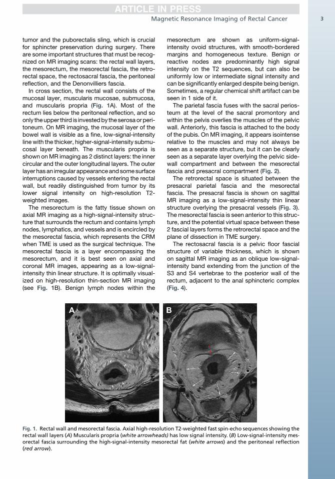
In cross section, the rectal wall consists of themucosal layer, muscularis mucosae, submucosa,and muscularis propria (Fig. 1A). Most of therectum lies below the peritoneal reflection, and soonly theupper third is investedby theserosaorperi-toneum. On MR imaging, the mucosal layer of thebowel wall is visible as a fine, low-signal-intensitylinewith the thicker, higher-signal-intensity submu-cosal layer beneath. The muscularis propria isshown onMR imaging as 2 distinct layers: the innercircular and the outer longitudinal layers. The outerlayer hasan irregular appearanceandsomesurfaceinterruptions caused by vessels entering the rectalwall, but readily distinguished from tumor by itslower signal intensity on high-resolution T2-weighted images.
The mesorectum is the fatty tissue shown onaxial MR imaging as a high-signal-intensity struc-ture that surrounds the rectum and contains lymphnodes, lymphatics, and vessels and is encircled bythe mesorectal fascia, which represents the CRMwhen TME is used as the surgical technique. Themesorectal fascia is a layer encompassing themesorectum, and it is best seen on axial andcoronal MR images, appearing as a low-signal-intensity thin linear structure. It is optimally visual-ized on high-resolution thin-section MR imaging(see Fig. 1B). Benign lymph nodes within the
Fig. 1. Rectal wall and mesorectal fascia. Axial high-resolutrectal wall layers (A) Muscularis propria (white arrowheadsorectal fascia surrounding the high-signal-intensity mesor(red arrow).
mesorectum are shown as uniform-signal-intensity ovoid structures, with smooth-borderedmargins and homogeneous texture. Benign orreactive nodes are predominantly high signalintensity on the T2 sequences, but can also beuniformly low or intermediate signal intensity andcan be significantly enlarged despite being benign.Sometimes, a regular chemical shift artifact can beseen in 1 side of it.
The parietal fascia fuses with the sacral perios-teum at the level of the sacral promontory andwithin the pelvis overlies the muscles of the pelvicwall. Anteriorly, this fascia is attached to the bodyof the pubis. On MR imaging, it appears isointenserelative to the muscles and may not always beseen as a separate structure, but it can be clearlyseen as a separate layer overlying the pelvic side-wall compartment and between the mesorectalfascia and presacral compartment (Fig. 2).
The retrorectal space is situated between thepresacral parietal fascia and the mesorectalfascia. The presacral fascia is shown on sagittalMR imaging as a low-signal-intensity thin linearstructure overlying the presacral vessels (Fig. 3).The mesorectal fascia is seen anterior to this struc-ture, and the potential virtual space between these2 fascial layers forms the retrorectal space and theplane of dissection in TME surgery.
The rectosacral fascia is a pelvic floor fascialstructure of variable thickness, which is shownon sagittal MR imaging as an oblique low-signal-intensity band extending from the junction of theS3 and S4 vertebrae to the posterior wall of therectum, adjacent to the anal sphincteric complex(Fig. 4).
ion T2-weighted fast spin-echo sequences showing the) has low signal intensity. (B) Low-signal-intensity mes-ectal fat (white arrows) and the peritoneal reflection
Fig. 2. Axial high-resolutionT2-weightedfast spin-echosequence. Parietal fascia is seen anterolaterally as a thinlinear low-intensity layer at the lateral side of the pelvis.
Fig. 4. Sagittal T2-weighted fast spin-echo sequenceobtained in a 56-year-old man. Rectosacral fascia isseen as an oblique thick low-signal-intensity bandextending posteriorly from the sacrum (S3–S4) to therectum (white arrows). Notice the peritoneal reflec-tion (black arrow).
Costa-Silva & Brown4
The peritoneal reflection is easily seen onsagittal MR imaging as a low-signal-intensity thinlinear structure that extends over the surface ofthe bladder posteriorly to its point of attachment
Fig. 3. Sagittal T2-weighted fast spin-echo sequenceobtained in a 62-year-old man with a rectal carcinomatreated with chemoradiation shows the posteriorfascial layers of the pelvis. Posterior mesorectal fascia(white arrowheads) is shown anteriorly and the presac-ral fascia is shown posteriorly (white arrows). There isfluid in the retrorectal space seenbetweenthese fascias.
onto the rectum. In men, the attachment site ofthe peritoneal reflection is the junction of the uppertwo-thirds and lower one-third of the rectum(see Fig. 4). In women, this site of attachmenthas more anatomic variations. The relationshipbetween the rectal tumor and the peritoneal reflec-tion is important in staging of the rectal cancer,because the tumors with invasion through the peri-toneal reflection are categorized as stage T4a.The Denonvilliers fascia is a well-developed
fascia that derives from the urogenital septumduring embryonal development. It forms a charac-teristic anterior surface of the mesorectum, on itslower part, and it is visible on axial MR imagingas a low-signal-intensity layer, adjacent to theprostate in men and as the rectovaginal septumbehind the posterior vaginal wall in women(Fig. 5). Inferiorly, the septum extends to the peri-neal body.
IMAGING TECHNIQUE
Before the development of the modern phased-array pelvic surface coil, endorectal coils werethe only method of obtaining high-resolutionimages of pelvic anatomic structures. The use ofendorectal coils was limited in rectal cancerassessment, because of near-field artifact andluminal distortions created by the coils in direct
Fig. 5. Axial T2-weighted high-resolution fast spin-echo showing the Denonvilliers fascia (arrowheads)posteriorly relative to the prostate gland. TME com-prises the rectum surrounded by complete mesorec-tum fat within an intact mesorectal fascial envelopeincluding Denonvilliers fascia anteriorly. This fascia isseen below the point of attachment of the peritonealreflection onto the anterior surface of rectum.
Magnetic Resonance Imaging of Rectal Cancer 5
contact with the rectal wall. Further problems withendorectal coils, as with any endoluminal tech-niques such as endoanal ultrasonography, werethe inability to evaluate stenosing tumors and to
Table 1Recommended MR imaging parameters
SequenceFOV(mm)
RepetitiTime/EchTime
Sagittal T2From one pelvic sidewall
to the other
TSE/23 250 3500/125
Axial T2Iliac crest to pelvic floor
TSE/22 420 3500/80
Axial T2Perpendicular to the
rectumOblique axial T2To cover lymph node
territory
TSE/16 160 3500/120
Coronal T2Low tumors parallel to
anal canal
TSE/16 160 3500/120
Abbreviations: NSA, number of signal averages; SENSE, sensitiData from Brown G, Daniels IR, Richardson C, et al. Technique
MRI for rectal cancer. Br J Radiol 2005;78(927):245–51.
fully evaluate the entire mesorectal lymph nodedrainage territory.
The introduction of phased-array coil systemsimproved staging of rectal cancer, which alongwith fast spin-echo T2-weighted sequencesenabled high-resolution imaging oriented tothe relevant surgical anatomic planes. Thesephased-array surface coils combined with a veryhigh spatial resolution allowed detailed evaluationof the rectal wall and depiction of the surroundingimportant anatomy.
The technique for acquisition of the sequenceshas been previously described,19 and the parame-ters are shown in Table 1.
A 1.5-T system is generally used with phased-array coils, which allow greater coverage of theanatomy when compared with the endorectalcoils. The experience with a 3-T system in thehigh-resolution protocol is still limited, but it islikely that there are few or no benefits with thissystem for the staging of rectal cancer.20
Patients must be fully informed about the lengthof time required for MR imaging scanning(between 20 and 30 minutes) and must be posi-tioned in the supine position in the scanner. Thereis no need to use purgative bowel preparation orenemas.19,21 Antispasmodic agents are used toreduce pelvic small bowel motion. Gadolinium-enhanced T1-weighted imaging and fat saturationare not necessary, because these have not beenshown to improve the diagnostic accuracy forstaging of rectal cancer.22
ono
Slice (mm) Matrix NSA Time (min:s)
3 interleaved 304 � 512 4 6:36
5 interleaved 352 � 512 1 1:38 SENSE 2
3 interleaved 256 � 512 6 5:36
3 interleaved 256 � 512 6 5:36
vity encoding for fast MR imaging; TSE, turbo spin-echo.s and trouble-shooting in high spatial resolution thin slice
Costa-Silva & Brown6
Initial localization images in the sagittal planesare needed to plan the high-resolution images(Fig. 6A–C). For this reason, the first series tobe acquired is a sagittal, 25-cm field-of-view(FOV), 3-mm-thick T2-weighted sequence from1 pelvic sidewall to the other, which enables iden-tification of the primary tumor. It is essential thatthe referring surgeon has accurately indicatedthe tumor position (low, mid, or high rectal)to help proper planning of the sequences. Thesecond series consists of large FOV axial
Fig. 6. Sagittal localization image (A) used to plan the axiaold woman with a low-rectum adenocarcinoma (white arrand axial high-resolution T2-weighted images (D) in a 49-yadenocarcinoma. Note that the high-resolution T2-weigtumor is obtained perpendicular to the long axis of the recin A and C). Note also that the axial sequences must be exmake sure to cover the draining nodes and tumor deposi
sections (4–5 mm) of the whole pelvis. These first2 sequences allow an overview of the pelvis andenable the primary tumor to be located withregard to the distance from the anal verge andpuborectalis sling, but these are not adequatefor T staging or nodal characterization, whichare best undertaken using the high-resolutionsequences described later.The high-spatial-resolution sequence com-
prises a T2-weighted thin axial section (3 mm)through the rectal tumor and adjacent tissues.
l high-resolution T2-weighted images (B) in a 54-year-ows in A). (C, D) Sagittal initial localization image (C)ear-old woman with a pediculate midrectummucinoushted thin-section axial sequence through the rectaltum at the level of the tumor (orange and green linestended 4 to 5 cm above the superior edge of tumor tots.
Magnetic Resonance Imaging of Rectal Cancer 7
These sequences are angled perpendicular to thelong axis of the rectum at the level of the tumor,using a 16-cm FOV (see Fig. 6B, D). The perpen-dicular plane is necessary; otherwise, the imagesmay be misinterpreted because of partial volumeeffects. This sequence must be extended to atleast 5 cm above the superior edge of the tumorto ensure coverage of draining nodes and tumordeposits (see Fig. 6A, C). Knowledge of the distri-bution pattern of mesorectal nodes may assist inpreoperative radiotherapy planning if the tumorshows high-risk features.
The T2-weighted thin-section coronal sequenceis optional for the upper-rectum and midrectumtumor, but mandatory for patients with low rectalcancers. This sequence consists of high-spatial-resolution thin sections (3 mm) parallel to thelong axis of the rectum at the level of the tumorand it shows the levator muscles, including thepuborectal, and the sphincter complex in relation-ship to the rectal wall (Fig. 7).
The routine use of diffusion-weighted imaging(DWI) is gradually increasing and preliminary datasuggest that apparent diffusion coefficient (ADC)values may reflect tumor aggressiveness, but it isnot clear whether this provides additional prog-nostic information compared with conventionalT2-weighted high-resolution imaging and whetherADC values could influence treatment decisions.23
The published data suggest that it is unlikely thatthe addition of DWI alters staging accuracy
Fig. 7. Sagittal initial localization image (A) used to plansequences (B) in a 61-year-old woman with a low rectal adeThe coronal sequence is parallel to the long axis of the rectA) and shows the levator muscles (white arrows), includcomplex in relationship to the rectal wall.
because of substantial overlap in ADC valuesbetween benign and malignant processes.23–25
Water diffusion is a physical process that resultsfrom the thermally driven, random motion of watermolecules.26,27 In a glass of water, moleculesundergo free, thermally agitated diffusion (witha three-dimensional Gaussian distribution). Thewidth of the Gaussian distribution expands withtime, and the average square of this width perunit time gives the units of the ADC. In tissues,apparent diffusion is observed because the move-ment of water molecules is modified by their inter-actions with cell membranes and macromoleculesin an environment. The restriction to flow of watermolecules is determined by tissue cellularity andthe integrity of the cell membrane. Tumors typi-cally display restricted diffusion because of theirhypercellularity.28
In our standard 1.5-T GE Signa HDxt system(General Electric Medical Systems, Milwaukee,WI), we use an 8-channel cardiac coil, with a spin-echo echo planar imaging sequence (repetitiontime [TR], 4500 milliseconds; echo time [TE], 80milliseconds; flip angle, 90�; FOV, 280–360 mm;16numberof averages; slice thickness, 4mm; inter-slice gap, 0.4 mm; acquisition matrix, 128 � 100)with bandwidth of 1.953 Hz/pixel, with an imagingtime of DWI of 3 minutes 36 seconds for 20 slices,b value (0, 500, 1000 s/mm2), and 3 directions.DWI is performed in the axial plane. The motion-probing gradient pulses are placed in the x-axis,
the coronal high-resolution thin-section T2-weightednocarcinoma extending to the anal canal (red arrows).um at the level of the tumor (orange and green lines ining the puborectal (black arrows), and the sphincter
Costa-Silva & Brown8
y-axis, and z-axis (all planes). The parameters areshown in Table 2.
IMAGING INTERPRETATIONStaging of the Primary Rectal Tumor
The American Joint Committee on Cancer guide-lines, seventh edition, defines the criteria for thestaging of primary rectal tumors. The process ofT staging, T substaging, and N staging is shownin Table 3. The T staging is based on the invasionof the primary tumor through the rectal wall and itsrelationship to the submucosa and muscularispropria. Therefore, the best results for stagingrectal cancer have been obtained through carefulinterpretation of thin-section, high-resolution, andsmall FOV T2-weighted images obtained perpen-dicular to the rectal wall.
T Staging
MR imaging should be analyzed by the radiologistassessing the primary rectal tumor in terms ofstage; the degree of invasion outside the muscula-ris propria (extramural extension); and relationshipto the mesorectal fascia, anal sphincter, and pelvicsidewall. The radiologist should first describe theheight of the tumor, ideally from the anal verge,because this is a useful reference point for thereferring surgeon, followed by the length of thetumor. The description of the morphologic ap-pearances of the tumor must be categorizedas polypoid, ulcerating, hemicircumferential, orcircumferential (Fig. 8). On T2-weighted high-resolution images, a nonmucinous tumor is shownas a lesion of intermediate signal intensity with noareas of high signal intensity. Mucinous tumorsappear as fluidlike high signal intensity on theseT2-weighted images (Fig. 9). The following step
Table 2Diffusion-weighted imaging sequence parameters (PGermany; and General Electric platforms, Milwaukee
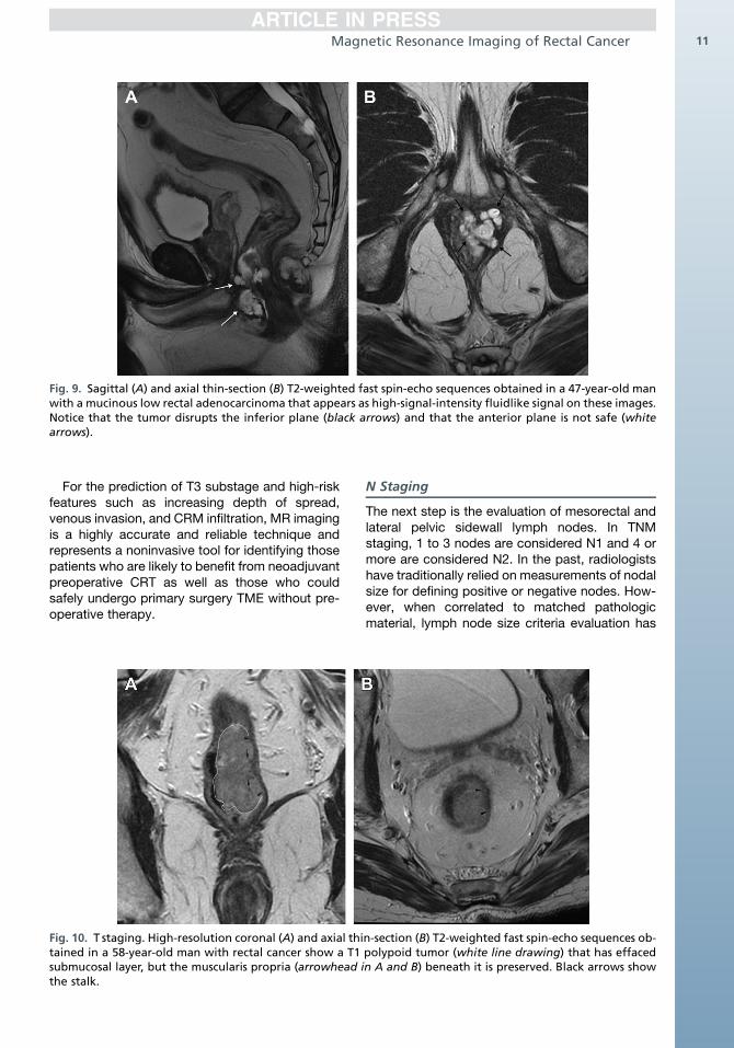
is the identification of the invasive portion of thetumor, which is the most worrisome area, corre-sponding to the most invasive portion of the tumoron the rectal wall and to the site of ulceration of theluminal component, at the same location.T1 tumors are those in which there is invasion of
the submucosa, represented by the identificationof abnormal signal intensity replacing the normalsubmucosa (Fig. 10). The best way to distinguishT1 (submucosal invasion only) from T2 tumors(extension to the muscular layer) is to show partialpreservation of the high signal intensity of thesubmucosa layer beneath the intermediate signalintensity of the tumor.T2 tumors are those with extension into themus-
cularis propria,where abnormal intermediate signalintensity with partial-thickness or full-thicknessinvolvement can be appreciated, but there is noextension of intermediate signal intensity to themesorectal fat (Fig. 11). T3 tumors manifest asa broad bulge or nodular projection of abnormalintermediate signal intensity extending beyondmuscularis propria in the mesorectal fat (Fig. 12).We do not recommend interpreting features suchas desmoplastic reaction as tumor because thisresults in overstaging.21,29 We advocate the useof T3 substaging as clinically more relevant thandistinguishing T3 from T4 spread. The T substagetakes into account the prognostic importance ofdepth of extramural spread from the outermostedge of the muscularis propria (extramural tumorextension) and enables recognition that a T3atumor has an identical prognosis to a T2 tumor.The T3 stratification was originally developed byHermanek as a modification of the TNM classifica-tion according to the followingdepthsof extramuralspread: T3a less than 1mmspread, T3b 1 to 5mm,T3c greater than 5–15 mm, and T3d greater than
hillips, Best, The Netherlands; Siemens, Erlangen,, Wisconsin.)
Siemens General Electric
220 280
256 138 � 256 128 � 100
3100 4500
71 80
2 2
8 16
5 4
trace 3 scan trace 3 scan trace
SPAIR STIR
750 0,350,750 0,500, 1000
STIR, short-tau inversion recovery.
Table 3TNM staging of rectal tumors on MR imaging
TNM Stage Description
Tx Primary tumor cannot be evaluated
T0 No evidence of invasive primary tumor
T1 Invasion of submucosa by tumor; abnormal signal intensity has replaced part but not allof the submucosa
T2 Invasion but not penetration of muscularis propria; intermediate signal intensity inmuscularis propria
T3 Invasion through muscularis propria; broad bulge or nodular projection of intermediatesignal intensity extending beyond muscularis propria
a <1 mm beyond the muscularis propria
b 1–5 mm beyond the muscularis propria
c >5 and �15 mm beyond the muscularis propria
d >15 mm beyond the muscularis propria
T4 Invasion of other organs
a Abnormal signal intensity extends into adjacent organs through peritoneal reflection
b Tumor invades visceral peritoneum
N0 No nodal metastasis
N1 1–3 perirectal or pericolic involved nodes
N2 �4 perirectal or pericolic involved nodes
Mx Cannot be assessed
M0 No metastasis
M1 Distant metastasis
Magnetic Resonance Imaging of Rectal Cancer 9
15 mm. Both imaging and histopathologic assess-ment of T3 substage are performed by measuringthe extramural tumor extension into themesorectalfat in millimeters beyond the outermost edge ofmuscularis propria (see Table 3). T4 tumors aredistinguished according to invasion of adjacentorgans or structures (T4a) or have perforated theperitoneal reflection (T4b) (Figs. 13 and 14).
MR imaging is a well-established technique inthe staging of advanced rectal cancers. Commonsites of local infiltration of adjacent structuresshould be evaluated when examining advancedlocal tumors.21 For mid and upper tumors, anteriorinvasion can involve the bladder, uterus, or sem-inal vesicles, as well as the peritoneum; lateralextension can involve the pelvic sidewall; andposterior extension can involve the sacrum. Forlow tumors, the common sites for stage T4 infil-tration are the pelvic floor structures; the analsphincter; the levator muscles; and the prostate,vagina, and coccyx.
Special attention should be given to lower rectaltumors, because they represent a greater chal-lenge for the colorectal surgeon than the higher-level tumors. The conventional TNM stagingsystems for colorectal cancers are insufficient inthese cases, because they do not account for
the anatomic considerations and the fact that themesorectal envelope tapers at this level. Wepropose a special staging system that takes intoaccount the relevant local anatomy, with the aimof providing more information to help the surgeondefine the most appropriate plane (Table 4).21,30,31
The margin positivity rate for abdominoperinealexcision has been reported to be as much as30%, compared with 10% for low anterior resec-tions. The concept of the 2 planes for rectal cancerallows the surgeon and radiologist to plot a care-fully defined route map for preoperative stagingand to plan surgery.30,32,33
For the evaluation of the potential CRM involve-ment, measurements are taken from the outermostradial border of the tumor and its distance to themesorectal fascia. The same distances must bemeasured for extramural venous invasion andtumor satellite/deposits (Box 1). A potentially posi-tive margin is defined as tumor lying within 1 mm(<1mm) of themesorectal fascia.7,8Measurementsare also taken when lymph nodes show definitefeatures of malignant replacement (mixed signalintensity and extension through the nodal capsuleby tumor), extramural vascular invasion, and tumordeposit or satellites may all result in a positivemargin if lyingwithin 1mmof themesorectal fascia.
Fig. 8. Same patient as Fig. 6A, B. Sagittal (A) and axial high-resolution thin-section T2-weighted (B) sequencesobtained in a 49-year-old woman with a midrectum adenocarcinoma. Note the stalk seen as a low-signal-intensitylinear structure (black arrowheads in A and B) and the polypoid morphology appearance of this tumor thatprotrudes into the rectal lumen. (C, D) Sagittal (C) and axial T2-weighted (D) sequences in a 62-year-old womanwith a circumferential rectal tumor. Notice the extramural venous invasion (white arrow in D) and a normallymph node (black arrow in D). The red line corresponds to the CRM distance.
Costa-Silva & Brown10
This measurement is important for the preventionof local recurrence after TME, because there isstrong evidence that neoplastic involvement ofthe CRM is closely related to a high recurrencerate.6,8,14,34 However, lymph nodes are a rarecause of circumferential margin involvement anda cause of margin involvement in only 1% to 2%of resected specimens, therefore defining amarginas involved must be based only on the clearevidence of a node with definite malignant features(mixed signal and irregularity of the nodal border).35
Extramural vascular invasion is an importantand independent prognostic feature that can bereadily identified on MR imaging.36,37 It is definedby the presence of tumor itself beyond the muscu-laris propria, within the vessels, which are visual-ized as signal flow-void tubular structures onT2-weighted spin-echo sequences lying perpen-dicular to the rectal wall. Tumor invasion of thevessels is seen as irregular expansion of the extra-mural vessels by intermediate tumor signal in-tensity (Fig. 15).
Fig. 9. Sagittal (A) and axial thin-section (B) T2-weighted fast spin-echo sequences obtained in a 47-year-old manwith a mucinous low rectal adenocarcinoma that appears as high-signal-intensity fluidlike signal on these images.Notice that the tumor disrupts the inferior plane (black arrows) and that the anterior plane is not safe (whitearrows).
Magnetic Resonance Imaging of Rectal Cancer 11
For the prediction of T3 substage and high-riskfeatures such as increasing depth of spread,venous invasion, and CRM infiltration, MR imagingis a highly accurate and reliable technique andrepresents a noninvasive tool for identifying thosepatients who are likely to benefit from neoadjuvantpreoperative CRT as well as those who couldsafely undergo primary surgery TME without pre-operative therapy.
Fig. 10. T staging. High-resolution coronal (A) and axial thitained in a 58-year-old man with rectal cancer show a T1submucosal layer, but the muscularis propria (arrowhead ithe stalk.
N Staging
The next step is the evaluation of mesorectal andlateral pelvic sidewall lymph nodes. In TNMstaging, 1 to 3 nodes are considered N1 and 4 ormore are considered N2. In the past, radiologistshave traditionally relied on measurements of nodalsize for defining positive or negative nodes. How-ever, when correlated to matched pathologicmaterial, lymph node size criteria evaluation has
n-section (B) T2-weighted fast spin-echo sequences ob-polypoid tumor (white line drawing) that has effacedn A and B) beneath it is preserved. Black arrows show
Fig. 11. T2 tumor. Sagittal (A) and axial high-resolution thin-section (B) T2-weighted fast spin-echo images ina 72-year-old woman with midrectal cancer show a polypoid lesion (white line drawing in A and B) that extendsinto the muscularis propria (arrowhead in A and arrows in B) with partial-thickness involvement, but there is noextension to the mesorectal fat. Notice that this tumor contains some mucin (black arrow in A).
Costa-Silva & Brown12
been proved to be unreliable.38,39 We therefore donot recommend evaluating lymph nodes bymeasuring size, because there is substantial over-lap related to enlarged benign reactive nodes.Moreover, metastatic disease is frequently ob-served in nodes less than 5mm in diameter. Brownand colleagues38 mapped individual nodes har-vested on pathology to the in vivo counterpartsand reported that uniform smooth-bordered nodeswith homogeneous signal intensity were nearlyalways benign. The presence of irregular bordersor mixed signal intensity strongly correlatedwith node positivity, with an overall accuracy of85% when tested prospectively using a high-
Fig. 12. T3 tumor. (A, B) T2-weighted sagittal (A) and obliqNote 8 mm of extension (red line) beyond outer muscle c
resolution technique (Fig. 16).39 Kim and col-leagues40 later confirmed these observationsfrom a retrospective series and noted that thepresence of a mottled heterogenic pattern wasassociated with 50% sensitivity and 95% speci-ficity for malignant involvement. Their study alsoconfirmed the relevance of spiculated or indistinctborders, with sensitivities of 45% and 36%, andspecificities of 100% and 100%, respectively.The clinical significance and management of
involved pelvic sidewall lymph nodes in rectalcancer remains controversial. The MERCURYstudy group evaluated 325 patients and foundthat 38 had suspicious pelvic sidewall nodes
ue axial (B) images showing annular stage T3c tumor.oat (white arrows in A and B).
Fig. 13. T4 tumor. Axial high-resolution thin-section T2-weighted fast spin-echo images in an 82-year-old manwith midrectal cancer show perforation and invasion through the peritoneal reflection (white arrows). The blackarrows show the mesorectal fascia.
Magnetic Resonance Imaging of Rectal Cancer 13
identified on baseline scans.41 After a minimum of5-year follow-up, these investigators found thatMR imaging-suspected nodal pelvic involvementwas associated with a worse 5-year overallsurvival (OS) and DFS for patients undergoingprimary surgery, without preoperative therapy.However, among patients who received preopera-tive radiotherapy, the presence of MR imaging-
Fig. 14. T4b tumor. Sagittal (A) and axial (B) T2-weightedthe prostate gland (white arrows in A). In (B), black arrow
suspected pelvic sidewall lymph nodes did notnegatively affect survival. There were also strongassociations between mesorectal nodal involve-ment detected by MR imaging and other adversefeatures such as extramural venous invasion, sug-gesting that suspicious pelvic sidewall nodeson MR imaging represented a more advanceddisease state. MR imaging detection of nodal
sequences showing a huge mucinous tumor invadings show the mucin pools.
Table 4Stages of low rectal tumors on MR imaging
Stage 1 Tumor confined to bowel wall but does not extend through full thickness; intact outer musclecoat
Stage 2 Tumor replaces muscle coat but does not extend into intersphincteric plane
Stage 3 Tumor invades intersphincteric plane or lies within 1 mm of levator muscle
Stage 4 Tumor invades external anal sphincter and is within 1 mm and beyond levators with orwithout invading adjacent organs signal intensity in muscularis propria
Data from Taylor FG, Swift RI, Blomqvist L, et al. A systematic approach to the interpretation of preoperative staging MRIfor rectal cancer. AJR Am J Roentgenol 2008;191(6):1827–35.
Costa-Silva & Brown14
disease in the pelvic sidewall compartment istherefore an adverse feature but does not seemto be independently prognostic for patient survival(Fig. 17).
Prognostic Factors Depicted by MR Imaging
The prospectively conducted MERCURY study ofconsecutive patients who have rectal cancerenabled validation of the MR imaging stagingfactors against the risks of local recurrence,distant failure, or both (Box 2). Based on theseoutcome data, the next generation of clinical trialsis being designed to specifically address thoserisks, for example, the use of neoadjuvant therapyfor patients who are at risk of distant metastasesrather than local recurrence (Fig. 18).
POSTTREATMENT EVALUATION: HOW CANMR IMAGING RESTAGE RECTAL CANCERAFTER CRT?
The standard treatment of advanced colorectalcancer includes performing neoadjuvant long-course CRT to downstage the tumor, followed byTME surgery, which have reduced local recur-rence and improved rates of curative resection.9,42
Assessment of treatment efficacy has principallyrelied on histopathologic evaluation of irradiatedspecimens after TME. These studies have shownthat posttreatment pathologic T and N stage (ypTand ypN, respectively, where y represents postra-diation therapy specimens and p pathologic
Box 1Distances to be obtained in relationship tomesorectal fascia (ie, potential MR imagingCRM, outer margin [mrCRM])
Main tumor to mrCRM
T3 substage suspicious lymph node to mrCRM
Extramural venous invasion to mrCRM
Tumor satellite/deposit to mrCRM
staging) can predict local recurrence, DFS, andOS, and several studies have shown that theyare independent prognostic factors. Some retro-spective studies have shown that complete patho-logic response after preoperative CRT followed bydefinitive surgical resection for advanced rectalcancer resulted in decreased recurrence andimproved DFS.43 Tumor downstaging has beenregarded as a marker for tumor radiosensitivity.
MR Imaging Technique
The MR imaging technique used for the posttreat-ment assessment must be the same as the oneused in the pretreatment evaluation. T2 high-resolution images are recommended to distinguishtumor from fibrosis for accurate MR TRG staging.Comparison with pretreatment images is essen-tial. Images should be acquired using the sameangles as those used in the pretreatment scansto enable comparison, and pretreatment imagesare used to help locate the treated tumor, whichmay be difficult to visualize in patients who havehad a good therapeutic response.
DWI on the Postneoadjuvant TreatmentEvaluation
As described earlier, DWI is a functional MRimaging technique that uses differences in theextracellular movement of water protons todiscriminate between tissues of varying cellularity.In tissues with normal cellularity, water protonscan diffuse relatively freely, which results ina loss of signal on DWI. Conversely, in tissueswith increased cellularity (tumor), diffusion of wateris restricted, resulting in persistent high signal onDWI. DWI has been proposed as a means of pre-dicting the response to treatment. Barbaro andcolleagues,44 in a prospective study of 62 consec-utive patients, assessed the value of ADCs ob-tained from MR imaging before and after CRT forrectal cancer as treatment response predictorsand found that low pretreatment ADCs of lessthan 1.0 � 10�3 mm2/s correlated with poor TRG
Fig. 15. Extramural venous invasion. 45-year-old woman with rectal cancer. Axial high-resolution T2-weightedimages showing extramural venous invasion (white arrows in A and B) seen as irregular expansion of the extra-mural vessels with tissue with the same intermediate signal as the primary tumor. (C) The vessel returned to itsnormal diameter and the normal flow void is seen (black arrow).
Magnetic Resonance Imaging of Rectal Cancer 15
scores. These investigators also observed thatduring treatment, the preoperative ADCs as wellas the mean percentage of increase in tumorADC was significantly greater in the respondersthan in the nonresponders. They showed thata preoperative ADC of 1.4� 10�3 mm2/s or greaterhad a positive and negative predictive value of78.9% and 61.8%, respectively, to indicatetherapeutic response.44 On the other hand,Curvo-Semedo and colleagues45 failed to showa relationship between pre-CRT ADC, post-CRTADC, or DADC measurements that could beused to differentiate between patients with a CRand residual tumor. Radiation-induced proctitisand fibrosis were significant and independentpredictors of diffusion restriction in patientsachieving pathologic CR after treatment with neo-adjuvant CRT for locally advanced rectal cancer,and pre-CRT tumor volume was observed to affectboth variables.24 Overall, many results regarding
Fig. 16. Lymph node involvement. (A, B) 59-year-old man wimages show obviously malignant nodes (white arrows) w
DWI and ADC have been conflicting, and furtherwork is necessary to validate the final impact ofDWI on the management of patients undergoingchemoradiotherapy treatment.
MR Imaging Interpretation After CRT
On post-CRT T2-weighted MR imaging, areasof fibrosis appear as very low signal intensity,whereas areas of residual tumor have intermediatesignal, similar to the baseline scans. Careful reviewof the high-resolution images enables identifica-tion of small foci of intermediate-signal-intensityviable tumor, within the fibrosis. Some tumorsdevelop a colloid response with mucin production,which is shown on T2-weighted images as very-high-signal-intensity pools. The recommendedevaluation of the MR imaging after CRT isdescribed in Box 3. The use of a standardizedapproach for pathologic evaluation must be
ith rectal cancer. T2-weighted sagittal (A) and axial (B)ith irregular borders and mixed signal intensity.
Fig. 17. Pelvic sidewall lymph node involvement. (A, B) 43-year-old patient, with T3d stage rectal cancer. Sagittal(A) and axial (B) images depicted irregular margins and a mottled heterogenic pattern (white arrows in A and B)in the right pelvic sidewall. (B) The arrowhead shows a small 4 mm malignant mesorectal node with irregularmargins and mixed texture.
Costa-Silva & Brown16
implemented to allow comparison between theresults of various treatment approaches. Asa consequence, reassessment of MR imagingscans after preoperative therapy has implicationsfor surgical planning, timing of surgery, sphinc-ter preservation, deferral of surgery for goodresponders, and development of further preopera-tive treatments for radiologically identified poorresponders. Up to 24% of patients undergoingthis treatment have pathologic CR in the finalspecimen. MR imaging is being investigated ina prospective trial not only as a means of identi-fying patients with a potential CR but also formonitoring continued tumor regression usingserial MR follow-up imaging. In the clinical setting,the subgroup of patients with unfavorable post-treatment MR imaging parameters seem to be at
Distance to the mesorectal fascia/potential CRMless than 1 mm
Low tumor extending into the intersphinctericplane or beyond (TME mesorectal plane ofdissection is unsafe)
Peritoneal involvement
Factors predicting distant failure
Extramural tumor spread greater than 5 mm
Extramural venous invasion
Poor TRG
a higher risk of local or systemic recurrence aftera standard TME resection. The surgeon may bewarned of this risk preoperatively, and an ex-tended dissection may need to be performed toavoid a potentially involved CRM. In future studies,this group could be considered for further therapysuch as intensified or extended systemic chemo-therapy, a radiotherapy boost, extension of thesurgical resection, or more intensive postoperativefollow-up.46
Histopathologic Tumor Grade Response
The use of preoperative CRT modifies the macroand microscopic aspects of the tumor appearanceof rectal cancer. The features of treated tumorsinclude marked fibrosis with or without replace-ment of neoplastic cells by inflammatory cells,and possible development of mucin productionpools.Histopathologists grade tumor response in
3 ways: first, assessment of the status of theCRM, followed by the evaluation of the depth oftumor spread and nodal status (ypT and ypNstage), and then, by evaluating TRG. TRG analysismay be considered a useful predictor of outcomein addition to T stage. T staging has limitationsbecause it cannot always adequately showthe extension of tumor regression (Box 4). Forexample, a tumor may regress so much that onlya few viable tumor cells remain outside the rectalwall, so assigning pT3 status may not, on its own,fully reflect the true extent of tumor regression.TRG as a measurement of response to neoadju-
vant therapy was first described by Mandard andcolleagues47 in 1994, in esophageal carcinomas
Fig. 18. An example of MR imaging evaluation being used to tailor treatment of patients in current internationalclinical trials of preoperative treatments. EMVI, extramural venous invasion; TEMS, transanal endoscopicmicrosurgery.
Magnetic Resonance Imaging of Rectal Cancer 17
on a scale from 1 to 5 based on the presence ofresidual viable tumor cells and the extent offibrosis. The Mandard modified grading, used forthe evaluation of post-CRT rectal cancer, is
Box 3MR imaging evaluation after CRT
Description of the morphologic appearances oftumor, including any mucinous or necroticcomponents
Height of treated tumor from the anal vergecompared with baseline pretreatment images
Length of treated tumor from the anal vergecompared with baseline pretreatment images
MR imaging T stage and T substage, taking intoaccount the depth of extramural spread
MR imaging TRG
Distance to the potential CRM and whether thisappears potentially involved or clear
Presence of extramural venous invasion
Mesorectal and pelvic sidewall lymph nodestaging
Data from Patel UB, Taylor F, Blomqvist L, et al.Magnetic resonance imaging-detected tumor re-sponse for locally advanced rectal cancer predictssurvival outcomes: MERCURY experience. J Clin Oncol2011;29(28):3753–60.
defined as follows: grade 1 is the absence ofresidual tumor and fibrosis extending through thedifferent layers of the rectal wall; grade 2 is definedby the presence of rare residual tumor cells scat-tered throughout the fibrosis; grade 3 is character-ized by an increase in the number of residual viabletumor cells, but the fibrosis still predominates;grade 4 shows residual tumor outgrowing thefibrosis; and grade 5 is characterized by absenceof any tumor regression.13
How to Assess Tumor Response Using MRImaging
Imaging methods for assessing response afteroncologic therapies continue to evolve. Althoughmorphologic evaluation is the standard of refer-ence, the added value of diffusion-weighted MRtechniques is being evaluated. Histopathologic
Box 4Histopathologic evaluation of tumor response
Assessment of the status of the CRM
Evaluation of the depth of tumor spread andnodal status (ypT and ypN stage)
Measurement of the extent of extramural spreadbeyond the muscularis propria
Evaluation of TRG
Costa-Silva & Brown18
results and survival outcomes are always the stan-dard of reference and validation, respectively.Various studies reported that MR imaging can beaccurately used to evaluate CRT, and severaldifferent methods have been proposed for assess-ing response on MR imaging.48,49
These methods include posttreatment MRimaging T staging (ymrT [T stage on MR imagesobtained after CRT]), MR imaging-based tumorregression grading (mrTRG), volume reductionbetween baseline and after treatment, and modi-fied Response Evaluation Criteria in Solid Tumors(mRECIST)50 measurement (Box 5).
T Staging After CRT
ymrT (T stage onMR images obtained after CRT) isbased on the interpretation of local extent ofpersistent tumor signal intensity relative to thelayers of bowel wall on axial high-resolutionT2-weighted images. Tumor response is repre-sented by the replacement of tumor signal bylow-signal-intensity fibrosis or the developmentof high-signal-intensity mucin pools; such areasare not considered to be tumor, and T stageis based on defining the extent of residualintermediate-signal-intensity tumor signal onhigh-resolution T2-weighted scans.
MR Imaging TRG Analysis
MR imaging TRG is based on principles similar tothe pathologic TRG system originally describedby Mandard and Dworak, and the degree of tu-mor replacement by fibrotic stroma is deter-mined.14,21,47,51,52 This is the most importantmethod for evaluating tumor response. TheMERCURY study group14 has shown that it hasbeen possible to develop an MR imaging-basedtumor regression grading (mrTRG) system byapplying the principles of histopathology TRG(Table 5). The tumor is assessed to determine ifit is fibrous or if tumor signal intensity predomi-nates. For this evaluation, a comparison between
Box 5Imaging methods for the assessment ofchemoradiation or radiation therapies
Posttreatment MR imaging T staging
MR imaging-based tumor regression grading(mrTRG)
Volume reduction between baseline and aftertreatment
Modified Response Evaluation Criteria in SolidTumors (mRECIST) measurement
the post-CRT and the baseline axial high-resolution images is necessary to determine theproportion of tumor that has become of low signalintensity, representing fibrosis, and the proportionthat remains with intermediate signal intensity,representing viable tumor.
MR Imaging Volumetric Analysis
Maximal height is measured on a sagittal section,and transverse maximum diameters are assessedon axial slices. Tumor volume is obtained by multi-plying tumor length, width, and height, and thepercentage of volume reduction is calculated.Another way to calculate volume is to outline thetumors on a work station. The baseline scans arecompared with the post-CRT scans.
MR Imaging mRECIST
Although RECIST criteria is considered a powerfulmethod of evaluating response, inconsistenciesarise in measuring geometrically irregular tumors,such as rectal cancers. Because measurement ofmultilobulated tumors cannot be reproduced,interobserver variability may be unacceptablyhigh.44 Maximum tumor length is best measuredon sagittal images before and after treatment.CR is defined as complete disappearance oftumor. Partial response to treatment is definedas at least a 30% decrease in tumor length, takingas reference the baseline tumor length. Progres-sion of the disease is defined as at least a 20%increase in tumor length, and stable disease isdefined as neither sufficient shrinkage to qualifyfor partial response nor sufficient increase toqualify for progression of the disease.This method has the major limitation of not
taking into account extramural shrinkage orprogression, which may be of greater prognosticor clinical relevance.Patel and colleagues49 compared the vari-
ous methods of identifying good versus poorresponders with the histopathologic standardsof T stage (ypT) and tumor regression grading(TRG) and concluded that favorable and unfavor-able histopathology were predicted by both ymrTand mrTRG, and therefore recommended theseas optimal parameters for posttreatment assess-ment of rectal cancers treated with CRT.
WHAT DOES THE PHYSICIAN NEEDS TOKNOW?The Surgeon’s View
With the evolution of surgical techniques (TME)and the shift to neoadjuvant CRT in advancedrectal cancers, high-resolution MR plays a pivotal
Table 5TRG of rectal tumor on MR imaging
Grade Response
Grade 1 Complete radiologic response No evidence of tumor signal intensity or fibrosis only
Grade 2 Good response Dense hypointense fibrosis, minimal residual tumor
Grade 3 Moderate response Mixed fibrosis/mucin and intermediate signalrepresenting residual tumor, but fibrosis stillpredominates
Grade 5 No response Tumor has the same appearance as baseline
Data from Patel UB, Taylor F, Blomqvist L, et al. Magnetic resonance imaging-detected tumor response for locallyadvanced rectal cancer predicts survival outcomes: MERCURY experience. J Clin Oncol 2011;29(28):3753–60.
Magnetic Resonance Imaging of Rectal Cancer 19
role for the surgeon. In the setting of primary rectaltumors, MR imaging is used to stage and identifypatients at risk of recurrence who may benefitfrom preoperative CRT, more extensive surgery,or both.
A surgeon dealing with rectal cancers initiallywants to differentiate between rectal tumorsconfined within the rectal wall and those thatextend beyond the muscularis propria. The depthof invasion outside the muscularis propria mustbe assessed, because it has a prognostic value.Radiation therapy produces little survival benefitand results in significant morbidity when used totreat stage T1 to T2 or favorable-risk early-stageT3a/T3b tumors (<5 mm invasion outside the mus-cularis propria, MR extramural venous invasion[EMVI] negative, and mrCRM negative). However,radiation therapy has an important role in moreadvanced-stage T3c/T3d tumors (>5 mm invasionoutside the muscularis propria), in which the risk oflocal and distant treatment failure increases.
The prognostic heterogeneity of stage T3 tu-mors is well known. The MERCURY study groupreported that the mean difference betweenthe MR-derived and histopathologically derivedmaximal extramural depth of the tumor was�0.05 mm � 3.85 mm, so MR and histopathologicassessments of tumor spread were consideredequivalent. Therefore, the ability of MR imagingto accurately separate patients according to prog-nostic risk by depth of spread is robust.11
Another important point is the relationship of thetumor to the potential CRM. Incomplete surgicalremoval of the circumferential tumor spread isbelieved to be the main cause of local recurrenceafter resection of rectal cancer. Several studieshave shown that MR imaging is a consistent andreproducible technique, with high specificity(92%) for predicting a negative CRM.7,8,53 High-resolution MR imaging with a phased-array coil
accurately predicted the distance from the tumorto the circumferential mesorectal plane of re-section. A tumor-free margin measured on MRimaging of at least 1 mm can predict a histologicfree margin with a high degree of certainty. TheMR imaging prediction of the tumor-free marginis therefore reliable.
In relationship to the nodal staging, it is impor-tant to know which patients need to receive CRTand to map local nodal spread within and beyondthe mesorectum. Although histopathologic nodaldisease is an independent poor prognostic factorin patients with rectal cancer, and adverselyaffects survival, this does not hold true for MRimaging assessment.
Evaluation of local depth of spread, presence orabsence of EMVI and CRM status using MRimaging have been shown to be more importantand more easily reproducible than MR imagingassessment of nodal status. Furthermore, encap-sulated lymph nodes are a rare cause of circumfer-ential margin involvement occurring in only 1.3%of patients compared with direct tumor spread ortumor nodules as a cause of CRM involvement.
In surgical planning, the ability to consistentlydefine a safe plane for both the radial and distalmesorectal dissection is desirable to ensurecomplete tumor and nodal clearance at surgery,thus reducing the risk of local pelvic recurrence.High-spatial-resolution MR imaging has proveduseful in showing local tumor extent, particularlyin relation to the mesorectal fascia.7,11 Areas atrisk of involvement after resection must bedescribed in detail by the radiologist; importantexamples are the peritoneal reflection, rectosacralfascia, and presacral fascia.
Another important topic for the surgeon is lowrectal cancer. The relation of the tumor to thesphincter complex and the ability to achieve clearradial and distal margins is key to the success of
Costa-Silva & Brown20
the operation. MR imaging can help the surgeonpredetermine the planes of surgical excision,because the worst outcomes related to tumorperforation and margin positivity rates have beenobserved when the surgeon has committed tothe mesorectal plane of surgery for the distalTME dissection instead of approaching thedissection in the extralevator plane at the outset.Preoperative MR imaging planning can preventthis situation. Various studies, including theMERCURY study, Dutch TME trial, the MRC CLA-SICC, and Leeds studies have shown that lowrectal cancer treated with TME plane surgerywith abdominoperineal excision resulted in sur-gical wasting and ensuing perforation of the tu-mors around the level of the puborectalis sling.These tumors were always more locally advancedthan similar-height tumors undergoing anteriorresection and had a worse outcome, as measuredby margin involvement and perforation rates.54–59
Therefore, accurate staging is required to deter-mine the need for neoadjuvant therapy or anenhanced surgical procedure such as extralevatorabdominoperineal excision or anteriorly enhancedabdominoperineal excision surgery (Fig. 19). It istherefore important to give a detailed descriptionof the radial extent of the tumor to the mesorectalplane and intersphincteric plane by carefulassessment of the sagittal, axial, and coronalimages. The radiologist needs to give detailedinformation to the surgeon regarding the potentialsurgical planes available for resection that enableclear margins.
Fig. 19. T2-weighted axial (A) and coronal (B) high-resoltumor. These images show the potential planes of surenhanced abdominoperineal excision (yellow line), interstion (blue line).
After CRT, the radiologist must evaluate tumorresponse to CRT, the T staging and substaging,N staging and CRM status, by measuring thedistance between the CRM and the tumor. Theextent of tumor regression carries prognosticsignificance and is related to OS and DFS.
The Radiation Oncologist’s View
After the diagnosis of advanced rectal cancer, it isimportant to show the radiation oncologist theplanning volumes for radiation therapy. A cleardescription of the tumor height and its relationshipto the anal verge, puborectalis sling, and promon-tory is desirable, as is the distribution of nodalinvolvement.Modern three-dimensional radiotherapy is
based on CT. For rectal cancer, this therapy relieson target definition on CT, which is not the optimalimaging modality. The major limitation of CT is itslow inherent contrast resolution. Targets definedby MR imaging could facilitate more accurate defi-nition of tumor volumes than CT. O’Neill andcolleagues60 reported that tumor volumes definedon MR imaging were smaller, shorter, and furtherfrom the anal sphincter than CT-based volumes.This finding is relevant because sphincter/pelvicfloor dysfunction is one of the predominant dose-limiting causes of toxicity for low-lying rectaltumors, and sphincter dysfunction is both doseand volume dependent. This situation remainsa major challenge for patients and clinicians, butradiotherapy planning may result in smaller
ution images of a 52-year-old man with a low rectalgical excision: TME anterior resection (yellow line),phincteric abdominoperineal/ultralow anterior resec-
Magnetic Resonance Imaging of Rectal Cancer 21
treatment volumes, which could lead to a reductionin dose to organs at risk and facilitate doseescalation.
It is increasingly becoming a common practiceto use MR imaging in radiotherapy treatmentplanning for target volume delineation, becausecommercial radiotherapy treatment planningsystems are capable of performing image registra-tion or fusion between images acquired. Theadvantage of MR imaging compared with CT isits superior ability to discriminate soft tissues.This improved accuracy could lead to better tumorcontrol as well as sparing of normal tissues. Thetarget contours must include the rectal tumor itselfand the MR imaging-based mesorectal and lateralsidewall lymph nodes with malignant involvement.
The Medical Oncologist’s View
Preoperative identification of metastatic disease isof great value. Patients with synchronous meta-static disease may be submitted to different treat-ment pathways, which include different regimes ofneoadjuvant chemotherapy, synchronous meta-stasectomy, and even metastasectomy beforeresection of the primary tumor.
For the medical oncologist, the prognostic fac-tors are of significant importance.
The following are defined as MR imaging goodprognosis:
� CRM clear on preoperative (tumor >1 mm tothe mesorectal fascia)7,11
� No evidence of extramural venous invasion36
� Early MR imaging T stage (T2 or less, T3a,T3b) regardless of N stage12,61
� For low rectal tumors, good prognosis wasdefined as MR imaging stage 1 or 2 lowrectal, namely tumor not encroaching intothe intersphincteric plane or levators56
These prognostic factors are being reevaluatedin many centers in terms of the best selection ofpatients who should receive preoperative CRT.There is a common use, in many centers, of routineneoadjuvant CRT for all T3 tumors; however,Taylor and colleagues,12 in a multicenter study, re-ported that preoperative MR imaging could selectpatients with rectal cancer who have a good prog-nosis, which, with optimal preoperative selectionand good-quality TME surgery, could achieve localrecurrence rates of 1.7% in MR imaging-definedT3a/b regardless of nodal status.
MR imaging in identification of poor prognosticfactors in rectal cancer is important.11,36,40 How-ever, the implications for preoperative stagingand management have not been fully determinedand are under evaluation in large multicenter
phase 2 and phase 3 trials. Hunter and col-leagues62 studied 236 patients and, from the MRimaging identification of depth of spread, extra-mural venous invasion, and CRM status, hypothe-sized that these risk factors would be linked witha higher risk of developing synchronous meta-static disease. They found that when MR imagingidentified high-risk patients (extramural venousinvasion, >5 mm extramural invasion, involvedCRM, or intersphincteric plane involved by tumorin low rectal tumors) a higher rate of synchronousmetastatic disease was shown (20.7% in the high-risk group vs 4.2% in the low-risk group; odds ratio6.0). It is hypothesized that more intensivepreoperative investigation on initial staging, in-cluding fluorodeoxyglucose-positron emissiontomography/computed tomography and liver MRimaging may improve outcomes, because ofearlier selection of patients requiring chemo-therapy and metastasectomy. More trials areneeded to confirm the possibility of using induc-tion neoadjuvant chemotherapy and the benefitof new regimes in MR imaging to identify high-risk patients.
SUMMARY
The introduction of MR imaging has helped inidentifying prognostic staging information inpatients diagnosed with rectal tumors. Rectalcancer staging is based on defining pertinentanatomy of the rectum and the surrounding struc-tures. The primary goal of imaging methods is toaccurately stage rectal cancer and to identify riskfactors for local recurrence to offer a tailoredtreatment, based on individual prognosis. High-resolution T2-weighted imaging is the most impor-tant sequence in the MR imaging evaluation ofrectal tumors, giving the opportunity to differen-tiate between tumors confined to the rectal walland those that extend beyond the muscularispropria. The validated high-risk factors identifiableusing MR imaging that are related to poor prog-nosis are: stage T3c or more, mesorectal fasciainvolvement, extramural venous invasion, andlow rectal tumors. By definition, if a rectal tumorlies within 1 mm of the mesorectal fascia onhigh-resolution MR imaging scans, the surgicalmargin is deemed to be involved. No imagingmodality is sufficiently reliable for determining thenodal status to plan treatments solely based onthe presence or absence of nodes, and it is there-fore advocated that more reliable measures suchas extramural spread, venous invasion, and CRMstatus are used to guide treatment decisions.
Restaging locally advanced rectal cancer afterneoadjuvant treatment is becoming relevant, and
Costa-Silva & Brown22
patients who achieve good response have a favor-able prognosis. Some investigations are nowfocused on conservative management of rectalcancer, increasing the demand for radiologic eval-uation of response to chemoradiation to distin-guish responding from nonresponding tumors.DWI seems to be a promising tool for facilitatingpredictions and monitoring the response to CRTfor rectal cancer.MR imaging of the rectum has emerged as the
first-line imaging tool for multidisciplinary teamdecisions.
REFERENCES
1. Siegel R, Naishadham D, Jemal A. Cancer statistics,
2013. CA Cancer J Clin 2013;63:11–30.
2. Ferlay J, Shin HR, Bray F, et al. Estimates of world-
wide burden of cancer in 2008: GLOBOCAN 2008.
Int J Cancer 2010;127(12):2893–917.
3. Center MM, Jemal A, Smith RA, et al. Worldwide
variations in colorectal cancer. CA Cancer J Clin
2009;59(6):366–78.
4. Nagtegaal ID, Quirke P. What is the role for the
circumferential margin in the modern treatment of
rectal cancer? J Clin Oncol 2008;26(2):303–12.
5. Heald RJ, Ryall RD. Recurrence and survival after
total mesorectal excision for rectal cancer. Lancet
1986;1(8496):1479–82.
6. Quirke P, Durdey P, Dixon MF, et al. Local recurrence
of rectal adenocarcinoma due to inadequate surgical
resection: histopathological study of lateral tumour
spread and surgical excision. Lancet 1986;2(8514):
996–9.
7. MERCURY Study Group. Diagnostic accuracy of
preoperative magnetic resonance imaging in pre-
dicting curative resection of rectal cancer: prospec-
tive observational study. BMJ 2006;333(7572):779.
8. Taylor FG, Quirke P, Heald RJ, et al. One millimetre is
the safe cut-off for magnetic resonance imaging
prediction of surgical margin status in rectal cancer.
Br J Surg 2011;98(6):872–9.
9. Sauer R, Becker H, Hohenberger W, et al. Preopera-
tive versus postoperative chemoradiotherapy for
rectal cancer. N Engl J Med 2004;351(17):1731–40.
10. Hiotis SP, Weber SM, Cohen AM, et al. Assessing
the predictive value of clinical complete response
to neoadjuvant therapy for rectal cancer: an analysis
of 488 patients. J Am Coll Surg 2002;194(2):131–5
[discussion: 135–6].
11. MERCURY Study Group. Extramural depth of tumor
invasion at thin-section MR in patients with rectal
cancer: results of the MERCURY study. Radiology
2007;243(1):132–9.
12. Taylor FG,QuirkeP,HealdRJ, et al. Preoperative high-
resolution magnetic resonance imaging can identify
good prognosis stage I, II, and III rectal cancer
bestmanagedby surgery alone: a prospective, multi-
center, European study that recruited consecutive
patients with rectal cancer. Ann Surg 2011;253(4):
711–9.
13. Bouzourene H, Bosman FT, Seelentag W, et al.
Importance of tumor regression assessment in pre-
dicting the outcome in patients with locally advanced
rectal carcinoma who are treated with preoperative
radiotherapy. Cancer 2002;94(4):1121–30.
14. Patel UB, Taylor F, Blomqvist L, et al. Magnetic reso-
nance imaging-detected tumor response for locally
advanced rectal cancer predicts survival outcomes:
MERCURY experience. J Clin Oncol 2011;29(28):
3753–60.
15. Habr-Gama A, Perez RO, Nadalin W, et al. Operative
versus nonoperative treatment for stage 0 distal
rectal cancer following chemoradiation therapy:
long-term results. Ann Surg 2004;240(4):711–7
[discussion: 717–8].
16. Maas M, Beets-Tan RG, Lambregts DM, et al. Wait-
and-see policy for clinical complete responders
after chemoradiation for rectal cancer. J Clin Oncol
2011;29(35):4633–40.
17. Havenga K, Enker WE, Norstein J, et al. Improved
survival and local control after total mesorectal
excision or D3 lymphadenectomy in the treatment
of primary rectal cancer: an international analysis
of 1411 patients. Eur J Surg Oncol 1999;25(4):
368–74.
18. Iafrate F, Laghi A, Paolantonio P, et al. Preoperative
staging of rectal cancer with MR imaging: correla-
tion with surgical and histopathologic findings.
Radiographics 2006;26(3):701–14.
19. Brown G, Daniels IR, Richardson C, et al. Tech-
niques and trouble-shooting in high spatial resolu-
tion thin slice MRI for rectal cancer. Br J Radiol
2005;78(927):245–51.
20. Maas M, Lambregts DM, Lahaye MJ, et al. T-staging
of rectal cancer: accuracy of 3.0 TeslaMRI compared
with 1.5 Tesla. Abdom Imaging 2012;37(3):475–81.
21. Taylor FG, Swift RI, Blomqvist L, et al. A systematic
approach to the interpretation of preoperative
staging MRI for rectal cancer. AJR Am J Roentgenol
2008;191(6):1827–35.
22. Vliegen RF, Beets GL, von Meyenfeldt MF, et al.
Rectal cancer: MR imaging in local staging–is
gadolinium-based contrast material helpful? Radi-
ology 2005;234(1):179–88.
23. Curvo-Semedo L, Lambregts DM, Maas M, et al.
Diffusion-weighted MRI in rectal cancer: apparent
diffusion coefficient as a potential noninvasive
marker of tumor aggressiveness. J Magn Reson
Imaging 2012;35(6):1365–71.
24. Jang KM, Kim SH, Choi D, et al. Pathological corre-
lation with diffusion restriction on diffusion-weighted
imaging in patients with pathological complete
response after neoadjuvant chemoradiation therapy
Magnetic Resonance Imaging of Rectal Cancer 23
for locally advanced rectal cancer: preliminary
results. Br J Radiol 2012;85(1017):e566–72.
25. Lambregts DM, Maas M, Riedl RG, et al. Value of
ADCmeasurements for nodal staging after chemora-
diation in locally advanced rectal cancer–a per lesion
validation study. Eur Radiol 2011;21(2):265–73.
26. Le Bihan D. Molecular diffusion nuclear magnetic
resonance imaging. Magn Reson Q 1991;7(1):1–30.
27. Bammer R. Basic principles of diffusion-weighted
imaging. Eur J Radiol 2003;45(3):169–84.
28. Qayyum A. Diffusion-weighted imaging in the
abdomen and pelvis: concepts and applications.
Radiographics 2009;29(6):1797–810.
29. Brown G, Richards CJ, Newcombe RG, et al. Rectal