Magnetically sensitive light-induced reactions in cryptochrome are consistent with its proposed role as a magnetoreceptor Kiminori Maeda a,1 , Alexander J. Robinson a,1 , Kevin B. Henbest a , Hannah J. Hogben b , Till Biskup b , Margaret Ahmad c,d , Erik Schleicher e , Stefan Weber e , Christiane R. Timmel a,2 , and P. J. Hore b,2 a Department of Chemistry, University of Oxford, Centre for Advanced Electron Spin Resonance, Inorganic Chemistry Laboratory, Oxford OX1 3QR, United Kingdom; b Department of Chemistry, University of Oxford, Physical and Theoretical Chemistry Laboratory, Oxford OX1 3QZ, United Kingdom; c Université Paris VI, 4 Place Jussieu, 75005 Paris, France; d Pennsylvania State University, Media, PA 19063; and e Institute of Physical Chemistry, Albert-Ludwigs-Universität Freiburg, 79104 Freiburg, Germany Edited by* Nicholas J. Turro, Columbia University, New York, NY, and approved January 27, 2012 (received for review November 17, 2011) Among the biological phenomena that fall within the emerging field of “quantum biology” is the suggestion that magnetically sensitive chemical reactions are responsible for the magnetic com- pass of migratory birds. It has been proposed that transient radical pairs are formed by photo-induced electron transfer reactions in cryptochrome proteins and that their coherent spin dynamics are influenced by the geomagnetic field leading to changes in the quan- tum yield of the signaling state of the protein. Despite a variety of supporting evidence, it is still not clear whether cryptochromes have the properties required to respond to magnetic interactions orders of magnitude weaker than the thermal energy, k B T . Here we de- monstrate that the kinetics and quantum yields of photo-induced flavin—tryptophan radical pairs in cryptochrome are indeed magne- tically sensitive. The mechanistic origin of the magnetic field effect is clarified, its dependence on the strength of the magnetic field mea- sured, and the rates of relevant spin-dependent, spin-independent, and spin-decoherence processes determined. We argue that crypto- chrome is fit for purpose as a chemical magnetoreceptor. magnetic compass ∣ magnetoreception ∣ migratory birds ∣ quantum biology ∣ radical pair mechanism O riginally identified in plants (1), and subsequently found in organisms ranging from bacteria to insects and mammals, cryptochromes are blue-light photoreceptor proteins with a vari- ety of functions including entrainment of circadian rhythms and, in plants, light-dependent regulation of growth and development [reviewed in Ref. (2)]. They have high sequence-homology and structural similarity to their evolutionary ancestors, the DNA photolyases (3, 4) and all members of the cryptochrome/photo- lyase family contain the redox-active cofactor flavin adenine dinucleotide (FAD). Cryptochromes were proposed as potential magnetoreceptors by Ritz et al. in 2000 in an attempt to explain the mechanism by which migratory birds are able to sense the di- rection of the Earth’s magnetic field for the purpose of navigation (5). Based on an earlier suggestion by Schulten (6), and drawing on the known magnetic sensitivity of radical-pair reactions in vitro [reviewed in Ref. (7)], this idea has gained considerable support. Eleven years after the original suggestion, cryptochrome remains the only candidate radical-pair magnetoreceptor. Photoreduction of the fully oxidized state of FAD in most proteins of the cryptochrome/photolyase family appears to be mediated by electron transfer along a conserved triad of trypto- phan (Trp) residues to give a flavosemiquinone radical, FAD •− or FADH • , together with a radical derived from the terminal resi- due of the Trp-triad (8–11) (Fig. 1). At least in plants, where the photo-active functions of cryptochromes are best understood, the flavosemiquinone form of the protein is thought to be the signaling state (12, 13). If the quantum yield of this state were de- pendent on the direction of the Earth’s magnetic field, then in prin- ciple cryptochrome could act as a compass sensor. The evidence accumulated over the last 11 years in support of the cryptochrome hypothesis has been reviewed in Refs. (14, 15). Hitherto, crypto- chrome photochemistry has not been shown to be magnetically sensitive. Cryptochromes have also attracted attention as potential mediators of biological effects of extremely low frequency (ELF) electromagnetic fields. Five observations are pertinent here. (i) Epidemiology suggests a weak association between increased risks of childhood leukemia and long-term exposure to 50∕60 Hz ELF fields stronger than 0.4 μT (16, 17). On this evidence, the International Agency for Research on Cancer (a part of the World Health Organization) has classified ELF magnetic fields as “possibly carcinogenic to humans” (18). The UK National Radiological Protection Board (now the Health Protection Agency), however, concluded in 2001 that there was no compel- ling evidence for carcinogenicity (19). (ii) Disruption of circadian timing has been associated with susceptibility to cancer (20). (iii) Cryptochromes are key components in the transcriptional regulation of mammalian circadian clocks (although there is no evidence that they function as photoreceptors or for the invol- vement of radicals) (2). (iv) The radical-pair mechanism is cur- rently the only physically plausible mechanism by which magnetic interactions that are orders of magnitude weaker than k B T can affect chemical reactions (7, 21). (v) Of the various radical-pair systems considered as possible mediators of biological magnetic- field effects, cryptochromes are the most likely candidates given their known (photo-)chemical and physical properties (22). In the following, we demonstrate that photo-induced radical pairs in a cryptochrome (Cry-1 from the plant Arabidopsis thali- ana, AtCry) are sensitive in vitro to weak applied magnetic fields. Comparing the behavior of AtCry with that of Escherichia coli photolyase (EcPL), we determine the reaction steps responsible for the magnetic-field effect, elucidate what appears to be the principal spin-decoherence mechanism, obtain estimates of the rates of these processes, and provide evidence that radical pairs in cryptochromes could have the properties required to respond to Earth-strength (approximately 50 μT) fields at physiological temperatures. Author contributions: C.R.T. and P.J.H. designed research; K.M., A.J.R., and K.B.H. performed research;K.M., A.J.R., K.B.H., H.J.H., T.B., E.S., S.W., C.R.T., and P.J.H. discussed the data; M.A., E.S., and S.W. expressed the proteins; K.M., A.J.R., K.B.H., H.J.H., T.B., C.R.T., and P.J.H. analyzed data; and P.J.H. wrote the paper. The authors declare no conflict of interest. *This Direct Submission article had a prearranged editor. 1 These authors contributed equally to this work. 2 To whom correspondence may be addressed. E-mail: [email protected] or [email protected]. This article contains supporting information online at www.pnas.org/lookup/suppl/ doi:10.1073/pnas.1118959109/-/DCSupplemental. 4774–4779 ∣ PNAS ∣ March 27, 2012 ∣ vol. 109 ∣ no. 13 www.pnas.org/cgi/doi/10.1073/pnas.1118959109
Transcript
Magnetically sensitive light-induced reactionsin cryptochrome are consistent with itsproposed role as a magnetoreceptorKiminori Maedaa,1, Alexander J. Robinsona,1, Kevin B. Henbesta, Hannah J. Hogbenb, Till Biskupb, Margaret Ahmadc,d,Erik Schleichere, Stefan Webere, Christiane R. Timmela,2, and P. J. Horeb,2
aDepartment of Chemistry, University of Oxford, Centre for Advanced Electron Spin Resonance, Inorganic Chemistry Laboratory, Oxford OX1 3QR,United Kingdom; bDepartment of Chemistry, University of Oxford, Physical and Theoretical Chemistry Laboratory, Oxford OX1 3QZ, United Kingdom;cUniversité Paris VI, 4 Place Jussieu, 75005 Paris, France; dPennsylvania State University, Media, PA 19063; and eInstitute of Physical Chemistry,Albert-Ludwigs-Universität Freiburg, 79104 Freiburg, Germany
Edited by* Nicholas J. Turro, Columbia University, New York, NY, and approved January 27, 2012 (received for review November 17, 2011)
Among the biological phenomena that fall within the emergingfield of “quantum biology” is the suggestion that magneticallysensitive chemical reactions are responsible for the magnetic com-pass of migratory birds. It has been proposed that transient radicalpairs are formed by photo-induced electron transfer reactions incryptochrome proteins and that their coherent spin dynamics areinfluenced by the geomagnetic field leading to changes in the quan-tum yield of the signaling state of the protein. Despite a variety ofsupporting evidence, it is still not clear whether cryptochromes havethe properties required to respond to magnetic interactions ordersof magnitude weaker than the thermal energy, kBT . Here we de-monstrate that the kinetics and quantum yields of photo-inducedflavin—tryptophan radical pairs in cryptochrome are indeed magne-tically sensitive. Themechanistic origin of themagnetic field effect isclarified, its dependence on the strength of the magnetic field mea-sured, and the rates of relevant spin-dependent, spin-independent,and spin-decoherence processes determined. We argue that crypto-chrome is fit for purpose as a chemical magnetoreceptor.
Originally identified in plants (1), and subsequently found inorganisms ranging from bacteria to insects and mammals,
cryptochromes are blue-light photoreceptor proteins with a vari-ety of functions including entrainment of circadian rhythms and,in plants, light-dependent regulation of growth and development[reviewed in Ref. (2)]. They have high sequence-homology andstructural similarity to their evolutionary ancestors, the DNAphotolyases (3, 4) and all members of the cryptochrome/photo-lyase family contain the redox-active cofactor flavin adeninedinucleotide (FAD). Cryptochromes were proposed as potentialmagnetoreceptors by Ritz et al. in 2000 in an attempt to explainthe mechanism by which migratory birds are able to sense the di-rection of the Earth’s magnetic field for the purpose of navigation(5). Based on an earlier suggestion by Schulten (6), and drawing onthe known magnetic sensitivity of radical-pair reactions in vitro[reviewed in Ref. (7)], this idea has gained considerable support.Eleven years after the original suggestion, cryptochrome remainsthe only candidate radical-pair magnetoreceptor.
Photoreduction of the fully oxidized state of FAD in mostproteins of the cryptochrome/photolyase family appears to bemediated by electron transfer along a conserved triad of trypto-phan (Trp) residues to give a flavosemiquinone radical, FAD•− orFADH•, together with a radical derived from the terminal resi-due of the Trp-triad (8–11) (Fig. 1). At least in plants, where thephoto-active functions of cryptochromes are best understood,the flavosemiquinone form of the protein is thought to be thesignaling state (12, 13). If the quantum yield of this state were de-pendent on the direction of the Earth’s magnetic field, then in prin-ciple cryptochrome could act as a compass sensor. The evidence
accumulated over the last 11 years in support of the cryptochromehypothesis has been reviewed in Refs. (14, 15). Hitherto, crypto-chrome photochemistry has not been shown to be magneticallysensitive.
Cryptochromes have also attracted attention as potentialmediators of biological effects of extremely low frequency (ELF)electromagnetic fields. Five observations are pertinent here. (i)Epidemiology suggests a weak association between increasedrisks of childhood leukemia and long-term exposure to 50∕60 HzELF fields stronger than 0.4 μT (16, 17). On this evidence, theInternational Agency for Research on Cancer (a part of theWorld Health Organization) has classified ELF magnetic fieldsas “possibly carcinogenic to humans” (18). The UK NationalRadiological Protection Board (now the Health ProtectionAgency), however, concluded in 2001 that there was no compel-ling evidence for carcinogenicity (19). (ii) Disruption of circadiantiming has been associated with susceptibility to cancer (20).(iii) Cryptochromes are key components in the transcriptionalregulation of mammalian circadian clocks (although there isno evidence that they function as photoreceptors or for the invol-vement of radicals) (2). (iv) The radical-pair mechanism is cur-rently the only physically plausible mechanism by which magneticinteractions that are orders of magnitude weaker than kBT canaffect chemical reactions (7, 21). (v) Of the various radical-pairsystems considered as possible mediators of biological magnetic-field effects, cryptochromes are the most likely candidates giventheir known (photo-)chemical and physical properties (22).
In the following, we demonstrate that photo-induced radicalpairs in a cryptochrome (Cry-1 from the plant Arabidopsis thali-ana, AtCry) are sensitive in vitro to weak applied magnetic fields.Comparing the behavior of AtCry with that of Escherichia coliphotolyase (EcPL), we determine the reaction steps responsiblefor the magnetic-field effect, elucidate what appears to be theprincipal spin-decoherence mechanism, obtain estimates of therates of these processes, and provide evidence that radical pairsin cryptochromes could have the properties required to respondto Earth-strength (approximately 50 μT) fields at physiologicaltemperatures.
Author contributions: C.R.T. and P.J.H. designed research; K.M., A.J.R., and K.B.H.performed research; K.M., A.J.R., K.B.H., H.J.H., T.B., E.S., S.W., C.R.T., and P.J.H. discussedthe data; M.A., E.S., and S.W. expressed the proteins; K.M., A.J.R., K.B.H., H.J.H., T.B., C.R.T.,and P.J.H. analyzed data; and P.J.H. wrote the paper.
The authors declare no conflict of interest.
*This Direct Submission article had a prearranged editor.1These authors contributed equally to this work.2To whom correspondence may be addressed. E-mail: [email protected][email protected].
This article contains supporting information online at www.pnas.org/lookup/suppl/doi:10.1073/pnas.1118959109/-/DCSupplemental.
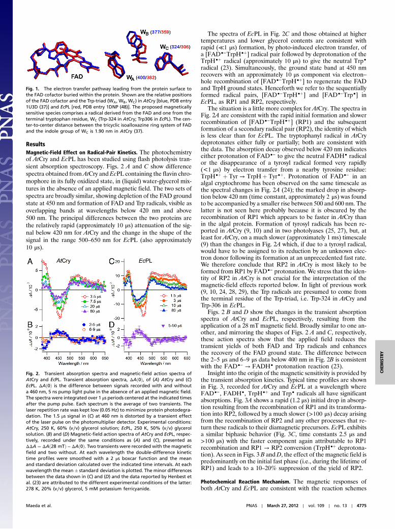
ResultsMagnetic-Field Effect on Radical-Pair Kinetics. The photochemistryof AtCry and EcPL has been studied using flash photolysis tran-sient absorption spectroscopy. Figs. 2 A and C show differencespectra obtained from AtCry and EcPL containing the flavin chro-mophore in its fully oxidized state, in (liquid) water-glycerol mix-tures in the absence of an applied magnetic field. The two sets ofspectra are broadly similar, showing depletion of the FAD groundstate at 450 nm and formation of FAD and Trp radicals, visible asoverlapping bands at wavelengths below 420 nm and above500 nm. The principal differences between the two proteins arethe relatively rapid (approximately 10 μs) attenuation of the sig-nal below 420 nm for AtCry and the change in the shape of thesignal in the range 500–650 nm for EcPL (also approximately10 μs).
The spectra of EcPL in Fig. 2C and those obtained at highertemperatures and lower glycerol contents are consistent withrapid (≪1 μs) formation, by photo-induced electron transfer, ofa [FAD•−TrpH•þ] radical pair followed by deprotonation of theTrpH•þ radical (approximately 10 μs) to give the neutral Trp•
radical (23). Simultaneously, the ground state band at 450 nmrecovers with an approximately 10 μs component via electron–hole recombination of [FAD•−TrpH•þ] to regenerate the FADand TrpH ground states. Henceforth we refer to the sequentiallyformed radical pairs, [FAD•−TrpH•þ] and [FAD•−Trp•] inEcPL, as RP1 and RP2, respectively.
The situation is a little more complex for AtCry. The spectra inFig. 2A are consistent with the rapid initial formation and slowerrecombination of [FAD•−TrpH•þ] (RP1) and the subsequentformation of a secondary radical pair (RP2), the identity of whichis less clear than for EcPL. The tryptophanyl radical in AtCrydeprotonates either fully or partially; both are consistent withthe data. The absorption decay observed below 420 nm indicateseither protonation of FAD•− to give the neutral FADH• radicalor the disappearance of a tyrosyl radical formed very rapidly(<1 μs) by electron transfer from a nearby tyrosine residue:TrpH•þ þ Tyr → TrpHþ Tyr•þ. Protonation of FAD•− in analgal cryptochrome has been observed on the same timescale asthe spectral changes in Fig. 2A (24); the marked drop in absorp-tion below 420 nm (time constant, approximately 2 μs) was foundto be accompanied by a smaller rise between 500 and 600 nm. Thelatter is not seen here probably because it is obscured by therecombination of RP1 which appears to be faster in AtCry thanin the algal protein. Formation of tyrosyl radicals has been re-ported in AtCry (9, 10) and in two photolyases (25, 27), but, atleast for AtCry, on a much slower (approximately 1 ms) timescale(9) than the changes in Fig. 2A which, if due to a tyrosyl radical,would have to be assigned to its reduction by an unknown elec-tron donor following its formation at an unprecedented fast rate.We therefore conclude that RP2 in AtCry is most likely to beformed fromRP1 by FAD•− protonation. We stress that the iden-tity of RP2 in AtCry is not crucial for the interpretation of themagnetic-field effects reported below. In light of previous work(9, 10, 24, 28, 29), the Trp radicals are presumed to come fromthe terminal residue of the Trp-triad, i.e. Trp-324 in AtCry andTrp-306 in EcPL.
Figs. 2 B and D show the changes in the transient absorptionspectra of AtCry and EcPL, respectively, resulting from theapplication of a 28 mT magnetic field. Broadly similar to one an-other, and mirroring the shapes of Figs. 2 A and C, respectively,these action spectra show that the applied field reduces thetransient yields of both FAD and Trp radicals and enhancesthe recovery of the FAD ground state. The difference betweenthe 2–5 μs and 6–9 μs data below 400 nm in Fig. 2B is consistentwith the FAD•− → FADH• protonation reaction (23).
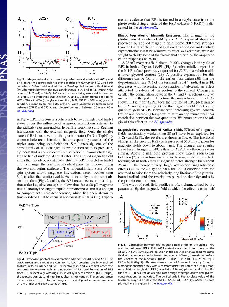
Insight into the origin of the magnetic sensitivity is provided bythe transient absorption kinetics. Typical time profiles are shownin Fig. 3, recorded for AtCry and EcPL at a wavelength whereFAD•−, FADH•, TrpH•þ and Trp• radicals all have significantabsorptions. Fig. 3A shows a rapid (1.2 μs) initial drop in absorp-tion resulting from the recombination of RP1 and its transforma-tion into RP2, followed by a much slower (>100 μs) decay arisingfrom the recombination of RP2 and any other processes that re-turn these radicals to their diamagnetic precursors. EcPL exhibitsa similar biphasic behavior (Fig. 3C, time constants 2.5 μs and>100 μs) with the faster component again attributable to RP1recombination and RP1 → RP2 conversion (TrpH•þ deprotona-tion). As seen in Figs. 3 B andD, the effect of the magnetic field ispredominantly on the initial fast phase (i.e., during the lifetime ofRP1) and leads to a 10–20% suppression of the yield of RP2.
Photochemical Reaction Mechanism. The magnetic responses ofboth AtCry and EcPL are consistent with the reaction schemes
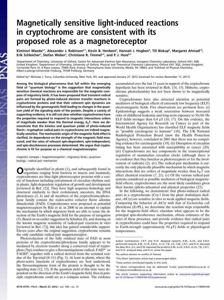
Fig. 1. The electron transfer pathway leading from the protein surface tothe FAD cofactor buried within the protein. Shown are the relative positionsof the FAD cofactor and the Trp-triad (WA, WB, WC) in AtCry [blue, PDB entry1U3D (37)] and EcPL [red, PDB entry 1DNP (48)]. The proposed magneticallysensitive species comprises a radical derived from the FAD and one from theterminal tryptophan residue, WC (Trp-324 in AtCry; Trp306 in EcPL). The cen-ter-to-center distance between the tricyclic isoalloxazine ring system of FADand the indole group of WC is 1.90 nm in AtCry (37).
Fig. 2. Transient absorption spectra and magnetic-field action spectra ofAtCry and EcPL. Transient absorption spectra, ΔAð0Þ, of (A) AtCry and (C)EcPL. ΔAð0Þ is the difference between signals recorded with and withouta 460 nm, 5 ns pump light pulse in the absence of an applied magnetic field.The spectra were integrated over 1 μs periods centered at the indicated timesafter the pump pulse. Each spectrum is the average of two transients. Thelaser repetition rate was kept low (0.05 Hz) to minimize protein photodegra-dation. The 1.5 μs signal in (C) at 460 nm is distorted by a transient effectof the laser pulse on the photomultiplier detector. Experimental conditions:AtCry, 250 K, 60% (v∕v) glycerol solution; EcPL, 250 K, 50% (v∕v) glycerolsolution. (B) and (D) Magnetic-field action spectra of AtCry and EcPL, respec-tively, recorded under the same conditions as (A) and (C), presented asΔΔA ¼ ΔAð28 mTÞ − ΔAð0Þ. Two transients were recorded with the magneticfield and two without. At each wavelength the double-difference kinetictime profiles were smoothed with a 2 μs boxcar function and the meanand standard deviation calculated over the indicated time intervals. At eachwavelength the mean� standard deviation is plotted. The minor differencesbetween the data shown in (C) and (D) and the data reported by Henbest etal. (23) are attributed to the different experimental conditions of the latter:278 K, 20% (v∕v) glycerol, 5 mM potassium ferricyanide.
Maeda et al. PNAS ∣ March 27, 2012 ∣ vol. 109 ∣ no. 13 ∣ 4775
CHEM
ISTR
Y
in Fig. 4. RP1 interconverts coherently between singlet and tripletstates under the influence of magnetic interactions internal tothe radicals (electron-nuclear hyperfine couplings) and Zeemaninteractions with the external magnetic field. Only the singletstate of RP1 can revert to the ground state (FADþ TrpH) byelectron-hole recombination, the corresponding reaction of thetriplet state being spin-forbidden. Simultaneously, one of theconstituents of RP1 changes its protonation state to give RP2,a process that is not subject to spin-selection rules and which sing-let and triplet undergo at equal rates. The applied magnetic fieldalters the time-dependent probability that RP1 is singlet or tripletand so changes the fractions of radical pairs that proceed alongthe two competing pathways. The nonequilibrium state of thespin system allows magnetic interactions much weaker thankBT to alter the reaction yields. As indicated by the transient ab-sorption data (Figs. 2 and 3), the RP1 reactions occur on a 10 μstimescale; i.e., slow enough to allow time for a 50 μT magneticfield to modify the singlet-triplet interconversion and fast enoughto compete with spin-decoherence, which has been shown bytime-resolved EPR to occur in approximately 10 μs (11). Experi-
mental evidence that RP1 is formed in a singlet state from thephoto-excited singlet state of the FAD cofactor (1FAD�) is dis-cussed in the SI Appendix.
Kinetic Regulation of Magnetic Responses. The changes in thephotochemical kinetics of AtCry and EcPL reported above areproduced by applied magnetic fields some 500 times strongerthan the Earth’s field. To shed light on the conditions under whichcryptochrome might be sensitive to much weaker fields, we havesought to clarify some of the factors that determine the amplitudeof the responses at 28 mT.
A 28 mT magnetic field elicits 10–20% changes in the yield ofRP2 in both AtCry and EcPL (Fig. 3), substantially larger thanthe 3–4% effects previously reported for EcPL in a solution witha lower glycerol content (23). A possible explanation for thisdifference can be found in the earlier observation (30) that thedeprotonation rate (kf ) of the terminal TrpH•þ radical in EcPLdecreases with increasing concentration of glycerol, an effectattributed to release of the proton to the solvent. Changes inkf alter the competition between the kb and kf reactions (Fig. 4)and so have the potential to tune the magnetic-field effect. Asshown in Fig. 5 for EcPL, both the lifetime of RP1 (determinedby the kb and kf steps, Fig. 4) and the magnetic-field effect on thequantum yield of RP2 increase with increasing glycerol concen-tration and decreasing temperature, with an approximately linearcorrelation between the two quantities. We comment on the ori-gin of this effect in the SI Appendix.
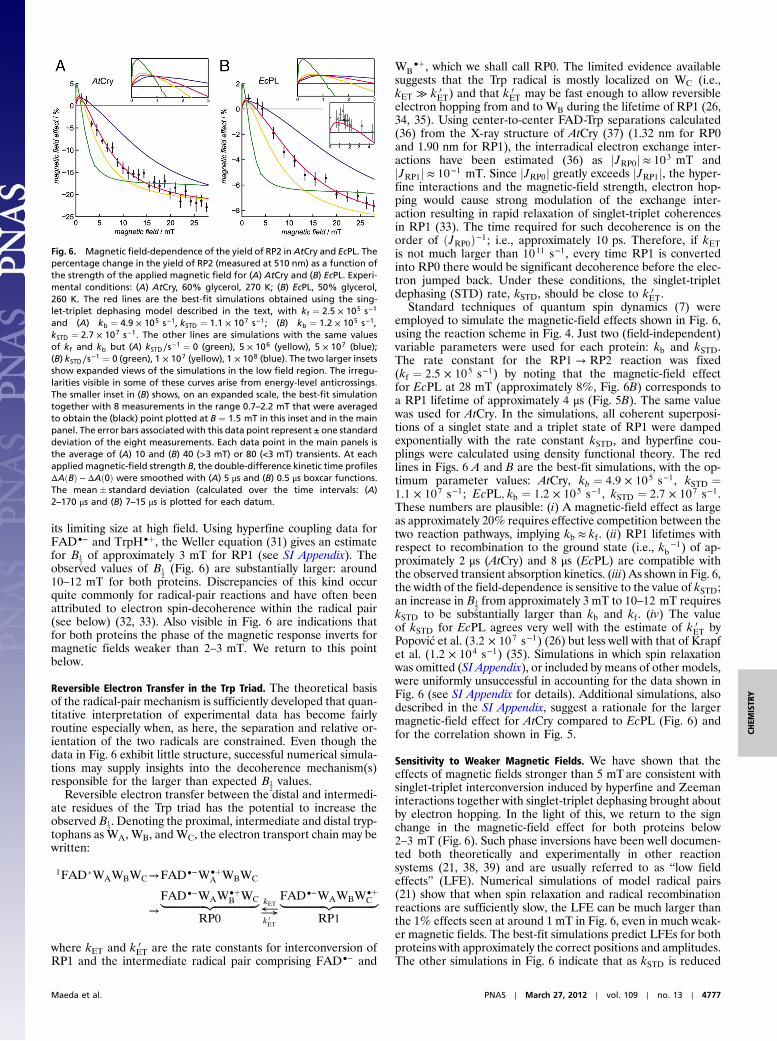
Magnetic-field Dependence of Radical Yields. Effects of magneticfields substantially weaker than 28 mT have been explored forAtCry and EcPL; the results are shown in Fig. 6. The fractionalchange in the yield of RP2 (as measured at 510 nm) is given formagnetic fields down to about 1 mT. The changes are roughlythree times stronger for AtCry than for EcPL but otherwise rathersimilar. Above 5 mT, both proteins show typical radical-pairbehavior (7): a monotonic increase in the magnitude of the effect,leveling off in both cases at magnetic fields stronger than about25 mT. The comparatively large asymptotic magnetic-fieldeffects (>20% for AtCry and >8% for EcPL) observed here areassumed to arise from the relatively long lifetime of the protein-bound radicals and the restrictions placed on their dynamics bythe protein environment.
The width of such field-profiles is often characterized by theparameter B1
2, the magnetic field at which the effect reaches half
Fig. 3. Magnetic-field effects on the photochemical kinetics of AtCry andEcPL. Transient absorption kinetic time profiles of (A) AtCry and (C) EcPL bothrecorded at 510 nm with and without a 28 mTapplied magnetic field. (B) and(D) Differences between the two signals shown in (A) and in (C), respectively:ΔΔA ¼ ΔAð28 mTÞ − ΔAð0Þ. 200 ns boxcar smoothing was used to produce(B) and (D); no smoothing was used for (A) and (C). Experimental conditions:AtCry, 270 K in 60% (v∕v) glycerol solution; EcPL, 250 K in 50% (v∕v) glycerolsolution. Similar traces for both proteins were observed at temperaturesbetween 240 K and 275 K and glycerol contents between 25% and 65%(SI Appendix).
Fig. 4. Proposed photochemical reaction schemes for AtCry and EcPL. Theblack arrows and species are common to both proteins; the blue and redfeatures refer to AtCry and EcPL, respectively. kb and kf are first-order rateconstants for electron–hole recombination of RP1 and formation of RP2from RP1, respectively. Although RP2 in AtCry is here drawn as [FADH•Trp•],the protonation state of the Trp radical is not certain. The curved greenarrows indicate the coherent, magnetic field-dependent interconversionof the singlet and triplet states of RP1.
Fig. 5. Correlation between the magnetic-field effect on the yield of RP2and the lifetime of RP1 in EcPL. (A) Transient absorption kinetic time profilesof EcPL in 50% (v∕v) glycerol solution in the absence of an applied magneticfield at the temperatures indicated. Recorded at 600 nm, these signals reflectthe kinetics of the reactions: TrpH•
þ→ Trp• þ Hþ and 1½FAD•−TrpH•þ� →
FADþ TrpH (Fig. 4). Lifetimes were extracted from such data by fitting toa monoexponential decay with a constant offset. (B) Effect of a 28 mT mag-netic field on the yield of RP2 (recorded at 510 nm) plotted against the life-time of RP1 (measured at 600 nm) over a range of temperatures and glycerolconcentrations, as indicated. The vertical axis is the absolute value of thefractional magnetic-field effect (MFE): jΔAð28 mTÞ − ΔAð0Þj∕ΔAð0Þ. The dataplotted here are given in the SI Appendix.
4776 ∣ www.pnas.org/cgi/doi/10.1073/pnas.1118959109 Maeda et al.
its limiting size at high field. Using hyperfine coupling data forFAD•− and TrpH•þ, the Weller equation (31) gives an estimatefor B1
2of approximately 3 mT for RP1 (see SI Appendix). The
observed values of B12(Fig. 6) are substantially larger: around
10–12 mT for both proteins. Discrepancies of this kind occurquite commonly for radical-pair reactions and have often beenattributed to electron spin-decoherence within the radical pair(see below) (32, 33). Also visible in Fig. 6 are indications thatfor both proteins the phase of the magnetic response inverts formagnetic fields weaker than 2–3 mT. We return to this pointbelow.
Reversible Electron Transfer in the Trp Triad. The theoretical basisof the radical-pair mechanism is sufficiently developed that quan-titative interpretation of experimental data has become fairlyroutine especially when, as here, the separation and relative or-ientation of the two radicals are constrained. Even though thedata in Fig. 6 exhibit little structure, successful numerical simula-tions may supply insights into the decoherence mechanism(s)responsible for the larger than expected B1
2values.
Reversible electron transfer between the distal and intermedi-ate residues of the Trp triad has the potential to increase theobservedB1
2. Denoting the proximal, intermediate and distal tryp-
tophans as WA, WB, andWC, the electron transport chain may bewritten:
1FAD�WAWBWC→FAD•−W•þA WBWC
→FAD•−WAW•þ
B WC|fflfflfflfflfflfflfflfflfflfflfflfflfflfflfflffl{zfflfflfflfflfflfflfflfflfflfflfflfflfflfflfflffl}
where kET and k 0ET are the rate constants for interconversion of
RP1 and the intermediate radical pair comprising FAD•− and
WB•þ, which we shall call RP0. The limited evidence available
suggests that the Trp radical is mostly localized on WC (i.e.,kET ≫ k 0
ET) and that k 0ET may be fast enough to allow reversible
electron hopping from and to WB during the lifetime of RP1 (26,34, 35). Using center-to-center FAD-Trp separations calculated(36) from the X-ray structure of AtCry (37) (1.32 nm for RP0and 1.90 nm for RP1), the interradical electron exchange inter-actions have been estimated (36) as jJRP0j ≈ 103 mT andjJRP1j ≈ 10−1 mT. Since jJRP0j greatly exceeds jJRP1j, the hyper-fine interactions and the magnetic-field strength, electron hop-ping would cause strong modulation of the exchange inter-action resulting in rapid relaxation of singlet-triplet coherencesin RP1 (33). The time required for such decoherence is on theorder of ðJRP0Þ−1; i.e., approximately 10 ps. Therefore, if kETis not much larger than 1011 s−1, every time RP1 is convertedinto RP0 there would be significant decoherence before the elec-tron jumped back. Under these conditions, the singlet-tripletdephasing (STD) rate, kSTD, should be close to k 0
ET.Standard techniques of quantum spin dynamics (7) were
employed to simulate the magnetic-field effects shown in Fig. 6,using the reaction scheme in Fig. 4. Just two (field-independent)variable parameters were used for each protein: kb and kSTD.The rate constant for the RP1 → RP2 reaction was fixed(kf ¼ 2.5 × 10⁵ s−1) by noting that the magnetic-field effectfor EcPL at 28 mT (approximately 8%, Fig. 6B) corresponds toa RP1 lifetime of approximately 4 μs (Fig. 5B). The same valuewas used for AtCry. In the simulations, all coherent superposi-tions of a singlet state and a triplet state of RP1 were dampedexponentially with the rate constant kSTD, and hyperfine cou-plings were calculated using density functional theory. The redlines in Figs. 6 A and B are the best-fit simulations, with the op-timum parameter values: AtCry, kb ¼ 4.9 × 10⁵ s−1, kSTD ¼1.1 × 10⁷ s−1; EcPL; kb ¼ 1.2 × 10⁵ s−1, kSTD ¼ 2.7 × 10⁷ s−1.These numbers are plausible: (i) A magnetic-field effect as largeas approximately 20% requires effective competition between thetwo reaction pathways, implying kb ≈ kf . (ii) RP1 lifetimes withrespect to recombination to the ground state (i.e., kb
−1) of ap-proximately 2 μs (AtCry) and 8 μs (EcPL) are compatible withthe observed transient absorption kinetics. (iii) As shown in Fig. 6,the width of the field-dependence is sensitive to the value of kSTD;an increase in B1
2from approximately 3 mT to 10–12 mT requires
kSTD to be substantially larger than kb and kf . (iv) The valueof kSTD for EcPL agrees very well with the estimate of k 0
ET byPopović et al. (3.2 × 10⁷ s−1) (26) but less well with that of Krapfet al. (1.2 × 10⁴ s−1) (35). Simulations in which spin relaxationwas omitted (SI Appendix), or included by means of other models,were uniformly unsuccessful in accounting for the data shown inFig. 6 (see SI Appendix for details). Additional simulations, alsodescribed in the SI Appendix, suggest a rationale for the largermagnetic-field effect for AtCry compared to EcPL (Fig. 6) andfor the correlation shown in Fig. 5.
Sensitivity to Weaker Magnetic Fields. We have shown that theeffects of magnetic fields stronger than 5 mTare consistent withsinglet-triplet interconversion induced by hyperfine and Zeemaninteractions together with singlet-triplet dephasing brought aboutby electron hopping. In the light of this, we return to the signchange in the magnetic-field effect for both proteins below2–3 mT (Fig. 6). Such phase inversions have been well documen-ted both theoretically and experimentally in other reactionsystems (21, 38, 39) and are usually referred to as “low fieldeffects” (LFE). Numerical simulations of model radical pairs(21) show that when spin relaxation and radical recombinationreactions are sufficiently slow, the LFE can be much larger thanthe 1% effects seen at around 1 mT in Fig. 6, even in much weak-er magnetic fields. The best-fit simulations predict LFEs for bothproteins with approximately the correct positions and amplitudes.The other simulations in Fig. 6 indicate that as kSTD is reduced
Fig. 6. Magnetic field-dependence of the yield of RP2 inAtCry and EcPL. Thepercentage change in the yield of RP2 (measured at 510 nm) as a function ofthe strength of the applied magnetic field for (A) AtCry and (B) EcPL. Experi-mental conditions: (A) AtCry, 60% glycerol, 270 K; (B) EcPL, 50% glycerol,260 K. The red lines are the best-fit simulations obtained using the sing-let-triplet dephasing model described in the text, with kf ¼ 2.5 × 10⁵ s−1
and (A) kb ¼ 4.9 × 10⁵ s−1; kSTD ¼ 1.1 × 10⁷ s−1; (B) kb ¼ 1.2 × 10⁵ s−1;kSTD ¼ 2.7 × 10⁷ s−1. The other lines are simulations with the same valuesof kf and kb but (A) kSTD∕s−1 ¼ 0 (green), 5 × 10⁶ (yellow), 5 × 10⁷ (blue);(B) kSTD∕s−1 ¼ 0 (green), 1 × 10⁷ (yellow), 1 × 10⁸ (blue). The two larger insetsshow expanded views of the simulations in the low field region. The irregu-larities visible in some of these curves arise from energy-level anticrossings.The smaller inset in (B) shows, on an expanded scale, the best-fit simulationtogether with 8 measurements in the range 0.7–2.2 mT that were averagedto obtain the (black) point plotted at B ¼ 1.5 mT in this inset and in the mainpanel. The error bars associated with this data point represent ± one standarddeviation of the eight measurements. Each data point in the main panels isthe average of (A) 10 and (B) 40 (>3 mT) or 80 (<3 mT) transients. At eachapplied magnetic-field strength B, the double-difference kinetic time profilesΔAðBÞ − ΔAð0Þ were smoothed with (A) 5 μs and (B) 0.5 μs boxcar functions.The mean� standard deviation (calculated over the time intervals: (A)2–170 μs and (B) 7–15 μs is plotted for each datum.
Maeda et al. PNAS ∣ March 27, 2012 ∣ vol. 109 ∣ no. 13 ∣ 4777
the initial slope increases causing the maximum LFE to shift toprogressively lower fields. Thus the effect of a 50 μT magneticfield on the yield of RP2 could be significantly larger than impliedby the best-fit simulations in Fig. 6 if singlet-triplet dephasing(and any other significant decoherence mechanisms) were suffi-ciently slow.
DiscussionThe cryptochrome hypothesis of radical-pair magnetoreceptionwas proposed 11 years ago. We present here direct evidence thatcryptochromes can exhibit the magnetically sensitive photochem-istry that is the essential prerequisite for a magnetic compasssensor. Magnetic-field effects on the quantum yields of radicalsproduced in AtCry in viscous solution of about þ1% in a 1 mTmagnetic field, and about −25% in a 30 mT field, have beendetected. The change in phase observed for fields weaker than2–3 mT is the signature of the low field effect which, under ap-propriate conditions, could permit significant responses to Earth-strength magnetic fields.
High glycerol concentrations and reduced temperatures havebeen used here to optimize the observed magnetic responses.One can only speculate about the environment of an avian cryp-tochrome in a magnetoreceptor cell, but it seems most likely thatthe proteins would have to be both immobilized and aligned inorder to show the anisotropic magnetic responses essential for acompass detection mechanism (5). Restricted molecular motion,leading to slower spin-decoherence, should also be favorable.Both factors prompted our use of mixed aqueous solvents witha higher viscosity than pure water. [Other reasons for using gly-cerol-water mixtures were (i) to regulate the competition betweendeprotonation of TrpH•þ and spin-selective recombination ofRP1 in EcPL (Fig. 5); (ii) to allow the use of temperatures below273 K for the same reason; and (iii) to stabilize the protein againstaggregation and precipitation.] Restricted motion could comeabout in different and probably more efficient ways in vivo, forexample by tethering to membrane proteins or cytoskeletal fila-ments (40), binding to signaling partners (41–43) and/or cofactors[e.g., ATP (37, 44)], dimerization (45), etc. It does not seem un-reasonable to conjecture that under optimum conditions in vivo,the effect of a 50 μT magnetic field could be substantially largerthan observed in the present study. Simulations suggest that thelow field effect can, under the right conditions, be as large as10–20% (21). Too little is known about light-dependent crypto-chrome signaling in general, and magnetoreception in particular,to say whether this would be large enough to form the basis of aviable magnetoreceptor.
It is evident from our results that a significant magnetic-fieldeffect from a cryptochrome-based radical pair requires kineticcompetition on a 1–10 μs timescale, between spin-selective elec-tron-hole recombination, and spin-independent formation of thesignaling state. If these processes were much faster than 1 μs therewould be insufficient time for the geomagnetic field to influencethe spin dynamics; if they were much slower, the effect wouldalmost certainly be attenuated by spin-decoherence (14). It seemsunlikely, however, that the conformational changes needed togenerate the signaling state [probably rearrangement of the C-terminal domain (46)] could be as fast as 10 μs. Our results showthat formation of a secondary species (RP2) from the magneti-cally sensitive radical pair RP1, [FAD•−TrpH•þ], avoids the needfor an abnormally rapid protein rearrangement or exceptionallyslow spin-decoherence. Protonation of the FAD•− radical (asmay occur in AtCry) or deprotonation of the TrpH•þ radical [asoccurs in EcPL (23, 30)] allows the magnetic-field effect on RP1to be “stored” in the form of a changed quantum yield of the
much longer lived state RP2, from which the signaling state cansubsequently be generated. The fact that there is no need for thereactions of RP2 to be magnetically sensitive means that itslifetime can greatly exceed its spin-decoherence time without illeffect.
There are now two members of the cryptochrome/photolyasefamily that show magnetic responses. Given the very differentbiological functions of EcPL (DNA repair) and AtCry (regulationof growth and development, entrainment of circadian rhythms)and the fact that neither bacteria nor plants appear to have spe-cialized magnetoreceptors, we suggest that magnetic sensitivityis a general feature of this protein family and that the resultsdescribed here may be extrapolated to bird cryptochromes (andpossibly even human cryptochromes).
Materials and MethodsProtein Preparation. The expression and purification of EcPL (as a mutant thatdoes not bind the methenyltetrahydrofolate cofactor) are described else-where (23, 47). To ensure the FAD cofactor was in its fully oxidized state,the protein was pretreated with potassium ferricyanide as described pre-viously (23). Glycerol was added to approximately 130 μM protein samplesin 50 mM Hepes buffer at pH 7.0 with 100 mM KCl to obtain solutions con-taining 20–50% glycerol (v∕v). Unlike our previous study of EcPL (23), potas-sium ferricyanide was not added to the samples to reoxidize photoreducedflavin.
AtCry (full length cryptochrome-1) was expressed in Sf21 cells using arecombinant baculovirus expression vector system and purified by Ni-NTAaffinity chromatography. Glycerol was added to approximately 150 μMprotein samples in Tris/HCl buffer at pH 7.5 with 500 mM NaCl and 250 mMimidazole to obtain solutions containing 20–60% glycerol (v∕v).
Transient Absorption Spectroscopy. Protein samples (approximately 250 μL)were cooled in a cryostat (Oxford Instruments, Optistat CF) with the tempera-ture controlled to within 0.1 K. The sample was held in a quartz cuvette (Hell-ma 104.002F QS; 10 mm path length, internal dimensions 2 × 10 × 45 mm) atthe center of the cryostat. Magnetic-field pulses of approximately 4 ms dura-tion, synchronized with the laser flash, were generated using home-builtHelmholtz coils. The maximum magnetic field at the position of the samplewas 29 mT. Samples were not shielded from the Earth’s magnetic field. Ra-dical pairs in EcPL and AtCry were generated by flash photolysis using a dyelaser (Sirah Cobra) pumped by a Nd:YAG laser (Continuum Surelite 1). Thelaser dye was Coumarin 460 (Exciton Inc.) in analytical grade methanol (FisherScientific). The Nd:YAG laser produced 5 ns pulses with energy approximately100 mJ and repetition rate 1 Hz, tuned by means of a Q-switch delay to pro-duce 5–7 mJ, 460 nm pulses (FWHM 37 nm) from the dye laser. Probe lightfrom a 300 W xenon arc lamp (Oriel) was passed through a water filter to cutout infrared components and through long-pass filters to remove unwantedwavelengths and then to the sample in a direction orthogonal to the pumppulses. Pump and probe beams were controlled by mechanical shutters toobtain a 0.05 Hz repetition rate to reduce photodegradation of the light-sensitive samples. Probe light was collected using a monochromator (Oriel77250), fed into a photomultiplier tube (Hamamatsu R928) and from thereto an oscilloscope (Iwatsu-LeCroy Waverunner LT342L). Data were trans-ferred to a personal computer and analyzed using IGOR PRO (Wavemetrics,Inc.) software.
Spin Dynamics Simulations. The magnetic field-dependence of radical-pairreaction yields (Fig. 6) was calculated from the equation of motion of theradical-pair spin density operator in Liouville space including coherent spindynamics, decoherence processes, and chemical reactivity by means of appro-priate superoperators. Singlet-triplet dephasing was introduced as proposedby Shushin (33). Further details are given in the SI Appendix.
ACKNOWLEDGMENTS. We thank S. M. Lea, J. Lillington and A. Bowen for dis-cussions and N. Baker and P. Stehle for technical assistance. P.J.H. and C.R.T.were supported by the Electromagnetic Fields Biological Research Trust,the Defense Advanced Research Projects Agency (QuBE: N66001-10-1-4061),and the Engineering and Physical Sciences Research Council. S.W. and E.S.were funded by Deutsche Forschungsgemeinschaft Grant WE2376/41.
1. Ahmad M, Cashmore AR (1993) HY4 gene of A. thaliana encodes a protein with
characteristics of a blue-light photoreceptor. Nature 366:162–166.
2. Chaves I, et al. (2011) The cryptochromes: Blue light photoreceptors in plants and
animals. Annu Rev Plant Biol 62:335–364.
3. Weber S (2005) Light-driven enzymatic catalysis of DNA repair: A review of recent
biophysical studies on photolyase. Biochim Biophys Acta 1707:1–23.
4. Sancar A (2008) Structure and function of photolyase and in vivo enzymology: 50th
anniversary. J Biol Chem 283:32153–32157.
4778 ∣ www.pnas.org/cgi/doi/10.1073/pnas.1118959109 Maeda et al.
5. Ritz T, Adem S, Schulten K (2000) Amodel for photoreceptor-basedmagnetoreceptionin birds. Biophys J 78:707–718.
6. Schulten K, Swenberg CE, Weller A (1978) A biomagnetic sensory mechanism based onmagnetic field modulated coherent electron spin motion. Z Phys Chem NF 111:1–5.
7. Rodgers CT (2009) Magnetic field effects in chemical systems. Pure Appl Chem81:19–43.
8. Gindt YM, et al. (1999) Origin of the transient electron paramagnetic resonancesignals in DNA photolyase. Biochemistry 38:3857–3866.
9. Giovani B, Byrdin M, Ahmad M, Brettel K (2003) Light-induced electron transfer in acryptochrome blue-light photoreceptor. Nat Struct Biol 10:489–490.
10. Zeugner A, et al. (2005) Light-induced electron transfer in Arabidopsis cryptochrome-1correlates with in vivo function. J Biol Chem 280:19437–19440.
11. Biskup T, et al. (2009) Direct observation of a photoinduced radical-pair intermediatein a cryptochrome DASH blue-light photoreceptor. Angew Chem Int Ed 48:404–407.
12. Banerjee R, et al. (2007) The signaling state of Arabidopsis cryptochrome 2 containsflavin semiquinone. J Biol Chem 282:14916–14922.
13. Bouly JP, et al. (2007) Cryptochrome blue light photoreceptors are activated throughinterconversion of flavin redox states. J Biol Chem 282:9383–9391.
14. Rodgers CT, Hore PJ (2009) Chemical magnetoreception in birds: A radical pairmechanism. Proc Natl Acad Sci USA 106:353–360.
15. Liedvogel M, Mouritsen H (2010) Cryptochromes—a potential magnetoreceptor:What do we know and what do we want to know? J Roy Soc Interface 7:S147–S162.
16. Ahlbom A, et al. (2000) A pooled analysis of magnetic fields and childhood leukaemia.Brit J Cancer 83:692–698.
17. Greenland S, et al. (2000) A pooled analysis of magnetic fields, wire codes, and child-hood leukemia. Epidemiology 11:624–634.
18. International Agency for Research on Cancer (2002) Static and extremely low-fre-quency (ELF) electric and magnetic fields. IARC monographs on the evaluation ofcarcinogenic risks to humans, vol. 80 (IARC, Lyon).
19. Advisory Group on Non-ionising Radiation (2001) AGNIR (2001) ELF electromagneticfields and the risk of cancer. Report of an Advisory Group on Non-ionising Radiation.Documents of the NRPB, Vol. 12 (National Radiological Protection Board, Chilton,Oxon, UK).
20. Reddy AB, Wong GKY, O’Neill J, Maywood ES, Hastings MH (2005) Circadian clocks:Neural and peripheral pacemakers that impact upon the cell division cycle. MutatRes 574:76–91.
21. Timmel CR, Till U, Brocklehurst B, McLauchlan KA, Hore PJ (1998) Effects of weakmagnetic fields on free radical recombination reactions. Mol Phys 95:71–89.
22. Lagroye I, Percherancier Y, Juutilainen J, Poulletier De Gannes F, Veyret B (2011) ELFmagnetic fields: Animal studies, mechanisms of action. Prog Biophys Mol Biol107:369–373.
23. Henbest KB, et al. (2008) Magnetic-field effect on the photoactivation reaction ofEscherichia coli DNA photolyase. Proc Natl Acad Sci USA 105:14395–14399.
24. Langenbacher T, Immeln D, Dick B, Kottke T (2009) Microsecond light-induced protontransfer to flavin in the blue light sensor plant cryptochrome. J Am Chem Soc131:14274–14280.
25. Aubert C, Mathis P, Eker APM, Brettel K (1999) Intraprotein electron transfer betweentyrosine and tryptophan in DNA photolyase from anacystis nidulans. Proc Natl Acad SciUSA 96:5423–5427.
26. Popović DM, Zmiric A, Zaric SD, Knapp EW (2002) Energetics of radical transfer in DNAphotolyase. J Am Chem Soc 124:3775–3782.
27. Weber S, et al. (2002) Photoactivation of the flavin cofactor in Xenopus laevis (6–4)photolyase: Observation of a transient tyrosyl radical by time-resolved electron para-magnetic resonance. Proc Natl Acad Sci USA 99:1319–1322.
28. Li YF, Heelis PF, Sancar A (1991) Active-site of DNA photolyase—tryptophan-306 isthe intrinsic hydrogen-atom donor essential for flavin radical photoreduction andDNA-repair in vitro. Biochemistry 30:6322–6329.
29. Brettel K, Byrdin M (2010) Reaction mechanisms of DNA photolyase. Curr Opin StructBiol 20:693–701.
30. Byrdin M, et al. (2004) Intraprotein electron transfer and proton dynamics duringphotoactivation of DNA photolyase from E. coli: Review and new insights from an“inverse” deuterium isotope effect. Biochim Biophys Acta 1655:64–70.
31. Weller A, Nolting F, Staerk H (1983) A quantitative interpretation of the magnetic-field effect on hyperfine-coupling-induced triplet formation from radical ion-pairs.Chem Phys Lett 96:24–27.
32. Miura T, Maeda K, Arai T (2006) The spin mixing process of a radical pair in low mag-netic field observed by transient absorption detected nanosecond pulsed magneticfield effect. J Phys Chem A 110:4151–4156.
33. Shushin AI (1991) The effect of the spin exchange interaction on SNP and RYDMRspectra of geminate radical pairs. Chem Phys Lett 181:274–278.
34. Woiczikowski PB, Steinbrecher T, Kubar T, Elstner M (2011) Nonadiabatic QM/MMsimulations of fast charge transfer in Escherichia coli DNA photolyase. J Phys ChemB 115:9846–9863.
35. Krapf S, Koslowski T, Steinbrecher T (2010) The thermodynamics of charge transfer inDNA photolyase: Using thermodynamic integration calculations to analyse the kineticsof electron transfer reactions. Phys Chem Chem Phys 12:9516–9525.
36. Efimova O, Hore PJ (2008) Role of exchange and dipolar interactions in the radical pairmodel of the avian magnetic compass. Biophys J 94:1565–1574.
37. Brautigam CA, et al. (2004) Structure of the photolyase-like domain of cryptochrome 1from Arabidopsis thaliana. Proc Natl Acad Sci USA 101:12142–12147.
38. Eveson RW, Timmel CR, Brocklehurst B, Hore PJ, McLauchlan KA (2000) The effects ofweak magnetic fields on radical recombination reactions in micelles. Int J Radiat Biol76:1509–1522.
39. Brocklehurst B (1976) Spin correlation in geminate recombination of radical ions inhydrocarbons. 1. Theory of magnetic-field effect. J Chem Soc Faraday Trans II72:1869–1884.
40. Kirschvink JL, Winklhofer M, Walker MM (2010) Biophysics of magnetic orientation:Strengthening the interface between theory and experimental design. J R Soc Inter-face 7:S179–S191.
41. Zuo Z, Liu H, Liu B, Liu X, Lin C (2011) Blue light-dependent interaction of CRY2 withSPA1 regulates COP1 activity and floral initiation in Arabidopsis. Curr Biol 21:841–847.
42. Liu B, Liu H, Zhong D, Lin C (2010) Searching for a photocycle of the cryptochromephotoreceptors. Curr Opin Plant Biol 13:578–586.
43. Partch CL, Sancar A (2005) Photochemistry and photobiology of cryptochromeblue-light photopigments: The search for a photocycle. Photochem Photobiol81:1291–1304.
44. Bouly J, et al. (2003) Novel ATP-binding and autophosphorylation activity associatedwith Arabidopsis and human cryptochrome-1. Eur J Biochem 270:2921–2928.
45. Sang Y, et al. (2005) N-terminal domain-mediated homodimerization is required forphotoreceptor activity of Arabidopsis cryptochrome 1. Plant Cell 17:1569–1584.
46. Kondoh M, et al. (2011) Light-induced conformational changes in full-length Arabi-dopsis thaliana cryptochrome. J Mol Biol 413:128–137.
47. Schleicher E, et al. (2005) Light-induced reactions of Escherichia coli DNA photolyasemonitored by Fourier transform infrared spectroscopy. FEBS J 272:1855–1866.
48. Park H-W, Kim S-T, Sancar A, Deisenhofer J (1995) Crystal structure of DNA photolyasefrom Escherichia coli. Science 268:1866–1872.
Maeda et al. PNAS ∣ March 27, 2012 ∣ vol. 109 ∣ no. 13 ∣ 4779
CHEM
ISTR
Y
1
SUPPORTING INFORMATION
Magnetically sensitive light‐induced reactions in cryptochrome are consistent with its proposed role as a magnetoreceptor
Kiminori Maedaa,1, Alexander J. Robinsona,1, Kevin B. Henbesta, Hannah J. Hogbenb, Till Biskupb, Margaret Ahmadc, Erik Schleicherd, Stefan Weberd, Christiane R. Timmela,2 & P. J. Horeb,2
aDepartment of Chemistry, University of Oxford, Centre for Advanced Electron Spin Resonance, Inorganic Chemistry Laboratory, Oxford, UK.
bDepartment of Chemistry, University of Oxford, Physical & Theoretical Chemistry Laboratory, Oxford, UK.
cUniversité Paris VI, 4 Place Jussieu, 75005 Paris, France and Pennsylvania State University, Media, Pennsylvania 19063, USA.
dInstitute of Physical Chemistry, Albert‐Ludwigs‐Universität Freiburg, 79104 Freiburg, Germany.
1These authors contributed equally to this work.
2To whom correspondence may be addressed. E‐mail: [email protected] or christiane.timmel@ chem.ox.ac.uk
Contents 1. Magnetic field effects on radical kinetics
2. Flavintryptophan photoreaction mechanism
3. Correlation between the magnetic field effect on the yield of RP2 and the
lifetime of RP1 in EcPL.
4. Calculation of B1/2
5. Simulation of magnetic field effects
2
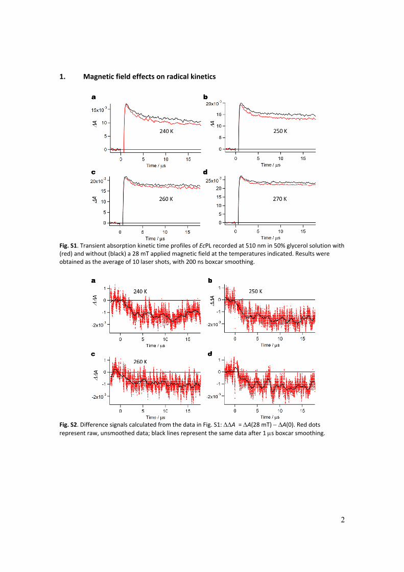
1. Magnetic field effects on radical kinetics
Fig. S1. Transient absorption kinetic time profiles of EcPL recorded at 510 nm in 50% glycerol solution with (red) and without (black) a 28 mT applied magnetic field at the temperatures indicated. Results were obtained as the average of 10 laser shots, with 200 ns boxcar smoothing.
Fig. S2. Difference signals calculated from the data in Fig. S1: A = A(28 mT) A(0). Red dots represent raw, unsmoothed data; black lines represent the same data after 1 s boxcar smoothing.
3
Fig. S3. Transient absorption kinetic time profiles of EcPL recorded at 510 nm in 65% glycerol solution with (red) and without (black) a 28 mT applied magnetic field at the temperatures indicated. Results were obtained as the average of 10 laser shots, with 200 ns boxcar smoothing.
Fig. S4. Difference signals calculated from the data in Fig. S3: A = A(28 mT) A(0). Red dots represent raw, unsmoothed data; black lines represent the same data after 1 s boxcar smoothing.
4
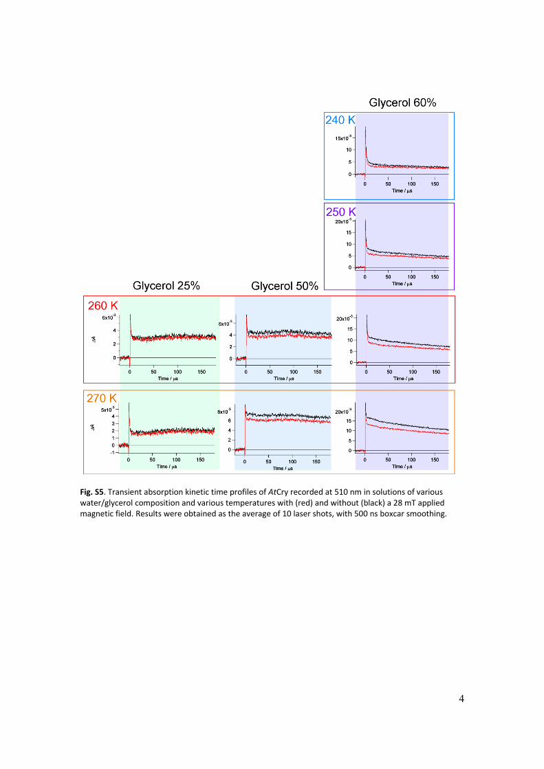
Fig. S5. Transient absorption kinetic time profiles of AtCry recorded at 510 nm in solutions of various water/glycerol composition and various temperatures with (red) and without (black) a 28 mT applied magnetic field. Results were obtained as the average of 10 laser shots, with 500 ns boxcar smoothing.
5
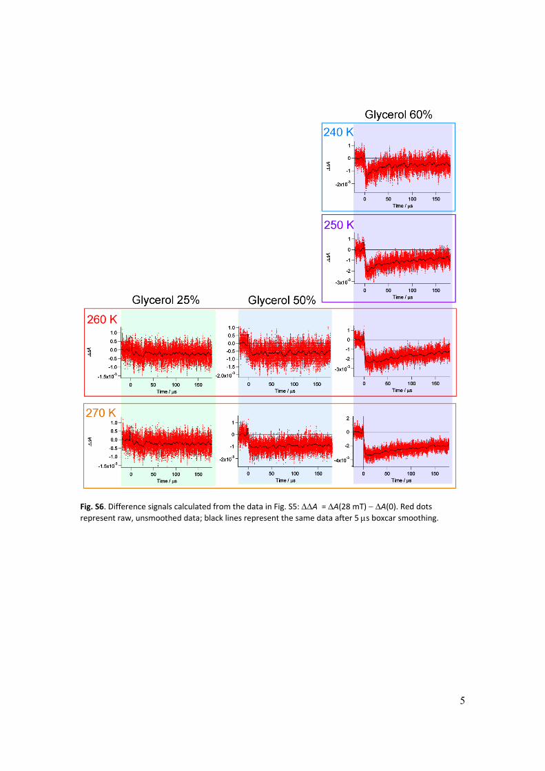
Fig. S6. Difference signals calculated from the data in Fig. S5: A = A(28 mT) A(0). Red dots represent raw, unsmoothed data; black lines represent the same data after 5 s boxcar smoothing.
6
2. Flavintryptophan photoreaction mechanism
Fig. 4 shows RP1 as formed in a singlet state from the photo‐excited singlet state of the
FAD cofactor (1FAD*) in AtCry and EcPL. Much of the photochemistry of free (i.e. not
protein‐bound) flavins proceeds from the photo‐excited triplet state formed by
intersystem crossing from the excited singlet. Although in principle possible in AtCry and
EcPL, this reaction pathway is excluded by the observation that an applied magnetic
field of intensity exceeding the hyperfine interactions reduces the yield of RP2 (Figs 2
and 3) and correspondingly increases the proportion of RP1 that returns directly to the
ground state (1). Direct formation of RP1 from 1FAD*, rather than via 3FAD* probably
has the advantage of a larger quantum yield because intersystem crossing is likely to be
slower than electron transfer and so would compete less effectively with other
processes that quench 1FAD*, e.g. fluorescence.
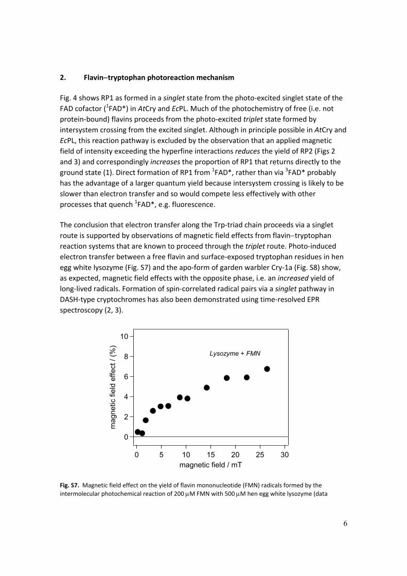
The conclusion that electron transfer along the Trp‐triad chain proceeds via a singlet
route is supported by observations of magnetic field effects from flavintryptophan reaction systems that are known to proceed through the triplet route. Photo‐induced
electron transfer between a free flavin and surface‐exposed tryptophan residues in hen
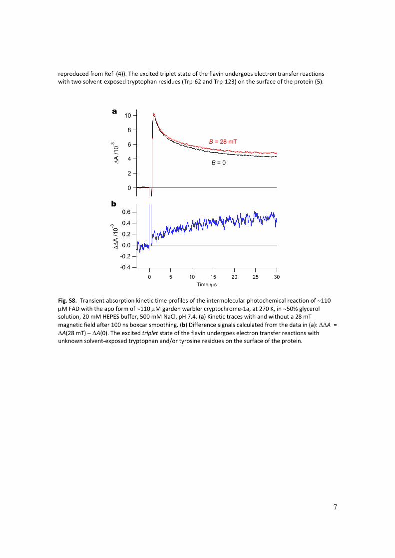
egg white lysozyme (Fig. S7) and the apo‐form of garden warbler Cry‐1a (Fig. S8) show,
as expected, magnetic field effects with the opposite phase, i.e. an increased yield of
long‐lived radicals. Formation of spin‐correlated radical pairs via a singlet pathway in
DASH‐type cryptochromes has also been demonstrated using time‐resolved EPR
spectroscopy (2, 3).
10
8
6
4
2
0
mag
netic
fiel
d ef
fect
/ (
%)
302520151050magnetic field / mT
Lysozyme + FMN
Fig. S7. Magnetic field effect on the yield of flavin mononucleotide (FMN) radicals formed by the
intermolecular photochemical reaction of 200 M FMN with 500 M hen egg white lysozyme (data
7
reproduced from Ref (4)). The excited triplet state of the flavin undergoes electron transfer reactions with two solvent‐exposed tryptophan residues (Trp‐62 and Trp‐123) on the surface of the protein (5).
10
8
6
4
2
0
A /1
0-3
302520151050
Time /s
0.6
0.4
0.2
0.0
-0.2
-0.4
A
/10
-3
B = 0
B = 28 mT
a
b
Fig. S8. Transient absorption kinetic time profiles of the intermolecular photochemical reaction of 110 M FAD with the apo form of 110 M garden warbler cryptochrome‐1a, at 270 K, in 50% glycerol solution, 20 mM HEPES buffer, 500 mM NaCl, pH 7.4. (a) Kinetic traces with and without a 28 mT
magnetic field after 100 ns boxcar smoothing. (b) Difference signals calculated from the data in (a): A = A(28 mT) A(0). The excited triplet state of the flavin undergoes electron transfer reactions with unknown solvent‐exposed tryptophan and/or tyrosine residues on the surface of the protein.
8
3. Correlation between the magnetic field effect on the yield of RP2 and the lifetime of RP1 in EcPL.
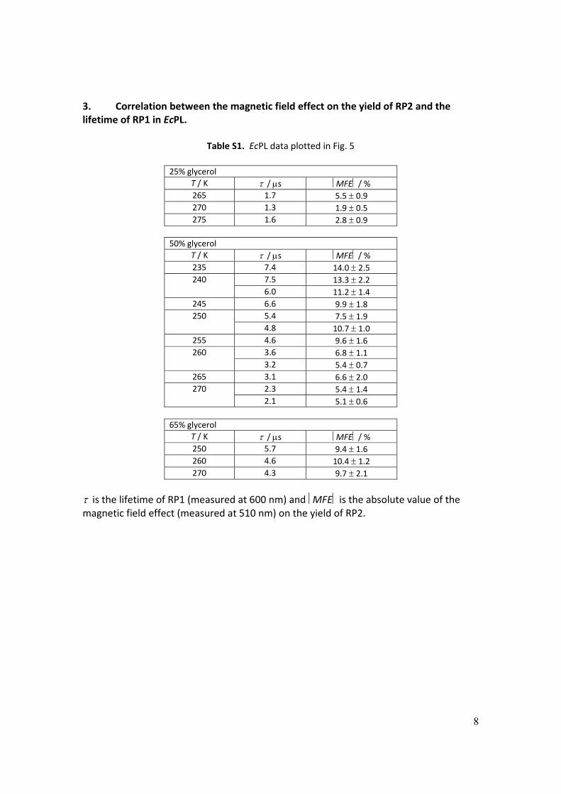
Table S1. EcPL data plotted in Fig. 5
25% glycerol
T / K / s MFE / % 265 1.7 5.5 0.9 270 1.3 1.9 0.5 275 1.6 2.8 0.9
50% glycerol T / K / s MFE / % 235 7.4 14.0 2.5 240 7.5 13.3 2.2
6.0 11.2 1.4 245 6.6 9.9 1.8 250 5.4 7.5 1.9
4.8 10.7 1.0 255 4.6 9.6 1.6 260 3.6 6.8 1.1
3.2 5.4 0.7 265 3.1 6.6 2.0 270 2.3 5.4 1.4
2.1 5.1 0.6
65% glycerol T / K / s MFE / % 250 5.7 9.4 1.6 260 4.6 10.4 1.2 270 4.3 9.7 2.1
is the lifetime of RP1 (measured at 600 nm) and MFE is the absolute value of the magnetic field effect (measured at 510 nm) on the yield of RP2.
9
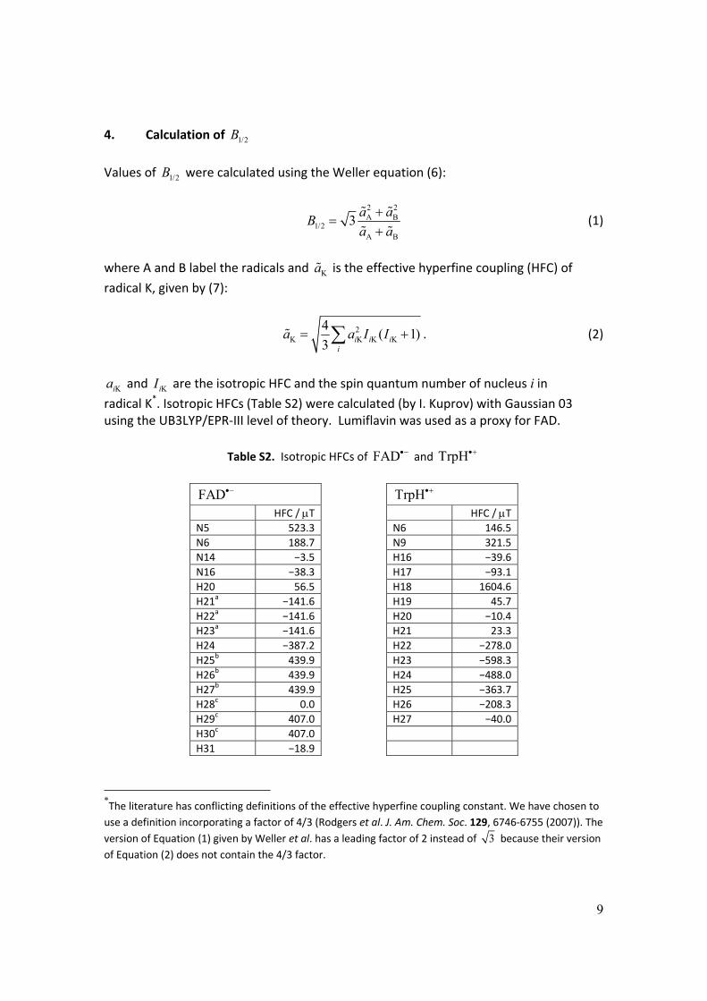
4. Calculation of 1/2B
Values of 1/2B were calculated using the Weller equation (6):
2 2A B
1/2A B
3a a
Ba a
(1)
where A and B label the radicals and Ka is the effective hyperfine coupling (HFC) of
radical K, given by (7):
2K K K K
4( 1)
3 i i ii
a a I I . (2)
Kia and KiI are the isotropic HFC and the spin quantum number of nucleus i in
radical K*. Isotropic HFCs (Table S2) were calculated (by I. Kuprov) with Gaussian 03 using the UB3LYP/EPR‐III level of theory. Lumiflavin was used as a proxy for FAD.
Table S2. Isotropic HFCs of FAD and TrpH
FAD TrpH
HFC / T HFC / T N5 523.3 N6 146.5
N6 188.7 N9 321.5
N14 −3.5 H16 −39.6
N16 −38.3 H17 −93.1
H20 56.5 H18 1604.6
H21a −141.6 H19 45.7
H22a −141.6 H20 −10.4
H23a −141.6 H21 23.3
H24 −387.2 H22 −278.0
H25b 439.9 H23 −598.3
H26b 439.9 H24 −488.0
H27b 439.9 H25 −363.7
H28c 0.0 H26 −208.3
H29c 407.0 H27 −40.0
H30c 407.0
H31 −18.9
*The literature has conflicting definitions of the effective hyperfine coupling constant. We have chosen to
use a definition incorporating a factor of 4/3 (Rodgers et al. J. Am. Chem. Soc. 129, 6746‐6755 (2007)). The
version of Equation (1) given by Weller et al. has a leading factor of 2 instead of 3 because their version
of Equation (2) does not contain the 4/3 factor.
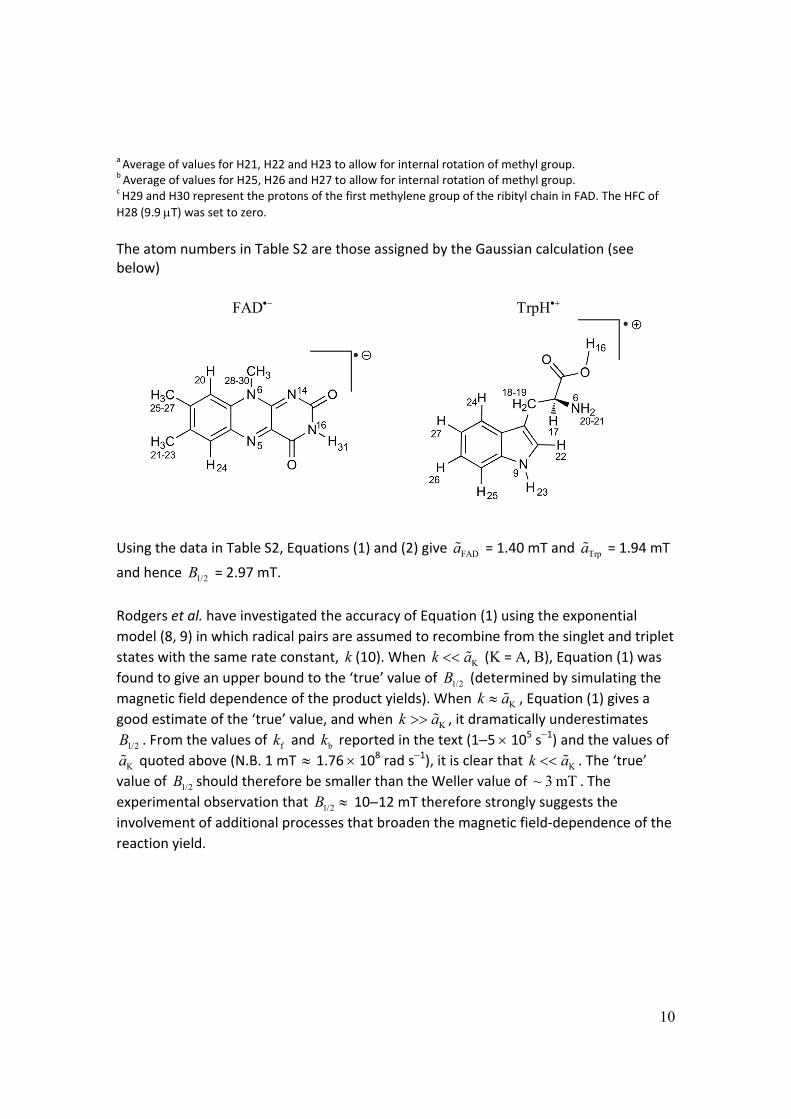
10
a Average of values for H21, H22 and H23 to allow for internal rotation of methyl group. b Average of values for H25, H26 and H27 to allow for internal rotation of methyl group. c H29 and H30 represent the protons of the first methylene group of the ribityl chain in FAD. The HFC of
H28 (9.9 T) was set to zero. The atom numbers in Table S2 are those assigned by the Gaussian calculation (see below)
FAD TrpH
Using the data in Table S2, Equations (1) and (2) give FADa = 1.40 mT and Trpa = 1.94 mT
and hence 1/2B = 2.97 mT.
Rodgers et al. have investigated the accuracy of Equation (1) using the exponential
model (8, 9) in which radical pairs are assumed to recombine from the singlet and triplet
states with the same rate constant, k (10). When Kk a (K = A, B), Equation (1) was found to give an upper bound to the ‘true’ value of 1/2B (determined by simulating the
magnetic field dependence of the product yields). When Kk a , Equation (1) gives a
good estimate of the ‘true’ value, and when Kk a , it dramatically underestimates
1/2B . From the values of fk and bk reported in the text (15 105 s1) and the values of
Ka quoted above (N.B. 1 mT 1.76 108 rad s1), it is clear that Kk a . The ‘true’
value of 1/2B should therefore be smaller than the Weller value of ~ 3 mT . The
experimental observation that 1/2B 1012 mT therefore strongly suggests the
involvement of additional processes that broaden the magnetic field‐dependence of the
reaction yield.
11
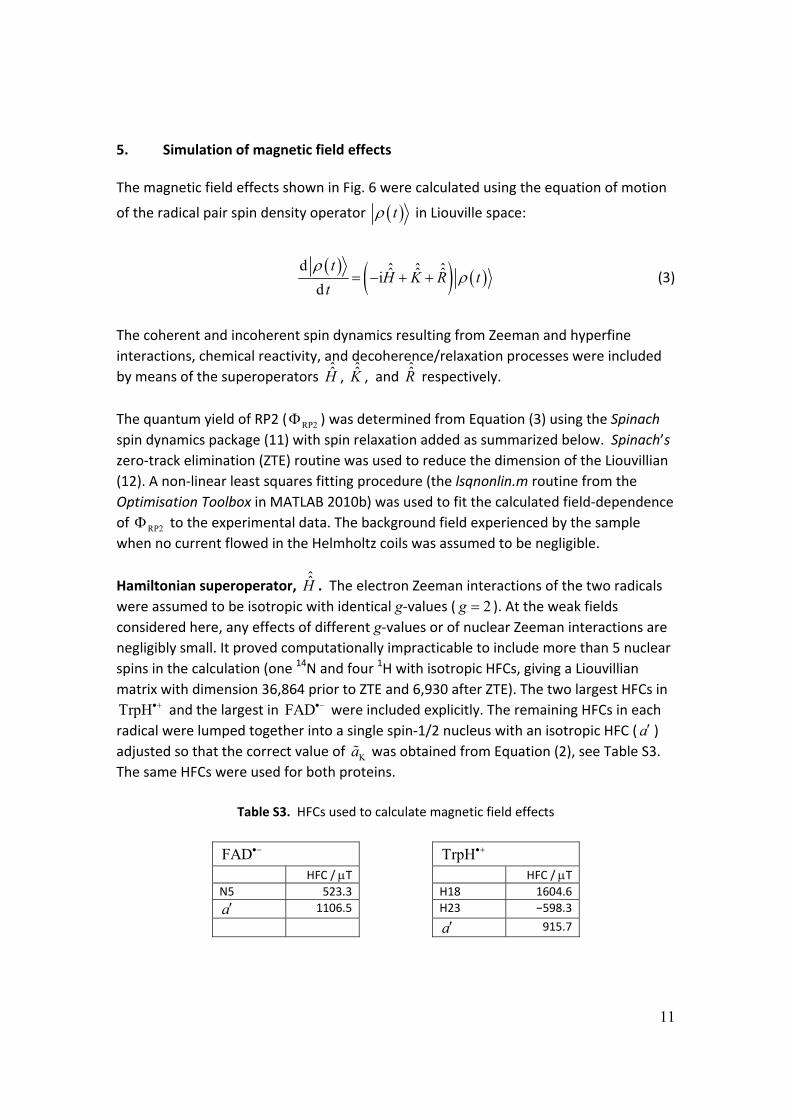
5. Simulation of magnetic field effects The magnetic field effects shown in Fig. 6 were calculated using the equation of motion
of the radical pair spin density operator t in Liouville space:
d ˆ ˆ ˆˆ ˆ ˆid
tH K R t
t
(3)
The coherent and incoherent spin dynamics resulting from Zeeman and hyperfine
interactions, chemical reactivity, and decoherence/relaxation processes were included
by means of the superoperators ˆ̂
H , ˆ̂
K , and ˆ̂R respectively.
The quantum yield of RP2 ( RP2 ) was determined from Equation (3) using the Spinach
spin dynamics package (11) with spin relaxation added as summarized below. Spinach’s
zero‐track elimination (ZTE) routine was used to reduce the dimension of the Liouvillian
(12). A non‐linear least squares fitting procedure (the lsqnonlin.m routine from the
Optimisation Toolbox in MATLAB 2010b) was used to fit the calculated field‐dependence
of RP2 to the experimental data. The background field experienced by the sample
when no current flowed in the Helmholtz coils was assumed to be negligible.
Hamiltonian superoperator, ˆ̂
H . The electron Zeeman interactions of the two radicals
were assumed to be isotropic with identical g‐values ( 2g ). At the weak fields
considered here, any effects of different g‐values or of nuclear Zeeman interactions are
negligibly small. It proved computationally impracticable to include more than 5 nuclear
spins in the calculation (one 14N and four 1H with isotropic HFCs, giving a Liouvillian
matrix with dimension 36,864 prior to ZTE and 6,930 after ZTE). The two largest HFCs in
TrpH and the largest in FAD were included explicitly. The remaining HFCs in each
radical were lumped together into a single spin‐1/2 nucleus with an isotropic HFC ( a ) adjusted so that the correct value of Ka was obtained from Equation (2), see Table S3.
The same HFCs were used for both proteins.
Table S3. HFCs used to calculate magnetic field effects
FAD TrpH HFC / T HFC / T N5 523.3 H18 1604.6
a 1106.5 H23 −598.3
a 915.7
12
Kinetic superoperator, ˆ̂
K . The calculation was based on the reaction scheme in Fig. 4,
ignoring the slow reactions that return RP2 to the ground state and assuming that RP1 is
formed instantaneously at 0t in a pure singlet state. Spin‐selective electronhole recombination of the singlet radical pair (rate constant bk ) and spin‐independent
formation of RP2 from RP1 (rate constant fk ) were included by means of Haberkorn
Relaxation superoperator, ˆ̂R . Initial simulations were performed to investigate the
magnetic responses of the [FAD TrpH ] radical pair in the absence of relaxation
processes. fk was fixed at 5 12.5 10 s (as described in the text), bk was varied to cover
the range b f0.5 / 6k k , and the quantum yield of RP2, RP2( )B , was determined for
magnetic fields, B , in the range 0 50 mTB . The percentage low and high field
effects (LFE and HFE) were calculated as
RP2 RP2
RP2
max[ ( )] (0)100%
(0)
B
and RP2 RP2
RP2
(0) (50 mT)100%
(0)
(4)
respectively, with the results shown in Fig. S9.
Fig. S9. Dependence on b f/k k of the calculated percentage LFE (black) and HFE (red) magnetic field
effects for [FAD TrpH ] with
5 1f 2.5 10 sk , STD 0k .
13
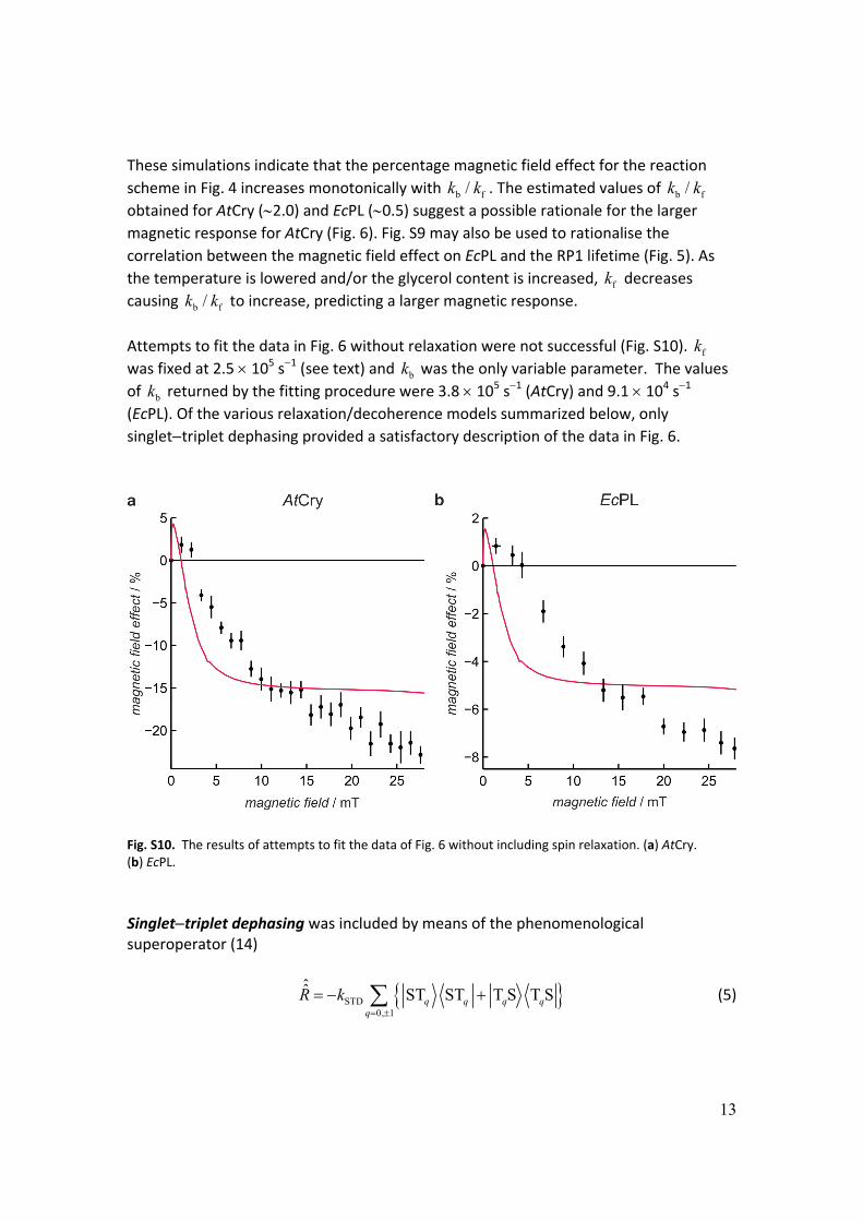
These simulations indicate that the percentage magnetic field effect for the reaction
scheme in Fig. 4 increases monotonically with b f/k k . The estimated values of b f/k k
obtained for AtCry (2.0) and EcPL (0.5) suggest a possible rationale for the larger magnetic response for AtCry (Fig. 6). Fig. S9 may also be used to rationalise the
correlation between the magnetic field effect on EcPL and the RP1 lifetime (Fig. 5). As
the temperature is lowered and/or the glycerol content is increased, fk decreases
causing b f/k k to increase, predicting a larger magnetic response.
Attempts to fit the data in Fig. 6 without relaxation were not successful (Fig. S10). fk
was fixed at 2.5 105 s1 (see text) and bk was the only variable parameter. The values
of bk returned by the fitting procedure were 3.8 105 s1 (AtCry) and 9.1 104 s1 (EcPL). Of the various relaxation/decoherence models summarized below, only
singlettriplet dephasing provided a satisfactory description of the data in Fig. 6.
Fig. S10. The results of attempts to fit the data of Fig. 6 without including spin relaxation. (a) AtCry. (b) EcPL.
Singlettriplet dephasing was included by means of the phenomenological superoperator (14)
STD0, 1
ˆ̂ST ST T S T Sq q q q
q
R k
(5)
14
The results are described in the text. Agreements with the data (Fig. 6) of comparable
quality were obtained with the three rate constants bk , fk , and STDk scaled identically.
Fixing the value of fk as described in the text allowed a unique fit.
Modulation of anisotropic HFCs. Spin relaxation arising from modulation of anisotropic HFCs by isotropic rotational diffusion was included by means of Redfield theory (15, 16).
The optimization variables were bk , fk and the rotational correlation time, c .
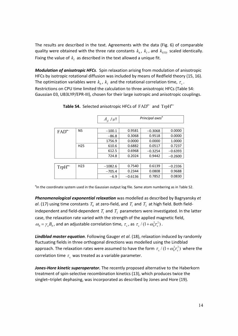
Restrictions on CPU time limited the calculation to three anisotropic HFCs (Table S4: Gaussian 03, UB3LYP/EPR‐III), chosen for their large isotropic and anisotropic couplings.
Table S4. Selected anisotropic HFCs of FAD and TrpH
aIn the coordinate system used in the Gaussian output log file. Same atom numbering as in Table S2.
Phenomenological exponential relaxation was modelled as described by Bagryansky et
al. (17) using time constants 0T at zero‐field, and 1T and 2T at high field. Both field‐
independent and field‐dependent 1T and 2T parameters were investigated. In the latter
case, the relaxation rate varied with the strength of the applied magnetic field,
0 e 0B , and an adjustable correlation time, c , as 2 2c 0 c/ (1 ) .
Lindblad master equation. Following Gauger et al. (18), relaxation induced by randomly fluctuating fields in three orthogonal directions was modelled using the Lindblad
approach. The relaxation rates were assumed to have the form 2 2c 0 c/ (1 ) where the
correlation time c was treated as a variable parameter.
Jones‐Hore kinetic superoperator. The recently proposed alternative to the Haberkorn treatment of spin‐selective recombination kinetics (13), which produces twice the
singlettriplet dephasing, was incorporated as described by Jones and Hore (19).
15
Averaging of anisotropic hyperfine interactions. Rotational diffusion in the viscous solutions used to obtain the data in Fig. 6 may not be sufficiently fast to average completely the anisotropic HFCs. Simulations were therefore performed without added relaxation, with spherical averaging over uniform distributions of radical pair orientations, using the HFC tensors in Table S4. Various combinations of the above approaches were also explored. References 1. Henbest KB, et al. (2008) Magnetic‐field effect on the photoactivation reaction of
Escherichia coli DNA photolyase. Proc. Natl. Acad. Sci. USA 105:14395‐14399. 2. Weber S, et al. (2010) Origin of light‐induced spin‐correlated radical pairs in
cryptochrome. J. Phys. Chem. B 114:14745‐14754. 3. Biskup T, et al. (2011) Time‐resolved EPR identifies unexpected electron transfer
in cryptochrome. Angew. Chem. Int. Ed.:in press. 4. Miura T, Maeda K, Arai T (2003) Effect of coulomb interaction on the dynamics of
the radical pair in the system of flavin mononucleotide and hen egg‐white lysozyme (HEWL) studied by a magnetic field effect. J. Phys. Chem. B 107:6474‐6478.
5. Hore PJ, Kaptein R (1983) Proton nuclear magnetic‐resonance assignments and surface accessibility of tryptophan residues in lysozyme using photochemically induced dynamic nuclear‐polarization spectroscopy. Biochemistry 22:1906‐1911.
6. Weller A, Nolting F, Staerk H (1983) A quantitative interpretation of the magnetic‐field effect on hyperfine‐coupling‐induced triplet formation from radical ion‐pairs. Chem. Phys. Lett. 96:24‐27.
7. Schulten K, Bittl R (1986) Probing the dynamics of a polymer with paramagnetic end groups by magnetic‐fields. J. Chem. Phys. 84:5155‐5161.
8. Brocklehurst B (1976) Spin correlation in geminate recombination of radical ions in hydrocarbons. 1. Theory of magnetic‐field effect. J. Chem. Soc. Faraday Trans. II 72:1869‐1884.
10. Rodgers CT, Norman SA, Henbest KB, Timmel CR, Hore PJ (2007) Determination of radical re‐encounter probability distributions from magnetic field effects on reaction yields. J. Am. Chem. Soc. 129:6746‐6755.
11. Hogben HJ, Krzystyniak M, Charnock GTP, Hore PJ, Kuprov I (2011) Spinach ‐ a software library for simulation of spin dynamics in large spin systems. J. Magn. Reson. 208:179‐194.
12. Kuprov I (2008) Polynomially scaling spin dynamics II: Further state‐space compression using Krylov subspace techniques and zero track elimination. J. Magn. Reson. 195:45‐51.
16
13. Haberkorn R (1976) Density matrix description of spin‐selective radical pair reactions. Mol. Phys. 32:1491‐1493.
14. Shushin AI (1991) The effect of the spin exchange interaction on SNP and RYDMR spectra of geminate radical pairs. Chem. Phys. Lett. 181:274‐278.
15. Goldman M (2001) Formal theory of spin‐lattice relaxation. J. Magn. Reson. 149:160‐187.
16. Kuprov I (2011) Diagonalization‐free implementation of spin relaxation theory for large spin systems. J. Magn. Reson. 209:31‐38.
17. Bagryansky VA, Borovkov VI, Molin YN (2007) Quantum beats in radical pairs. Russ. Chem. Rev. 76:493‐506.
18. Gauger EM, Rieper E, Morton JJL, Benjamin SC, Vedral V (2011) Sustained quantum coherence and entanglement in the avian compass. Phys. Rev. Lett. 106:040503.
19. Jones JA, Hore PJ (2010) Spin‐selective reactions of radical pairs act as quantum measurements Chem. Phys. Lett. 488:90‐93.