Ulysses Lins Instituto de Microbiologia Prof. Paulo de Góes, Universidade Federal do Rio de Janeiro Magnetotactic bacteria: biology, diversity and biotechnology Instituto de Microbiologia UFRJ BIOTA – FAPESP 2014
Transcript
Ulysses Lins
Instituto de Microbiologia Prof. Paulo de Góes, Universidade Federal do Rio de Janeiro
Magnetotactic bacteria: biology,
diversity and biotechnology
Instituto de Microbiologia UFRJ
BIOTA – FAPESP 2014
Magnetotaxis
Ability of some bacteria to orientate and navigate along magnetic
field lines
Magnetotactic bacteria orientate along filed lines because of
magnetosomes
Magnetosomes
Cocci
Rod-shaped Multicellular
Spirilla
They are microaerophilic or anaerobes
Morphology, phylogeny and metabolism
Function of magnetotaxis
To help bacteria to orientate and navigate along the
geomagnetic filed lines and towards more favorable regions
Magnetic crystal
Magnetite (Fe3O4) Greigite (Fe3S4)
Magnetosomes
Membrane
Magnetic crystal
Magnetosomes
•Magnetosomes consist of a magnetic crystal enveloped
by a lipid-protein membrane
Magnetosomes and biological control • The membrane defines a region of the cytoplasm where bacteria control growth, size
and purity of the crystals through the proteins.
• The iron atoms are transported from the environment to the magnetosome vesicle
where the crystal is nucleated and grows.
Jogler & Schüler, 2007
Magnetosomes and controlled morphology
Prismatic
Bullet-shaped
Cubo-octahedra
Inorganic magnetite
Morphology produced by bacteria
• Bacteria control the morphology of the magnetosomes.
• They facilitate the growth of specific faces to modify the morphology.
•If we understand the control mechanism we can try to mimic it.
Magnetosomes and morphology
Inorganic magnetite
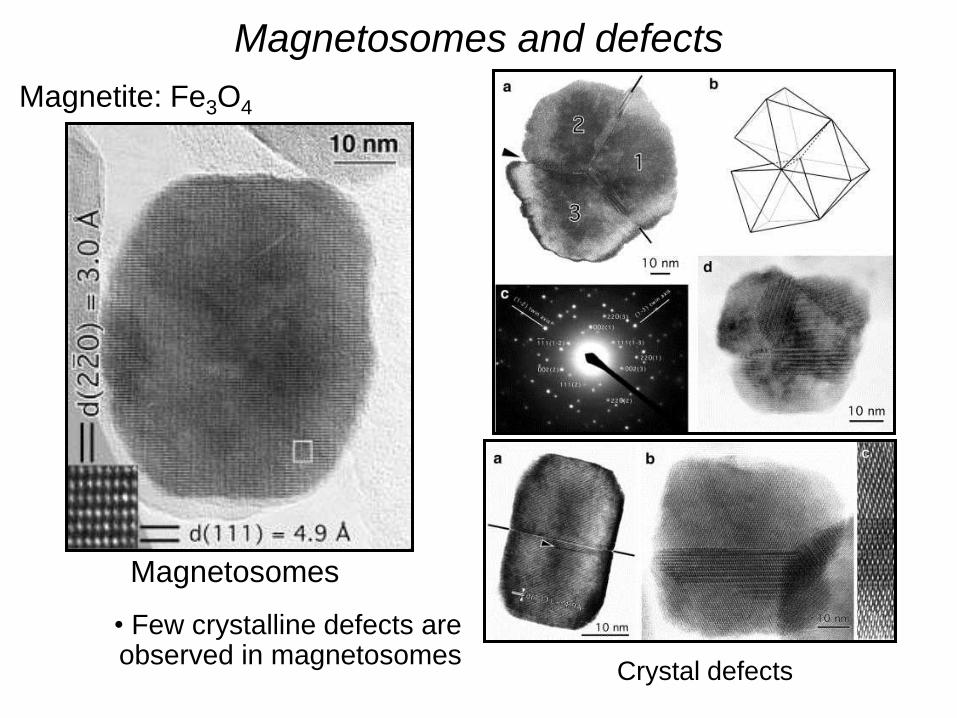
Magnetosomes and defects
Magnetite: Fe3O4
Crystal defects
Magnetosomes
• Few crystalline defects are observed in magnetosomes
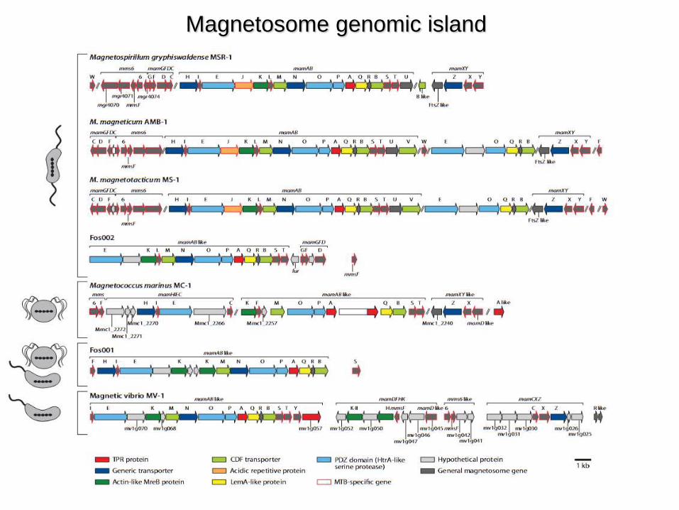
Magnetosome genomic island
A membrana do magnetosomo é uma
invaginação da membrana plasmática
50 nm
From: Komeili et al., Science 2006
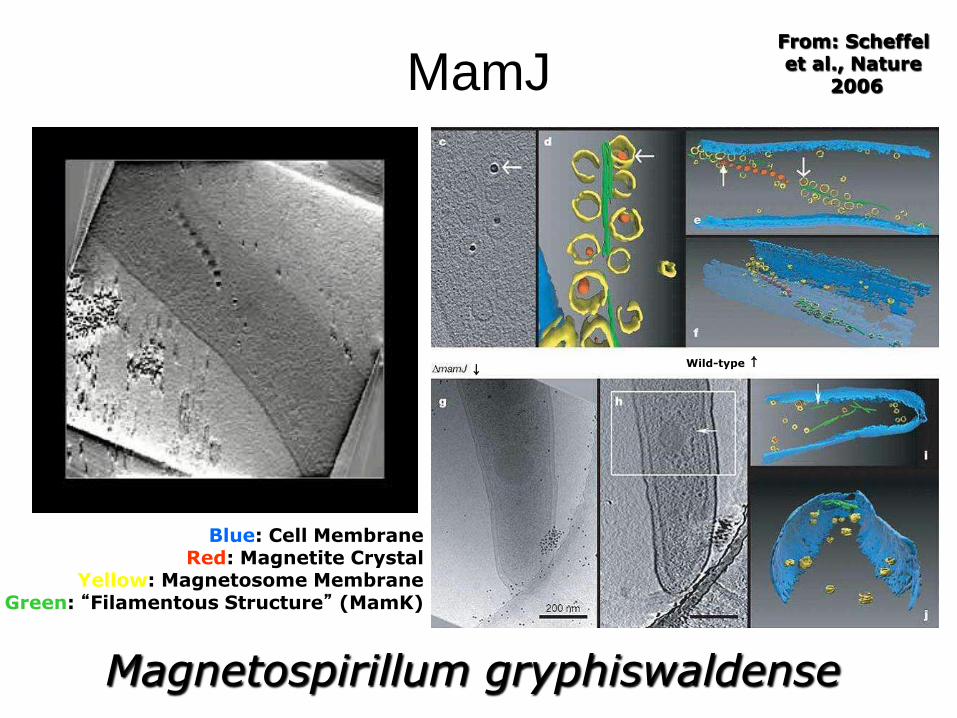
Magnetospirillum magneticum
Protein function in the control of the
magnetosome chain
MamK – actin-like protein similar to MreB
It is essential for the formation of the chain
From: Komeili et al., Science 2006
Magnetospirillum magneticum
MamJ – acidic protein associated with the filamentous structure (MamK)
16S rRNA gene sequencing for Candidatus Magnetoglobus
multicellularis
Magnetic vibrio
Magnetic coccus
Candidatus Magnetoglobus multicellularis
MMP 1991
Candidatus Magnetobacterium bavaricum
FISH
Abreu et al., 2007
Candidatus Magnetoglobus multicellularis
•Each cell contains 60-100
greigite (Fe3S4) magnetosome
Abreu et al., 2008
What to do with all these data?
• Understand ecology to....
• Develop a culture medium
Genome of Candidatus Magnetoglobus
multicellularis
Genome coverage 23x
Length (bp) 12.459.246
G+C content (%) 37.27
Coding density (%) 77%
Average of ORF length (bp) 914
Number of Contigs 3.706
Total number of ORFs 10.639
Number of known protein ORFs 2.792
Number of partial ORFs 26
Number of truncated ORFs 140
Number of hypothetical ORFs 7520
rRNA 3
rRNA 16s 1
rRNA 23s 1
rRNA 5s 1
tRNA 46
KEGG matches 71%
InterPro matches 77%
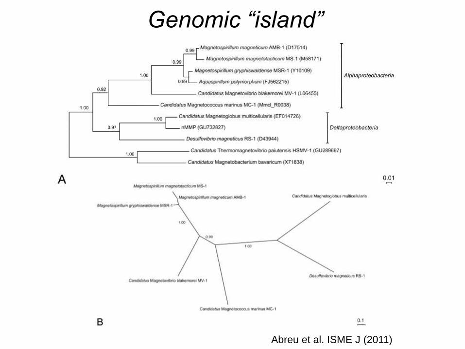
Genomic “island”
Abreu et al. ISME J (2011)
Genomic “island”
Figure: ORFs organization in two contigs containing putative MAI related genes in Candidatus Magnetoglobus multicellularis and MAI homologous regions in cultivated MTB. MTB genes with homologous sequences in Ca. M. multicelullaris are represented in black and genes without homology in white.
Magnetosome biomineralization
Ca. M. multicellularis
BW-1 g
BW-1 m
Desulfovibrio
M. marinus
Ca. M. bavaricum
Ca. M. blakemorei
M. magnetotacticum
MamABEMP1Q1g concatenated genes
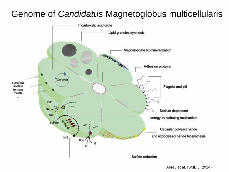
Genome of Candidatus Magnetoglobus multicellularis
Abreu et at. ISME J (2014)
Abreu et al. Microsc. Microanal 2013
Cell adhesion molecules and magnetosomes distribution
Cell adhesion molecules and phylogeny
Abreu et at. ISME J (2014)
Cultivation
Enrichment cultures Abreu et at. ISME J (2014)
Culture of ‘Candidatus Magnetoglobus multicellularis’
Ecological role
Abreu et at. ISME J (2014)
Lefevre et al., Science 2011
Genes for magnetosomes in strain BW-1
Lefevre et al., Science 2011
Desulfamplus magnetomortis strain BW-1
Produce greigite and magnetite
Lefevre et al., Science 2011
Desulfamplus magnetomortis cepa BW-1
Produce greigite and magnetite
• Optimization and cultivation
of magnetotactic bacteria
• Diversity and evolution of
magnetotaxis
• Applications of
magnetosomes
Monophyletic origin of magnetotaxis
Lefevre et al., Environm Microbiol 2013a
Lefevre et al., Environm Microbiol 2013b
Genes for greigite magnetosomes in Deltaproteobacteria
Magnetosome Associated Deltaproteobacteria
Lefevre et al., Environm Microbiol 2013b
Feo transport genes
Magnetosome Associated Deltaproteobacteria
Lefevre et al., Environm Microbiol 2013b
Cytoskeleton
Magnetosome Associated Deltaproteobacteria
Magnetospirillum
• Optimization and cultivation
of magnetotactic bacteria
• Diversity and evolution of
magnetotaxis
• Applications of
magnetosomes
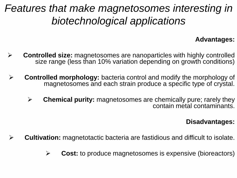
Features that make magnetosomes interesting in
biotechnological applications
Advantages:
Controlled size: magnetosomes are nanoparticles with highly controlled size range (less than 10% variation depending on growth conditions)
Controlled morphology: bacteria control and modify the morphology of magnetosomes and each strain produce a specific type of crystal.
Chemical purity: magnetosomes are chemically pure; rarely they contain metal contaminants.
Disadvantages:
Cultivation: magnetotactic bacteria are fastidious and difficult to isolate.
Cost: to produce magnetosomes is expensive (bioreactors)
Magnetovibrio blakemorei strain MV-1
Marine vibrio with a polar flagellum and a single chain of elongated magnetite magnetosomes
Cryo-electron tomography of Magnetovibrio blakemorei MV-1
Abreu et al. (2013) J. Struc. Biol.
Magnetovibrio blakemorei strain MV-1 Optimization of growth
Silva et al. (2013) Appl Environm. Microbiol.
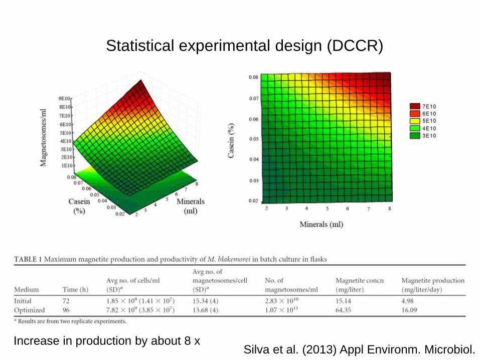
Statistical experimental design (DCCR)
Silva et al. (2013) Appl Environm. Microbiol. Increase in production by about 8 x
Cost analysis
Silva et al. (2013) Appl Environm. Microbiol.
Increase in cost by about 2 x
Net increase in production/cost by about 4 x
Growth in Bioreactor
Silva et al. (2013) Appl Environm. Microbiol.
Immobilization of lipases
Lipase from Rhizomucor miehei
Post-docs Ana Carolina de Araújo
Fernanda de Ávila Abreu
PhD students Fernando Pereira de Almeida
Karen Tavares Silva Viviana Morillo
Master students
Jefferson Bomfim Silva Cypriano Pedro Ernesto Lopes Leão
Undergraduate students Marina Chao Campello
Mayara Gil de Castro Santos
Technical support Danielle da Silva Moreira
Tarcísio Nascimento Correa
Laboratory
Dr. Alioscka Souza – NIH, USA
Dr. Richard Leapman – NIH, USA
Dra. Ana Tereza Vasconcelos – LNCC
Dr. Luiz Gonzaga - LNCC
Dr. Bechara Kachar, NIDCD, NIH, USA
Dr. Christofer Lefrévre - Saint-Paul, Durance Cedex, France
Dr. David Pignol - Saint-Paul, Durance Cedex, France
Dra. Denise Guimarães Freire – Instituto de Química – UFRJ
Dr. Dennis Bazylinski - University of Nevada, USA
Dr. Marcos Farina – Instituto Ciências Biomédicas – UFRJ
Dra. Melissa Limoeiro Estrada Gutarra – Escola de Química – UFRJ
Dr. Richard Frankel, CalPol State Univ., San Luis Obispo, USA
Collaborators
Financial support
4th International Meeting on Magnetotactic Bacteria , 2014