Magnolol Inhibits Human Glioblastoma Cell Proliferationthrough Upregulation of p21/Cip1
LI-CHING CHEN,† YU-CHI LIU, ) YU-CHIH LIANG,‡ YUAN-SOON HO,‡ AND
WEN-SEN LEE*,†,§,^
†Graduate Institute of Medical Sciences, ‡Department of Biomedical Technology, §Department ofPhysiology, School of Medicine, Taipei Medical University, Taipei, Taiwan, )Department of
Ophthalmology, Taipei Veterans General Hospital, Taipei, Taiwan, and ^Cancer Research Center,Taipei Medical University and Hospital, Taipei, Taiwan
Previously, we demonstrated that magnolol isolated from the bark of Magnolia officinalis has
anticancer activity in colon, hepatoma, and leukemia cell lines. In this study, we show that magnolol
concentration dependently (0-40 μM) decreased the cell number in a cultured human glioblastoma
cancer cell line (U373) and arrested the cells at the G0/G1 phase of the cell cycle. Magnolol
treatment decreased the protein levels of cyclins A and D1 and increased p21/Cip1, but not cyclins
B and D3, cyclin-dependent kinase (CDK)2, CDK4, CDC25C, Weel, p27/Kip1, and p53. The CDK2-
p21/Cip1 complex was increased, and the CDK2 kinase activity was decreased in the magnolol-
treated U373. Pretreatment of U373 with p21/Cip1 specific antisense oligodeoxynucleotide pre-
vented the magnolol-induced increase of p21/Cip1 protein levels and the decrease of DNA
synthesis. Magnolol at a concentration of 100 μM induced DNA fragmentation in U373. Our findings
suggest the potential applications of magnolol in the treatment of human brain cancers.
Malignant glioblastomas are the most common primary braintumor. Despite remarkable advances in surgical techniques andtreatment options, including chemotherapy and radiotherapy,the prognosis of malignant glioblastomas is still poor because oftheir diffuse invasion of the brain parenchyma, which is difficultfor total surgical resection (1). In fact, the 5 year survival rate forthe most malignant type, glioblastoma multiforme, is only 2%.Curative therapy without damaging the affected brain parench-yma is very difficult due to the infiltrative growth patterns and thenatural resistance to chemotherapy of malignant glioblasto-mas (2). The standard chemotherapy regimen for patients witholigodendroglioma involves combined treatment with procarba-zine, lomustine, and vincristine (3, 4). Each of these drugs wasdiscovered several decades ago with central neurotoxic sideeffects (5-7). Obviously, there is an urgent need for new ther-apeutic strategies in treating malignant glioblastomas.
Magnolol (MAG), a hydroxylated biphenyl compound alsoknown as Hou p’u among Chinese herbalists, has been reportedto have anticancer activity (8, 9). Our previous studies demon-strated that MAG suppressed proliferation of cultured humancolon and liver cancer cells by inhibiting DNA synthesis andactivating apoptosis (10). Similar results were also reported byYang et al. showing that MAG inhibited proliferation of humanlung squamous carcinoma CH27 cells at lower concentrations(10-40 μM) and induced apoptosis at higher concentrations
(80-100 μM) (11). Recent studies demonstrated thatMAGcouldinhibit hepatic metastasis from an experimental liver and spleenmetastasismodel using L5178Y-ML25 lymphomaand reduce theincidence of pulmonary metastases in the lung metastasis modelusing B16-BL6 melanoma (12). These findings suggest the poten-tial for MAG in cancer chemoprevention. Although the anti-tumor effects of MAG have been investigated in various cancercell lines, little is known about its effect on the malignant gliomasof the central nervous system.
Here, we showed that MAG inhibited the proliferation ofglioblastoma cells in vitro. These experimental findings reportedbelow highlight the molecular mechanisms of MAG-inducedantigrowth activity in U373 cells.
MATERIALS AND METHODS
Cell Lines and Cell Culture. The human astrocytoma cell line, U373,was obtained from the American Type Culture Collection (Rockville,MD) and cultured in 75 cm2 culture flasks (Greiner, Frickenhausen,Germany). Cells were grown in minimum essential medium with Eagle’ssalts (MEM; Invitrogen Corp.) containing glutamine (2 mM), 1% non-essential amino acids (NEAA), sodium pyruvate (1 mM), and 10% fetalbovine serum (FBS) in a humidified incubator (37 �C and 5% CO2).
Reagents. MAGwas purchased from Pharmaceutical Industry, Tech-nology and Development Center (Taiwan). Antibodies against cyclin A,cyclin D1, cyclin-dependent kinase 4 (CDK4), CDC25C, Wee1, P21, P53,and CHK1 were purchased from Santa Cruz Biotechnology, Inc. (SantaCruz, CA). Antibodies against p27 and CDK2 were purchased from BDBioscience Pharmingen (San Diego, CA). Antibodies against cyclin B andcyclinD3were purchased fromTransductionLaboratories (San Jose,CA).Anti-p-CHK1 antibody was purchased from Cell Signaling Technology,
*To whom correspondence should be addressed. Tel:þ886-2-2739-1775. Fax: þ886-2-2739-1775. E-mail: [email protected].
7332 J. Agric. Food Chem., Vol. 57, No. 16, 2009 Chen et al.
Inc. (Danvers, MA). Anti-G3PDH antibody was purchased from JacksonImmunoResearch Laboratories Inc. (West Grove, PA).
Determination of Cell Growth Curve. Human U373 (1�105) cellswere plated in 35 mm Petri dishes and grown inMEM supplemented with10%FBS.Dimethyl sulfoxide (DMSO), 0.05% (v/v), without (control) orwith MAG was added. The incubation medium was changed daily untilcell count. The cell number was determined by using the MTT assay.
Trypan Blue Exclusion Assay. As previously described (13), cellviability was estimated by the trypan blue exclusion assay at 3 or 6 daysafterMAG (0-40 μM) treatment. Briefly, humanU373 (1�105) cells wereplated in the 6 cmPetri dishes and grown inMEMsupplementedwith 10%FBS. The culture medium and MAG were changed daily until the trypanblue exclusion assay.
[3H]Thymidine Incorporation. As previously described (10), U373cells at a density of 1�104 cells/cm3 were plated in 24 well plates andgrown in growth medium (MEM plus 10% FBS). After the cells hadgrown to 70% confluence, they were rendered quiescent by incubationin MEM containing 0.04% FBS for 24 h. The U373 cells were thentreated with MAG in MEM supplemented with 10% FBS for theindicated time points. During the last 3 h of the incubation,[3H]thymidine was added at 1 μCi/mL (1 μCi = 37 kBq). Incorporated[3H]thymidine was extracted in 0.2 N NaOH and measured in a liquidscintillation counter.
Flow Cytometric Analysis (14). At 24 h after the cells were plated,the medium was removed. Cells were washed three times with PBS andthen incubated with medium containing 0.04% FBS for 24 h. After serumstarvation, the cells were then challenged with medium containing 10%FBS. The stages of cell cycle were determined by flow cytometic analysis.
Cells were incubated with 50 μg/mL propidium iodide (Sigma), and DNAcontent was measured using a FACSCAN laser flow cytometric analysissystem (Becton Dickenson, San Jose, CA). Ten thousand cells wereanalyzed for each sample.
Western Blot Analysis. Western blot analysis was performed asdescribed previously (15). Briefly, cell lysates were prepared, electrotrans-ferred, immunoblotted with antibodies, and then visualized by incubatingwith the colorigenic substrates [nitro blue tetrazolium (NBT) and5-bromo-4-chloro-3-indolyl-phosphate (BCIP)] (Kirkegaard & PerryLaboratories, MD). The levels of G3PDH protein were detected and usedas the control for equal protein loading.
Immunoprecipitation (IP) and Kinase Activity Assay. As pre-viously described (16), theMAG-treated cells were lysed in Rb lysis buffer[137 mM NaCl, 20 mM Tris, pH 7.9, 10 mM NaF, 5 mM EDTA, 1 mMEGTA, 10% (v/v) glycerol, 1% triton X-100, 1 mM sodium orthovana-date, 1 mM sodium pyrophosphate, 100 μM β-glycerophosphate, 1 mMPMSF, 10 μg/mL aprotinin, and 10 μg/mL leupeptin] and immunopreci-pitated with anti-CDK2 or anti-CDK4 antibody (2 μg/mL). The proteincomplexes in beads were washed twice with Rb lysis buffer and then oncewithRb kinase assay buffer. The levels of phosphorylated ofRb (for pRb),histone H1 (for CDK2), and glutathione s-transferase-Rb fusion protein(for CDK4) were measured by incubating the beads with 40 μL of hot Rbkinase solution [0.25 μL (2 μg) of Rb-GST fusion protein (Santa CruzBiotechnology), 0.5 μL of (γ-32P) ATP (Amersham), 0.5 μL of 0.1 mMATP, and 38.75 μL of Rb kinase buffer] at 37 �C for 30 min and thenstopped by boiling the samples in SDS sample buffer for 5 min. Thesamples were analyzed by 12% SDS-PAGE, and the gel was then driedand subjected to autoradiography.
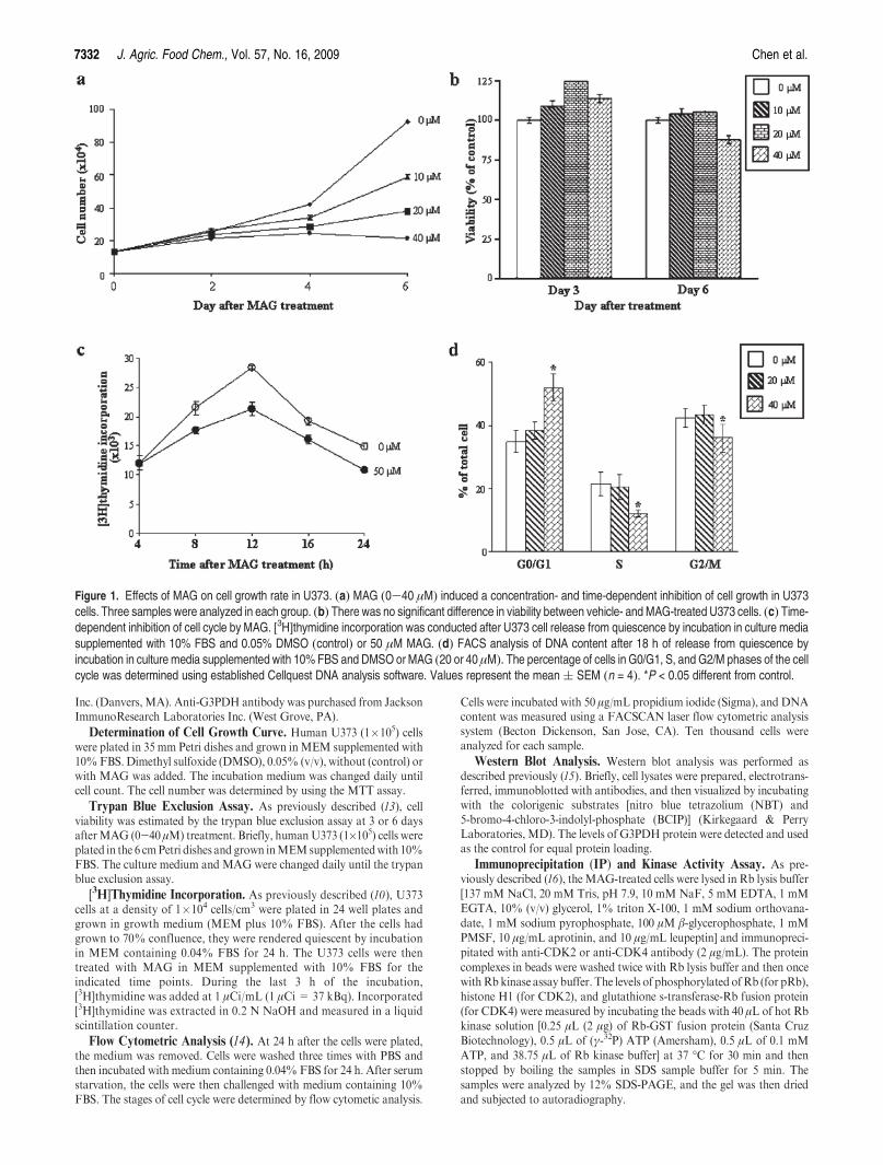
Figure 1. Effects of MAG on cell growth rate in U373. (a) MAG (0-40 μM) induced a concentration- and time-dependent inhibition of cell growth in U373cells. Three samples were analyzed in each group. (b) There was no significant difference in viability between vehicle- and MAG-treated U373 cells. (c) Time-dependent inhibition of cell cycle by MAG. [3H]thymidine incorporation was conducted after U373 cell release from quiescence by incubation in culture mediasupplemented with 10% FBS and 0.05% DMSO (control) or 50 μM MAG. (d) FACS analysis of DNA content after 18 h of release from quiescence byincubation in culture media supplemented with 10%FBS and DMSO orMAG (20 or 40 μM). The percentage of cells in G0/G1, S, and G2/M phases of the cellcycle was determined using established Cellquest DNA analysis software. Values represent the mean( SEM (n = 4). *P < 0.05 different from control.
DNA Fragmentation Analysis. The DNA was isolated from U373cells treatedwith orwithoutMAGand incubated overnight with nuclease-free proteinase K at 55 �C in 10 mM Tris, pH 7.5, 150 mM EDTA, and0.5%sodiumdodecyl sulfate (SDS). TheDNAwas thenused for detectionof DNA laddering as described previously (15).
Antisense Oligonucleotide. The p21/Cip1-specific antisense (50-TC-CCCAGCCGGTTCTGACAT-30) and sense (50-ACCTGTGCTCCGA-CACGTCT-30) phosphothioates were designed as previously des-cribed (17), synthesized, and purified using high-performance liquidchromatography by Genset. Antisense or sense p21/Cip1 was added toU373 at a final concentration of 20 nM at 16 h before the cell waschallengedwith 10%FBS and 50 μMMAG treatment for additional 18 h.
Reverse Transcriptase-Polymerase Chain Reaction (RT-PCR)Analysis. The RT-PCR assays for p21/Cip1 gene expression wereperformed as described previously (18, 19). The U373 cells were treatedwith MAG for 24 h. Total RNAs were isolated from U373 cells usingTrizol reagent according to the manufacturer’s protocol (Life Technolo-gies, Inc.). The cDNA was amplified from 1 μg of total RNA using aSuperScript one-step RT-PCR with platinum Taq system (Life Technol-ogies, Inc.). PCR was conducted for 30 cycles in thermal controller.Primers used for amplification were as follows: p21/Cip1 50-ATTAG-CAGCGGAACAAGGA GTCAGACAT-3 and 50-CTGTGAAAGA-CACAGAACAGTACAGGGT-30; G3PDH 50-GACCCCTTCATTG-ACCTCAAC-30 and 50-GATGACCTTGCCCACAGCCTT-30. Eachamplification cycle consisted of 0.5 min at 94 �C for denaturation, 0.5 minat 55 �C for primer annealing, and 1min at 72 �C for extension. In all of theamplification procedures, reverse transcriptase-free control assays con-sisting of the amplification mixture, the RNA sample, and distilled waterin place of reverse transcriptase were used to check for possible contam-ination of the RNA samples with DNA. After PCR amplification, the
fragments were stained with ethidium bromide and analyzed by agarosegel electrophoresis.
Statistics. All data were expressed as the mean value( standard errorof the mean (SEM). Three to four samples were analyzed in eachexperiment. Comparisons were subjected to one-way analysis of variancefollowed by Fisher’s least significant difference test. Significance wasaccepted at P < 0.05.
RESULTS AND DISCUSSION
MAG Induces G0/G1 Cell Cycle Arrest in Human U373 Cells.
To study the effect of MAG on cell growth rate, U373 cells werecultured for 6 days without or with MAG (0-40 μM), and thecells were then harvested and counted. As shown in Figure 1a,MAG (0-40 μM) induced a concentration-dependent inhibitionof growth in human U373 cells. To confirm that the MAG-induced decrease in cell number of U373 cells was not due to celldeath, we conducted a viability assay by treating the cells withU373 cells for 3 or 6 days at the concentrations (5-40μM)used inthe study of cell growth inhibition. Trypan blue exclusion assaysindicated that there was no significant difference in cell viabilitybetween control and MAG-treated U373 cells (Figure 1b), sug-gesting that there was an inhibitory effect of MAG on themechanisms for cell division in the subcultured U373 cells. Tofurther examine the actions of MAG on cell cycle regulatorymechanisms, the U373 cells were switched to media with 0.04%FBS for 24 h to render them quiescent and to synchronize theirmitotic activities at theG0/G1 phase. The synchronized cells werethen returned to culture media supplemented with 10% FBS,
Figure 2. Effect of MAG on the levels of cell cycle regulatory proteins. Proteins were extracted from the cultured U373 cells at 24 h after MAG treatment andprobed with proper dilutions of specific antibodies. (a) MAG (0-40 μM) concentration dependently decreased the protein levels of cyclins A and D1 but notcyclin B, cyclin D3, CDK2, CDK4, CDC25C, and Wee1. Results from a representative experiment are shown. Values shown in parentheses represent therelative protein abundance of cyclin A and D1. (b) MAG increased the protein levels of p21/Cip1 but not p27/Kip1 and p53. The membrane was probed withanti-G3PHD antibody to verify equivalent protein loading. (c) MAG concentration dependently increased the levels of p21/Cip1 mRNA. Top panel:A representative measurement of the levels of p21/Cip1 mRNA. Bottom panel: Quantitative results of p21/Cip1 mRNA, which were adjusted withcorresponding G3PDH mRNA level and expressed as a percentage of control. Values represent the means( SEM (n = 3). *P < 0.05 different from control.
7334 J. Agric. Food Chem., Vol. 57, No. 16, 2009 Chen et al.
and at various times thereafter, they were harvested for[3H]thymidine incorporation and flow cytometric analyses. Asillustrated in Figure 1c, MAG (50 μM) treatment caused areduction of thymidine incorporation into U373 cells during theS phase of the cell cycle. Figure 1d shows that MAG induced asignificant accumulation of cells in the G0/G1 phase of the cellcycle, suggesting that the observed growth inhibitory effect ofMAG in theU373 was due to an arrest of DNA replication at theG0/G1 of the cell cycle.
Alterations in Cell Cycle Activity. To investigate the molecularmechanisms underlying MAG-induced G0/G1 arrest, the cellswere switched to media with 0.04% FBS for 24 h to render themquiescent at the G0/G1 phase. The cells were then returned toculture media supplemented with 10% FBS and 0.05% DMSOwithout or with MAG (20-40 μM), and at 24 h thereafter, theywere harvested for protein extraction and Western blot analysesto examine the effects of MAG on the expression of cell cycleregulatory proteins. It has been generally believed that progres-sion of cell cycle activity is regulated by coordinated successiveactivationof certainCDKs,whichoccurs late in theG1phase andis instrumental in the transition from the G1 to the S phase(20,21). This CDK activation is in turnmodulated by associationwith a number of regulatory subunits called cyclins and with agroup of CDK-inhibitory proteins designated CKIs (22). Cyclinshave been identified as cyclins A, D1, D3, and E, whereas the
most common CDKs are CDK2 and CDK4. As shown inFigure 2a, the protein levels of cyclins A and D1, but not cyclinD3, CDK2, and CDK4, were decreased in the MAG-treatedU373 cells. Because the CDK activity can be controlled by agroup of CKIs, we examined the protein levels of p21/Cip1 andp27/Kip1, two known CKIs, in the MAG-treated U373 cells.As shown inFigure 2b, the protein levels of p21/Cip1, but not p27/Kip1, were increased in theMAG-treatedU373 cells as comparedwith the DMSO-treated cells (control). The protein levels of p53inU373were not affected by theMAG treatment (Figure 2b).Wealso examined the change of the mRNA level of p21/Cip1(Figure 2c) in the MAG-treated U373 cells. As illustrated inFigure 2c, MAG (20-40 μM) concentration dependently in-creased the levels of p21/Cip1 mRNA.
Previous studies have demonstrated that p21/Cip1 arrests thecell cycle at the G0/G1 phase through binding and inactivatingthe CDKs (20, 21, 23). In accord with the established notion thatp21/Cip1 is a CDK inhibitor, we found that the formation of thep21/Cip1-CDK2 complexwas increased and the assayableCDK2activitywas decreased in theMAG-treatedU373 cells (Figure 3a).In contrast, the formation of p21/Cip1-CDK4 complex and theCDK4 activity were not significantly changed (if there is anychange). We further examined the levels of pRb protein, whichcan bind to the E2F-1 transcription factor and prevent it frominteracting with the cells transcription machinery. As shown in
Figure 3. Involvement of p21/Cip1 in the MAG-induced decrease of thymidine incorporation in U373 cells. (a) MAG increased the formation of CDK2-p21/Cip1, but not CDK4-p21/Cip1, complex in U373 cells. The CDK2, but not CDK4, activity was decreased byMAG. Results from a representative experiment areshown. Values shown in parentheses represent the formation of the CDK2-p21/Cip1 complex. The CDK2 and CDK4 kinase activities were determined asdescribed in theMaterials and Methods. Results from a representative experiment are shown. Values (means(SEM; n = 3) shown in parentheses representthe kinase activity of CDK2.WB,Western blot analysis. (b)MAG decreased the levels of phosporylated Rb (pRb) but not phosphorylated CHK1 (pCHK1) andtotal CHK1 protein. Results from a representative experiment are shown. The membrane was probed with anti-G3PHD antibody to verify equivalent proteinloading. (c) Antisense p21/Cip1 oligonucleotide (AS)was added to U373 cells at a final concentration of 20 nMat 16 h before the cell was challengedwith 10%FBS and 50 μMMAG for an additional 21 h. Pretreatment of U373 cells with AS p21/Cip1, but not S p21/Cip1, prevented the MAG-induced increase of p21/Cip1 protein level (top panel: Results from a representative experiment are shown. Values shown in parentheses represent the means( SEM of p21/Cip1protein levels from three experiments) and decrease of [3H]thymidine incorporation (bottom panel: Values represent the means( SEM; n = 4). *p < 0.05 vscontrol. #p < 0.05 vs 50 μM MAG-treated. AS, antisense oligonucleotide; and S, sense oligonucleotide.
Figure 3b, MAG treatment decreased the levels of phosphory-lated Rb protein.
It has been indicated that the G2/M cell cycle checkpoint istightly regulated by theCdc2/Cyclin B,which is required for entryinto mitosis (24). In the MAG-treated U373 cells, the level ofcyclin B protein was not changed significantly (Figure 2a). More-over, MAG treatment did not cause any significant change of thelevels of Wee1 and CDC25C protein, two major CDC2 activityregulators, and of CHK1 protein, which is essential for the G2/MDNA damage-induced checkpoint (Figure 3b). Taken together,these data suggest that MAG arrested the U373 cells at the G0/G1, but not G2/M, phase of the cell cycle.
A number of studies have suggested that p21/Cip1 does havetumor suppressor properties in human glioblastoma due to itshigh expression level found in several human brain tumors(22, 25). Replication-deficient adenovirus was utilized as anexpression vector to transfer exogenous p53 and p21/Cip1cDNAs into the U373 glioma cells and demonstrated that over-expression of p21/Cip1 induced a cell cycle arrest but did notinduce apoptosis (26), suggesting that p21/Cip1 might protect thecells from p53-mediated programmed cell death. To furtherdemonstrate that the increased p21/Cip1 expression observed intheMAG-treated U373 correlated withG0/G1 arrest, the experi-ment illustrated in Figure 3c was carried out. As shown inFigure 3c, MAG treatment increased the p21/Cip1 protein levelsand decreased the [3H]thymidine incorporation in U373 cells.However, treatment of the cells with a p21/Cip1 antisense oligo-nucleotide, which blocked the p21/Cip1 induction, prevented theMAG-induced decrease of [3H]thymidine incorporation. In con-trast, a p21/Cip1 sense oligonucleotide, which did not affect theexpression of p21/Cip1, failed to prevent the MAG-induceddecrease of [3H]thymidine incorporation. Accordingly, we con-cluded that MAG induced an increase in p21/Cip1 expression,which in turn inhibited the CDK2 enzyme activities and led to theimpairment of U373 in the transition from the G1 to the S phase.
Previously, we demonstrated thatMAG inhibited proliferationof liver and colon cancer cell lines with p53 wild-type (Hep-G2
and COLO-205) or mutated (Hep-3B and HT-29), suggestingthat p53 is not required for the MAG-induced proliferationinhibition in cancer cell lines (10). Although p21/Cip1 has beenrecognized to be a critical downstream effector of p53 protein,some studies have shown that p21/Cip1 can also be inducedthrough a p53-independent pathway (27). The p53 gene in U373cell line has a point mutation at codon 273 (CGTf CAT). Thismutation leads to replacement of an arginine by a histidine atcodon 273 on exon5. In the present study, a highbasal level of p53protein was detected in the untreatedU373 cells.MAG at a rangeof concentrations (0-100 μM) did not increase the level of p53protein (Figures 2b and 4c), suggesting that p53 might not beinvolved in the MAG-induced proliferation inhibition and theoccurrence of apoptosis in U373. Instead, our data indicate thatthe induction of p21/Cip1 expression might contribute to theMAG-induced proliferation inhibition in U373. Moreover,MAG increased p21/Cip1 at both mRNA and protein levels(Figure 2b,c). A similar result was demonstrated by a previousstudy from another group showing that hypoxia increased thelevel of p21 protein, but not p53 protein, in U373 cells (28).
Effect of MAG on DNA Fragmentation and CKI Protein Levels
of U373Cells.We further examined theMAGeffect onU373 cellswhen the concentration of MAG was increased. As illustratedin Figure 4a, the thymidine incorporation was dramaticallydecreased in the U373 cells treated withMAG at a concentrationof 100 μM. Figure 4b shows that the DNA laddering effect wasobserved in U373 cells treated with MAG at a concentration of100 μM for 48 h. We also examined the CKI protein levels inU373 treated with higher concentrations of MAG. As illustratedin Figure 4c, MAG at a range of concentrations (0-100 μM)concentration dependently increased the levels of p21. However,the levels of p27 protein were not changed significantly at lowerconcentrations of MAG (0-80 μM) but were dramaticallyincreased when the MAG concentration reached 100 μM. Incontrast, the levels of p53 protein were not changed significantlyby MAG treatment even at a concentration of 100 μM. It seemsthat the induction of p27/Kip1 protein was correlated with the
Figure 4. MAG inducesDNA fragmentation in U373. (a)MAGat a concentration of 100μM induced a dramatic decrease of [3H]thymidine incorporation in U373cells. Values represent the means( SEM (n = 4). *p < 0.05 vs control. (b) A typical DNA ladder pattern associated with apoptosis was observed in U373 cellstreated with 100μMMAG for 48 h. (c)MAG (0-100μM) concentration dependently increased the protein levels of p21/Cip1 but not p53. The levels of p27/Kip1protein in U373 were not changed significantly by MAG at lower concentrations (0-80 μM) but were increased dramatically at a higher concentration (100 μM).
7336 J. Agric. Food Chem., Vol. 57, No. 16, 2009 Chen et al.
occurrence of apoptosis inU373 cells.Recently, it has been shownthat overexpression of p27/Kip1 induces apoptosis and inhibitstumor formation in vitro and in vitro (29).Moreover, cotransduc-tion of p27/Kip1 strongly augments Fas ligand- and caspase-8-mediated apoptosis in U-373 (30). The dramatically increasedp27/Kip protein levels were observed in the U373 treated with ahigher concentration (100 μM) of MAG but not lower concen-trations (e40 μM), suggesting that the p27/Kip1 proteinmight beinvolved in the MAG-induced apoptosis but not cell growtharrest.
In conclusion, multifocal glioblastomas constitute an increas-ingly diagnosed subgroup of glioblastoma multiforme, the mostmalignant primary brain tumor in adults. Because glioblastomamultiforme is resistant to all currently used treatments, to searchfor new therapeutic strategies is urgent. The current experimentalmodels used in discovering new antiglioma therapies includeeither murine gliomas (31), which have biologic characteristicsthat are very different from those of human gliomas, or humanglioma cell lines grafted subcutaneously onto the flanks ofimmunodeficient mice (14, 16, 32). However, in such models,diffuse invasiveness into the brain parenchyma, the hallmark ofthe malignant human glioma phenotype, is no longer valid.Migrating glioma cells are known to escape from chemothera-peutic agent-induced apoptosis and thus the compounds thatrestore the apoptosis-resistant cells not only delay the invasion ofcancer cells into brain parenchyma but also enhance the sensitiv-ity of these slowly migrating cells (33). The pharmacokinetics ofMAG in rats demonstrated thatMAGcan cross the blood-brainbarrier (BBB), and there is no significant difference amongvarious regions of the brain after 10 min of MAG (5 mg/kg,i.v.) administration; themean concentration ofMAG in the brainwas approximately 4-fold as high as that in the plasma (34).Although the functional BBB has been identified for a long time,the complexity and relevance of the blood-brain-tumor barrierhas been recognized recently as an important factor that limitsthe effective treatment of central nervous system neoplasms.A previous report has demonstrated that MAG could relievethe heatstroke-induced cerebral ischemic injury through inhibit-ing of free radical formation (35). This finding demonstratedagain thatMAG could cross the BBB and act in the brain. In thisstudy, we observed that MAG suppresses proliferation of cul-tured human U373 cells by inhibiting DNA synthesis andactivating apoptosis. Our study provides the basis of molecularmechanisms for MAG in brain cancer treatment. Taken togetherwith previous studies, our present study suggested that use ofMAG in the treatment of glioblastoma showed several advan-tages that included (1) increased the occurrence of apoptosis, (2)induced the cell cycle arrest, and (3) crossed the BBB. Theuniversality of MAG in the inhibition of cancer cell proliferationwould make it a very attractive agent for cancer chemotherapy.
(1) Surawicz, T. S.; Davis, F.; Freels, S.; Laws, E. R., Jr.; Menck, H. R.Brain tumor survival: Results from the National Cancer Data Base.J. Neuro-Oncol. 1998, 40 (2), 151–160.
(2) Cairncross, J. G.;Ueki, K.; Zlatescu,M.C.; Lisle, D.K.; Finkelstein,D. M.; Hammond, R. R.; Silver, J. S.; Stark, P. C.; Macdonald, D.
R.; Ino, Y.; Ramsay, D. A.; Louis, D. N. Specific genetic predictorsof chemotherapeutic response and survival in patients with anaplas-tic oligodendrogliomas. J. Natl. Cancer Inst. 1998, 90 (19), 1473–1479.
(3) Heiss, W. D.; Turnheim, M.; Mamoli, B. Combination chemother-apy of malignant glioma. Effect of postoperative treatment withCCNU, vincristine, amethopterine and procarbazine. Eur. J. Cancer1978, 14 (11), 1191–1202.
(4) Kesari, S.; Schiff, D.; Drappatz, J.; LaFrankie, D.; Doherty, L.;Macklin, E. A.; Muzikansky, A.; Santagata, S.; Ligon, K. L.;Norden, A. D.; Ciampa, A.; Bradshaw, J.; Levy, B.; Radakovic,G.; Ramakrishna, N.; Black, P. M.; Wen, P. Y. Phase II study ofprotracted daily temozolomide for low-grade gliomas in adults.Clin.Cancer Res. 2009, 15 (1), 330–337.
(5) Postma, T. J.; van Groeningen, C. J.; Witjes, R. J.; Weerts, J. G.;Kralendonk, J. H.; Heimans, J. J. Neurotoxicity of combinationchemotherapy with procarbazine, CCNU and vincristine (PCV) forrecurrent glioma. J. Neuro-Oncol. 1998, 38 (1), 69–75.
(6) Neyns, B.; Sadones, J.; Chaskis, C.; De Ridder, M.; Keyaerts, M.;In’T Veld, P.; Michotte, A. The role of chemotherapy in thetreatment of low-grade glioma;A review of the literature. ActaNeurol. Belg. 2005, 105 (3), 137–143.
(7) Perilongo, G. Considerations on the role of chemotherapy andmodern radiotherapy in the treatment of childhood low gradeglioma. J. Neuro-Oncol. 2005, 75 (3), 301–307.
(8) Fujita, M.; Itokawa, H.; Sashida, Y. Studies on the components ofMagnolia obovata Thunb. II. On the components of the methanolextract of the bark. Yakugaku Zasshi 1973, 93 (4), 422–428.
(9) Lee, D. H.; Szczepanski, M. J.; Lee, Y. J. Magnolol inducesapoptosis via inhibiting the EGFR/PI3K/Akt signaling pathway inhuman prostate cancer cells. J. Cell Biochem. 2009, 106 (6), 1113–1122.
(10) Lin, S. Y.; Liu, J. D.; Chang, H. C.; Yeh, S. D.; Lin, C. H.; Lee,W. S.Magnolol suppresses proliferation of cultured human colon and livercancer cells by inhibiting DNA synthesis and activating apoptosis.J. Cell Biochem. 2002, 84 (3), 532–544.
(11) Yang, S. E.; Hsieh,M. T.; Tsai, T. H.; Hsu, S. L. Effector mechanismof magnolol-induced apoptosis in human lung squamous carcinomaCH27 cells. Br. J. Pharmacol. 2003, 138 (1), 193–201.
(12) Ikeda, K.; Sakai, Y.; Nagase, H. Inhibitory effect of magnolol ontumour metastasis in mice. Phytother. Res. 2003, 17 (8), 933–937.
(13) Ho, Y. S.; Lee,H.M.;Mou, T. C.;Wang, Y. J.; Lin, J. K. Suppressionof nitric oxide-induced apoptosis by N-acetyl-L-cysteine throughmodulation of glutathione, bcl-2, and bax protein levels. Mol.Carcinog. 1997, 19 (2), 101–113.
(14) Arai, T.; Joki, T.; Akiyama,M.; Agawa,M.;Mori, Y.; Yoshioka, H.;Abe, T. Novel drug delivery system using thermoreversible gelationpolymer for malignant glioma. J. Neuro-Oncol. 2006, 77 (1), 9–15.
(15) Lee, W. S.; Chen, R. J.; Wang, Y. J.; Tseng, H.; Jeng, J. H.; Lin, S.Y.; Liang, Y. C.; Chen, C. H.; Lin, C. H.; Lin, J. K.; Ho, P. Y.; Chu,J. S.; Ho,W. L.; Chen, L. C.; Ho, Y. S. In vitro and in vivo studies ofthe anticancer action of terbinafine in human cancer cell lines:G0/G1 p53-associated cell cycle arrest. Int. J. Cancer 2003, 106 (1),125–137.
(16) Kim, K. J.; Wang, L.; Su, Y. C.; Gillespie, G. Y.; Salhotra, A.; Lal,B.; Laterra, J. Systemic anti-hepatocyte growth factor monoclonalantibody therapy induces the regression of intracranial gliomaxenografts. Clin. Cancer Res. 2006, 12 (4), 1292–1298.
(17) Chen, R. J.; Lee, W. S.; Liang, Y. C.; Lin, J. K.; Wang, Y. J.; Lin, C.H.; Hsieh, J. Y.; Chaing, C. C.; Ho, Y. S. Ketoconazole induces G0/G1 arrest in human colorectal and hepatocellular carcinoma celllines. Toxicol. Appl. Pharmacol. 2000, 169 (2), 132–141.
(18) Aoki, S.; Kong, D.; Suna, H.; Sowa, Y.; Sakai, T.; Setiawan, A.;Kobayashi, M. Aaptamine, a spongean alkaloid, activates p21promoter in a p53-independent manner. Biochem. Biophys. Res.Commun. 2006, 342 (1), 101–106.
(19) Suzui, M.; Masuda, M.; Lim, J. T.; Albanese, C.; Pestell, R. G.;Weinstein, I. B. Growth inhibition of human hepatoma cells byacyclic retinoid is associated with induction of p21(CIP1) andinhibition of expression of cyclin D1. Cancer Res. 2002, 62 (14),3997–4006.
(20) Yu, D. H.; Macdonald, J.; Josephs, S.; Liu, Q.; Nguy, V.; Tor, Y.;Wong-Staal, F.; Li, Q. X.MDDD, a 4,9-diazapyrenium derivative, isselectively toxic to glioma cells by inducing growth arrest at G0/G1independently of p53. Invest. New Drugs 2006, 24 (6), 489–498.
(21) Komata, T.; Kanzawa, T.; Takeuchi, H.; Germano, I. M.; Schreiber,M.; Kondo, Y.; Kondo, S. Antitumour effect of cyclin-dependentkinase inhibitors (p16(INK4A), p18(INK4C), p19(INK4D), p21-(WAF1/CIP1) and p27(KIP1)) on malignant glioma cells. Br. J.Cancer 2003, 88 (8), 1277–1280.
(22) Krex, D.; Mohr, B.; Appelt, H.; Schackert, H. K.; Schackert, G.Genetic analysis of a multifocal glioblastoma multiforme: A suitabletool to gain new aspects in glioma development. Neurosurgery 2003,53 (6), 1377-1384; discussion 1384.
(23) Harmalkar, M. N.; Shirsat, N. V. Staurosporine-induced growthinhibition of glioma cells is accompanied by altered expression ofcyclins, CDKs and CDK inhibitors. Neurochem. Res. 2006, 31,685–692.
(24) Huang, T. S.; Shu, C. H.; Chao, Y.; Chen, S. N.; Chen, L. L.Activation of MAD 2 checkprotein and persistence of cyclin B1/CDC 2 activity associate with paclitaxel-induced apoptosis in humannasopharyngeal carcinoma cells. Apoptosis 2000, 5 (3), 235–241.
(25) Begnami, M. D.; Rushing, E. J.; Evangelista, R.; Santi, M.; Quezado,M. Evaluation of RB gene and cyclin-dependent kinase inhibitors P21and P27 in pleomorphic xantoastrocytoma. Int. J. Surg. Pathol. 2006,14 (2), 113–118.
(26) Gomez-Manzano, C.; Fueyo, J.; Kyritsis, A. P.; McDonnell, T. J.;Steck, P. A.; Levin, V. A.; Yung, W. K. Characterization of p53 andp21 functional interactions in glioma cells en route to apoptosis.J. Natl. Cancer Inst. 1997, 89 (14), 1036–1044.
(27) Michiell, P.; Chedid, M.; Lin, D.; Pierce, J. H.;Mercer, W. E.; Givol,D. Induction ofWAF1/CIP1 by a p53-independent pathway.CancerRes. 1994, 54 (13), 3391–3395.
(28) Bell, H. S.; Whittle, I. R.; Bader, S. A.;Wharton, S. B. Discovery of aperinecrotic 60 kDa MDM2 isoform within glioma spheroids andglioblastoma biopsy material. Neuropathol. Appl. Neurobiol. 2005,31 (2), 191–202.
(29) Wang, X.; Gorospe, M.; Huang, Y.; Holbrook, N. J. p27Kip1overexpression causes apoptotic death of mammalian cells. Onco-gene 1997, 15 (24), 2991–2997.
(30) Shinoura, N.; Furitsu, T.; Asai, A.; Kirino, T.; Hamada, H. Co-transduction of p27Kip1 strongly augments Fas ligand- and caspase-8-mediated apoptosis in U-373MG glioma cells. Anticancer Res.2001, 21 (5), 3261–3268.
(31) Ahmadi, R.; Urig, S.; Hartmann, M.; Helmke, B. M.; Koncarevic,S.; Allenberger, B.; Kienhoefer, C.; Neher, M.; Steiner, H. H.;Unterberg, A.; Herold-Mende, C.; Becker, K. Antiglioma activityof 2,20:60,20 0-terpyridineplatinum(II) complexes in a rat model;Effects on cellular redox metabolism. Free Radical Biol. Med.2006, 40 (5), 763–778.
(32) Koller, E.; Propp, S.; Zhang, H.; Zhao, C.; Xiao, X.; Chang, M.;Hirsch, S. A.; Shepard, P. J.; Koo, S.; Murphy, C.; Glazer, R. I.;Dean, N. M. Use of a chemically modified antisense oligonucleotidelibrary to identify and validate Eg5 (kinesin-like 1) as a target forantineoplastic drug development. Cancer Res. 2006, 66 (4), 2059–2066.
(33) Manero, F.; Gautier, F.; Gallenne, T.; Cauquil, N.; Gree, D.;Cartron, P. F.; Geneste, O.; Gree, R.; Vallette, F. M.; Juin, P. Thesmall organic compound HA14-1 prevents Bcl-2 interaction withBax to sensitize malignant glioma cells to induction of cell death.Cancer Res. 2006, 66 (5), 2757–2764.
(34) Tsai, T. H.; Chou, C. J.; Chen, C. F. Pharmacokinetics and braindistribution of magnolol in the rat after intravenous bolus injection.J. Pharm. Pharmacol. 1996, 48 (1), 57–59.
(35) Chang, C. P.; Hsu, Y. C.; Lin, M. T. Magnolol protects againstcerebral ischaemic injury of rat heatstroke. Clin. Exp. Pharmacol.Physiol. 2003, 30 (5-6), 387–392.
Received May 4, 2009. Revised manuscript received July 20, 2009.
Accepted July 21, 2009. This research was supported by a grant from the
National Science Council of the Republic of China (NSC96-2320-