64
Major Causes of Morbidity and Mortality in SLE

Major Causes of Morbidity and Mortality in SLE

Patient EM
• EM, an 18-year-old Black female presents to the Emergency Department with acute onset of confusion and hallucinations
• Her parents report that she has been complaining of “fatigue” for the last 6 months and has lost 5 pounds. An antinuclear antibody test (ANA) ordered by her primary physician last week was strongly positive
• Abnormal physical findings include a low-grade fever of 100°F and several small oral ulcers
• Labs: strongly positive anti-dsDNA antibody, borderline anti-Sm and normal levels of C3 and C4
• EM develops disorganized thinking, lack of orientation, agitation, and delusions (consistent with acute confusional state). She is admitted to the hospital

Patient EM
• Addressing EM’s symptoms involves:
- Exclusion of secondary causes of confusion (infectious, metabolic, drug-induced, vascular)
- Imaging and lumbar puncture to help to determine cause
- Measurement of antiphospholipid antibodies which can, in some patients, alter the management plan
• Patient is treated with steroids and hydroxychloroquine
• Management with steroids/immunosuppression is complicated by an episode of Escherichia coli (E. coli) pyelonephritis in the hospital
• When an 18-year-old is seen at the emergency department, the physician usually addresses the acute problem and the teenager goes back to normal life; however, EM’s journey is different1
1. Sacks JJ, Helmick CG, Langmaid G, Sniezek JE. MMWR Morb Mortal Wkly Rep. 2002;51(17);371-374.
Introduction
• Major causes of morbidity in systemic lupus erythematosus (SLE) - Neuropsychiatric
- Renal
- Cardiovascular
- Other (bone related, malignancy, infections, hematologic)
• Mortality in SLE

Neuropsychiatric Lupus (NPSLE)
• 19 case definitions of neuropsychiatric manifestations
• Most commonly: - Cognitive dysfunction
- Headache
- Psychiatric disorders (anxiety, psychosis*, depression)
- Seizures*
- Stroke (may be associated with antiphospholipid antibodies)
- Peripheral neuropathies
*Part of the classification criteria for SLE.
Bertsias GK, Boumpas DT. Nat Rev Rheumatol. 2010;6:358-367.
Epidemiology of Neuropsychiatric Lupus
• Cumulative incidence is ~30%–40%
• In early disease - ~20% of patients already have atrophy on brain MRI
- ~10% have focal lesions
• Not all neuropsychiatric manifestations in lupus patients are directly attributable to lupus. Two-thirds may be due to other causes
Sanna G, Bertolaccini ML, Cuadrado MJ, et al. J Rheumatol. 2003;30(5):985-992; Muscal E, Brey R. Neurol Clin. 2010;28(1):61-73.

Correct Attribution of Neuropsychiatric Events is Critical—Consider Other Causes
- Infections
- Medications and toxins
Prescription medications
Illicit drugs
Dietary supplements
Alternative and complementary therapies
- Cardiovascular
Hypertension
Ischemic stroke
Hemorrhagic stroke
- Other
• Non-SLE disease-related etiologies of neuropsychiatric symptoms that should be considered

Radiologic Findings (CT and MRI)
• Atrophy (most common) • Vascular abnormalities
• Demyelination • Inflammation
Image courtesy of the Rheumatology Image Bank
A. The initial MRI scan with fluid-attenuated inversion-recovery reveals multiple high-intensity areas in the deep white matter.
Katsumata Y, Kawaguchi Y, Yamanaka H. J Rheumatol. 2011;38;2689.
B. 4 months later, there is significant cerebral atrophy, characterized by a loss of brain volume, along with multiple high-intensity areas.
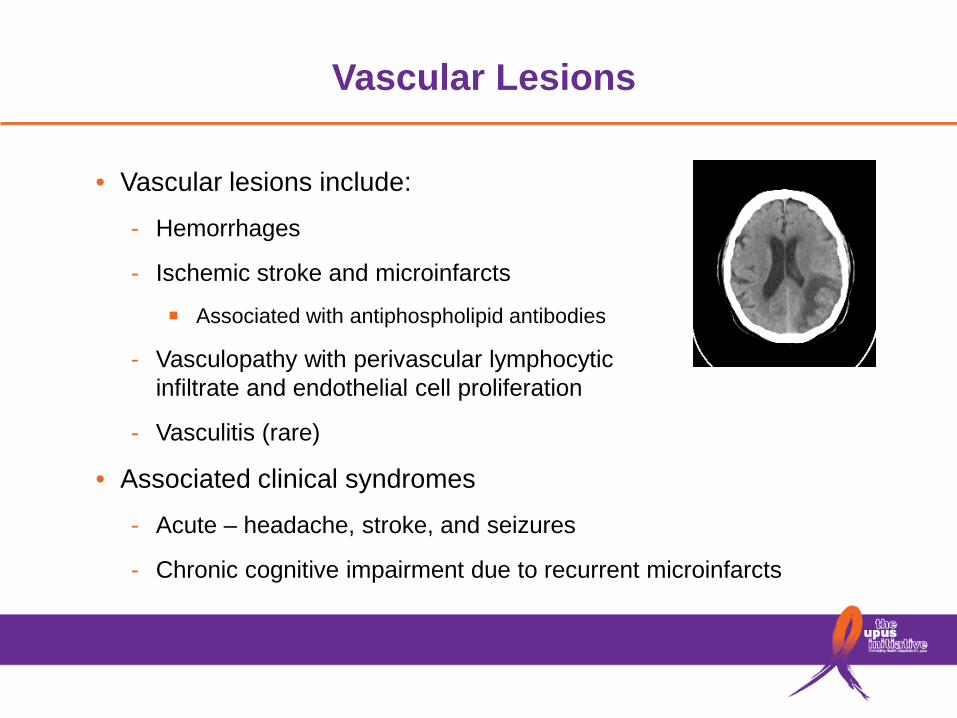
Vascular Lesions
• Vascular lesions include:
- Hemorrhages
- Ischemic stroke and microinfarcts
■ Associated with antiphospholipid antibodies
- Vasculopathy with perivascular lymphocytic infiltrate and endothelial cell proliferation
- Vasculitis (rare)
• Associated clinical syndromes
- Acute – headache, stroke, and seizures
- Chronic cognitive impairment due to recurrent microinfarcts

Injury to the Brain Parenchyma
• Diffuse central nervous system syndromes often wax and wane - Acute confusional state, psychosis, and mood disorders
- Suggests temporary neuronal dysfunction
• Cerebrospinal fluid analysis may indicate local inflammation - Increased lymphocytes and proinflammatory cytokines
- Elevated protein levels and autoantibodies
• Specific autoantibodies have been associated with neuronal toxicity
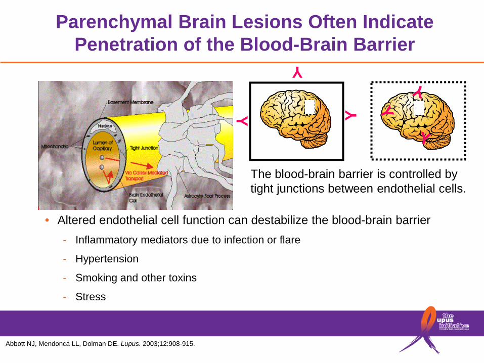
Parenchymal Brain Lesions Often Indicate Penetration of the Blood-Brain Barrier
• Altered endothelial cell function can destabilize the blood-brain barrier - Inflammatory mediators due to infection or flare
- Hypertension
- Smoking and other toxins
- Stress Y
Y Y
The blood-brain barrier is controlled by tight junctions between endothelial cells.
Abbott NJ, Mendonca LL, Dolman DE. Lupus. 2003;12:908-915.

Cognitive Dysfunction Is Common in Lupus Patients
• Observed in 50%–80% of lupus patients
• Problems with: - Attention
- Concentration
- Memory
- Word-finding
• Attribution of cognitive dysfunction to lupus is difficult
“I have to squeeze my brain really hard to get a thought out!”
Benedict RH, Shucard JL, Zivadinov R, Shucard DW. Neuropsychol Rev. 2008;18(2):149-166.

Many Causes of Cognitive Dysfunction in Lupus
Cognitive Dysfunction
Strokes
Neuronal toxicity (antibodies, cytokines)
Vasculitis
Antiphospholipid syndrome
Medications
Depression/anxiety
Metabolic dysfunction
Thrombotic thrombocytopenic
purpura
Sleep disorders

Peripheral Nervous System Involvement
• Neuropathies (motor or autonomic) or myasthenia gravis-like syndrome
• SLE/myasthenia overlap is associated with anti-acetylcholine receptor antibodies
• Circulating antibodies and inflammatory mediators have direct access to peripheral nerves

Transverse Myelitis
• Transverse myelitis is a rare, late manifestation of SLE but can occur at presentation
• Most patients, but not all, demonstrate a sensory level with spastic weakness and sphincter dysfunction
Simeon-Aznar CP, Tolosa-Vilella C, Cuenca-Lugue R, Jordana-Comajuncosa R, Ordi-Ros J, Bosch-Gil JA. Rheumatology. 1992;31(8):555-558. Espinosa G, Mendizábal A, Minguez S, et al. Semin Arthritis Rheum. 2010;39(4):246-256. Birnbaum J, Petr M, Thomson R, Izbudak I, Kerr D. Arthritis Rheum. 2009;60(11):3378-3387.

Transverse Myelitis
(a) Sagittal T2-weighted, gadolinium-enhanced MRI of the spine of a 38-year-old female SLE patient showing cord enlargement and hyperintense signal in the C2, C4–C6, and C7–T1 spinal cord (arrows), consistent with longitudinal spinal myelitis
(b) Posttreatment MRI of the spine
demonstrates complete resolution of the T2 hyperintense signal
Goh YP, Naidoo P, Ngian GS. Clin Radiol. 2013;68(2):181-191.

Neuropsychiatric Lupus—Identifying the Cause Will Determine Treatment
• NPSLE manifestations may occur during periods of disease quiescence in other organs
• Correct ascertainment and attribution is critical - For example, an ischemic stroke due to long-standing
diabetes and hypertension should not be treated with immunosuppression
• Immunosuppression for inflammatory manifestations
• Traditional drugs for headache, seizures, stroke, and mood disorders
• Stress management and psychotherapy

Conclusions—Neuropsychiatric Lupus
• The most common causes of neuropsychiatric involvement are non-lupus related. Rule-out other causes first
• NPSLE encompasses a broad range of clinical presentations and pathologies
- Vascular lesions can cause both acute focal and chronic diffuse impairment
- Autoantibodies and other proinflammatory molecules that cross the blood-brain barrier may have direct effects on neurons, resulting in altered cellular function or death
- Peripheral nerves are exposed to the circulation
• Correct diagnosis is critically important to ensure that appropriate therapy is used

Patient EM
• Resolution of symptoms and decrease in anti-dsDNA antibodies over 6–8 weeks is followed by steroid taper over the next 6 months. She was maintained on hydroxychloroquine and followed every 3 months but is lost to follow-up after 2 years
• 3 years later, at age 23 she presents with fever and joint pains after returning from a trip to Jamaica. In the last 3 days she has noticed mild swelling of both ankles
• Anti-dsDNA antibodies have significantly increased since her last visit. Both C3 and C4 are decreased below normal
• Urinalysis reveals 300 mg/dL proteinuria and 5 WBC/hpf. Her serum creatinine is normal
Epidemiology of Lupus Nephritis
• Prevalence: 30%–65% in adults and 80% in children
• 10% annual incidence in 1 large cohort
• More frequent and severe in children, Blacks, Hispanics, and males
• Strong predictor of morbidity and mortality
Patel M, Clarke AM, Symmons Y. Arthritis Rheum. 2006;54(9):2963-2969. Hiraki LT, Feldman CH, Liu J. Arthritis Rheum. 2012;64(8):2669-2676. Danchenko N, Satia JA, Anthony MS. Lupus. 2006;15:308-318. Petri M. Lupus. 2005;14(12):970-973.

Nephritis Is Induced by Renal Deposition of Antibodies
Davidson A, Berthier C, Kretzler M. In: Dubois' Lupus Erythematosus and Related Syndromes (8th Ed.). Philadelphia, PA: Saunders; 2013:237-255.
Anatomy of the glomerulus, consisting of a tuft of capillary loops fed by the afferent arteriole. The tuft is held together by the mesangium. The enlarged capillary loop shows the components of the glomerular filtration barrier. The barrier is formed by the glycocalyx, fenestrated endothelial cells (End), glomerular basement membrane (GBM), podocyte foot processes (Pod and FP), and slit diaphragm (SD). The podocyte layer is contiguous with the parietal epithelial layer (PEp), which is surrounded by the Bowman capsule. Immune deposits may be found on either side of the GBM (SubEnd or SubEp) or in the mesangium (Mes).
Anatomy of the glomerulus
Tubular and vascular deposits may also occur.
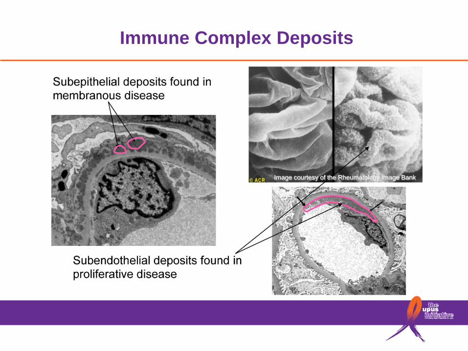
Immune Complex Deposits
Image courtesy of the Rheumatology Image Bank
Clinical Diagnosis of SLE Nephritis
• Increase in proteinuria is most common - Measured by spot protein:creatinine ratio >0.5 or 24-hour
collection >500 mg/24 hours
- The absolute increase in proteinuria that defines a nephritis flare is arbitrary
• Microscopic abnormalities on urinalysis - White cells or red blood cells >5 cells/hpf in the absence of
infection or other causes
- Cellular casts (white cell or red cell)
- White cells and red blood cells are seen more frequently than casts
Hahn BH, McMahon MA, Wilkinson A, et al. Arthritis Care Res. 2012;64(6):797-808.

Lupus Renal Pathology
• Renal biopsy is used routinely to evaluate disease type and severity and to direct management
• All patients with clinical evidence of active lupus nephritis, and previously untreated, should have a kidney biopsy (unless strongly contraindicated)
• Treatment is based on biopsy results - Proliferative disease is treated more aggressively than
mesangial and membranous disease because it progresses more rapidly and is more likely to cause chronic damage

Renal Pathology—International Society of Nephrology Scores*
• Class I – Minimal mesangial glomerulonephritis – deposits but normal light microscopy
http://www.fondazionedamico.org/biopsiarenale_atlas/
*2002 International Society of Nephrology/Renal Pathology Society (ISN/RPS). Images I-III: courtesy of www.fondazionedamico.org/biopsiarenale_atlas.
• Class II – Mesangial proliferative glomerulonephritis
• Class III – Focal glomerulonephritis involving <50% of glomeruli
Renal Pathology—International Society of Nephrology Scores*
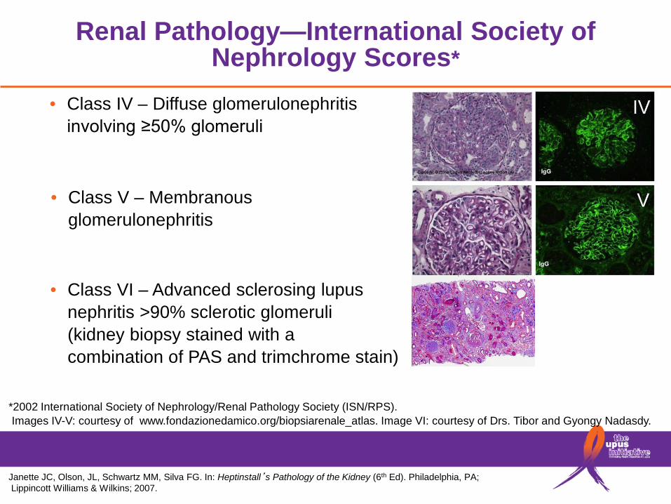
• Class IV – Diffuse glomerulonephritis involving ≥50% glomeruli
*2002 International Society of Nephrology/Renal Pathology Society (ISN/RPS). Images IV-V: courtesy of www.fondazionedamico.org/biopsiarenale_atlas. Image VI: courtesy of Drs. Tibor and Gyongy Nadasdy.
Janette JC, Olson, JL, Schwartz MM, Silva FG. In: Heptinstall’s Pathology of the Kidney (6th Ed). Philadelphia, PA; Lippincott Williams & Wilkins; 2007.
• Class V – Membranous glomerulonephritis
• Class VI – Advanced sclerosing lupus nephritis >90% sclerotic glomeruli (kidney biopsy stained with a combination of PAS and trimchrome stain)
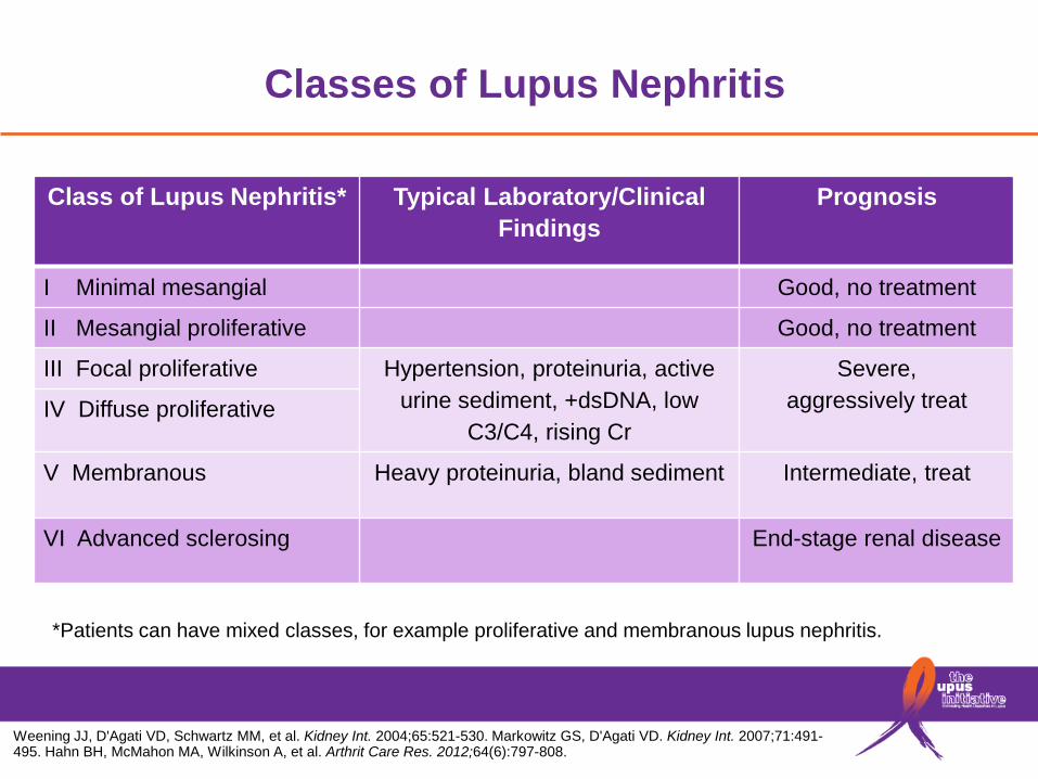
Classes of Lupus Nephritis
Class of Lupus Nephritis*
Typical Laboratory/Clinical Findings
Prognosis
I Minimal mesangial Good, no treatment
II Mesangial proliferative Good, no treatment
III Focal proliferative Hypertension, proteinuria, active urine sediment, +dsDNA, low
C3/C4, rising Cr
Severe, aggressively treat IV Diffuse proliferative
V Membranous Heavy proteinuria, bland sediment Intermediate, treat
VI Advanced sclerosing End-stage renal disease
*Patients can have mixed classes, for example proliferative and membranous lupus nephritis.
Weening JJ, D'Agati VD, Schwartz MM, et al. Kidney Int. 2004;65:521-530. Markowitz GS, D'Agati VD. Kidney Int. 2007;71:491-495. Hahn BH, McMahon MA, Wilkinson A, et al. Arthrit Care Res. 2012;64(6):797-808.
Progression to End-Stage Renal Disease
• 10%–30% progress within 15 years
• Rate of end-stage renal disease (ESRD) in the United States due to SLE appears to be increasing (especially in younger age groups, Blacks, and the Southeast)
• Mortality rates from ESRD are stable
• 5-year mortality of children with ESRD is 22%
• Many disparities exist in access to treatment and transplantation
Hiraki LT, Feldman CH, Liu J, et al. Arthritis Rheum. 2012;64(8):2669-2676. Hiraki LT, Lu B, Alexander SR, et al. Arthritis Rheum. 2011;63(7):1988-1997. Costenbader KH, Desai A, Alarcón GS, et al. Arthritis Rheum. 2011;63(6):1681-1688.

Video of Dr. Bevra Hahn and Liz Shaw Stabler (Patient)
University of California Los Angeles,
School of Medicine


Treatment of Proliferative Lupus Nephritis Classes III/IV
• Induction – intensive immunosuppression to reduce inflammation by controlling immunologic causes of injury:
• Immunosuppression with either cyclophosphamide or high-dose mycophenolate mofetil and steroids is superior to steroids alone
• Mycophenolate mofetil is preferred in patients who desire to preserve fertility
• The ACR guidelines recommend mycophenolate mofetil in Blacks over cyclophosphamide as the drug of first choice
• The ACR guidelines recommend a 3-day IV pulse of steroid as part of induction of therapy
• Induction therapy is recommended for 6 months
Hahn BH, McMahon MA, Wilkinson A, et al. Arthritis Care Res. 2012;64(6):797-808.
Treatment of Proliferative Lupus Nephritis Classes III/IV
• Maintenance – longer period of less-intensive therapy to prevent flare - Mycophenolate mofetil is the current standard of care;
azathioprine can be used as an alternative
- Length of time needed is not well defined (>3 years)
• Adjunct therapy - Hydroxychloroquine
- Angiotensin-converting enzyme (ACE) inhibitors
- Control blood pressure to goal of ≤130/80 mm
Hahn BH, McMahon MA, Wilkinson A, et al. Arthritis Care Res. 2012;64(6):797-808.
Pure Membranous Nephritis
• 50% of patients are serologically inactive at presentation
• Supportive treatment
- ACE inhibitors can decrease proteinuria - Hypercoagulability requires treatment on an individualized basis - Rigorous control of blood pressure - Aggressive treatment of dyslipidemia
• Immunosuppression (mycophenolate mofetil) and steroids (prednisone) are used for patients with nephrotic range proteinuria or progressive disease
• When patients present with a mixed type pathological process, the treatment is tailored to the more aggressive type of process (Class III or IV–V)
Hahn BH, McMahon MA, Wilkinson A, et al. Arthritis Care Res. 2012;64(6):797-808.
Limitations of Current Therapies
• Toxicity
- Infections (especially in leukopenic patients)
- Infertility (cyclophosphamide)
- Malignancy – bladder (cyclophosphamide), cervical dysplasia
- Multiple toxicities of long-term or high-dose steroid use
• Efficacy
- Remission rates ~50%
- Relapse rates 30%–50% by 2–3 years
- Rates of ESRD due to SLE are increasing in the United States, especially in Blacks
Costenbader KH, Desai A, Alarcón GS, et al. Arthritis Rheum. 2011;63:1681-1688.

Risks for Developing End-Stage Renal Disease
• Demographics - Younger age or male gender
- Poverty
• Clinical features - Hypertension
- Autoantibodies and low complement
- Abnormal renal function at presentation
• Delay in treatment
• Failure to respond to treatment, or flare after remission
Franco C, Yoo W, Franco D, Xu Z. Bull NYU Hosp Jt Dis. 2010;68(4):251-256.
Monitoring to Minimize Future Complications
• Address factors that contribute to a poor outcome - Treat hypertension aggressively - Consider the use of ACE inhibitors and angiotensin II receptor
blockers (ARBs) - Address psychosocial factors
• Manage long-term atherosclerosis risks • Prevent adverse effects of medications
- Consider prophylaxis for infections - Ensure yearly PAP test and other cancer screening as
clinically indicated - For patients taking cyclophosphamide, interventions to
prevent infertility and bladder toxicity should be considered - Manage bone health
Conclusions—SLE Nephritis
• Nephritis is a common manifestation of SLE
• Proliferative nephritis is the most common form
• Treatment of proliferative disease involves induction of remission followed by maintenance immunosuppression
• Membranous nephritis is not a benign condition, and treatment is indicated in patients with significant proteinuria
• Current therapies are toxic and insufficiently effective, and ESRD still ensues in 10%–30% of patients
Patient EM
• EM responds to high-dose mycophenolate mofetil and prednisone. She is maintained on low-dose mycophenolate mofetil and 5 mg prednisone daily for 2 years, and is then switched to azathioprine as she wants to get pregnant
• She gains 50 pounds over this time, which she is unable to lose
• 2 subsequent arthritic flares are treated with moderate-dose prednisone. She is maintained on hydroxychloroquine and prednisone 7.5 mg/day
• She requires an ACE inhibitor for mild hypertension and at age 36 develops type 2 diabetes. Her HbA1C is always above normal
• At age 43 she presents to the Emergency Department with central chest pain on exertion and is found to have an inferior myocardial infarction
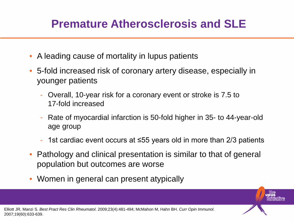
Premature Atherosclerosis and SLE
• A leading cause of mortality in lupus patients
• 5-fold increased risk of coronary artery disease, especially in younger patients
- Overall, 10-year risk for a coronary event or stroke is 7.5 to 17-fold increased
- Rate of myocardial infarction is 50-fold higher in 35- to 44-year-old age group
- 1st cardiac event occurs at ≤55 years old in more than 2/3 patients
• Pathology and clinical presentation is similar to that of general population but outcomes are worse
• Women in general can present atypically
Elliott JR, Manzi S. Best Pract Res Clin Rheumatol. 2009;23(4):481-494; McMahon M, Hahn BH. Curr Opin Immunol. 2007;19(60):633-639.
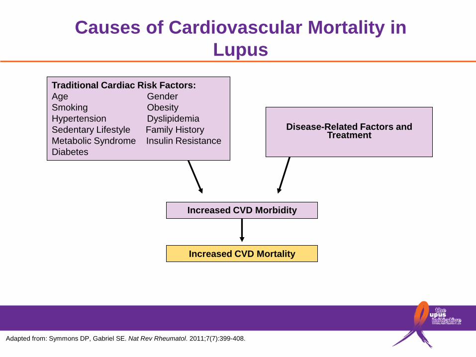
Causes of Cardiovascular Mortality in Lupus
Disease-Related Factors and
Treatment
Increased CVD Morbidity
Increased CVD Mortality
Adapted from: Symmons DP, Gabriel SE. Nat Rev Rheumatol. 2011;7(7):399-408.
Traditional Cardiac Risk Factors: Age Gender Smoking Obesity Hypertension Dyslipidemia Sedentary Lifestyle Family History Metabolic Syndrome Insulin Resistance Diabetes
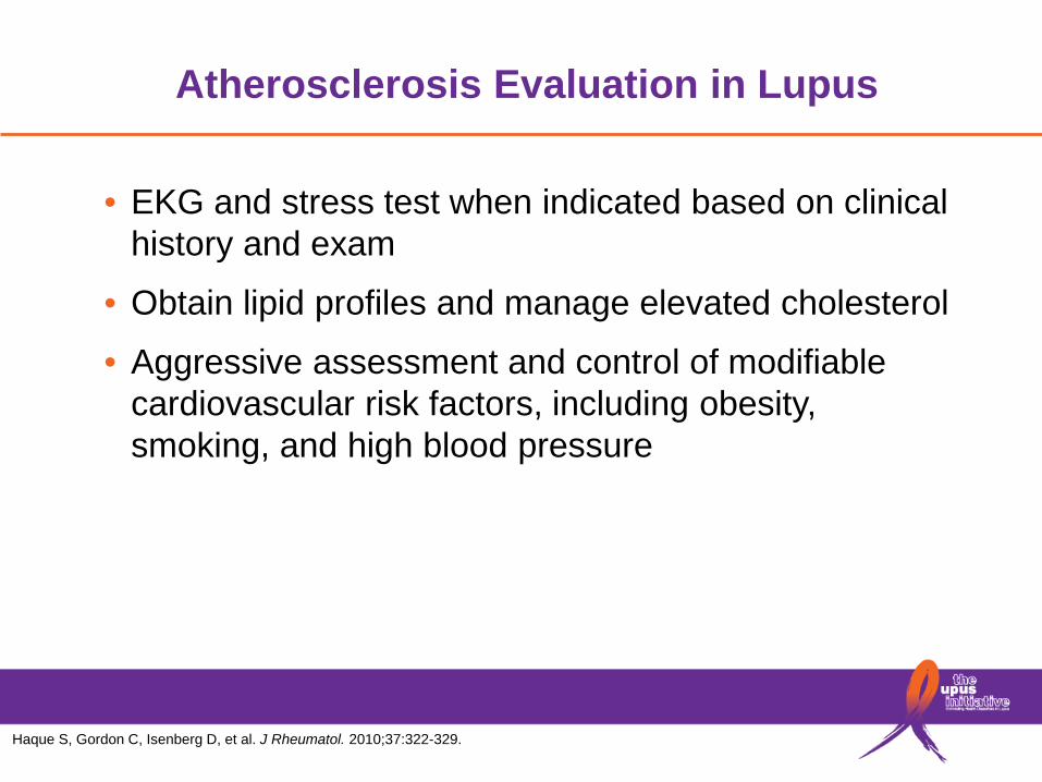
Atherosclerosis Evaluation in Lupus
• EKG and stress test when indicated based on clinical history and exam
• Obtain lipid profiles and manage elevated cholesterol
• Aggressive assessment and control of modifiable cardiovascular risk factors, including obesity, smoking, and high blood pressure
Haque S, Gordon C, Isenberg D, et al. J Rheumatol. 2010;37:322-329.

Other Morbidities to Consider
• Bone related
• Malignancy
• Infections
• Hematologic

Bone Health in Women with Lupus
• Osteonecrosis, a rare condition in healthy individuals, is a major cause of morbidity in some lupus patients. Patients with this condition often require surgical intervention
• Women with lupus are nearly 5 times more likely to experience a fracture from osteoporosis than those without lupus
• Likely contributors to this increased risk include: - Glucocorticoid use
- Sun avoidance (contributing to vitamin D deficiency)
- Disease-related mechanisms
Ramsey-Goldman R, Dunn JE, Huang CF, et al. Arthritis Rheum. 1999;42(5):882-890. Grossman J, Gordon R, Ranganath VK, et al. Arthritis Care Res. 2010;62(11):1515-1526. Gladman DD, Chaudhry-Ahluwalia V, et al. J Rheumatol. 2001;28(10):2226.
Bone Health in Women with Lupus
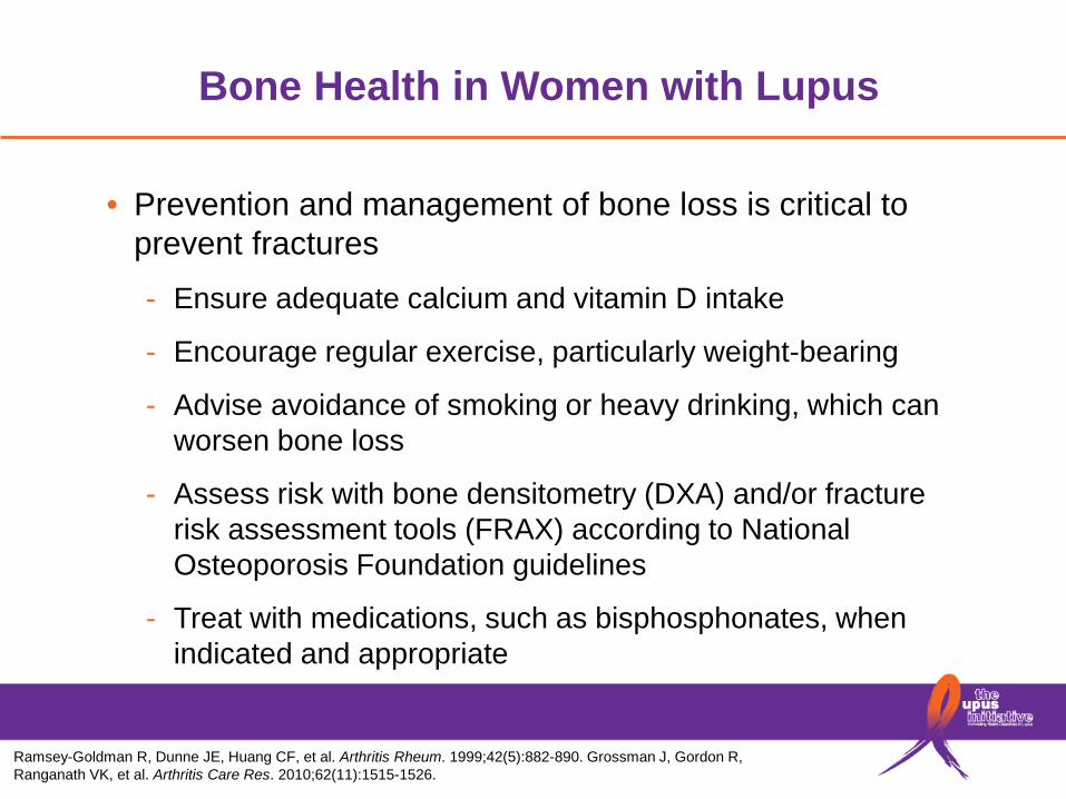
• Prevention and management of bone loss is critical to prevent fractures - Ensure adequate calcium and vitamin D intake
- Encourage regular exercise, particularly weight-bearing
- Advise avoidance of smoking or heavy drinking, which can worsen bone loss
- Assess risk with bone densitometry (DXA) and/or fracture risk assessment tools (FRAX) according to National Osteoporosis Foundation guidelines
- Treat with medications, such as bisphosphonates, when indicated and appropriate
Ramsey-Goldman R, Dunne JE, Huang CF, et al. Arthritis Rheum. 1999;42(5):882-890. Grossman J, Gordon R, Ranganath VK, et al. Arthritis Care Res. 2010;62(11):1515-1526.

Increased Malignancy Risk with SLE
*Data shown are for 23 participating sites in N America, Europe, Iceland, and Asia. The total number of patients was 9547 (76,948 patient-years). The calendar period was 1958–2000. In addition to the categories presented, the total included the following cancers: 21 nonmelanoma skin, 18 primary unknown, 15 head and neck, 12 kidney, 7 central nervous system, 5 esophagus, 5 connective tissue, 3 larynx or mediastinum, 2 small intestine, 2 other female genitourinary, 1 adrenal gland. †Determined using the Poisson distribution. ‡Includes 7 multiple myeloma and 6 lymphoid malignancies not otherwise specified. §Includes invasive cancers; the only cancer registry data that include both invasive and in situ cervical neoplasms are data from the Saskatchewan Cancer Centre.
§
Cancers observed and expected, with standardized incidence ratio (SIR) and 95% confidence intervals (95% CI)*
Bernatsky S. Boivin JF, Joseph L, et al. Arthritis Rheum, 2005;52(5):1481-1490.

Infections and SLE
• Infections are a significant cause of hospitalizations and death
• Risk for infection is increased by: - Active disease
- Immunosuppressive therapies
- Leukopenia/lymphopenia
- Low complement
Bernatsky S, Boivin JF, Joseph L, et al. Arthritis Rheum. 2006;54(8):2550-2557. Staples PJ, Gerding DN, Decker JL, Gordon RS. Arthritis Rheum. 1974;17(1):1-10. Ginzler E, Dvorkina O. In: Wallace DJ, Hahn B. Dubois’ Lupus Erythematosus (7th Ed). Philadelphia, PA; Walters Kluwer Health: Lippincott Williams & Wilkins, 2007.901-10. Sacks JJ, Helmick CG, Langmaid G, Sniezek JE. MMWR Morb Mortal Wkly Rep. 2002;51(17):371-374.

Infections and SLE
• Organisms - Bacterial (respiratory, urinary tract, and skin)
- Viruses (herpes zoster, human papillomavirus)
- Opportunistic (pneumocystis pneumonia, fungi)
• Opportunities for prevention - Vaccinations (inactivated influenza, pneumococcal,
no live vaccines)
- Screening for tuberculosis, hepatitis
- Pneumocystis pneumonia prophylaxis for patients on more intensive immunosuppressive therapies

Hematologic Manifestations in Lupus— Peripheral Blood Cytopenias
• Any or all of the major lineages can be affected - Anemia
- Leukopenia ■ Neutropenia
■ Lymphopenia
- Thrombocytopenia
• Treatment depends upon identifying cause and assessing severity

Hematologic Manifestations in Lupus—Anemia
• Anemia is very common in lupus and often multi-factorial - 25% mild (hematocrit 30%–35%)
- 8% moderate (hematocrit 25%–29%)
- 4% severe (hematocrit <25%)
(cause not attributed)
• Most common causes - Anemia of chronic inflammatory disease
- Anemia associated with renal disease (low erythropoietin)
- Iron deficiency
Bertoli AM, Vila LM, Apte M, et al. Rheumatology. 2007;46:1471-1476. Kao AH, Manzi S, Ramsey-Goldman R. Lupus. 2004; 13(11):865-868.

Hematologic Manifestations in Lupus—Anemia
• Hemolytic anemia (an ACR classification criteria) - Relatively rare, ranging from 5%–13%
- Requires evidence of hemolysis (low haptoglobin and increased reticulocytes)
- Coombs positivity (antibodies to red blood cells) alone much more common, as high as 40%
Bertoli AM, Vila LM, Apte B, et al. Rheumatology. 2007;46:1471-1476. Kao AH, Manzi S, Ramsey-Goldman R. Lupus. 2004; 13(11):865-868.

Hematologic Manifestations in Lupus— Leukopenia and Lymphopenia
• Leukopenia
- Defined as <4000 cells/µL
- Usually an element of neutropenia
- Prevalence of up to 50% sometime during course
• Lymphopenia - Defined as <1500 cells/µL
- May be present in absence of leukopenia
- Prevalence of up to 60%–70% sometime during course
Kao AH, Manzi S, Ramsey-Goldman R. Lupus. 2004;13:865-868.

Hematologic Manifestations in Lupus—Thrombocytopenia
• Defined as <100,000 platelets/µL • Seen in 10%–25% of patients but severe (<50,000 ) less than
10% • Causes
- From lupus ■ Antiplatelet antibodies ■ Antiphospholipid antibodies ■ Thrombotic thrombocytopenic purpura/microangiopathic hemolytic
anemia
- From complications ■ Drug-induced bone marrow suppression ■ Infection
Levine AB, Erkan D. Curr Rheumatol Rep. 2011;13:291-299.

EM—What Could We Have Done Better?
• Education and attention to psychosocial factors - Advise sun protection: year-round use of SPF-45 or higher, clothing
that is UV impenetrable and avoidance of UV exposure when possible
- Encourage weight loss and exercise
- Encourage compliance with clinic visits and medications
• Keep vaccinations up to date • Monitor for early detection of flares • Minimize steroid use • Treat cardiac risk factors aggressively • Monitor bone health

Reducing Adverse Events in Lupus
• Management of risks
- Cardiovascular disease
- Infection
- Fracture
- Cancer
• Hydroxychloroquine used as a background therapy
- Reduce mortality
- Decrease incidence of diabetes
- Antithrombotic effects
- Favorable lipid effects
Broder A, Putterman C. J Rheumatol. 2013;40(1):30-33. Tang C, Godfrey T, Stawell R, Nikpour M. Intern Med J. 2012 Jul 25. [Epub ahead of print]
Mortality Rate in SLE is 2–3 Times Higher than General Population
• Death rates have decreased by 60% in the United States since the 1970s, especially for infections and renal disease
• Risks of death increased in females, Blacks, and younger-onset patients
• Most common causes of death in SLE patients in the United States - Heart disease and stroke (1.7 x general population)
- Hematologic malignancies and lung cancer (2.1 x general population)
- Infections (5 x general population; also a common cause of hospitalization)
- Renal disease (7.9 x general population)
Bernatsky S, Boivin JF, Joseph L, et al. Arthritis Rheum. 2006;54(8):2550-2557.

Conclusions—Mortality and Morbidity in SLE
• Mortality and morbidity in SLE involves - Active disease
- Infectious consequences of chronic immunosuppressive therapy
- Medication toxicities
- Long-term sequelae of inflammation
• Each of these needs to be proactively addressed in order to achieve optimal long-term outcomes for individual patients

Bibliography

Slide 3 Reference Sacks JJ, Helmick CG, Langmaid G, Sniezek JE. Trends in deaths from systemic lupus erythematosus US 1979-1998. MMWR Morb Mortal Wkly Rep. 2002;51:371-374. Slide 5 Reference Bertsias GK, Boumpas DT. Pathogenesis, diagnosis and management of neuropsychiatric SLE manifestations. Nat Rev Rheumatol. 2010;6:358-367. Slide 6 References Muscal E, Brey R. Neurological manifestations of systemic lupus erythematosus in children and adults. Neurol Clin. 2010;28:61–73. Sanna G, Bertolaccini ML, Cuadrado MJ, et al. Neuropsychiatric manifestations in systemic lupus erythematosus: Prevalence and association with antiphospholipid antibodies. J Rheumatol. 2003;30:985-992. Slide 8 Reference Katsumata Y, Kawaguchi Y, Yamanaka H. Rapid progressive cerebral atrophy in systemic lupus erythematosus. J Rheumatol. 2011;38;2689. Slide 11 Reference Abbott NJ, Mendonca LL, Dolman DE. The blood-brain barrier in systemic lupus erythematosus. Lupus. 2003;12: 908-915. Slide 12 Reference Benedict RH, Shucard JL, Zivadinov R, Shucard DW. Neuropsychological impairment in systemic lupus erythematosus: Comparison with multiple sclerosis. Neuropsychol Rev. 2008;18:149–166. .

Slide 15 References Birnbaum J, Petri M, Thompson R, Izbudak I, Kerr D. Distinct subtypes of myelitis in systemic lupus erythematosus. Arthritis Rheum. 2009;60:3378-3387. Espinosa G, Mendizábal A, Mínguez S, et al. Transverse myelitis affecting more than 4 spinal segments associated with systemic lupus erythematosus: Clinical, immunological, and radiological characteristics of 22 patients. Seminars in Arthritis and Rheumatism. 2010;39:246-256. Simeon-Aznar CP, Tolosa-Vilella C, Cuenca-Luque R, Jordana-Comajuncosa R, Ordi-Ros J, Bosch-Gil JA. Transverse myelitis in systemic lupus erythematosus: Two cases with magnetic resonance imaging. Rheumatology. 1992;31:555-558. Slide 16 Reference Goh YP, Naidoo P, Ngian GS. Imaging of systemic lupus erythematosus. Part I: CNS, cardiovascular, and thoracic manifestations. Clinical Radiology. 2013;68:181-191. Slide 20 References Danchenko N, Satia JA, Anthony MS. Epidemiology of systemic lupus erythematosus: a comparison of worldwide disease burden. Lupus. 2006;15:308-318. Hiraki LT, Feldman CH, Liu J, et al. Prevalence, incidence, and demographics of systemic lupus erythematosus and lupus nephritis from 2000 to 2004 among children in the US Medicaid beneficiary population. Arthritis Rheum. 2012;64:2669-2676. Patel M, Clarke AM, Bruce IN, Symmons DP. The prevalence and incidence of biopsy-proven lupus nephritis in the UK: Evidence of an ethnic gradient. Arthritis Rheum. 2006;54:2963-2969. Petri M. Lupus in Baltimore: evidence-based ‘clinical pearls’ from the Hopkins Lupus Cohort. Lupus. 2005;14:970-973.

Slide 21 Reference Davidson A, Berthier C, Kretzler M. Pathogenetic Mechanisms in Lupus Nephritis (Chapter 18). In: Dubois' Lupus Erythematosus and Related Syndromes (8th Ed.). Philadelphia, PA 2013: 237–255. Slide 23 Reference Hahn BH, McMahon MA, Wilkinson A, et al. American College of Rheumatology guidelines for screening, treatment, and management of lupus nephritis. Arthritis Care Res (Hoboken). 2012;64:797-808. Slide 26 Reference Janette JC, Olson, JL, Schwartz MM, Silva FG, editors. Heptinstall’s Pathology of the Kidney. 6th ed. Phil, PA; Lippincott Williams & Wilkins, 2007. Slide 27 References Hahn BH, McMahon MA, Wilkinson A, et al. American College of Rheumatology guidelines for screening, treatment, and management of lupus nephritis. Arthrit Care Res. 2012;64:797-808. Markowitz GS, D'Agati VD. The ISN/RPS 2003 classification of lupus nephritis: an assessment at 3 years. Kidney Int. 2007;71:491-495. Weening JJ, D'Agati VD, Schwartz MM, et al. The classification of glomerulonephritis in systemic lupus erythematosus revisited. Kidney Int. 2004;65:521-30. Slide 28 References Costenbader KH, Desai A, Alarcón GS, et al. Trends in the incidence, demographics, and outcomes of end-stage renal disease due to lupus nephritis in the US from 1995 to 2006. Arthritis Rheum. 2011;63:1681-1688. Hiraki LT, Feldman CH, Liu J, et al. Prevalence, incidence, and demographics of systemic lupus erythematosus and lupus nephritis from 2000 to 2004 among children in the US Medicaid beneficiary population. Arthritis Rheum. 2012;64:2669-2676.

Slide 28 References (cont.) Hiraki LT, Lu B, Alexander SR, et al. End-stage renal disease due to lupus nephritis among children in the US, 1995-2006. Arthritis Rheum. 2011;63:1988-1997. Slide 31 Reference Hahn BH, McMahon MA, Wilkinson A, et al. American College of Rheumatology guidelines for screening, treatment, and management of lupus nephritis. Arthritis Care Res (Hoboken). 2012;64:797-808. Slide 32 Reference Hahn BH, McMahon MA, Wilkinson A, et al. American College of Rheumatology guidelines for screening, treatment, and management of lupus nephritis. Arthritis Care Res (Hoboken). 2012;64:797-808. Slide 33 Reference Hahn BH, McMahon MA, Wilkinson A, et al. American College of Rheumatology guidelines for screening, treatment, and management of lupus nephritis. Arthritis Care Res (Hoboken). 2012;64:797-808. Slide 34 Reference Costenbader KH, Desai A, Alarcón GS, et al. Trends in the incidence, demographics and outcomes of end-stage renal disease due to lupus nephritis in the U.S., 1995-2006. Arthritis Rheum. 2011;63:1681-1688. Slide 35 Reference Franco C, Yoo W, Franco D, Xu Z. Predictors of end stage renal disease in African Americans with lupus nephritis. Bull NYU Hosp Jt Dis. 2010;68:251-256. Slide 39 References Elliott JR, Manzi S. Cardiovascular risk assessment and treatment in systemic lupus erythematosus. Best Pract Res Clin Rheumatol. 2009;23:481-494. McMahon M, Hahn BH. Atherosclerosis and systemic lupus erythematosus—Mechanistic basis of the association. Curr Opin Immunol. 2007;19:633-639.

Slide 40 Reference Adapted from: Symmons DP, Gabriel SE. Nat Rev Rheumatol. 2011;7:399-408. Slide 41 Reference Haque S, Gordon C, Isenberg D, et al. Risk factors for clinical coronary heart disease in systemic lupus erythematosus: the lupus and atherosclerosis evaluation of risk (LASER) study. J Rheumatol. 2010;37:322-329. Slide 43 References Grossman JM, Gordon R, Ranganath VK, et al. American College of Rheumatology 2010 recommendations for the prevention and treatment of glucocorticoid-induced osteoporosis. Arthritis Care & Research. 2010; 62:1515-1526. Ramsey-Goldman R, Dunn JE, Huang CF, et al. Frequency of fractures in women with systemic lupus erythematosus: comparison with United States population data. Arthritis Rheum. 1999;42:882-890. Gladman DD, Chaudry-Ahluwalia V, Ibanez D, Bogoch E, Urowitz MB. Outcomes of symptomatic osteonecrosis in 95 patients with systemic lupus erythematosus. J Rheumatol. 2001;28:2226-2229. Slide 44 References Grossman J, Gordon R, Ranganath VK, et al. American College of Rheumatology 2010 recommendations for the prevention and treatment of glucocorticoid-induced osteoporosis. Arthritis Care & Research. 2010; 62):1515-1526. Ramsey-Goldman R, Dunn JE, Huang CF, et al. Frequency of fractures in women with systemic lupus erythematosus: comparison with United States population data. Arthritis Rheum. 1999;42:882-890. Slide 45 Reference Bernatsky S. Boivin JF, Joseph L, et al. An international cohort study of cancer in systemic lupus erythematosus. Arthritis Rheum. 2005;52:1481–1490.

Slide 46 References Bernatsky S, Boivin JF, Joseph L, et al. Mortality in systemic lupus erythematosus. Arthritis Rheum. 2006;54:2550-2557. Ginzler E, Dvorkina O. Infections in systemic lupus erythematosus (Chapter 45). In: Wallace DJ, Hahn B. Dubois’ Lupus Erythematosus. 7th ed. Philadelphia, PA; Walters Kluwer Health: Lippincott Williams & Wilkins, 2007:901-910. Sacks JJ, Helmick CG, Langmaid G, Sniezek JE. Trends in deaths from systemic lupus erythematosus, 1979–1998. MMWR Morb Mortal Wkly Rep. 2002;51:371–374. Staples PJ, Gerding DN, Decker JL, Gordon RS. Incidence of infections in systemic lupus erythematosus. Arthritis Rheum. 1974;17:1-10. Slide 49 References Bertoli AM, Vila LM, Apte M, et al. Systemic lupus erythematosus in a multiethnic US cohort LUMINA LI: Anemia as a predictor of disease activity and damage accrual. Rheumatology. 2007;46:1471-1476. Kao AH, Manzi S, Ramsey-Goldman R. Review of ACR hematologic criteria in systemic lupus erythematosus. Lupus. 2004;13: 865-868. Slide 50 References Bertoli AM, Vila LM, Apte M, et al. Systemic lupus erythematosus in a multiethnic US cohort LUMINA LI: Anemia as a predictor of disease activity and damage accrual. Rheumatology. 2007;46:1471-1476. Kao AH, Manzi S, Ramsey-Goldman R. Review of ACR hematologic criteria in systemic lupus erythematosus. Lupus. 2004;13: 865-868.
Slide 51 Reference Kao AH, Manzi S, Ramsey-Goldman R. Review of ACR hematologic criteria in systemic lupus erythematosus. Lupus. 2004;13: 865-868. Slide 52 Reference Levine AB, Erkan D. Clinical assessment and management of cytopenias in lupus patients. Curr Rheumatol Rep. 2011;13:291-299. Slide 54 References Broder A, Putterman C. Hydroxychloroquine use is associated with lower odds of persistently positive antiphospholipid antibodies and/or lupus anticoagulant in systemic lupus erythematosus. J Rheumatol. 2013;40:30-33. Tang C, Godfrey T, Stawell R, Nikpour M. Hydroxychloroquine in lupus: emerging evidence supporting multiple beneficial effects. Intern Med J. 2012;Jul 25. Slide 55 Reference Bernatsky S, Boivin JF, Joseph L, et al. Mortality in systemic lupus erythematosus. Arthritis Rheum. 2006;54:2550-2557.


