ORIGINAL RESEARCH published: 31 March 2021 doi: 10.3389/fimmu.2021.645528 Frontiers in Immunology | www.frontiersin.org 1 March 2021 | Volume 12 | Article 645528 Edited by: Chanvit Leelayuwat, Khon Kaen University, Thailand Reviewed by: Steven Thomas Cox, Anthony Nolan, United Kingdom Jack D. Bui, University of California, San Diego, United States *Correspondence: María Carmen Molina [email protected]Specialty section: This article was submitted to NK and Innate Lymphoid Cell Biology, a section of the journal Frontiers in Immunology Received: 23 December 2020 Accepted: 23 February 2021 Published: 31 March 2021 Citation: Toledo-Stuardo K, Ribeiro CH, Canals A, Morales M, Gárate V, Rodríguez-Siza J, Tello S, Bustamante M, Armisen R, Matthies DJ, Zapata-Torres G, González-Hormazabal P and Molina MC (2021) Major Histocompatibility Complex Class I-Related Chain A (MICA) Allelic Variants Associate With Susceptibility and Prognosis of Gastric Cancer. Front. Immunol. 12:645528. doi: 10.3389/fimmu.2021.645528 Major Histocompatibility Complex Class I-Related Chain A (MICA) Allelic Variants Associate With Susceptibility and Prognosis of Gastric Cancer Karen Toledo-Stuardo 1 , Carolina H. Ribeiro 1 , Andrea Canals 2,3 , Marcela Morales 1 , Valentina Gárate 1 , Jose Rodríguez-Siza 1 , Samantha Tello 1 , Marco Bustamante 4 , Ricardo Armisen 5 , Douglas J. Matthies 6 , Gerald Zapata-Torres 6 , Patricio González-Hormazabal 7 and María Carmen Molina 1 * 1 Immunology Program, Faculty of Medicine, Institute of Biomedical Sciences (ICBM), University of Chile, Santiago, Chile, 2 Biostatistics Program, School of Public Health, University of Chile, Santiago, Chile, 3 Academic Direction, Clínica Santa María, Santiago, Chile, 4 Department of Surgery (Oriente), Hospital del Salvador, University of Chile, Santiago, Chile, 5 Center of Genetics and Genomics, Faculty of Medicine Clínica Alemana, Institute for Sciences and Innovations in Medicine (ICIM), Universidad del Desarrollo, Santiago, Chile, 6 Department of Inorganic and Analytical Chemistry, Faculty of Chemical and Pharmaceutical Sciences, University of Chile, Santiago, Chile, 7 Human Genetics Program, Institute of Biomedical Sciences (ICBM), University of Chile, Santiago, Chile Gastric cancer (GC) is the fifth most prevalent type of cancer worldwide. Gastric tumor cells express MICA protein, a ligand to NKG2D receptor that triggers natural killer (NK) cells effector functions for early tumor elimination. MICA gene is highly polymorphic, thus originating alleles that encode protein variants with a controversial role in cancer. The main goal of this work was to study MICA gene polymorphisms and their relationship with the susceptibility and prognosis of GC. Fifty patients with GC and 50 healthy volunteers were included in this study. MICA alleles were identified using Sanger sequencing methods. The analysis of MICA gene sequence revealed 13 MICA sequences and 5 MICA-short tandem repeats (STR) alleles in the studied cohorts We identified MICA ∗ 002 ( ∗ A9) as the most frequent allele in both, patients and controls, followed by MICA ∗ 008 allele ( ∗ A5.1). MICA ∗ 009/049 allele was significantly associated with increased risk of GC (OR: 5.11 [95% CI: 1.39–18.74], p = 0.014). The analysis of MICA-STR alleles revealed a higher frequency of MICA ∗ A5 in healthy individuals than GC patients (OR = 0.34 [95% CI: 0.12–0.98], p = 0.046). Survival analysis after gastrectomy showed that patients with MICA ∗ 002/002 or MICA ∗ 002/004 alleles had significantly higher survival rates than those patients bearing MICA ∗ 002/008 (p = 0.014) or MICA ∗ 002/009 (MICA ∗ 002/049) alleles (p = 0.040). The presence of threonine in the position MICA-181 (MICA ∗ 009/049 allele) was more frequent in GC patients than controls (p = 0.023). Molecular analysis of MICA-181 showed that the presence of threonine provides greater mobility to the protein than arginine in the same position (MICA ∗ 004), which could explain, at least in
Transcript
ORIGINAL RESEARCHpublished: 31 March 2021
doi: 10.3389/fimmu.2021.645528
Frontiers in Immunology | www.frontiersin.org 1 March 2021 | Volume 12 | Article 645528
Major Histocompatibility ComplexClass I-Related Chain A (MICA)Allelic Variants Associate WithSusceptibility and Prognosis ofGastric CancerKaren Toledo-Stuardo 1, Carolina H. Ribeiro 1, Andrea Canals 2,3, Marcela Morales 1,
Valentina Gárate 1, Jose Rodríguez-Siza 1, Samantha Tello 1, Marco Bustamante 4,
Ricardo Armisen 5, Douglas J. Matthies 6, Gerald Zapata-Torres 6,
Patricio González-Hormazabal 7 and María Carmen Molina 1*
1 Immunology Program, Faculty of Medicine, Institute of Biomedical Sciences (ICBM), University of Chile, Santiago, Chile,2 Biostatistics Program, School of Public Health, University of Chile, Santiago, Chile, 3 Academic Direction, Clínica Santa
María, Santiago, Chile, 4Department of Surgery (Oriente), Hospital del Salvador, University of Chile, Santiago, Chile, 5Center
of Genetics and Genomics, Faculty of Medicine Clínica Alemana, Institute for Sciences and Innovations in Medicine (ICIM),
Universidad del Desarrollo, Santiago, Chile, 6Department of Inorganic and Analytical Chemistry, Faculty of Chemical and
Pharmaceutical Sciences, University of Chile, Santiago, Chile, 7Human Genetics Program, Institute of Biomedical Sciences
(ICBM), University of Chile, Santiago, Chile
Gastric cancer (GC) is the fifth most prevalent type of cancer worldwide. Gastric tumorcells express MICA protein, a ligand to NKG2D receptor that triggers natural killer (NK)cells effector functions for early tumor elimination.MICA gene is highly polymorphic, thusoriginating alleles that encode protein variants with a controversial role in cancer. Themaingoal of this work was to study MICA gene polymorphisms and their relationship with thesusceptibility and prognosis of GC. Fifty patients with GC and 50 healthy volunteers wereincluded in this study. MICA alleles were identified using Sanger sequencing methods.The analysis of MICA gene sequence revealed 13 MICA sequences and 5 MICA-shorttandem repeats (STR) alleles in the studied cohorts We identified MICA∗002 (∗A9) asthe most frequent allele in both, patients and controls, followed by MICA∗008 allele(∗A5.1). MICA∗009/049 allele was significantly associated with increased risk of GC (OR:5.11 [95% CI: 1.39–18.74], p = 0.014). The analysis of MICA-STR alleles revealed ahigher frequency of MICA∗A5 in healthy individuals than GC patients (OR = 0.34 [95%CI: 0.12–0.98], p = 0.046). Survival analysis after gastrectomy showed that patientswith MICA∗002/002 or MICA∗002/004 alleles had significantly higher survival rates thanthose patients bearing MICA∗002/008 (p = 0.014) or MICA∗002/009 (MICA∗002/049)alleles (p = 0.040). The presence of threonine in the position MICA-181 (MICA∗009/049allele) was more frequent in GC patients than controls (p = 0.023). Molecular analysisof MICA-181 showed that the presence of threonine provides greater mobility to theprotein than arginine in the same position (MICA∗004), which could explain, at least in
Toledo-Stuardo et al. MICA Variants in Gastric Cancer
part, some immune evasion mechanisms developed by the tumor. In conclusion, ourfindings suggest that the study of MICA alleles is crucial to search for new therapeuticapproaches and may be useful for the evaluation of risk and prognosis of GC andpersonalized therapy.
Gastric cancer (GC) is the fifth most common neoplasm andthe third leading cause of cancer-related death worldwide(1), accounting for 783,000 deaths in 2018 according toGLOBOCAN data.
The absence of early clinical tools for GC diagnosis decreasesthe success of therapies and survival of patients, which raisesthe question of the necessity to explore novel biologicalbiomarkers of the disease (2–4). In this context, the majorhistocompatibility complex class I-related protein A (MICA)may provide relevant information about the pathological changesin the gastrointestinal mucosa. The healthy gastric mucosaexpress low levels of this protein, while the tumor tissueoverexpresses it (5–7). MICA expression in the carcinogenesisprocess is regulated by transcriptional, translational and/or post-translational modification mechanisms (8, 9) that could beassociated withHelicobacter pylori or Epstein Barr-virus infection(10, 11).
MICA is a ligand to natural killer group 2D (NKG2D),an activating receptor (12) that is important for the anti-tumor immune response (13). NKG2D is expressed by cytotoxiclymphocytes, including natural killer (NK) cells, γδ T cells,and CD8+ T cells. NK cells constitute the first line of defenseagainst intracellular pathogens; they also contribute to themaintenance of mucosal homeostasis and the developmentof efficient immune responses against cancer (14). NKG2Dbinding to MICA on target cells triggers NK cell cytolyticactivation, which results in target cell lysis through the releaseof granzyme and perforin by the effector cell (15). However,tumors have developed several strategies to evade the immuneresponse, such as the proteolytic shedding of MICA from the cellmembrane (13). The resulting soluble form of MICA induces thedownregulation of NKG2D receptor on NK cells, compromisingthe immune response mediated by these and other cytolyticcells (16).
The human MICA gene is highly polymorphic; indeed, ithas been reported more than 110 MICA alleles that encodeover 100 protein variants (http://hla.alleles.org/data/mica.html).The human MICA gene is located in chromosome 6p21.3,46 kb distant from HLA-B, and consists of six exons: exon1–6 encode the leader peptide, the three extracellular proteindomains (α1, α2, and α3), the transmembrane region, andthe hydrophobic cytoplasmatic tail, respectively (17). Exon 5contains a short tandem repeat (STR) with a variable numberof GCT triplet repeats, which encode alanine (Ala). A secondnomenclature for MICA alleles, MICA-STR (18), has thusemerged based on the presence of these repetitions; for instance,MICA∗A5 consists of five repetitions of GCT. Such STR
affects the length of the transmembrane region, as in the caseof MICA∗A9, which contains nine GCT repetitions, whichconsequently codifies nine residues of alanine (18). Interestingly,the MICA∗A5.1 (prototype MICA∗008) variant produces aninsertion of guanine at position 952 (Chr6: 31380161-31380162on Assembly GRCh37) in the transmembrane region, changingthe reading frame to a pre-mature stop codon and generatinga protein with a GPI anchor, which results in the recruitmentof MICA to exosomes and release of the protein in the form ofextracellular vesicles (EVs) (19, 20).
MICA polymorphisms have been previously studied in cancer,especially the MICA-129 residue, which is associated to thepresence of methionine (Met) or valine (Val), where MICA-129 Met has shown a strong interaction with NKG2D, leadingto the downregulation of the receptor more efficiently thanMICA-129 Val (21). On the other hand, MICA-129 Val hasbeen associated to an increase in soluble MICA (sMICA) levelsin multiple myeloma, which induces NKG2D downregulationand contributes to immune evasion (16). Furthermore, severalMICA transmembrane regions, based on the GCT repetitions,have been associated to higher susceptibility to certain typesof cancers. For instance, MICA∗A9 allele has been proposedto confer a risk for GC (22), while MICA∗A5.1 allele maybe associated with increased susceptibility to oral squamouscell carcinoma in Japanese patients (23). Since MICA allelesvary among human populations and generate proteins withdifferent biological properties that may result in variable diseasesusceptibility, we decided to study MICA gene polymorphismsand susceptibility to gastric cancer and their relationship with thetumor progression.
MATERIALS AND METHODS
Patients and Healthy Controls SamplesGastric Cancer Patients Tissue SamplesDuring June 2011 and August 2015, a total of 50 patients (17female, 33 male) aged 65.4± 10.3 years (range, 49–78 years) withconfirmed diagnosis of gastric adenocarcinoma and treated at theDepartment of Gastrointestinal Surgery, Hospital del Salvador(Santiago, Chile), were enrolled in this study. Fresh primarygastric tumor tissue samples were obtained at the time of surgeryand immediately processed for genomic DNA extraction.
Healthy Controls Blood SamplesBlood samples were obtained from 50 healthy donors (24female, 26 male) aged 47.3 ± 15.6 years (range, 30–61 years)without prior gastrointestinal diseases or any type of cancer, wholived in Santiago, Chile. Blood samples were collected in 4mL
Frontiers in Immunology | www.frontiersin.org 2 March 2021 | Volume 12 | Article 645528
Toledo-Stuardo et al. MICA Variants in Gastric Cancer
EDTA-containing vacutainer tubes (BD Biosciences, San José,CA, USA) for genomic DNA extraction.
Ethical ConsiderationsThis study was approved by the Committee on Human EthicsInvestigation of the Faculty of Medicine, University of Chile, andthe Committee on Scientific Ethics of the Metropolitan HealthService of the Chilean Government. All patients and healthyvolunteers signed a written Informed Consent for tissue andblood donation. Name and identification of patients and controlswere omitted and rendered anonymous before data analysis.
Clinicopathological DataThe clinicopathological information included patient gender,age, primary tumor location, tumor size, Lauren’s histologicalclassification, Bormann classification (24), lymph nodemetastasis and tumor stage according to the American JointCommittee on Cancer, AJCC, 7th edition (25). Histopathologicalanalysis was carried out by the team of pathologists fromHospitaldel Salvador (Santiago, Chile). The histological differentiationgrade was based on the World Health Organization (WHO)classification (26). Patient survival was assessed for 36 monthsafter surgery or until death due to tumor-specific disease.
Extraction and Purification of GenomicDNAThe genomic DNA was obtained from ∼30mg of tumor tissueusing the E.Z.N.A Tissue DNA kit (Omega, Bio-tek, USA),following the manufacturer’s instructions. The genomic DNAfrom blood samples was isolated using the salting out methodpreviously described by Subbarayan PR and collaborators (27).
Measurement of DNA Purity and IntegrityThe genomic DNA purity was assessed according to the mean
260/280 nm ratio in a SynergyTM
microplate reader (Biotek,Winooski, VT, USA). The genomic DNA integrity was verifiedby electrophoresis using a 1% agarose gel (40mg of analyticalgrade Agarose, 40mL of Tris-borate-EDTA buffer) with 0.5µL of ethidium bromide solution (Sigma-Aldrich/Merck KGaA,Darmstadt, Germany). The DNA bands were visualized using aUV transilluminator.
MICA GenotypingTo amplify theMICA gene of GC patients and healthy volunteersby PCR, we used two pairs of specific primers to 2-3 and 4-5exons of the MICA gene, and MICA genotyping was carriedout using bidirectional Sanger sequencing methods. Forwardprimer (2-3 exons): 5′ TGAAATCCTCGTTCTTGTCCCTTTGC 3′, Reverse primer (2-3 exons): 5′ AGGGTCCTCTACTTGCCCTGATTAC 3′; Forward primer (4-5 exons): 5′
TCAGCCAGAGTGAGAACAGTGAAGA 3′, Reverse primer(4-5 exons): 5′ TCATCCCCTGTTATGGAAGCCTTGTC 3′. ThePCR reactions were analyzed by 0.8% agarose gel electrophoresis.The amplicons were purified using the Wizard SV Gel andPCR Clean-Up System Purification Kit (Promega, USA). Theamplified products were sequenced on an ABI PRISM-3500 XLsequencer (Applied Biosystems, Foster City, CA). The genomic
sequences of each sample were analyzed using Chromas 2.4Viewer and were manually verified. These sequences werethen compared with MICA allelic genotype sequences obtainedfrom the IPD-IMGT/HLA database (28) (http://hla.alleles.org/data/mica.html). MICA allelic genotypes were obtainedaccording to reference sequences of specific MICA alleles. Thepresence of heterozygous alleles in the sequences was assignedby Chromas and verified in the electropherogram. The exon 5was carefully analyzed, manually, due to the presence of STR ormicrosatellites with different lengths, especially in heterozygouspatients, as Sanger sequencing of this exon frequently resultsin base overlap. To validate our methodology, we analyzed twoavailable cell lines with known MICA alleles, which includedAGS ATCC CRL-1739 human gastric cancer cell line (which ishomozygous for MICA∗010) (29) and PC-3 ATCC CRL-1435human prostate cancer cell line (which is heterozygous forMICA∗008 and ∗012) (30). MICA∗009 and MICA∗049 allelesdiffer in one amino acid in the exon 6. Since the differentialresidue was not distinguished in this study, this allele was namedas MICA∗009/049; accordingly, allele MICA∗002/009 was alsonamed MICA∗002/049.
Molecular AnalysisMICA protein sequence and its isoforms were retrieved fromUniProtKB1 (31) (https://www.uniprot.org) in its FASTA format.A protein-protein BLAST (32) (http://blast.ncbi.nlm.nih.gov)was further performed to search for a protein template with thehighest sequence identity to MICA, which resulted in PDB code1HYR (17) with a 2.7 Å resolution. A sequence alignment withthis template was then carried out using Clustal Omega server(33) (https://www.ebi.ac.uk). This target sequence alignment wasfed on the Swiss-Model server (34) (https://swissmodel.expasy.org) to build the models of the proteins, which were thensolvated in silico with water using a rectangular shape andneutralized with ions to a concentration of 0.15M of NaClto mimic physiological environment using the CHARMM-GUIserver (35). Periodic boundary conditions were applied, anda minimization procedure was performed using a maximumnumber of 5,000 steps of conjugated gradient followed by 2,500steps of steepest descent run. Next, the NVT ensemble wasused for subsequent equilibration steps at 303.15K using atimestep of 0.001 ps for 125 ps. NPT dynamic runs were usedfor production runs with a timestep of 0.004 ps for a period of500 ns. The simulations were carried out using Amber14 (36)suite of programs.
Statistical AnalysisDescriptive analysis of demographic data and clinicopathologicalcharacteristics of cases and controls were performed usingmedian and interquartile range for quantitative variablesand absolute and percentage frequency distributions forcategorical variables.
The distribution of MICA-sequence alleles and MICA-STRalleles among cases and controls, and the proportion of eachallele among cases and controls were compared using Fisher’sExact Test. In addition, logistic regressions were adjusted forthe prediction of GC by comparing each allele with the others,
Frontiers in Immunology | www.frontiersin.org 3 March 2021 | Volume 12 | Article 645528
Toledo-Stuardo et al. MICA Variants in Gastric Cancer
TABLE 1 | Demographic data and clinicopathological characteristics of gastriccancer patients and controls.
Demographic
data
Controls
n = 50
Gastric patients
n = 50
Median/n IQR/% Median/n IQR/%
Age 49 (30-61) 67 (49-78)
Gender
Female 24 (48.0%) 17 (34.0%)
Male 26 (52.0%) 33 (66.0%)
Clinicopathological characteristics n %
Location of tumor Cardia 10 (21.3%)
No cardia 35 (74.5%)
Both 2 (4.3%)
Tumor size, cm ≤5 18 (36.0%)
>5 32 (64.0%)
Lauren’s classification Intestinal type 27 (54.0%)
Diffuse type 13 (26.0%)
Mixed type 10 (20.0%)
Borrmann’sclassification
I (Polypoid/fungating) 2 (4.0%)
II (Superficialspreading)
3 (6.0%)
III (Ulcerating) 23 (46.0%)
IV (Linitis plastica) 11 (22.0%)
V (Unclassified) 5 (10.0%)
Not documented 6 (12.0%)
Peritoneal cytology Positive 5 (10.0%)
Negative 39 (78.0%)
Not documented 6 (12.0%)
TNM staging I, II 8 (16.3%)
III, IV 41 (83.7%)
Helicobacter pylori Positive 23 (46.0%)
Negative 27 (54.0%)
IQR, Interquartile range.
obtaining the odds ratio (OR) with their respective confidenceintervals (95%, CI).
The distribution of genotypes of each MICA residue betweencases and controls was also compared using Fisher’s Exact Test.
Survival curves of GC patients, according to the presence ofMICA alleles, were obtained using the Kaplan-Meier method andcompared using the Log-rank test.
Statistical analysis was performed using Stata 14 software, anda p-value < 0.05 was considered significant.
RESULTS
Clinicopathological Characteristics of GCPatientsPatients demographic characteristics and clinicopathologicalfeatures of tumors are described in Table 1. Tumor size wasgiven as the maximum tumor diameter measured on the freshlyresected stomach. Most of GC patients (64%) presented with a
tumor size higher than 5 cm. In 74.5% of patients, the tumors hadcardia location. According to Lauren’s histological classification,27 out of 50 patients had intestinal type GC, 13 had diffuse typeGC, and 10 patients presented with mixed type GC. Borrmann’sclassification showed that the gastric tumors were mainly at stageIII (46%), followed by stage IV (11%), which is related to theTNM staging classification, where 41 patients were found at stageIII-IV. Additionally, 46% of the patients showed Helicobacterpylori infection.
MICA Allelic Frequency in GC Patients andHealthy IndividualsThe analysis of MICA gene allowed us to identify 13 MICA-sequence alleles and 5 MICA-STR alleles in the studiedpopulation (Table 2). The distribution of MICA allelicfrequencies identified in this study was different betweenGC patients and healthy volunteers (p = 0.024). We observedthat the most frequent MICA allele found in both, patients andcontrols was the MICA∗002 (∗A9) allele, which was followed bythe MICA∗008 (∗A5.1) allele. Together, both alleles representedmore than 50 percent of all the alleles identified in our analysis.As shown in Supplementary Table 1, the MICA genotypefrequency distribution in GC patients and healthy individualsdid not show significant differences. The MICA∗002/008(∗A9/A5.1) heterozygous genotype was the most common inboth groups, representing 18.2 and 20% in GC patients andhealthy controls, respectively.
We found that the MICA∗009/049 allele frequency wassignificantly higher in GC patients than in healthy controls(p = 0.007), with a OR: 5.11 (95% CI: 1.39–18.74, p = 0.014).Additionally, we detected a higher frequency of MICA∗A5 allelein healthy individuals than in GC patients, with a OR= 0.34 [95%CI: 0.12–0.98, p= 0.046) (Table 2). These results indicate that thepresence of the MICA∗A5 allele may be a protective factor in thistype of tumor.
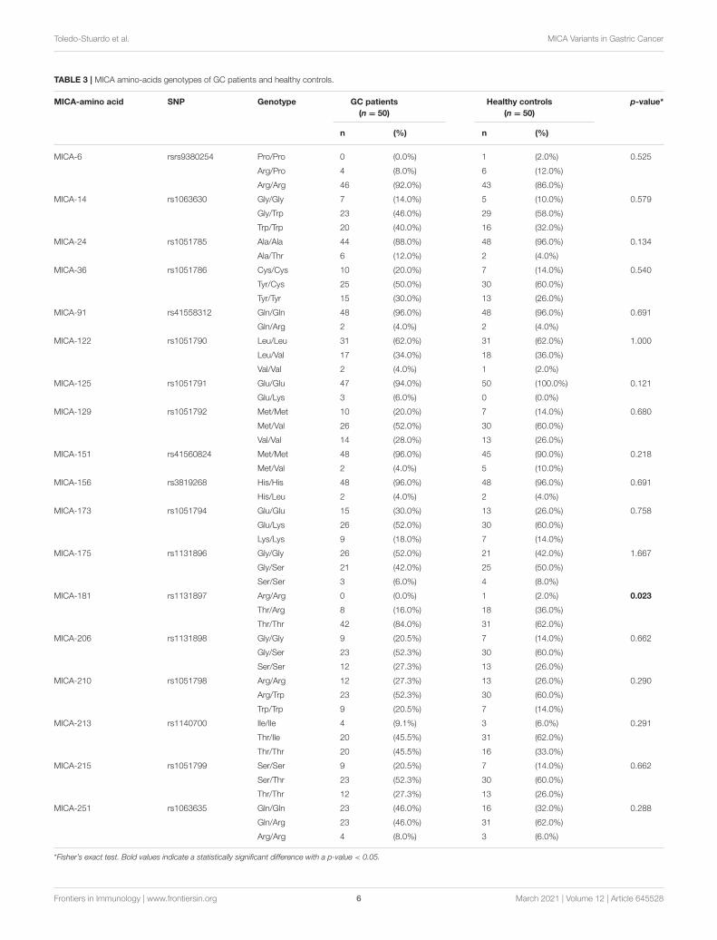
MICA Polymorphisms and Developmentof GCWe next analyzed the genotypes of the main amino-acids ofMICA ectodomain in GC patients and healthy controls (Table 3).The results showed that the only amino-acid that varies betweenpatients and controls was the residue in position 181 (p= 0.023),which corresponds to the substitution of threonine by arginine.This change defines the difference between MICA∗004 allele(arginine) and MICA∗009/049 allele (threonine). We observedthat the majority of GC patients had a Thr/Thr genotype (42/50),while the Arg/Arg genotype was not identified in these patients.Patients with a Thr/Thr or Thr/Arg genotype did not show asignificant difference in the tumor size or differentiation grade.
Additionally, we evaluated the possible association betweenclinical features related to the risk of developing GC and theMICA-129 polymorphism, since in position 129 resides one ofthe most studied residues that determine a variable affinity toNKG2D receptor. We found that MICA-129 single nucleotidepolymorphism (SNP) (rs1051792) showed a strong linkagedisequilibrium with other SNPs related to residues 36, 173, 206,
Frontiers in Immunology | www.frontiersin.org 4 March 2021 | Volume 12 | Article 645528
*Odds ratio (OR) of gastric cancer for each allele compared to the rest of alleles. The table included 44 of 50 patients whose alleles have been identified.NA, Not applicable; it corresponds to an allele without single tandem repetitions. Bold values indicate a statistically significant difference with a p-value < 0.05.
210, and 215. We did not identify associations between thesegenotypes and tumor size or differentiation grade. The presenceof positive peritoneal cytology was identified in only five patientsand, for this reason, it was not possible to classify the patients intogroups according to this pathological characteristic, althoughpositive peritoneal cytology was detected only in patients withMet/Val and Val/Val MICA-129 genotype (data no shown).
We studied the relevance of the MICA alleles, bothMICA-sequence and MICA-STR, on the clinicopathologicalcharacteristics of the tumor. We observed that the majorityof patients with the MICA∗A9/A6 heterozygote alleleshowed mainly a poorly differentiated tumor (p = 0.031)(Supplementary Table 2), indicating that the presence of thisgenotype is related to an advanced stage of the tumor, asalso observed previously (37). However, we did not find anassociation between the presence of the most frequent MICA-sequence alleles (∗002, ∗008 and ∗009/049) and tumor size ordifferentiation grade (Supplementary Table 2).
Survival of GC Patients Based on MICAAllelesWe analyzed the overall survival of GC patients according toMICA alleles during 36 months after gastrectomy. We excludedthe patients with a TNM stating I and II to avoid conflictinginterpretations due to the possibility of a higher survival inpatients with a lesser advanced disease. First, we classified the
patients based on the main alleles found among them, whichincluded patients homozygous for MICA∗002 or MICA∗008and heterozygous for MICA∗002/009 (MICA∗002/049),MICA∗002/004, or MICA∗002/008. The Kaplan-Meier curvesand Log-rank test showed significant differences amongthese groups of patients. MICA ∗002 and MICA∗002/004patients showed a higher survival rate than MICA∗002/008patients (p = 0.014) or MICA∗002/009 (MICA∗002/049) (p =
0.040). The survival distributions of homozygous patients forMICA∗002 or heterozygous patients for MICA∗002/004 weresimilar to that of homozygous patients for MICA∗008 (p =
0.070). Likewise, no differences in the survival distributionsof patients with MICA∗002/008 compared to MICA∗002/009(MICA∗002/049) or MICA∗008/008 alleles could be detected(p= 0.611 and p= 0.909, respectively), neither between patientswith MICA∗002/009 (MICA∗002/049) and those homozygousfor MICA∗008 (p= 0.883) (Figure 1A).
We also compared the survival curves of GC patients basedon MICA-STR alleles (Figure 1B) and MICA-129 genotype(Figure 1C), which did not reach statistical significance(p = 0.057 and p = 0.175, respectively). Therefore, the MICAsequence, and not MICA-STR, may have a prognostic value inthis type of cancer.
Molecular Analysis of MICA-181 ResidueSince we found that the MICA-181 residue showed differencesbetween GC patients and controls (Table 3), and the fact that
Frontiers in Immunology | www.frontiersin.org 5 March 2021 | Volume 12 | Article 645528
Toledo-Stuardo et al. MICA Variants in Gastric Cancer
FIGURE 1 | Kaplan-Meier curves for overall survival of GC patients withtumors at III and IV TNM staging, according to MICA-sequence, MICA-STRand MICA-129 genotype. The p-values were calculated by the Log-rank test.(A) The survival probability of GC patients with MICA*002/002 (n = 4) andMICA*002/004 (n = 4) alleles were significantly higher than that of
(Continued)
FIGURE 1 | MICA* 002/008 (n = 7) (p = 0.014) and MICA*002/009(MICA*002/049) alleles (n = 4) (p = 0.040). The comparison betweenMICA*002/002 or MICA*002/004 and MICA*008/008 (n = 3) did not showsignificance (p = 0.070). (B) The survival probability of GC patients withMICA*A9/A9 (n = 4), MICA*A9/A5.1 (n = 7), MICA*A9/A6 (n = 9),MICA*A5.1/A6 (n = 3) and MICA*A5.1/A5.1 (n = 3) did not show significantdifferences (p = 0.057). (C) The survival probability of GC patients with theMet/Met genotype (n = 8), Met/Val (n = 22) and Val/Val (n = 11) did not showstatistical differences (p = 0.175).
FIGURE 2 | RMSD variation in MICA variant simulations. Black colored linerepresents MICA*004 (Arg181 variant), while red colored line representsMICA*009/049 (Thr181 variant). After 200 ns, MICA*004 remains constant,but MICA*009/049 displayed a less restrictive movement.
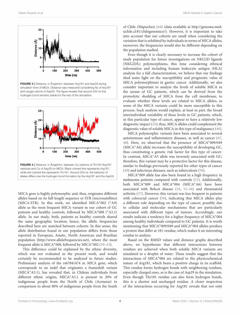
this residue defines the MICA∗009/049 (Thr181) and MICA∗004(Arg181) alleles, we decided to perform a molecular analysisof these proteins to evaluate the impact of this amino-acidresidue in the dynamic properties of MICA. Molecular dynamicssimulations of both proteins displayed a different behaviorduring the simulation time of 500 ns. MICA∗009/049 displayedgreater mobility compared to MICA∗004, which revealed a morerestricted movement at the end of the simulation time, duringwhich the change in the root mean square deviation (RMSD)values remained almost constant, as observed in Figure 2.
During the simulation, we noticed that the Arg181 residueestablished an electrostatic interaction with Asp29 (Figure 3),which, according to the simulations, is responsible for theflattening of the curve for MICA∗004. Based on this observation,it was possible to measure a distance of Cα carbons of bothresidues (Arg181 vs. Thr181) with Asp29 in order to compare theimportance of this interaction. We observed a higher distance forThr181 than Arg181 around 300 ns (Figure 4). This implies thatThr181 does not accomplish a stabilizing interaction, probablydue to its shorter side chain, as compared to Arg181. Thisconfirmed that there is a greater movement in MICA∗009/049, asThr181 does not display the electrostatic interaction with Asp29.
DISCUSSION
MICA, one of the main ligands to NKG2D receptor, has beenconsidered an immunological target due to its participation inimmune evasion mechanisms in cancer (38), including GC (5).
Frontiers in Immunology | www.frontiersin.org 7 March 2021 | Volume 12 | Article 645528
Toledo-Stuardo et al. MICA Variants in Gastric Cancer
FIGURE 3 | Distance, in Ångström, between Arg181 and Asp29 duringsimulation time of MICA. Distance was measured considering Nε of Arg181and oxygen atoms of Asp29. The figure reveals that around 200 ns thishydrogen bond remains stable for the rest of the simulation.
FIGURE 4 | Distance, in Ångström, between Cα carbons of Thr181/Arg181residues and Cα of Asp29 on MICA. Black colored line represents Arg181,while red colored line represents Thr181. Around 200 ns, the behavior ofalleles differs due the hydrogen bond formation by the Arg181 and the Asp29.
MICA gene is highly polymorphic and, thus, originates differentalleles based on its full-length sequence or STR (microsatellites)(MICA-STR). In this work, we identified MICA∗002 (∗A9)allele as the most frequent MICA variant in our cohort of GCpatients and healthy controls, followed by MICA∗008 (∗A5.1)allele. In our study, both, patients as healthy controls sharedthe same geographic location; hence, the allelic frequenciesdescribed here are matched between cohorts. In this sense, theallele distribution found in our population differs from thosereported in European, Asiatic, North American and Brazilianpopulation (http://www.allelefrequencies.net), where the mostfrequent allele is MICA∗008, followed by MICA∗002 (39–43).
This difference could be explained by the ethnic diversity,which was not evaluated in the present work, and wouldcertainly be recommended to be analyzed in future studies.Preliminary analysis of the rs67841474 in MICA gene, whichcorresponds to an indel that originates a frameshift variant(MICA∗A5.1), has revealed that, in Chilean individuals fromdifferent ethnic origins, MICA∗A5.1 is present in 20% ofindigenous people from the North of Chile (Aymaras) incomparison to about 80% of indigenous people from the South
of Chile (Mapuches) (44) (data available at http://genoma.med.uchile.cl:81/chilegenomico/). However, it is important to takeinto account that our cohorts are small when considering thevariation that is exhibited by individuals in terms ofMICA alleles;moreover, the frequencies would also be different depending onthe population studied.
Even though it is clearly necessary to increase the cohort ofstudy population for future investigations on NKG2D ligands(NKG2DL) polymorphisms, this time considering ethnicalinformation and including human leukocyte antigen (HLA)analysis for a full characterization, we believe that our findingsshed some light on the susceptibility and prognostic value ofMICA polymorphisms in gastric cancer. Additionally, we alsoconsider important to analyze the levels of soluble MICA inthe serum of GC patients, which can be derived from theproteolytic shedding of MICA from the cell membrane, toevaluate whether these levels are related to MICA alleles, assome of the MICA variants could be more susceptible to thisprocess. Such analysis would explain, at least in part, the broadinterindividual variability of these levels in GC patients, which,in this particular type of cancer, appear to have a relatively lowdiagnostic impact (45); thus, MICA alleles could complement thediagnostic value of soluble MICA in this type of malignancy (46).
MICA polymorphic variants have been associated to severalautoimmune and inflammatory diseases, as well as cancer (47,48). Here, we observed that the presence of MICA∗009/049(MICA∗A6) allele increases the susceptibility of developing GC,thus constituting a genetic risk factor for this type of cancer.In contrast, MICA∗A5 allele was inversely associated with GC;therefore, this variant may be a protective factor for this disease,similar to findings previously reported for other types of cancer(49) and infectious diseases, such as tuberculosis (50).
MICA∗009 allele has also been found in a high frequency inmelanoma patients compared with controls (51). Additionally,both MICA∗009 and MICA∗004 (MICA∗A6) have beenassociated with Behcet disease (18, 52–56) and rheumatoidarthritis (57). However, this variant was less frequent in patientswith colorectal cancer (58), indicating that MICA alleles playa different role depending on the type of cancer, possibly dueto cellular and molecular mechanisms that are particularlyassociated with different types of tumors. Accordingly, ourresults indicate a tendency for a higher frequency of MICA∗004among healthy individuals compared to GC patients. It is worthmentioning that MICA∗009/049 and MICA∗004 alleles producea protein that differ at 181-residue, which makes it an interestingresidue to analyze.
Based on the RMSD values and distance graphs describedabove, we hypothesize that different interactions betweenresidues are achieved when both soluble MICA variants aresimulated in a droplet of water. These results suggest that theinteractions of MICA∗004 are related to the physicochemicalnature of Arg181, which bears a positive charge in its scaffold.This residue forms hydrogen bonds with neighboring residues,especially charged ones, as is the case of Asp29 in the simulation.Even though Thr181 residue can also form hydrogen bonds,this is a shorter and uncharged residue. A closer inspectionof the interactions occurring for Arg181 reveals that not only
Frontiers in Immunology | www.frontiersin.org 8 March 2021 | Volume 12 | Article 645528
Toledo-Stuardo et al. MICA Variants in Gastric Cancer
FIGURE 5 | Electrostatic potential map for the crevice formed by residues His3 and Asp29 (yellow sticks) on MICA. The electrostatic potential map is given inkT/e units.
is a coulombic interaction with Asp29 reinforced by hydrogenbonds, but it also displays a cation-π interaction with a terminalhistidine residue (His3) at 4.5 Å. These interactions seem to bestrong enough to keep the Arg181 stack in a crevice formed bythese polar residues. In the case of Thr181 in MICA∗009/049,these interactions are not observed during the simulation time,resulting in a more freely movement compared to Arg181. Thisobservation implies that the change in only one amino acid canrender a completely different dynamic behavior for both proteins.Figure 5 displays the Arg181 in the crevice formed by Asp29 andHis3, which, in turn, displays a negative electrostatic potentialsurface, where Arg181 remains occluded. This is not observedwith Thr181, which is not able to visit this site.
The MICA∗009-HLA B∗50, B∗51 or B∗52 haplotypes havebeen described in previous studies (59–61). Accordingly, HLAB∗51 was found to be more frequent in Helicobacter pylori-positive pediatric patients with active gastritis and duodenal ulcer(62) and the HLA B∗52 antigen has shown to be associatedwith lymph node metastasis in gastric cancer (63). Therefore,both HLA-B alleles and MICA∗009/049 could to be factor riskin gastric cancer, so we suggest to consider this information forfuture studies.
When we analyzed the clinical characteristics of GC patientsin relation to MICA polymorphisms, we observed that MICA-129, MICA-181 and MICA-sequence alleles were not related totumor size, TNM stating or tumor differentiation grade in thistype of cancer, whereas, tumors fromMICA∗A9/A6 heterozygotepatients, who thus possess combinations of MICA∗002 andMICA∗009/049 or MICA∗004, showed poorly differentiatedtumors. These observations suggest that the transmembraneregion of MICA may play a relevant role in GC progress. Wesuspect that the length of MICA transmembrane region, relativeto the presence of six or more alanine residues, could implicatean increased susceptibility for proteolytic shedding of MICA bymetalloproteases in the tumor microenvironment. If this is thecase, the soluble levels of MICA would increase, which could
negatively modulate the NKG2D receptor, favoring immuneevasion mechanisms.
Here, we have also analyzed whether MICA variants affectthe prognosis of GC. Our results showed that GC patientscarrying theMICA∗002 allele have better survival rates comparedto GC patients carrying other MICA alleles. It has beenpreviously demonstrated that the SNP rs9266825, which is partof MICA∗002, ∗007, ∗018, ∗017, ∗001 alleles, is associated withincreased survival rates in non-small cell lung cancer patients(64). Nevertheless, our study demonstrates, for the first time, theoverall survival rates of GC patients based on the whole sequenceof MICA gene.
We propose that certain MICA alleles have the potential tobe more expressed on gastric tumor cells, depending on theircellular microenvironment, which may favor a better or worseimmune response mediated by NK cells. According to our data,MICA∗002 and MICA∗004 alleles could be highly expressedon the surface of gastric tumor cells, which would triggerNKG2D receptor activation and an effective NK cell response.This is supported by other studies, which indicate that high-cell surface MICA/B expression in cancers of the digestive tractwas associated with increased patient survival (65). In contrast,the levels of other protein variants, including MICA∗009 andMICA∗008, may be lower on tumor cell surface due to theirshedding by metalloproteinases or release into the tumor milieuin extracellular vesicles, reducing target cell interaction withNK cells through the NKG2D receptor, thus affecting NKcell-mediated cytotoxicity. Support for this hypothesis comesfrom reports showing that patients with gastric tumors withhigh MICA expression had higher overall survival and disease-free-survival than patients bearing tumors with low MICAexpression (66).
Our findings indicate that certain MICA alleles could havedifferent effects on clinicopathological features of gastric tumor,such as the differentiation grade of tumor, as well as on patientoverall survival after potentially curative gastrectomy, which
Frontiers in Immunology | www.frontiersin.org 9 March 2021 | Volume 12 | Article 645528
Toledo-Stuardo et al. MICA Variants in Gastric Cancer
may depend on the tumor microenvironment and regulation ofMICA protein expression. In this sense, it will be interesting toanalyze, in future studies, whether soluble MICA levels presentany relationship with MICA alleles in GC patients.
In conclusion, MICA∗009/049 allele increases thesusceptibility to gastric cancer, whereas, MICA∗A5 allelehas a protective effect in this disease. MICA∗002/002 andMICA∗002/004 patients have higher survival rates thanMICA∗002/008 and MICA∗002/009 (MICA∗002/049) aftersurgery. Therefore, we consider that functional studies of MICAvariants may help elucidate the mechanisms by which MICAconfers protection or risk to GC development and prognosis,which may be a useful tool for the development of noveltherapeutical approaches to treat this disease.
DATA AVAILABILITY STATEMENT
The raw data supporting the conclusions of this article will bemade available by the authors, without undue reservation, to anyqualified researcher.
ETHICS STATEMENT
The studies involving human participants were reviewed andapproved by Committee on Human Ethics Investigation of theFaculty of Medicine, University of Chile, and the Committeeon Scientific Ethics of the Metropolitan Health Service of theChilean Government. The patients/participants provided theirwritten informed consent to participate in this study.
AUTHOR CONTRIBUTIONS
KT-S, CHR, PG-H, GZ-T, and MCM interpreted the data andwrote the manuscript. AC analyzed the data. RA interpreted thedata. GZ-T, DM, KT-S, MM, VG, JR-S, ST, and MB performedthe bioinformatics analysis and laboratory experiments. MCMsupervised the work. All authors contributed to manuscriptrevision and approved the submitted version.
FUNDING
This work was supported by the National Agency for ResearchandDevelopment (ANID)/Scholarship Program/DOCTORADOBECASCHILE/2017Grant 21171812, ENLACE-VID ENL013/17(University of Chile), Biomedical Sciences Institute (ICBM)Funding Grant 2020 (University of Chile) and REDES180146UDECHILE from ANID and FONDECYT Grant 1171484.
ACKNOWLEDGMENTS
The authors would like to thank Mr. Bastián Jerez, Ms. JuanaOrellana, Ms. Ruth Mora, and Ms. Nancy Fabres for theirinvaluable expert technical collaboration.
SUPPLEMENTARY MATERIAL
The Supplementary Material for this article can be foundonline at: https://www.frontiersin.org/articles/10.3389/fimmu.2021.645528/full#supplementary-material
REFERENCES
1. Bray F, Ferlay J, Soerjomataram I, Siegel RL, Torre LA, Jemal A. Globalcancer statistics 2018: GLOBOCAN estimates of incidence and mortalityworldwide for 36 cancers in 185 countries. CA Cancer J Clin. (2018) 68:394–424. doi: 10.3322/caac.21492
2. Nakamura S, Kanda M, Kodera Y. Incorporating molecular biomarkers intoclinical practice for gastric cancer. Expert Rev Anticancer Ther. (2019) 19:757–71. doi: 10.1080/14737140.2019.1659136
3. Nakamura S, Kanda M, Shimizu D, Sawaki K, Tanaka C, Hattori N, et al.STRA6 expression serves as a prognostic biomarker of gastric cancer. CancerGenomics Proteomics. (2020) 17:509–16. doi: 10.21873/cgp.20207
4. Kanda M, Suh YS, Park DJ, Tanaka C, Ahn SH, Kong SH, et al. Serumlevels of ANOS1 serve as a diagnostic biomarker of gastric cancer: aprospective multicenter observational study. Gastric Cancer. (2020) 23:203–11. doi: 10.1007/s10120-019-00995-z
5. Ribeiro CH, Kramm K, Galvez-Jiron F, Pola V, Bustamante M, Contreras HR,et al. Clinical significance of tumor expression of major histocompatibilitycomplex class I-related chains A and B (MICA/B) in gastric cancer patients.Oncol Rep. (2016) 35:1309–17. doi: 10.3892/or.2015.4510
6. Groh V, Bahram S, Bauer S, Herman A, Beauchamp M, Spies T. Cell stress-regulated human major histocompatibility complex class I gene expressedin gastrointestinal epithelium. Proc Natl Acad Sci U S A. (1996) 93:12445–50. doi: 10.1073/pnas.93.22.12445
7. Allegretti YL, Bondar C, Guzman L, Cueto Rua E, Chopita N, FuertesM, et al. Broad MICA/B expression in the small bowel mucosa:a link between cellular stress and celiac disease. PLoS ONE. (2013)8:e73658. doi: 10.1371/journal.pone.0073658
8. Fattahi S, Golpour M, Amjadi-Moheb F, Sharifi-Pasandi M,Khodadadi P, Pilehchian-Langroudi M, et al. DNA methyltransferases
and gastric cancer: insight into targeted therapy. Epigenomics.(2018)10:1477–97. doi: 10.2217/epi-2018-0096
9. Garrido-Tapia M, Hernandez CJ, Ascui G, Kramm K, Morales M, Ga RateV, et al. STAT3 inhibition by STA21 increases cell surface expression ofMICB and the release of soluble MICB by gastric adenocarcinoma cells.Immunobiology. (2017) 222:1043–51. doi: 10.1016/j.imbio.2017.05.009
10. Polakovicova I, Jerez S, Wichmann IA, Sandoval-Borquez A, Carrasco-VelizN, Corvalan AH. Role of microRNAs and exosomes in helicobacter pyloriand Epstein-Barr virus associated gastric cancers. Front Microbiol. (2018)9:636. doi: 10.3389/fmicb.2018.00636
11. Wang R, Liu K, Chen XZ, group Sr. Associations between gastric cancerrisk and virus infection other than Epstein-Barr virus: the protocol ofa systematic review and meta-analysis based on epidemiological studies.Medicine (Baltimore). (2019) 98:e16708. doi: 10.1097/MD.0000000000016708
12. Risti M, Bicalho MD. MICA and NKG2D: is there animpact on kidney transplant outcome? Front Immunol. (2017)8:179. doi: 10.3389/fimmu.2017.00179
13. Chitadze G, Bhat J, Lettau M, Janssen O, Kabelitz D. Generation of solubleNKG2D ligands: proteolytic cleavage, exosome secretion and functionalimplications. Scand J Immunol. (2013) 78:120–9. doi: 10.1111/sji.12072
14. Poggi A, Benelli R, Vene R, Costa D, Ferrari N, Tosetti F, et al. Human gut-associated natural killer cells in health and disease. Front Immunol. (2019)10:961. doi: 10.3389/fimmu.2019.00961
15. Wensveen FM, Jelencic V, Polic B. NKG2D: A master regulator of immunecell responsiveness. Front Immunol. (2018) 9:441. doi: 10.3389/fimmu.2018.00441
16. Zingoni A, Vulpis E, Cecere F, Amendola MG, Fuerst D, SaribekyanT, et al. MICA-129 dimorphism and soluble MICA are associatedwith the progression of multiple myeloma. Front Immunol. (2018)9:926. doi: 10.3389/fimmu.2018.00926
Frontiers in Immunology | www.frontiersin.org 10 March 2021 | Volume 12 | Article 645528
Toledo-Stuardo et al. MICA Variants in Gastric Cancer
17. Li P, Morris DL, Willcox BE, Steinle A, Spies T, Strong RK. Complex structureof the activating immunoreceptor NKG2D and its MHC class I-like ligandMICA. Nat Immunol. (2001) 2:443–51. doi: 10.1038/87757
18. Mizuki N, Ota M, Kimura M, Ohno S, Ando H, Katsuyama Y, et al. Tripletrepeat polymorphism in the transmembrane region of the MICA gene: astrong association of six GCT repetitions with Behcet disease. Proc Natl AcadSci U S A. (1997) 94:1298–303. doi: 10.1073/pnas.94.4.1298
19. Ashiru O, Boutet P, Fernandez-Messina L, Aguera-Gonzalez S,Skepper JN, Vales-Gomez M, et al. Natural killer cell cytotoxicity issuppressed by exposure to the human NKG2D ligand MICA∗008that is shed by tumor cells in exosomes. Cancer Res. (2010)70:481–9. doi: 10.1158/0008-5472.CAN-09-1688
20. Ashiru O, Lopez-Cobo S, Fernandez-Messina L, Pontes-Quero S, Pandolfi R,Reyburn HT, et al. A GPI anchor explains the unique biological features ofthe common NKG2D-ligand allele MICA∗008. Biochem J. (2013) 454:295–302. doi: 10.1042/BJ20130194
21. Isernhagen A, Malzahn D, Bickeboller H, Dressel R. Impact of the MICA-129Met/Val Dimorphism on NKG2D-Mediated Biological Functions andDisease Risks. Front Immunol. (2016) 7:588. doi: 10.3389/fimmu.2016.00588
22. Lo SS, Lee YJ, Wu CW, Liu CJ, Huang JW, Lui WY. The increase of MICAgene A9 allele associated with gastric cancer and less schirrous change. Br JCancer. (2004) 90:1809–13. doi: 10.1038/sj.bjc.6601750
23. Tamaki S, Sanefuzi N, Ohgi K, Imai Y, Kawakami M, Yamamoto K, et al. Anassociation between the MICA-A5.1 allele and an increased susceptibility tooral squamous cell carcinoma in Japanese patients. J Oral Pathol Med. (2007)36:351–6. doi: 10.1111/j.1600-0714.2007.00539.x
24. Dai X, Zhang X, Yu J. Clinicopathological features and Borrmannclassification associated with HER2-positive in primary gastric cancer. ClinExp Gastroenterol. (2019) 12:287–94. doi: 10.2147/CEG.S212895
25. Washington K. 7th edition of the AJCC cancer staging manual: stomach. AnnSurg Oncol. (2010) 17:3077–9. doi: 10.1245/s10434-010-1362-z
26. Watanabe H, Jass JR, Sobin LH in collaboration with pathologists in 8countries. Histological typing of oesophageal and gastric tumours, 2ndedition. In: WHO International Histological Classification of Tumors. Berlin:Springer-Verlag (1990).
27. Subbarayan PR, Sarkar M, Ardalan B. Isolation of genomic DNA from humanwhole blood. Biotechniques. (2002) 33:1231. doi: 10.2144/02336bm10
29. Li Z, Groh V, Strong RK, Spies T. A single amino acid substitutioncauses loss of expression of a MICA allele. Immunogenetics. (2000) 51:246–8. doi: 10.1007/s002510050039
30. Chitadze G, Lettau M, Bhat J, Wesch D, Steinle A, Furst D, et al.Shedding of endogenous MHC class I-related chain molecules A and Bfrom different human tumor entities: heterogeneous involvement of the “adisintegrin and metalloproteases” 10 and 17. Int J Cancer. (2013) 133:1557–66. doi: 10.1002/ijc.28174
31. UniProt C. UniProt: a worldwide hub of protein knowledge.Nucleic Acids Res.(2019) 47:D506–D15. doi: 10.1093/nar/gky1049
33. Sievers F, Wilm A, Dineen D, Gibson TJ, Karplus K, Li W, et al. Fast, scalablegeneration of high-quality protein multiple sequence alignments using ClustalOmega.Mol Syst Biol. (2011) 7:539. doi: 10.1038/msb.2011.75
34. Guex N, Peitsch MC, Schwede T. Automated comparativeprotein structure modeling with SWISS-MODEL and Swiss-PdbViewer: a historical perspective. Electrophoresis. (2009) 30(Suppl.1):S162–73. doi: 10.1002/elps.200900140
35. Jo S, Kim T, Iyer VG, Im W. CHARMM-GUI: a web-basedgraphical user interface for CHARMM. J Comput Chem. (2008)29:1859–65. doi: 10.1002/jcc.20945
36. Case DA, Babin V, Berryman JT, Betz RM, Cai Q, Cerutti DS, et al. AMBER
14. San Francisco, CA: University of California (2014).37. Feng F, Liu J, Wang F, Zheng G, Wang Q, Liu S, et al. Prognostic
value of differentiation status in gastric cancer. BMC Cancer. (2018)18:865. doi: 10.1186/s12885-018-4780-0
38. Schmiedel D, Mandelboim O. NKG2D ligands-critical targetsfor cancer immune escape and therapy. Front Immunol. (2018)9:2040. doi: 10.3389/fimmu.2018.02040
39. Klussmeier A, Massalski C, Putke K, Schafer G, Sauter J, SchefzykD, et al. High-throughput MICA/B genotyping of over two millionsamples: workflow and allele frequencies. Front Immunol. (2020)11:314. doi: 10.3389/fimmu.2020.00314
40. Wang WY, Tian W, Zhu FM, Liu XX, Li LX, Wang F. MICA,MICB polymorphisms and linkage disequilibrium with HLA-Bin a Chinese mongolian population. Scand J Immunol. (2016)83:456–62. doi: 10.1111/sji.12437
41. Yamakawa RH, Saito PK, Gelmini GF, da Silva JS, Bicalho MDG,Borelli SD. MICA diversity and linkage disequilibrium with HLA-B allelesin renal-transplant candidates in southern Brazil. PLoS ONE. (2017)12:e0176072. doi: 10.1371/journal.pone.0176072
42. Wenda S, Fae I, Sanchez-Mazas A, Nunes JM, Mayr WR, FischerGF. The distribution of MICA alleles in an Austrian population:evidence for increasing polymorphism. Hum Immunol. (2013)74:1295–9. doi: 10.1016/j.humimm.2013.06.013
43. Lucas D, Campillo JA, Lopez-Hernandez R, Martinez-Garcia P, Lopez-Sanchez M, Botella C, et al. Allelic diversity of MICA gene and MICA/HLA-Bhaplotypic variation in a population of the Murcia region in southeasternSpain.Hum Immunol. (2008) 69:655–60. doi: 10.1016/j.humimm.2008.07.011
44. Verdugo RA, Di Genova A, Herrera L, Moraga M, Acuna M, Berrios S,et al. Development of a small panel of SNPs to infer ancestry in Chileansthat distinguishes Aymara and Mapuche components. Biol Res. (2020)53:15. doi: 10.1186/s40659-020-00284-5
45. Jiang X, Huang JF, Huo Z, Zhang Q, Jiang Y, Wu X, et al. Elevation ofsoluble major histocompatibility complex class I related chain A protein inmalignant and infectious diseases in Chinese patients. BMC Immunol. (2012)13:62. doi: 10.1186/1471-2172-13-62
46. Zhao P, Chen D, Cheng H. Prognostic significance of soluble majorhistocompatibility complex class I-related chain A (sMICA) in gastric cancer.Br J Biomed Sci. (2018) 75:203–5. doi: 10.1080/09674845.2018.1505188
47. Wang Q, Zhou X. Associations of MICA polymorphisms withinflammatory rheumatic diseases. Open Rheumatol J. (2015)9:94–100. doi: 10.2174/1874312901409010094
48. Onyeaghala G, Lane J, Pankratz N, Nelson HH, Thyagarajan B, WalcheckB, et al. Association between MICA polymorphisms, s-MICA levels, andpancreatic cancer risk in a population-based case-control study. PLoS ONE.(2019) 14:e0217868. doi: 10.1371/journal.pone.0217868
49. Ji M,Wang J, Yuan L, Zhang Y, Zhang J, DongW, et al. MICA polymorphismsand cancer risk: a meta-analysis. Int J Clin Exp Med. (2015) 8:818–26.
50. Chen E, Chen C, Chen F, Yu P, Lin L. Positive association between MIC genepolymorphism and tuberculosis in Chinese population. Immunol Lett. (2019)213:62–9. doi: 10.1016/j.imlet.2019.07.008
51. Campillo JA, Lopez-Hernandez R, Martinez-Banaclocha H, Bolarin JM,Gimeno L, Mrowiec A, et al. MHC class I chain-related gene a diversity inpatients with cutaneous malignant melanoma from southeastern Spain. DisMarkers. (2015) 2015:831864. doi: 10.1155/2015/831864
52. Wallace GR, Verity DH, Delamaine LJ, Ohno S, Inoko H, Ota M, et al.MIC-A allele profiles and HLA class I associations in Behcet’s disease.Immunogenetics. (1999) 49:613–7. doi: 10.1007/s002510050656
53. Carapito R, Shahram F, Michel S, Le Gentil M, Radosavljevic M, MeguroA, et al. On the genetics of the Silk Route: association analysis of HLA,IL10, and IL23R-IL12RB2 regions with Behcet’s disease in an Iranianpopulation. Immunogenetics. (2015) 67:289–93. doi: 10.1007/s00251-015-0841-6
54. Mizuki N, Meguro A, Tohnai I, Gul A, Ohno S, Mizuki N. Association ofmajor histocompatibility complex class I chain-related gene A and HLA-BAlleles with Behcet’s disease in Turkey. Jpn J Ophthalmol. (2007) 51:431–6. doi: 10.1007/s10384-007-0473-y
55. Lee YH, Song GG. Associations between major histocompatibilitycomplex class I chain-related gene A polymorphisms andsusceptibility to Behcet’s disease. A meta-analysis. Z Rheumatol. (2015)74:714–21. doi: 10.1007/s00393-014-1536-3
56. Eyerci N, Balkan E, Akdeniz N, Keles S. Association of MICAalleles and human leukocyte antigen B in Turkish patients
Frontiers in Immunology | www.frontiersin.org 11 March 2021 | Volume 12 | Article 645528
Toledo-Stuardo et al. MICA Variants in Gastric Cancer
diagnosed with Behcet’s disease. Arch Rheumatol. (2018) 33:352–7. doi: 10.5606/ArchRheumatol.2018.6704
57. Wang Y, Li S, Chen C, Luo Q, Li Y, Liu L, et al. MICB∗002 andMICB∗014 protect against rheumatoid arthritis, whereas MICA∗009 andMICA∗A6 are associated with rheumatoid arthritis in a Hainan Han Chinesepopulation. Int J Rheum Dis. (2019) 22:90–5. doi: 10.1111/1756-185X.13302
58. Ding W, Ma Y, Zhu W, Pu W, Zhang J, Qian F, et al. MICA(∗)012:01 Allele facilitates the metastasis of KRAS-mutant colorectalcancer. Front Genet. (2020) 11:511. doi: 10.3389/fgene.2020.00511
59. Jarduli LR, Alves HV, de Souza VH, Uaska Sartori PV, Fava VM, deSouza FC, et al. Association of MICA and HLA-B alleles with leprosyin two endemic populations in Brazil. Int J Immunogenet. (2021) 48:25–35. doi: 10.1111/iji.12518
60. Cambra A, Munoz-Saa I, Crespi C, Serra A, Etxagibel A, Matamoros N, et al.MICA-HLA-B haplotype diversity and linkage disequilibrium in a populationof Jewish descent from Majorca (the Balearic Islands). Hum Immunol. (2009)70:513–7. doi: 10.1016/j.humimm.2009.04.005
61. Cha CH, Sohn YH, Oh HB, Ko SY, ChoMC, Kwon OJ. MICB polymorphismsand haplotypes with MICA and HLA alleles in Koreans. Tissue Antigens.(2011) 78:38–44. doi: 10.1111/j.1399-0039.2011.01694.x
62. Gonen S, Sari S, Kandur Y, Dalgic B, Soylemezoglu O. Evaluation of humanleukocyte antigen class I and Ii antigens in helicobacter pylori-positivepediatric patients with active gastritis and duodenal ulcer. Arq Gastroenterol.(2017) 54:297–9. doi: 10.1590/s0004-2803.201700000-62
63. Ogoshi K, Tajima T, Mitomi T, Tsuji K. HLA antigens are candidate markersfor prediction of lymph node metastasis in gastric cancer. Clin Exp Metastasis.(1996) 14:277–81.
64. Xu J, Tian S, Yin Z, Wu S, Liu L, Qian Y, et al. MicroRNA-binding site SNPs inderegulated genes are associated with clinical outcome of non-small cell lungcancer. Lung Cancer. (2014) 85:442–8. doi: 10.1016/j.lungcan.2014.06.010
65. Zhao Y, Chen N, Yu Y, Zhou L, Niu C, Liu Y, et al. Prognostic value ofMICA/B in cancers: a systematic review andmeta-analysis.Oncotarget. (2017)8:96384–95. doi: 10.18632/oncotarget.21466
66. Chen Y, Lin WS, Zhu WF, Lin J, Zhou ZF, Huang CZ, et al. TumorMICA status predicts the efficacy of immunotherapy with cytokine-inducedkiller cells for patients with gastric cancer. Immunol Res. (2016) 64:251–9. doi: 10.1007/s12026-015-8743-0
Conflict of Interest: The authors declare that the research was conducted in theabsence of any commercial or financial relationships that could be construed as apotential conflict of interest.