Page 1
MALARIA AND BACTERIAL CO-INFECTIONS: A STUDY
AMONG CHILDREN PRESENTING WITH FEBRILE
ILLNESSES IN ACCRA.
BY
RAYMOND BEDU AFFRIM
(10397193)
THIS THESIS IS SUBMITTED TO THE UNIVERSITY OF
GHANA, LEGON, IN PARTIAL FULFILMENT OF THE
REQUIREMENT FOR THE AWARD OF MPHIL
MICROBIOLOGY DEGREE
JUNE 2015
University of Ghana http://ugspace.ug.edu.gh
Page 2
i
DECLARATION
I hereby declare that this thesis is my original work and has not been presented for a
degree in any other institution. I have duly acknowledged references made to other
authors’ work in the reference section of the thesis.
Student:
Signature:……………………………………………Date………/.………/…..……
Mr. Raymond Bedu Affrim
Supervisors:
Signature:…………………………………………… Date………/.………/…..……
Rev. Professor. Patrick Ferdinand Ayeh-Kumi
Department of Microbiology, University of Ghana School of Biomedical and Allied
Health Sciences, College of Health Sciences.
Signature:…………………………………………… Date………/.………/…..……
Professor Ben Gyan
Department of Immunology, Noguchi Memorial Institute of Medical Research,
University of Ghana.
University of Ghana http://ugspace.ug.edu.gh
Page 3
ii
DEDICATION
To the memory of my mother Miss Florence Akua Kofituo. Thank you for the gift of
education, advice and care which has brought me this far.
To all children in deprived settings burdened with malaria and bacterial co-infections
who are unable to seek quality healthcare. You are the inspiration for such studies;
there is definitely light at the end of the tunnel.
University of Ghana http://ugspace.ug.edu.gh
Page 4
iii
ACKNOWLEDGEMENT
I give thanks unto the Almighty God for giving me strength and wisdom to carry out
this project.
My profound appreciation goes to my supervisory team, Professor Ben Gyan, Rev.
Professor Patrick Ayeh-Kumi, Dr. Simon Attah and Dr. Patience Tetteh-Quacoo, I say
a big thank you for your constructive criticisms and encouragements without which this
work would not have been a success. I am grateful to Professor Dorothy Yeboah Manu,
Head of Microbiology Department of NMIMR for giving me the permission to use
laboratory facilities in the Department for my bench work. To the Head of Microbiology
Department - SBAHS, Dr Theophilus Adiku for your constructive guidance and advice
for the successful completion of this work. Many thanks go to Professor Eric Sampane-
Donkor, Dr. Japhet Opintan, Dr. Korang Larbi, Rita Ofosu Agyemang and all the staff
and students of the Medical Microbiology Department, School of Biomedical and Allied
Health Sciences for their moral support and encouragement.
I acknowledge with deep appreciation, the indispensable help from my nephew Mr.
Francis Dzidefo Krampa. Special thanks to Mr. Lorenzo Arkyeh, Christian Bonsu,
Emelia Danso, Elias Asuming-Brempong and all staff of the Microbiology Department,
NMIMR for being accommodative and lending their hands of support.
I wish to express my heartfelt gratitude to all my siblings, and especially my children
for their immense contribution towards my education. I am indebted to Mr. Thomas
Dankwah, Richael Mills, Dominic Edu and John Nyarko of the Central Laboratory,
KBTH for their assistance in lab analysis.
Finally, am grateful to the NMIMR for allowing me to access the Postgraduate Fund in
order to research further into the immunologic aspects of this study.
University of Ghana http://ugspace.ug.edu.gh
Page 5
iv
TABLE OF CONTENTS
DECLARATION ............................................................................................................. i
DEDICATION ................................................................................................................ii
ACKNOWLEDGEMENT ............................................................................................ iii
LIST OF TABLES ........................................................................................................vii
LIST OF FIGURES ......................................................................................................vii
LIST OF ABBREVIATIONS ..................................................................................... viii
ABSTRACT ................................................................................................................... ix
CHAPTER ONE ............................................................................................................. 1
INTRODUCTION .......................................................................................................... 1
1.1 BACKGROUND .............................................................................................. 1
1.2 PROBLEM STATEMENT ................................................................................... 3
1.3 JUSTIFICATION / RATIONALE ........................................................................ 4
1.4 AIM ....................................................................................................................... 5
1.4a Specific Objectives .......................................................................................... 5
CHAPTER TWO ............................................................................................................ 6
LITERATURE REVIEW ............................................................................................... 6
2.1 MALARIA ............................................................................................................ 6
2.1.1 Historical notes ............................................................................................... 6
2.1.2 Etiology .......................................................................................................... 6
2.1.3 Life cycle ........................................................................................................ 7
2.1.4 Transmission ........................................................................................... 10
2.1.5 The Vector .............................................................................................. 11
2.1.6 Clinical manifestation ............................................................................. 11
2.1.7 Epidemiology .......................................................................................... 12
2.1.8 Diagnosis ................................................................................................. 13
2.1.9 Treatment ................................................................................................ 14
University of Ghana http://ugspace.ug.edu.gh
Page 6
v
2.2 BACTERAEMIA ........................................................................................... 15
2.2.1 Bacteraemia episodes .............................................................................. 15
2.2.2 Common etiologic agents of bacteraemia ............................................... 16
2.2.3 Diagnosis of bacteremia .......................................................................... 20
2.2.4 Management of bacteremia .................................................................... 20
2.3 MALARIA AND BACTERIAL CO-INFECTION ....................................... 21
CHAPTER THREE ...................................................................................................... 23
METHODOLOGY ....................................................................................................... 23
3.1 STUDY DESIGN ................................................................................................ 23
3.2 STUDY SITES .................................................................................................... 23
3.3 STUDY POPULATION ..................................................................................... 24
3.3.1 Inclusion Criterion ........................................................................................ 24
3.3.2 Exclusion Criteria ......................................................................................... 24
3.4 SAMPLE SIZE DETERMINATION .................................................................. 25
3.5 SAMPLING METHODOLOGY ........................................................................ 25
3.6 CONSENT AND QUESTIONNAIRE ................................................................ 25
3.7 SAMPLE COLLECTION AND PROCESSING ................................................ 25
3.8 LABORATORY ANALYSIS ............................................................................. 26
3.8.1 Haematology ................................................................................................. 26
3.8.2 Parasitological processing ............................................................................ 26
3.8.3 Widal test ...................................................................................................... 27
3.8.4 Blood culture ................................................................................................ 28
3.8.5 Stool culture .................................................................................................. 28
3.8.6 Biochemical identification ............................................................................ 28
3.8.7 Biochemical characterization (API 20E) ...................................................... 29
3.8.8 Antimicrobial Susceptibility Testing (AST) .......................................... 30
3.9 DATA HANDLING AND STATISTICAL ANALYSIS ................................... 31
University of Ghana http://ugspace.ug.edu.gh
Page 7
vi
CHAPTER FOUR ......................................................................................................... 32
RESULTS ..................................................................................................................... 32
4.1 ENROLMENT ............................................................................................... 32
4.2 STUDY PARTICIPANTS ............................................................................. 33
4.3 CLINICAL CHARACTERISTICS ................................................................ 34
4.4 LABORATORY FINDINGS ......................................................................... 35
4.5 RISK FACTORS ............................................................................................ 38
CHAPTER FIVE .......................................................................................................... 39
DISCUSSIONS ............................................................................................................. 39
5.1 LIMITATAIONS ........................................................................................... 45
2.5 CONCLUSION .............................................................................................. 45
5.3 RECOMMENDATIONS ............................................................................... 46
REFERENCES ............................................................................................................. 47
APPENDIX A: CONSENT FORM ............................................................................. 69
APPENDIX B: QUESTIONNAIRE ............................................................................ 72
APPENDIX C: MEDIA AND STANDARD SOLUTIONS ........................................ 73
APPENDIX D: STAINING PROCEDURES ............................................................... 78
APPENDIX E: BIOCHEMICAL TESTS ..................................................................... 79
University of Ghana http://ugspace.ug.edu.gh
Page 8
vii
LIST OF TABLES
Table 3.1 Antibiotic Disc Concentrations used for the isolated organisms
Table 4.1 Distribution of participants from the three sites studied.
Table 4.2 Demographic characteristics of the participants.
Table 4.3 Associations between clinical features and co-infection in participants.
Table 4.4 Haematological features of single and co-infections among participants
Table 4.5 Isolates from positive blood culture
Table 4.6a Antimicrobial susceptibility profiles of the Staphylococcus aureus isolates.
Table 4.6b Antimicrobial susceptibility profiles of the Enterobactereacae isolates.
Table 4.7 Risk factors for invasive bacterial infections.
LIST OF FIGURES
Figure 2.1. Overview of Plasmodium's life cycle
Figure 2.2. Global distribution of Malaria. SOURCE: WHO, 2012.
Figure 3.1 Map of Greater Accra Region showing the 16 districts
University of Ghana http://ugspace.ug.edu.gh
Page 9
viii
LIST OF ABBREVIATIONS
API Analytical Profile Index
BA Blood Agar
BD Becton and Dickinson
BF Blood Film
BHI Brain Heart Infusion
CA Chocolate Agar
CDC Centre for Disease Control
EDTA Ethylene Diamine Tetraacetic Acid
GHS Ghana Health Service
GSS Ghana Statistical Service
NTS Non-Typhoidal Salmonella
iNTS Invasive Non-Typhoidal Salmonella
Mac Mackonkey
NMCP National Malaria Control Programme
PMI President’s Malaria Initiative
PML Princess Marie Louis
RDT Rapid Diagnostic Test
SS Salmonella-Shigella
UNICEF United Nations International Children’s Fund
WHO World Health Organization
University of Ghana http://ugspace.ug.edu.gh
Page 10
ix
ABSTRACT
Background: Malaria predisposes children in areas where malaria is endemic to
concurrent bacteraemia. In the tropics, co-infections of both diseases are prevalent and
are the leading causes of paediatric hospital admissions, morbidity and mortality.
Methods: A cross-sectional study was conducted to investigate the prevalence of co-
infection of malaria and bacterial bloodstream infections among 232 children under 13
years who reported to three healthcare facilities in Accra and Dodowa with conditions
of febrile illnesses suspected to be malaria. The study was conducted between the
months of May and December 2014.
Results: Out of 1187 eligible febrile children, only 232 (19.55%) who tested positive
for malaria were included in the study. They comprised 121 males and 111 females.
Blood and stool specimens were taken for haematological analysis and culture for the
identification of pathogenic bacteria after malaria diagnosis. Descriptive data were
summarised and chi-square analysis was used in testing for associations. Fever
(76.72%), anaemia (69.39%) and vomiting (49.56%) were the commonest symptoms of
clinical visits. Of the 232 children tested, blood cultures were positive in 5.6% (13/232)
for bacterial agents and there were no bacteria isolated from stool cultures. Anaemia,
parasitaemia and white blood cell counts were high but not associated with co-infection
after chi-square analysis. Co-infection of malaria and bacteraemia was associated with
children who never patronised food from outside their homes. Other risk factors were in
high frequencies but were not associated with co-infections.
Conclusion: These results may suggest co-infection of bacteraemia and malaria,
however non-typhoidal Salmonella may not be associated with malaria in the present
study.
University of Ghana http://ugspace.ug.edu.gh
Page 11
1
CHAPTER ONE
INTRODUCTION
1.1 BACKGROUND
Febrile illnesses remain the leading cause of paediatric mortality and morbidity
especially in sub-Saharan Africa (Bryce et al., 2005; Crill et al., 2006). According to
the World Health Organization (WHO), febrile illness is an acute illness characterized
by a rise in body temperature. Malaria and bacteraemia are among the commonest
causes of these febrile illnesses and are of major public health importance in developing
countries (WHO/CDR 1995; Berkley et al., 2005; Bryce et al., 2005; Uneke, 2008).
Worldwide, an estimated 76% of the under-fives’ deaths occur due to undiagnosed
invasive bacterial infections (Christopher et al., 2013), and in Africa, bloodstream
bacterial infections are responsible for 1 out of 6 deaths in children before their fifth
birthday (Blomberg et al., 2007). Berkley et al. (2005), have documented that, in malaria
endemic areas, 11% of the children admitted with fever are found to have bacteraemia
and 12% of these children will die because malaria was over diagnosed at the expense
of other causes of fever.
Malaria, a mosquito borne infectious disease infects both humans and primates, and is
caused by parasites of the genus Plasmodium (Warren, 1993). Globally, it remains the
most important disease in tropical and sub-tropical countries, posing a huge burden on
health and economic development. It has also been a major obstacle to sustainable
development by the world’s poorest regions (Gallup and Sachs, 2001). Approximately 198
million cases of malaria were reported at the end of 2013 with 584,000 deaths (Bassat
et al., 2015).
University of Ghana http://ugspace.ug.edu.gh
Page 12
2
Most bacterial infections are widespread but more prevalent in regions where sanitary
conditions are poor and may invade the bloodstream after a wide variety of focal
infections. Transient bacteraemia is usually non-alarming but may progress to
septicaemia which can be life-threatening when immediate medical attention is not
given (Meremikwu et al., 2005). Septicaemia is a bloodstream infection usually caused
by pathogenic bacteria and together with bacteraemia may be collectively referred to as
invasive bacterial infections. Varieties of bacteria found to cause febrile illnesses in
children include Staphylococcus spp, Streptococcus spp, Enterobacter spp, Escherichia
coli, Klebsiella pneumoniae, Pseudomonas spp, Enterococcus spp, Neisseria
meningitides, Salmonella spp, Moraxella catarrhalis, Haemophilus influenzae and
Campylobacter spp (Bandyopadhyay et al., 2002; Tintinalli et al., 2004; Wald and
Minkowski, 1980). Among the commonly reported bacterial etiologic agents isolated
from African children with bacteremia are; Salmonella species, Streptococcus
pneumonia and other Gram-negative bacteria (Bronzan et al., 2007; Were et al., 2011;
Shaw, 2008; Graham, 2000; Walsh et al., 2000; Enwere et al., 2006; Roca et al., 2006;
Sigauque et al., 2009).
Several studies have associated invasive bacterial infections with high mortality in
children with severe malaria in sub-Saharan Africa (Bronzan et al., 2007; Berkley et al.,
2009; Were et al., 2011). Both malaria and bacteraemia mainly affect young children in
the sub region and represent the principal cause of hospital admission, hence a massive
burden for the under-resourced health facilities. Together, they account for more than
half of all paediatric cases on admission to hospitals (Berkley et al., 2005). Comparing
all co-infections of malaria and bacterial agents, non-typhoidal Salmonella (NTS), a
species of Salmonella is consistently reported as the main bloodstream bacterial
infections seen in African children with severe P. falciparum malaria (Gordon et al.,
University of Ghana http://ugspace.ug.edu.gh
Page 13
3
2008, Bronzan et al., 2007; Oundo et al., 2002; Mackenzie et al., 2010; Mtove et al.,
2011). Malaria has long been alleged to increase the risk of bacterial infections and may
contribute especially to the seasonality of NTS disease (Smith, 1982; Kariuki et al.,
2006).
The clinical presentations of children with bloodstream infection are generally; fever,
difficult breathing, tachycardia, malaise, inability to feed or lethargy, although those
with asymptomatic bacteraemia are not likely to show any signs of illness (Meremikwu
et al., 2005). Since antiquity, clinicians have had difficulty in differentiating invasive
bacteraemia from malaria based on clinical presentations alone due to these overlapping
clinical features especially in the early stages (Cox et al., 1996; Nsutebu et al 2002).
Their differentiation thus requires appropriate laboratory investigations for
confirmations as fever or changes in body temperature is frequently associated with
malaria in many endemic areas and therefore treated accordingly. This may have led to
considerable overestimation of the incidence of malaria whereas bacteraemia remains
unsuspected and the causative agent unrecognised (Evans et al., 2004).
1.2 PROBLEM STATEMENT
About 20-50% of all hospital admissions are a consequence of malaria with high case-
fatality rates due to late presentation and inadequate management (WHO/UNICEF,
2003). Malaria and NTS results in substantial burden of illness and death (Morpeth,
2009). In malaria endemic regions, the presence of bacterial etiologic agents in addition
to favourable environmental factors such as improper sewage disposal, poor personal
hygiene, poverty, and rapidly increasing urbanization may facilitate co-infection of
these diseases (Morpeth, 2009; Keong and Sulaiman, 2006).
University of Ghana http://ugspace.ug.edu.gh
Page 14
4
Ghana records nearly 25-40% of all outpatient clinic visits for malaria, most of which is
diagnosed clinically (WHO/UNIFEC, 2005). The presentations of malaria which
include fever and general weakness are nonspecific and may well be due to other
bacterial infections (Luxemburger et al., 1998). It is difficult clinically to differentiate
malaria from bacterial infections such as NTS or typhoid without appropriate laboratory
investigations. Moreover, many facilities lack the laboratory capacity to undertake
bacterial culture in routine investigations of these bacterial infections. In Africa,
including Ghana, most cases of malaria and bacterial co-infections are diagnosed on the
basis of clinical symptoms and treatment is presumptive without laboratory
confirmation.
1.3 JUSTIFICATION / RATIONALE
Predictors of positive blood culture is crucial for clinicians to ensure a timely and
appropriate management response. The present study will provide an epidemiological
data on co-infection of malaria with bacterial agents in Accra. Data from this study may
aid development of preventive strategies including active surveillance systems to control
and manage co-infections as well as contribute to implementation of the guided
empirical treatment of common bacteria isolates causing febrile illnesses at the study
sites.
University of Ghana http://ugspace.ug.edu.gh
Page 15
5
1.4 AIM
To investigate the prevalence of malaria and bacterial co-infections in children.
1.4a Specific Objectives
To determine the prevalence of malaria and bacterial bloodstream infections in
the study population.
To determine the haematological indicators of single and co-infections of
malaria and bacteria bloodstream infections
To determine the risk factors associated with malaria and bacterial co-infections.
University of Ghana http://ugspace.ug.edu.gh
Page 16
6
CHAPTER TWO
LITERATURE REVIEW
2.1 MALARIA
2.1.1 Historical notes
Malaria derived its name from the Italian word “Mal’aria” which means “bad air”, as
the disease was associated with marshy areas. Malaria is an ancient disease and was
previously described in Chinese medical writing (CDC, 2012). Some other earlier
references to the disease include the accounts of the Hippocrates who described the
symptoms of Malaria (Boyd, 1949). In 1880 Charles Louis Alphonse Laveran, a French
Army surgeon in Algeria discovered Malaria parasites in the blood of a patient and 18
years later, Dr Ronald Ross, a British medical officer in India discovered that the
causative agent of malaria was transmitted by mosquitoes. Subsequently, Giovanni
Battista Grassi, an Italian Professor confirmed the vector to be Anopheles mosquitoes
(CDC, 2012).
2.1.2 Etiology
Malaria is caused by intraerythrocytic protozoan parasites belonging to Plasmodium
species (phylum Apicomplexa). Human malaria is caused by five different species of
Plasmodium: P. falciparum, P. malariae, P. ovale, P. vivax and P. knowlesi (Alam,
2014). The species differ in their geographical distribution, morphology, immune
response, relapse patterns and drug response. P. falciparum causes tropical malaria, P.
vivax and P. ovale cause tertian malaria whilst P. malariae causes quartan malaria
(Harinasuta et al., 1988). Relapses are characteristic in P. vivax and P. ovale infections.
University of Ghana http://ugspace.ug.edu.gh
Page 17
7
The most widespread species are P. vivax and P. falciparum, the latter is attributable to
the severest forms of malaria whilst infections of other species are rarely life-threatening
(Carpenter et al., 1991; Breman, 2004). P. ovale is restricted to West Africa sub-region
whereas P. malariae is found worldwide at low prevalence (Carter and Mendis, 2002).
Occasionally, humans become infected with a zoonotic species, P. knowlesi (Daneshvar
et al., 2009; Chin, et al., 1968; Cox-Singh et al., 2008).
2.1.3 Life cycle
The malaria parasite has a complex life cycle divided into three stages; the exo-erythr
ocytic or pre-erythrocytic stage which usually occurs in the liver, the erythrocytic stage
which occurs in the erythrocytes, and the sexual stage (sporogony) which occurs in the
mosquito. The exo-erythrocytic and the erythrocytic stages constitute the asexual cycle
(schizogony) (Figure 2.1).
Figure 2.1 Overview of Plasmodium's life cycle. (CDC, 2006).
University of Ghana http://ugspace.ug.edu.gh
Page 18
8
2.1.3a Schizogony (Asexual stage)
2.1.3a1 Exo-erythrocytic cycle (Human Liver stages)
Infective sporozoites from the salivary gland of the female Anopheles mosquito are
introduced into the human bloodstream during a blood meal. From the bloodstream, the
sporozoites invade hepatocytes where they remain for one or two weeks (prepatent
period) and undergo asexual replication known as exo-erythrocytic schizogony to
develop into schizonts (NIH, 2007; Khan and Lai 1999). The schizonts contain
thousands of merozoites which are released into the bloodstream. It is estimated that
each Plasmodium falciparum sporozoite can give rise to up to 40,000 merozoites. In P.
vivax and P. ovale, some injected sporozoites may differentiate into stages called
hypnozoites which may remain dormant in the liver cells for some time only to undergo
schizogony causing relapse of disease when the red cells are invaded. This period of
maturation is usually not accompanied by any clinical illness (Khan andLai, 1999).
2.1.3a2 Erythrocytic cycle (Human Blood stages)
Merozoites released from schizonts invade erythrocytes in the bloodstream, within 1-2
minutes. According to Miller et al. (2002), merozoites enter erythrocytes by a complex
invasion process divided into four phases: (a) initial recognition and reversible
attachment of the merozoite to the erythrocyte membrane; (b) reorientation and junction
formation between the apical end of the merozoite (irreversible attachment) and the
release of substances from the rhoptry and microneme organelles, leading to formation
of the parasitophorous vacuole; (c) movement of the junction and invagination of the
erythrocyte membrane around the merozoite accompanied by removal of the merozoite's
surface coat; and (d) resealing of the parasitophorous vacuole and erythrocyte
membranes after completion of merozoite invasion (Miller et al., 2002; Tuteja, 2007).
University of Ghana http://ugspace.ug.edu.gh
Page 19
9
Upon entry, the merozoites use hemoglobin as source of energy and this point are
transformed into trophozoites. The early trophozoite is often referred to as the ‘ring
form’, because of its characteristic morphology (Figure 2.1). The trophozoite enlarges
and is accompanied by highly active metabolism including glycolysis of large amounts
of imported glucose, the ingestion of host cytoplasm and the proteolysis of hemoglobin
into constituent amino acids. Malaria parasites cannot degrade the heme by-product and
free heme is potentially toxic to the parasite. Therefore, during hemoglobin degradation,
most of the liberated heme is polymerized into hemozoin. It then undergoes multiple
rounds of nuclear division without cytokinesis resulting in the formation of schizonts
(Miller et al., 2002).
Each mature schizont produces up to about 36 merozoites and these are released after
lysis of the RBC to invade other uninfected RBCs (NIH, 2007). The release of
erythrocytic merozoites coincides with the sharp increases in body temperature during
the progression of the disease, and the repetitive intra-erythrocytic cycle of invasion–
multiplication–release–invasion continues, taking about 48h in P. falciparum, P. ovale
and P. vivax infections and 72h in P. malariae infection (Tuteja, 2007). This occurs
somewhat synchronously and the merozoites are released at approximately the same
time of the day and continue until it is brought under control by the immune system or
by antimalarial drugs. The contents of the infected RBC that are released upon its lysis
stimulate the production of tumor necrosis factor (TNF) and other cytokines, which are
responsible for the characteristic clinical manifestations of the disease (Tuteja, 2007).
Merozoites of some Plasmodium species show a distinct preference for erythrocytes of
certain age. For instance, merozoites of P. vivax attack young immature RBCs called
reticulocytes, those of P. malariae attack the older erythrocytes while those of P.
falciparum indiscriminately enter into any available erythrocyte (Aikawa et al., 1980).
University of Ghana http://ugspace.ug.edu.gh
Page 20
10
A small proportion of the merozoites in the red blood cells eventually differentiate to
produce micro- and macrogametocytes after a variable number of cycles. The
gametocytes have no further activity within the human host but are essential for
transmitting the infection to new hosts through female Anopheles mosquitoes (Figure
2.1).
2.1.3b Sporogony (sexual stage) - Mosquito stages
A mosquito taking a blood meal from an infected individual may ingest gametocytes
into its midgut. These gametes fuse and undergo fertilization which occurs in the
mosquito’s stomach, producing zygotes. The zygotes develop into motile, elongated
ookinetes, which penetrate the mosquito’s mid-gut wall and develop into oocysts. The
oocysts grow, divide, and rupture, releasing sporozoites that travel to the mosquito’s
salivary glands for onward transmission into another host (Figure 2-1).
The sporzoites are found in the salivary glands after 10–18 days and thereafter the
mosquito remains infective for 1–2 months. Thus the infectious cycle can repeat once
the mosquito feeds on another human host (Tuteja, 2007).
2.1.4 Transmission
Malaria is transmitted through; the injection of sporozoites during mosquito bites, blood
transfusion and vertical transfer of the parasites from infected mothers to their children
before or during birth (congenital malaria) (Hoffman, 1996). However, the main mode
of transmission responsible for majority of the cases seen worldwide is through the bite
of an infected female Anopheles mosquito.
University of Ghana http://ugspace.ug.edu.gh
Page 21
11
2.1.5 The Vector
Out of 400 species of Anopheles mosquitoes, about 60 are capable of transmitting
malaria under natural conditions, 30 of which are of major importance. The major
vectors are Anopheles gambiae species complex and A. funestus. These species
generally bite late in the night, are indoor resting, and are most common in the rural and
peri-urban areas. Transmission is proportional to the density of the vector, number of
times of bites each day, the survival of the vector after feeding and a human host
(reservoir). A. gambiae is the most infective vector, they are tough, long lived, naturally
occurring in high densities and bite humans frequently.
2.1.6 Clinical manifestation
Malaria presents with symptoms such as fever, headache, muscle pain, vomiting, rapid
breathing, coughs and convulsions (Warrell et al., 1990). Symptoms may begin with
indefinite malaise, a slow rising fever lasting several days, chills, headache, nausea, and
ends with profuse sweating. After a period free of fever, the cycle of chills, fever and
sweating is repeated every one to three days (Cheesbrough, 1987). Malaria can cause
anaemia which may be severe particularly in young children. Severe malaria can cause
black water fever, cerebral malaria, pulmonary oedema (rare but often fatal), and
hypoglycaemia which is being increasingly reported in patients with severe malaria,
especially children and pregnant women (Waller et al., 1995; Murphy and Breman,
2001).
University of Ghana http://ugspace.ug.edu.gh
Page 22
12
2.1.7 Epidemiology
2.1.7a Global Distribution of Malaria
Malaria is the 3rd leading cause of death for children under five years worldwide,
after pneumonia and diarrheal diseases (WHO, 2013). Malaria affects a wide number
of countries and has a broad distribution in both the subtropics and the tropics. Sub-
Saharan Africa remains most heavily burdened, other areas of high endemicity
include; India, Brazil and Sri Lanka as shown in figure 2. In Africa and some part of
India, the disease occurs both in urban and rural areas and is most prevalent during
rainy seasons.
An estimated 3.3 billion people are at risk with nearly 90% of all malaria deaths
occurring among children in Africa. Out of about 198 million cases of malaria
recorded worldwide in 2013, 584 000 died (WHO, 2014).
Figure 2.2: Global distribution of Malaria. SOURCE: WHO, 2012.
University of Ghana http://ugspace.ug.edu.gh
Page 23
13
2.1.7b Malaria situation in Ghana
In Ghana, malaria is perennial in all parts of the country, with seasonal variations that
are more pronounced in the Northern sector. Ghana’s entire population of 24.2 million
(2010 Census) is at risk of malaria infection (PMI, 2012), but children under five years
of age and pregnant women are at higher risk of severe illness due to lowered immunity.
Between 3.1 and 3.5 million cases of clinical malaria are reported in public health
facilities each year, of which 900,000 cases are in children under five years (USAID,
2009). The WHO recently estimated total malaria-attributable child deaths at 14,000 per
year in Ghana (WHO, 2008). The intensity of malaria transmission ranges from May-
October in the Northern part of the country but may be longer in the forest zones. Peak
levels of malaria infection in the population may persist for two-three months into the
dry season (Ahmed, 1989).
2.1.8 Diagnosis
Malaria is diagnosed clinically based on a patient’s signs and symptoms upon physical
examination. However, the non-specific nature of symptoms, which overlap with other
common infections remain a major challenge to clinical diagnosis. This can impair
diagnostic specificity leading to the promotion of indiscriminate use of antimalarial
agents (Mwangi et al., 2005; McMorrow et al., 2008; Bhandari et al., 2008b). It is
therefore necessary to confirm clinical findings with appropriate laboratory tests.
Patients diagnosed with malaria are generally categorized as having either
uncomplicated (<250000 parasites/µl) or severe malaria (>250000 parasites/µl).
University of Ghana http://ugspace.ug.edu.gh
Page 24
14
2.1.8a Diagnostic Assays
Malaria is diagnosed in the laboratory via microscopy, rapid diagnostic tests (RDT)
(Holland et al., 2005) and quantitative buffy coat (QBC) method (Bhandari et al.,
2008b).
Conventional microscopic diagnosis requires staining thin and thick peripheral blood
smears with Giemsa to give the parasites a distinctive appearance. This technique
remains the gold standard for laboratory confirmation of malaria. Serological methods
such as indirect immunofluorescence (IFA) or enzyme-linked immunosorbent assay
(ELISA) do not detect current infection but rather measures past exposure.
Molecular diagnostic methods such as polymerase chain reaction (PCR) is most useful
for confirming the species of malarial parasite after the diagnosis but are not used
routinely due to cost and technicality.
2.1.9 Treatment
Uncomplicated malaria may be treated with oral antimalarial agents; severe malaria
requires parenteral therapy. The WHO recommends artemisinin combination therapy
(ACT) as first line therapy for uncomplicated malaria (USAID, 2009) and parenteral
artesunate (a derivative of arthemisinin) is the most potent agent for the treatment of
severe malaria (Dondorp et al., 2005a; Dondorp et al., 2010). Artemisinin is a
sesquiterpene lactone extracted from the leaves of Artemisia annua (sweet wormwood),
which has been used for centuries in China for the treatment of fever. It is an effective
and rapidly acting agent for elimination of blood stage parasites, with a broad spectrum
of activity against asexual forms from young rings to mature schizonts, as well as
gametocytes of P. falciparum. The drug seems to inhibit an essential calcium adenosine
University of Ghana http://ugspace.ug.edu.gh
Page 25
15
triphosphatase, PfATP6, outside the food vacuole of the parasite (Eckstein-Ludwig et
al., 2003).
2.2 BACTERAEMIA
Bacteraemia is the invasion and circulation of bacteria through the vascular system. A
more severe form, septicaemia results when circulating bacteria multiply at a rate that
exceeds their removal by phagocytes (Berger, 1983). It is characterized by fever, chills,
malaise and toxicity (Parrillo, 1993). Bacteraemia may progress to other infections such
as meningitis and endocarditis (Parrillo, 1993) if left untreated.
Earlier study in Ghana found that, invasive bacterial infections were associated with a
mortality of about 40%, with NTS and Staphylococcus aureus being among the most
common organisms isolated (Evans et al., 2004). Streptococcus pneumoniae and
Haemophilus influenzae are also responsible for deaths in children with bacterial
bloodstream infections (Berkley et al., 2005). These two organisms have also been
associated with occult bacteraemia (unsuspected bacteraemia) in apparently healthy
children younger than 2 years of age with positive blood cultures (Berger, 1983).
2.2.1 Bacteraemia episodes
Bacteraemia may be described as transient, intermittent or continuous depending on
their entry into the circulatory system (Mahon et al., 2000). Transient bacteraemia
occurs when normal flora are displaced from their usual sites into the blood (LeFrock et
al., 1973). Intermittent bacteraemia involves the periodic passing of bacteria from an
infected part of the body into the blood. Continuous bacteraemia generally occurs
through intravascular infections such as endocarditis or through catheterization and
indwelling cannulas (Musher et al., 2000).
University of Ghana http://ugspace.ug.edu.gh
Page 26
16
2.2.2 Common etiologic agents of bacteraemia
A wide range of bacteria are responsible for bloodstream infections. These organisms
however differ from one locality to the other with varying antimicrobial susceptibility
patterns (Meremikwu et al., 2005). Contrary to some studies that have named Gram
positive organisms as the commonest isolates in neonates (Phiri et al., 2005), Ayoola et
al have established that Gram negative organisms are more common than the Gram
positive organisms (Ayoola et al., 2003). In Ghana, NTS and S. aureus are the
predominant aetiological agents of bloodstream infections (Nielsen et al., 2012). Some
bacteria responsible for bloodstream infections in children are as follows:
2.2.2a Non typhoidal Salmonellae (NTS)
Non typhoidal Salmonellae is a common cause of bloodstream infection among African
children according to several studies conducted in Africa (Graham et al., 2000;
Ikumapayi et al., 2007). The important strains include Salmonella Enteritidis,
Salmonella Choleraesuis, and Salmonella Typhimurium (Brooks et al., 2007). NTS is
one of the three major causes of invasive disease in children below the age of three
(Ikumapayi et al., 2007) resulting in high morbidity and eventual death (Oundo et al.,
2002; Vaagland et al., 2004). NTS infections occur worldwide and their mode of
transmission is oro-faecal. NTS causes self-limiting gastroenteritis in healthy
individuals in developed countries (Kariuki et al., 2006) whilst in sub- Saharan Africa,
it causes bloodstream infection in children and adults and may lead to death if prompt
and appropriate antimicrobial therapy is not given (Graham et al., 2000; Gordon et al.,
2002). A study conducted by Feasey et al. (2012) suggests that fatality from NTS
infections ranged from 20 to 25%. Kariuki et al (2006) found out that NTS was
responsible for 51.2% of bloodstream infection in Kenya. In Ghana, Nielson et al (2012)
and Evans et al., (2004) reported 53.3% and 43% respectively.
University of Ghana http://ugspace.ug.edu.gh
Page 27
17
2.2.2b Salmonella Enterica serova Typhi (Salmonella Typhi)
Typhoid fever also known as enteric fever is found worldwide and accounts for several
cases of morbidities and mortalities. They are common in developing countries where
sanitary conditions are very poor (Evans et al., 2004). Typhoid infections may be mild
or severe but can sometimes be life threatening if proper attention is not given.
Transmission of enteric fever is oro-faecal through the ingestion of contaminated food
or water. In Ghana, typhoid fever is predominant in areas with poor sanitary conditions
(Nielsen et al., 2012; Acquah et al., 2013).
2.2.2c Other Enterobacteriaceae
Enterobacteriaceae is the general group of bacteria that colonize the gastrointestinal
tract. They are the most significant contributors to intestinal infections, which are among
the most frequent diseases in the developing world. Examples include Escherichia coli,
Klebsiella spp, Enterobacter spp, Salmonella and Shigella (Kayser et al., 2005).
Enterobacteriaceae have important pathogenicity factors namely; endotoxins,
exotoxins, invasins and colonizing factors which aid in their adaptation in the
gastrointestinal tract.
When a host is immunosuppressed, Escherichia coli may reach into blood stream and
cause sepsis (Mahon et al., 2000). E. coli is among the leading causes of meningitis in
infants (Brooks et al., 2007).
Klebsiella pneumoniae causes a small proportion (about 1%) of bacterial pneumonias
but may also occasionally result in urinary tract infections, bacteraemia and other extra-
pulmonary infections.
University of Ghana http://ugspace.ug.edu.gh
Page 28
18
2.2.2d Staphylococcus aureus
Staphylococcus aureus is a major pathogen causing pyogenic and toxin mediated
infections in humans. It is a part of the normal microbiota of the nose, skin, mouth, and
other parts of the body. It is ubiquitous and a common cause of most superficial and
invasive infections notably, skin and soft-tissue infections, endovascular infections,
septicaemia, endocarditis and wound infections (Del Rio et al., 2009; Hakeem et al.,
2013).
Bloodstream infections caused by S. aureus are often difficult to treat, and therefore
associated with relatively high morbidity and mortality (Shinefield et al., 2002; Naber,
2009). Findings from a study conducted in Ghana by Evans et al. (2004) have reported
S. aureus (29%) as one of the major etiologic agents. This agrees with studies conducted
by Meremikwu (2005) and Awoniyi et al., (2009) in Nigeria that have also reported S.
aureus as a major organism isolated representing 48.7% and 28% respectively. In
Mozambique S. aureus (39%) was found as one of the major pathogens isolated from
neonates with bloodstream infections followed by group B Streptococcus (20%)
(Sigaúque et al., 2009). Another study conducted by Tsering et al (2011) in India also
revealed that 97% cases of septicaemia in children was caused by S. aureus.
2.2.2e Streptococcus pneumoniae
Streptococcus pneumoniae is an important pathogen that causes diseases ranging from
upper respiratory tract infections to severe invasive diseases such as pneumonia,
septicaemia and meningitis (Bogaert et al., 2004; Donkor et al., 2013). Transmission of
pneumococcus is usually by direct contact with contaminated respiratory secretions and
is highest in young children.
University of Ghana http://ugspace.ug.edu.gh
Page 29
19
Streptococcus pneumoniae is the leading cause of death in children less than five years
(Isaacman et al., 2010) with about 1.2 million new cases of pneumococci infections
emerging annually (Bogaert et al., 2004; Donkor et al., 2013). Nielsen et al. in 2012
conducted a study in Ghana and concluded that S. pneumoniae (9.1%) was among the
frequent isolates in blood amongst Ghanaian children.
2.2.2f Haemophilus influenzae
H. influenzae belongs to normal bacterial flora of the respiratory tract and a major cause
of several invasive and non -invasive infections (García-Cobos et al., 2008). Diseases
caused by H. influenzae include childhood pneumonia, meningitis, septicaemia, acute
otitis media and epiglottitis (Tristram et al., 2007; Resman et al., 2011). Hib is
commonly found in the nose and throat of healthy individuals living in areas where
vaccination is not carried out. Almost all children who are not vaccinated are exposed
to Hib by the age five.
2.2.2g Neisseria meningitidis
Neisseria meningitidis, commonly called meningococcus is found in the mucosa of the
oropharynx of humans and a natural colonizer of the upper respiratory tract (Stephens
et al., 2007; Caugant and Maiden, 2009). Humans are the only natural reservoir and
therefore infection is spread from man to man, the nasopharynx is the site from which
meningococci are transmitted through droplet secretions or via aerosols from an infected
person to a susceptible individual (Rosenstein et al., 2001). Meningococcal diseases
have repeatedly caused epidemics (Greenwood, 2007) and are still a global health
problem affecting all ages, and a leading cause of bacterial meningitis and septicaemia
(Thompson et al., 2006; Antignac et al., 2003).
University of Ghana http://ugspace.ug.edu.gh
Page 30
20
2.2.3 Diagnosis of bacteremia
Prompt diagnosis and effective treatment is often required for bloodstream infection to
prevent complications (Meremikwu et al., 2005; Prabhu et al., 2010). Bacteriological
examination is therefore very essential for diagnosis of bacteraemia. Although blood
cultures remain the gold standard for diagnosis, its’ limitation is that most diagnostic
procedures may take up to a week to complete and may cause some clinicians to depend
on empirical treatment (Omoregie et al., 2009).
2.2.3a Blood Cultures
Blood cultures are mainly employed in the diagnosis of bacteraemia (Prabhu et al.,
2010). Physical signs and symptoms may be useful in identifying patients but have
limited specificity (Kamga et al., 2011). Culture and isolation of specific pathogen in
bacteriological cultures is definitive diagnosis for suspected cases of bacteraemia
(Meremikwu et al., 2005). Positive blood cultures, though the gold standard for
diagnosing bacteraemia, may give false negative results in neonates even when there are
strong clinical suggestions of infection. This is attributable to the fact that antibiotics
administered to mothers during pregnancy may suppress the growth of bacteria in
culture, yet the neonate may have clinical symptoms and laboratory findings may not
indicate bacteraemia or septicaemia (Kaufman and Fairchild, 2004).
2.2.4 Management of bacteremia
Antibiotics are used to treat bacterial bloodstream infections worldwide. A wide range
of antimicrobials including cephalosporins, aminoglycosides, flouroquinolones and
cabarpenems have been successfully used in treatment. Results of bacteriological
cultures and antimicrobial susceptibility tests may take about five days, necessitating
University of Ghana http://ugspace.ug.edu.gh
Page 31
21
initial empirical treatment of the infection. This practice however can add up to the
already existing problems of antimicrobial resistance. Antimicrobial resistance may
occur when selected antimicrobial agents are over prescribed. In order to develop
effective guidelines for empirical antimicrobial treatment, knowledge of the type of
etiologic agents and the pattern of antibiotic resistance are fundamental (Berkley, 2005).
2.3 MALARIA AND BACTERIAL CO-INFECTION
An association between malaria and susceptibility to invasive bacterial infection has
been known for almost a century (Dondorp et al., 2005a), and has been repeatedly
documented in different settings across Sub-Saharan Africa (Cook and Zumla, 2009;
Medana et al. 2011). This association was first described for malaria and non-Typhoid
Salmonella (NTS) bacteraemia (Dondorp et al., 2005b), which remains the most
frequent cause of malaria associated bacteraemia in many studies, but also includes
susceptibility to other Gram negative bacteria. (Cook et al., 2009; Haldar et al., 2007).
The linking of NTS to malaria has been documented from studies in Africa (Berkley et
al., 1999; Oundo et al., 2002). Oundo et al. (2002) realized in their study in Kenya that,
septicaemia infection caused by some species of Salmonella were mostly common and
severe at peak season of malaria than any other time.
It has been observed that malaria infection was often associated with NTS bacteraemia
even in countries where NTS infection was very rare in healthy individuals (Brown et
al. 2001). Supporting the concept that the malaria was the cause of the susceptibility to
NTS infection, observations in British Guyana demonstrated that once malaria was
cured with quinine, co-infected individuals were often able to spontaneously clear NTS
infection without additional treatment.
University of Ghana http://ugspace.ug.edu.gh
Page 32
22
Studies of the epidemiology of malaria-NTS co-infection have clearly shown that the
incidence of malaria and NTS bacteraemia are strongly correlated (Cook et al., 2009;
Medana et al., 2011; Maneerat et al., 2000) whereas stool carriage of NTS is not as
closely related to the incidence of NTS bacteraemia. Where malaria transmission has
declined over time, similar trends have been observed in NTS bacteremia (Cook et al.,
2009; Maneerat et al., 2000).
In Kenyan children, nearly two-thirds of cases of bacteraemia were attributable to the
effect of malaria when malaria transmission was at its highest levels (Cook et al., 2009).
NTS has been reported as one of the most common causes of community acquired
bacteremia in children presenting to hospital in Kenya, second only to S. pneumoniae.
However, the association between malaria and bacteremia extends only to NTS and
some other common Gram negative organisms. Gram positive bacteria (Cook et al.,
2009) have not been implicated. High case fatality rates have been reported for patients
hospitalised with malaria and bacterial co-infections, suggesting that mortality may be
increased, (Haldar et al., 2007; Medana et al., 2002; Garcia et al., 1999)
Clinical observations have prompted speculation that malaria may cause susceptibility
to bacteraemia through immunoparesis (Haldar et al., 2007) impairment of phagocytic
cell function (Maude et al., 2009; Essuman et al., 2010) complement consumption
(Maude et al., 2009) or increased gut permeability (Haldar et al., 2007). Several
subsequent studies have suggested that increased susceptibility to NTS bacteraemia may
persist after clearance of microscopically detectable malaria infection (Brown et al.,
2001; Haldar et al., 2007; White et al., 2009) or that susceptibility is greater at moderate
than high parasite density (White et al., 2009; Maude et al., 2009). Other studies have
suggested that the association is particularly strong in the case of severe malarial anemia
(Medana et al. 2011; Dorovini et al., 2011; Garcia et al, 1999; Maude et al., 2009).
University of Ghana http://ugspace.ug.edu.gh
Page 33
23
CHAPTER THREE
METHODOLOGY
3.1 STUDY DESIGN
The study was a cross-sectional investigation conducted between the months of May
and December 2014 among children who reported to various healthcare facilities in and
around Accra with conditions of febrile illnesses and were suspected to have malaria.
Prior to commencement of the study, ethical clearance was sought from the College of
Health Sciences, University of Ghana and the Ghana Health Service.
3.2 STUDY SITES
The study was conducted in three hospitals within the Greater Accra region, Ghana. The
region is divided into 16 districts with a population of 4.01 million. Two of the study
sites, the Maamobi and the Princess Marie Louis Children’s (PML) Hospitals are located
within the Accra metropolitan area whilst the Shai Osu-Doku District Hospital in
Dodowa is located in the Tema metropolis. The sites combined have a total bed capacity
of about 300 and a doctor to patient ratio of 1:5,000 (Ministry of Health, Ghana -
unpublished reports). All the three facilities provide healthcare services to in-patients
and out-patients under various specialties including medicine and surgery. The Princess
Marie Louis Children’s (PML) Hospital serves as one of the main paediatric referral
centres in the country. It sees approximately 200 out-patient cases daily in all specialties.
University of Ghana http://ugspace.ug.edu.gh
Page 34
24
Figure 3.1: Map of Greater Accra Region showing the 16 districts (Source: Dodowa
Malaria Research Center).
3.3 STUDY POPULATION
The population investigated were febrile children below 13 years of age seeking medical
care at both inpatient and outpatient departments. The participants were selected
according to the following criteria:
3.3.1 Inclusion Criterion
Children below 13 years presenting with malaria or symptoms of fever whose parents
consented.
3.3.2 Exclusion Criteria
The study excluded children above 13 years, children on antibiotics and anti-malarial
therapy, children of parents who did not consent, and those in critical conditions as
severe anaemia.
University of Ghana http://ugspace.ug.edu.gh
Page 35
25
3.4 SAMPLE SIZE DETERMINATION
Sample size was calculated using n = z²p (1-p) / e²
Where n = the minimum sample size, z = the standard score, p = the known prevalence
of co-infection in children and e = the allowable error margin. Using the prevalence of
11% (Berkeley et al, 2005) at 95% Confidence level (z = 1.96, e = 5%), the minimum
number of study participants that were enrolled for the study was 100.
3.5 SAMPLING METHODOLOGY
Health personnel at the three study sites assisted in educating parents of all eligible
participants and addressed questions and concerns. Participating children were then
stratified into malarial and non-malarial cases after screening with RDT. All participants
who have sufficient data for inclusion criteria were recruited into the study.
3.6 CONSENT AND QUESTIONNAIRE
Participation in the study was on a voluntary basis. Written informed consent was
obtained from parents and guardians who were willing to include their wards. The
children with their guardian were assured of confidentiality of the information they
provided. A structured assessment form was used to obtain the clinical history
regarding febrile illness, demographic data and risk factors (Appendix A and B).
3.7 SAMPLE COLLECTION AND PROCESSING
Three (3) mL of venous blood was collected from each patient aseptically with a needle
and syringe. Out of this, 1mL was transferred into a sterile EDTA tube for
University of Ghana http://ugspace.ug.edu.gh
Page 36
26
haematological analysis, malaria diagnosis and serological analysis (widal test). The
remaining 2mL of blood was directly inoculated into 18 mL of Thioglycollate broth
(Oxoid, UK) for blood culture.
Fresh stool samples were collected from the patients into Selenite F broth (10 mL/tube),
and the tubes containing samples were transported to the laboratory for bacteriological
culture.
3.8 LABORATORY ANALYSIS
3.8.1 Haematology
The haematological parameters for each patient were measured using automated
haematology analyzer (sysmex 21N, Germany). These included haemoglobin level,
total white blood cell (WBC) counts, total red blood cells (RBCs) counts, mean cell
volume (MVC), platelet counts, neutrophil counts, lymphocyte counts and eosinophil
counts.
3.8.2 Parasitological processing
3.8.2a Rapid diagnostic test
The First Response (Premier Medical Corp., India) rapid diagnostic test (RDT) kit was
used in screening all blood samples for malaria. In performing this test, 20µL of whole
blood and 10µL of a buffer were added to the sample well of the test kit. It was incubated
for 15 minutes and the results were read immediately. Positive tests had a purple band
on both the test and control regions. The negatives had no bands on the test regions.
University of Ghana http://ugspace.ug.edu.gh
Page 37
27
3.8.2b Thick and thin blood smears
Thick and thin films of peripheral blood were prepared to examine malaria parasites.
For the thick film, a small drop (5-8uL) of blood was placed at one end of a microscope
slide, evenly smeared and dried to identify malaria parasites. Thin blood films were
prepared similarly, however, the small drop of blood at one end of the microscope slide
was evenly spread out to cover almost the entire length of the slide using a spreader.
The blood films were thoroughly air-dried and the thin film was fixed with absolute
methanol for species identification (Appendix D).
The blood films were then stained with freshly prepared 20% Giemsa solution (in
phosphate buffer) left to stain for 20 minutes, followed by rinsing carefully under slow
running tap water. The slides were air-dried and examined with immersion oil under
light microscope (X100 objective –Primo-Star Zeiss, Germany) for the presence of
malaria parasites and species identification. Malaria parasites were counted against 200
White Blood Cells (WBCs) to obtain the parasite density expressed as parasite per
microliters (µL) of blood.
3.8.3 Widal test
The widal agglutination test was performed on all blood samples by the rapid slide
titration method (Lynch and Raphael, 1983) using commercial antigen suspension
(Cypress Diagnostic, Belgium) for the somatic (O) and flagella (H) antigens. To
perform this test, 50µL of test serum was placed in 2 circles on a glass slide and equal
volumes each of positive controls and normal saline also added in different circles. A
drop each of O or H antigens were added to the test serum in each circle and then to the
negative and positive controls. The content of each circle was mixed and spread to the
University of Ghana http://ugspace.ug.edu.gh
Page 38
28
entire circle after which it was rocked gently for 1 minute and observed for
agglutination.
3.8.4 Blood culture
Blood culture was done manually by inoculation into Thioglycollate broth and incubated
aerobically at 35°C for 7 days and examined visually daily for evidence of bacterial
growth. Indicators of bacterial growth that were used included; turbidity of blood-broth
mixture, growth of microcolonies, haemolysis, colour changes and gas production.
After 24hours of incubation, all cultures showing growth or no growth were sub-
cultured onto solid media plates of MacConkey agar, and Blood agar and incubated at
37°C aerobically for 24 hours. In the case where Thioglycollate broth showed no growth
up to day 7, subcultures were repeated from the broth on day 7 before it was discarded.
All isolates from the subcultures were Gram stained and identified.
3.8.5 Stool culture
Stool samples were cultured on Salmonella-Shigella (SS) agar after pre-enrichment in
Selenite F broth as described by Brooks et al. (2006). The cultures were incubated for
24 hours at 37ºC and observed for the growth of non-lactose fermenters.
3.8.6 Biochemical identification
Colonies from solid agar plates were subjected to biochemical tests and further
identification by the API 20E (bio-Mérieux, Inc. France). TSI agar was used to
determine the ability of bacteria to ferment glucose and/or lactose and their ability to
produce hydrogen sulphide or other gases. Presumptive colonies have alkaline (red)
slants and acid (yellow) butts, with or without H2S production (blackened agar). For
University of Ghana http://ugspace.ug.edu.gh
Page 39
29
urease test, the production of ammonia from urea was shown by a change in the phenol
red indicator from yellow to pink. Salmonella species are typically urease-negative.
Oxidase test was used to determine the presence of an enzyme cytochrome oxidase,
which catalyses the oxidation of reduced cytochrome by molecular oxygen. In
Simmon’s citrate agar, the use of citrate as a sole carbon source was indicated by the
production of ammonia and a change in the colour of the medium from green to blue.
Testing for indole production is important in the identification of Enterobacteria. Indole
production was tested by Kovac’s reagent and a positive test was indicated by red
coloured compound. Motility was indicated by turbidity extending out from the line of
stab inoculation. Non-motile organisms grew only in the inoculated area.
Gram positive bacteria were identified using the coagulase and catalase test. Catalase
activities were detected with BD catalase reagent droppers (BD, Maryland, USA),
according to the manufacturer’s instruction (Appendix D).
3.8.7 Biochemical characterization (API 20E)
Further confirmation of the isolates was carried out with a commercial bacterial
identification kit such as the Analytical Profile Index, API 20E strip kit (bio-Mérieux,
Inc., France). The API is a miniaturized panel version of the conventional procedures
used for the identification of Enterobacteria and other Gram-negative bacteria. The
reagents used included API NaCl 0.85% medium, API 20E reagent kit, Zn reagent,
oxidase and mineral oil. To prepare the strips, an incubation box (tray and lid) was used
and 5 ml of distilled water were distributed into microcupules of the tray to create humid
atmosphere. To prepare the inoculums, single well isolated colony was removed from
an isolation plate and emulsified in 5 ml of AI 0.85% NaCl in order to achieve a
homogeneous bacterial suspension. Anaerobiosis in the tests arginine dihydrolase
University of Ghana http://ugspace.ug.edu.gh
Page 40
30
(ADH), lysine decarboxylase (LDC), ornithine decarboxylase (ODC), H2S and
urea(URE) was maintained by overlaying with mineral oil. The incubation box was
closed and incubated at 36±2°C for 24 h as described by the manufacturer. The strains
were identified and the number codes generated were interpreted on the results obtained
with the API 20E kit using the identification chart supplied by the manufacturer.
For quality Control, Escherichia coli ATCC 25922, Pseudomonas aeruginosa ATCC
27853 and methicillin resistant Staphylococcus aureus ATCC 9213 were set up together
with the test organism to control media, biochemical tests, and potency of antibiotic
discs.
3.8.8 Antimicrobial Susceptibility Testing (AST)
Susceptibilities to various antimicrobial testing was carried out on Mueller Hinton agar
as described by the Kirby–Bauer disc diffusion method (Bauer et al., 1966) and
interpreted by the Clinical Laboratory Standards Institute guidelines (CLSI, 2013).
3.8.8a Inoculum preparation for AST
Plates and antibiotic discs were brought to room temperature before use. Four to five
colonies were touched with a straight wire loop and emulsified in peptone until the
turbidity was similar to that of 0.5% McFarland standard.
3.8.8b Inoculation and Application of Antibiotic discs
A sterile cotton swab was dipped into the inoculum and rotated against the wall of the
tube to remove excess volume of the inoculum. The entire surface of the agar plate was
swabbed evenly in three directions. The inoculated plates were air-dried, and antibiotic
discs (Oxoid, UK) were placed on the agar using flamed forceps and were gently pressed
University of Ghana http://ugspace.ug.edu.gh
Page 41
31
down to ensure contact. Once applied, the discs were not removed. The antibiotic discs
used are as shown in table 3.1.
3.8.8c Incubation and Reading
Plates were incubated aerobically at 37°C and diameters of zones of inhibitions were
measured with a caliper after the 24-hour incubation period. Measured zones of
inhibitions were compared with zone diameter interpretative chart (CLSI, 2013).
Table 3.1 Antibiotic Disc Concentrations Used for the isolated organisms
Gram Positive Bacteria Gram Negative Bacteria
Antibiotic Concentration
(µg/disc) Antibiotic
Concentration
(µg/disc)
Cefoxitin 30 Ampicillin 25
Penicillin 10 Tetracycline 50
Erythromycin 15 Cotrimoxazole 25
Cefuroxime 30 Gentamicin 10
Gentamicin 10 Cefuroxime 30
Ciprofloxacin 5 Chloramphenicol 30
Ceftriaxone 30
Meropenem 10
3.9 DATA HANDLING AND STATISTICAL ANALYSIS
All data were handled confidentially. Clinical and laboratory data sheets were completed
by the investigator only. Data was stored in bound folders and put under lock until data
entry. During data entry, database files were protected under password. All data was
cross-checked for correction of errors that might arise during the course of data entry.
Data was entered into a database and analysed descriptively using MS excel and MS
access. Measures of central tendency, frequency tables and bar charts were used in data
summary.
University of Ghana http://ugspace.ug.edu.gh
Page 42
32
CHAPTER FOUR
RESULTS
4.1 ENROLMENT
In total, 1187 children presenting with fever, convulsion, anaemia, diarrhoea and
vomiting reported to health centres at the three sites during the span of the study. An
initial 246 children were recruited but excluding incomplete questionnaires and
inadequate specimens provided, 232 valid participants were enrolled. All enrolled
participants were malaria positive cases diagnosed through preliminary RDT screening
and then confirmed through microscopy. Consenting parents or guardians completed
questionnaires while participants provided blood and stool for cultures. The
participation for the study at the three sites ranged from 15.52% (Maamobi) to 58.19%
(Dodowa) as shown in table 4.1. One hundred and ninety-seven participants (84.91%)
were from urban communities whilst 35(15.09%) came from peri-urban communities
few kilometres from the study sites.
Table 4.1: Distribution of participants from the three sites studied.
SITE Number consented
Percentage of study
participants
Dodowa 135 58.19%
P.M.L 61 26.29%
Maamobi 36 15.52%
University of Ghana http://ugspace.ug.edu.gh
Page 43
33
4.2 STUDY PARTICIPANTS
Of the participants, 52.2% were males. The most occurring age group for males was 9-
13 years whilst that of females was 0- 2 years. All participants of school going age (3-5
years for pre-school and 6-13years for basic school) were in school and came from
various locations within the environs of the three study sites. The demographic
characteristics of the participating children are shown in table 4.2.
Table 4.2 Demographic characteristics of the participants.
SITE Number Percentage (%)
Sex
Male 121 52.16
Female 111 47.84
Age group
0-2 60 25.86
3-5 53 22.84
6-8 54 23.28
9-13 65 28.02
Education
Schooling 172 74.14
Not schooling 60 25.86
University of Ghana http://ugspace.ug.edu.gh
Page 44
34
4.3 CLINICAL CHARACTERISTICS
The most common malaria associated symptoms leading to hospital visit among the
study participants were fever, anaemia and vomiting. There were no significant
differences between the clinical presentation of patients and single or co-infection as
shown in table 4.3
Table 4.3: Associations between clinical features and co-infection in
participants.
Co-infections
x2 P-value Yes No
Severity of parasitemia
Mild 2 75 *
Moderate 7 93 1.75 0.30
Severe 4 51 1.62 0.23
Fever
Yes 11 168 0.46 0.73
No 2 51 *
Vomiting
Yes 7 108 0.10 0.78
No 6 111 *
Anaemia
Yes 9 152 0.00 1.00
No 4 67 *
Convulsion
Yes 0 25 1.66 0.37
No 13 194 *
Diarrhoea
Yes 5 60 0.75 0.36
No 8 159 *
Sickling
Yes 0 24 1.42 0.62
No 13 219 *
University of Ghana http://ugspace.ug.edu.gh
Page 45
35
4.4 LABORATORY FINDINGS
Of the 232 participants who tested positive for P. falciparum after RDT and microscopy,
the median level of parasiteamia was 88,000/µL (range: 6000/µL-380,000/µL).
Children aged 9-13 years had higher levels of parasiteamia compared to all other age
groups. There were no associations between parasiteamia and clinical presentation. The
haematological features of the participants are shown in table 3.
Table 4.4: Haematological features of single and co-infections among participants
Parasitemia
(<250000parasite/µl)
Hyperparasitemia
(>250000
parasite/µl))
Co-infection
(malaria +
bacteraemia)
n=196 n=36 n=13
Haemoglobin
Non-anaemic 35 34 4
Anaemic 145 2 9
Severe anaemia 16 0 0
WBC counts
Low 0 0 0
Normal 122 15 5
High 74 21 8
Neutrophil %
Low 15 3 2
Normal 133 26 10
High 50 5 1
Lymphocyte %
Low 59 22 3
Normal 126 12 8
High 11 2 2
University of Ghana http://ugspace.ug.edu.gh
Page 46
36
Shown in table 4.4 are the isolates from positive blood cultures. The proportion of blood
cultures yielding a clinically significant positive result was 5.6% (13/232) after
identification using appropriate biochemical tests and API 20E. Enterobacteria were
isolated in 8 positive while the remaining 5 were Staphylococcus aureus. Blood and
stool cultures from the participants were negative for salmonella species.
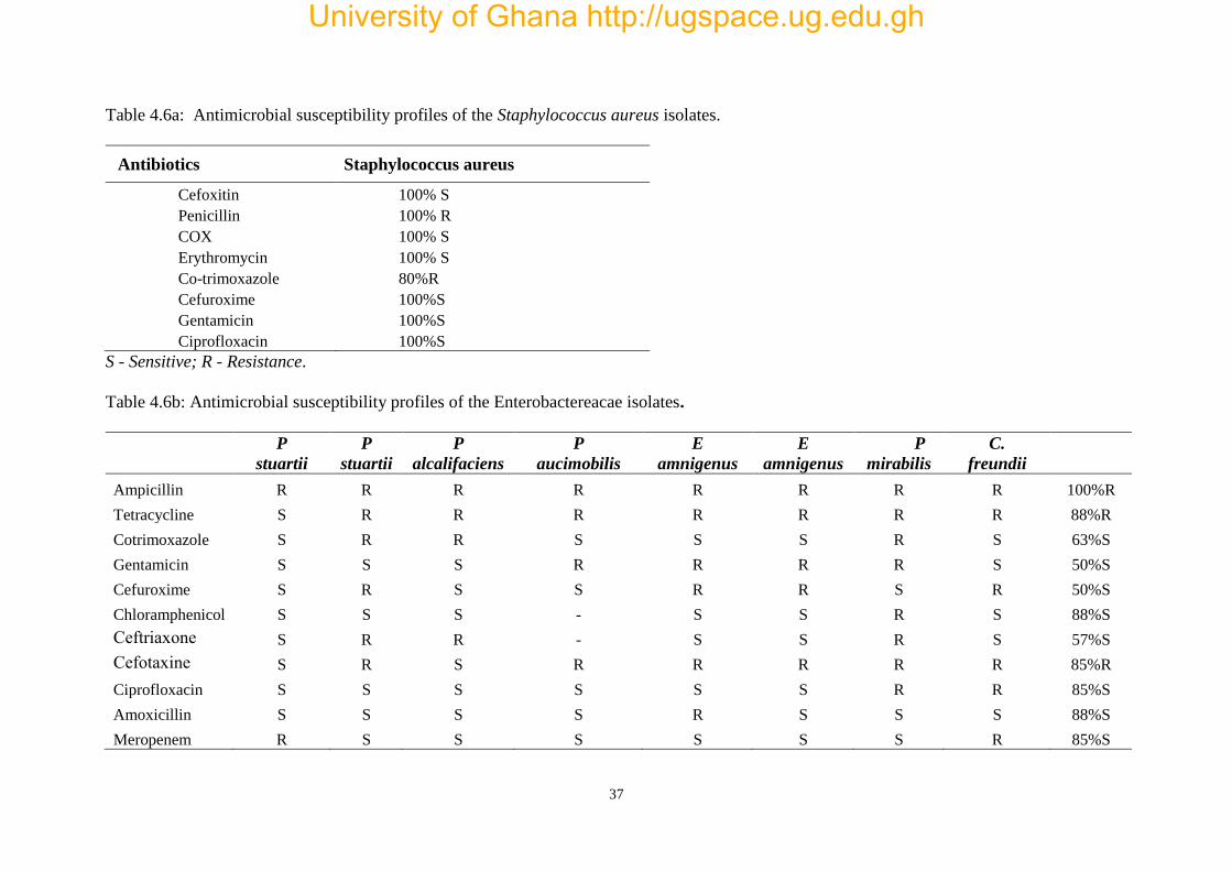
All the S. aureus isolates showed complete resistance to penicillin (100%) whiles 80%
resistance was for Co-trimoxazole. The Enterobacteriacae were completely resistant to
ampicillin. Tetracycline and ceftriaxone also shows high resistance. The etiologic
agents of bacteraemia are shown in table 4.4 and their corresponding antimicrobial
susceptibility patterns are shown in tables 4.5a and 4.5b.
Table 4.5: Isolates from positive blood culture
Organisms Total isolates Age groups
Gram Negative Bacteria
Citrobacter freundii
Providencia stuartii
Providencia alcalifaciens
Enterobacter amnigenus
Proteus mirabilis
Pseudomonas aucimobilis
Enterobacter amnigenus
1
2
1
1
1
1
1
0-2
0-2, 6-8
0-2
3-5
6-8
9-13
9-13
Gram positive Organisms
Staphylococcus aureus 5 3-5, 6-8, 9-13
University of Ghana http://ugspace.ug.edu.gh
Page 47
37
Table 4.6a: Antimicrobial susceptibility profiles of the Staphylococcus aureus isolates.
Antibiotics Staphylococcus aureus
Cefoxitin 100% S
Penicillin 100% R
COX 100% S
Erythromycin 100% S
Co-trimoxazole 80%R
Cefuroxime 100%S
Gentamicin 100%S
Ciprofloxacin 100%S
S - Sensitive; R - Resistance.
Table 4.6b: Antimicrobial susceptibility profiles of the Enterobactereacae isolates.
P
stuartii
P
stuartii
P
alcalifaciens
P
aucimobilis
E
amnigenus
E
amnigenus
P
mirabilis
C.
freundii
Ampicillin R R R R R R R R 100%R
Tetracycline S R R R R R R R 88%R
Cotrimoxazole S R R S S S R S 63%S
Gentamicin S S S R R R R S 50%S
Cefuroxime S R S S R R S R 50%S
Chloramphenicol S S S - S S R S 88%S
Ceftriaxone S R R - S S R S 57%S
Cefotaxine S R S R R R R R 85%R
Ciprofloxacin S S S S S S R R 85%S
Amoxicillin S S S S R S S S 88%S
Meropenem R S S S S S S R 85%S
University of Ghana http://ugspace.ug.edu.gh
Page 48
38
4.5 RISK FACTORS
Table 4.6 shows differences in proportions of the risk factors usually associated with invasive
bacterial infections. More than half of the children ate outside their homes frequently, and of
those who had toilet facilities at home, only 24.8% (31 out of 125) had flush toilets (water closet).
Recent antibiotic use within four weeks prior to the study was also minimal (14.65%).
Table 4.7. Risk factors for invasive bacterial infections.
*reference
Co-infections
x2 P-value Yes No
Recent antibiotic use
Yes 1 33 *
No 12 186 0.53 0.70
Sickling
Yes 0 24 1.59 0.37
No 13 195 *
Toilet facility at home
Yes 7 6 0.00 1.00
No 118 101 *
Type of toilet facility
Bush/polytene 1 14 0.00 1.00
KVIP 10 175 0.37 0.69
WC 2 30 *
Eating out
Never 7 42 10.28 <0.05
Occasionally 3 44 1.90 0.18
Regularly 3 133 *
Water source
Packaged 9 105 0.60 1.00
Borehole/tap 4 107 0.26 1.00
Well/Rain 0 7 *
University of Ghana http://ugspace.ug.edu.gh
Page 49
39
CHAPTER FIVE
DISCUSSIONS
The purpose of this study was to determine the prevalence of co-infection of malaria and
bacteraemia and also to assess the risk factors for bacterial infections within selected
communities in the Accra sub-metro. Malaria coupled with bacterial infections are a major public
health concern as the tendency of misdiagnosis is eminent and therefore tends to increase
morbidity and mortality.
More children reported to the clinics with febrile illnesses suspected to be malaria than adults.
They lived within the environs of the study sites and those who were of school going age were
enrolled in various schools. The finding of more children especially from Dodowa and Maamobi
adds to anecdotal reports indicating that children are more at risk of febrile illnesses suspected
to be malaria compared to adults. Age is an influential factor in malaria infection and may
contrast Svenson et al. (1995) who found that the severity of malaria was not age dependent.
Health survey from the PML Children’s hospital also indicate that the causes of frequent visits
to clinics was febrile related illnesses all of which was suspected malaria. Their increased risk of
malaria could be attributable to; immature immunity especially in children under the age of 2
years (WHO, 1996; Rijkers et al., 1998; Klein Klouwenberg and Blont, 2008) and multiple
exposures to the vector bites since children may not take necessary precautions about the vector
when they engage in outdoors activities.
University of Ghana http://ugspace.ug.edu.gh
Page 50
40
Both diseases have similar symptoms characterized by fever, weakness, body weight loss,
anaemia and in some cases gastrointestinal disturbances (Niikura et al., 2008; Samal and Sahu,
1991). The similarity in symptoms is one of the causes of difficulties that arises in the preliminary
diagnosis of the diseases and there is no report from the Ministry of Health, Ghana, on the status
of co-infections in the country. Bacterial co-infections in malaria positive children was evaluated
based on culture and identification with API20E. Thirteen (13) out of the 232 participants had
dual malaria and bloodstream bacterial infections, representing 5.6% co-infection. Surprisingly,
none of the cultured samples were positive for NTS despite being an important co-infection with
malaria and where many of the participants did not have potable water and toilet facilities in their
homes. The finding of no positive cultures for NTS is in huge contrast to other studies in the
country and elsewhere in Africa that have associated the susceptibility of NTS to malaria
infection, while others have related the two as most common in the tropics and subject to
misdiagnosis (Evans et al., 2004; Nielson et al., 2012; Bronzan et al., 2007; Lepage et al., 1987;
Maitland et al., 2006; Nesbitt et al., 1989).
Staphylococcus aureus was the only Gram positive organism among the six different organisms
isolated from blood cultures, all others were Gram negative. S. aureus may not necessarily be
attributed to malaria, instead, it confirms its implications in community-acquired bacteraemia
among children in rural sub-Saharan Africa. Recent investigations elsewhere in Africa show that
S. aureus was the most frequent cause of bacteraemia caused by Gram-positive organisms in
infants and young children presenting at a hospital in Nigeria (Johnson et al. 2008) and
Mozambique (Sigauque et al. 2009). These infections were single-infections and not co-infection
with malaria. It also agrees to findings from other developing countries that suggest S. aureus to
University of Ghana http://ugspace.ug.edu.gh
Page 51
41
be a major aetiological agent of septicaemia (Evans et al., 2004; Meremikwu et al., 2005; Hill et
al., 2007; Kizito et al., 2007; Komolafe and Adegoke, 2008) which constitute a significant threat
to child survival in these areas. The reason why S. aureus was predominant cannot be explained
from this study, however, it’s implications in community-acquired bacteraemia among children
at a rural sub-Saharan are highlighted. All of the S. aureus were resistant to penicillin (Table
4.5a). Usage of penicillin is therefore not appropriate for the treatment of Staphylococcal
systemic infections as it may lead to treatment failure. Cefoxitin, Erythromycin, Co-trimoxazole,
Gentamicin and Ciprofloxacin are more effective drugs of choice as the organisms showed no
resistance to these groups of antibiotics.
Several published reports have also demonstrated that Gram-negative organisms either exceed
or rival Gram-positive organisms in bloodstream infections in both adults and children from
African countries (Gordon et al., 2002; Ayoola et al., 2003; Archibald et al., 2003) as observed
in this study. It is unknown why Gram negative organisms were mostly implicated in
bloodstream infections but a likely reason that could explain this occurrence is that the Gram
Negative isolates belong to the group of Enterobacteriacae that usually colonize the gut and
progress to cause systemic invasion. It is noteworthy that, all the laboratory requisition forms of
the participants specified for malaria tests and there were no requests for blood associated
bacterial infections. Nevertheless, the isolation of some bacteria in the blood may indicate missed
diagnosis which would consequently lead to mistreatment. This may add to growing evidence
that, much attention is not paid in cases of malaria related co-infections in Ghana as their clinical
symptoms may present in the same way. Systemic bacteraemia reported herein in children with
University of Ghana http://ugspace.ug.edu.gh
Page 52
42
Plasmodium falciparum malaria makes the identification of concurrent infections a prerequisite
for adequate treatment given their overlap in clinical signs.
With the abundance of affordable antimalarial medicines, self-diagnosis of malaria is often the
main diagnostic approach used by many Ghanaians for febrile illnesses. The second clinical
diagnosis is often sought after the self-diagnosis and medication has failed (Nyamongo, 2002).
While prompt and accurate diagnosis of malaria is part of effective disease management, many
other etiologic agents could be responsible for febrile presentations. If these presentations are
accurately diagnosed, it could help to reduce indiscriminate use of antimalarial especially in
young children and improve differential diagnosis of febrile illness. It is hence important to
supplement febrile conditions with blood cultures to ensure accurate diagnosis despite their
confounding clinical symptoms.
The data presented support accumulating evidence that invasive bacterial infections are
important concurrent infections in paediatric populations with malaria. Studies from the Gambia
have found community acquired invasive bacteraemia infections to be higher in children aged 2-
29 months compared to adults (Enwere et al., 2006). A similar study at a rural hospital in
Mozambique showed an increased risk of community acquired bacteraemia in children <3 years
of age compared to adults (Sigauque et al., 2009). These finding of higher bacteraemia in children
may not be comparable to results from this study as adults were not included and the few isolates
obtained from blood cultures were not enough to draw definitive conclusions. The absence of
NTS isolates in positive blood cultures also imply that, results from this study show no
significance in associating NTS co-infections with malaria as other authors (Bronzan et al., 2007;
Evans et al., 2004) have previously demonstrated.
University of Ghana http://ugspace.ug.edu.gh
Page 53
43
On the other hand, the frequent diagnosis of malaria without routine bacterial cultures to diagnose
infections in most primary and secondary health facilities, some clinicians may be tempted to
give alternative remedies to other infections that mimic malaria. This may lead to unguided
empirical treatment especially with antibiotics, which, in turn, may result into antimicrobial
resistance and can affect a patient’s rate of recovery. There is therefore further emphasizes the
need for supplementary tests in ensuring accurate diagnosis before commencement of therapy.
Anaemia has been shown to be prevalent in areas where malaria is endemic (Le Hung et al.,
2005). The association between malaria and anaemia has been well documented (Adam et al.,
2005; Huddle et al., 1999; Kagu et al., 2007; Mayor et al., 2007; Muhangi et al., 2007; Ouma et
al., 2007; Tarimo, 2007). Malaria is characterized by a drop in the level of haemoglobin, resulting
from the destruction of erythrocytes and as expected, the results from this study shows that
anaemia was predominant among the participants. In Sub-Saharan Africa where malaria is
endemic, the association between anaemia and malaria is so strong that anaemia is often taken
as a proxy indicator of the malaria control programmes (Le Hung et al., 2005).
Although no mortality was recorded during the span of the study, the prevalence of anaemia was
high and may be disadvantageous in malaria positive patients, potentially leading to severe
anaemia as more RBCs are invaded and lysed when infection progresses without treatment. It is
worth mentioning that the 6.9% severe anaemia observed in of children from this study is a
frequent complication of Plasmodium falciparum infections that occurs in young children
(Breman, 2001) with a case-fatality rate reaching 23% in malaria holoendemic areas (Obonyo et
al., 2007). This study could not establish any associations between malaria and anaemia despite
the high proportions of anaemic children. A reason that accounted for this was the lack of a
University of Ghana http://ugspace.ug.edu.gh
Page 54
44
malaria negative control group. However, some studies have reported no association (Le Hung
et al., 2005; Stoltzfus et al., 2000; Stoltzfus et al., 1997) between malaria and anaemia. A study
by Mato (1998) among Yanomami Amerindian population from the Southern Venezuelan
Amazon also found no association between malaria and anaemia.
WBC counts are essential in predicting the health status or immunocompetence of a person and
are lower to the normal counts during malaria. While increased counts indicate infections,
reduced WBC counts, on the other hand, suggest an increased susceptibility to infections.
Interestingly, this study found high WBC counts in co-infected children compared to children
with only malaria and showed that, the WBC counts of the children did not fall below the
standard value as generally anticipated. It contrasts studies that suggest that children who test
positive for P. falciparum and those with high parasite densities are associated with low WBC
counts (Omalu et al 2008). The reasons for this disparity cannot be discerned from the study,
however, a risk factor for NTS infection in malaria is attributable to the reduced WBC counts
which impair the defense of the body and subsequently, immunity to diseases. Since no lower
counts of WBCs were recorded, it may be a likely reason why NTS was not isolated after culture
as it is arguable that the immune system of the children was not impaired to an extent of
predisposing them to NTS bacteraemia. This conclusion is however based on thin evidence.
Among the risk factors used to access possible transmission of bacterial of NTS among the
participants were; parasitaemia, source of drinking water, sickle cell disease, younger age, lack
of in-house toilet facilities, and lack of potable water. Surprisingly, participants who never
patronized food from outside their homes were associated with bacteraemia (p<0.01). All the
other factors showed no association.
University of Ghana http://ugspace.ug.edu.gh
Page 55
45
5.1 LIMITATAIONS
This study is subject to several limitations. To begin with, the study was conducted in urban areas
which are known to have lesser prevalence of bacterial infections compared to rural centres.
Choosing study sites from rural areas would have been preferable for prevalence studies
involving NTS.
Secondly, molecular techniques involving PCR are more sensitive and would have been more
accurate in diagnosis compared to blood cultures since some studies have argued that the
sensitivity of culture from 1mL of blood is minimal thus giving only 1CFU/mL for organisms
such as NTS. Furthermore, failure to collect other parameters such as temperature, BMI, blood
sugar and the comparatively lower sample size are other limitations that prevent the study from
drawing a definitive conclusion.
2.5 CONCLUSION
In conclusion, malaria has gained more recognition through research compared to bacterial
etiologic agents. Similarities in the clinical presentations of both diseases may influence
clinicians to pay particular attention to the management of malaria in patients while co-infections
may be missed out on the first diagnosis. This study predominantly found S. aureus and
Enterobacteria to be responsible for co-infections in malaria patients. NTS which has been
previously reported by various authors may not be associated with malaria in the population
studied. Although none of the stool cultures was positive for bacteria, there is the need to provide
safe drinking water, good toilet facilities and practice adequate personal hygiene in order to
University of Ghana http://ugspace.ug.edu.gh
Page 56
46
prevent the transmission of bacterial infections, especially in areas where malaria is endemic
areas.
5.3 RECOMMENDATIONS
Knowledge of the prevalence, risk factors of NTS and malaria co-infections in Ghana,
particularly in high risk areas is critical in misdiagnosis of malaria regionally and globally. There
is the need for clinicians, public health officials GHS/Policy makers to include invasive bacterial
infection such as NTS in routine diagnosis of persons presenting with febrile illnesses.
Malaria still remains a public health concern especially in children. In the environs of the study
sites where drainage is poor and mosquito breeding habitats have an extensive distribution,
parents and caregivers should protect their children with long lasting insecticide treated bed nets.
Furthermore, preventive strategies among the general public should include; promotion of inbuilt
toilet facilities at home to maximise personal hygiene. Provision of potable water for
communities without access to running tap water.
Traditional microbiological culture methods employed to detect systemic bacteraemia are often
time consuming and have modest sensitivity. Therefore, molecular methods may be useful in
accurately diagnosing bacteraemia.
University of Ghana http://ugspace.ug.edu.gh
Page 57
47
REFERENCES
Acquah, S. E., Quaye, L., Sagoe, K., Ziem, J. B., Bromberger, P. I. and Amponsem, A. A. (2013).
Susceptibility of bacterial etiological agents to commonly-used antimicrobial agents in children
with sepsis at the Tamale Teaching Hospital. BMC Infect Dis, 13, 89.
Adam, I., Khamis, A.H. and Elbashir, M.I. (2005) Prevalence and risk factors for anaemia in
pregnant women of eastern Sudan. Transactions of the Royal Society of Tropical Medicineand
Hygiene 99, 739‐743
Ahmed, K. (1989). Epidemiology of malaria in Ghana. Ghana Med. J. 23, 190-196.
Aikawa, M., Suzuki, M., and Gutierrez, Y. (1980). Pathology of malaria. Malaria, 2, 47-102.
Alam, A. (2014). Serine proteases of malaria parasite Plasmodium falciparum: potential as
antimalarial drug targets. Interdisciplinary perspectives on infectious diseases, 2014.
Antignac, A., Ducos-Galand, M., Guiyoule, A., Pirès, R., Alonso, J. M. and Taha, M.-K. (2003).
Neisseria meningitidis strains isolated from invasive infections in France (1999–2002):
phenotypes and antibiotic susceptibility patterns. Clin Infect Dis, 37, 912-920.
Archibald, L. K., Kazembe, P. N., Nwanyanwu, O., Mwansambo, C., Reller, L. B., & Jarvis, W.
R. (2003). Epidemiology of bloodstream infections in a bacille Calmette-Guerin-vaccinated
pediatric population in Malawi. Journal of Infectious Diseases, 188(2), 202-208.
Awoniyi, D., Udo, S. and Oguntibeju, O. (2009). An epidemiological survey of neonatal sepsis
in a hospital in Western Nigeria. Afr J Microbiol Research, 3, 385-389.
University of Ghana http://ugspace.ug.edu.gh
Page 58
48
Ayoola, O. O., Adeyemo, A. A. and Osinisu, K. (2003). Aetiological agents,clinical features and
outcome of septicaemia in infants in Ibadan. WAJM, 22.
Bandyopadhyay, S, Bergholte J, Blackwell CD, Friedlander JR, Hennes H. (2002). Risk of
serious bacterial infection in children with fever without a source in the post–haemophilus
influenzae Era when antibiotics Are reserved for culture-proven bacteremia. Arch Pediatr
Adolesc Med, 156:512–517.
Bassat, Q., Ogutu, B., Djimde, A., Stricker, K., and Hamed, K. (2015). Tailoring a Pediatric
Formulation of Artemether-Lumefantrine for Treatment of Plasmodium falciparum Malaria.
Antimicrobial Agents and Chemotherapy, 59(8), 4366-4374.
Bauer, A.W., Kirby, W.M.M., Sherris, J.C., Turck, M. (1966): Antibiotic susceptibility testing
by a standardized single disk method. Am. J. Clin. Pathol. 45: 493-496.
Berger, S. A (1983) Pseudobacteremia due to contaminated alcohol swabs. J Clin Microbiol
18(4): 974-975.
Berkley, J, Mwarumba S, Bramham K, Lowe B, Marsh K. (1999). Bacteremia complicating
severe malaria in children. Trans. R. Soc. Trop. Med. Hyg. 93:283–286.
Berkley, J. A., Bejon, P., Mwangi, T., Gwer, S., Maitland, K., Williams, T. N., and Newton, C.
R. (2009). HIV infection, malnutrition, and invasive bacterial infection among children with
severe malaria. Clinical infectious diseases, 49(3), 336-343.
University of Ghana http://ugspace.ug.edu.gh
Page 59
49
Berkley, J. A., Maitland, K., Mwangi, I., Ngetsa, C., Mwarumba, S., Lowe, B. S., Newton, C. R.
J. C., Marsh, K., Scott, J. A. G., English, M., (2005). Use of clinical syndromes to target
antibiotic prescribing in seriously ill children in malaria endemic area. BMJ, 10:1136.
Bhandari, A., Dratler, S., Raube, K., and Thulasiraj, R. D. (2008a). Specialty care systems: A
pioneering vision for global health. Health Affairs, 27(4), 964-976.
Bhandari, P. L., Raghuveer, C. V., Rajeev, A, and Bhandari, P. D. (2008b). Comparative study
of peripheral blood smear, Quantitative buffy coat and Modified centrifuged blood smear in
malaria diagnosis. Indian J Pathol Microbiol; 51(1): 108-12
Blomberg, B., Manji, K. P., Urassa, W. K., Tamim, B. S., Mwakagile, D. S., Jureen, R., and
Maselle, S. Y. (2007). Antimicrobial resistance predicts death in Tanzanian children with
bloodstream infections: a prospective cohort study.BMC infectious diseases, 7(1), 1.
Bogaert, D., De Groot, R. and Hermans, P. (2004). Streptococcus pneumoniae colonisation: the
key to pneumococcal disease. Lancet Infect Dis, 4, 144-154.
Boyd, M. F. (Ed.). (1949). Malariology: A comprehensive survey of all aspects of this group of
diseases from a global standpoint (Vol. 2). Saunders.
Breman, J. G. (2001). The Ears of the Hippopotamus: Manifestations, Determinants, and
Estimates of the Malaria Burden. American Journal of Tropical Medicine and Hygiene.;64(Suppl.
12):1–11.
University of Ghana http://ugspace.ug.edu.gh
Page 60
50
Breman, J. G., Alilio, M. S., and Mills, A. (2004). Conquering the intolerable burden of malaria:
what’s new, what’s needed: a summary. The American journal of tropical medicine and
hygiene, 71(2 suppl), 1-15.
Bronzan, R. N., Taylor, T. E., Mwenechanya, J., Tembo, M., Kayira, K., Bwanaisa, L., and Phiri,
A. (2007). Bacteremia in Malawian children with severe malaria: prevalence, etiology, HIV
coinfection, and outcome. Journal of Infectious Diseases, 195(6), 895-904.
Brooks, G. F., Carroll, K. C., Butel, J. S., Morse, S. A. (2007) Medical Microbiology. 24th
Edition. McGraw-Hill Companies, Inc. San Francisco. Pp. 145-150.
Brown, H., Rogerson, S., Taylor, T., Tembo, M., Mwenechanya, J., Molyneux, M., & Turner,
G. (2001). Blood-brain barrier function in cerebral malaria in Malawian children. The American
journal of tropical medicine and hygiene,64(3), 207-213.
Bryce, J., Boschi-Pinto, C., Shibuya, K., Black, R. E., & WHO Child Health Epidemiology
Reference Group. (2005). WHO estimates of the causes of death in children. The
Lancet, 365(9465), 1147-1152.
Carpenter, C. C., Pearson, G. W., Mitchell, V. S., & Oaks Jr, S. C. (Eds.). (1991). Malaria:
Obstacles and Opportunities. National Academies Press.
Carter, R., and Mendis, K. N. (2002). Evolutionary and historical aspects of the burden of
malaria. Clinical microbiology reviews, 15(4), 564-594.
Caugant, D. A., and Maiden, M. C. (2009). Meningococcal carriage and disease—population
biology and evolution. Vaccine, 27, B64-B70.
University of Ghana http://ugspace.ug.edu.gh
Page 61
51
CDC. (2012). CDC Health Information for International Travel 2012. Oxford University Press,
New York, USA
Cheesbrough, M. (1987). Medical Laboratory Manual for Tropical Countries. Volume 1 (No.
Ed. 2). Tropical Health Technology.
Chin, W., Contacos, P. G., Collins, W. E., Jeter, M. H., and Alpert, E. (1968). Experimental
mosquito-transmission of Plasmodium knowlesi to man and monkey. The American Journal of
Tropical Medicine and Hygiene, 17(3), 355-358.
Christopher, A., Mshana, S. E., Kidenya, B. R., Hokororo, A., and Morona, D. (2013).
Bacteremia and resistant gram-negative pathogens among under-fives in Tanzania. Ital J Pediatr,
39(1), 27.
Clinical and Laboratory Standards Institute (CLSI). (2013). M100-S23. Performance standards
for antimicrobial susceptibility testing: 23rd informational supplement. CLSI, Wayne, PA.
Cook, G.C. and Zumla, A.I. (eds.) Manson's Tropical Diseases (Saunders Elsevier, 2009).
Cox-Singh, J., Davis, T. M., Lee, K. S., Shamsul, S. S., Matusop, A., Ratnam, S., ... and Singh,
B. (2008). Plasmodium knowlesi malaria in humans is widely distributed and potentially life
threatening. Clinical infectious diseases, 46(2), 165-171.
Daneshvar, C., Davis, T. M., Cox-Singh, J., Rafa'ee, M. Z., Zakaria, S. K., Divis, P. C., and
Singh, B. (2009). Clinical and laboratory features of human Plasmodium knowlesi infection.
Clinical infectious diseases, 49(6), 852-860.
University of Ghana http://ugspace.ug.edu.gh
Page 62
52
Del Rio, A., Cervera, C., Moreno, A., Moreillon, P. and Miró, J. M. (2009). Patients at risk of
complications of Staphylococcus aureus bloodstream infection. Clin Infect Dis, 48, S246-S253.
Dondorp, A, Nosten, F., Stepniewska, K., Day, N., White, N., S. E. A. Q. A. M. A. T. (2005a).
Artesunate versus quinine for treatment of severe falciparum malaria: a randomised trial. Lancet;
366:717–25.
Dondorp, A. M., Desakorn, V., Pongtavornpinyo, W., Sahassananda, D., Silamut, K.,
Chotivanich, K., Newton, P. N., Pitisuttithum, P., Smithyman, A. M., White, N. J., Day, N. P.
(2005b). Estimation of the total parasite biomass in acute falciparum malaria from plasma
PfHRP2. PLoS Med, 2(8), e204.
Dondorp, A. M., Fanello, C. I., Hendriksen, I. C., Gomes, E., Seni, A., Chhaganlal, K. D., ... and
AQUAMAT group. (2010). Artesunate versus quinine in the treatment of severe falciparum
malaria in African children (AQUAMAT): an open-label, randomised trial. The Lancet,
376(9753), 1647-1657.
Donkor, E. S., Adegbola, R. A., Wren, B. W. and Antonio, M. (2013). Population biology of
Streptococcus pneumoniae in West Africa: multilocus sequence typing of serotypes that exhibit
different predisposition to invasive disease and carriage. PloS one, 8, e53925.
Dorovini-Zis, K., Schmidt, K., Huynh, H., Fu, W., Whitten, R. O., Milner, D., ... & Taylor, T.
E. (2011). The neuropathology of fatal cerebral malaria in malawian children. The American
journal of pathology, 178(5), 2146-2158.
University of Ghana http://ugspace.ug.edu.gh
Page 63
53
Eckstein-Ludwig, U., Webb, R. J., Van Goethem, I. D. A., East, J. M., Lee, A. G., Kimura, M.,
and Krishna, S. (2003). Artemisinins target the SERCA of Plasmodium falciparum. Nature,
424(6951), 957-961.
Enwere, G., Biney, E., Cheung, Y., Zaman, S. M., Okoko, B., Oluwalana, C., and Cutts, F. T.
(2006). Epidemiologic and clinical characteristics of community-acquired invasive bacterial
infections in children aged 2–29 months in The Gambia. The Pediatric infectious disease journal,
25(8), 700-705.
Essuman, V. A., Ntim-Amponsah, C. T., Astrup, B. S., Adjei, G. O., Kurtzhals, J. A., Ndanu, T.
A., & Goka, B. (2010). Retinopathy in severe malaria in Ghanaian children–overlap between
fundus changes in cerebral and non-cerebral malaria. Malar J, 9, 232.
Evans, J. A., Adusei, A., Timmann, C., May, J., Mack, D., Agbenyega, T., ... & Frimpong, E.
(2004). High mortality of infant bacteraemia clinically indistinguishable from severe malaria.
Qjm, 97(9), 591-597.
Feasey, N. A., Dougan, G., Kingsley, R. A., Heyderman, R. S., and Gordon, M. A. (2012).
Invasive non-typhoidal salmonella disease: an emerging and neglected tropical disease in Africa.
The Lancet, 379(9835), 2489-2499.
Gallup, J. L., and Sachs, J. D. (2001). The economic burden of malaria. The American journal
of tropical medicine and hygiene, 64(1 suppl), 85-96.
University of Ghana http://ugspace.ug.edu.gh
Page 64
54
García, F., Cebrián, M., Dgedge, M., Casademont, J., Bedini, J. L., Neves, O., Grau, J. M. (1999).
Endothelial cell activation in muscle biopsy samples is related to clinical severity in human
cerebral malaria. Journal of Infectious Diseases, 179(2), 475-483.
García-Cobos, S., Campos, J., Cercenado, E., Román, F., Lázaro, E., Pérez-Vázquez, M., De
Abajo, F. and Oteo, J. 2008. Antibiotic resistance in Haemophilus influenzae decreased, except
for β-lactamase-negative amoxicillin-resistant 67 isolates, in parallel with community antibiotic
consumption in Spain from 1997 to 2007. Antimicrob Agents Chemother, 52, 2760-2766.
Gill, J., Kumar, R., Todd, J., & Wiskin, C. (2006). Methicillin-resistant Staphylococcus aureus:
awareness and perceptions. Journal of hospital infection, 62(3), 333-337.
Gordon, M. A., Banda, H. T., Gondwe, M., Gordon, S. B., Boeree, M. J., Walsh, A. L., ... and
Molyneux, M. E. (2002). Non-typhoidal salmonella bacteraemia among HIV-infected Malawian
adults: high mortality and frequent recrudescence. Aids, 16(12), 1633-1641.
Gordon, M. A., Graham, S. M., Walsh, A. L., Wilson, L., Phiri, A., Molyneux, E., & Molyneux,
M. E. (2008). Epidemics of invasive Salmonella enterica serovar enteritidis and S. enterica
Serovar typhimurium infection associated with multidrug resistance among adults and children
in Malawi. Clinical Infectious Diseases, 46(7), 963-969.
Graham, S. M., Molyneux, E. M., Walsh, A. L., Cheesbrough, J. S., Molyneux, M. E. and Hart,
C. A. (2000). Nontyphoidal Salmonella infections of children in tropical Africa. Pediatr Infect
Dis J, 19, 1189-1196.
University of Ghana http://ugspace.ug.edu.gh
Page 65
55
Greenwood, B. (2007). The changing face of meningococcal disease in West Africa. Epidemiol
Infect, 135, 703-705.
Hakeem, L., Laing, R. B., Tonna, I., Douglas, J. G. and Mackenzie, A. R. (2013). Invasive
Staphylococcus aureus infections in diabetes mellitus. Br J Diabetes Vasc Dis, 13, 164-177.
Haldar, K., Murphy, S. C., Milner Jr, D. A., & Taylor, T. E. (2007). Malaria: mechanisms of
erythrocytic infection and pathological correlates of severe disease. Annu. Rev. Pathol. Mech.
Dis., 2, 217-249.
Harinasuta, T., Bunnag, D., Wernsdorfer, W. H., & McGregor, I. (1988). The clinical features
of malaria. Malaria: principles and practice of malariology. Volume 1., 709-734.
Hill, P. C., Onyeama, C. O., Ikumapayi, U. N., Secka, O., Ameyaw, S., Simmonds, N., Donkor,
S. A., Howie, S. R., Tapgun, M. and Corrah, T. (2007). Bacteraemia in patients admitted to an
urban hospital in West Africa. BMC Infect Dis, 7, 2.
Hoffman, S. L. (1996). Malaria vaccine development: a multi-immune response approach.
American Society for Microbiology (ASM).
Holland, C. A., & Kiechle, F. L. (2005). Point-of-care molecular diagnostic systems—past,
present and future. Current opinion in microbiology, 8(5), 504-509.
Huddle, J. M., Gibson, R. S., & Cullinan, T. R. (1999). The impact of malarial infection and diet
on the anaemia status of rural pregnant Malawian women. European journal of clinical nutrition,
53(10), 792-801.
University of Ghana http://ugspace.ug.edu.gh
Page 66
56
Ikumapayi, U. N., Antonio, M., Sonne-Hansen, J., Biney, E., Enwere, G., Okoko, B., Oluwalana,
C., Vaughan, A., Zaman, S. M. and Greenwood, B. M. (2007). Molecular epidemiology of
community-acquired invasive non-typhoidal Salmonella among children aged 2–29 months in
rural Gambia and discovery of a new serovar, Salmonella enterica Dingiri. J Med Microbiol, 56,
1479-1484.
Isaacman, D. J., Mcintosh, E. D. and Reinert, R. R. (2010). Burden of invasive pneumococcal
disease and serotype distribution among Streptococcus pneumoniae isolates in young children
in Europe: impact of the 7-valent pneumococcal conjugate vaccine and considerations for future
conjugate vaccines. Int J Infect Dis, 14, e197-e209.
Johnson, A. W. B., Osinusi, K., Aderele, W. I., Gbadero, D. A., Olaleye, O. D., & Adeyemi-
Doro, F. A. (2008). Etiologic agents and outcome determinants of community-acquired
pneumonia in urban children: a hospital-based study. Journal of the National Medical
Association, 100(4), 370-385.
Kagu, M. B., Kawuwa, M. B., & Gadzama, G. B. (2007). Anaemia in pregnancy: a cross-
sectional study of pregnant women in a Sahelian tertiary hospital in Northeastern Nigeria.
Journal of Obstetrics and Gynaecology, 27(7), 676-679.
Kamga, H. L. F., Njunda, P. F., Nde, A. L., Assob, J. C. N., Nsagha, D. S. and Weledji, P. (2011).
Prevalence of septicaemia and antibiotic sensitivity pattern of bacteria isolate at the University
Teaching Hospital, Yaounde. Afr J Clin Exp Microbiol, 12, 2 - 8.
University of Ghana http://ugspace.ug.edu.gh
Page 67
57
Kariuki, S., Revathi, G., Kariuki, N., Kiiru, J., Mwituria, J. and Hart, C. A. (2006).
Characterisation of community acquired non-typhoidal Salmonella from bacteraemia and
diarrhoeal infections in children admitted to hospital in Nairobi, Kenya. BMC Microbiol, 6, 101.
Kaufman, D. and Fairchild, K. D. (2004). Clinical microbiology of bacterial and fungal sepsis in
very-low-birth-weight infants. Clin Microbiol Rev, 17, 638-680.
Kayser, F. H., Bienz, K. A., Eckert, J., & Zinkernagel, R. M. (2005). General bacteriology.
Medical microbiology. New York: Kayser Microbiology, 146-309.
Keong, B. C. M., & Sulaiman, W. (2006). Typhoid and malaria co-infection-an interesting
finding in the investigation of a tropical fever. Malaysian J Med Sci, 13, 74-5.
Khan, N. and Lai. A. (1999). “The malaria website: pathology/immune response.” . Biology
Department, Brown University.
Kizito, M., Mworozi, E., Ndugwa, C. and Serjeant, G. R. (2007). Bacteraemia in homozygous
sickle cell disease in Africa: is pneumococcal prophylaxis justified? Arch Dis Child, 92, 21-23.
Klein Klouwenberg, P., & Bont, L. (2008). Neonatal and infantile immune responses to
encapsulated bacteria and conjugate vaccines. Clinical and Developmental Immunology, 2008.
Komolafe, A. O. and Adegoke, A. A. (2008). Incidence of bacterial Septicaemia in Ile-Ife
Metropolis, Nigeria. Malays J Microbiol, 4(2), 51- 61.
Komolafe, A. O., & Adegoke, A. A. (2008). Incidence of bacterial septicaemia in Ile-Ife
metropolis, Nigeria. Malaysian Journal of Microbiology, 4(2), 51-61.
University of Ghana http://ugspace.ug.edu.gh
Page 68
58
Le Hung, Q., de Vries, P. J., Giao, P. T., Binh, T. Q., Nam, N. V., and Kager, P. A. (2005).
Anemia, malaria and hookworm infections in a Vietnamese ethnic minority.
LeFrock, JL, Ellis, CA, Turchik, JB, Weinstein, L (1973) Transient bacteremia associated with
sigmoidoscopy. N Engl J Med 289(9): 467-469.
Lepage, P., Bogaerts, J., Van Goethem, C., Ntahorutaba, M., Nsengumuremyi, F., Hitimana, D.,
& Levy, J. (1987). Community-acquired bacteraemia in African children. The Lancet,
329(8548), 1458-1461.
Luxemburger, C., Nosten, F., Kyle, D., Kiricharoen, L, Chongsuphajasiddhi, T. and White, N.
(1998). Clinical features cannot predict a diagnosis of malaria or differentiate the infecting
species in children living in an area of low transmission. Trans R Soc Trop Med Hyg; 92:45-49.
Lynch, M.J. and S.S. Raphael, 1983. Immunology and Serology. Lynch's Medical Laboratory
Technology, Saunders, Philadelphia.
Mackenzie, G., Ceesay, S. J., Hill, P. C., Walther, M., Bojang, K. A., Satoguina, J., and
Greenwood, B. M. (2010). A decline in the incidence of invasive non-typhoidal Salmonella
infection in The Gambia temporally associated with a decline in malaria infection.
Mahon, CR, Manuselis, G. (2000). Textbook of Diagnostic Microbiology. 2nd
Edition. Elsevierʼs
Health Sciences, Philadelphia. Pp 997-1009.
Maitland, K., Berkley, J. A., Shebbe, M., Peshu, N., English, M., & Newton, C. R. C. (2006).
Children with severe malnutrition: can those at highest risk of death be identified with the WHO
protocol?. PLoS Med, 3(12), e500.
University of Ghana http://ugspace.ug.edu.gh
Page 69
59
Maneerat, Y., Viriyavejakul, P., Punpoowong, B., Jones, M., Wilairatana, P., Pongponratn, E.,
& Udomsangpetch, R. (2000). Inducible nitric oxide synthase expression is increased in the brain
in fatal cerebral malaria. Histopathology, 37(3), 269-277.
Mato, S. P. (1998). Anemia and malaria in a Yanomami Amerindian population from the
southern Venezuelan Amazon. The American journal of tropical medicine and hygiene 59(6):
998-1001.
Maude, R. J., Dondorp, A. M., Sayeed, A. A., Day, N. P., White, N. J., & Beare, N. A. (2009).
The eye in cerebral malaria: what can it teach us?. Transactions of the Royal Society of Tropical
Medicine and Hygiene, 103(7), 661-664.
Mayor, A., Aponte, J. J., Fogg, C., Saúte, F., Greenwood, B., Dgedge, M., and Alonso, P. L.
(2007). The epidemiology of malaria in adults in a rural area of southern Mozambique. Malaria
journal, 6(1), 3.
McMorrow, M. L., Masanja, M. I., Abdulla, S. M., Kahigwa, E., and Kachur, S. P. (2008).
Challenges in routine implementation and quality control of rapid diagnostic tests for malaria–
Rufiji District, Tanzania. The American journal of tropical medicine and hygiene, 79(3), 385-
390.
Medana, I. M., Day, N. P., Sachanonta, N., Mai, N. T., Dondorp, A. M., Pongponratn, E., ... &
Turner, G. D. (2011). Coma in fatal adult human malaria is not caused by cerebral oedema. Malar
J, 10, 267.
University of Ghana http://ugspace.ug.edu.gh
Page 70
60
Meremikwu, M. M., Nwachukwu, C. E., Asuquo, A. E., Okebe, J. U., & Utsalo, S. J. (2005).
Bacterial isolates from blood cultures of children with suspected septicaemia in Calabar, Nigeria.
BMC infectious diseases, 5(1), 1.
Miller, L. H., Baruch, D. I., Marsh, K., & Doumbo, O. K. (2002). The pathogenic basis of
malaria. Nature, 415(6872), 673-679.
Morpeth, S. C., Ramadhani, H. O., & Crump, J. A. (2009). Invasive non-typhi Salmonella
disease in Africa. Clinical Infectious Diseases, 49(4), 606-611.
Mtove, G., Amos, B., Nadjm, B., Hendriksen, I. C., Dondorp, A. M., Mwambuli, A., and Deen,
J. (2011). Decreasing incidence of severe malaria and community-acquired bacteraemia among
hospitalized children in Muheza, north-eastern Tanzania, 2006–2010. Malar J, 10(320), 1475-
2875.
Muhangi, L., Woodburn, P., Omara, M., Omoding, N., Kizito, D., Mpairwe, H., and Elliott, A.
M. (2007). Associations between mild-to-moderate anaemia in pregnancy and helminth, malaria
and HIV infection in Entebbe, Uganda. Transactions of the Royal Society of Tropical Medicine
and Hygiene, 101(9), 899-907.
Murphy, S. C., and Breman, J. G. (2001). Gaps in the childhood malaria burden in Africa:
cerebral malaria, neurological sequelae, anemia, respiratory distress, hypoglycemia, and
complications of pregnancy. The American journal of tropical medicine and hygiene, 64(1
suppl), 57-67.
University of Ghana http://ugspace.ug.edu.gh
Page 71
61
Musher, D. M., Alexandraki, I., Graviss, E. A., Yanbeiy, N, Eid, A., Inderias, L. A., Phan, H.
M., Solomon, E. (2000). Bacteremic and nonbacteremic pneumococcal pneumonia. A
prospective study. Medicine (Baltimore) 79(4): 210-221.
Mwangi, T. W., Mohammed, M., Dayo, H., Snow, R. W., and Marsh, K. (2005). Clinical
algorithms for malaria diagnosis lack utility among people of different age groups. Tropical
Medicine and International Health, 10(6), 530-536.
Naber, C. K. (2009). Staphylococcus aureus bacteremia: epidemiology, pathophysiology, and
management strategies. Clin Infect Dis, 48, S231-S237.
National Institutes of Health (NIH), National Institute of Allergy and Infectious Diseases (2007).
Understanding Malaria: Fighting an Ancient Scourge. NIH Publication No. 07-7139.
National Institutes of Health (NIH). (2007). “Understanding malaria: fighting an ancient
scourge.” U.S. Department of Health and Human Services.
Nesbitt, A., & Mirza, N. B. (1989). Salmonella septicaemias in Kenyan children. Journal of
tropical pediatrics, 35(1), 35-39.
Nielsen, M. V., Sarpong, N., Krumkamp, R., Dekker, D., Loag, W., Amemasor, S., Agyekum,
A., Marks, F., Huenger, F., Krefis, A. C., Hagen, R. M., Adu-Sarkodie, Y., May, J. and Schwarz,
N. G. (2012). Incidence and characteristics of bacteraemia among children in Rural Ghana. PloS
one, 7, e44063.
University of Ghana http://ugspace.ug.edu.gh
Page 72
62
Niikura, M., Kamiya, S., Kiyoshi, K., Fumie., K. (2008). Coinfection with Nonlethal Murine
Malaria Parasites Suppresses Pathogenesis Caused by Plasmodium berghei NK65. J. Immunol.
180: 6877-6884.
NMCP/GHS, 2008. Strategic Plan for Malaria Control in Ghana (2008-2015).
Nsutebu, E. F., Ndumbe, P. M., & Koulla, S. (2002). The increase in occurrence of typhoid fever
in Cameroon: overdiagnosis due to misuse of the Widal test?. Transactions of the Royal Society
of Tropical Medicine and Hygiene, 96(1), 64-67.
Nyamongo, I. K. (2002). Health care switching behaviour of malaria patients in a Kenyan rural
community. Social science and medicine. 54 377-386.
Obonyo, C. O., Vulule, J., Akhwale, W. S., & Grobbee, D. E. (2007). In-hospital morbidity and
mortality due to severe malarial anemia in western Kenya. The American journal of tropical
medicine and hygiene, 77(6 Suppl), 23-28.
Omalu, I. C., Oguche, S., Gyang, V. P., Akindigh, T. M., Egah, D. Z., and Gokop, B. (2008).
Standard white blood cell count for malaria density estimation: A need for review? Annals of
Tropical Medicine and Public Health, 1(1), 29.
Omoregie, R., Egbe, C. A., Ogefere, H. O., Igbarumah, I. and Omijie, R. E. (2009). Effects of
Gender and Seasonal Variation on the Prevalence of Bacterial Septicaemia Among Young
Children in Benin City, Nigeria. Libyan J Med, 4, 153 - 157.
University of Ghana http://ugspace.ug.edu.gh
Page 73
63
Ouma, P., Van Eijk, A. M., Hamel, M. J., Parise, M., Ayisi, J. G., Otieno, K., and Slutsker, L.
(2007). Malaria and anaemia among pregnant women at first antenatal clinic visit in Kisumu,
western Kenya. Tropical Medicine and International Health, 12(12), 1515-1523.
Oundo, J. O., Muli, F., Kariuki, S., Waiyaki, P. G., Iijima, Y., Berkley, J., and Lowe, B. (2002).
Non-typhi salmonella in children with severe malaria. East African medical journal, 79(12), 633-
639.
Parrillo, J. E. (1993). Pathogenetic mechanisms of septic shock. New England Journal of
Medicine, 328(20), 1471-1477.
Phiri, A., Milledge, J., Calis, J. C. J., Graham, S. M., Wilson, L. K., Soko, D., ... & Molyneux,
E. M. (2006). Aetiology of neonatal sepsis at QECH, Blantyre: 1996-2001. Malawi Medical
Journal, 17(3), 92-96.
PMI, 2012. Malaria Operational Plan – FY 2012 (Year 5): Ghana.
Prabhu, K., Bhat, S., & Rao, S. (2010). Bacteriologic profile and antibiogram of blood culture
isolates in a pediatric care unit. Journal of laboratory physicians, 2(2), 85.
Resman, F., Ristovski, M., Ahl, J., Forsgren, A., Gilsdorf, J. R., Jasir, A., ... & Riesbeck, K.
(2011). Invasive disease caused by Haemophilus influenzae in Sweden 1997–2009; evidence of
increasing incidence and clinical burden of non‐type b strains. Clinical Microbiology and
Infection, 17(11), 1638-1645.
University of Ghana http://ugspace.ug.edu.gh
Page 74
64
Rijkers, G. T., Sanders, E. A. M., Breukels, M. A., & Zegers, B. J. M. (1998). Infant B cell
responses to polysaccharide determinants. Vaccine, 16(14), 1396-1400.
Roca, A., Sigauque, B., Quintó, L., Mandomando, I., Valles, X., Espasa, M., ... & Levine, M.
(2006). Invasive pneumococcal disease in children <5 years of age in rural Mozambique.
Tropical Medicine & International Health, 11(9), 1422-1431.
Rosenstein, N. E., Perkins, B. A., Stephens, D. S., Popovic, T. and Hughes, J. M. (2001).
Meningococcal disease. N Engl J Med, 344, 1378-1388.
Samal, K. K., & Sahu, C. S. (1991). Malaria and Widal reaction. The Journal of the Association
of Physicians of India, 39(10), 745-747.
Shaw, A., Reddy, E. and Crump, J. (2008). Etiology of community-acquired bloodstream
infections in Africa. In: 46th Annual Meeting of the Infectious Diseases Society of America.
Washington, D.C., 20.
Shinefield, H., Black, S., Fattom, A., Horwith, G., Rasgon, S., Ordonez, J., Yeoh, H., Law, D.,
Robbins, J. B. and Schneerson, R. (2002). Use of a Staphylococcus aureus conjugate vaccine in
patients receiving hemodialysis. N Engl J Med, 346, 491-496.
Sigauque, B., Roca, A., Mandomando, I. (2009). Community-acquired bacteremia among
children admitted to a rural hospital in Mozambique. Pediatr Infect Dis J; 28:108–13.
Smith, D. C. (1982). The rise and fall of typhomalarial fever: I. Origins. Journal of the history of
medicine and allied sciences, 37(2), 182-220.
University of Ghana http://ugspace.ug.edu.gh
Page 75
65
Stephens, D. S., Greenwood, B. and Brandtzaeg, P. (2007). Epidemic meningitis,
meningococcemia, and Neisseria meningitidis. Lancet, 369, 2196-2210.
Stoltzfus, R. J., Chwaya, H. M., Montresor, A., Albonico, M., Savioli, L., & Tielsch, J. M.
(2000). Malaria, hookworms and recent fever are related to anemia and iron status indicators in
0-to 5-y old Zanzibari children and these relationships change with age. The Journal of nutrition,
130(7), 1724-1733.
Stoltzfus, R. J., Chwaya, H. M., Tielsch, J. M., Schulze, K. J., Albonico, M. and Savioli, L.
(1997) Epidemiology of iron deficiency anemia in Zanzibari schoolchildren: the importance of
hookworms. Am. J. Clin. Nutr. 65:153-159.
Svenson, J. E., MacLean, J. D., Gyorkos, T. W., & Keystone, J. (1995). Imported malaria:
clinical presentation and examination of symptomatic travelers. Archives of Internal
Medicine, 155(8), 861-868.
Tarimo, D. S. (2007). Appraisal on the prevalence of malaria and anaemia in pregnancy and
factors influencing uptake of intermittent preventive therapy with sulfadoxine-pyrimethamine in
Kibaha district, Tanzania.
Thompson, M. J., Ninis, N., Perera, R., Mayon-White, R., Phillips, C., Bailey, L., Harnden, A.,
Mant, D. and Levin, M. (2006). Clinical recognition of meningococcal disease in children and
adolescents. Lancet, 367, 397-403.
University of Ghana http://ugspace.ug.edu.gh
Page 76
66
Tintinalli, J. E., Kellen, G. D., Stapczynski, J. S. (2004). Causes of bacteremia in children have
been reported as P. Bacteremia, sepsis, and meningitis in children. In Emergency Medicine: A
Comprehensive Study Guide. 6th edition.:735–742.
Tristram, S., Jacobs, M. R. and Appelbaum, P. C. (2007). Antimicrobial resistance in
Haemophilus influenzae. Clin Microbiol Rev, 20, 368-389.
Tsering, D. C., Chanchal, L., Pal, R. and Kar, S. (2011). Bacteriological profile of septicemia
and the risk factors in neonates and infants in sikkim. J Glob Infect Dis, 3, 42-5.
Tuteja, R. (2007). Malaria− an overview. FEBS Journal, 274(18), 4670-4679.
Uneke, C. J. (2008). Concurrent malaria and typhoid fever in the tropics: the diagnostic
challenges and public health implications. Journal of Vector Borne Diseases, 45(2), 133.
USAID (2009). Presidential Malaria Initiative. Malaria Operational Plan – Year Three (FY
2010), Ghana (Draft Report). Pp 6-14.
Vaagland, H., Blomberg, B., Krüger, C., Naman, N., Jureen, R. and Langeland, N. (2004).
Nosocomial outbreak of neonatal Salmonella enterica serotype Enteritidis meningitis in a rural
hospital in northern Tanzania. BMC Infect Dis, 4, 35.
Wald, E. R., and Minkowski, J. M. (1980). Bacteremia in childhood. Southern medical journal,
73(7), 904-905.
University of Ghana http://ugspace.ug.edu.gh
Page 77
67
Waller, D., Krishna, S., Crawley, J., Miller, K., Nosten, F., Chapman, D., and White, N. J.
(1995). Clinical features and outcome of severe malaria in Gambian children. Clinical infectious
diseases, 21(3), 577-587.
Walsh, A. L., Phiri, A. J., Graham, S. M., Molyneux, E. M., and Molyneux, M. E. (2000).
Bacteremia in febrile Malawian children: clinical and microbiologic features. The Pediatric
infectious disease journal, 19(4), 312-319.
Warrell, D. A. (1993). Clinical features of malaria. Bruce-Chwatt's Essential malariology.
London, Boston, Melbourne, Auckland: Edward Arnold, 35-49.
Warrell, D. A., Molyneux, M. E., & Beales, P. F. (1990). Severe and complicated malaria. World
Health Organization Division of Control of Tropical Diseases. Transactions of the Royal Society
of tropical medicine and hygiene, 84(Supplement 2).
Were, T., Davenport, G. C., Hittner, J. B., Ouma, C., Vulule, J. M., Ong'echa, J. M., and Perkins,
D. J. (2011). Bacteremia in Kenyan children presenting with malaria. Journal of clinical
microbiology, 49(2), 671-676.
White, V. A., Lewallen, S., Beare, N. A., Molyneux, M. E. and Taylor, T. E. (2009). Retinal
pathology of paediatric cerebral malaria in Malawi. PL05 One 4, e4317
WHO. (2008). World malaria report 2008. World Health Organization.
WHO. (2014). World malaria report 2013. World Health Organization.
University of Ghana http://ugspace.ug.edu.gh
Page 78
68
WHO/CDR (1995). Management of Childhood Illness: Assess and Classify the Sick Child Age
2 Months Up to 5 Years. Atlanta, Georgia: WHO Division of Diarrheal and Acute Respiratory
Disease Control and UNICEF.
WHO/UNICEF. (2005). World malaria report 2005: Roll Back Malaria. World Health
Organization and UNICEF.
WHO/UNICEF. Africa Malaria Report 2003
World Health Organization. (1996). Perinatal mortality: a listing of available information.
World Health Organization. (2013). Children: reducing mortality. Geneva (Fact Sheet No. 178).
University of Ghana http://ugspace.ug.edu.gh
Page 79
69
APPENDIX A: CONSENT FORM
UNIVERSITY OF GHANA MEDICAL SCHOOLCOLLEGE OF HEALTH SCIENCES
DEPARTMENT OF MICROBIOLOGY, P.O. BOX 4236
KORLE-BU, ACCRA, GHANA
Participant’s Name: ___________________________________
Participant’s ID No: ___________________________________
MALARIA AND BACTERIA CO-INFECTIONS: A STUDY AMONG
CHILDREN PRESENTING WITH FEBRILE ILLNESSES IN ACCRA.
Background
Malaria afflicts about 90 countries and territories in the tropical and subtropical regions and
almost one half of them are in Africa. WHO estimates 300 to 500 million malaria cases
worldwide annually, with about 90% from Africa. In addition, the estimated annual mortality
attributed to malaria ranges from 700,000 to 2.7 million globally and 75% of them are African
children and expectant mothers.
Non-typhi Salmonella (NTS) is among the three most common pathogens causing bacteremia
in children and adults in sub-Sahara Africa (Shaw, 2008). NTS bacteremia in Africa is highly
associated with other diseases (particularly HIV, malaria and sickle cell) and malnutrition.
Malaria has long been suspected to increase the risk of NTS infection in children. Thus it is
very common to see patients undergoing both malaria and typhoid treatment even if the
diagnosis has not been confirmed.
In malaria endemic region, there exists an association between falciparum malaria and
salmonella bacteremia and this often confuses diagnosis and delays appropriate management.
Both malaria and typhoid fever share social circumstances which are imperative to their
transmission. The clinical presentation of NTS bacteremia is non-specific and in the absence of
blood culture, may be confused with other febrile illness such as malaria. Co-infection with
NTS has been associated with increased malaria mortality.
University of Ghana http://ugspace.ug.edu.gh
Page 80
70
Purpose of study
The purpose of this study is to determine the prevalence of the co-infection of malaria and non-
typhoidal salmonella infection in children.
What is required of you
You will be asked to provide some information on your child such as age, locality, educational
level, duration of febrile condition, sources of water for domestic usage, etc.
Peripheral blood from finger prick will be taken then; 3mL of venous blood may also be taken
from your child if your child meets that criterion. After this your child will be asked to provide
stool into the container that will be given you.
All participants are to provide both specimen types assigned to the group except under special
conditions where either urethral swab or urine specimen may be accepted from male participants.
Risk
The clinical records of your child for the past six (6) months will be reviewed. However,
information obtained about you will be confidential and will not be by any means discussed
publicly against your name. You will be assigned an ID number which will be used in public
discussion and not your name. Collection of the vaginal and urethral swab specimens is invasive
which may be painful and uncomfortable. You are assured that these specimens will be taken by
a specialist and so will not be very much painful or affect you negatively. You may spend some
more time than normal check-up times but you are fully assured that there will not be any
unnecessary time wasting.
Benefits
The possible benefit for you participating will be getting tested for the infection of Malaria and
non-Typhoidal Salmonella free of charge. This will aid your doctor’s decision and subsequently
appropriate treatment. Also, the information obtained from the study will reveal the state of co-
infection in the community which will help select appropriate measures to diagnose, prevent and
control its occurrence.
University of Ghana http://ugspace.ug.edu.gh
Page 81
71
Terms of participation
Participation is voluntary and so you are not under any obligation to participate.
You may decide to withdraw from the study at any time without having to give any
explanation/reason.
Your refusal to take part in the study will not influence the study or your subsequent
health care delivery.
All information obtained from you will be confidential.
You will not receive any financial compensation for participation.
Signing
A: TO BE COMPLETED BY RESEARCHER
B: TO BE COMPLETED BY PARTICIPANT
SOCIOECONOMIC AND RISK ASSESSMENT QUESTIONNAIRE
Participants ID no. ________________________
I have fully explained to _______________________________________________________as
a participant; the procedure, risk and benefits of the above described study. I have also addressed
all his/her questions and concerns.
____________________________________ ________________ _____________
Name Sign/thumbprint Date
I _______________________________________________________ have been fully informed
of the above described study in a language that I fully understand. By signing this form, I give
my consent to participate in the study.
____________________ ____________________________________________
Date Sign/thumbprint
University of Ghana http://ugspace.ug.edu.gh
Page 82
72
APPENDIX B: QUESTIONNAIRE
Target Number: ……………….
A: Personal data:
Name of Patient: ……………………………………………………………………
Location of residence……………………………………………………………….
Age: [ ] (0- 2) years [ ] (3- 5) years [ ] (6- 8) years [ ] (9- 13) years
Sex: [ ] 1: Male 2: Female
B: Clinical data:
Fever: [ ] 1: Yes 2: No Vomittng: [ ] 1: yes 2: No
Diarrhoea: [ ] 1: Yes 2: No Sickling status: [ ] 1: Positive 2: Negative
Anaemia: [ ] 1: Yes 2: No Convulsion: [ ] 1: Yes 2: No
Antibiotic use [ ] 1:Yes 2:No
1. Educational background: (a) None (b) Primary (c) J.S.S.
Name them ___________________________________________________
Environmental
1. Number of times you have eaten outside in the past weeks : ……………………
2. Number of persons in households …………………………..
3. Do you have toilet facility in house…….if yes….state which one……………………….
4. What is the sources of household water [ ]tapwater [ ]borehole [ ]river
[ ]packaged water Others………………
Date _______________________________________________________
Signature/thumbprint __________________________________________
University of Ghana http://ugspace.ug.edu.gh
Page 83
73
APPENDIX C: MEDIA AND STANDARD SOLUTIONS
Preparation of agar media and standard solutions for culture, identification and antimicrobial
susceptibility testing of bacteria isolates
The following media and standard solutions were aseptically prepared according to
manufacturer`s instructions using sterile distilled water. Where necessary the media and
solutions were autoclaved at 121oC and 15 psi pressure for 15 min.
With the agar plates, dehydrated powders were dissolved in appropriate volumes of distilled
water according to manufacturer`s instructions. Substances were mixed thoroughly and gently
heated to completely dissolved and autoclaved. When cooled to about 50-55oC, approximately
25ml volumes were dispensed into 90 cm sterile Petri dishes, left to set and agar surfaces dried.
For the agar slopes, dehydrated powders were dissolved in appropriate volumes of distilled water
according to manufacturer`s instructions. Substances were mixed thoroughly and gently heated
to completely dissolve. Appropriate volumes were then dispensed into appropriate tubes before
autoclaving. Autoclaved tubes were slanted at appropriate gradients during setting for the agar
slopes.
Quality and sterility of prepared media were ascertained by inoculating randomly selected media
with Pseudomonas and Escherichia coli positive control strains.
a) Mueller Hinton Agar
Composition gm/Ltr
Beef infusion solids 2.0
Acid Hydrolysed Casein 17.5
Starch 1.5
Agar No.1 17.0
pH 7.3 ± 0.1
Preparation
Prepared according to the manufacturer`s (BIOTEC) instructions. When cooled to about 55 oC,
approximately 25 ml volumes were dispensed into 90 cm sterile Petri dishes, allowed to set and
agar surfaces dried.
University of Ghana http://ugspace.ug.edu.gh
Page 84
74
b) MacConkey Agar
Composition gm/Ltr
Peptic digest animal tissue 20.0
Lactose 10.0
Bile salts 5.0
NaCl 5.0
Neutral red 0.075
Agar 12.0
pH 7.4 ± 0.2
Preparation
Prepared according to the manufacturer`s (OXOID) instructions. When cooled to about 55 oC,
approximately 25 ml volumes were dispensed into 90 cm sterile Petri dishes, allowed to set and
agar surfaces dried.
c) Blood Agar
Composition i) Blood Agar Base gm/Ltr
Beef Extract 10.0
Tryptose 10.0
NaCl 5.0
Agar 15.5
ii) 5-10 % Sheep Blood 50 ml/Ltr pH 7.3± 0.2
Preparation
Prepared according to the manufacturer`s instructions. When cooled to about 55 oC,
approximately 25 ml volumes were dispensed into 90 cm sterile Petri dishes, allowed to set and
agar surfaces dried.
d) Nutrient Agar
Composition gm/Ltr
Peptone 5.0
Beef/Yeast Extract 3.0
NaCl 5.0
Agar 15.0
pH 7.0 ± 0.2
University of Ghana http://ugspace.ug.edu.gh
Page 85
75
PreparationPrepared according to the manufacturer`s instructions. When cooled to about 55
oC, approximately 25 ml volumes were dispensed into 90 cm sterile Petri dishes, allowed to set
and agar surfaces dried.
e) Triple Sugar Iron (TSI)
Composition gm/Ltr
Peptone 20.0
Yeast extract 3.0
`Lab-Lemco powder` 3.0
NaCl 5.0
Lactose 10.0
Glucose 1.0
Ferric citrate 0.03
Sodium thiosulphate 0.3
Phenol red q.s
Agar 12.0
pH 7.4± 0.2
Preparation
Prepared according to the manufacturer`s (OXOID) instructions. Completely dissolved mixtures
were dispensed into appropriate tubes before they were autoclaved. The tubes were slanted at
appropriate gradient before setting.
f) Urea slope
Composition gm/100ml
Glucose 1.0
Peptone 1.0
NaCl 5.0
Di-sodium phosphate 1.2
Potassium dihydrogen phosphate 0.8
Phenol red 0.012
Agar 15.0
pH 6.8 ± 0.2
Preparation
Prepared according to the manufacturer`s (OXOID) instructions. The completely dissolved
mixture was autoclaved and allowed to cool. 5ml of filtered sterilized 40% urea solution was
then added aseptically to the autoclaved mixture before dispensed into sterile tubes and sloped.
University of Ghana http://ugspace.ug.edu.gh
Page 86
76
g) Citrate Agar Slant
Composition gm/Ltr
Magnesium sulfate (heptahydrate) 0.2
Ammonium di-hydrogen phosphate 1.0
Di-potassium phosphate 1.0
Sodium citrate (dehydrate) 2.0
NaCl 5.0
Bromothymol blue 0.08
Agar 15.0
pH 6.9
Preparation
Prepared according to the manufacturer`s (OXOID) instructions. Completely dissolved mixtures
were dispensed into appropriate tubes before they were autoclaved. The tubes were slanted at
appropriate gradient before setting.
h) Peptone water
Composition gm/Ltr
Peptone 10.0
NaCl 5.0
pH 7.1± 0.2
Preparation
Prepared according to the manufacturer`s (BIOTEC) instructions. Completely dissolved
mixtures were dispensed into appropriate tubes before they were autoclaved.
i) Kovac`s indole reagent
Composition
p-dimethylaminobenzaldehyde 5.0g
Amyl alcohol 75ml
Conc. HCl 25ml
Preparation
The aldehyde was first dissolved in the alcohol by warming the mixture gently in a water bath.
The mixture was allowed to cool and the acid carefully added and then kept in a brown bottle to
protect it from sun light.
University of Ghana http://ugspace.ug.edu.gh
Page 87
77
j) McFarland 0.5 Turbidity Standard (per 100ml)
Composition
1ml Conc. H2SO4
0.5g Dihydrate barium chloride (BaCl2.2H2O)
Preparation
1ml of Conc. H2SO4 was added to 99 ml of distilled water and thoroughly mixed for 1% v/v
solution of H2SO4 . 0.5g of Dihydrate barium chloride (BaCl2.2H2O) was dissolved in 50ml
distilled water for 1% v/v solution of barium chloride. 0.6ml of the prepared 1% v/v barium
chloride solution was then added to 99.4ml of the prepared 1% v/v H2SO4 solution. Solutions
thoroughly mixed and dispensed into capped tubes.
University of Ghana http://ugspace.ug.edu.gh
Page 88
78
APPENDIX D: STAINING PROCEDURES
a) Gram stain
The dried smear was fixed and covered with crystal violet stain for 30 seconds. The stain was
quickly washed off with clean water and all the water was tipped off. The smear was then covered
with Lugol’s iodine for 30 seconds, after which it was washed with clean water. It was then
decolourized rapidly with acetone-alcohol and washed immediately with clean water. The smear
was then covered with neutral red stain for 2 minutes and then washed. Then the back of the
slide was wiped clean and placed in a draining rack for the smear to air-dry. The smear was then
examined microscopically first with 40X objective to check the staining and to see the
distribution of the material. Then it was examined with the oil immersion objective to look for
bacteria and cells.
Results
Gram positive bacteria……………………………………………………Dark purple
Yeast cells………………………………………………………………...Dark purple
Gram negative bacteria……………..…………………………………….Pale to dark red
Nuclei of pus cells………………………………………………………...Red
Epithelial cells………………………..…………………………………...Pale red
b) Giemsa stain
Giemsa stain is used to differentiate nuclear and/or cytoplasmic morphology of platelets, RBCs,
WBCs and parasites. It is the most dependable stain for blood parasites particularly in thick films
is the Giemsa stain containing azure B. The stain must be diluted for use with water buffered to
PH 7.2.
Procedure
Allow the blood film to air dry thoroughly for one hour. Stain with diluted Giemsa stain 1:20 for
20 minutes .Wash by placing film in buffered water for 2 to 5minnutes. Let air dry in a vertical
position and examine using x40 then oil immersion.
Results:
Erythrocytes stain pink, platelets show a light pale pink, lymphocyte cytoplasm appear sky blue
and leucocyte nuclear chromatin stain magenta.
University of Ghana http://ugspace.ug.edu.gh
Page 89
79
APPENDIX E: BIOCHEMICAL TESTS
a) CATALASE TEST
Three millilitres of hydrogen peroxide solution was poured into a test tube. A sterile glass rod
was then used to remove a good growth of the test organism and immersed into the hydrogen
peroxide solution. Immediate bubbling was looked out for.
b) COAGULASE TEST
The plasma was diluted 1 in 10 in physiological saline. Three small test tubes were taken and
labeled as follows:
T = test organism
Pos = positive control
Neg = negative control
Then 0.5 ml of the diluted plasma was pipetted into each tube. Five drops of the test organism
culture was added to the tube labeled ‘T’, 5 drops of the positive control was added to the tube
labeled ‘Pos’ and 5 drops of sterile broth was added to the tube labeled ‘Neg’. After mixing
gently, the 3 tubes were incubated at 35ºC and examined for clotting after an hour.
c) OXIDASE TEST
Principle:
Cytochrome oxidase is an enzyme found in some bacteria that transfers electrons to oxygen, the
final electron acceptor in some electron transport chains. Thus, the enzyme oxidizes reduced
cytochrome c to make this transfer of energy.
The cytochrome oxidase test uses dyes such as p- phenylenediamine dihydrochloride that
substitute for oxygen as artificial electron acceptors. In the reduced state the dye is colorless
however in the presence of cytochrome oxidase and atmospheric oxygen p- phenylenediamine
is oxidized, forming indophenol blue.
University of Ghana http://ugspace.ug.edu.gh
Page 90
80
Quality Control Used:
Positive Control: Pseudomonas aeruginosa (ATCC 27853).
Negative Control: E. coli (ATCC 25922).
Procedure:
1. Two to three drops of the reagent was dropped onto a filter paper strip.
2. Using a sterile plastic loop, a pure colony of the test organism was smeared onto the area on
the filter paper containing the reagent.
3. Bacterial colonies observed to have developed a deep blue color at the inoculation
site within 10 seconds were considered to have a cytochrome oxidase activity. The test
organism was therefore positive for oxidase test.
d) INDOLE TEST
Principle:
Indole, a benzyl pyrrole, is one of the metabolic degradation products of the amino acid
tryptophan. Indole production is an important characteristic in the identification of many
species of microorganisms being particularly useful in separating Escherichia coli (positive)
from members of the Klebsiella- enterobacter-Hafnia-Serratia group (mostly negative). The
indole test is based on the formation of a red complex when indole reacts with the aldehyde
group of p-dimethylaminobenzaldehyde.
Quality Control Used:
Positive Control: Escherichia coli (ATCC 25922)
Negative Control: Klebsiella pneumoniae (ATCC 700603)
Procedure:
The test organism was inoculated into MIO medium and incubated at 35ºC for 18-24hrs
Two to three drops of Kovac’s reagent using indole reagent droppers were added to the
medium after incubation.
University of Ghana http://ugspace.ug.edu.gh
Page 91
81
The development of a bright fuchsia red colour at the interface of the reagent and the MIO
medium within seconds after adding the reagent is indicative of the presence of indole and was
interpreted as a positive test
e) CITRATE TEST
Principle:
This test is commonly used as part of a group of tests that distinguish between members of the
Enterobacteriaceae based on their metabolic by-products. The citrate test screens a bacterial
isolate for the ability to utilize citrate as its carbon and energy source. A positive diagnostic
test rests on the generation of alkaline by-products of citrate metabolism. A subsequent increase
in the pH of the medium is demonstrated by the color change of a bromothymol blue pH
indicator.
In most common formulation, citrate is the sole source of carbon in the Simmons citrate medium
while inorganic ammonium salt (NH4H2PO4) is the sole fixed nitrogen source. When an organic
acid such as citrate is used as a carbon and energy source, alkaline carbonates and bicarbonates
are produced. The visible presence of growth on the medium and the change in pH indicator
color due to the increased pH are the signs that an organism can import citrate and use it as a
sole carbon and energy source; such organisms are considered to be citrate positive.
Citrate, a Krebs cycle intermediate, is generated by many bacteria; however, utilization of
exogenous citrate requires the presence of citrate transport proteins. Upon uptake by the cell,
citrate is cleaved by citrate lyase to oxaloacetate and acetate. The oxaloacetate is then
metabolized to pyruvate and CO2. Further metabolic breakdown is dependent upon the pH
of the medium. Under alkaline conditions, pyruvate is metabolized to acetate and formate. The
carbon dioxide that is released will subsequently react with water and the sodium ion in the
medium to produce sodium carbonate, an alkaline compound that will raise the pH. In
addition, ammonium hydroxide is produced when the ammonium salts in the medium are used
as the sole nitrogen source.
University of Ghana http://ugspace.ug.edu.gh
Page 92
82
Procedure:
Simmons citrate medium was prepared according to the manufacturer. Tubes were allowed to
attain room temperature prior to inoculation. Fresh pure cultures were for inoculation. A single
isolated colony was stabbed to the bottom of the tube and lightly streaked the surface of the
slant. The cap was placed loosely on the tube since citrate utilization requires oxygen.
Inoculated tubes were incubated for 18 to 24hrs at 35°C in an ambient condition.
f) TRIPLE SUGAR IRON (TSI) AGAR FERMENTATION
Principle:
TSI Agar is used for the determination of carbohydrate fermentation and hydrogen sulfide
production in the identification of Gram-negative bacilli. TSI Agar contains three sugars
(dextrose, lactose and sucrose), phenol red for the detection of carbohydrate fermentation and
ferrous ammonium sulfate for the detection of hydrogen sulfide production (indicated by
blackening in the butt of the tube).Carbohydrate fermentation is detected by the presence of gas
and a visible color change (from red to yellow) of the pH indicator, phenol red. The production
of hydrogen sulfide is also indicated by the presence of a precipitate that blackens the medium
in the butt of the tube.
Procedure:
The medium was prepared based on the manufacturer’s instructions. Tubes were allowed to
attain room temperature before inoculation. Fresh pure cultures were used for inoculation. A
selected single isolated colony was stabbed to the bottom of the tube after which the surface of
the slant was lightly streaked. The cap of tube was placed loosely and inoculated tubes were
incubated for 18 to 24hrs at 35°C in an ambient condition
University of Ghana http://ugspace.ug.edu.gh
Page 93
83
G). THE A.P.I.20E SYSTEM
Principle
The API20E contains microamounts of prepared media, which when inoculated will give results
similar to the macromolecular testing. The biochemical tests carried out by this system are as
follows:
1. Orthonitrophenolgalactosidase
2. Arginine dehydrolase
3. Lysine decarboxylase
4. Ornithine decarboxylase
5. Citrate
6. Hydrogen sulfide
7. Urease
8. Tryptophan deaminase
9. Indole
10. Voges-Proskauer
11. Gelatin
12. Glucose
13. Mannitol
14. Inositol
15. Sorbitol
16. Rhamnose
17. Saccharose/sucrose
18. Melibiose
19. Amygdaline
20. Arabinose
Setting up and reading an API strip
Underneath each strip is an incubation chamber consisting of a series of wells. These were filled
with distilled water at the start of the test.
Using aseptic technique, a colony of the unknown isolate was transferred to a small volume of
sterile distilled water, emulsified and vertex for 3 minutes.
The suspension was used to inoculate each of the chambers of the API strip.
Most wells were filled only to the opening of the cup.
Some reactions occur in the absence of air. A drop of mineral oil was placed over these cups.
The test was incubated overnight at thirty-seven degrees Celsius.
University of Ghana http://ugspace.ug.edu.gh
Page 94
84
It is important to check the purity of the suspension. This was done by inoculating a loopful onto
a non-selective medium. If more than one bacterium was present, positive results from each
member of the mixture will accumulate leading to an incorrect identification. The purity plate
was streaked for single colonies.
It was incubated along with the API test strip.
Following incubation numerous colonies have developed on the plate. They were all of the same
type, so we may assume the suspension was pure.
Each reaction on the developed API strip was observed and scored using the API interpretation
table. This revealed if a test was negative or positive.
Having established which results were negative and which were positive, the reactions were
placed in groups of three and each group was given a score - called an octal score - from zero to
seven. The final group included the oxidase test which was performed separately.
If all three reactions were negative the score is zero. If the first reaction in the group was positive
the test scored one point. The second test, if positive, scored two points and a positive in the third
test scored four points. It infers that a single positive in the third test can be differentiated from
a positive in each of the first two tests, which scored one plus two points. By using this system
of numbering, every possible combination of results has a unique score from zero to seven.
University of Ghana http://ugspace.ug.edu.gh