73
1 Ethiopian Public Health Institute Federal Ministry of Health November, 2017 Ethiopia Malaria Laboratory Diagnosis External Quality Assessment Scheme Guidelines 2 nd Edition
1
Ethiopian Public Health Institute Federal Ministry of Health
November, 2017 Ethiopia
Malaria Laboratory Diagnosis External
Quality Assessment Scheme Guidelines
2nd Edition

2
Ethiopian Public Health Institute Federal Ministry of Health
January 2018 Ethiopia
Malaria Laboratory Diagnosis External
Quality Assessment Scheme Guidelines
2nd Edition

I
FOREWORD
The Ethiopian Public Health Institute (EPHI) is mandated by the Federal Ministry of Health (FMOH) to
protect and promote the health of Ethiopian people by addressing priority public Health and Nutrition
problems through problem-solving research, public health emergency management, establishing and
maintaining quality laboratory system. To realize the stated mission, EPHI is working hard to improve the
quality of laboratory services across the health system in the country to tackle all diseases in general and
the major diseases including HIV/AIDS, tuberculosis and malaria in particular.
Malaria is among the leading cause of morbidity and mortality in Ethiopia. Plasmodium falciparum and P.
vivax are the two most dominant malaria parasites in Ethiopia and are prevalent in all malaria endemic
areas with their relative frequency varying in time and space within a given geographical range.
Approximately 60% of the total population lives in areas at risk of malaria. According to Ethiopia’s Federal
Ministry of Health (FMOH), in 2009 Ethiopian Fiscal Year (EFY), the total number of laboratory confirmed
plus clinical malaria cases were 1,747,251. In particular, the monthly pattern showed an increase in the
first five months of the fiscal year reaching the highest peak in November, followed by a decrease until
April. A total of 374 deaths were recorded in the same period, with a Case Fatality Rate (CFR) of 0.02%.
Out of the total 1,747,251 malaria cases reported in the fiscal year, 1, 276,371 (73%) were confirmed by
either microscopy or rapid diagnostic tests (RDT), out of which 1, 059,829 (83%) were Plasmodium
falciparum (PF) and 216,542(17%) were Plasmodium vivax (PV). When we look at the trend with the
regard to parasite type over the year, Plasmodium falciparum is steadily increasing while P. vivax is
decreasing.
In 2003 to increase population’s access to health, the FMOH launched a countrywide program – Health
Service Extension Program (HSEP) that focuses on the delivery of 17 essential health packages. Malaria
has been one of the packages that are under implementation with enhanced advocacy, communication
and social mobilization that dramatically increased the number of service seekers for both diagnostic and
treatment services. The FMOH has developed the 2014-2020 National Strategic Plan (NSP) which is built
on the achievements of 2011-2015 strategic plan, and, through sustained control, will move towards
malaria elimination through an integrated community health approach. This will be achieved through
continued provision of malaria prevention tools (LLINs and IRS). Increased diagnosis and case detection,
increased access to treatment, and will only be possible as part of a community mobilization effort.

II
A successful Malaria Prevention and Control Program is dependent on availability of high quality
laboratory diagnostic services. Early diagnosis and prompt treatment is one of the main strategies in
malaria prevention and control. Malaria diagnosis based on clinical signs and symptoms alone is not
specific and usually leads to excessive use of anti-malarial drugs. Therefore parasite-based diagnosis is an
important part of the case management of malaria. The National Laboratory Quality System (NLQS)
Operational Plan was developed by EPHI in December 2006 to establish a system for ensuring high quality
laboratory services for diseases such as HIV, TB, and malaria. In order to support and facilitate, quality
assurance of blood film microscopy and RDT for malaria, and particularly focusing on the implementation
of external quality assessment (EQA), this comprehensive Malaria Laboratory Diagnosis EQA scheme
Guidelines has been developed. This standalone guideline serves as framework for implementation of
National EQA scheme for malaria laboratory diagnosis. Competence in the area of microscopy and RDT is
a necessity for laboratory technicians, health extension workers and other health workers providing
service to malaria cases. The use of this guideline alongside other contemporary guidelines will be
instrumental in the national effort to strengthen integrated quality assurance of laboratory activities and
ultimately improve quality of life of the general public.
EPHI calls all partners – governmental, non-governmental and private alike to provide support in the
proper use of this guideline in malaria diagnostic laboratories the future strengthen the fight against
Malaria.
Ebba Abate (PhD)
Director General, Ethiopian Public Health Institute (EPHI)

III
ACKNOWLEDGMENT
The development and the revision of this standalone Malaria Laboratory Diagnosis External Quality
Assessment Implementation Manual was made possible through contribution of the professionals and
institutions listed below:
The following individuals are acknowledged for Critical review and revision of the guideline:
Abnet Abebe EPHI
Gonfa Ayana EPHI
Adisu Kebede EPHI
Habtamu Asrat EPHI
Desalegn Tesema EPHI
Abeba G/Tsadik EPHI
Adino Desale EPHI
Sindew Mekasha EPHI
Tesfay Abreha ICAP in Ethiopia
Leykun Demeke ICAP in Ethiopia
Fanuel Zewdu ICAP in Ethiopia
Abebe Tadesse ICAP in Ethiopia
Mekonnen Tadesse ICAP in Ethiopia
Daniel W/senbet ICAP in Ethiopia
Kinde Mulatu ICAP in Ethiopia
The following individuals are acknowledged for developing the first edition of this guideline.
Gudeta Tibesso EPHI
Getachew Belay EPHI
Bereket Hailegiorgis ICAP in Ethiopia
Leykun Demeke ICAP in Ethiopia
Sintayehu G/Sellasie ICAP in Ethiopia
Gonfa Ayana EPHI
Leulseged Takele EPHI
Markos Sleshi EPHI

IV
Moges Kassa EPHI
Sindew Mekasha EPHI
Adisu Kebede EPHI
Hussien Mohammed EPHI
Tilahun Muchie EPHI
Abnet Abebe EPHI
Zenebe Melaku ICAP in Ethiopia
Samuel Girma ICAP in Ethiopia
Richard Reithinger USAID/PMI Ethiopia
Yamo Ouma IMaD/AMREF
Nicole Whitehurst IMaD/MCDI
The generous financial and technical support of ICAP at Columbia University through funding from PMI
Ethiopia was paramount importance to do serial expert and consultative meetings to revise this guideline.
We are indebted to PMI for covering the cost of printing the guidelines.

V
TABLE OF CONTENTS FOREWORD ...................................................................................................................................... I
ACKNOWLEDGMENT ....................................................................................................................... II
TABLE OF Contents ......................................................................................................................... V
LIST OF TABLES ............................................................................................................................... VI
LIST OF FIGURES ............................................................................................................................ VII
ABBREVIATIONS ............................................................................................................................. IX
1 INTRODUCTION ................................................................................................................... 1
1.1 Background ........................................................................................................................ 1 1.2 Quality Assurance (QA) of Malaria Microscopy and RDTs.............................................. 3
2 EQA METHODS AND LABORATORY NETWORK FOR MALARIA LABORATORY DIAGNOSIS . 5
2.1 Panel Testing ...................................................................................................................... 5 2.1.1 Roles and responsibilities ........................................................................................ 6 2.1.2 Source of Panel Slides ............................................................................................. 8
2.1.3 Registration of Participant Laboratories ................................................................. 8 2.1.4 Design and Production of Panel Slides ................................................................... 9
2.1.5 Packaging and Shipment of Slides .......................................................................... 9 2.1.6 Analysis and Feedback ............................................................................................ 9
2.2 Blinded Rechecking ......................................................................................................... 11
2.2.1 Roles and Responsibilities .................................................................................... 12
2.2.2 Slide Storage in the Health Facility....................................................................... 14 2.2.3 Sample Size for Rechecking ................................................................................. 15 2.2.4 Slide Selection and processing Technique ............................................................ 15
2.2.5 Result Analysis ...................................................................................................... 17 2.3 On-site Supervision (for Microscopy and RDT) .............................................................. 22
2.3.1 Roles and Responsibilities .................................................................................... 23 2.3.2 On-site Supervision for Malaria RDT ................................................................... 26 2.3.3 Procedure for Malaria Microscopy On-site Supervision....................................... 28 2.3.4 Procedure for Malaria RDT On-site Supervision .................................................. 29
3 ANNEXES ........................................................................................................................... 30
Annex-A Participant Laboratory Registration Form for Panel Testing ................................ 30 Annex-B Instruction, Result Reporting and Feedback Form for Malaria Microscopy Panel
Testing......................................................................................................................................... 31 B.1. Instructions for Reading Malaria Slide Panel Testing .................................................... 31
B.2. Result Reporting Form at the Health Facility for Reading Malaria Slide Panel Testing 32 B.3. Feedback Reporting Form for Reading Malaria Slide Panel Testing ............................. 33
Annex C. Blinded rechecking result recording and feedback forms .......................................... 34 C.1. Selected Slide Result Recording Form for Rechecking .................................................. 34 C.2. Slide Reader Result Record Form for Rechecking (2nd Reader) ................................... 35 C.3. Slide Reader Result Record Form for Rechecking (3rd Reader for Discordant Result) . 36
VI
C.4. Performance Notification Form ...................................................................................... 37 C.5. Annual Feedback Form for Participant Health Facility in Blinded Rechecking............. 39
Annex D. SOPs and checklist to Conduct On-site Supervision.................................................. 40 D.1. SOP to Conduct On-site Supervision ............................................................................. 40 D.2. Supervisory Checklist for Malaria Microscopy Laboratory Service .............................. 43 D.3. Supervisory Checklist for Malaria RDT Service ............................................................ 51
Annex E. Trouble Shooting for Malaria Microscopy Examination ............................................ 56
Annex F. Quality Indicators for Malaria Laboratory Diagnosis ................................................. 58 F.1. Quality Indicators for Malaria Microscopy ..................................................................... 58 F.2. Quality Indicators for Malaria RDT ................................................................................ 59
4 REFERENCES .................................................................................................................... ..60
VII
LIST OF TABLES
Table 1 Scoring on Panel Slides .................................................................................................................. 9
Table 2 Interpretation of Scoring Panel Slide Results .............................................................................. 10
Table 3 Grading of Laboratory Performance Based on Result of Panel Slides ......................................... 10
Table 4 Result Recording as Positive or Negative on a 2x2 Table Format ................................................ 17
Table 5 Example of Result Analysis ........................................................................................................... 18
Table 6 Grading Performance of Slide Rechecking Cycle ......................................................................... 19
Table 7 Result recording for monitoring the accuracy of the differentiation of P. falciparum and non P.
falciparum ................................................................................................................................................. 20
Table 8 Grading Performance of Species identification ........................................................................ 21

VIII
LIST OF FIGURES
Figure 1: Structure of Panel testing ............................................................................................................ 6
Figure 2: Structure of random blinded rechecking ................................................................................... 12
Figure 3: Structure of Onsite Evaluation for Malaria Microscopy ............................................................ 23
IX
ABBREVIATIONS
ACT Artemisinin Based Combination Therapy
AMREF African Medical and Research Foundation
DNA Deoxyribose Nucleic Acid
EFY Ethiopian Fiscal Year
EPHI Ethiopian Public Health Institute
EQA External Quality Assessment
FIND Foundation for Innovative New Diagnostics
GFATM Global Fund to Fight AIDS, Tuberculosis and Malaria
GoE Government of Ethiopia
HIV Human Immunodeficiency Virus
HEWs Health Extension Workers
HEWS Health Extension Worker Supervisor
HSDP Health Sector Development Program
HSEP Health Service Extension Program
IPTp Intermittent Preventive Treatment of Pregnant women
IRS Indoor Residual Spray
ITNs Insecticide Treated Bed Nets
MCDI Medical Care Development International
NEQAS National External Quality Assurance Scheme
NLQS National Laboratory Quality System
RLCBD Regional Laboratory Capacity Building Directorate
NRL National Reference Laboratory
PCR Polymerase Chain Reaction
Pf Plasmodium falciparum
PMI President’s Malaria Initiative

X
PT Proficiency Test
Pv Plasmodium vivax
QA Quality Assurance
QBC Quantitative Buffy Coat
QC Quality Control
QI Quality Improvement
QO Quality Officer
RDT Rapid Diagnostic Tests
REQAS Regional External Quality Assessment Scheme
RRL Regional Reference Laboratory
SOP Standard Operating Procedure
TB Tuberculosis
TOT Training of Trainers
USAID The United States Agency for International Development
WHO World Health Organization

1
1 INTRODUCTION
1.1 Background
Malaria is caused by a parasite called Plasmodium, which is transmitted via the bite of infected female
anopheles mosquitoes. In the human body, parasites multiply in the liver, and then infect red blood cells.
Symptoms of malaria include fever, headache, and vomiting, and usually appear between 10 and 15 days
after an infected mosquito bite. If not treated, malaria can quickly become life-threatening by disrupting
the blood supply to vital organs.
Malaria is a serious public health problem in many parts of the world, exacting an unacceptable toll on
the health and economic welfare of the world’s poorest communities. There were large reductions in the
number of malaria cases and deaths between 2000 and 2015. According to the latest estimates, between
2000 and 2015, malaria case incidence was reduced by 41% and malaria mortality rates by 62%. At the
beginning of 2016, malaria was considered to be endemic in 91 countries and territories, down from 108
in 2000. Much of the change can be attributed to the wide-scale deployment of malaria control
interventions. Despite this remarkable progress, malaria continues to have a devastating impact on
people’s health and livelihoods. Updated estimates indicate that 212 million (range 148–304 million)
cases occurred globally in 2015, leading to 429 000 deaths (range 235 000–639 000), most of which were
in children aged under 5 years in Africa. Recognizing the need to hasten progress in reducing the burden
of malaria, WHO developed the Global Technical Strategy for Malaria 2016–2030, which sets out a vision
for accelerating progress towards malaria elimination. The WHO strategy is complemented by the Roll
Back Malaria advocacy plan, Action and investment to defeat malaria 2016–2030.
Malaria is among the leading cause of morbidity and mortality in Ethiopia. Plasmodium falciparum and P.
vivax are the two most dominant malaria parasites in Ethiopia and are prevalent in all malaria endemic
areas with their relative frequency varying in time and space within a given geographical range. The
major malaria vector incriminated in Ethiopia is Anopheles arabiensis; in some areas A. pharoensis, A.
funestus and A. nili also play minor role in transmission of malaria. Approximately 60% of the total
population lives in areas at risk of malaria. According to Ethiopia’s Federal Ministry of Health (FMOH), in
2009 Ethiopian Fiscal Year (EFY), the total number of laboratory confirmed plus clinical malaria cases
were 1,747,251. In particular, the monthly pattern showed an increase in the first five months of the

2
fiscal year reaching the highest peak in November, followed by a decrease until April. A total of 374
deaths were recorded in the same period, with a Case Fatality Rate (CFR) of 0.02%.
Out of the total 1,747,251 malaria cases reported in the fiscal year, 1, 276,371 (73%) were confirmed by
either microscopy or rapid diagnostic tests (RDT), out of which 1, 059,829 (83%) were Plasmodium
falciparum (PF) and 216,542(17%) were Plasmodium vivax (PV). When we look at the trend with the
regard to parasite type over the year, Plasmodium falciparum is steadily increasing while P. vivax is
decreasing.
The main objective of the malaria prevention, control and elimination program in Ethiopia is to reduce
morbidity and prevent mortality by applying intervention strategies that are suited to the local
epidemiological situation of the disease. Early diagnosis and prompt treatment is one of the main
strategies in malaria prevention and control.
Malaria diagnosis based on clinical signs and symptoms alone is not specific and usually leads to excessive
use of anti-malarial drugs. Therefore parasite-based diagnosis is an important part of the case
management of malaria particularly in a context with multiple species, and WHO recommends that the
demonstration of parasites should form the basis for treating malaria in all cases except young children in
areas of very high endemicity and during the control phase of malaria epidemics and emergencies.
There are different methods for detecting malaria parasites, including malaria microscopy, rapid
diagnostic tests (RDTs; for detection of parasite antigens), enzymatic immunoassays or
immunofluorescence techniques for detection of antibodies to malaria, Quantitative Buffy Coat (QBC)
and Polymerase Chain Reaction (PCR; for malaria parasite DNA detection). PCR is currently the most
accurate test and can identify low levels of infection not detectable by other methods. However, logistical
and cost constraints have prevented this approach to be used routinely in an operational setting.
The diagnosis of malaria by conventional microscopy remains the gold standard for malaria diagnosis;
although it requires highly-skilled personnel and may have a lower sensitivity than the recent molecular
techniques. Microscopy is inexpensive (once the microscope is purchased), accurate and reliable, and
can be used for species differentiation, parasite quantification, management of severe disease and
investigating treatment failures. Maintaining a proper setting and standards of competency of
laboratory personnel are vital parts of malaria microscopy performance. Although RDTs can provide
rapid results at health post level, evidence shows that current RDT accuracy in the field is variable for
3
reasons such as lack of RDT lot testing after purchasing, relatively short shelf life (i.e. 18 months on
average),and exposure to high temperatures during transport and storage.
The National Laboratory Quality System (NLQS) Operational Plan was developed by EPHI in December
2006 to establish a system for ensuring high quality laboratory services for diseases such as HIV, TB, and
malaria. The development of this external quality assessment guideline for both malaria microscopy and
malaria RDTs is important to establish External Quality Assessment (EQA) scheme for malaria diagnosis
at different levels of the health care system.
An acceptable malaria microscopy and RDT service should provide results that are consistently accurate
and timely enough to have a direct impact on treatment. This requires a comprehensive and active EQA
scheme. This guideline is designed primarily to assist managers of malaria control programs and health
facility based laboratory services to develop and maintain a sustainable EQA scheme on malaria
Microscopy and RDTs.
Health facilities at all level of the tier system that are involved in malaria case management must
participate in EQA. This extend from health posts staffed by Health Extension Workers (HEWs) to health
centers and district/regional/referral/federal hospitals, and health facilities must be networked to the
next level health facility to implement EQA activities in a sustainable fashion.
The purposes and benefits of introducing an EQA scheme are multiple and of mutual interest to both
organizers and participants. The scheme monitors performance of each testing point over time, and
identifies those testing facilities that require interventions to improve and bring their performance up to
the accepted quality standard.
1.2 Quality Assurance (QA) of Malaria Microscopy and RDTs
QA is a system designed to improve the reliability and efficiency of laboratory services. The components
of a QA scheme for malaria diagnosis are:
a) Quality Control (QC): A systematic internal monitoring of work practices, technical
procedures, equipment, and materials including quality of stains.

4
b) External Quality Assessment (EQA): A process which allows participant laboratories to
assess their capabilities by comparing their results with those of other laboratories in the
network. This can be achieved through panel testing or blinded rechecking of slides for
microscopy; and review of laboratory performance by on-site supervision for both
microscopy and RDTs.
c) Quality Improvement (QI): A process by which the components of microscopy and RDT
diagnostic services are analyzed with the aim of identifying and permanently correcting any
deficiencies. Data collection, data analysis, and creative problem solving are skills used in
this process.
The primary aim of the malaria microscopy and RDT QA scheme are to ensure the service is:
• Managed by competent and motivated staff.
• Supported by effective training and supervision that maintains a high level of staff competency
and performance.
• Supported by a logistics system that provides and maintains an adequate and uninterrupted
supply.
The specific objectives of the QA scheme for malaria diagnosis are to:
• Improve the overall performance of professionals at each level of the laboratory services.
• Sustain the highest level of accuracy (in sensitivity and specificity) in confirming the presence of
parasites.
• Monitor systematically laboratory procedures, reagents and equipment.
A QA scheme must be:
• Realistic, feasible and sustainable.
• Compatible with the different situations and needs of each country.
• A catalyst for change to a culture of quality.
• Able to promote the best quality in the prevailing circumstances.
5
A QA scheme should appropriately recognize and accredit good performance; identify laboratories and
laboratory personnel with serious problems that lead to poor performance; establish regional or national
benchmarks for quality diagnosis; and establish central monitoring of indicators including accuracy,
equipment and reagent performance, stock control and workload. This guideline is prepared to
standardize EQA for microcopy and RDT along the health delivery system of the Ethiopian FMOH.
2 EQA METHODS AND LABORATORY NETWORK FOR MALARIA LABORATORY DIAGNOSIS
There are three EQA methods for evaluating performance of malaria laboratory diagnosis namely panel
testing, blinded rechecking and Onsite evaluation.
2.1 Panel Testing
Panel testing refers to the process by which laboratories (known as the “test laboratories”) performs
malaria microscopy on a set of prepared slides received from the National and Regional Laboratories.
This exercise can check both the laboratories’ staining quality as well as the ability of technicians to
recognize and identify malaria parasites present.
Panel slides to be prepared for EQA consist of 10 stained slides but in cases involving poor staining
performance at a test site, an alternative approach is to include both stained and unstained films so as to
be able to evaluate proficiency in malaria microscopy. The unstained panel slides should be examined
within a week of the smear prepared. The panel should consist of high-quality blood slides, representing
all malaria parasite species prevalent species in the country, various parasite densities, mixed infections
and negative slides. The National or Regional Laboratories must provide feedback to the test laboratories,
including scoring for accuracy of the results as well as suggestions as to the likely explanations for any
errors and ways to improve performance. A major advantage of panel testing is that it provides a rapid
picture of the proficiency of many laboratories in Ethiopia and specific Regional States. Distribution of the
same panel to different laboratories will identify sites most in need of improvement and will allow
comparison between sites. Panel testing is conducted three times per year.

6
Figure 1: Structure of Panel testing
2.1.1 Roles and responsibilities
2.1.1.1 Ethiopian Public Health Institute
Prepare well characterized and validated blood film slides and distribute to RRLs, Sub
Regional Laboratories, Federal Hospitals, Uniformed Referral Hospitals, and Tikur Anbesa
specialized and teaching hospital.
Prepare and send feedback results to RRLs, Sub Regional Laboratories, Federal Hospitals,
Uniformed Referral Hospitals, and Tikur Anbesa specialized teaching hospital and other
authorized bodies within two weeks.
7
Follow the implementation of corrective actions and provide training and technical
support
Present summary report of PT performance to national laboratory technical working group
one month after each of the PT cycles when found appropriate
Provide consolidated bi-annually summary report to FMOH
Organize annual review meetings.
2.1.1.2 Regional Reference Laboratories
Prepare or borrow well characterized and validated blood film slides and distribute to sub-
regional laboratories, EQA Centers, health facility laboratories which are not participating
in blinded rechecking, and health facility laboratories in malaria elimination districts.
Prepare or borrow well characterized and validated blood film slides and distribute to
uniformed peripheral laboratories if requested for support.
Prepare and send feedback to participant laboratories within two weeks of result receipt.
Participate in panel testing organized by EPHI
Take corrective actions for identified gaps and report to EPHI within two weeks.
Follow the implementation of corrective actions and provide training and technical
support to, sub-regional laboratories.
Provide consolidated summary report to RHB and EPHI after each round of PT
implementation in the region
Organize review meetings annually.
2.1.1.3 Sub-Regional Laboratories
Participate in PT organized by EPHI and/or RRLs
Take corrective actions on identified gaps and report to EPHI and/or RRLs within two
weeks.
2.1.1.4 Uniformed services (Army, Federal Police and Federal Prison hospital laboratories)
Prepare or borrow well characterized and validated blood film slides and distribute to
peripheral laboratories which are not participating in blinded rechecking
Participate in PT program organized by EPHI if not involved on blinded rechecking
8
Take corrective actions for the identified gaps and report to EPHI within two weeks.
Send feedback results to participant laboratories within two weeks after the arrival of the
slides.
Follow the implementation of corrective actions and provide technical support to
participant peripheral laboratories.
Co-Work with RRL to distribute PT to uniformed peripheral laboratories.
Provide consolidated bi-annually summary report to EPHI.
2.1.1.5 Federal hospital Laboratories
Participate in PT program organized by EPHI.
Take corrective actions on the identified gaps and report to EPHI within two weeks.
2.1.1.6 Peripheral Laboratories
Participate in PT conducted by RRLs /uniformed referral hospitals
Take corrective actions on the identified gaps and report to EPHI/RRLs/uniformed service
laboratories within two weeks
2.1.2 Source of Panel Slides
Panel slides should be prepared and available both at National and Regional Laboratories. Slide banks
should contain, as a minimum, slides of all the malaria species found in Ethiopia, as well as blood slides
that have been confirmed as malaria negative. The number of slides of each category should be based on
the relative parasite prevalence encountered by the malaria control program. The size of the bank must
be determined by a needs assessment, characteristics of the QA system and available resources.
2.1.3 Registration of Participant Laboratories
Health facility laboratories at all levels of the public health laboratory system in the public and private
sectors which provide malaria microscopy are eligible to participate in the panel testing. The health
facility submitting the registration form (annex A) for participating in the PT scheme will receive a unique
code number, which is a common one to all NEQAS activities.

9
2.1.4 Design and Production of Panel Slides
Slides for PT use should be prepared with a standardized method where all slides are characterized and
validated by a minimum of six expert readers (WHO level 1) and molecular techniques. The established
national slide bank is a resource center for getting PT slides for this purpose. PT slides may be prepared
at regional level which could be validated only by the available expert readers and molecular techniques.
PT slides will contain 10 stained slides composed of malaria negative and positive slides with different
species, stage, and density.
2.1.5 Packaging and Shipment of Slides
Slides will be packed for distribution to the participant laboratories using standard procedures for
handling hazardous material. The reporting formats, instruction letters and other additional information
will be packed separately. The PT slides shipped from the national archive for the EQA program should
be returned together with the results to the National/Regional EQA coordinating centers. Shipment of
slides and results will be conducted using appropriate courier system.
2.1.6 Analysis and Feedback
Data entry, cleaning and analysis will be conducted at national and regional levels after receiving of
results. Feedback for participant laboratories (see Annex B.3) will be sent within 15 days up on scoring
the results. The scoring system is explained in Tables 1 to 3. A final summary report will be discussed
and improvement plan will be developed for appropriate corrective actions.
Result Scoring for Panel Testing
Table 1 Scoring on Panel Slides
Key diagnostic Criteria Value
Positive slide reported as negative or vice versa 0 points per slide
Positive slide reported correctly as positive 3 points per slide
Positive slide reported with correct parasite species
identification
3 points per slide
Positive slide reported with correct parasite stage
identification
2 points per slide
Positive slide reported with correct parasite load 2 points per slide
Negative slide report correctly negative 10 points per slide
10
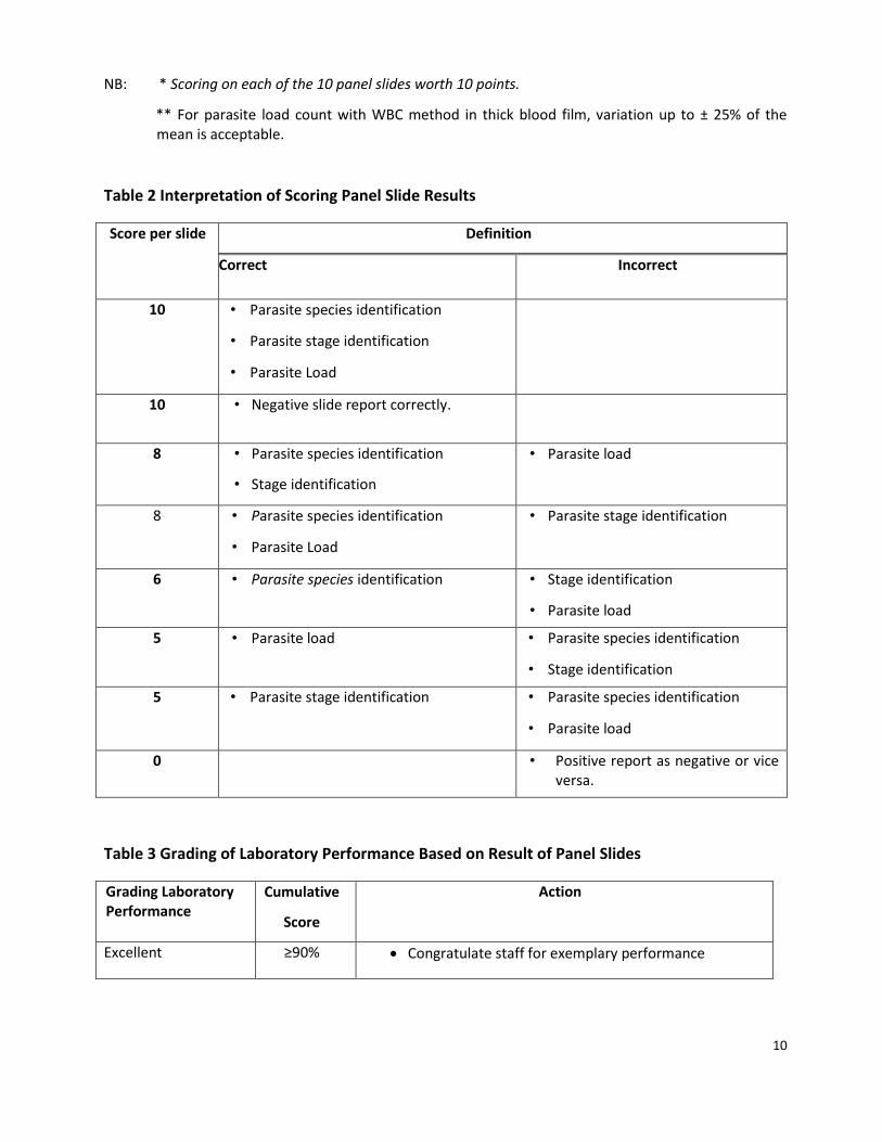
NB: * Scoring on each of the 10 panel slides worth 10 points.
** For parasite load count with WBC method in thick blood film, variation up to ± 25% of the mean is acceptable.
Table 2 Interpretation of Scoring Panel Slide Results
Score per slide Definition
Correct Incorrect
10 • Parasite species identification
• Parasite stage identification
• Parasite Load
10 • Negative slide report correctly.
8 • Parasite species identification
• Stage identification
• Parasite load
8 • Parasite species identification
• Parasite Load
• Parasite stage identification
6 • Parasite species identification
• Stage identification
• Parasite load
5 • Parasite load
• Parasite species identification
• Stage identification
5 • Parasite stage identification • Parasite species identification
• Parasite load
0 • Positive report as negative or vice versa.
Table 3 Grading of Laboratory Performance Based on Result of Panel Slides
Grading Laboratory Performance
Cumulative
Score
Action
Excellent ≥90% Congratulate staff for exemplary performance

11
Very Good 80<90% Staff should be congratulated for very good performance and told to ‘maintain it’.
Good 70<80%
Staff should be congratulated for good performance
and the need for ‘further improvement’
Check staff competency
Consider on the job training based on staff’s weakness
Check reagent quality
Check the microscope
Poor
<70%
Staff should be informed of poor and the need for
‘immediate action for improvement’
Arrange immediate on-site supervision.
Check staff competency
Consider on the job training based on staff’s weakness
Check Reagent quality
Check the Microscope
Regular follow-up for corrective action
2.2 Blinded Rechecking
Blinded rechecking refers to the process by which a random selection of slides collected from the
“testing” laboratories is reexamined at a higher level laboratory. Slides are checked for quality of blood
film preparation, quality of staining, and accuracy of the result. Rechecking reflects the true performance
of laboratories offering routine diagnostic services at health facility level. The purpose of the exercise is
to allow a statistically valid assessment of the proficiency of the peripheral laboratories
Rechecking may detect malaria misdiagnosis in routine work and assess the overall quality of testing. This
should not be considered a criticism of the person who performed the routine examination. Misdiagnosis
in routine examination is frequently caused by different reasons such as high workload, poor equipment
and not necessarily lack of skill by the reader. Each round of rechecking must be followed by feedback in
the form of written report, showing details of incorrect scorings and offering suggestions for quality
improvement (corrective actions).

12
Figure 2: Structure of random blinded rechecking
2.2.1 Roles and Responsibilities
2.2.1.1 Ethiopian Public Health Institute
Coordinate the implementation of blinded rechecking program in the country
Follow the implementation of corrective actions and provide training and technical
support.
Compile and present summary reports on the program implementation to the national
laboratory technical working group when found necessary
Provide consolidated report annually to the FMOH
Organize annual review meetings

13
2.2.1.2 Regional Reference Laboratories
Coordinate the implementation of blinded rechecking program in their respective
region
Perform blinded rechecking for University Hospitals, EQA center laboratories and
peripheral laboratories (which are not covered by EQA centers and sub-regional
laboratories).
Conduct blind rechecking for uniformed peripheral laboratories if requested a
support by uniformed services.
Resolve discrepant blinded rechecking results from EQA centers and sub-regional
laboratories
Send feedback results to participant laboratories within two weeks after the arrival
of the slide at RRL.
Follow the implementation of corrective actions and provide training and technical
support to participant laboratories in their respective region.
Provide consolidated report quarterly to RHB and EPHI
Organize annual review meetings.
2.2.1.3 Sub-regional and EQA Center Laboratories
Participate in blinded rechecking program organized by RRLs (Not applicable for
sub-regional laboratories).
Sub regional and EQA Center Laboratories take corrective actions and report to
RRLs.
Perform blinded rechecking to peripheral laboratories.
Send feedback results to participant laboratories within two weeks after the arrival
of the slides.
Follow the implementation of corrective actions and provide technical support to
participant peripheral laboratories.
Store and select positive and negative blood film slides as indicated in this guideline
(applicable only for EQA center laboratories)
Provide consolidated report quarterly to RRLs

14
2.2.1.4 Uniformed services (Army, Federal Police and Federal Prison hospital laboratories)
Perform blinded rechecking to peripheral laboratories.
Send feedback results to participant laboratories within two weeks after the arrival
of the slides.
Follow the implementation of corrective actions and provide technical support to
uniformed peripheral laboratories.
Work in collaboration with RRL if blind rechecking for uniformed peripheral
laboratories may be conducted by RRL.
Store and select positive and negative Blood film slides as indicated in this guideline
Provide consolidated report quarterly to EPHI
2.2.1.5 Federal hospital Laboratories
Federal hospital laboratories are not participating in blind rechecking program.
2.2.1.6 Peripheral laboratories
Store and select positive and negative blood film slides as indicated in this
guideline
Participate in blinded rechecking program organized by the RRLs/sub-regional
laboratories/ EQA Sites or Uniformed referral hospitals.
Take corrective actions and report to RRLs/ sub-regional/EQA centers laboratories
or uniformed referral hospitals.
2.2.2 Slide Storage in the Health Facility
• Store all positive and negative slides in a slide box away from excessive heat and humidity
until the slides have been selected.
• Store slides consecutively according to laboratory number so there is a direct link between the
results in the laboratory register and the slide location.
• Stored slides should be free from immersion oil. Remove the oil by either gently wiping the film
with lens tissue or leaving the slides overnight with the smear side facing down on ordinary tissue
paper.

15
• Slides must have laboratory numbers clearly visible. Slides without laboratory numbers cannot be
used for validation purposes.
• Results should not be written on slides; these slides cannot be used for validation purposes.
2.2.3 Sample Size for Rechecking
In accordance with the WHO recommendation, a minimum of 10 slides per month are required for
blinded rechecking purpose; 5 negatives and 5 positives.
2.2.4 Slide Selection and processing Technique
The success of blinded rechecking is critically dependent on correct selection of slide samples.
Microscopy slides for rechecking must be selected from the laboratory register and not directly from the
slide storage boxes.
2.2.4.1 Systematic Slide Selection Technique
Thirty slides per health facility should be re-examined every three months for accuracy. The following
selection technique should be applied during sampling (See also example 1):
o Ten stained malaria slides are selected each month to determine accuracy: 5 positive slides
and 5 negative slides.
o If less than 10 slides are examined in the facility, select all slides for rechecking.
o If the number of positive slides examined is less, make up the difference with negative slides.
o Ideally malaria slides should be stored for 1 month and the selection made before discarding
the slides. The slide selection procedure will be conducted on weekly basis by the laboratory
head/quality officer using the procedure described above (select slide from registration book
and note the serial number - put a mark on the register book to identify the selected slides).
o During collection of selected slides, the supervisors should counter check the conformity of
the selected slides with the laboratory registration book.
o Slides should always be stored for at least 1 week, to allow for patient follow up. If slides are
selected weekly, select as follows:

16
Example 1: Slide selection technique to select 5 negative slides:
1. Count the number of negative slides per month; For example the total negative slides are 62.
2. Divide by 5 and round up. 62 : 5 = 12.4 = 12 (rounded)
3. Take 12 small pieces of paper and number them 1, 2, 3 . . . 12.
4. Mix them in a container and pick one, for example 3; Start at the 3rdnegative slide in the register,
and select that one. Select every 12thnegative after that; for example slides 3rd, 15th, 27th, 39th,
51stnegative slides
If you do not get enough slides (i.e. either due to loss or broken slide while storing), keep
selecting each 12th slide a second time around.
Follow the same procedure for the positive slides. If the five positive slides cannot be selected, make up the difference with negative slides.
Week 1 - randomly select 2 positive slides and 1 negative slide
Week 2 - randomly select 1 positive slide and 1 negative slide
Week 3 - randomly select 1 positive slide and 1 negative slide
Week 4 - randomly select 1 positive slide and 2 negative slides
These numbers are the minimum sample size required for statistical analysis (see below). More slides can
be selected provided there is sufficient capacity for accurate rechecking of all slides. Either the site
supervisor or the facility laboratory personnel should transfer the data of the collected 30 selected slides
of each participating health facility laboratory from the laboratory registration book into appropriate
form of Annex C.1.
2.2.4.2 Slide processing
• The quality officer or responsible personnel should complete the code number of collected slides
in Annex C.2 to provide it to the laboratory personnel (2nd readers) for rechecking and result
recording.
• The quality officer or responsible personnel should identify discrepant result(s) and give the
discrepant slide(s) and blank form of Annex C.3 to third laboratory personnel in the laboratory. If
discrepant result exists between the second and third reader, the two readers will jointly review

17
the slides and reach consensus. Any slide with unresolved discrepant results should be sent to the
higher level laboratory for final decision.
• Give feedback to the testing site with comments and recommendations for appropriate
corrective actions using performance notification form (Annex C.4) within a maximum of two
weeks’ time.
• Send compiled summary report of the participant sites to the higher tier for further analysis and
possible quality improvement interventions.
NB:
- The Regional Laboratories are the higher level laboratories that control the quality services
of Sub-regional laboratories, nearby Universities health facility laboratories, and EQA site
laboratories.
- Laboratories in the private sector (stand alone, medium clinic, higher Clinic, and hospital
laboratories) will be provided with similar services by the respective Regional Laboratories
or EQA site laboratories designated for the purpose by the Regional Laboratories.
2.2.5 Result Analysis
Table 4 Result Recording as Positive or Negative on a 2x2 Table Format
Rechecking labs, Sub-regional and RRLs
Positive Negative Total
Source
laboratory
Positive A B A+B
Negative C D C+D
Total A+C B+D A+B+C+D
A = number of slides reported as positive by both readers (True positive) B = number of slides reported as positive in routine testing by the laboratory but found to be
negative by the cross-checker (false positives)
C = number of slides reported as negative in routine testing by the laboratory but found to be
positive by the cross-checker (false negatives)
D = number of slides reported as negative by both readers (True negative)
Results are analyzed as:

18
1. Percentage of slides in agreement, i.e. percentage of positive slides correctly identified and
percentage of negative slides correctly identified:
% Agreement = True positive + True negative= (A+D) x 100
Total A+B+C+D
2. False positive rate (% false positives)
False positive rate = False positive x 100 = B x100
True positive + False Positive A+B
3. False negative rate (% false negatives)
False Negative Rate=False Negative x100 = Cx100
True Negative + False Negative D+C
Table 5 Example of Result Analysis
Rechecking labs, Sub-regional and RRLs
Positive Negative Total
Source
laboratory
Positive A (8) B (2) A+B (10)
Negative C (1) D (19) C+D (20)
Total A+C (9) B+D (21) A+B+C+D (30)
% Slide Agreement (Detection) = True positive + True negative= (A+D) x 100% = (8+19) x 100%= 90%
Total A+B+C+D 30
False positive rate = False positive x 100 = B x100% =2x100%=20%
True positive + False Positive A+B 10
False Negative Rate=False Negative x100 = Cx100%= 1 X 100%=5%
True Negative + False Negative D+C 20

19
Table 6 Grading Performance of Slide Rechecking Cycle
Grade % of slide
Agreement
(Detection)
Action
Excellent ≥90% .Congratulate staff for exemplary performance
Very good 80<90%
• Staff should be congratulated for very good performance and
told to maintain their performance
• Identify any breach for improvement
Good 70<80%
• Staff should be congratulated for good performance and the
need for ‘further improvement’
• Conduct regular on-site Supervision
• Check staff competency
• Check reagent quality and the microscope
• Consider on the job training based on staff’s weakness
Poor
<70%
• Staff should be informed of poor performance and the need
for ‘immediate action for improvement’
• Arrange immediate on-site Supervision.
• Check staff competency
• Consider intensive on the job training based on staff’s
weakness
• Check reagent quality and the microscope
• Regular follow-up for corrective action
NB:
1. ‘Error’ stand for any positive result reported as negative, or any negative result reported as
positive..
2. Any EQA performance persistently static or a progressive decreasing pattern in the percentage
agreement is an alarming sign that indicates the corrective action has not been effective and
should be reviewed immediately.
3. Any EQA performance above the previous once is encouraging and still needs follow ups.
20
Table 7 Result recording for monitoring the accuracy of the differentiation of P. falciparum and non P. falciparum
Rechecking labs, Sub-regional and RRLs
P. falciparum
Present
P. falciparum
NOT present
Total
Source
laboratory
P. falciparum
Present
A B A+B
P. falciparum
NOT present
C D C+D
Total A+C B+D A+B+C+D
Where:
A=number of slides reported as containing P.falciparum (either as a single or mixed
infection) by both readers
B= number of slides reported as containing P.falciparum only in routine testing by the
laboratory but the presence of P.falciparum was not confirmed by the cross-checker
(incorrect species identification)
C= number of slides reported as P.falciparum not present in routine testing by the laboratory but
found to be present by the cross-checker,either as a single or mixed infection (incorrect
species identification)
D =number of slides reported as not containing P.falciparum by both readers
NB:
1. For specific species, % agreement is calculated from only positive slides reported by the facility
2. species identification % agreement can be calculated for all malaria parasite species including
mixed infections
% Species identification Agreement= (A+D)x100%
A+B+C+D
21
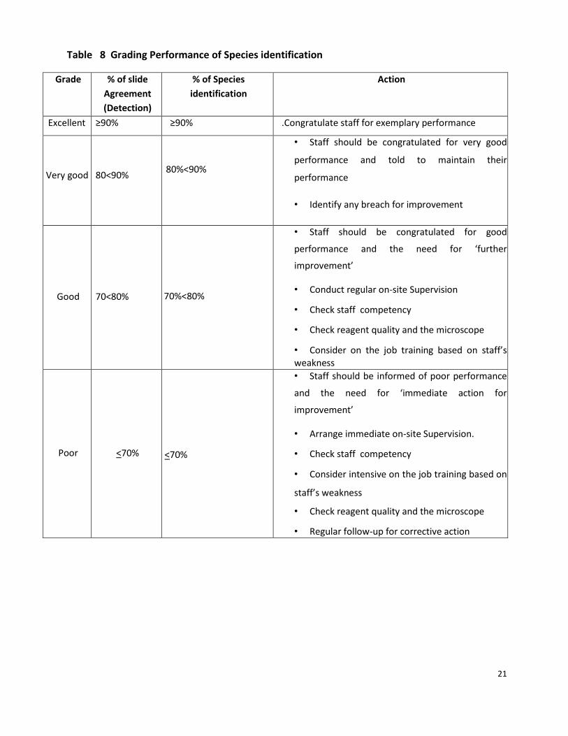
Table 8 Grading Performance of Species identification
Grade % of slide
Agreement
(Detection)
% of Species
identification
Action
Excellent ≥90% ≥90% .Congratulate staff for exemplary performance
Very good 80<90%
80%<90%
• Staff should be congratulated for very good
performance and told to maintain their
performance
• Identify any breach for improvement
Good 70<80%
70%<80%
• Staff should be congratulated for good
performance and the need for ‘further
improvement’
• Conduct regular on-site Supervision
• Check staff competency
• Check reagent quality and the microscope
• Consider on the job training based on staff’s weakness
Poor
<70%
<70%
• Staff should be informed of poor performance
and the need for ‘immediate action for
improvement’
• Arrange immediate on-site Supervision.
• Check staff competency
• Consider intensive on the job training based on
staff’s weakness
• Check reagent quality and the microscope
• Regular follow-up for corrective action
22
2.3 On-site Supervision (for Microscopy and RDT)
On-site supervision of malaria microscopy and RDT requires regular supervisory visits to obtain a realistic
picture of laboratory conditions and practices for malaria microscopy and RDT use. On-site supervision
includes a comprehensive assessment of laboratory organization, equipment, adequacy and storage of
supplies, reagent quality, availability and usage of SOPs, reading and reporting of results and infection
control measures using a supervisory checklist. On-site supervision is the ideal way to obtain a realistic
assessment of the skills practiced in the testing laboratory/facility, to provide problem solving strategies
and corrective action, and assess the need for training. The supervision includes assessment of test
performance, provision of on-site training and strengthening of services.
Malaria microscopy on-site supervision is conducted in accordance with NEQAS two times a year by
quality officers and malaria experts and others working on malaria quality improvement. Onsite
supervision provides an opportunity for basic supervision, including assessment of laboratory supplies
storage and inventory, basic procedures, availability of functional equipment, quality of reagents,
training status of the laboratory staff, review of laboratory practical skills, work load, safety and waste
disposal system, performance of internal QC and result recordkeeping practice. A major advantage of on-
site supervision is the ability to identify sources of errors detected by panel testing or rechecking and to
implement appropriate measures to resolve problems.
Sufficient time must be allotted for the visit to include observation of all the work associated with
malaria microscopy, including preparing films, staining, reading of films by the laboratory personnel and
examining a few stained positive and negative films by supervisors to observe the quality of film
preparation and staining as well as condition of microscope.
23
Figure 3: Structure of Onsite Evaluation for Malaria Microscopy
2.3.1 Roles and Responsibilities
2.3.1.1 Ethiopian Public Health Institute
Set supervision standards; develop/review checklists
Conduct on-site evaluation to all RRLs, Sub regional laboratories, federal hospitals
and Uniformed services (Army, Federal Police and Federal Prison hospital
laboratories) based on their PT performance.
Conduct on-site evaluation for any health facility to evaluate the overall program as
needed.
24
Send feedback on time to RRLs, RRLs, Sub regional laboratories, federal hospitals
and Uniformed services, RHB and other authorized bodies within two weeks of data
arrival at EPHI
Follow the implementation of corrective actions and provide training and technical
support
Prepare report on the performance of laboratories based on on-site evaluation to
the national laboratory technical working group when found necessary.
Provide consolidated bi-annually summary report to FMOH
Organize review meetings annually.
2.3.1.2 Regional Reference Laboratories
Participate in on-site evaluation conducted by EPHI
Take corrective actions for the identified gaps and report to EPHI within two weeks.
Conduct on-site evaluation for, university hospitals, EQA centers and sub-regional
laboratories.
Conduct on-site evaluation to laboratories which are not covered by sub-regional
and EQA center laboratories.
Conduct on-site evaluation for uniformed peripheral laboratories if requested a
support by uniformed services.
Conduct on-site evaluation for any health facility in their respective region to
evaluate the overall program as needed.
Send feedback to participant laboratories within two weeks of data arrival at
regional laboratory
Follow the implementation of corrective actions and provide training and technical
support to participant laboratories.
Provide consolidated bi-annually summary report to RHB and EPHI
Provide timely data and documentation to facilitate on-site supervision by EPHI
Organize review meetings annually.
2.3.1.3 Sub Regional Laboratories and EQA centers
Participate in on-site evaluation conducted by RRLs and EPHI(for Sub regional
laboratories only)

25
Take corrective actions for the identified gaps and report to RRLs/EPHI within two
weeks
Conduct on-site evaluation to peripheral laboratories.
Send feedback to participant laboratories within two weeks of data arrival at the
EQA sites and sub-regional laboratories.
Follow the implementation of corrective actions and provide training and technical
support to participant laboratories.
Provide consolidated bi-annually summary report to RRLs
2.3.1.4 Uniformed services (Army, Federal Police and Federal Prison hospital laboratories)
Participate in on-site evaluation conducted by EPHI
Take corrective actions and report to EPHI.
Conduct on-site evaluation to uniformed peripheral laboratories.
Send feedback to participant laboratories within two weeks of data arrival
Follow the implementation of corrective actions and provide training and technical
support to participant laboratories.
Work in collaboration with RRL if on-site evaluation for uniformed peripheral
laboratories may be conducted by RRL.
Provide consolidated bi-annually summary report to EPHI
2.3.1.5 Federal hospital Laboratories
Participate on on-site evaluation conducted by EPHI
Take corrective actions for the identified gaps and report to EPHI
Provide all the necessary data and documents to EPHI to facilitate on-site
supervision
2.3.1.6 Peripheral laboratories
Participate in on-site evaluation conducted by RRLs/EQA sites/Uniformed
services/sub-regional Laboratories
Take corrective actions for the identified gaps and report to RRLs/EQA
sites/Uniformed services/sub-regional Laboratories within two weeks.

26
Provide all the necessary data and documents to facilitate on-site supervision by
RRLs/EQA sites/Uniformed services/sub-regional Laboratories
2.3.1.7 Health Posts
Health Posts are responsible for provision of malaria laboratory diagnosis using
RDTs.
Health posts are expected to implement basic QA activities for malaria diagnosis
and must be involved in REQAS through onsite supervision by the higher tier
laboratory.
Health posts are expected to implement standard supply chain management
systems for RDTs.
2.3.2 On-site Supervision for Malaria RDT
On-site supervision for malaria RDT should be performed two times a year by HEWs Supervisor (HEWS)
and others working on malaria RDT quality improvement. On-site supervision provides an opportunity for
assessment of RDT supplies storage conditions, inventory, and basic procedures of RDT including sample
collection, skill of HEW to perform RDTs, internal quality control, result interpretation, recording and
reporting, safety practice and waste disposal, and need of retraining by using a supervisory checklist
(Annex D.3). A major advantage of on-site supervision is the ability to identify sources of errors and
provide on-site corrective actions to improve the quality of test results and implement appropriate
measures to resolve problems.
2.3.2.1 Supervisory checklist
Every EQA scheme will need to have checklists to assist laboratory supervisors during the on-site visits
and standardize collection and analysis of data for subsequent remedial action. Checklists may be revised
in the light of problems that are identified during such visits.
A comprehensive list of all operational elements to be observed will help to ensure consistency in
laboratory evaluations and provision of immediate feedback to facilitate rapid corrective action. It also
serves as means of documentation of the visit and a record of current conditions and actions needed.
The checklist should be completed during the visit and discussed with the test performer before the
supervisor leaves the health facility. Filled checklists should be submitted to the onsite supervision
organizer after completion of each visit.

27
Feedback containing need for corrective action or additional resources should be reported to each
respective health facility through the recommended channel of communication and a consolidated
summary report need to be submitted to EPHI and Regional Health Bureaus.
Supervisory checklists for on-site supervision of malaria diagnosis (microscopy and RDT) are provided in
Annexes D.2 and D.3. These checklists contain open, non-leading questions and recommended
observations along with objective criteria for acceptable practices. By using open, non-leading questions,
as well as direct observation of daily practices, the supervisor can assess how well the laboratory
personnel understand proper procedures, and is not just providing the expected “yes” response. These
detailed checklists provide a template that may be adapted to meet the specific needs of EQA at each
level. The preferred format should include simple, objective “Yes/No” evaluation criteria, yielding data
that can easily be entered into a database for long term tracking and comparing performance.
Documentation of any significant problems requires development of strategies and activities for
improvement of quality.
2.3.2.2 General Activities to be considered for On-site Supervision
• Make a schedule for site visits.
• Form a supervisory team.
• Prepare necessary materials like the check list and feedback report.
• Arrange logistics for the site visit.
• Conduct on-site supervision.
• Review the previous site supervision feedback (if available).
• Provide EQA feedback, investigate any poor performances, and make corrective action and
follow-up.
NB: SOP for on-site supervision is explained in Annex D.1.
2.3.2.3 Resource Requirements for On-site Supervision:
• Logistics (vehicles and per diem).
• Supervisors.
• Standard Checklist.
• Format for immediate feedback.

28
• Fax, e-mail and web based services for communication.
2.3.3 Procedure for Malaria Microscopy On-site Supervision
Tasks to be done by supervisory team:
• EPHI and Regional Laboratories are coordinating bodies for malaria laboratory diagnosis
EQA in their respective operational setting.
• For EQA activity by EPHI, the National Quality Officers arrange the logistics including checklists,
and select the supervisory team members one month prior to the starting day of the on-site
supervisory visit by communicating with the National Quality Manager.
• For EQA activities at regional, sub-regional and EQA sites, Quality Officers at each level arrange
the logistics including checklists, and select the supervisory team members one month prior to
the starting day of the on-site supervisory visit.
• The respective Quality Officers inform selected supervisors 15 days prior to the starting date of
onsite supervision and identify team leaders of each supervisory team.
• The respective Quality Officers arrange orientation session for the supervisory team, prepare
official letters for each supervisory team and communicate with the participating health facility
one week before the starting date of the onsite supervision.
• The supervisory teams participate in the orientation session a day before the starting date of
onsite supervision and collect the checklist and other items needed for the onsite visit.
• At the health facility level, the supervisors follow the SOP to Conduct On-site Supervision (see
Annex-D.1).
• The team leaders of each supervisory team submit the completed checklist to the respective
Quality Officers after completing the onsite supervision.
• The National and Regional, sub-regional and EQA sites Quality Officers prepare feedback and
corrective action needed for each participating laboratory within a 15 days after the visit and also
compile summary reports all participant sites, and report to the EPHI and Regional Health
Bureaus.
•
29
• The respective Quality Officers develop site specific corrective action plan to address the
identified gaps, and lead its implementation to strengthen the malaria diagnosis services based
on the recommendations stated in the report.
• The respective Quality Officers follow the implementation of the feedback/corrective actions
given.
2.3.4 Procedure for Malaria RDT On-site Supervision
• Regional Laboratories are the coordinating bodies for malaria RDT EQA in their respective
operational setting.
• In consultation with Regional Laboratories, Zonal laboratory experts and/or Zonal malaria experts
provide technical and logistical support for HEWS and others working on malaria RDT quality
improvement including provision of orientation sessions prior to the initiation of the onsite
supervision activity.
• All HEWSs need to participate in the orientation sessions before performing onsite evaluation and
need to collect the relevant checklists and formats.
• At the health posts, the HEWSs need to follow the standard SOP to Conduct On-site Supervision
(See annex-D.1).
• The HEWS submits a copy of the completed checklist to the Zonal laboratory expert and/or Zonal
malaria expert after completing the onsite supervision within two weeks of the visit.
• The HEWS is also expected to prepare and give feedback and takes corrective action for each
participating site within 15 days of the visit.
• The Zonal laboratory expert and/or Zonal malaria expert sends a compiled summary report of
each participant sites to the RRL, Zonal Health Departments and Regional Health Bureaus.
• Based on the recommendations and corrective action stated in the report the Regional
Laboratory, Zonal Health Department, District Health Office and the HEWS plan to take corrective
actions for the major gaps identified and continue to strengthen the malaria RDT services.
• The Regional Laboratory, sub regional laboratory and EQA sites provides a summarized report of
findings from onsite supervision to the National Quality Manager every six months.

30
3 ANNEXES
Annex-A Participant Laboratory Registration Form for Panel Testing
Region_______________________________
City/Town__________________________
Facility name_______________________
Office phone________________________
Fax__________________________________
Address of 1st Contact
• Name_____________________________________
• Job Title__________________________________
• Mobile____________________________________
• Fax_________________________________________
• E-mail________________________________________
Address of 2nd Contact
• Name_____________________________________
• Job Title__________________________________
• Mobile____________________________________
• Fax_________________________________________
• E-mail________________________________________
You may Contact
P. O. Box 1242/5654 Tel: +251 11-2751522/2753470 +251 11- 2754744 E-mail: [email protected]

31
Annex-B Instruction, Result Reporting and Feedback Form for Malaria Microscopy Panel Testing
B.1. Instructions for Reading Malaria Slide Panel Testing
1. Before proceeding to reading the slide, read the instructions and all forms carefully
2. Make sure that the panel contains 10 slides and are properly labeled.
3. Read all the ten panel slides as if you examined routine clinical blood film samples.
4. Heath facility level laboratory staff who routinely reads malaria slides of patients is expected to
read and report the reading of panel slides in a similar way.
5. Laboratories are expected to properly handle and return the panel slides.
6. Record your finding appropriately.
7. Submit the completed result form to the responsible body using fax, e-mail or post with a contact
address of the respective institutions.
Caution
o Panel slides should be treated as if potentially infectious.

32
B.2. Result Reporting Form at the Health Facility for Reading Malaria Slide Panel Testing
Region ___________________
Zone_____________________
Woreda___________________
Name of health facility/ laboratory ____________________________________
Date received by the health facility laboratory __________________ Received by_________________
Slide ID Negative Positive
Remark
Species Stage
Parasite
Load
Date read: _________________ Date reported __________________
Name and signature of the reader:__________________________
33
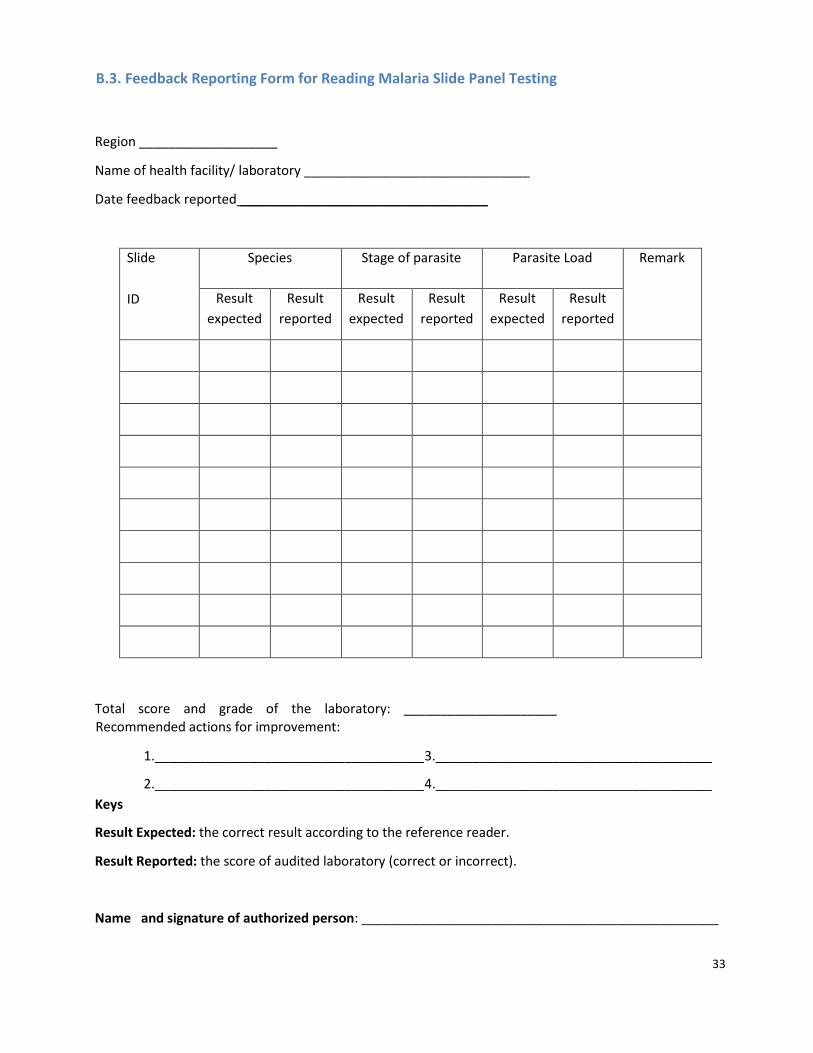
B.3. Feedback Reporting Form for Reading Malaria Slide Panel Testing
Region ___________________
Name of health facility/ laboratory _______________________________
Date feedback reported __________________________________
Slide
ID
Species Stage of parasite Parasite Load Remark
Result
expected
Result
reported
Result
expected
Result
reported
Result
expected
Result
reported
Total score and grade of the laboratory: _____________________ Recommended actions for improvement:
1._____________________________________
2._____________________________________
3.______________________________________
4.______________________________________
Keys
Result Expected: the correct result according to the reference reader.
Result Reported: the score of audited laboratory (correct or incorrect).
Name and signature of authorized person: _________________________________________________
34
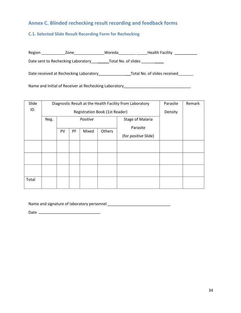
Annex C. Blinded rechecking result recording and feedback forms
C.1. Selected Slide Result Recording Form for Rechecking
Region ___________Zone______________Woreda________ _____Health Facility __________
Date sent to Rechecking Laboratory _____Total No. of slides __ ____
Date received at Rechecking Laboratory __Total No. of slides received_______
Name and Initial of Receiver at Rechecking Laboratory_______________________________
Slide
ID.
Diagnostic Result at the Health Facility from Laboratory
Registration Book (1st Reader)
Parasite
Density
Remark
Neg. Positive Stage of Malaria
Parasite
(for positive Slide) PV PF Mixed Others
Total
Name and signature of laboratory personnel
Date

35
C.2. Slide Reader Result Record Form for Rechecking (2nd Reader)
Rechecking Laboratory____________________________
Region __________Zone______________Woreda__________ Health Facility_____________
Total slides Received Source
Name of laboratory personnel, who examine the slides
Slide
ID
2nd Reader result (At the Rechecking Lab.) Parasite
Density
Slide quality grading Remark
Neg. Positive Stage
(for positive Slide)
Excellent Good Poor
PV PF Mixed Others
Total
NB: -Quality of blood film includes size and thickness of the film and quality of the staining.
Name of 2nd reader Signature________________ Date_________________
General comment ______________________________________________________________________ _____________________________________________________________________________________
Key for Slide quality grading
Excellent Gross appearance: Both thin and thick film prepared on the same slide, thick film 10 mm diameter, newsprint read under thick film before staining, 10 mm from frosted end and thick film, 10 mm between thick and a thin film with distinct head, body and tail. Microscopic appearance: Demonstrates RBCs lysed in thick film and a monolayer of RBCs, with normal and abnormal morphology in thin film. Staining allows the trophozoites, gametocytes and/or schizonts and the white blood cells to be clearly distinguished against the background.
Good Gross appearance: Thick film with irregular and uneven thickness, thin film with uneven tail, too thick, too wide or too long. Microscopic appearance: Demonstrates RBCs lysed in thick film and a monolayer of RBCs, with normal and abnormal morphology in thin film. Staining allows the trophozoites, gametocytes and/or schizonts and the white blood cells to be clearly distinguished against the background.
Poor Gross appearance: Film with ragged tail, too thick, too wide or too long with uneven thickness. Microscopic appearance: Distorted appearance of the RBCs, malaria parasite and the white cells. Difficult to spot fields with monolayer of cells on thin film, lack of white blood cells to be clearly distinguished against the background and no properly lysed RBCs in thick film.

36
C.3. Slide Reader Result Record Form for Rechecking (3rd Reader for Discordant Result)
Rechecking Laboratory____________________________
Region __________Zone______________Woreda__________ Health Facility_____________
Total slides Received Source
Name of laboratory personnel, who examine the slides
Slide
ID
3rd Reader result (At the Rechecking Lab.) Parasite
Density
Slide quality grading Remark
Neg. Positive Stage
(for positive Slide)
Excellent Good Poor
PV PF Mixed Others
Total
NB: -Quality of blood film includes size and thickness of the film and quality of the staining.
Name of 3rd reader Signature________________ Date_________________
General comment ______________________________________________________________________ _____________________________________________________________________________________
Key for Slide quality grading
Excellent Gross appearance: Both thin and thick film prepared on the same slide, thick film 10 mm diameter, newsprint read under thick film before staining, 10 mm from frosted end and thick film, 10 mm between thick and a thin film with distinct head, body and tail. Microscopic appearance: Demonstrates RBCs lysed in thick film and a monolayer of RBCs, with normal and abnormal morphology in thin film. Staining allows the trophozoites, gametocytes and/or schizonts and the white blood cells to be clearly distinguished against the background.
Good Gross appearance: Thick film with irregular and uneven thickness, thin film with uneven tail, too thick, too wide or too long. Microscopic appearance: Demonstrates RBCs lysed in thick film and a monolayer of RBCs, with normal and abnormal morphology in thin film. Staining allows the trophozoites, gametocytes and/or schizonts and the white blood cells to be clearly distinguished against the background.
Poor Gross appearance: Film with ragged tail, too thick, too wide or too long with uneven thickness. Microscopic appearance: Distorted appearance of the RBCs, malaria parasite and the white cells. Difficult to spot fields with monolayer of cells on thin film, lack of white blood cells to be clearly distinguished against the background and no properly lysed RBCs in thick film.

37
C.4. Performance Notification Form
Notification No: ___________ Code No. _________________
To: ______________________________________
From: ___________________________________
I. Total No. of slides with correct reading
IV. Grading of performance by % of Agreement
Excellent (>90%)
Very good (80-90%
Good (70-80%)
Poor (≤70%)
% of false positive
% of false negative
II. Total number of slide with discordant results
III. Type of discordance:
# Positive diagnosed as negative
# Negative diagnosed as positive
# Species misdiagnosis
III) Recommendation
General ______________________________________________________________________________
Specific_______________________________________________________________________________
_____________________________________________________________________________________
Feedback Summary Table
Slide ID. Result Slide Quality Remark
Correctly
read
Discordant
Pos. Report
as Neg.
Neg. Report
as Pos.
Species
Misdiagnosed
Total
Go
od Po
or

38
Name and signature of authorized personnel: ____________________________________________
Date: ______________
Key for Slide quality grading
Excellent Gross appearance: Both thin and thick film prepared on the same slide, thick film 10 mm diameter, newsprint read under thick film before staining, 10 mm from frosted end and thick film, 10 mm between thick and a thin film with distinct head, body and tail. Microscopic appearance: Demonstrates RBCs lysed in thick film and a monolayer of RBCs, with normal and abnormal morphology in thin film. Staining allows the trophozoites, gametocytes and/or schizonts and the white blood cells to be clearly distinguished against the background.
Good Gross appearance: Thick film with irregular and uneven thickness, thin film with uneven tail, too thick, too wide or too long. Microscopic appearance: Demonstrates RBCs lysed in thick film and a monolayer of RBCs, with normal and abnormal morphology in thin film. Staining allows the trophozoites, gametocytes and/or schizonts and the white blood cells to be clearly distinguished against the background.
Poor Gross appearance: Film with ragged tail, too thick, too wide or too long with uneven thickness. Microscopic appearance: Distorted appearance of the RBCs, malaria parasite and the white cells. Difficult to spot fields with monolayer of cells on thin film, lack of white blood cells to be clearly distinguished against the background and no properly lysed RBCs in thick film.

39
C.5. Annual Feedback Form for Participant Health Facility in Blinded Rechecking
1. % Agreement in the four consecutive rechecking scheme
2.
100%
95%
90%
85%
80%
75%
70%
1stRechecking 2ndRechecking 3rdRechecking 4th rechecking
Rechecking Cycle
2. % False Negative in the four consecutive slide rechecking cycle
50%
40%
30%
20%
10% 0%
1stRechecking 2ndRechecking 3rd Rechecking 4th rechecking
Rechecking Cycle
3. %False Positive in the four consecutive slide rechecking cycle
50%
40%
30%
20%
10%
0%
1stRechecking 2ndRechecking 3rd Rechecking 4th rechecking
Rechecking Cycle
% A
gree
men
t
% F
alse
Neg
ativ
e
% F
alse
Po
siti
ve

40
Annex D. SOPs and checklist to Conduct On-site Supervision
D.1. SOP to Conduct On-site Supervision
Purpose:
• To assess site performance and compliance with quality assurance implementation.
• To provide on-site supervision and support.
• To monitor process improvement.
• To gather data.
Scope:
• To be used for guiding onsite supervision of malaria diagnosis at Health facility level by national,
regional, sub regional, EQA sites, zonal and facility level supervisors.
Materials required:
• A new checklist (for general laboratory).
• Previous checklist (if available).
• EQA reports.
Procedure:
• Introduce yourself to the laboratory head (HEW at Health Post) and state the purpose of your
visit.
• Ask the laboratory head (HEW at Health Post) if you could meet have brief meeting with
him/her as well as the quality officer in private.
• Encourage the laboratory head and quality officer (HEW at Health Post) to feel comfortable.
Spend a little time making general enquiries into the overall health facility laboratory (Health
Post) activities.
• Discuss the previous site visit; check if the action items are completed and implemented.
41
• Ask the health facility laboratory head (HEW at Health Post) the questions on the checklist and
write down the scores.
• Discuss EQA report feedback, investigate any poor performances. Check if corrective actions
have been implemented. If not, ask for reasons or constraints to implementing corrective
actions, arrange to provide necessary support, and agree on corrective actions to be
implemented and followed up.
• Ask the health facility laboratory head (HEW at Health Post) to show you the laboratory (Health Post). During this time, pay special attention to observing the following:
-
-
-
-
Effective and efficient work flow
Staff competency
Use of SOPs
Equipment maintenance and
completed records
- Use of quality controls and
completed records
- Complete correction logs
- Inventory system
- Adherence to safety - Adequate infrastructure
This is an opportunity to ensure the questionnaire answers match the reality in the laboratory, as well as
to provide on-site supervision.
• Ensure the checklist is complete and scores added correctly. • Conduct an exit interview with the manager. Provide praise where appropriate. Discuss
challenging areas and non-compliance, and make recommendations.
• If there are critical problems in the laboratory, discuss these with the laboratory head and agree
on actions for immediate implementation (such cases should be reported to the national
laboratory quality manager for follow up).
• It is important to build good relations with the laboratory head and staff. The visit should be
positive, encouraging and supportive.
Total time of site visit will vary per laboratory. However, a supervisory visit for minimum of 6hours is
recommended.
42
Outcomes and follow up:
• The supervisor will review the supervision result and send reports back to the sites within 15
days.
• The report will include recommendations for process improvement, and laboratory heads
(HEWs) are expected to implement such activities.
• All activities will be followed up during the next site visit.
• Where health facility laboratories (Health Posts) have poor performance, arrangements will be
made by the quality officers (HEWS) for the provision of intensive on-site mentorship.
• Where laboratories perform poorly and do not comply with recommendations, a letter of non-
compliance will be sent to the laboratory and hospital medical officer (Woreda Health Office).
• Wherever necessary, the NRL/RRL will provide assistance and support.
• The National Quality Manager and Regional Quality Officer will compile an annual summary
report and submit it to the EPHI and Regional Health Bureau, respectively.

43
D.2. Supervisory Checklist for Malaria Microscopy Laboratory Service
Region______________ Zone_____________ Woreda______________
Name of health facility ______________________
Name of laboratory department head________________
Tel. No_________________ Fax____________________________
Date of onsite supervision conducted_______________________
I. Training
No Questions Responses
Total No. Of lab
staff___
Number of laboratory
personnel trained on
malaria microscopy___
Name of
trained staff
in the fiscal
year
When was
s/he trained?
(mm/yyyy
EC)
How long was
the training? (#
of Days)
Who provided
the training?
(Organization)
Comments
Comments
II. Malaria microscopy laboratory format and supplies
Are the following
malaria microscopy
formats and other
materials available?
Items 1 = Available and being used
2 = Available, but not used
3 = Not Available
Malaria microscopy guideline
SOP for malaria microscopy
Laboratory result log book
Job aids
Weakly /monthly report form for malaria

44
Are the following
reagents and other
Item 1 = Available and being
used
Enough
for the
Laboratory
commodities available?
2 = Available, but not
used
3= Not Available
4= Not Applicable
coming 4
Months
1= Yes
2=No
Absolute methanol
Absorbent cotton wool
Beaker/volumetric flask
Binocular microscope with electric
source of light
Brown bottle
Distilled water
Drying rack
Funnel
Giemsa powder/Giemsa stain stock
solution
Glass beads
Glycerol
Immersion oil
Timer
Lens cleaning solution
Lens paper
Measuring cylinder
Microscope slides
Glass-writing pen/lead pencil
Slide boxes
Staining rack
Staining jar
Tally counter
Tissue paper
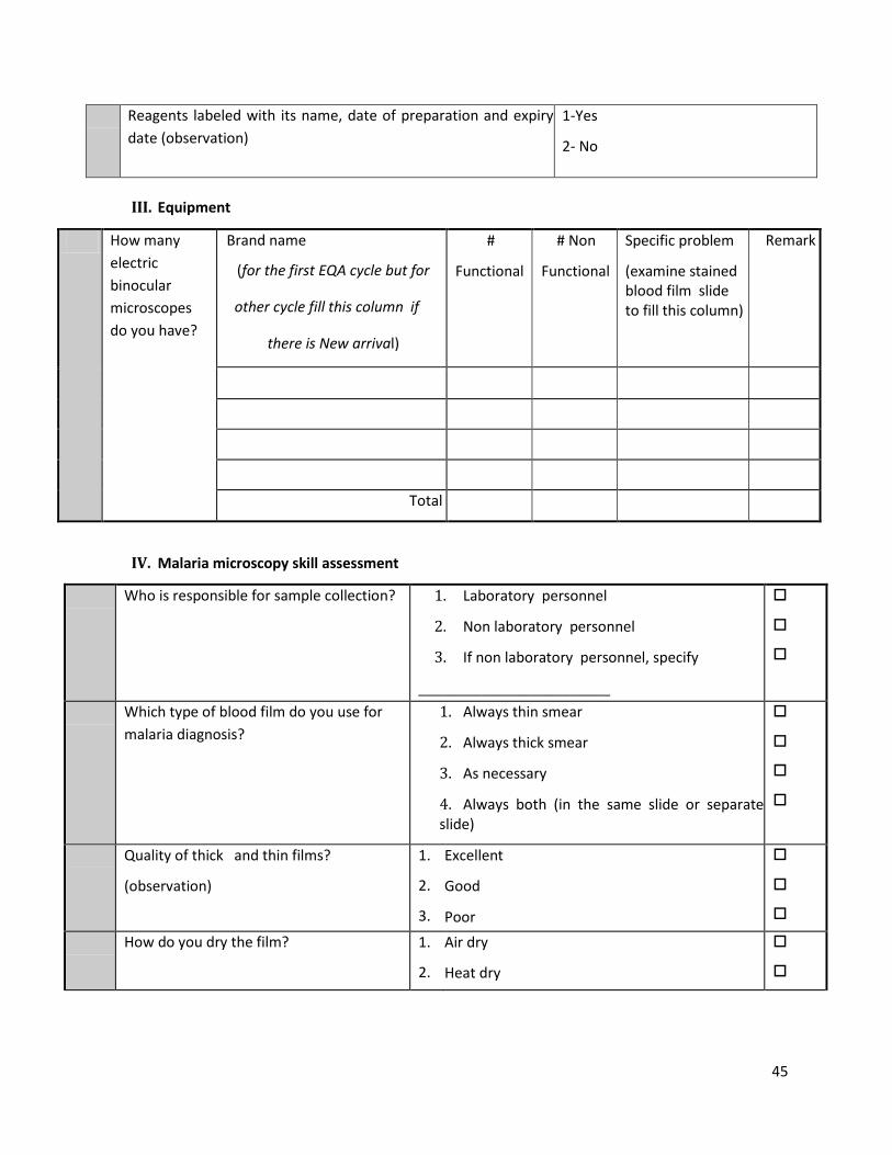
45
Reagents labeled with its name, date of preparation and expiry
date (observation)
1-Yes
2- No
III. Equipment
How many
electric
binocular
microscopes
do you have?
Brand name
(for the first EQA cycle but for
other cycle fill this column if
there is New arrival)
#
Functional
# Non
Functional
Specific problem
(examine stained blood film slide to fill this column)
Remark
Total
IV. Malaria microscopy skill assessment
Who is responsible for sample collection? 1. Laboratory personnel
2. Non laboratory personnel
3. If non laboratory personnel, specify
________________________
Which type of blood film do you use for
malaria diagnosis?
1. Always thin smear
2. Always thick smear
3. As necessary
4. Always both (in the same slide or separate slide)
Quality of thick and thin films?
(observation)
1.
2.
3.
Excellent
Good
Poor
How do you dry the film? 1.
2.
Air dry
Heat dry

46
Which part of the film (thin or thick) do you
fix?
1.
2.
3.
Thin
Thick
Both
How many fields do you examine to report
a negative result (no parasites)?
1.
2.
3.
4.
5.
<25
50
100
200
__________
Do you report positive results by identifying
species and parasite stages?
1.
2.
3.
4.
Species only
Stages only
Both
None
Do you quantify positive results (parasite
density)?
1. Yes
2. No 3. if Yes, which method specify __________________________
When using WBC method, how many WBC
do you count to quantify a parasite load?
1.
2.
3.
4.
5.
50 WBC
100 WBC
200 WBC
500 WBC
Not applicable
Do you clean the microscope or objective
lenses prior to starting microscope reading
and at the end of the day?
1.
2.
Yes
No
What do you use for microscope lens
cleaning?
1.
2.
3.
4.
Cotton
Lens paper
Tissue paper
Other ____________________
Which reagent do you use for blood film
staining?
1.
2.
3.
Giemsa
Wright
If other, specify_____________?
For Giemsa stain

47
Do you prepare the stock reagent or use
ready-made reagent?
1.
2.
Preparing reagent
Readymade reagent
How often do you prepare the working
reagent?
1.
2.
3.
Every 24 hrs
Prior to staining
Other specify_____________
What is the commonly used reagent
container to store the stock stain?
1.
2.
Brown bottle
Any transparent bottle
3. If other, specify ____________
Where do you store the stock reagent? 1.
2.
Away from direct sunlight and moisture in
lockable cabinet
Other, specify_________________
Have you ever interrupted malaria
laboratory services due to shortages of
reagents, supplies and microscope
problem?
1. Yes
2. No
If yes
1. Cause of interruption____________
2. For how long ______________________
3. How many times in the last 4 months ________
Have you experienced some difficulties
with your microscope during the last 4
months?
1=y
2=
If y
1.
2.
3.
es
No
es,
with the Stage with
the objective
other specify,_________________________
Do you have an inventory list of supplies
and stains?
1.
2.
Yes
No
How often do you receive supplies like
stains and others?
1.
2.
3.
Monthly
Every 6 months
Once a year
48
Do you have difficulties receiving your
supplies?
1.
2.
3.
Yes
No
If yes, why _____________________________
Do you store patient blood (with EDTA)
known to have parasites
1.
2.
Yes
No
Do you keep slides for rechecking? 1.
2.
3.
Yes
No
If no why?
__________________________________
Have you been supervised in the past 6
months?
1.
2.
3.
Yes
No
If yes specify the supervisor _______________
Is a standard laboratory register book in
use?
1.
2.
3.
Yes
No
If not why? ________________
Is a standard laboratory request form in
use?
1.
2.
3.
Yes
No
If not why? ________________________
V. Quality Assurance
Internal Quality Control (QC) Practiced
Do you prepare positive and negative slides
for reagent quality control purposes?
1.
2.
Yes
No, if no why? __________________
When do you conduct internal quality control
for malaria microscopy?
.
1. Weekly
2. Monthly
3. Upon opening of new batch_
4. During unusual staining results
5. Others, Specify _________
Are stained slides ever rechecked by a person
in the laboratory? 1.
2.
Yes
No, if no why? __________________

49
EQA practiced
Are stained slides validated regularly, and
feedback obtained? 1. Yes
2. No, if no why? __________________
Do you participate in an EQA scheme, and is
feedback obtained 1. Yes
2. No, if no why? ____________________
VI. Safety and waste Disposal
Are gloves and gowns worn while
performing the procedure?
1. Yes
2. No, if no why? __________________________
Are a safety box/sharp container and
non-sharp container available and
placed in the right position?
1. Yes
2. No, if no why? __________________________
Is the working area clean and
decontaminated before/after
procedures?
1.
2.
Yes
No, if no why?
___________________________
Is waste disposed of in the appropriate
container (sharp material to sharp
container and non-sharps to non-sharp
container)?
1.
2. Yes
No, if no why? ___________________
3. How many blood film slides have been examined during the last four months?
Year Positive Negative Total
Malaria Other
Hemoparasite
(specify)
Pf Pv Mixed
Pf and Pv
Others
50
4. SUPERVISORS’ COMMENTS (best practices, major problems identified, suggested solutions) on
MALARIA MICROSCOPY
BEST PRACTICES: ___________________________________________________________________________
_____________________________________________________________________________________________________
MAJOR PROBLEM IDENTIFIED: __________________________________________________________________________
_____________________________________________________________________________________________________
SUGGESTED SOLUTIONS: _
___________________________________________________________________________
_____________________________________________________________________________________________________
SUPERVISORS
NAMESIGNATURE
1._____________________________ ______________________
2._____________________________ ______________________
3._____________________________ ______________________

51
D.3. Supervisory Checklist for Malaria RDT Service
Region______________ Zone_____________ Woreda______________
Name of Health Post supervised______________________
Name responsible staff________________ Tel.
No/Fax/P.O.Box______________
Date of onsite supervision conducted___________________
I. Training
No Questions Responses
Total No. of staff___
Number of staff
trained on RDT____
Name of
trained staff
in the fiscal
year
When was
s/he
trained?
(mm/yyyy
EC)
How long was
the training? (#
of Days)
Who (the
organization)
Provided the
training?
Comments
Comments
II. Document and supplies
Are the following
malaria RDT
documents available?
Items 1 = Available and being used
2 = Available, but not used
3 = Not Available
Malaria RDT guideline
SOP for malaria RDT
52
Malaria RDT log book
Malaria RDT job aid
Weekly/monthly report form
Are the following RDT
Kits and consumables
available?
Items 1 = Available and
being used
2 = Available, but
not used
3= Not available
Enough for the
coming 4
months 1= Yes
2=No
Multi species RDT kit
Single species RDT kit
Sterile blood lancet
Timer
Absorbent cotton wool
Labeling pen/pencil
70% Ethanol
III. Malaria RDT Skill Assessment, Storage and Inventory
Observe the actual sample collection
procedure
• Collect the necessary items before
blood collection
• Disinfect the finger before pricking
• Wipe away the first drop
• Collect adequate volume of blood
1. Adheres to the test procedure
2. Doesn’t adhere to the test procedure
3. If not , list problem identified
___________________________
___________________________

53
Observe the actual test performing
procedure.
• Reads expiry date before opening
the kit
• Dispenses correct volume of blood
1. Adheres to the test procedure
2. Does not adhere to the test procedure
3. If not, list problem identified
___________________________
to proper well
• Keeps exact time of result reading
• Correctly interprets the result
• Correctly records the result
____
_______________________
Whom do you consult if you encounter a
problem with RDT performance?
1.
2.
3.
HEW supervisor
Zonal malaria expert
Other, specify__________________
How often do you receive supplies like
RDT kits and consumables?
1.
2.
3.
4.
Monthly
Every 6 months
Once a year
Other, specify_____________________
Do you have difficulties receiving your
supplies?
1.
2.
3.
Yes
No
If yes, why ___________________________
Do you check the expiry date of the RDT
before performing the test?
1.
2.
Yes
No
Do you store RDT kits according to the
manufacturer’s instructions?
1.
2.
Yes
No, if no why? _____________________
Do you have an inventory system to
control stock outs of RDTs and
consumables?
1.
2.
Yes
No, if no why? ____________________
Have you ever been supervised during
the past four months?
1.
2.
3.
Yes
No
If yes, specify the supervisor _____________
IV. Safety and waste disposal

54
Gloves and gowns worn while
performing RDTs
1. Yes
2. No, if no why? _________________________
Safety box/sharps container and
nonsharps container available and
placed in the right position?
1. Yes
2. No, if no why? ____________________
Working area clean and
decontaminated before/after test
procedure?
1.
2.
Yes
No, if no why?
____________________________
Waste disposed of in the appropriate
container (sharps material to sharps
container and non-sharps to non-
sharps container).
1.
2. Yes
No, if no why? ______________________
3. How many RDT have been performed during the last four months?
Year Positive Negative Total
Pf Pv Others
4. SUPERVISORS’ COMMENTS (best practices, major problems identified, suggested solutions) on
MALARIA RDT
BEST PRACTICE :
___________________________________________________________________________
_____________________________________________________________________________________________________
_____________________________________________________________________________________________________
MAJOR PROBLEM IDENTIFIED:
______________________________________________________________
___________________________________________________________________________
__________________________

55
___________________________________________________________________________
__________________________
SUGGESTED SOLUTIONS: ______________________________________________________________________
_____________________________________________________________________________________________________
_____________________________________________________________________________________________________
SUPERVISORS
NAMESIGNATURE
1._____________________________ ______________________
2._____________________________ ______________________
3. ____________________________ ______________________

56
Annex E. Trouble Shooting for Malaria Microscopy Examination
Misdiagnosis
Possible causes Notes Suggested actions
• Very low parasite density
• Very low parasite density may cause false negative results unless 100 fields are examined before reporting as negative.
• Because low parasite densities can be difficult to detect on occasions it is correct for a clinician to request a reexamination in some cases.
• The laboratory staff should read at least 100 fields.
• Consider retraining of the Laboratory staff.
• Low skill level by the laboratory staff
• Low laboratory staff competency.
• Lack of refresher training.
• Low laboratory staff motivation.
• Consider retraining of the Laboratory staff.
• Proper mentorship of the laboratory staff.
• Pressure on laboratory staff by clinical staff to find malaria parasites when there is a clinical suspicion of malaria.
• Some clinical staff can be critical of laboratories (and assume poor quality slide examination) that report negative findings in patients with symptoms consistent with malaria.
• The clinical staff should be fully aware of the laboratory QC results – if the QC results are good then the clinical staff should trust the results of the laboratory.
• Laboratory staff choosing to report negative slides as ‘weakly positive’ because they believe this is ‘safer’.
• A major problem that can be caused by either (1) lack of skill or confidence; (2) pressure from clinicians; (3) substandard equipment, e.g. sub-quality microscope and reagents, lack of electrical supply.
• Laboratory staff retraining to increase their skill and confidence.
• Discuss with the clinical staff regarding the laboratory diagnostic procedure and the patient’s condition.
• Ensure quality of microscope and reagents, and access to a power supply.

57
• Artifacts such as stain deposit
• (Precipitation of Giemsa stain solution) may be incorrectly interpreted as malaria parasites.
• Staining with dilute Giemsa stain older than 24hrs after preparation; using poorly cleaned slides; fungus contaminated slides; non filtered Giemsa working solution and shaking the stock Giemsa solution immediately before dilution.
• Prepare diluted Giemsa stain immediately and filter before use.
• Use only new slides or slides that have been fully cleaned.
• Never use slides that have become contaminated by fungus.
• Retrain staff in good laboratory technique and recognition of artifacts.
• Howell-Jolly bodies
• Caused by poor laboratory reading skill; • Retrain laboratory staff.
• and Platelets misidentified as malaria parasites
• Platelets are less of a problem as laboratory staff is familiar with their morphology; laboratory staff can be less familiar with Heinz bodies.
•
• High workload causing the laboratory personnel to examine slides too quickly.
• The maximum workload capacity of laboratory personnel should not be exceeded. It is also important to note that malaria slides are often examined by a laboratory during a peak period during the day rather than evenly distributed over the whole day. The laboratory workload capacity needs to be particularly managed during these peak workload periods.
• Improve facility workload management.
• Poor quality thick and thin blood films preparation
• Technical incompetency of laboratory personnel
• Retrain laboratory staff
• Poor quality microscope(s)
• The sensitivity of malaria microscopy is directly dependent upon the quality of the microscope, and the quality of the illumination. In particular using a mirror microscope on a cloudy day will significantly reduce sensitivity.
• Upgrade the microscope(s) and provide electrical illumination.
• Poor quality stains • For maximum sensitivity malaria parasites should be identified by a combination of morphology and color. This requires a good quality stain and correct staining technique.
• Purchase only high quality stains.
• Poor staining technique
• Assuming a good quality stain is used, then poor staining can be attributed to staining technique.
• Retrain laboratory staff in staining methodology.

58
Annex F. Quality Indicators for Malaria Laboratory Diagnosis
F.1. Quality Indicators for Malaria Microscopy
• SOPs and job and bench aids for malaria microscopy diagnosis are in place.
• Qualified staff
o Trained laboratory personnel on malaria microscopy
Functional equipment
o Microscope in good working order
o Availability of maintenance and cleaning records
o Functional timer and tally counter
Reagent preparation and storage
o Fresh working reagent from stock solution are used daily
o Reagent stored according to the manufacturer instructions (Giemsa stain should
be stored in brown bottle)
o Clearly labeled reagent
Quality control
o Every new batch reagents regularly checked using known positive and negative
blood films and documented
• EQA (External Quality Assessment)
o EQA participation and documentation
o Mechanisms or process for implementing corrective actions are in place
• Correct blood film specimen
o Completed request
o Labeled with unique ID and matched with the request
• Safety and waste disposal
o Protective clothing, such as gloves and laboratory coat are used.
o Working space is clear, clean, and ventilated
o Running water is available and adequate
o Apparatus are available for disposal of sharps and other contaminated
materials.

59
F.2. Quality Indicators for Malaria RDT
• SOP and job aid in place
• Checked RDTs are used
• Trained personnel is working on malaria RDT
• RDTs are properly stored and transported
• In-time RDTs are used
• EQA participation
• Mechanisms or process for implementing corrective actions are in place
• All used RDTs are discarded in a safe place for incineration
60
4 REFERENCES
1. World Health Organization. World Malaria report 2016.
http://apps.who.int/iris/bitstream/10665/252038/1/9789241511711-eng.pdf?ua=1. Accessed on 11
November, 2017.
2. Zwang J, Olliaro P, Barennes H, Bonnet M, Brasseur P, Bukirwa H, et.al. Efficacy of artesunate-
amodiaquine for treating uncomplicated Plasmodium falciparum malaria in sub-Saharan Africa: a
multi-center analysis. Malar J 2009; 8:203.
3. World Health Organization. Factsheet on the World Malaria Report 2014.
http://www.who.int/malaria/media/world-malaria-report-2016/en/. Uploaded on December
2016. Accessed on November,15 2017.
4. FMOH (2006). National Five-Year Strategic Plan for Malaria Prevention and Control in Ethiopia
(2006-2010).
5. FMOH (2014). National Malaria Strategic Plan 2014-2020. pp 80.
6. FMOH/ANVR/WHO (2007). Entomological profile of malaria in Ethiopia.
7. FMOH (2008) - Ethiopian National Malaria Indicator Survey 2007-Technical Summary.
8. FMOH/EPHI/TLCP (2008). Guidelines for Quality Assurance of Smear Microscopy for Tuberculosis
Diagnosis (DRAFT).
9. IMaD Ghana MOH (2008). Draft of National Guideline for Laboratory Diagnosis of Malaria.
10. SNNPRS Regional Health Bureau/Malaria Consortium (2008). Implementation Guideline for
Malaria Microscopic Diagnosis Quality Assurance. Awassa, September 2008.
11. FMOH (2004). Malaria Diagnosis and Treatment Guidelines for Health Workers in Ethiopia. 2nd
Edition. Addis Ababa, July 2004.
12. WHO/SEARO/WPRO. Malaria Light Microscopy. Creating a Culture of Quality a Manual for Quality
Assurance of Malaria Microscopy Problem (DRAFT).
13. FMOH/EPHI (2009). Master Plan for the Public Health Laboratory System in Ethiopia (2009-2013).
Second Edition. Addis Ababa, January 2009.
14. WHO/USAID (2000). New Perspectives Malaria Diagnosis: report of a joint WHO/USAID informal
consultation. Geneva, October 1999.
15. WHO (2009). Malaria Microscopy Quality Assurance Manual. Version 1, February 2009.
16. FMOH 2009 EFY(2016/2017). Annual Performance Report. Version 1.

61


