Editor: Associate Professor Dr. Ngeow Wei Cheong BDS (Mal), FFDRCSIre (Oral Surgery), FDSRCS (Eng), AM (Mal)Department of Oral & Maxillofacial Surgery,Faculty of Dentistry, University of Malaya,50603 Kuala Lumpur, Malaysia.E-mail: [email protected]
Editorial Advisory Board:We wish to express our sincere thanks to all members of the Editorial Advisory Board who gave their time willingly toreview article as well as to assist with the editorial work of this journal.
Dr. How Kim Chuan Professor Dr. Michael Ong Professor Dr. Phrabhakaran NambiarDr. Chai Wen Lin Dr. Elise Monerasinghe Dr. Haizal HussainiDr. Seow Liang Lin Dr. Lam Jac Meng Dr. Shahida Said
The Editor of the Malaysian Dental Association wishes to acknowledge the tireless efforts of the following referees toensure that the manuscripts submitted are up to standard.
Dato’ Dr. Chin Chien Tet Prof. Dr. Ong Siew Tin Prof. Dato’ Dr. Ishak Abdul RazakProf. Dr. Toh Chooi Gait Dr. Roslan Abdul Rahman Prof. Dr. Ghazali Mat Nor Prof. Dr. Michael Ong Dr. Lam Jac Meng Prof. Dr. Rahimah Abdul Kadir Dr. Haizal Hussaini Dr. Elise Monerasinghe Prof. Dr. Tara Bai Taiyeb AliDr. How Kim Chuan Dr. Sadna Rajan Prof. Dr. Phrabhakaran NambiarDr. Ismadi Ishak Dr. Ajeet Singh Prof. Dr. Nasruddin JaafarDr. Shahida Said Dr. Roslan Saub Prof. Dr. Nik Noriah Nik HusseinDr. Roszalina Ramli Dr. Norliza Ibrahim Prof. Dr. Zainal Ariff Abdul RahmanDr. Norintan Ab. Murat Pn. Rathiyah Ahmad Prof. Dr. Dasan SwaminathanDr. Ros Anita Omar Dr. Siti Mazlipah Ismail Prof. Dr. Rosnah Mohd ZainDr. Zamros Yuzadi Dr. Chai Wen Lin Assoc. Prof. Dr. Datin Rashidah EsaDr. Siti Adibah Othman Dr. Wong Mei Ling Assoc. Prof. Dr. Raja Latifah Raja JallaludinDr. Reginald Sta Maria Dr. Dalia Abdullah Assoc. Prof. Dr. Khoo Suan PhaikDr. Seow Liang Lin Dr. Chew Hooi Pin Assoc. Prof. Dr. Tuti Ningseh Mohd DomDr. Noriah Hj. Yusoff Dr. Lew Chee Kong Dr. Nor Adinar BaharuddinDr. Yap Yoke Yong Dr. Sharifah Tahirah Al-Junid Dr. Nurshaline Pauline Hj KipliDr. Nor Azwa Hashim Dr. Wey Mang Chek Dr. Zeti Adura Che Abd. Aziz
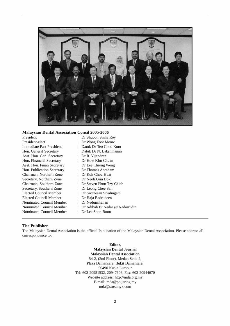
Malaysian Dental Association Concil 2005-2006President : Dr Shubon Sinha RoyPresident-elect : Dr Wong Foot MeowImmediate Past President : Datuk Dr Teo Choo Kum Hon. General Secretary : Datuk Dr N. LakshmananAsst. Hon. Gen. Secretary : Dr R. VijendranHon. Financial Secretary : Dr How Kim ChuanAsst. Hon. Finan Secretary : Dr Lee Chiong WengHon. Publication Secretary : Dr Thomas Abraham Chairman, Northern Zone : Dr Koh Chou HuatSecretary, Northern Zone : Dr Neoh Gim BokChairman, Southern Zone : Dr Steven Phun Tzy ChiehSecretary, Southern Zone : Dr Leong Chee SanElected Council Member : Dr Sivanesan SivalingamElected Council Member : Dr Haja BadrudeenNominated Council Member : Dr NedunchelianNominated Council Member : Dr Adibah Bt Nadar @ Nadarrudin Nominated Council Member : Dr Lee Soon Boon
The PublisherThe Malaysian Dental Association is the official Publication of the Malaysian Dental Association. Please address allcorrespondence to:
Editor,Malaysian Dental Journal
Malaysian Dental Association54-2, (2nd Floor), Medan Setia 2,
Plaza Damansara, Bukit Damansara,50490 Kuala Lumpur
Aim And ScopeThe Malaysian Dental Journal covers all aspects of work in Dentistry and supporting aspects of Medicine. Interactionwith other disciplines is encouraged. The contents of the journal will include invited editorials, original scientific arti-cles, case reports, technical innovations. A section on back to the basics which will contain articles covering basic sci-ences, book reviews, product review from time to time, letter to the editors and calendar of events. The mission is to pro-mote and elevate the quality of patient care and to promote the advancement of practice, education and scientific researchin Malaysia.
PublicationThe Malaysian Dental Journal is an official publication of the Malaysian Dental Association and is published half year-ly (KDN PP4069/12/98)
SubscriptionMembers are reminded that if their subscription are out of date, then unfortunately the journal cannot be supplied. Sendnotice of change of address to the publishers and allow at least 6 - 8 weeks for the new address to take effect. Kindly usethe change of address form provided and include both old and new address. Subscription rate: Ringgit Malaysia 60/- foreach issue, postage included. Payment in the form of Crossed Cheques, Bank drafts / Postal orders, payable to MalaysianDental Association. For further information please contact :
The Publication SecretaryMalaysian Dental Association
54-2, (2nd Floor), Medan Setia 2, Plaza Damansara, Bukit Damansara,50490 Kuala Lumpur
Back issuesBack issues of the journal can be obtained by putting in a written request and by paying the appropriate fee. Kindly sendRinggit Malaysia 50/- for each issue, postage included. Payment in the form of Crossed Cheques, Bank drafts / Postalorders, payable to Malaysian Dental Association. For further information please contact:
The Publication SecretaryMalaysian Dental Association
54-2, (2nd Floor), Medan Setia 2, Plaza Damansara, Bukit Damansara,50490 Kuala Lumpur
Membership and change of addressAll matters relating to the membership of the Malaysian Dental Association including application for new membershipand notification for change of address to and queries regarding subscription to the Association should be sent to HonGeneral Secretary, Malaysian Dental Association, 54-2 (2nd Floor) Medan Setia 2, Plaza Damansara, Bukit Damansara,50490 Kuala Lumpur. Tel: 603- 20951532, 20951495, 20947606, Fax: 603- 20944670, Website Address:http://www.mda.org.my. Email: [email protected] or [email protected]
DisclaimerStatements and opinions expressed in the articles and communications herein are those of the author(s) and not neces-sarily those of the editor(s), publishers or the Malaysian Dental Association. The editor(s), publisher and the MalaysianDental Association disclaim any responsibility or liability for such material and do not guarantee, warrant or endorse anyproduct or service advertised in this publication nor do they guarantee any claim made by the manufacturer of such prod-uct or service.
Malaysian Dentist: Tax implications, penalties for non compliance and tax planning aspectsKF Choong 6
Expression of p53 and PCNA at the tumour invasive front of oral squamous cell carcinomaBTF George, RB Zain, SKS Kumar 10
The Expert says………Tumour markers in a nut shellHM Hussaini 18
Drug-induced pemphigus in Wilson diseaseS Ram, SKS Kumar, RB Zain, NP Kipli, Lee GK, Ching LL 20
A radiographic study of mandibular third molar development in a local orthodontic populationST Loke, SK Tee 24
Level of knowledge, perception and practices in relation to oral health promotion among final year trainee dental nursesNA Azli, AT Zamzuri 37
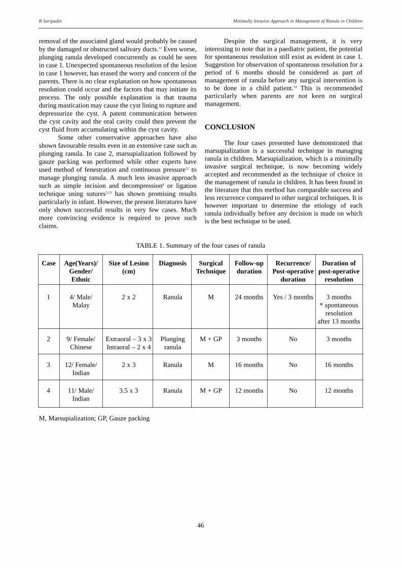
Minimally invasive approach in management of ranula in childrenB Saripudin 44
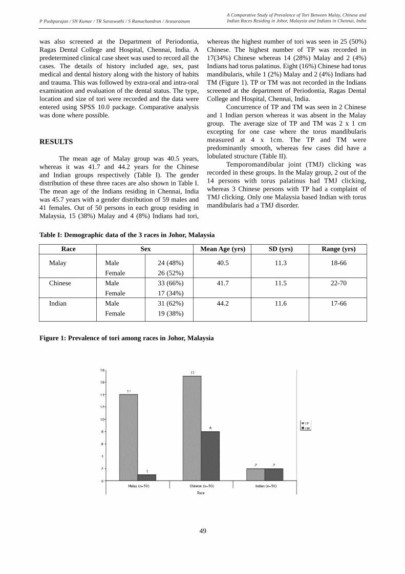
A comparative study of prevalence of tori between Malay, Chinese and Indian races residing in Johor,Malaysia and Indians in Chennai, IndiaP Pushparajan, S Nalin Kumar, TR Saraswathi, S Ramachandran, Arasaratnam 48
Dental management of patients with prosthetic joints: A reviewWL Chai, CK Yong, WC Ngeow 53
Manifestation of systemic diseases in the periodontium – a report of two cases.YK Chan 58
Instructions to contributors 62
Survey on readership of Malaysian Dental Journal 64
MDJ cover page: H&E pictures show intraepithelial split of pemphigus vulgaris in original magnifications of 40X and100X. Note the basal cells separated from the epithelium (arrows). Two illustrations below show DIF positive for IgG and DIF positive for fibrinogen.
The cover picture is courtesy of Professor Dr. Rosnah Zain. Taken from article “Drug-induced pemphigus in Wilsondisease”
It is indeed an honour to be invited by thePublication Secretary, Dr. M. Thomas Abraham to becomethe editor of the 2005/06 issue of the Malaysian DentalJournal. Dr. Abraham has done a great job in reviving theMalaysian Dental Journal, coming out with not only oneissue but two issues per year. It is indeed a difficult task forme to follow. Nevertheless, I take this new challenge as anew learning opportunity. Hopefully I can equal Dr.Abraham’s contribution if not better.
For a start, I hope to continue the good work thatDr. Abraham has established. Hence it is my commitmentto make sure that 2 issues of the Malaysian Dental Journalwill be published during my tenure as the Editor. I am gladto announce that this is the first of the 2 issues I am com-mitted to publish. We hope to be able to come up with thenext issue before the next Annual General Meeting in June2006.
Most dental professional journals restrict the contents to matters pertaining to research, updates, tips andnews in dentistry. A few well read journals like the BritishDental Journal, Journal of the American DentalAssocaition and Dental Updates do include non-scientificjournals like articles on the world-wide web to keep dentists abreast with the ever fast changing world. I too,believe we shall not restrain ourselves if we can receivegood contribution/articles that may improve our professionas well as quality of life. For a start, I am glad to includean article on taxation for dentists. It is specially commis-sioned by a good friend of mine, Associate Professor Dr.Choong Kwai Fatt who gladly share with us his knowledgeof income taxation and how we can get the best out of theallowances provided under the Malaysian Law. If therespond is good, we hope to come up with articles onfinancial planning as I personally feel that these topicswere less taught in dental schools, yet they in fact becomean important part of our life once we start working.
The Malaysian Dental Journal (formerly the DentalJournal of Malaysia), has been published for decades.Unfortunately, we have no feedback of the quality of pub-lications as well as feedback on what dentists want to read.Prominent journal like the British Dental Journal conduct-ed surveys on its readership. As a matter of fact, its surveyeven looked into dentists’ lifestyle in order to cater foradvertisers. So, you do see advertisement on car for exam-ple in this dental journal. It is my hope to do the same inorder to improve on any shortcomings. I have included atthe end of this journal a set of questionnaire survey formswhich I hope readers will send back to the Editorial Officeof the Malaysian Dental Journal. Some of you may haveseen the forms being circulated around at some CDE pro-grammes. We hope those who have not responded will doso.
One feature that we are experimenting with thisissue of Malaysian Dental Journal is the incorporation of asection called: The Specialist says….. This is a commission column especially to discuss some learningissues on selected topics. We hope the busy practitionerswill find that they learn something even if the are not interested to read the whole content of a related article.
Lastly, I would like to extend my gratitude to themembers of the MDJ Editorial Advisory Board. I couldhave never done all the editorial work without their tirelessassistance. Also grateful thanks to all the referees whohave kindly review articles for the Malaysian DentalJournal.
Thank you.
Associate Professor Dr. Ngeow Wei CheongEditorMalaysian Dental Journal 2005/06
Malaysian Dentist: Tax Implications, Penalties for Non Compliance and Tax Planning Aspects
Choong KF Associate Professor & Tax Consultant Faculty of Business and Accountancy, University of Malaya, Kuala Lumpur.
INTRODUCTION
Malaysian dentists exercising employment incomehave to file in the income tax return Form BE latest by 30 April 2006 to the Central Processing Center atAras 12, Menara C, Persiaran MPAJ, Jalan Pandan Utama,Pandan Indah, 55100 Kuala Lumpur. Under the selfassessment system, the difference between the actualincome tax payable and the total monthly tax deducted byemployer also has to be accounted to the Inland RevenueBoard (IRB) collection branch by 30 April 2006. If there isexcess payment, the tax authorities would refund thisamount by crediting the taxpayer’s bank based on thedetails provided by taxpayer.
Employment Income
Dentists exercising employment with the privatesector will report the gross employment income as statedin the EA Form.1 If entertainment allowances or travelingallowances are provided by the employer, then these enter-tainment expenses and traveling expenses incurred by theemployee in carrying out his/her duties are tax deductible.Only the net amount is reported.
Dentists exercising employment with the government sector however have the following incomeexempted from their income tax:
i) Fixed Housing Allowance;ii) Special Housing Allowance;iii) Fixed Public Service Allowance;iv) Critical Allowance;v) Free Utilities Allowance;vi) Mobile Phone Facility;vii) Driving License Allowance;viii) Wilayah Housing Allowance.
The gross employment income reported in the ECForm has to exclude the above and reported the net sum inthe tax return.
Interest Income
Interest income derived from commercial banks isnot required to be disclosed in the return Form BE as suchinterest income is either exempted or already taxed atsource (5% tax) by the bank.
Interest income derived from the following sourceswill be taxed:
(a) Loans to relatives or friends;(b) Convertible loan stocks/debentures/bonds(c) Convertible loan stocks issued by companies listed
on MESDAQ.
Dividend Income
Only taxable dividend income is required to bereported to the tax authorities. The dividend income will bestated in the tax voucher as follow:
Gross Tax 28% Net dividenddividend
RM RM RM1,000 280 720
The amount included in the Form BE is the grossdividend of RM 1,000 and the tax deductible of RM280 isdeducted from the income tax payable computed, includedin Column E11 of Form BE.
Rental Income
Rental income is reported in the tax return Form BEat the net amount, after deducting the following expensesthat are incurred wholly and exclusively in the productionof rental income. These expenses are:(i) quit rent and assessment;(ii) cost of repairs and maintenance of the property;(iii) insurance premium on fire/burglary;(iv) cost of supervision and rental collection;
Malaysian Dentist: Tax implications, penalties for non compliance and tax planning aspects KF Choong
Maximising donation and tax relief
Individuals embarking on the following charitable activities will be given tax deductions.
Section Description Individual44(6) Cash donation to Government, or local authority √44(6) Cash donation to approved institution or organization √44(6A) Artifact, manuscript or painting to the Government, State Government √44(8) Cash donation to public or school libraries (restricted to RM20,000) √44(9) Cash or contribution in kind for provision of facilities in public places for disabled persons √44(10) Cash or medical equipment to approved healthcare facility (restricted to RM20,000) √44(11) Painting to national or state art galleries √
The following are tax reliefs available to the taxpayer:
YA 2005Reliefs RMPersonal (self) 8,000Medical expenses for parents (restricted to) 5,000Basic supporting equipment for taxpayer, spouse, children or parents (restricted to) 5,000Disabled taxpayer 6,000Fees for education in technical, industrial, scientific, technological or vocational fields – only apply to that individual and restricted to 5,000Medical fees for taxpayer, spouse and children for serious diseases (restricted to) (includes RM500 for complete medical examination) 5,000Purchase of books, magazine, journals for taxpayer, spouse, child 700Interest expense incurred on acquisition of first residential property (restricted to) 2,000Wife relief (if housewife or wife elects joint assessment) 3,000Disabled wife3,500[Husband relief: if husband elects joint assessment with wife Disabled husband 3,500]Child relief: Disabled (RM5,000 per child) √: Local universities, colleges or similar establishments (RM4,000 per child) √: Overseas universities, colleges or similar establishments (RM 1,000 per child) √: Others (RM1,000 per child) √EPF and life insurance premium (restricted to) 6,000Education or medical insurance – taxpayer, spouse, children (restricted to) 3,000EPF annuity insurance (restricted to) 1,000
(v) cost of obtaining a new tenant to replace the old ten-ant;
(vi) interest paid on loan facility taken to finance theproperty;
(vii) cost of renewing the rental agreement and other mis-cellaneous expenses.
If the properties are in joint name of husband andwife, then each will be responsible to report their share ofthe net rental income in the respective tax return.
Foreign Source Income
Income received from outside Malaysia will be taxexempt by virtue of para 28 of sch 6 of the Income Tax Act1967. It is not required to be reported.
8
Malaysian Dentist: Tax implications, penalties for non compliance and tax planning aspects KF Choong
Tax Rebate
Tax rebates are given preferential tax treatment ascompared to tax relief. It is deducted from the income taxpayable. The following are the tax rebates available to dentists:(a) Religious payment
A rebate shall be granted for any zakat, fitrah or anyother payment of Islamic religious dues which areobligatory and paid in the basis period of 2005. Thepayment must be evidenced by a receipt issued bythe Pusat Zakat of the respective States.
(b) Personal computerA rebate of RM500 shall be granted to an individualin respect of purchase of a personal computer in2005. The claim has to be substantiated by a receipt.The rebate of RM500 would not be granted:(i) for the following 4 years;(ii) where the personal computer was used for busi-
ness; or(iii) where such rebate has been granted to spouse.
Income Tax Payable
The income tax payable is arrived at after taking thechargeable income and multiplying it with the scale taxranges between 0% – 28%.
Aggregate income (from all source of income) xx- Donation (x)
Total Income xx- Tax relief (x)
Chargeable income xx
Income tax rate (range of 0% - 28%)
Income tax payable x- Rebate
Zakat & Fitrah (for Muslim dentists) xComputer (500)Net income tax payable (A) xx
- Tax credit on dividend income (x)- Total monthly tax deducted
(as per Form EA/EC) (x)Final tax payable (A – B) x
Refund of excess tax paid (B – A) x
Where there is final tax payable, the dentist needs to fill in the remittance slip (CP 501) at the end of Form BE togetherwith the payment to either a commercial bank (Public Bank or Bumiputra Commerce) or the IRB’s collection branchlocated at:
SEMENANJUNG MALAYSIA SABAH & WP LABUAN SARAWAK
Tingkat Bawah, Block 8A, Tingkat Bawah Aras 1Komplex Bangunan Kerajaan, Wisma Bandaraya Wisma Ting Pek KhiingJalan Duta Jalan Masjid Lama No. 1, Jalan PadunganKuala Lumpur Kota Kinabalu Kuching
The final tax payment has to be paid latest by 30 April 2006. Failing which, a late payment penalty of 10% will beimposed.
Where there is excess tax payment, the excess will be refunded by the tax authorities via the bank account provided bythe taxpayer. The taxpayer however is required to write in to IRB’s collection branch if the excess amount is notreceived by 31 December 2006. The relevant addresses are:
SEMENANJUNG MALAYSIA SABAH & WP LABUAN SARAWAK
Lembaga Hasil Dalam Negeri Lembaga Hasil Dalam Negeri Lembaga Hasil Dalam NegeriMalaysia Malaysia MalaysiaCawangan Pungutan, Tingkat 15, Unit Pungutan Tingkat Bawah , Unit Pungutan Blok 8A Komplex Bangunan Kerajaan, Wisma Bandaraya Aras 1, 3, 6, 7 & 8Jalan Duta Karung Berkunci 11061 Jalan Masjid Lama Wisma Ting Pek Khiing
50990 Kuala Lumpur. 88600 Kota Kinabalu No. 1, Jalan Padungan93100 Kuching
9
Malaysian Dentist: Tax implications, penalties for non compliance and tax planning aspects KF Choong
Responsibility of Keeping Records
The self assessment regime of taxation requires thetaxpayer to keep and retain in safe custody the statement ofincome and expenditure, Form EA/EC, income tax pay-ment, invoices, vouchers and receipts that are necessary toverify the particulars in the return Form BE for a period of7 years. Failure to do so will result in non deductibility ofsuch expenditure, a fine of between RM300 to RM10,000and/or imprisonment for one year.4 The tax authorities willcarry out tax audits by visiting the taxpayer’s premisesonce in every five years to ensure the computation and therecords are retained in accordance to the Income Tax Act1967 (the Act). Penalties and/or additional notices ofassessment will be imposed after the tax audit if thereexists non compliance with the Act.
Submission of Return
The Act imposes strict penalty for non submissionof income tax return Form BE, that is 3 times of theincome tax payable,5 notwithstanding the fact that monthly tax deductions have been made by the employer.The deduction of monthly tax does not constitute a reasonable excuse and has no relevance to the submissionof returns.
In hardcore cases, where a taxpayer has consistently failed to submit his/her income tax returns forseveral years, the tax authorities, in practice, would prosecute the taxpayer and if convicted, the taxpayer willbe liable to a fine of between RM200 and RM2,000 and/orimprisonment for a period of 6 months for each year of nonsubmission. In addition, the court will further order thetaxpayer to submit the returns within 30 days from theorder and will accordingly be liable for the income tax.
Tax Planning
Employment income exercised by a dentist has notmuch scope of tax planning. The significant tax planningtool will be to forgo the annual bonus or allowances inexchange for an increased contribution by the employer tothe Employees’ Provident Fund (EPF). Under the existingtax regime of section 34(4), the employer will be given atax deduction against their business income up to 19% ofthe employee’s salary. This tax planning tool only be effective if it is from the employer’s contribution and notthe employee’s contribution.
Employees will have a much lower income taxpayable by forgoing the receipt of annual bonuses andallowances. The amount is now represented by the EPFcontribution to the Employees Provident Fund. Employeesmay now withdraw 30% of the accumulated fund in EPFto purchase residential homes or retain it to enjoy an annual dividend of 5%, compounded annually. This returnof 5% is superior to the interest income derived from commercial banks.
Alternatively, dentists having to pay for child carefacilities personally which is not tax deductible mayarrange with the employer for the provision of child carefacility by the employer as this is a tax free benefit toemployee.
CONCLUSION
Living in a modern society is very stressful anddemanding. Dentists are required to keep abreast with thetechnical knowledge in his/her profession and also famil-iarize themselves with the self assessment regime of taxa-tion. Failure to do so will result in unnecessary moneybeing incurred on penalties or additional income taxpayable. The alternative solution is to seek professional taxadvice when embarking on filling in of the tax return.
REFERENCES
1. C1 of B5 Form.2. Section 103 of the Act.3. A18 to A20 of Form BE.4. Section 119A of the Act.5. Section 112(3) of the Act.6. Section 112(1), 112(2A) of the Act.7. Section 1B(1)(b)(i) benefits.
Additional Reading:1. Choong Kwai Fatt (2005), ‘How To Fill In Your Income Tax
Form B’, InfoWorld.2. Choong Kwai Fatt (2004), ‘ Tax Planning For Employees’,
Sweet and Maxwell Asia.
Address for Correspondence:
Dr. Kwai Fatt ChoongAssociate ProfessorFaculty of Business AccountancyUniversity of MalayaWebpage : www.kwaifatt.comE-mail : [email protected]
10
MALAYSIAN DENTAL JOURNAL
Expression of p53 and PCNA at the Tumour Invasive Front of Oral SquamousCell Carcinoma
BTF George, MClinDent Specialist in Oral Pathology and Oral Medicine Hospital Umum, Kuching, Sarawak
RB Zain, MS Professor in Oral Pathology and Oral Medicine Department of Oral Pathology, Oral Medicine & Periodontology,Faculty of Dentistry, University of Malaya
SKS Kumar, MDSc Research Assistant, Department of Oral Pathology, Oral Medicine & Periodontology, Faculty of Dentistry,University of Malaya
ABSTRACT The tumour invasive front of oral squamous cell carcinoma (OSCC) has been shown to have prognostic significance. The aim of this study was to evaluate the expression of p53 and PCNA (Proliferating cellnuclear antigen) at the invasive front of OSCC and to determine their association with certain clinicopathologicfactors. METHODS: The study sample consisted of biopsies from 27 patients diagnosed with OSCC in buccalmucosa. Immunohistochemistry was used to investigate the expression of p53 and PCNA. RESULTS: The expression of p53 and PCNA was detected in 92.6% (25) and 100% (27) cases respectively. In general, the predominant distribution of immunoreactivity for p53 at the tumour invasive front with almost sparing of the central keratinising areas was observed to be similar to that for PCNA. The present study also suggests that thereis no relationship between expression of p53 and PCNA with TNM staging. However there appears to be a relationship between expression of p53 and PCNA with both modified Broders malignancy grading and “patternof invasion”. CONCLUSION: p53 and PCNA are well expressed at the invasive front of oral squamous cell carcinoma. Thus, it is feasible to use these markers in future studies to look into these markers as prognostic indicators.KEYWORDS: Tumour, Invasive front, SCC, Markers.
INTRODUCTION
Oral cancer is a serious global public health problem with an annual incidence of about 200,000 ofwhich up to two-thirds occur in developing countries.1
Although there have been significant advances in themulti-modal treatment of the disease, the prognosis fororal cancer has not improved significantly.2,3
The present study has focused on the tumour invasive front areas of oral squamous cell carcinoma(OSCC) because these parts have been observed to reflectimportant biological events, like morphological andmolecular characteristics of the tumour, which may be ofprognostic significance.4,5
Mutation of the p53 tumour suppressor gene, alsoknown as the “guardian of genome”,6 has been reported asone of the most common event in cancer.7 This mutationmay lead to an increase in the pool of proliferating cellsand also the probability of neoplastic transformation.8
PCNA (Proliferating cell nuclear antigen) plays an essential role in DNA replication and has been suggestedas a marker of proliferating cells.9 p53 may act as a complimentary marker to PCNA given that PCNA reactivity defines the growth fraction of a tumour and p53reactivity demonstrate the irreversible malignant changehaving occurred inside this fraction.10
The aim of the present study is to determine anyassociation between expression of p53 and PCNA at thetumour invasive front of OSCC in the buccal mucosa withcertain clinicopathologic features which have establishedprognostic significance, namely TNM clinical staging,11
conventional malignancy grading as in modified Brodersgrading12 and “pattern of invasion”.4,5 This is the first studyin Malaysia which looks into the expression of PCNA andp53 at the tumour invasive front. This preliminary study isin accordance to the first phase in a hierarchy of prognosisstudy proposed by Hall and Going in 1999.13
Expression of p53 and PCNA at the Tumour Invasive Front of Oral Squamous Cell CarcinomaBTF George / RB Zain / SKS Kumar
This study utilised the immunohistochemicalmethod to investigate the expression of p53 and PCNA atthe tumour invasive front. This is because tissue morphological changes observed under light microscopeare now recognised as a comparatively late consequence ofkey molecular events that have initiated pathologicalchange, and the immunohistochemical studies are used tolink these specific regulatory proteins in either their normal or mutated forms with these tissue changes.14
MATERIALS AND METHODS
The sample for this study was obtained from thearchives of the Department of Oral Pathology, OralMedicine and Periodontology, Faculty of Dentistry,University of Malaya, consisted of 27 cases of untreatedprimary tumours in the buccal mucosa that had been diagnosed histopathologically as oral squamous cell carcinoma (OSCC). These specimens had been fixed in10% buffered formalin and then embedded in paraffinwax.
Histopathological grading
This was carried out using the modified Brodersmalignancy grading12 and “pattern of invasion”.4,5
Broders malignancy grading (Modified)12
The modified Broders system took into account asubjective assessment of the degree of keratinisation, cel-lular and nuclear pleomorphism and mitotic activity of thetumour population, and the tumours were then graded aswell differentiated (grade 1), moderately differentiated(grade 2), and poorly differentiated (grade 3) oral squa-mous cell carcinoma.
Invasive front grading
The evaluation of “pattern of invasion” was carriedout at the most invasive part of tumours (defined as 3-6 celllayers at the advancing front of tumours). A score of 1 to 4is given based on the cohesiveness of the tumour at theinvasive front15 (Refer Table 1). A high score indicated apoor prognosis and a low score, a good prognosis.
Immunostaining procedures
The immunostaining was carried out using theavidin biotin peroxidase technique. Heat mediated antigenretrieval was performed by incubating sections (4µ thick)in 0.01M citrate buffer at pH 6.0 in a microwave oven setat 100°C for 20 minutes. The primary antibodies used wereDO-7 (Dako) and PC-10 (Dako) for p53 and PCNArespectively, while the controls were from a known case ofOSCC that reacted positively to p53 and PCNA.
Immunohistochemical grading
The immunoreactivity for both PCNA and p53were graded only at the invasive front area by integratingthe staining intensity and proportion of tumour cellsstained described in previous studies (16), with the aid ofan image analyser (Refer Table 2).
TNM staging
The TNM staging used in the present study wasbased on the guidelines provided for lip and oral cavitycarcinomas found in the 4th edition of the InternationalUnion Against Cancer (IUCC) TNM guidelines11.
RESULTS
The present study consisted of histological specimens taken from 27 patients diagnosed earlier with primary OSCC in the buccal mucosa. The age of thesepatients ranged from a minimum of 47 years to a maximumof 76 years. The mean age is 61.6 years (standard deviation= 9.9). This sample consists of 18 (66.7%) female and 9male (33.3%) patients, while the ethnic distribution is madeup of 22 (82%) Indians, 3 (11%) Malays and 2 (7%)Chinese. The patients in this series had TNM clinical stages:II, III and IV. There were 5 (18.5%) patients clinicallystaged at II, 6 (22.2%) at stage III and 16 (59.3%) at stageIV. There were no patients at TNM stage I in this study.Fourteen (51.9%) patients had well differentiated carcinoma(grade 1), 9 (33.3%) had a moderately differentiated tumour(grade 2), while the remaining 4 (14.8%) had a poorly dif-ferentiated SCC (grade 3). The evaluation of the morphologic parameter of “pattern of invasion” at thetumour invasive front revealed 9 (33.3%) specimens with ascore of 2, and 13 (48.1%) had a score of 3 while theremaining 5 (18.5%) had a score of 4.
Expression of p53 at the tumour invasive front ofOSCC in buccal mucosa
Immunohistochemically detectable p53 proteinaccumulation was observed in 25 (92.6%) cases. Twocases (7.4%) did not demonstrate any expression of p53 atthe tumour invasive front. All reactions with a distinctnuclear staining were considered positive, irrespective ofthe intensity of the immunoreactivity. A striking accumulation of p53 positive tumour cells were seen at thetumour invasive front as well as in peripheral layers ofinvading tumour islands (Refer Table 3; Figure 1).However, the central keratinizing areas in most tumourswere p53 negative.
12
Expression of p53 and PCNA at the Tumour Invasive Front of Oral Squamous Cell CarcinomaBTF George / RB Zain / SKS Kumar
Table 1: Criteria of scoring used for “pattern of invasion” 4,5
Score Criteria
1 Pushing, well delineated infiltrating borders
2 Infiltrating, solid cords, bands and/or strands
3 Small groups or cords of infiltrating cells (n<15)
4 Marked and widespread cellular dissociation in small groups and/ or in single cells (n < 15)
Table 2: Criteria for the grading of tumours for p53 and PCNA immunoreactivity:
Grading P53/PCNA immunoreactivity
Strong (3) Strong nuclear immunostaining in >50% of the ITF cells
Moderate (2) Strong nuclear immunostaining in 10% to 50% or moderate nuclear immunostaining in > 50% of the ITF cells
Weak (1) Moderate nuclear immunostaining in 10% to 50% or weak nuclear immunostaining in any proportion of the ITF cells
Expression of p53 and PCNA at the Tumour Invasive Front of Oral Squamous Cell CarcinomaBTF George / RB Zain / SKS Kumar
Figure 1 Immunoreactivity of p53 at the tumour invasive front of SCC in buccal mucosa. A striking accumulation of p53 positive tumour cells is seen atthe tumour invasive front as well as in peripheral layers of invading tumourislands. However, the central keratinizing areas in most tumours are virtuallyp53 negative. Arrows showing tumour invasive front. T – Tumour area; CT –Connective tissue area. (Original magnification X50).
Figure 2 Expression of PCNA at the tumour invasive front of SCC in buccal mucosa. Generally the distribution of immunoreactivity for PCNAis observed to be similar to that of p53. An intense nuclei staining forPCNA is seen in tumour cells at the deep infiltrating margins as well asin the periphery of invading epithelial nests. Keratinized cells are however not stained. Arrows showing tumour invasive front. T – Tumourarea; CT – Connective tissue area. (Original magnification X50).
14
Expression of p53 and PCNA at the Tumour Invasive Front of Oral Squamous Cell CarcinomaBTF George / RB Zain / SKS Kumar
Expression of PCNA at the tumour invasive front ofOSCC in buccal mucosa
PCNA expression was observed in all 27 cases inthis study. Again only reactions with a distinct nuclearstaining were considered positive, irrespective of the intensity of the immunoreactivity. Generally, the distribu-tion of immunoreactivity for PCNA was observed to besimilar to that of p53. An intense nuclei staining for PCNAwas seen in tumour cells at the deep infiltrating margins aswell as in the periphery of invading epithelial nests (ReferTable 4; Figure 2). Keratinized cells were however notstained.
Relationship between the immunohistochemicalexpression of p53 and PCNA at tumour invasive front of OSCCs in buccal mucosa with selected clinicopathologic parameters
TNM clinical staging
The relationship between p53 and PCNA expres-sion with TNM clinical staging is shown in Figures 3 and4 respectively. For p53, there was an increase in grades 2and 3 staining in TNM IV as compared to TNM II and III.For PCNA, while TNM II and III samples showed quiteintense staining of grade 2 and 3, TNM IV showed thepresence of grade 1 (less intense) staining.
The relationship between p53 and PCNA expres-sion with conventional malignancy grading is shown inFigures 5 and 6 respectively. Degree of expression or grad-ing for both p53 and PCNA is seen to generally increasewith decrease in differentiation of tumour as defined bymodified Broders grading.
“Pattern of invasion”
The relationship between p53 and PCNA expres-sion with “pattern of invasion” is shown in Figures 7 and 8respectively. The degree of expression of both p53 andPCNA is seen generally to increase with increase in thescore of “pattern of invasion”.
DISCUSSION
The sample size in the current study is small when compared to other case series of oral SCCs diagnosed frombiopsies seen at the Institute of Medical Research,Malaysia17-19 and the Faculty of Dentistry, University ofMalaya.20 However, the socio-demographic characteristics(age, gender and ethnicity) of this case series are in accordwith those from previous studies17-20 that OSCC is a diseaseof the older age group in Malaysia with an overall femalepreponderance, and predominantly involving the Indian
ethnic group. Previous studies have also observed that themost frequently encountered type of OSCC is the well dif-ferentiated type while the poorly differentiated type is theleast common.17-20 These distinct findings are also generally noted in the present study.
Distribution of p53 and PCNA
The distribution of p53 and PCNA immunoreactivityseen predominantly accumulating at the tumour invasivefront agrees well with previous observations and support thevalidity of assessing molecular activity at this area of oralcarcinoma.10,21,22 The generally similar immuno-localisationof p53 and PCNA staining at the tumour invasive frontappear to suggest that p53 protein expression is found inareas with proliferative activity and might indicate theinvolvement of the mutated form of the p53 protein in thealteration of the cell cycle regulation process.10,21,22
Therefore it has been suggested that p53 may act asa complementary marker to PCNA, since PCNA reactivitydefines the growth fraction of tumour and p53 reactivitydemonstrates the irreversible malignant change havingoccurred inside this fraction.10,22
These observations may however, also be due to accu-mulation of the wild type p53 induced by DNA damage orspecific viral protein binding. 21, 23-25 These findings are compatible with functions of p53 as an inducer of DNA repairand PCNA as a DNA repair protein. 21.26
Tumours without detectable p53, with a high PCNAindex, can also contain cells with mutations that result inproduction of truncated proteins that are not detectable orcells containing non sense mutation that leads to termina-tion of protein synthesis.21,27,28
The difficulty in interpreting the results from thisstudy is due partially to the complex biologies of p53 and PCNA proteins, and to the influence of immunohistochemical techniques on patterns ofimmunoreactivity.14 Thus, the use of immunohistochemicaltechniques requires care in interpretation, caution in drawing conclusions and to always consider the biology ofmolecules being investigated.29
Relationship between the immunohistochemicalexpression of p53 and PCNA at the tumour invasivefront of OSCC in buccal mucosa with selected clinicopathologic parameters
TNM clinical staging
The results from the present study appear to suggestthat there is no relationship between the expression of p53and PCNA at the tumour invasive front and TNM clinical staging. A similar finding has also been observed in previous study involving SCC of head and neck.30
Morawski et al.’s30 study however demonstrated their findings using statistical analysis (Spearman RankCorrelation Test) while the present study did not use anytest of significance due to its small sample size.
15
Expression of p53 and PCNA at the Tumour Invasive Front of Oral Squamous Cell CarcinomaBTF George / RB Zain / SKS Kumar
It is a known fact that there is some disagreementbetween observers regarding the determination of tumoursize and node status owing to the inherent subjectivity ofthe system.4,5 Furthermore, the clinical staging may sometimes turn out to be different from what is found afterexcision and histopathological examination (pTNM).31
Therefore some of patient’s data on clinical stagingobtained from their respective folders may be associatedwith uncertainty, which may in turn weaken the findingsand conclusions of study.
Previous studies on oral and head and neck squa-mous cell carcinoma have found that there is no correlation between expression of p53 at tumour invasivefront, and conventional malignancy grading30,32 whereas thepresent study appears to suggest otherwise. The two previ-ous studies demonstrated their findings using statistical analysis (Pearson and Spearman correlation test respectively) while the present study did not.
There has been suggestion that the lack of statistically significant correlation between expression ofp53 and clinicopathological parameters such as conventional malignancy grading may probably indicate amore important function for p53 in early phase of tumour generation rather than determining a prognosis-relatedparameter.32
The difference in findings may also be attributed tothe inherent subjectivity of the malignancy (Broders) grading system whereby there is no complete agreement on classification of individual tumours and histopathological specimens.4,5 However, attempts weremade to minimise these problems in the present study byregrading (conventional malignancy grading and “patternof invasion”) the samples independently by two examinersafter a training and calibration exercise.
The present study has observed a possible relation between expression of PCNA at the invasive front and conventional malignancy (modified Broders) grading. Thisfinding appears to concur with that of Morawski et al.(1999) study, which demonstrated the correlation usingstatistical analysis (Spearman Rank Correlation Test). Thisrelation seems logical, as modified Broders gradingdescribing the degree of cancerous cell differentiation considers cell proliferative properties.30
“Pattern of invasion”
The findings from the present study suggest thatthere is a relationship between expression of p53 andPCNA at tumour invasive front, and the histologicalparameter “pattern of invasion”.
“Pattern of invasion” has been reported as the most important single histological parameter in assessing theability of tumours to metastasize,33 thus making it an indicator of tumour aggressiveness.34 Therefore, it would
appear that the findings in this study may be of significance when one consider the important relationshipbetween “pattern of invasion” and clinical outcome,33,35
and also because of the very high level of interobserverreproducibility obtained with “pattern of invasion” (whencompared to other histological parameters of InvasiveFront Grading and Broders malignancy grading) (36,37).
SUMMARY AND CONCLUSION
This is a preliminary study with a small sample sizewith variable clinical stages and malignancy grades. No test of significance was carried out to confirm the relationship between expression of p53 and PCNA at thetumour invasive front with the selected clinico-pathologic parameters. Therefore the present findings are preliminaryin nature.
In conclusion, p53 and PCNA are well expressed atthe invasive front of oral squamous cell carcinoma. Thus,it is feasible to use these markers in future studies to lookinto these markers as prognostic indicators. However, tofurther validate the prognostic significance of any markerswill require the knowledge of the survival pattern of thesepatients. With such patterns, a comparison of the survivalpatterns between those with higher and lower p53 and/PCNA expressions at the tumour invasive front can furtherindicate its possible clinical significance. Thus, goodrecord keeping of follow-up of these patients by clinicianswill further enhance the clinical applicability of such laboratory findings.
ACKNOWLEDGEMENT
The authors would like to thank the University ofMalaya for the research grant (Vot F-00402001B). Wewould also like to thank the staff at the Department of OralPathology, Oral Medicine and Periodontology for theirtechnical assistance. Our gratitude also goes to ProfessorDr. Douglas A. Luke, UKM for his kind assistance.
REFERENCES
1. Parkin DM, Pisani P, Ferlay J. Estimates of the worldwideincidence of 25 major cancers in 1990. Int J Cancer 1999, 80:827-841.
2. Silverman S, Gorsky M. Epidemiologic and demographicupdate in oral cancer: California and national data – 1973 to1985. JADA 1990, 120: 495-499.
3. Mork J. 40 years of monitoring head and neck cancer inNorway-No good news. Anticancer Res 1998, 18: 3705-3708.
4. Bryne M. Is the invasive front of the oral carcinoma the mostimportant area for prognostication? Oral Dis 1998, 4: 70-77.
16
Expression of p53 and PCNA at the Tumour Invasive Front of Oral Squamous Cell CarcinomaBTF George / RB Zain / SKS Kumar
5. Bryne M., Boysen M., Alfsen CG, et al. The invasive front ofcarcinomas. The most important area for tumour prognosis?Anticancer Res 1998, 18: 4757-4764.
6. Lane DP. p53 guardian of genome. Nature 1992, 358: 15-16.7. Greenblatt MS, Bennett WP, Hollstein M, Harris CC.
Mutations in the p53 tumour suppressor gene: Clues to cancer aetiology and molecular pathogenesis. Cancer Res1994, 54: 4855-4878.
8. Raybaud-Diogene H, Tetu B, Morency R, Fortin A, Monteil,RA. p53 overexpression in head and neck squamous cell carcinoma: Review of the literature. Oral Oncol Eur J Cancer1996, 32B(3): 143-149.
9. Hall PA, Levison DA, Woods AL, et al. Proliferating cellnuclear antigen (PCNA) immunolocalization in paraffin sections: An index of cell proliferation with evidence ofderegulated expression in some neoplasm. J Pathol 1990,162: 285-294.
10. Lan HA, Zain RB, Saitoh M, Muramatsu Y, Shrestha P, MoriM. Proliferating cell nuclear antigen (PCNA) & p53 inepithelial dysplasia & squamous cell carcinoma of oralmucosa-a marker for poor tumour differentiation, increasingnuclear atypia & invasiveness? Anticancer Res 1996, 16:3059-3066.
11. Hermanek P, Sobin LH. eds. International Union AgainstCancer (UICC): TNM classification of malignant tumours.4th edn, 2nd revision. Berlin Heidleberg: Springer-Verlag.1992: 15-20.
12. Pindborg JJ, Reichart PA, Smith CJ, van der Waal I. (eds).World Health Organisation. Histological typing of cancer &precancer of the oral mucosa. 2nd edn. Berlin Heidleberg:Springer-Verlag. 1997: 1-40.
13. Hall PA, Going JJ. Predicting the future: a critical appraisalof cancer prognosis studies. Histopathology. 1999, 35: 489-494.
14. Mighell A. PCNA and p53. Oral Oncol. Eur. J. Cancer. 1995,31B (6): 403-404.
15. Helliwell TR, Woolgar JA. eds. Minimum dataset for headand neck carcinoma. London: Royal College of Pathologists.2000, 1-20.
16. Piffko J, Bankfalvi A, Tory K, et al. Molecular assessment ofp53 abnormalities at the invasive front of oral squamous cellcarcinomas. Head Neck. 1998, 20: 8-15.
17. Ramanathan K, Lakshimi S. Oral carcinoma in PeninsularMalaysia: Racial variation in the Indians, Malays, Chineseand Caucasians. Gann Monograph on Cancer Research 1976,18: 27-36.
18. Ng KH, Siar CH, Ramanathan K, Murugasu P,Chelvanayagam, PI. Squamous cell carcinoma of the oralmucosa in Malaysia-any change? Southeast Asian J TropMed Pub Health 1985, 16(4): 602-606.
19. Ng KH, Siar CH. Oral cancers in Malaysia and national oralcancer control programme. Dent J Malaysia 1997, 18(1&2):48-51.
20. Siar CH, Ng KH, Mah CF, Ling CC. Oral squamous cell carcinomas in Peninsular Malaysia. Asian Med J 1990,33(12): 697-703.
21. Van Heerden WFP, Van Rensburg EJ, Hemmer J,Raubenheimer EJ, Engelbrecht S. Correlation between p53mutation, p53 protein labelling and PCNA expression in oralsquamous cell carcinomas. Anticancer Res 1998, 18: 237-240.
22. Ibrahim SO, Lillehaug JR, Johannessen AC, Liavaag PG,Nilsen R, Vasstrand EN. Expression of biomarkers (p53,transforming growth factor alpha, epidermal growth factorreceptor, c-erbB-2/neu & the proliferative cell nuclear antigen) in oropharyngeal squamous cell carcinomas. OralOncol 1999, 35: 302-313.
23. Kastan MB, Onyekwere O, Sidransky D, Vogelstein B, CraigRW. Participation of p53 protein in the cellular response toDNA damage. Cancer Res 1991, 51: 6304-6311.
24. Save V, Nylander K, Hall PA. Why is p53 protein stabilizedin neoplasia? Some answers but many more questions! JPathol 1998, 184: 348-350.
25. Nylander K, Dabelsteen E, Hall PA. The p53 molecule and itsprognostic role in squamous cell carcinomas of the head andneck. J Oral Pathol Med 2000, 29: 413-25.
27. Bodner SM, Minna JD, Jensen SM, et al. Expression ofmutant p53 proteins in lung cancer correlates with the classof p53 gene mutation. Oncogene 1992, 7: 743-749.
28. Baas IO, Mulder JWR, Offerhaus GJA, Vogelstein B,Hamilton SR. An evaluation of 6 antibodies for immunohis-tochemistry of mutant p53 gene product in archival colorec-tal neoplasms. J Pathol 1994, 172: 5-12.
29. Mc Cormick D, Hall PA. The complexities of PCNA.Histopathology 1992, 21: 591-594.
30. Morawski K, Gabriel A, Namyslowski G, Ziolkowski A,Pietrawska V, Streplewska K. Clinical application of prolifer-ating cell nuclear antigen, oncoprotein p53 and tumour frontgrading analysis in patients operated on for laryngeal cancer.Eur Arch Otorhinolaryngol 1999, 256: 378-383.
31. Macluskey M, Ogden GR. An overview of the prevention oforal cancer and diagnostic markers of malignant change: 2.Markers of value in tumour diagnosis. Dent Update 2000, 27:148-152.
32. Piffko J, Bankfalvi A, Ofner D, et al. Expressions of p53 pro-tein in oral squamous cell carcinoma & adjacent non-tumourous mucosa of the floor of mouth: an archivalimmunohistochemical study using wet autoclave pre-treat-ment for antigen retrieval. J Oral Pathol Med 1995, 24: 337-42.
33. Crissman JD, Liu WY, Gluckman JL, Cummings G.Prognostic value of histopathologic parameters in squamouscell carcinoma of the oropharynx. Cancer 1984, 54: 2995-3001.
34. Woolgar JA, Scott J, Vaughan ED, Brown JS, West CR,Rogers S. Survival, metastasis and recurrence of oral cancerin relation to pathological features. Ann R Coll Surg Eng1995, 77: 325-331.
35. Odell EW, Jani P, Ahluwalia SM, Levison DA, Morgan PR.The prognostic value of individual histologic grading param-eters in small lingual squamous cell carcinomas. Cancer1994, 74: 789-794.
36. Anneroth G, Hansen LS. A methodologic study of histologicclassification & grading of malignancy in oral squamous cellcarcinoma. Scand J Dent Res 1984, 92: 448-468.
37. Bryne M, Nielsen K, Koppang HS, Dabelsteen E.Reproducibility of 2 malignancy grading systems withreportedly prognostic value for oral cancer patients. J OralPathol Med 1991, 20: 369-72.
17
Expression of p53 and PCNA at the Tumour Invasive Front of Oral Squamous Cell CarcinomaBTF George / RB Zain / SKS Kumar
Corresponding Author:
Professor Dr. Rosnah Binti ZainDepartment of Oral Pathology, Oral Medicine &Periodontology, Faculty of Dentistry, University of Malaya50603 Kuala Lumpur, Malaysia Tel : 03-79674896 Fax : 03-79674531E-mail : [email protected]
18
The Expert Says………
Tumour Markers in a Nut Shell
By Dr Haizal Mohd Hussaini
One day a general dental practitioner would be ableto say to his patient “Mr Big, I suggest you better stopsmoking, if you don’t, from my reading you will developed cancer in approximately 23 month and 15 daysfrom now…”. This conversation might seems like it coming from one of those Star Trek episode but with thetremendous amount of research being done in the area ofcancer, this conversation might not be in too distancefuture.
One of the key factors of success in any cancertreatment would be to be able to diagnose the lesion early (prognostic) and to predict how it is going to behave (predictive). Markers or tumour markers are very useful toindicate the presence of malignancy or the process ofmalignancy. They are frequently present due to alterationin the metabolism of cancerous cells and may be found ina body fluid such as blood, serum or in a tumour tissue.Among serum base tumour markers, the most well knownwhich has been use extensively is Prostate-Specific antigen (PSA). It is widely use to detect prostatic carcinoma, although not 100% fool proof, it has fulfillmost criteria of being a good tumour marker such as differentiating healthy and those with malignant tumour.Other serum base tumour markers such as Alpha-fetoProtein (AFP) and Carcinoembrionic antigen which isgood for detecting gastrointestinal related tumours. Thesetumour markers have been generally accepted due to itsless invasive nature and most private hospital in Malaysiainclude these markers in their routine screening package.Other tumour markers such as hormonal in testicular carcinoma (Choriogonadotrophic hormone), protein suchas Bence Jones protein in Myelomas are also good tumourmarkers since they are by products of the tumour itself. Incervical cancer, Pap smear has saved countless lives indetecting early step towards malignancy, although it ismore of a clinical process rather than tumour markers,most centre send their smear tissues for Human Herpes(HPV) markers since HPV inclusion in cervical cells marka step towards malignancy.
Unfortunately, in oral cancer there is still a longway to go. Most tumour markers in relation to oral cancerare concentrated around tissues or cellular markers. There are no satisfactory tumour markers that can be used routinely at the moment, to detect malignancy transformation. Most promising markers are around cellcycles regulator such as p53, as well as cellular growth andproliferation such as growth factors. Most articles that youwould come across usually dealt within these two types ofmarkers.
Our normal cell would need to divide in order tomaintain a healthy tissue; therefore it would go into acomplex process called cell cycle. The cell cycle is regulated by certain regulators (regulators such as Cyclinsand Ki67 proteins etc), and the cell would then be thoroughly checked for any errors before being allow toduplicate itself (p53, p21 DNA damage detection). If thereis any error with the cell DNA, p53 will ask the cell todestroy itself (apoptosis). If these regulators itself are atfault, you might then have a potentially cancerous cellduplicating. Growth factor receptor (EGF-R) and otherkinases which help the cell to proliferate are also seen tobe overproduced in cancerous tissue.
These are among the promising candidate for oralcancer tumour markers. Some papers also suggests that the cancerous cell at the fronline (tumour front) might give aclue on how the tumour would behave, hence giving aprognostic indicator for the patient. However, a lot ofresearch still being done in oral cancer tumour markers,and it will be a while until we have a good candidate.
It might seems like tumour markers articles andresearch are endless and they seem to be nowhere nearsolving the problems, the genetic and molecular network isintricate and each research is like a one piece of a big jigsaw puzzle. Perhaps one day we will be able to put thelast jigsaw on the puzzle and for once solve the oral cancer problem. For more in depth information, I wouldsuggest reading the following references.
Dr Haizal Mohd HussainiBDS (Mal), MDSc (Leeds), FDSRCS (Edin)Head Dept of Oral Pathology and Oral MedicineFaculty of Dentistry,Universiti Kebangsaan MalaysiaJalan Raja Muda Abdul Aziz50300 Kuala Lumpur, Malaysia.
1) NW Johnson, AW Ranasinghe, KAAS Warnakulasuriya.Potential malignant lesions and conditions of the mouth andoropharynx: natural history-cellular and molecular markersof risk. Eur J of Cancer Prev, 1993;2:31
2) A Bankfalvi, J Piffko. Prognostic and predictive factors inoral cancer: Role of the invasive tumour front. J Oral PatholMed 2000;29:291
20
MALAYSIAN DENTAL JOURNAL
Drug-Induced Pemphigus in Wilson Disease
S Ram, MDS Postgraduate Student Department of Oral Pathology, Oral Medicine and Periodontology, Faculty ofDentistry, University of Malaya, 50603, Kuala Lumpur, Malaysia.
SKShyam Kumar, MDSc Postgraduate Student Department of Oral Pathology, Oral Medicine and Periodontology,Faculty of Dentistry, University of Malaya, 50603, Kuala Lumpur, Malaysia.
RB Zain, MS (Mich), Fellow, AAOP (USA) Professor, Department of Oral Pathology, Oral Medicine andPeriodontology, Faculty of Dentistry, University of Malaya, 50603, Kuala Lumpur, Malaysia.
NP Kipli, FDSRCS (Eng) Lecturer, Department of Oral Pathology, Oral Medicine and Periodontology, Faculty ofDentistry, University of Malaya, 50603, Kuala Lumpur, Malaysia.
GK Lee, MRCP (UK), FRCP (Glas), FACG, M.D. (Mal) Professor, Department of Gastroenterology Faculty ofMedicine, University of Malaya, 50603, Kuala Lumpur, Malaysia.
LL Ching, MBChB, MRCP (UK) Lecturer, Department of Dermatology Faculty of Medicine, University of Malaya,50603, Kuala Lumpur, Malaysia.
ABSTRACT Wilson disease is an autosomal recessive abnormality in the hepatic excretion of copper characterised by a marked increase in the storage of copper by body tissues. Recurrent oral ulcerations in Wilsondisease are most commonly caused by drug therapy for the disease. Drug-induced pemphigus presenting as recurrent oral ulcerations in a case of Wilson disease on Penicillamine therapy is described. KEY WORDS: Wilson disease; Drug-induced pemphigus, Oral ulcers
INTRODUCTION
Wilson disease is a rare autosomal recessivedisorder characterised by a decrease in hepatic excretion ofcopper that results in toxic accumulation of the metal inliver, brain, kidney and cornea. Deficiency of the plasmacopper binding alpha globulin protein cerruloplasmin andan excessive absorption of copper from the intestine is thecharacteristic feature of this disease.1-4
Management of the disease requires lifelong medication with copper chelating agents, the most widelyused of which is penicillamine.1-3 However, penicillaminehas numerous side effects, one of which is drug-induced pemphigus (DIP).5,6
DIP is a well-established variety of pemphigus.Since the 1950s, evidence has shown that drugs may causeor exacerbate pemphigus. A drug origin should be considered in every new patient with pemphigus.7,8 Thepurpose of this report is to describe a rare, longstandingand challenging case of DIP in a patient with Wilson dis-ease on chronic penicillamine therapy.
CASE REPORT
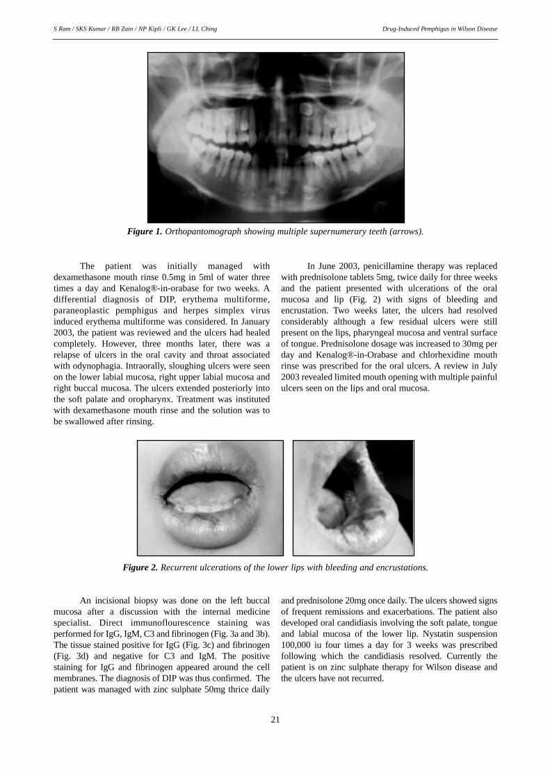
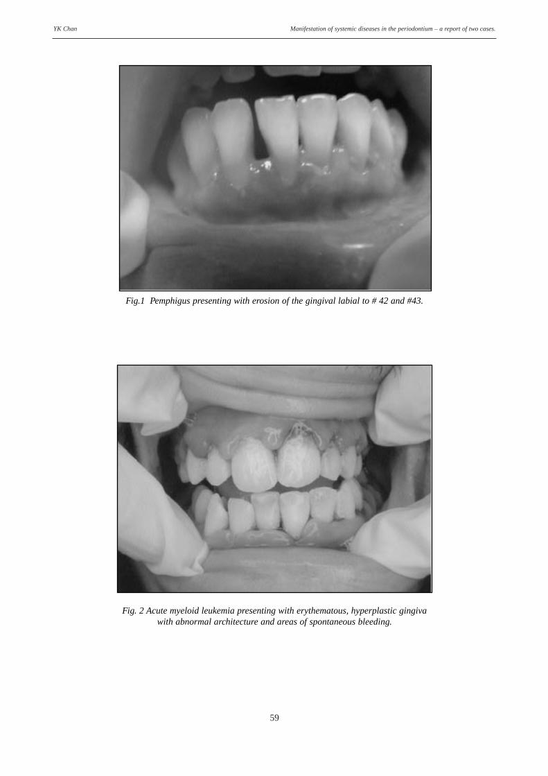
A 22-year-old Malaysian Chinese female wasreferred from the Faculty of Medicine, University ofMalaya to the Oral Medicine clinic, Faculty of Dentistry,University of Malaya in December 2002, with a complaintof recurrent painful oral ulcerations. The patient’s medicalhistory was significant for Wilson disease since 1999 andthe patient was on penicillamine and spirinolactone.Extraoral examination of the patient revealed no abnormalities. Intraorally, the patient had large sloughingulcers on the right buccal mucosa, right lower vestibuleand right upper vestibule. A few petechiae were seen onthe left lower labial mucosa and right soft palate. A bonyhard swelling of normal mucosal colour and measuringabout 0.5cm in diameter, was noticed on the right upperalveolar mucosa. Apart from these findings, a linguallyplaced supernumerary tooth was observed between 34 and35. Orthopantomographic examination revealed the presence of multiple supernumerary teeth located between13, 14; 15, 16; 23, 24; 25, 26; and 35, 36 (FDI numberingsystem) (Fig. 1).
Drug-Induced Pemphigus in Wilson DiseaseS Ram / SKS Kumar / RB Zain / NP Kipli / GK Lee / LL Ching
The patient was initially managed with dexamethasone mouth rinse 0.5mg in 5ml of water threetimes a day and Kenalog®-in-orabase for two weeks. Adifferential diagnosis of DIP, erythema multiforme,paraneoplastic pemphigus and herpes simplex virusinduced erythema multiforme was considered. In January2003, the patient was reviewed and the ulcers had healedcompletely. However, three months later, there was arelapse of ulcers in the oral cavity and throat associatedwith odynophagia. Intraorally, sloughing ulcers were seenon the lower labial mucosa, right upper labial mucosa andright buccal mucosa. The ulcers extended posteriorly intothe soft palate and oropharynx. Treatment was institutedwith dexamethasone mouth rinse and the solution was tobe swallowed after rinsing.
In June 2003, penicillamine therapy was replacedwith prednisolone tablets 5mg, twice daily for three weeksand the patient presented with ulcerations of the oralmucosa and lip (Fig. 2) with signs of bleeding and encrustation. Two weeks later, the ulcers had resolved considerably although a few residual ulcers were still present on the lips, pharyngeal mucosa and ventral surfaceof tongue. Prednisolone dosage was increased to 30mg perday and Kenalog®-in-Orabase and chlorhexidine mouthrinse was prescribed for the oral ulcers. A review in July2003 revealed limited mouth opening with multiple painfululcers seen on the lips and oral mucosa.
Figure 2. Recurrent ulcerations of the lower lips with bleeding and encrustations.
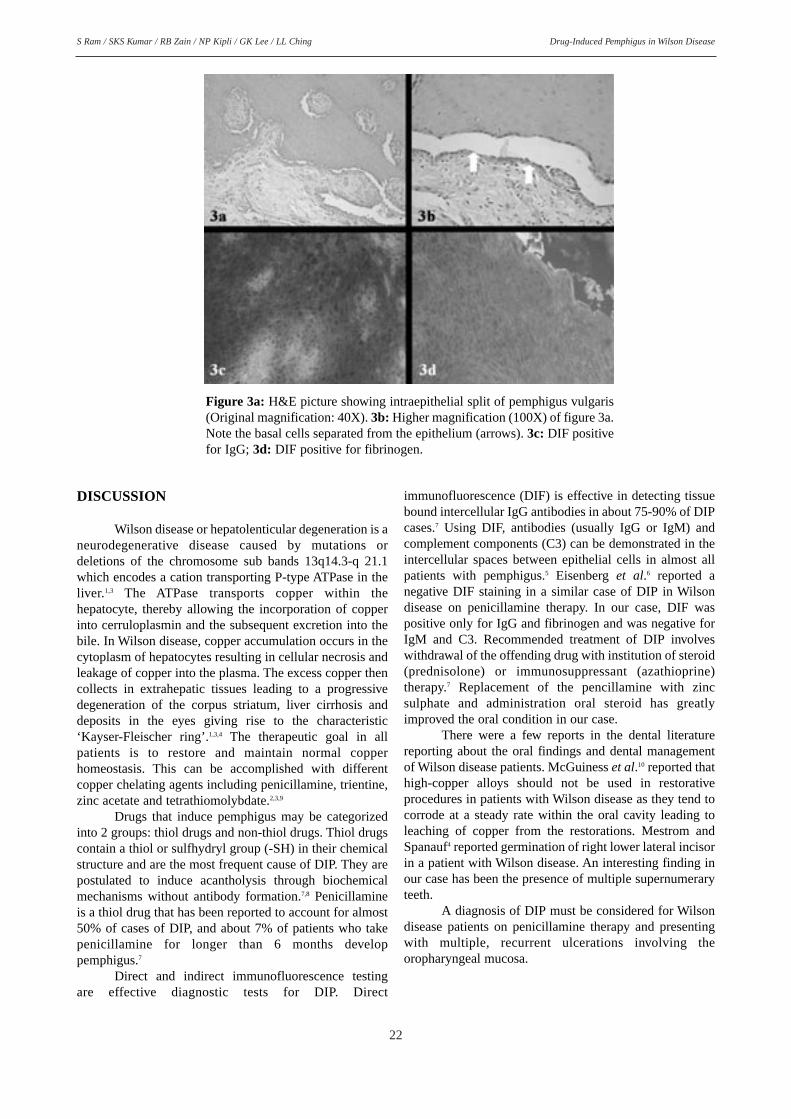
An incisional biopsy was done on the left buccalmucosa after a discussion with the internal medicine specialist. Direct immunoflourescence staining was performed for IgG, IgM, C3 and fibrinogen (Fig. 3a and 3b).The tissue stained positive for IgG (Fig. 3c) and fibrinogen(Fig. 3d) and negative for C3 and IgM. The positive staining for IgG and fibrinogen appeared around the cellmembranes. The diagnosis of DIP was thus confirmed. Thepatient was managed with zinc sulphate 50mg thrice daily
and prednisolone 20mg once daily. The ulcers showed signsof frequent remissions and exacerbations. The patient alsodeveloped oral candidiasis involving the soft palate, tongueand labial mucosa of the lower lip. Nystatin suspension100,000 iu four times a day for 3 weeks was prescribed following which the candidiasis resolved. Currently thepatient is on zinc sulphate therapy for Wilson disease andthe ulcers have not recurred.
22
Drug-Induced Pemphigus in Wilson DiseaseS Ram / SKS Kumar / RB Zain / NP Kipli / GK Lee / LL Ching
DISCUSSION
Wilson disease or hepatolenticular degeneration is a neurodegenerative disease caused by mutations or deletions of the chromosome sub bands 13q14.3-q 21.1which encodes a cation transporting P-type ATPase in theliver.1,3 The ATPase transports copper within the hepatocyte, thereby allowing the incorporation of copperinto cerruloplasmin and the subsequent excretion into thebile. In Wilson disease, copper accumulation occurs in thecytoplasm of hepatocytes resulting in cellular necrosis andleakage of copper into the plasma. The excess copper thencollects in extrahepatic tissues leading to a progressivedegeneration of the corpus striatum, liver cirrhosis anddeposits in the eyes giving rise to the characteristic‘Kayser-Fleischer ring’.1,3,4 The therapeutic goal in allpatients is to restore and maintain normal copper homeostasis. This can be accomplished with different copper chelating agents including penicillamine, trientine,zinc acetate and tetrathiomolybdate.2,3,9
Drugs that induce pemphigus may be categorizedinto 2 groups: thiol drugs and non-thiol drugs. Thiol drugscontain a thiol or sulfhydryl group (-SH) in their chemical structure and are the most frequent cause of DIP. They arepostulated to induce acantholysis through biochemicalmechanisms without antibody formation.7,8 Penicillamineis a thiol drug that has been reported to account for almost50% of cases of DIP, and about 7% of patients who takepenicillamine for longer than 6 months develop pemphigus.7
Direct and indirect immunofluorescence testing are effective diagnostic tests for DIP. Direct
immunofluorescence (DIF) is effective in detecting tissuebound intercellular IgG antibodies in about 75-90% of DIPcases.7 Using DIF, antibodies (usually IgG or IgM) andcomplement components (C3) can be demonstrated in theintercellular spaces between epithelial cells in almost allpatients with pemphigus.5 Eisenberg et al.6 reported a negative DIF staining in a similar case of DIP in Wilsondisease on penicillamine therapy. In our case, DIF waspositive only for IgG and fibrinogen and was negative forIgM and C3. Recommended treatment of DIP involveswithdrawal of the offending drug with institution of steroid(prednisolone) or immunosuppressant (azathioprine) therapy.7 Replacement of the pencillamine with zinc sulphate and administration oral steroid has greatlyimproved the oral condition in our case.
There were a few reports in the dental literaturereporting about the oral findings and dental managementof Wilson disease patients. McGuiness et al.10 reported thathigh-copper alloys should not be used in restorative procedures in patients with Wilson disease as they tend tocorrode at a steady rate within the oral cavity leading toleaching of copper from the restorations. Mestrom andSpanauf4 reported germination of right lower lateral incisorin a patient with Wilson disease. An interesting finding inour case has been the presence of multiple supernumeraryteeth.
A diagnosis of DIP must be considered for Wilson disease patients on penicillamine therapy and presentingwith multiple, recurrent ulcerations involving the oropharyngeal mucosa.
Figure 3a: H&E picture showing intraepithelial split of pemphigus vulgaris(Original magnification: 40X). 3b: Higher magnification (100X) of figure 3a.Note the basal cells separated from the epithelium (arrows). 3c: DIF positivefor IgG; 3d: DIF positive for fibrinogen.
23
Drug-Induced Pemphigus in Wilson DiseaseS Ram / SKS Kumar / RB Zain / NP Kipli / GK Lee / LL Ching
REFERENCES
1. Scheinberg IH. Wilson’s disease. In: Fauci SA, Braunwald E,Isselbacher KJ et al., editors. Harrison’s principles of internalmedicine, vol.2, 14th Ed. New York. McGraw-Hill1. 1998:2166-69.
2. Gitlin JD. Wilson disease. Gastroenterology. 2003; 125:1868-77.
3. Langner C, Denk H. Wilson Disease. Virchows Arch. 2004;445:111-8. Epub 2004 Jun 17.
4. Mestrom TJ, Spanauf AJ. The dental management of a partially edentulous patient suffering from hepatolenticulardegeneration (Wilson's disease). Aust Dent J. 1981; 26: 153-5.
5. Neville BW, Damm DD, Allen CM, Bouquot JE. Oral &Maxillofacial Pathology. 2nd ed. Philadelphia: WB Saunders.2002: 665-67.
6. Eisenberg E, Ballow M, Wolfe SH, Krutchkoff DJ, TanzerJM. Pemphigus- like mucosal lesions: a side effect of penicillamine therapy. Oral Surg Oral Med Oral Pathol. 1981;51: 409-14.
7. Scott DM. Pemphigus, drug-induced. 2001,http://www.emedicine.com,http://www.emedicine.com/derm/topic314.htm.
8. Brenner S, Bialy-Golan A, Ruocco V. Drug-induced pemphigus. Clin Dermatol. 1998; 16: 393-7.
9. Scheinberg IH, Jaffe ME, Sternleib I. The use of trientine inpreventing the effects of interrupting penicillamine therapy inWilson’s disease. N Engl J Med. 1987; 317: 209-13.
10. McGuiness JW, McInnes-Ledoux PM, Ferraro EF, Carr JC.Daily release of copper from dental alloy restorations in apatient with Wilson's disease. Oral Surg Oral Med OralPathol. 1987; 63: 511-4.
Corresponding author:
Professor Dr. Rosnah Binti ZainDepartment of Oral Pathology, Oral Medicine &Periodontology Faculty of Dentistry,University of Malaya 50603 Kuala Lumpur, Malaysia Tel: 00-603-7967 7403 Fax: 00-603-7967 4896 E-mail address: [email protected]
24
MALAYSIAN DENTAL JOURNAL
A Radiographic Study of Mandibular Third Molar Development in a LocalOrthodontic Population
ST Loke, M.Orth RCS Consultant Orthodontist, Orthodontic Specialist Unit, Alor Star, Kedah
SK Tee, BDS Dental Officer, Kulim Dental Clinic, Kedah
ABSTRACT The purpose of this study was to evaluate mandibular third molar development before and afterorthodontic treatment. There were 82 patients with mean age of 15.31 years (range 10.08 to 46.67) and a total of149 mandibular third molars at pre-treatment. The post-treatment sample was 16 mandibular third molars. Themost common angular position of mandibular third molar was mesioangular (79.9%), followed by vertical(15.4%), horizontal (2.7%) and distoangular (2.0%). Mesioangular was the most common position within genderand ethnic distribution. The angular position of mandibular third molars changed unpredictably as the rootdeveloped and eruption is not predictable even when there was sufficient space for eruption. Only one in four ofteeth that had sufficient space for eruption and full root formation erupted in alignment at recall. However, moreteeth (88.2%) were impacted when the space/width ratio was less than 1.00 compared with space/width ration of1.00 or more (46.2%). Root formation and eruption continued even when impaction occurs. The majority (81.3%)of the third molars that had erupted with part or entire crown at the occlusal level of the second molar had fullroot formation with closed apices at a mean age of 20.19 years. KEY WORDS: third molar, development, radiograph, orthodontic treatment
INTRODUCTION
Dental panoramic radiographs are routinely takenin the orthodontic clinic before orthodontic treatment is commenced. These radiographs give a good overall view of the dentition in the mandible and maxilla and are invaluable in orthodontic diagnosis and treatmentplanning, assessing treatment progress and detectingpathology. The majority of orthodontic patients are in thepre-teens and teenage age group when the third molars aredeveloping and it would be advantageous if we could predict the final outcome of these teeth taking into accountindividual growth and whether extractions were carriedout. The mandibular third molars would need to be preserved if it is judged to erupt successfully and have afunctional role in the dentition.
Some researchers have found that premolar extraction treatment has been associated with mesialmovement of the molars concomitant with an increase inthe eruption space for the third molars1,2,3,4,5 while othershave only small differences treated with and withoutextractions.6,7 The average age of mandibular third molaremergence varies from 17 to 21 years, but the roots are notfully formed until 18 to 25 years of age.8,9,10 The angulationof many of these teeth has been observed to change to a
more upright position and eventually erupted with fulldevelopment of the root and mandibular growth4,5,6,11,12 butthis is an unpredictable phenomenon. Capelli (1991)13 used cephalometric radiographs instead of orthopantomogramsto study third molar impaction in 60 patients who had firstpremolars extracted for orthodontic treatment. He foundthat mandibular growth was directly related to the positionof mandibular third molars with impactions more likely inthose with a predominance of vertical growth. Thus thirdmolar impaction could have been overdiagnosed in studiesexamining subjects where the root is not fully developed orthe subject has not achieved his/her potential growth. Themajority of orthodontic patients are still in their teens afterorthodontic treatment is completed thereby making it difficult to predict the final outcome of the third molars.
Dental age determination from tooth developmentis important from forensic, criminal and legal aspectswhen the chronological age is unknown or suspect.However, the correlation between chronological age anddental age as estimated from mandibular third molar ispoor.14,15,16 This is probably due to the large range, that is,about +/- 10 months, in individual maturity.14 There was asystematic underestimation of chronological age, the 95per cent confidence interval, which was about +/- 4 years.Third molar dental age estimation may still be useful to a
A Radiographic Study of Mandibular Third Molar Development in a Local Orthodontic PopulationST Loke / SK Tee
certain extent in specific cases if it is not possible to evaluate other teeth or there were no other better non-dental methods. It would be interesting to compare oursample of predominantly Asian ethnic origin with that ofCaucasians.14
Thus, the main objectives of this study were:
1. To assess third mandibular tooth development in termsof root development, tooth depth, angulation, toothwidth between sexes and in different ethnic groups.
2. To compare root development, tooth depth and angulation in groups with sufficient/insufficient spaceto erupt.
3. To evaluate mandibular third molar development pre-and post-orthodontic treatment.
4. To compare chronological age at different stages ofroot development with that of a Caucasian group.
METHODOLOGY
Case Selection:
1. Patients with at least one mandibular third molar wherecrown formation has completed. The pre-treatmentpanoramic radiograph must be of good quality. All dental panoramic radiographs (DPT) were taken withthe Proline Model 2002 in the Radiology department,Alor Star Hospital, Kedah.
2. Exclusion criteria:
• Cleft lip and palate patients.• History of extraction of mandibular permanent pre
molar/molar teeth.• Radiographs that were unclear or distorted
This is a retrospective, cross-sectional study of orthodontic patients seen in the Orthodontic unit, TelukWanjah dental clinic, Alor Star from 1992 to 2000. Therewere 82 cases that satisfied the selection criteria.
Reference lines from DPT:
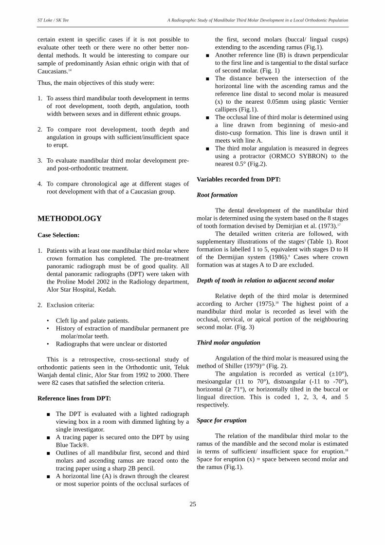
■ The DPT is evaluated with a lighted radiographviewing box in a room with dimmed lighting by asingle investigator.
■ A tracing paper is secured onto the DPT by usingBlue Tack®.
■ Outlines of all mandibular first, second and thirdmolars and ascending ramus are traced onto thetracing paper using a sharp 2B pencil.
■ A horizontal line (A) is drawn through the clearestor most superior points of the occlusal surfaces of
the first, second molars (buccal/ lingual cusps)extending to the ascending ramus (Fig.1).
■ Another reference line (B) is drawn perpendicularto the first line and is tangential to the distal surfaceof second molar. (Fig. 1)
■ The distance between the intersection of the horizontal line with the ascending ramus and thereference line distal to second molar is measured(x) to the nearest 0.05mm using plastic Vernier callipers (Fig.1).
■ The occlusal line of third molar is determined usinga line drawn from beginning of mesio-and disto-cusp formation. This line is drawn until itmeets with line A.
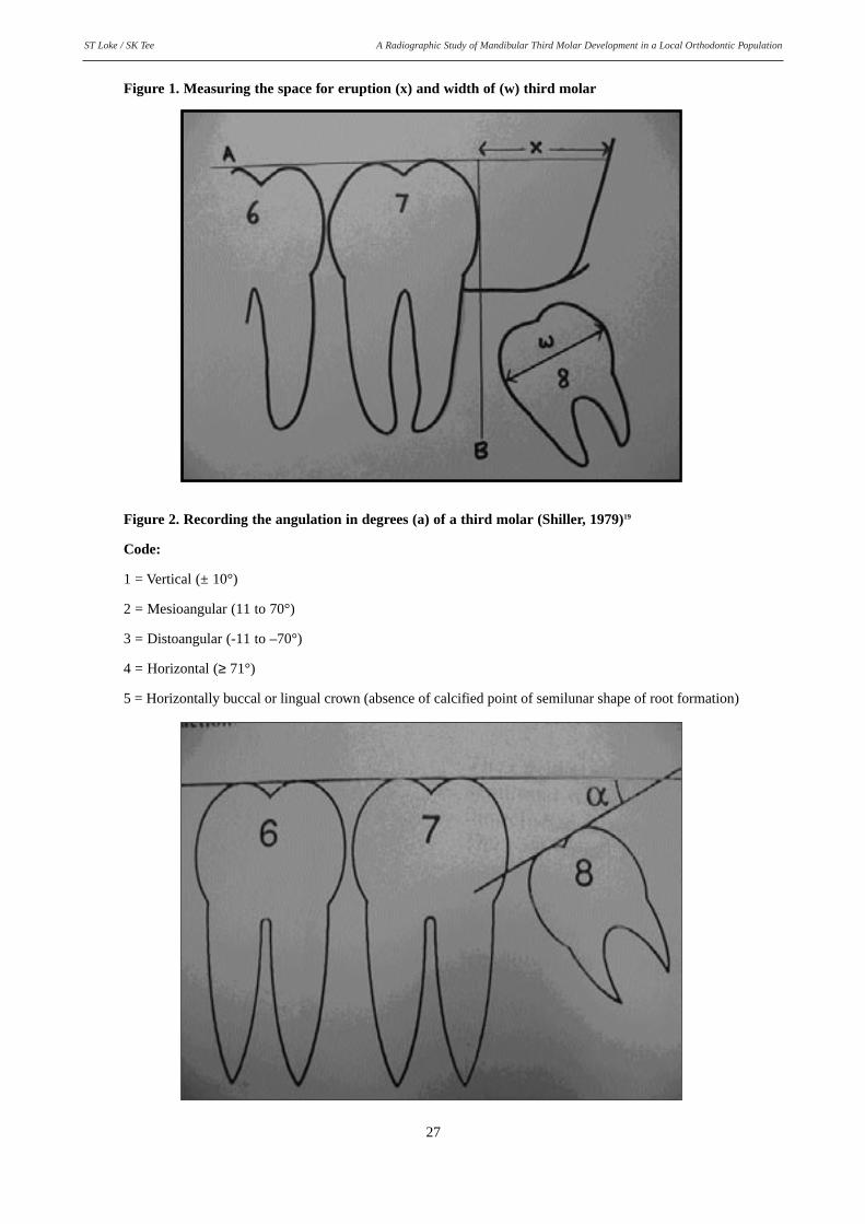
■ The third molar angulation is measured in degreesusing a protractor (ORMCO SYBRON) to the nearest 0.5° (Fig.2).
Variables recorded from DPT:
Root formation
The dental development of the mandibular thirdmolar is determined using the system based on the 8 stagesof tooth formation devised by Demirjian et al. (1973).17
The detailed written criteria are followed, with supplementary illustrations of the stages1 (Table 1). Rootformation is labelled 1 to 5, equivalent with stages D to Hof the Dermijian system (1986).8 Cases where crown formation was at stages A to D are excluded.
Depth of tooth in relation to adjacent second molar
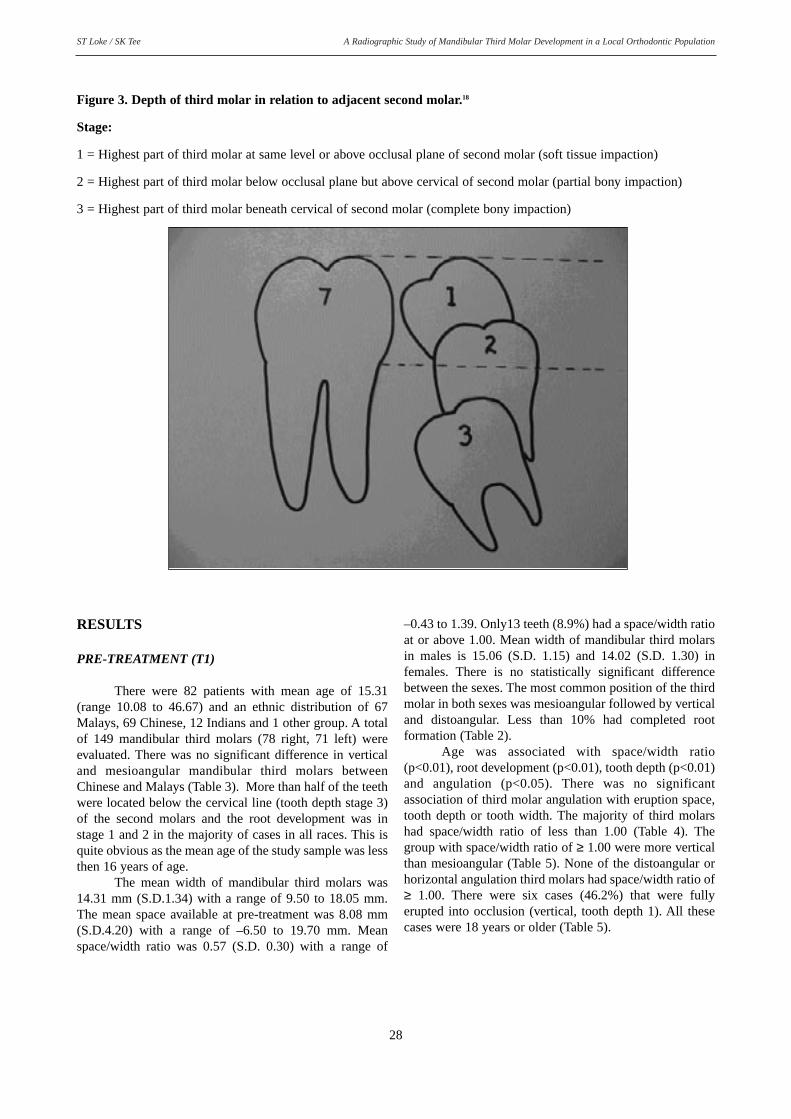
Relative depth of the third molar is determinedaccording to Archer (1975).18 The highest point of amandibular third molar is recorded as level with theocclusal, cervical, or apical portion of the neighbouringsecond molar. (Fig. 3)
Third molar angulation
Angulation of the third molar is measured using themethod of Shiller (1979)19 (Fig. 2).
The angulation is recorded as vertical (±10°),mesioangular (11 to 70°), distoangular (-11 to -70°),horizontal (≥ 71°), or horizontally tilted in the buccal orlingual direction. This is coded 1, 2, 3, 4, and 5respectively.
Space for eruption
The relation of the mandibular third molar to theramus of the mandible and the second molar is estimatedin terms of sufficient/ insufficient space for eruption.18
Space for eruption (x) = space between second molar andthe ramus (Fig.1).
25
A Radiographic Study of Mandibular Third Molar Development in a Local Orthodontic PopulationST Loke / SK Tee
Tooth width
Third molar width (w) is determined by measuringthe most bulbous points (mesial and distal contour) of thecrown to the nearest 0.05mm (Fig.1).
Space available between second molar and the ramus (x)
Mesiodistal width of third molar (w)
Selection of patients for recall post-treatment
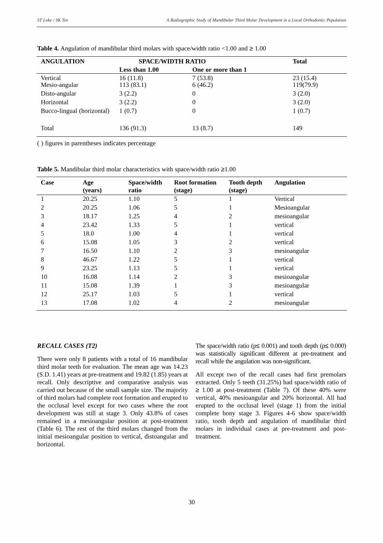
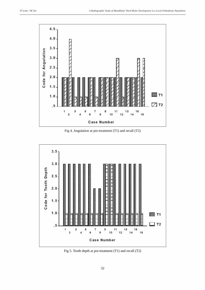
Patients with mandibular third molars that showedimpaction at pre-treatment were recalled after five years.Those molars that were already erupted into occlusionwere excluded. Patients have to be at least 18 years old atrecall. A DPT is taken at recall and the variables recordedas before. All these patients had completed orthodontictreatment with/ without extractions. There were 27 patientsthat satisfied the criteria but only 8 patients (total of 16third molars) attended the clinic for recall despite extensive efforts to contact the patients.
Measurement error
To assess measurement error in the tracing andrecording techniques, ten radiographs were randomlyselected, traced and measured on two separate occasionsby the same investigator. The correlation between the two measurements for space was 0.997 and for width was0.974. The mean measurement error for space was –4.00E-02 (S.D. 0.34) and for width was 0.14 (S.D. 0.29).
Statistical Analysis
Data is analysed using Statistical Package forSocial Sciences (SPSS) Version 10.0. Paired t test and correlation were used for intra-examiner reliability.Descriptive analysis, Pearson’s correlation and one-wayanalysis of variance (ANOVA) were carried out to test forsignificant difference between groups. Paired t test wascarried out to compare variables at pre-treatment (T1) andpost-treatment (T2). Association of age and root development was compared with that of Thorson and Hagg(1991).14
26
Table 1. Description of the root formation stages in molars.8
STAGE DESCRIPTION
The crown formation is completed down to the cementoenamel junction. The 1 pulp chamber has a trapezoidal form. Beginning of root formation is seen in the
form of a spikule.
Initial formation of the radicular bifurcation is seen in the form of either a 2 calcified point or a semilunar shape.
The root length is still less than the crown height.
The calcified region of the bifurcation has developed further down from its 3 semilunar stage to give the roots a more definite and distinct outline, with funnel-
shaped endings. The root length is equal to or greater than the crown height.
4 The walls of the distal root canal are now parallel.The apical end of the distal root canal is still partially open.
5 The apical end of the distal root canal is completely closed.The periodontal membrane has an uniform width around the root and the apex.
Space/width ratioSpace/width ratio =
A Radiographic Study of Mandibular Third Molar Development in a Local Orthodontic PopulationST Loke / SK Tee
Figure 1. Measuring the space for eruption (x) and width of (w) third molar
27
Figure 2. Recording the angulation in degrees (a) of a third molar (Shiller, 1979)19
Code:
1 = Vertical (± 10°)
2 = Mesioangular (11 to 70°)
3 = Distoangular (-11 to –70°)
4 = Horizontal (≥ 71°)
5 = Horizontally buccal or lingual crown (absence of calcified point of semilunar shape of root formation)
A Radiographic Study of Mandibular Third Molar Development in a Local Orthodontic PopulationST Loke / SK Tee
Figure 3. Depth of third molar in relation to adjacent second molar.18
Stage:
1 = Highest part of third molar at same level or above occlusal plane of second molar (soft tissue impaction)
2 = Highest part of third molar below occlusal plane but above cervical of second molar (partial bony impaction)
3 = Highest part of third molar beneath cervical of second molar (complete bony impaction)
28
RESULTS
PRE-TREATMENT (T1)
There were 82 patients with mean age of 15.31(range 10.08 to 46.67) and an ethnic distribution of 67Malays, 69 Chinese, 12 Indians and 1 other group. A totalof 149 mandibular third molars (78 right, 71 left) wereevaluated. There was no significant difference in verticaland mesioangular mandibular third molars betweenChinese and Malays (Table 3). More than half of the teethwere located below the cervical line (tooth depth stage 3)of the second molars and the root development was instage 1 and 2 in the majority of cases in all races. This isquite obvious as the mean age of the study sample was lessthen 16 years of age.
The mean width of mandibular third molars was14.31 mm (S.D.1.34) with a range of 9.50 to 18.05 mm.The mean space available at pre-treatment was 8.08 mm(S.D.4.20) with a range of –6.50 to 19.70 mm. Meanspace/width ratio was 0.57 (S.D. 0.30) with a range of
–0.43 to 1.39. Only13 teeth (8.9%) had a space/width ratioat or above 1.00. Mean width of mandibular third molarsin males is 15.06 (S.D. 1.15) and 14.02 (S.D. 1.30) infemales. There is no statistically significant differencebetween the sexes. The most common position of the thirdmolar in both sexes was mesioangular followed by verticaland distoangular. Less than 10% had completed root formation (Table 2).
Age was associated with space/width ratio(p<0.01), root development (p<0.01), tooth depth (p<0.01)and angulation (p<0.05). There was no significantassociation of third molar angulation with eruption space,tooth depth or tooth width. The majority of third molarshad space/width ratio of less than 1.00 (Table 4). Thegroup with space/width ratio of ≥ 1.00 were more verticalthan mesioangular (Table 5). None of the distoangular orhorizontal angulation third molars had space/width ratio of≥ 1.00. There were six cases (46.2%) that were fully erupted into occlusion (vertical, tooth depth 1). All thesecases were 18 years or older (Table 5).
A Radiographic Study of Mandibular Third Molar Development in a Local Orthodontic PopulationST Loke / SK Tee
Table 2. Gender distribution of angulation, root development and tooth depth.
There were only 8 patients with a total of 16 mandibularthird molar teeth for evaluation. The mean age was 14.23(S.D. 1.41) years at pre-treatment and 19.82 (1.85) years atrecall. Only descriptive and comparative analysis was carried out because of the small sample size. The majorityof third molars had complete root formation and erupted tothe occlusal level except for two cases where the rootdevelopment was still at stage 3. Only 43.8% of casesremained in a mesioangular position at post-treatment(Table 6). The rest of the third molars changed from theinitial mesioangular position to vertical, distoangular andhorizontal.
The space/width ratio (p≤ 0.001) and tooth depth (p≤ 0.000)was statistically significant different at pre-treatment and recall while the angulation was non-significant.
All except two of the recall cases had first premolarsextracted. Only 5 teeth (31.25%) had space/width ratio of≥ 1.00 at post-treatment (Table 7). Of these 40% were vertical, 40% mesioangular and 20% horizontal. All haderupted to the occlusal level (stage 1) from the initial complete bony stage 3. Figures 4-6 show space/widthratio, tooth depth and angulation of mandibular thirdmolars in individual cases at pre-treatment and post-treatment.
A Radiographic Study of Mandibular Third Molar Development in a Local Orthodontic PopulationST Loke / SK Tee
Table 6. Demographics at pre-treatment (T1) and post-treatment (T2)
TOOTH DEPTHSame level or above 0 14 (87.5)occlusal plane of M2(stage1)Below occlusal plane & 2 (12.5) 0above cervical line of M2 (stage2)Below cervical line of M2 14 (87.5) 2 (12.5)(stage3)
Total 16 16
( ) figures in parentheses indicates percentage
Table 7. Tooth depth and angulation at pre-treatment (T1) and recall (T2) in cases with space/width ratio ≥ 1.00.
A Radiographic Study of Mandibular Third Molar Development in a Local Orthodontic PopulationST Loke / SK Tee
Fig 4. Angulation at pre-treatment (T1) and recall (T2)
Fig 5. Tooth depth at pre-treatment (T1) and recall (T2)
32
A Radiographic Study of Mandibular Third Molar Development in a Local Orthodontic PopulationST Loke / SK Tee
Fig 6. Space/width ratio at pre-treatment (T1) and recall (T2)
Table 8. The mean age (in years) assessed by the different stages of root formation with the present study and figuresgiven by Thorson and Hagg (1991)14
Stage of root formation 1 2 3 4 5
GIRLSThorson & Hagg 13.75 15.40 17.30 19.50 20.70
(estimated dental age)Present study 13.65 14.69 15.76 17.85 24.58
A Radiographic Study of Mandibular Third Molar Development in a Local Orthodontic PopulationST Loke / SK Tee
DISCUSSION
Angulation
There was no statistically significant difference in angulation between males and females. Shiller (1979)19
found that if the initial inclination of a mesioangularmandibular third molar was 35° or less, then 13% becameupright within a year before the age of 20. In the presentstudy, all 16 teeth had inclination less than 35 degrees atpre-treatment and 4 teeth (25%) uprighted to a verticalposition at recall despite two teeth with space/width ratioof less than 1.00. But 31.1% changed to distoangular andhorizontal positions. Hattab et al. (1995)20 found that halfof the impacted mandibular third molars weremesioangular and 23% of these erupted by 20.4 years. Thiswas comparable to that of Venta et al. (1991)4 who showedthat more than 50% of mesioangular teeth remainedunerupted (p<0.001) while more than 85% of those thatwere vertical were partially or fully erupted (p<0.001). Alater study by Hattab (1997)11 found that 44% of initialmesioangular teeth uprighted at complete root formationand 37% became fully erupted while 15% were partiallyerupted.
Venta et al. (1997)21 developed a method (transparent measuring device) to predict lower third molareruption from the orthopantomogram. The predicted reliability and sensitivity of this device was 95%. He foundthat mesioangular deviation of more than 45° wouldremain impacted. Venta (1993)22 looked at five radiographic characteristics of mandibular third molars,namely, angulation, root formation, type of impaction,crown depth in bone and relative space to develop a predictive model at age 20. He found that the most important predictor of third molar eruption was the ‘typeof impaction’ (p<0.01). There was no significant differencebetween males and females. Haavikko et al. (1978)6
similarly found that most mandibular third molars emergedwhen the initial angle between second and third molar wasless than 10 degrees and this decreased to one in everythree cases when the initial angle increased to between 20and 30 degrees. With larger angles the prediction of eruption becomes more difficult and impaction increases.Castella et al. (1998)23 found that the more the tooth isdeveloped, the higher is the accuracy of predictionalthough two stages where the crown is fully formed orwhen the roots were 1/3 formed possessed high predictivevalue of impaction. He concluded that impaction was predictable both in extraction and non-extraction patientsalthough his results have to be interpreted with cautionbecause of the small sample size.
Richardson (1975)24 and Richardson et al. (1984)25
found that during development of the dentition, the position of the mandibular third molar continuouslychanges before eruption and uprighting occurs. Hattab(1997)11 found that a significant proportion of mesiallyimpacted mandibular third molars had changed their angulation and became fully erupted by the individualreached 24 years of age. Even those that originally presented at 35 degrees or more registered an average
decrease of 12 degrees in angulation although nonebecame upright. This was previously observed in the studyby Haavikko et al. (1978)6 who found that the third molar had a tendency to upright or changed to a more distoangular position during development both in the normal and extraction groups.
Tooth depth
In the present study, 87.5% of the third molars initially in partial or complete bony impaction (stage 2 and3) had reached occlusal level (stage 1) of crown depthwhen root formation was complete or near completion.Only two cases (12.5%) remained at stage 3 with root formation at stage 3. Venta et al. (1991)4 found that themajority of third molars (about 70%) initially completelyburied in bone (p<0.05) and about 30% of those in partialbony impaction remain unerupted. All those in soft tissueimpaction only were partially or fully erupted (p<0.01).
Space for eruption
Kim et al. (2003)5 found higher third molarimpaction (p<0.01) and smaller retromolar space(p<0.001) in both arches of the non-extraction patient thanin the extraction patients. However, they could not detectany difference in mandibular mesial movement betweenpatients with eruption and impaction of the mandibularthird molars suggesting that other variables could be equally important for the development of sufficient eruption space. Although Haavikko et al. (1978)6 foundthat 71.8% of mandibular third molars was unerupted inthe normal group and 65.0% in the extraction group at age19.5 years, they concluded that extraction of premolarsmerely accelerate but not promote eruption. Their findingssuggested that teeth with favourable angulations wouldeventually erupt.
Earlier studies have suggested that the probabilityof mandibular third molar eruption is directly related to theproportion of the third molar in front of the anterior borderof the ramus before eruption. However, Kim et al. (2003)5
found that as many as 60% of their subjects with eruptionspace ≤ 5 mm and 55% of those with a distance of ≤ 3.5mm erupted into occlusion. They suggested that the size ofthe third molar eruption space associated with increasedrisk of impaction might be smaller than the 23 mm as previously suggested.26,27,28 In the present study, at recall,two cases erupted into occlusion despite pre-treatmentspace/width ratio of less than 1.0. One case hadspace/width ratio increased to 0.59 from zero and the othercase increased from 0.58 to 0.70. The mean eruption spaceof the four cases that had erupted into occlusion was10.4mm at post-treatment and the mean width of the teethwas 13.7 mm. All those with sufficient space to erupt haderupted partially or completely to the occlusal level fromthe initial bony impaction stage. Of those with insufficientspace, 18.2% remained in bony impaction. This observation was comparable to that of Venta et al. (1991)4
who reported that about 40% of those with initial insufficient space to erupt remain unerupted.
34
A Radiographic Study of Mandibular Third Molar Development in a Local Orthodontic PopulationST Loke / SK Tee
Our pre-treatment findings; mean eruption space of13.3 mm with a mean space/width ratio of 0.96 (range 0.50to 1.33) in 12 cases where the mandibular third molarswere fully erupted into occlusion (vertical + tooth depth 1),contradicts the higher space requirements as observed bySchulhof (1976)26 and Ricketts (1979).28 Two of the cases(16.7%) had eruption space of less than 9 mm while theother ten (83.3%) had eruption space between 10 and 20mm. This was comparable to the findings of Kim et al.(2003).5 The mean age of our cases was 22.08 years androot development was nearly complete or completed (stage4 and 5).
Root development
Hattab (1997)11 studied mesially impactedmandibular third molars in young Jordaninan Arab adults(mean age 19.7 years) and he observed that 79% ofmandibular third molars had complete root formation. Allhad complete root formation by 24 years. In the presentstudy, 81.3% of the recall cases had complete root formation and reached the occlusal level at a mean age of20.19 years. The root formation of the other three casesprogressed to stage 3 and 4 with a mean age of 18.72 years.Venta et al. (1991)4 observed that although development ofthe root was already complete at the baseline, a number ofunerupted or partially erupted teeth erupted by the age of26 years implying eruptive activity still continued. Andabout 40% of those with initially incomplete developmentof the root remained unerupted. In the present study thetwo teeth with incomplete root development (stage 3)remained in complete bony impaction while the rest withcomplete root formation erupted.
Age estimation
Menzies-Clow (1984)15 used Gravely’s (1965)29
description of third molar crown formation to compareroot formation in 6-15 year olds in two centres. He extended this measurement of estimated dental age bymeasuring root formation in second molars. He concludedthat dental age was better correlated with chronologicalage in second molar root formation than third molar crownformation. This conclusion seems obvious since the stagesof root formation are more clearly defined than stages ofcrown formation as developed by Gravely (1965)29 usingoblique lateral jaw radiographs.
Thorson and Hagg (1991)14 followed the Demirjiansystem (1986)8 of eight stages of molar tooth formationusing panoramic radiographs and they transferred eachstage to a dental age using the figures from a referencesample of 4640 French-Canadian girls and boys byLevesque et al. (1981).30 They found that associationbetween dental age and chronological age was pooralthough precision was high. In girls, the mean differencebetween dental age and chronological age varied from – 6months to – 3.5 years, being largest in the oldest group. Inboys, the mean difference between dental age and chronological age was – 10 and – 17 months in the agegroups between 14.5 to 20.5 years. There was no
significant correlation in the older age groups. The chronological age of the patients in the present study whencompared to the dental age in patients in Thorson andHagg (1991)14 is comparable only in stage 1 and 2 of rootformation (Table 8). This may be attributed to the verysmall number of patients in the later stages of root formation.
Limitations of study
The pre-treatment sample is biased towards a larger younger age group. This is inevitable since themajority of patients seeking orthodontic treatment are inthe pre-teens and teens. Thus comparison of chronologicalage to root development in the later stages is difficult toprovide an accurate representation due to the small sample.The recall sample is too small for a more valid statisticalanalysis. Thus the analysis is limited to a descriptivenature. Any inferences made have to be taken with caution.
CONCLUSION
Our findings suggest that the dental panoramicradiograph is a useful radiograph to monitor developmentof the mandibular third molar besides being invaluable in diagnosis and treatment planning for orthodontictreatment. The most common position of mandibular thirdmolars was mesioangular in both males and females and inthe different ethnic groups; followed by vertical, horizontaland distoangular. Root formation and eruption throughbone continued even when the teeth remained or becomeimpacted and the majority of patients had complete rootdevelopment after 18 years. Positional changes and eruption of initially impacted mandibular third molars areunpredictable phenomena. Although it is difficult to predict the ultimate position of the third molar with a reliable degree of accuracy after root formation is complete, the space/width ratio and initial angulationappears to give some prognostic value. Chronological ageas compared to dental age estimation from root development of mandibular third molars is comparable toa Caucasian group when the root formation was at the initial stages.
RECOMMENDATION
Since there is no reliable way to predict the eruption of mandibular third molars in young patients, it is recommended that National Clinical Practice Guidelinesbe adhered to in the management of unerupted and/orimpacted third molar teeth. For example, the health servicein Scotland uses the Scottish Intercollegiate ‘Guidelines inthe management of unerupted and impacted third molarteeth’.31 A longer follow-up of orthodontic patients until atleast above 20 years of age may be advisable before a decision is made to extract asymptomatic impacted teethsince positional changes continued until root developmentand growth is complete.
35
A Radiographic Study of Mandibular Third Molar Development in a Local Orthodontic PopulationST Loke / SK Tee
1. Faubion BH. Effect of extraction of premolars on eruption ofmandibular third molars. J Am Dent Assoc 1968; 76:316-20
2. Kaplan R. Some factors related to mandibular third molarimpaction. Angle Orthod 1975; 45:153-8
3. Richardson ME. The relative effects of the extraction of various teeth on the development of mandibular third molars.Trans Eur Orthod Soc 1976; 79-85
4. Venta I, Murtomaa H, Turtola L, Meurman J, YlipaavalniemiP. Clinical follow-up study of third molar eruption from ages20 to 26 years. Oral Surg Oral Med Oral Pathol 1991;72:150-3
5. Kim TW, Artun J, Behbenani F, Artese F. Prevalence of thirdmolar impaction in orthodontic patients treated nonextractionand with extraction of 4 premolars. Am J Orthod DentofacialOrthop 2003; 123:138-45
6. Haavikko K, Altonen M, Mattila K. Predicting angulationaldevelopment and eruption of the lower third molar. AngleOrthodontist 1978; 48: 39
7. Graber TM, Kaineg TF. The mandibular third molar: its predictive status and role in lower incisor crowding. ProcFinn Dent Soc 1981; 77:37-44
8. Demirjian A. Dentition. In: Falkner F, Tanner JM (eds):Human Growth 2, 2nd ed, Bailliere, Tindall. London. 1986;269-298
9. Proffit WR, Fields HW. Early stages of development. In:Proffit WR, Fields HW, editors. Contemporary orthodontics.2nd ed. St. Louis: Mosby Year Book 1993; p. 56-86
10. Ash MM Jr. Development and eruption of the teeth. In: AshMM Jr., editor. Wheeler’s dental anatomy, physiology andocclusion. 7th ed. Philadelphia: WB Saunders; 1993. p. 24-45
11. Hattab F. Positional changes and eruption of impactedmandibular third molars in young adults. Oral Surg Oral MedOral Pathol Oral Radiol Endod 1997; 84:604-8
12. Venta I, Turtola l, Ylipaavalniemi P. Change in clinical statusof third molars in adults during 12 years of observation. JOral Maxillofac Surg 1999; 57:386-9
13. Capelli J Jr. Mandibular growth and third molar impaction inextraction cases. Angle Orthod 1991; 61:223-9
14. Thorson J and Hagg U. The accuracy and precision of thethird mandibular molar as an indicator of chronological age.Swed Dent J 1991; 15:15-22
15. Menzies Clow I. A radiographic survey of third molar development: A comparison. Br J Orthod 1984; 11:9-15
16. Haag U and Matsson L. Dental maturity as an indicator ofchronological age: The accuracy and precision of three methods. Eur J Orthod 1985; 7: 25-34
17. Demirjian A, Goldstein H., Tanner JA. A new system of dental age assessment. Hum Biol 1973; 45:211-27
18. Archer W. Oral and Maxillofacial Surgery 1975, Vol. 1, 5thEd., pp. 259-311. Philadelphia & London: Saunders
19. Shiller W. Positional changes in mesio-angular impactedmandibular third molars during a year. J Am Dent Assoc1979; 99:460
20. Hattab FN, Rawashdeh MA, Fahmy MS. Impaction status ofthird molars in Jordanian students. Oral Surg Oral Med OralPathol Oral Radiol Endod 1995; 79:24-9
21. Venta I, Murtomaa H, Ylipaavalniemi P. A device to predictlower third molar eruption Oral Surg Oral Med Oral PatholOral Radiol Endod 1997; 84:598-603
22. Venta I. Predictive model for impaction of lower third molars.Oral Surg Oral Med Oral Pathol 1993; 76:699-703
23. Castella P, Albright RH Jr, Straja S, Tuncay OC. Prediction ofmandibular third molar impaction in the orthodontic patientfrom a panoramic radiograph. Clin Orthod Res. 1998; 1:37-43
24. Richardson ME. The development of third molar impaction.Br J Orthod 1975; 2:231-4
25. Richardson E, Malhotra S, Semenya K. Longitudinal study ofthree views of mandibular third molar eruption in males. AmJ Orthod 1984; 86:119-29
26. Schulhof R. Third molars and orthodontic diagnosis. J ClinOrthod 1976; 10:272-81
27. Ricketts RM, Turley P, Chaconas S, Schulhof RJ. Third molarenucleation: diagnosis and technique. J Calif Dent Assoc1976; 4:52-7
28. Ricketts RM. Studies leading to the practice of abortion oflower third molars. Dent Clin North Am 1979; 23:393-411
29. Gravely J. A radiographic survey of third molar development.Br Dent J 1965; 2:397-401
30. Levesque GY, Demirjian A, Tanguay R. Sexual demorphismin the development, emergence and agenesis of the mandibular third molar. J Dent Res 1981; 60:1735-1741
31. Management of unerupted and impacted third molar teeth. ANational Clinical Guideline. Scottish IntercollegiateGuidelines Network. SIGN Publication 2000
Level Of Knowledge, Perception And Practices In Relation To Oral HealthPromotion Among Final Year Trainee Dental Nurses
NA Azli BDS (Baghdad), DDPHRCS (England), MSc. (London) Head, Examination Unit, Children’s Dental Centre & DentalTraining College Malaysia, No. 3, Sepoy Lines Road, 10450 Penang.
AT Zamzuri BDS (Malaya), DDPHRCS (England), MSc.(London), Cert.MedEd. (Dundee) Head, Basic Training Unit, Children’sDental Centre & Dental Training College Malaysia, No. 3, Sepoy Lines Road, 10450 Penang.
ABSTRACT Dental Nurses have an important role in oral health promotion. This study aims to examine the extentof knowledge, perception and practices of oral health promotion among the final year trainee dental nurses. Selfadministered questionnaires were distributed to a sample of 131 final year trainee dental nurses. The questionswere grouped under five domains measuring knowledge, perception, practices and perceived roles in oral healthpromotion, and problems in implementation of oral health promotion activities. Descriptive analysis was performed and the Chi-square test was initially used to examine association between variables. Majority oftrainees had good and moderate knowledge of oral health promotion (21.4% and 45.8% respectively). About 64%perceived oral health promotion as moderately important and substantially high percentage have good perception of their role (90.8%). Getting canteen operators to sell healthy food was perceived to be the main practical constraint. However teachers and school authorities were felt to be generally more cooperative compared to parents and children. Slightly more than half felt they have insufficient time to carry out other activities apart from achieving their work schedule. The main source of knowledge was reported to have beenreceived during their course of training (79.4%). Although findings of this study suggest that level of knowledge,perception and perceived roles to be associated with chair side practices of oral health promotion, however thisassociation was found to be statistically insignificant. In conclusion, the trainee dental nurses demonstratedappropriate knowledge of oral health promotion, perceived oral health promotion to be moderately importantand have a generally good perception of their role in promoting oral health. Subsequently, these findings suggestthat this group will carry out oral health promotion practices in their future work environment based on theirassimilation of knowledge and good perception.KEY WORDS: Oral health promotion, Knowledge
37
MALAYSIAN DENTAL JOURNAL
INTRODUCTON
Health is an emotive concept and is defined differently by many people. Many attempts have beenmade to define health. Health is both an individual and asocial responsibility that is best secured by collaborativeactions at all levels of society.1 World Health Organisation(WHO) in 1948 defined health as “a complete state ofphysical, mental and social well-being and not merely theabsence of disease and infirmity”. Following this concept,WHO in 1984 further defined health promotion as “theprocess of enabling people to increase control over and toimprove their health”. In an attempt to identify a framework for health promotion the WHO has recognizeda set of activities that are central to health promotionknown as the Ottawa Charter (1986).2 The five guidelinesas outlined in this charter are used in the implementationof oral health promotion activities.
Contrary to health education, health promotionprinciples seek to involve more than just the actions ofindividuals. It involves the population as a whole who arethen empowered to take control of their own health needs.3
Oral health promotion is an umbrella term which incorporates all types of oral health education. It not onlyencompasses the broader term that includes political,economic, social and behavioral processes which contribute to oral health, it still incorporates preventiveinterventions which can only be delivered by trained professionals.4
In 1994 the Ministry of Health set up the OralHealth Promotion Unit previously known as the NationalDental Health Educational Unit. Apart from the formulation of policies, this unit aims to promote, regulateand monitor oral health promotion efforts and activitiesnationwide. The School Dental Service, which was set upin 1950 forms an integral part of dental services rendered
Level Of Knowledge, Perception And Practices In Relation To Oral Health Promotion Among Final Year Trainee Dental NursesAzli NA / Zamzuri AT
to schoolchildren. Traditionally the focus of these servicesby the dental nurses has been primarily curative rather thanpromoting health. The school set up is seen to be an idealplace to promote oral health among the schoolchildren.Being the thrust of the school dental services, dental nurses have a pivotal role in promoting oral health in aschool setting. Therefore, they need to be empowered withthe knowledge, perceptions and skills in promoting oralhealth and health in general.
Topics on health promotion were added into the curriculum for Dental Nurses in the year 2000 as anadjunct to the preventive information they have acquired. Itwas initially introduced to the students in 2001 during theircourse of study in the second year, semester IV. With prop-er knowledge and attitude in oral health promotion, theycan play an important role in the health education of indi-viduals and groups, and be a role model themselves.5 Theaim of this study was to determine the level of knowledge acquired, the perception and practice of oralhealth promotion activities among the final year traineedental nurses with the following specific objectives:
i) To determine the extent of oral health promotionknowledge among the final year trainee dental nurses
ii) To determine their perception and perceived roles inpromoting health
iii) To identify practical constraints in conducting oralhealth promotion activities
iv) To identify sources of knowledge concerning oralhealth promotion
v) To examine the relationship between knowledge,perception, perceived roles and practices of oral healthpromotion
METHOD
A qualitative cross-sectional study was conducted involving all 131 final year trainee dental nurses (Intake2001). The questionnaire used was based on that used elsewhere.6 However slight modifications were made to 8questions to make it more acceptable to the students. Apre-test of the questionnaire involving 20 second yeartrainee dental nurses selected at random was done prior tothe actual study. The pre-test was made known to the participants and administered face to face. Time to complete the questionnaire was noted. Slight modifications were made to the questionnaire based uponresponses and the survey format was finalized.
The self-administered questionnaire has fivedomains: (i) knowledge in oral health promotion (ii) perception of oral health promotion (iii) oral health promotion practices (iv) perceived roles in oral health promotion and (v) problems in implementation of oral
health promotion activities. Demographic information wasalso recorded. This was not reported with due consideration that the sample comprises of students. Thescale used varies from a simple 2-point scale (Yes, No), asimple 3-point scale (agree, disagree, don’t know andnever, sometimes, always) to a 4-point scale (stronglyagree, agree, disagree, strongly disagree). The administration of the questionnaire was conducted in aclassroom setting facilitated by the researchers.Participation was voluntary but remained anonymous.
Knowledge, perception and perceived role in oralhealth promotion were analysed in relation to chair sideoral health promotion practices. The data were analysedusing the statistical software SPSS for Windows version12.0. Both descriptive and measures of association usingChi-Square test was used in the analysis. The significancelevel was set at p<0.05.
RESULTS
A pre-test of the questionnaire was conducted toensure reliability and validity for this group of subjectsalbeit it being used elsewhere.6 The mean time taken forrespondents to complete the questionnaire was 32 minutesas compared to 18 minutes in the actual study.Modifications were made to four questions based uponresponses from a total of 35 questions (11.0%).
A total of 131 final year trainee dental nurses participated in the study. The response rate received for thequestionnaires was 100%. However the overall completedresponse rate was comparable (99.9%). Knowledge on oralhealth promotion was rated as good, moderate and poorbased on the strength of their agreements on the five statements given. Table 1 indicates that only 21.4% hadgood knowledge and 45.8% had moderate A total of 97.0%of students agreed that parents and teachers are encouraged to be equally responsible towards the schoolchildren’s oral health, and 95.0% agreed that dental healthmessages should be included in general health messagesand activities.
Altogether 12 statements were used to measure thestudents’ perception of oral health promotion. The overallperception was rated as “not important”, “moderatelyimportant” and “very important”. It was noted that about64.0% perceived oral health promotion as “moderatelyimportant” and only 7.0% perceived oral health promotionas “not important” (Table 1). To instill awareness andresponsibility among school children towards oral healthwas perceived as “important” by all the students. They alsounanimously agreed that teachers’ cooperation are neededto upgrade the oral health status of schoolchildren, and thatthey must be explained the need to brush their teeth. Only26.0% perceived that school children could be trusted tolook after their teeth and gums.
Table 1 also indicates 90.8% of students had a goodperception on their role in oral health promotion based ontheir agreement to more than three out of five statements.
38
Level Of Knowledge, Perception And Practices In Relation To Oral Health Promotion Among Final Year Trainee Dental NursesAzli NA / Zamzuri AT
Slightly more than 9.0% students did not perceive theirrole in cooperating with the school about providinghealthy food at the canteen. Almost all had a good perception on their role in instilling good oral healthhabits.
The 4-point scale (strongly agree, agree, disagree,strongly disagree) used in the questionnaire was furtherreduced to a 2-point scale (agree, disagree). The greatestconstraint reported was getting cooperation from canteenoperators in selling healthy food (77.1%) (Table 2). Thenext constraint was getting parents involvement in theirchildren’s oral health, and equally difficult to get the children to be interested in their own oral health (65.7%and 64.1% respectively). However, more than half felt thatgetting cooperation from teachers and school authoritieswas not a constraint.
Majority reported having heard about oral healthpromotion (95.4%). The main source of knowledge on oralhealth promotion approaches was reported to have beenreceived during their course of training at the institution(79.4%). Other sources are as shown in Table 3.
To facilitate analysis for overall practice of oralhealth promotion at chair side, a score of 0, 1 and 2 weregiven to “never”, “sometimes” and “always” respectively.
In this study, a composite score of 0 - 9 were considered as“poor practice” and a score of 10 - 14 as “good practice”.Table 4 shows that 57.3% had “good practice” while42.7% had poor practice. These practices are shown inTable 5. About 85.0% reported getting the children torelax while talking to them and only 9.2% reported havingdiscussed other topics while providing treatment. Amongthe other topics discussed were pertaining to hobbies,ambitions, studies and favourite food.
Table 6 suggests that students with good and moderate knowledge on oral health promotion had betterpractice at chair side as compared to those with poorknowledge. The differences observed however, are not statistically significant (p =0.395).
An association between perception and practice oforal health promotion was observed as in Table 7. Studentswho perceived oral health promotion as “very important”reported to have good chair side practice and those whoseperception was rated “unimportant” had poor practice.However the findings were also found to be statistically insignificant (p=0.266).
There is an association between perceived role and practice as shown in Table 8 but the evidence to supportthis observation is insufficient (p=0.124).
39
TABLES AND FIGURES
Table 1. Knowledge, perception and perceived roles of oral health
Knowledge of Frequency (n= 131) Percentoral health promotion
Good 28 21.4
Moderate 60 45.8
Poor 43 32.8
Total 131 100.0
Perception of Frequency (n= 131) Percentoral health promotion
Very important 38 29.0
Moderately important (‘Important’?) 84 64.1
Not important 9 6.9
Total 131 100.0
Perceived role in Frequency (n= 131) Percentoral health promotion
Good perception 119 90.8
Poor perception 12 9.2
Total 131 100.0
Level Of Knowledge, Perception And Practices In Relation To Oral Health Promotion Among Final Year Trainee Dental NursesAzli NA / Zamzuri AT
Table 2. Practical constraints in conducting oral health promotion activities
Practical constraints Disagree Agree Totaln % n % n %
It is difficult to get teachers to cooperate in 70 53.4 60 45.8 130 99.2improving the oral health of schoolchildren
It is difficult to get parents to be involved in 45 34.3 86 65.7 131 100 their children’s oral health
It is difficult to get school authorities to cooperate 79 60.3 52 39.7 131 100in improving the oral health of schoolchildren
It is difficult to get canteen operators to cooperate 29 22.1 101 77.1 130 99.2in selling food that is good for health
It is difficult to get children to be interested in 47 35.9 84 64.1 131 100 taking care of their own dental health
Time is insufficient to carry out other activities apart 64 48.8 67 51.2 131 100from achieving schedule targets set by training institution
Table 3. Sources of knowledge concerning oral health promotion (how about using a bar chart here?)Just a suggestion
Sources of Knowledge* No. of Dental Nursing students (n= 131)
During course of training (DTC) 104 (79.4)
CDE talks at practical training centres (PTC) 76 (58)
Discussion with trained Dental Nurses at PTC 85 (64.9)
Discussion with Dental Officers at PTC 46 (35.1)
Television, mass media 66 (50.4)
Books/journals 75 (57.3)
* There may be more than one source of knowledge for each subjectPercentage in parenthesis
Table 4. Practice of oral health promotion
Practice of oral health Frequency (n= 131) Percent
Good practice 75 57.3
Poor practice 56 42.7
Total 131 100.0
40
Level Of Knowledge, Perception And Practices In Relation To Oral Health Promotion Among Final Year Trainee Dental NursesAzli NA / Zamzuri AT
Table 5. Chairside oral health promotion practices
OHP practices Never Sometimes Always Total
n % n % n % n %
I discussed with the child about his dental 6 4.6 80 61.1 45 34.3 131 100health after I have examined his teeth and gums
If a child has gingivitis, I use a mirror to show 19 14.5 68 51.9 44 33.6 131 100 him the plaque on his teeth
If a child has poor oral hygiene, I try to find out 8 6.1 41 31.3 82 62.6 131 100if he has any problems brushing his teeth
If a child still has caries, even after being case 17 13.0 42 32.0 72 55.0 131 100completed, I try to find out if he has been taking a lot of sweet foods
I try to get the children to relax while I’m talking 0 0 20 15.3 111 84.7 131 100to them, by smiling and being friendly
When I talk to children about keeping their teeth 21 16.0 82 62.6 28 21.4 131 100clean, I also tell them about keeping themselves clean
I discuss with the children about their family, 27 20.6 84 64.1 20 15.3 131 100diet and living conditions
While treating the children, do you ever 76 58.0 42 32.1 12 9.2 130 99.3discussed other topics?
Table 6. Knowledge and chair side oral health promotion practices
Knowledge of oral health promotion Practice of oral health promotion Total
Good practice Poor practice
Good 17 (60.7%) 11 (39.3%) 28 (100%)
Moderate 37 (61.7%) 23 (38.3%) 60 (100%)
Poor 21 (48.8%) 22 (51.2%) 43 (100%)
Total 75 (57.3%) 56 (42.7%) 131 (100%)
Table 7. Perception and practice of oral health promotion
Perception of oral health promotion Practice of oral health promotion Total
Good practice Poor practice
Very important 24 (63.2%) 14 (36.8%) 38 (100%)
Moderately important 48 (57.1%) 36 (42.9%) 84 (100%)
Not important 3 (33.3%) 6 ( 66.7%) 9 (100%)
Total 75 (57.3%) 56 (42.7%) 131 (100%)
Table 8. Perceived role and practice of oral health promotion
Perceived role in oral health promotion Practice of oral health promotion Total
Good practice Poor practice
Good perception 71 (59.7%) 48 (40.3%) 119 (100%)
Poor perception 47 (33.3%) 8 (66.7%) 12 (100%)
Total 75 (57.3%) 56 (42.7%) 131 (100%)
41
Level Of Knowledge, Perception And Practices In Relation To Oral Health Promotion Among Final Year Trainee Dental NursesAzli NA / Zamzuri AT
DISCUSSION
This study was based on the questionnaire used in aprevious study of similar topic but targeted towards traineddental nurses. For the purpose of this study, slight modifications were made to the questionnaire before andafter pre-testing.
The domain on activities being carried out in theschool was not considered in the present study since thetrainee dental nurses were not expected to participatedirectly with the school authorities in carrying out such
activities. The composite scores to attain overall measuresof knowledge, perception and practice did not considerweightage for each item in the domains but rather wasgiven arbitrarily. Since evaluation was made on the basis ofself-reported data, measurement error due to misinterpretation of questions and memory errors wereexpected to occur.7 Thus findings from this study are basedon these limitations.
As primary health care workers, school dental nurses have a key role in promoting oral health by encouraging positive health behaviours in school children.In the past, training of dental nurses did not include oralhealth promotion and even if present were often lackingand their levels of knowledge about fundamental issuesmay still not be good.8 The authors recommended updatingtheir oral health knowledge and practices.
Dental health education mainly focuses on modesof delivery that ensure knowledge is imparted. The acquisition of knowledge does not necessarily result inbehaviour change nor does good knowledge of oral healthpromotion necessarily relate to good practices. Howeverknowledge that has been assimilated is more likely to betranslated into practices.9 Assimilation of knowledge givesa sense of personal control over their oral health, thus theyare more likely to adopt self-care practices.
The knowledge of oral health promotion among thetrainees fell into the category of good and moderate ratherthan poor as observed in a similar study on trained dentalnurses.6 This is expected as this batch of trainees receiveda formal training in oral health promotion which was integrated into their curriculum unlike their counterparts.
In the same previous study on trained nurses,majority of both trainees and trained dental nurses perceived oral health promotion as moderately importantand more than half have demonstrated good oral healthpromotion practices. Similarly, those who have good perception of their role in oral health promotion are morelikely to carry out oral health promotion practices. Thusdespite not being trained formally in oral health promotionthe acquisition of experience and their positive perceptionof oral health promotion portrayed good practices.
Since auxiliary health personnel specialize in preventive information and health promotion, it is important that their own oral health knowledge is good andtheir oral health behaviour conforms to professional recommendations.10 With a proper knowledge and oralhealth behaviour, they can play an important role in healtheducation of individuals and groups,5 and act as role models for schoolchildren and teachers in their workplace.The concept of health promoting school is well establishedand it is important that oral health is addressed within thisframework.
Both descriptive and measures of association werecarried out in the analysis. The Chi-Square tests howeverdid not demonstrate the strength of the associationbetween the variables due to the small sample. It is recommended that a cohort study be carried out on thesame group of trainees after a period of actual workingexperience in order to examine the differences.
CONCLUSION
It may be concluded that the trainee dental nurseshave appropriate knowledge of oral health promotion,perceived oral health promotion to be moderately important and have a generally good perception of theirrole in promoting oral health. The findings also suggestthis group will carry out oral health promotion practicesbased on their assimilation of knowledge and good perception.
42
Level Of Knowledge, Perception And Practices In Relation To Oral Health Promotion Among Final Year Trainee Dental NursesAzli NA / Zamzuri AT
ACKNOWLEDGEMENT
The authors thank the Director General of HealthMalaysia for his permission to print this article. Theauthors also wish to extend their gratitude to Director ofOral Health Malaysia, Dr. Hjh Yun Rohani binti Hj. MdShaffie, Puan Naziah Md Jasin and last but not least to Dr.Catherine Chen Jean Ai for granting us the permission touse the questionnaire for this study,
REFERENCES
1. Schou L, Locker D. Principles of oral health promotion. InPine CM (ED) Community Oral health, London: ReedEducational & Professional Publishing Ltd., 1997 pp 177-187.
2. World Health Organisation. Ottawa Charter for health promotion. WHO 1986.
3. Dines A, Cribb A. Health promotion concepts and practice.Blackwell Scientific Publications, 1993:20-33
5. Uitenbrock DG, Schauls RMM, Tromp JAH, Kaut JH. Dentalhygienists’ influence on the patient’s knowledge, motivation,self care, and perception of change. Comm Dent OralEpidemiol 1989; 17: 87-90
6. Chen CJ. Knowledge, perception and practice of oral healthpromotion in schools among dental nurses in Sarawak. [MCDDissertation] Kuala Lumpur: University Malaya 1999
7. Greenhaulgh T. How to read a paper. The basics of evidencebased medicine. BMJ Publishing Group, BMA House,London. 1998
8. Mc Grath C, Moles DR, Holt RD, Bedi R. Training strategiesfor oral health promotion through school nurses and healthvisitors in the UK. Int J Health Edu 1998; 36: 43-45.
9. Freeman R, Maizels J, Wyllie M, Sheiham A. The relationship between health related knowledge, attitudes anddental health behaviours in 14-16 year old adolescents.Comm Dent Health 1993; 10: 397-404
10. Al-Ansari J, Honkala E, Honkala S. Oral health knowledgeand behaviour among male health sciences college studentsin Kuwait. BMC Oral Health. 2003; 3: 2
Address for Correspondence:
Dr. Naziah Ahmad AzliBDS (Baghdad), DDPHRCS (England), MSc. (London)Children’s Dental Centre & Dental Training CollegeMalaysia,No. 3, Sepoy Lines Road, 10450 Penang.Tel: 04-2299233Fax: 04-2296313
43
Minimally Invasive Approach in Management of Ranula in Children
B Saripudin, BDS, FDSRCS (Eng.), Paediatric Dental Specialist, Department of Paediatric Dentistry, Kuala Lumpur Hospital,50586 Jalan Pahang, Kuala Lumpur.
ABSTRACT Ranula is a disorder of the salivary gland that is usually presented as a swelling in the floor of themouth. It can be managed through a few different modalities but surgical management is the ultimate option.Among the various surgical techniques, surgical removal of the lesion together with the sublingual gland has beenthe most acceptable technique. However recent findings have found that a minimally invasive approach in surgical technique such as marsupialization was as successful in comparison, with less risk of recurrence. Thesefindings have led to inclination towards marsupialization as a better option in managing ranula in paediatric population. This paper presents four such cases including a plunging ranula, which was managed through such atechnique.KEYWORDS: Ranula, Surgery, Children.
MALAYSIAN DENTAL JOURNAL
INTRODUCTION
Ranula is a clinical term used for cystic swelling inthe floor of the mouth that resembles the frog underbellyas the name also suggests. It is actually a disorder of thesalivary gland, which is rather common in that region. It isthought to arise as a result of either retention of salivawithin the gland substance or extravasation of saliva that is collected in the submucosal tissue. Though it is commonly seen intraorally, it can be present as submandibular or neck swelling extraorally which is calledthe plunging ranula.1 The extraoral swelling developed asan extension of the intraoral lesion. It rarely causes anypain2 unless it suddenly enlarges or becomes infected.However, patients or their parents would complain ofenlarging lump that interrupt speech and masticatory function such as chewing food or feeding.2,3,4,5
Cases of ranula have been reported in numerous literatures since the late nineteenth century. It has beenreported to be seen in patients of various ages but a recentliterature has found that the majority of those cases wereseen in the second decade of life.6 A number of recent literature has described findings and management of ranula in infants and children.
Surgical management is the ultimate choice in managing these cases and it has been proven to be successful in eliminating the lesion. Surgical excision ofthe associated gland together with the cyst has beenregarded the best option among the surgical techniquessince the risk for recurrence is the least.6 In children however there has been inclination to manage these casesthrough a minimally invasive or conservative approach thathas resulted in comparable success.
The paper presents four interesting cases that weremanaged by the author through a minimally invasiveapproach including a case of plunging ranula. These caseswere reviewed for duration between 3 to16 months.
CASE REPORT
A 4-year old Malay boy was referred to theDepartment of Paediatric Dentistry for the management ofa swelling under his tongue that has been present since afew months ago. Its presence was noticed by his father butit was painless. It maintained its size for such period oftime and only caused some disturbances while chewingfood and talking. It has ruptured once and its size reduced.Intraoral examination has shown a bluish 2cm x 2cmlump on the right side of the floor of the mouth. It was asoft, non-tender and fluctuant lump on palpation. Theoverlying mucosa was normal. An intraoral radiograph ofan occlusal view did not show any remarkable findings.The lesion was diagnosed as ranula with the view of otherpossible differential diagnosis such as dermoid cyst andlymphangioma. The patient was managed with surgicalintervention through marsupialization under local anesthesia whereby the cyst content aspirated as viscoussubstance. The mucosa was unroofed to expose the cystlining. An incision was made through the lining to drainout the content. The cyst lining was then sutured to the oralmucosa to create a patent opening. Three weeks after thesurgery the patient was reviewed and the lump hadresolved. The lump recurred after 2 months to a lesserextent but an extraoral swelling had now developed. Thepatient was planned for another surgical intervention but
Minimally Invasive Approach in Management of Ranula in ChildrenB Saripudin
the father has requested for a delay of treatment. Thepatient only returned for follow-up one year later and theswelling remained the same. Another follow-up a monthlater has led to an unexpected spontaneous resolution ofthe lump.
A 9-year old Chinese girl was referred for management of swelling below the tongue, which wasnoticed by her mother since birth. There was no discomfortor disturbance related to the swelling except that it wasincreasing in size lately. On examination it was found thata non-tender extraoral swelling present at the right submandibular region about 3cm x 3cm in size. Intraorallya large soft tissue lesion was found in the floor of themouth about 2cm x 4cm in size. The extraoral lesion wasfound to be an extension of the intraoral lesion. The soft,fluctuant and non-tender lesion had normal overlyingmucosa. The lesion was diagnosed as plunging ranula.Marsupialization of lesion was done in which the cyst wasdecompressed and followed by suturing the cyst margin tothe normal mucosa. Ribbon gauze without any medicationwas then packed into the cystic cavity and left for sevendays. Healing was uneventful at one-week review and bothextraoral and intraoral lesion resolved. At one month’sreview, the wound edges were inflamed but there were noother obvious features of infection. There was no evidenceof recurrence at review 2 month later. The duration of follow-up was 3 months since the day of the surgery.
A 12-year old Indian girl was referred for the management of swelling on the left floor of the mouth thathad been noticed for the past few months ago. Generallythere was no complaint from the patient but the parentswere concerned. On examination a 2cm X 3 cm lesion wasnoted on the left side of the floor of the mouth. On bimanual palpation it was soft, fluctuant and non-tender.The lesion was limited to the sublingual region with awell-defined margin. A diagnosis of ranula was made andthe lesion was managed through marsupialization underlocal anaesthesia. An opening was made to drain out thecontent of the lesion. The cyst lining was then sutured tothe normal oral mucosa. The patient was reviewed afterone week and the wound was found healing well. Another follow-up at 3 month postoperatively has showncomplete resolution but the father claimed that theswelling has arisen and subsided consecutively from timeto time. Another follow-up that was done 16 months postoperatively did not show any evidence of recurrence.
An 11-year old Indian boy was referred for swellingof the whole floor of the mouth. Patient did not notice theswelling until during dental check-up in school two weeksearlier. On examination a large 3.5 cm x 3cm lesion wasnoted in the middle of the floor of the mouth. It was soft,fluctuant and non-tender to palpation. Aspiration of thelesion has given viscous, translucent whitish fluid. A diagnosis of ranula was then made in view of the clinicalfeatures. Marsupialization under local anaesthesia wasdone almost similar to the other cases. Ribbon gauze waspacked inside the cystic cavity and left for about a week.At one-week review, healing progressed satisfactorily.There was no evidence of recurrence as seen at 8 and 12months follow-up postoperatively.
The summary of the cases presented is as shown inTable 1.
DISCUSSION
Ranula is managed through various surgical techniques such as drainage, marsupialization, surgicalexcision of lesion alone or with removal of the salivarygland involved. All of these techniques has shown successin eliminating the lesion but the variety in surgical technique is possibly contributed by unclear understandingon the etiology and pathogenesis of ranula.1,5 There weresome evidence in the literature that suggested the development of ranula was due to obstruction of salivaryduct or extravasation of saliva into the submucosal tissuebut the possibility of other concept7 for its developmentshould not be neglected. Ranula might not necessarilyarise from only the sublingual gland. Some experts havefound that ranula arose from the submandibular gland orfrom extravasation of the accessory incisal gland and evenas subsequent to perforated submandibular duct.5
Despite the existence of various surgical techniques in management of ranula, recurrence is still experienced bypatient. This is possibly due to the variation in etiology andpathogenesis of ranula as mentioned above. Hence it is ofimportance to really identify the exact etiology of eachlesion before deciding on the surgical technique to be used.
A review on a series of cases managed through various surgical techniques to manage ranula has foundthat surgical removal of the sublingual gland has the leastrisk of recurrence.6 It was recommended then that removalof the sublingual gland is necessary in management of various clinical patterns of ranula. This is also supportedby an earlier report in the literature whereby the failure ratewith marsupialization was higher.2
Marsupialization is a minimally invasive approach inwhich the cyst is unroofed and the lining is sutured to theadjacent mucosa to create a patent communication with theoral cavity. This is among the earliest techniques in the man-agement of ranula but it became out of favour because ofrecurrence that occurred as early as 6 weeks postoperatively.8 However, in recent years this technique hasbeen found gaining acceptance in cases of children andinfants. Such technique has shown almost equal success andlesser recurrence in comparison to other techniques.9,10
Moreover, a number of literatures have shown evidenceagainst a more invasive approach such as surgical removal ofthe associated gland. The result of the current situation is feltto be contributed by reduction of recurrence through thetechnique that was modified. The modification is by addi-tional packing of the cyst cavity with ribbon gauze that is leftfor a period of time.8 This was evident in case 2 and 4.
Marsupialization is preferred than the surgicalremoval of sublingual gland because the latter would carrya greater risk of injuring the submandibular duct and thelingual nerve. Moreover in children the risk would increasebecause of relatively limited access to the floor of themouth. Nevertheless, any recurrence that occurred after
45
Minimally Invasive Approach in Management of Ranula in ChildrenB Saripudin
TABLE 1. Summary of the four cases of ranula
Case Age(Years)/ Size of Lesion Diagnosis Surgical Follow-up Recurrence/ Duration of Gender/ (cm) Technique duration Post-operative post-operative Ethnic duration resolution
1 4/ Male/ 2 x 2 Ranula M 24 months Yes / 3 months 3 monthsMalay * spontaneous
resolution after 13 months
2 9/ Female/ Extraoral – 3 x 3 Plunging M + GP 3 months No 3 monthsChinese Intraoral – 2 x 4 ranula
3 12/ Female/ 2 x 3 Ranula M 16 months No 16 monthsIndian
4 11/ Male/ 3.5 x 3 Ranula M + GP 12 months No 12 monthsIndian
M, Marsupialization; GP, Gauze packing
removal of the associated gland would probably be causedby the damaged or obstructed salivary ducts.11 Even worse,plunging ranula developed concurrently as could be seenin case 1. Unexpected spontaneous resolution of the lesionin case 1 however, has erased the worry and concern of theparents. There is no clear explanation on how spontaneousresolution could occur and the factors that may initiate itsprocess. The only possible explanation is that trauma during mastication may cause the cyst lining to rupture anddepressurize the cyst. A patent communication betweenthe cyst cavity and the oral cavity could then prevent thecyst fluid from accumulating within the cyst cavity.
Some other conservative approaches have alsoshown favourable results even in an extensive case such asplunging ranula. In case 2, marsupialization followed bygauze packing was performed while other experts haveused method of fenestration and continuous pressure12 tomanage plunging ranula. A much less invasive approachsuch as simple incision and decompression4 or ligationtechnique using sutures3,13 has shown promising resultsparticularly in infant. However, the present literatures haveonly shown successful results in very few cases. Muchmore convincing evidence is required to prove suchclaims.
Despite the surgical management, it is very interesting to note that in a paediatric patient, the potentialfor spontaneous resolution still exist as evident in case 1.Suggestion for observation of spontaneous resolution for aperiod of 6 months should be considered as part of management of ranula before any surgical intervention isto be done in a child patient.14 This is recommended particularly when parents are not keen on surgical management.
CONCLUSION
The four cases presented have demonstrated thatmarsupialization is a successful technique in managingranula in children. Marsupialization, which is a minimallyinvasive surgical technique, is now becoming widelyaccepted and recommended as the technique of choice inthe management of ranula in children. It has been found inthe literature that this method has comparable success andless recurrence compared to other surgical techniques. It ishowever important to determine the etiology of each ranula individually before any decision is made on whichis the best technique to be used.
46
Minimally Invasive Approach in Management of Ranula in ChildrenB Saripudin
1. de Visscher JGAM, van der Wal KGH, de Vogel PL. Theplunging ranula. J Cranio-Max.-Fac Surg. 1989;17:182-185.
2. Crysdale WS, Mendelsohn JD, Conley S. Ranulas-mucoceleof the oral cavity: experience in 26 children. Laryngoscope.1988; 98:296-298.
3. Itro A, Cassaro E, Marra G. Nonsurgical treatment of a sublingual ranula in a ten-month-old baby. J Clin PediatrDent. 1999; 24:31-33
4. Amin MA, Bailey BMW. Congenital atresia of the orifice ofthe submandibular duct: a report of 2 cases and review. Br JOral Maxillofac Surg. 2001; 39: 480-482.
5. Baurmarsh HD. Treating oral ranula: another case againstblanket removal of the sublingual gland. Br J Oral MaxillofacSurg. 2001; 39: 217-220.
6. Zhao YF, Jia Y, Chen XM, Zhang WF. Clinical review of 580ranula. Oral Surg Oral Med Oral Pathol Oral Radiol Endod.2004; 98:281-287.
7. Praetorius F, Hammarstrom L. A new concept of the pathogenesis of oral mucous cysts based on a study of 200cases. J Dent Assoc S Afr. 1992; 47: 226-231.
8. Baurmash HD. Marsupialization for treatment of oral ranula: a second look at the procedure. J Oral MaxillofacSurg. 1992; 50: 1274-1279.
9. Yuca K, Bayram I, Cankaya H, Caksen H, Kiroglu AF, KirisM. Pediatric intraoral ranulas: an analysis of nine cases.Tohoku J Exp Med. 2005; 205: 151-155.
10. Haberal I, Gocmen H, Samim E. Surgical management ofpediatric ranula. Int J Pediatr Otorhinolaryngol. 2004; 68:161-163.
11. Iida S, Kogo M, Tominaga G, Matsuya T. Plunging ranula asa complication of intraoral removal of a submandibularsialolith. Br J Oral Maxillofac Surg. 2001; 39:214-216.
12. Takagi S, Mizukawa N, Kimura T, Asaumi JI. Treatment of aplunging ranula with fenestration and continuous pressure.Br J Oral Maxillofac Surg. 2003; 41: 410-413.
13. Morton RP, Bartley JR. Simple sublingual ranulas:pathogenesis and management. J Otolaryngol.1995; 24: 253-254.
14. Pandit RT, Park AH. Management of pediatric ranula.Otolaryngol Head Neck Surg. 2002;127: 115-118.
Address for Correspondence:
Dr. Bahruddin SaripudinPaediatric Dental Specialist,Department of Paediatric Dentistry,50586 Jalan Pahang, Kuala Lumpur.Tel : 03-26155681
47
REFERENCES
A Comparative Study of Prevalence of Tori Between Malay, Chinesse andIndian Races Residing in Johor, Malaysia and Indians in Chennai, India
P Pushparajan. MDS Professor, Dept. of Periodontia.
SN Kumar. MDS. Lecturer, Dept. of Oral Pathology.
TR. Saraswathi. MDS, M.Sc (Lond) Professor & Head, Dept. of Oral Pathology.
S Ramachandran. MDS Principal, Ragas Dental college & Hospital
Arasaratnam. BDS. Dental Surgeon.
ABSTRACT Torus palatinus (TP) and torus mandibularis (TM) are two of the most common developmentalanomalies that present as intraoral exostoses. They are usually asymptomatic unless the overlying mucosa undergoes irritation or ulceration. In recent times, the presence of these tori has been associated with parafunctional disorders and temperomandibular problems. The objective of this article is to compare the prevalence of tori between Malay, Indian and Chinese people residing in Malaysia and Indians in Chennai, India.Screening of 150 consecutive persons was done in the Sentosa Dental Surgery centre in Johor Bahru, Malaysiaconstituting of 50 persons in each race of Malay, Chinese and Indians. A group of 100 Indian people was alsoscreened at the Department of Periodontia, Ragas Dental College and Hospital, Chennai, India. Out of 50 persons in each group residing in Malaysia, 15 (38%) Malay and 4 (8%) Indians had tori, whereas the maximumnumber of tori was seen in 25 (50%) Chinese. No Indian in Chennai presented with a torus. In the Malay group,2 out of the 14 persons with torus palatinus had TMJ clicking, whereas 3 Chinese persons with TP had a complaint of TMJ clicking. Only one Malaysia based Indian with torus mandibularis had a TMJ disorder. Anoverview of the incidence of tori in different races and populations of the world along with various hypothesesand current genetic concept on aetiology is included in this article. KEYWORDS: Torus palatinus, Torus mandibularis, Ethnics, Temporomandibular disorders.
48
MALAYSIAN DENTAL JOURNAL
INTRODUCTION
Torus palatinus (TP) and torus mandibularis (TM)are the most common bony exostoses occurring in specific intraoral locations.1 TP is an exophytic mass ofbone occurring usually in the midline of the hard palatewhereas TM is frequently seen in the canine/premolar areain the lingual aspect of the mandible. Usually they areasymptomatic unless the overlying mucosa undergoes secondary irritation or if these tori interfere with the construction of complete dentures.
Earlier reports have suggested vast variation in theoccurrence of tori in different races and ethnic groups.1,2
The prevalence of TP ranges from 0.4% to 66.5% whereasfor TM it was 0.5% to 63.4% as reported in English literature in different populations.1
Although asymptomatic in most patients, the presence of tori have been linked to parafunctional andtemperomandibular disorders.3,4 The purpose of this studyis to record the prevalence of tori between Malay, Indianand Chinese people residing in Malaysia and Indians inChennai and to compare the differences in outcome withregard to gender, size and shape of tori between the 3races.
MATERIALS AND METHODS
Screening of 150 consecutive adult persons constituting of 50 persons in each race of Malay, Chineseand Indians was done in the Sentosa Dental Surgery centrein Johor Bahru, Malaysia. A group of 100 Indian people
A Comparative Study of Prevalence of Tori Between Malay, Chinese andIndian Races Residing in Johor, Malaysia and Indians in Chennai, IndiaP Pushparajan / SN Kumar / TR Saraswathi / S Ramachandran / Arasaratnam
was also screened at the Department of Periodontia,Ragas Dental College and Hospital, Chennai, India. A predetermined clinical case sheet was used to record all thecases. The details of history included age, sex, past medical and dental history along with the history of habitsand trauma. This was followed by extra-oral and intra-oralexamination and evaluation of the dental status. The type,location and size of tori were recorded and the data wereentered using SPSS 10.0 package. Comparative analysiswas done where possible.
RESULTS
The mean age of Malay group was 40.5 years,whereas it was 41.7 and 44.2 years for the Chinese and Indian groups respectively (Table I). The gender distribution of these three races are also shown in Table I.The mean age of the Indians residing in Chennai, Indiawas 45.7 years with a gender distribution of 59 males and41 females. Out of 50 persons in each group residing inMalaysia, 15 (38%) Malay and 4 (8%) Indians had tori,
whereas the highest number of tori was seen in 25 (50%)Chinese. The highest number of TP was recorded in17(34%) Chinese whereas 14 (28%) Malay and 2 (4%)Indians had torus palatinus. Eight (16%) Chinese had torusmandibularis, while 1 (2%) Malay and 2 (4%) Indians hadTM (Figure 1). TP or TM was not recorded in the Indiansscreened at the department of Periodontia, Ragas DentalCollege and Hospital, Chennai, India.
Concurrence of TP and TM was seen in 2 Chineseand 1 Indian person whereas it was absent in the Malaygroup. The average size of TP and TM was 2 x 1 cmexcepting for one case where the torus mandibularis measured at 4 x 1cm. The TP and TM were predominantly smooth, whereas few cases did have a lobulated structure (Table II).
Temporomandibular joint (TMJ) clicking wasrecorded in these groups. In the Malay group, 2 out of the14 persons with torus palatinus had TMJ clicking,whereas 3 Chinese persons with TP had a complaint ofTMJ clicking. Only one Malaysia based Indian with torusmandibularis had a TMJ disorder.
49
Table I: Demographic data of the 3 races in Johor, Malaysia
Race Sex Mean Age (yrs) SD (yrs) Range (yrs)
Malay Male 24 (48%) 40.5 11.3 18-66
Female 26 (52%)
Chinese Male 33 (66%) 41.7 11.5 22-70
Female 17 (34%)
Indian Male 31 (62%) 44.2 11.6 17-66
Female 19 (38%)
Figure 1: Prevalence of tori among races in Johor, Malaysia
A Comparative Study of Prevalence of Tori Between Malay, Chinese and Indian Races Residing in Johor, Malaysia and Indians in Chennai, IndiaP Pushparajan / SN Kumar / TR Saraswathi / S Ramachandran / Arasaratnam
DISCUSSION
Torus palatinus has been observed and described asearly as 1724;1 however the term “torus palatinus” wascoined by Kupffer and Bessel-Hagen in 1879.1 The term“torus mandibularis was coined by Furst in 1908 but thefirst description of TM was reported in 1884.1 TP occursmore frequently in females with an average female: maleratio of 1.7: 1, whereas TM is reported to have a malepredilection in most of the studies.1,2 In our study TM hada male predilection in all the three races. TP was seen morein Chinese females whereas in Malay and Indian races itwas more prevalent in males, which is contrary to earlierreports. Both TP and TM tend to occur in 35-65 year agegroup in either gender.1,2,5 In our study, both TP and TMwere seen mainly in the age group of 25-50 years, which isin a relatively younger age group than reported earlier inliterature. The youngest person with a torus in this studywas a 17 year old male and the oldest was a 66 year oldmale, both in the Chinese race.
Concurrence of TP and TM has been reported earlier in various populations. Reichart et al reported theconcurrence TP and TM to be 0.7% in population ofGerman patients and 4.7% in a group of Thai patients.6
Haugen et al reported a concurrence of tori in 2.22% of5000 Norwegian patients.7 Chohayeb et al reported thepresence of both tori to be 8.4% of 448 female patients inAmerica.8 In this study, concurrence of TP and TM wasseen in only 2 Chinese and 1 Indian patients whereas it wasnot noted in the Malay group.
Race wise, Chohayeb et al reported that tori weredetected in 107 African Americans (35.4%), 20 Caucasians(32.3%), 13 Hispanics (30.2%), 14 Asians (38.9%), and 1of the 5 Native American (20%) in a study group of 448women. Torus palatinus were found in 69.7% of the studygroup whereas the percentage of torus mandibularis was38.7%.8 Shah et al reported that the prevalence of TorusPalatinus and Torus Mandibularis in 1000 Indian patientswas 9.5% and 1.4% respectively.9 Reichart et al reportedthat TP was recorded in 13.5% of 1317 German patientsand in 23.1% of 947 Thai patients. TM was recorded in
5.2% of the German patients and 9.4% of the Thaipatients.6 Tori have been reported in Saudi Arabian schoolchildren,10 Icelandic schoolchildren11 and Nigerianchildren.12 Tori have also been reported in Israelis,13
Brazilians,14 Peruvians15 and Ghanaians.16 In general, theprevalence of tori is reported to be higher in mongoloidsthan in caucosoids.2
In our study, the Chinese showed the highest number of tori (n=25; 50%) followed by the Malay (n=15;38%) with the remaining occurred in Indians (n=4; 8%).Both TP and TM were seen predominantly in Chinese. Inthe Malay, TP was more common than TM. Indians do nothave a tendency for tori formation as supported by the leastnumber of tori cases.
The aetiology of tori is suggested to be the result ofinterplay of genetic, environmental, functional, nutritionaland climatologic factors.1 Several studies have suggestedthat torus formation is influenced by stress from masticatory hyperfunction whereas others stated dietary habits and avitaminoses to be the underlyingcause.1,4,7,17 Habitual clenching of teeth, grinding and bruxism are the other proposed etiological factors.1,4 Sincethere were no tori in the Indian group screened in Chennai,India when compared to 4 cases in Indians residing inMalaysia, masticatory hyperfunction as a result of nutritional and diet variations perhaps should be considered as the environmental factors leading to tori formation in Indian residents of Malaysia.
Various theories and hypotheses have been put forthregarding the etiology of tori. Considering the genetic influence, an autosomal dominant mode of inheritance was suggested, which was not satisfactory dueto the improper penetration of TP and TM trait from onegeneration to the other.1 Eggen and Natvig proposed that Xlinked dominant or recessive mode of inheritance to be thecause for the variations in the prevalence of TP and TM.This hypothesis was deemed incomplete as it requires thetransmission of the torus trait from the male parent to allthe female offsprings in the X linked dominant model andonly males should have tori according to the X linkedrecessive model.18
50
Table II: Torus type among races in Johor, Malaysia
Race Torus Type
TP TM
Lobulated Smooth Lobulated Smooth
Malay 4 10 1 0
Chinese 1 16 1 7
Indian 0 2 1 1
TP – Torus Palatinus TM – Torus Mandibularis
A Comparative Study of Prevalence of Tori Between Malay, Chinese andIndian Races Residing in Johor, Malaysia and Indians in Chennai, IndiaP Pushparajan / SN Kumar / TR Saraswathi / S Ramachandran / Arasaratnam
Current genetic concepts have localized the problem to a mutation in the gene for low-density lipoprotein receptor-related protein 5 (LRP5) located inchromosome 11q12-13. The mutation in this gene is thesubstitution of valine for glycine at codon 171 denoted asLRP5V171. The presence of LRP5 mutation resulted inincreased bone growth signaling and causes high bonedensity, with the formation of torus mandibularis and toruspalatinus.19
More recently, a genetic model have been suggested whereby tori can be formed in particular populations with genetic predilection but with a thresholdvalue above which only individuals will be affected. Thishypothesis states genetic as well as environmental factorsconstituting a multifactorial system for the etiology oftori.13,20 The quasi-continuous genetic/threshold model proposes that the environmental factors responsible mustfirst reach a threshold level before the genetic factors canexpress themselves in the individual. Hence, both geneticand environmental factors determine liability, making thesystem multifactorial. These characteristics tend to clusterin families but genetic patterns are not clear cut and do notfit any dominant or recessive mode.7, 21
In this study, though the three races were residing inthe same region, the high incidence of tori in Chinese andMalay when compared to Indians suggests a geneticpredilection influenced by environmental factors such asdiet and parafuntional activity.
Sirirungrojying et al conducted a study to comparethe presence of oral tori and parafunctional activity(clenching, grinding teeth and/or bruxism) between temporomandibular disorder patients and control subjects.The results showed that the prevalence of TM and parafunctional activity was higher in temporomandibulardisorder patients than control patients. Kerdpon et alreported a strong association between clenching/grindingof teeth and the presence of TM in study group of 609 Thaipatients.4 Hence, TM might be useful as an indicator ofincreased risk of TMD in some patients.3,4
In our Malay group, 2 out of the 14 persons withtorus palatinus had TMJ clicking, whereas 3 Chinese persons with TP had a complaint of TMJ clicking. Eventhough only 2 Indians had TM, one of the persons wasrecorded to have a TMJ disorder. Further study needs to bedone to determine the relationship between TM & TP andtemporomandibular disorder in the Malaysian population.
CONCLUSION
Torus palatinus and torus mandibularis are the mostcommon intraoral bony exostoses occurring in specificintraoral locations in the jaws. However, the presence ofthese tori has been associated with parafunctional disorders and temperomandibular problems. The acceptedhypothesis for formation of tori states a multifactorial system with genetic as well as environmental factors constituting a quasi-continuous genetic/threshold model.In this study, though the three races were residing in thesame region, the high incidence of tori in Chinese andMalay when compared to Indians suggests a geneticpredilection.
REFERENCES
1) Antoniades DZ, Belazi M, Papanayiotou P. Concurrence oftorus palatinus with palatal and buccal exostoses: case reportand review of the literature. Oral Surg Oral Med Oral PatholOral Radiol Endod. 1998;85(5):552-7.
2) Jainkittivong A, Langlais RP. Buccal and palatal exostoses:prevalence and concurrence with tori. Oral Surg Oral MedOral Pathol Oral Radiol Endod. 2000 ;90(1):48-53.
3) Sirirungrojying S, Kerdpon D. Relationship between oral toriand temporomandibular disorders. Int Dent J. 1999;49 (2):101-4.
4) Kerdpon D, Sirirungrojying S. A clinical study of oral tori insouthern Thailand: prevalence and the relation to parafunc-tional activity. Eur J Oral Sci. 1999;107(1):9-13.
5) Komori T, Takato T. Time-related changes in a case of toruspalatinus. J Oral Maxillofac Surg. 1998;56(4):492-4.
6) Reichart PA, Neuhaus F, Sookasem M. Prevalence of toruspalatinus and torus mandibularis in Germans and Thai.Community Dent Oral Epidemiol. 1988;16(1):61-4
7) Haugen LK. Palatine and mandibular tori. A morphologicstudy in the current Norwegian population. Acta OdontolScand. 1992;50(2):65-77.
8) Chohayeb AA, Volpe AR. Occurrence of torus palatinus andmandibularis among women of different ethnic groups. Am JDent. 2001;14(5):278-80.
9) Shah DS, Sanghavi SJ, Chawda JD et al. Prevalence of toruspalatinus and torus mandibularis in 1000 patients. Indian JDent Res. 1992 Oct-Dec;3(4):107-10.
10) Salem G, Holm SA, Fattah R et al. Developmental oralanomalies among schoolchildren in Gizan region, SaudiArabia. Community Dent Oral Epidemiol. 1987;15(3):150-1
11) Axelsson G, Hedegaard B. Torus palatinus in Icelandicschoolchildren. Am J Phys Anthropol. 1985;67(2):105-12.
12) Sawyer DR, Taiwo EO, Mosadomi A. Oral anomalies inNigerian children. Community Dent Oral Epidemiol. 1984;12(4):269-73
13) Gorsky M, Raviv M, Kfir E et al. Prevalence of torus palatinus in a population of young and adult Israelis. ArchOral Biol. 1996;41(6):623-5
14) Bernaba JM. J Dent Res. Morphology and incidence of toruspalantinus and mandibularis in Brazilian indians. 1977;56(5):499-501.
15) Sawyer DR, Allison MJ, Elzay RP et al. A study of toruspalatinus and torus mandibularis in Pre-ColumbianPeruvians. Am J Phys Anthropol. 1979;50(4):525-6.
16) Bruce I, Ndanu TA, Addo ME. Epidemiological aspects oforal tori in a Ghanaian community.Int Dent J. 2004;54(2):78-82.
17) Eggen S. Torus mandibularis: an estimation of the degree ofgenetic determination. Acta Odontol Scand. 1989;47(6):409-15
18) Eggen S, Natvig B. Variation in torus mandibularis prevalence in Norway. A statistical analysis using logisticregression. Community Dent Oral Epidemiol. 1991; 19(1):32-5
19) Boyden LM, Mao J, Belsky J et al. High bone density due toa mutation in LDL-receptor-related protein 5. N Engl J Med.2002 16;346(20):1513-21.
20) Eggen S, Natvig B. Concurrence of torus mandibularis andtorus palatinus. Scand J Dent Res. 1994;102(1):60-3.
21) Seah YH. Torus palatinus and torus mandibularis: a review ofthe literature. Aust Dent J. 1995;40(5):318-21.
51
A Comparative Study of Prevalence of Tori Between Malay, Chinese and Indian Races Residing in Johor, Malaysia and Indians in Chennai, IndiaP Pushparajan / SN Kumar / TR Saraswathi / S Ramachandran / Arasaratnam
Address for Correspondence:
Dr. S. Nalin Kumar. MDS.Department of Oral Pathology Ragas Dental College & Hospital# 2/102, East Coast Road,Uthandi, Chennai – 119.Tamilnadu, India.
52
Dental Management of Patients with Prosthetic Joints: A Review
WL Chai. FDSRCS Lecturer, Faculty of Dentistry, University of Malaya, Kuala Lumpur
CK Yong. MS Ortho Lecturer, Department of Orthopaedic Surgery, University Malaya Medical Centre
WC Ngeow. FFDRCSI Lecturer, Faculty of Dentistry, University of Malaya, Kuala Lumpur
ABSTRACT Most prosthetic joint infections originate from wound contamination or haematogenous seeding fromdistant sites of infection. Bacteraemia may follow dental treatment but there is little evidence of it related to prosthetic joint infection. Nevertheless, controversy continues with regards to the effect of dental treatment inpatients with prosthetic joints. Dentists are always at dilemma as whether to prescribe prophylactic antibioticsprior to dental procedures. This article reviews current English literature on this dilemma. It is suggested thatdentists and orthopaedic surgeons in Malaysia adopt the latest recommendation by the American DentalAssociation and the American Academy of Orthopaedic Surgeons (ADA/AAOS). KEY WORDS: Dental treatment, Prosthetic joint, Bacteraemia.
53
MALAYSIAN DENTAL JOURNAL
INTRODUCTION
Replacement of diseased joints with prosthesisstarted about 5 decades ago. It has since become a majorachievement in orthopaedic surgery. Initially post-operative infection rates of 15 to 25% were reported.1-5
With intra-operative antibiotic prophylaxis, modern operation theatre designs and aseptic surgical techniques,prosthetic joint infection rate has dropped to 0.5 to 5%.6
The consequences of prosthetic joint infection are devastating, entailing prolonged hospitalisation and antibiotics, multiple surgeries, significant morbidity andmortality.7 Early prosthetic joint infections happen within2 months of operation, mostly from direct inoculation or airborne contamination. Late infection usually occurafter 2 months of operation and is usually the result ofhaematogenous seeding or contagious spread. Bacteraemiafrom surgical and dental treatments has been implicated inlate haematogenous spread.8,9 Understandable concernabout the disastrous consequences of prosthetic joint infection has led to advocacy of prophylactic use of antibiotic before a dental procedure. However, it is important to recognise that routine use of prophylacticantibiotic has little supporting evidence, and is not withoutits own adverse effect.
DENTAL TREATMENT BACTERAEMIAS
The role of dental treatment bacteraemia as thesource of prosthetic joint infection has been disputed.
The incidence of late infection in arthroplasties has been quoted as from less than 0.1%10 to 0.6%,11 with microorganisms from a dental source involved in from0.04%3 to 0.07%.11 It has been suggested that even thesevery small proportions may derive from bacteriologicalmethods that may be flawed12 and the actual incidence ofjoint infection secondary to dental treatment is perhapseven lower.13
It has earlier been suggested that bacteraemias cancause haematogenous seeding of total joint implants, bothin the early postoperative period and for many years following implantation.14 It has also been suggested thatthe most critical period is up to two years after jointreplacement.15 However, it has been shown too that bacteraemias may occur in the course of normal dailylife16,17,18,19 and concurrently with dental and medical procedures.18 Guntheroth suggested that more oral bacteraemias are spontaneously induced by daily eventsthan are dental treatment-induced.18
Moreover, the predominant pathogens in prosthetic joint infection are Staphylococcus aureus and Staphylococcus epidermidis, accounting for 54%.11
These microorganisms are more commonly found on theepidermis. In contrast, the commonest human oral floralike Streptococci viridans and Peptostreptococcus are onlyimplicated in 0.07% of prosthetic joint infection.20
Ching et al., found 4 cases of infected joints due to streptococci viridans but they were related to acute oralinfection, and not dental treatment-induced bacteraemia.21
Out of twenty-one reported prosthetic joint infections aftera dental procedure or infection, Thyne & Ferguson found
Dental Management of Patients with Prosthetic Joints: A ReviewWL Chai / CK Yong / WC Ngeow
only one case close to meeting criteria of being related todental treatment-induced bacteraemia.8 Bartzokas et al.,reported 4 cases of prosthetic joint infection caused by S.sanguis of the viridans group, with same strain isolatedfrom the mouth and from the infected prosthesis. Howeverall these four cases had history of caries and periodontaldisease and the prosthetic joint infections were thought notassociated to dental treatment-induced bacteraemia.22
On the other hand, Waldman et al., reported nineinfected total knee arthroplasties associated with dentalprocedures, five of which had predisposing systemic riskfactors, and all the dental procedures were extensive innature, with average time of 115 minutes.9 LaPorte et al.,reported of the 52 late infections identified among 2973patients after total hip arthroplasty, three (6%) were strongly associated with a dental procedure.Streptococcus viridans was identified in two cases andPeptostreptococcus in one. These were attributed to predisposing conditions, of which one patient had diabetesmellitus and another rheumatoid arthritis.20
DILEMMA FOR DENTISTS
The problem for the dentists is that mostorthopaedic surgeons still recommend antibiotic prophylaxis for dental treatment of their prosthetic jointpatients.2,23,24,25 Little26 has noted that if a consultation is obtained from the patient’s orthopaedic surgeon,prophylaxis will most likely be recommended. Normally,it is difficult for the dentist to go against such a recommendation when most other orthopaedic surgeonswould make the same recommendation. However, if thedentist follows the recommendation and the patient has aserious adverse effect to the antibiotic, the dentist may befaced with an emergency situation in the dental clinic, notwithstanding legal proceedings that may follow.
RECOMMENDATION OF PROFESSIONALBODIES ON ANTIBIOTIC PROPHYLAXISFOR DENTAL PATIENTS WITH TOTAL JOINTREPLACEMENT
Despite most of the orthopaedic and dental surgeons supported antibiotic prophylaxis in patients withprosthetic joints undergoing dental treatments, and manymore reviews against it,26,27,28 there are currently only twomain professional guidelines. One is the recommendationby the Working Party of the British Society forAntimicrobial Chemotherapy (BSAC) in 1992,29 and theother, a joint statement by the American DentalAssociation and the American Academy of OrthopaedicSurgeons (ADA/AAOS) first reported in 1997 and laterupdated in 2003.30,31
BSAC29 recommended against any use of antibioticprophylaxis in dental patient with prosthetic joint
replacement: “Advice of a Working Party of the BritishSociety for Antimicrobial Chemotherapy is that patientswith prosthetic joint implants (including total hip replacements) do not require antibiotic prophylaxis fordental treatment. The Working Party considers that it isunacceptable to expose patients to the adverse effects ofantibiotics when there is no evidence that such prophylaxis is of any benefit, but that those who developany intercurrent infection require prompt treatment with antibiotics to which the infecting organisms are sensitive”. The Working Party had commented that joint infectionshave rarely been shown to follow dental procedures andwere even more rarely caused by oral streptococci.
A slightly different stand was taken by theADA/AAOS in 1997:30 “Antibiotic prophylaxis is not indicated for dental patients with pins, plates and screws,nor is it routinely indicated for most dental patients withtotal joint replacements. However, it is advisable to consider premedication in a small number of patients whomay be at potential increased risk of haematogenous totaljoint infection”. The latest advisory statement issued byADA/AAOS, which is the first periodic update of the 1997statement and was recently published in the Journal of theAmerican Dental Association,31 again echoed the samefinding and recommendation. The 2003 statement includessome modifications of the classification of patients atpotential risk and of the incidence stratification of bacteremic dental procedures. The patients deemed to be atpotential increased risk of haematogenous total joint infection were categorised by ADA/AAOS into 3 groups,namely:31
a) All patients during the first two (2) years after prosthetic joint replacement.
b) Immunocompromised / immunosuppressed patients:c) Patients with co-morbidities
The immunocompromised / immunosuppressedpatients include those who suffer from drug-induced andradiation-induced immunosuppression and those who haveinflammatory arthropathies like rheumatoid arthritis,systemic lupus erythematosus.31
Patients with co-morbidities are those who suffer from malnutrition, haemophilia, HIV infection,insulin-dependent diabetes and malignancy and those whohave had previous prosthetic joint infections.31
ADA/AAOS also reviewed the latest incidencestratification of bacteraemic procedures in which procedure that cause bleeding were deems to be of highrisk.31 This includes dental extractions, periodontal procedures including surgery, subgingival placement ofantibiotic fibres/strips, scaling and root planning, probing,recall maintenance, dental implant placement and reimplantation of avulsed teeth, endodontic (root canal)instrumentation or surgery only beyond the apex, initialplacement of orthodontic bands but not brackets,intraligamentary and intraosseouslocal anaesthetic injections and prophylactic cleaning of teeth or implantswhere bleeding is anticipated.31
54
Dental Management of Patients with Prosthetic Joints: A ReviewWL Chai / CK Yong / WC Ngeow
ADA/AAOS emphasised that prophylaxis shouldbe considered for patients with total joint replacement thatmeet these criteria (medical condition & bloody dentalprocedure). They stressed that no other patients withorthopaedic implants should be considered for antibioticprophylaxis prior to dental treatment/procedures.31
However, they stressed that clinical judgement may indicate antibiotic use in selected circumstances that maycreate significant bleeding and hence bacteraemia inpatients of lower risk group.31
ORAL HEALTH CARE
The best measure in tackling late haematogenousspread prosthetic joint infection is by prevention. Forpatients undergoing total joint replacement, theADA/AAOS recommended that they should be in gooddental health prior to surgery.31 All patients planned forjoint replacement should first visit their dental practitioners as early as possible, and if possible, upontheir first orthopaedic consultation. This is important inour context as oral health status in the adult Malaysianpopulation is not good in general. Ninety five per cent ofthe adult population had caries experience, with the meandecayed, missing and filled teeth (DMFT) being 13.2.More than seventy two per cent (72.4%) of adults hadsome form of periodontal disease with 29 percent having pockets deeper than the normal 3 mm.32 In fact,periodontal disease among Malaysian adults is a problemof worrying magnitude to the dental professions.33 Thishappens despite Malaysian regularly brushes their teeth. Ina study by Esa et al.,34 it was reported that about 90% oftheir subjects brushed their teeth, of which 57% brushedtwice a day. Taiyeb Ali & Razak35 attribute poor oral healthstatus among Malaysian adults to the low priority for dental health and possibly the lack of dental consciousnessand knowledge among the general population. In addition,they postulated that increase in the retention of the dentition due to the reduction in tooth loss from dentalcaries as a result of nationwide fluoridation programme forthe past years and the widespread use of fluoridated toothpaste in this country, may have resulted in a greaternumber of teeth being exposed to the ravages of periodontal disease. Roberts et al.,19 have shown that simple tooth-brushing produced a detectable bacteraemiain 38.5% of 52 children and in older patients with periodontal disease, the proportion is probably higher.Such episodes of spontaneous bacteraemia have been quot-ed to happen up to 12 times a day, that is over 4000 per year.19 Thus, the risk of bacteraemia seems significant even in healthy adult Malaysian population due to the prevalence of periodontal disease.
No study has been done to evaluate the oral healthstatus of Malaysian patients prior to prosthetic jointreplacements. Nevertheless, based on data regarding oralhealth status in general, it is suspected that most of them
require some form of dental treatment. Most patients donot see the relationship between undergoing joint replacement and dental treatment. Most often they are atlost when referred to the dental clinics. It is therefore, theduty of dental surgeons to explain to the patients the asso-ciation between dental bacteraemia and possible infection to their prosthetic joints.
Patients who already have had a total joint arthroplasty should perform effective daily oral hygieneprocedures to remove plaque (e.g. manual or poweredtoothbrushes, interdental cleaners, oral irrigators) to establish and maintain good oral health. The risk of bacteraemia is noted to be far more substantial in a mouthwith ongoing inflammation than in one that is healthy andemploying these home-oral hygiene devices.30,31
Nevertheless, as for the dentists, they are concerned ofpossible risk of bacteraemia following dental treatment. Inessence, dental procedures that produced bleeding (mostoften extractions and periodontal/gum treatment) mayrequire antibiotic prophylaxis in patients deemed to be ofhigh risk.
Any patient who have had total joint prosthesisreplacement and suffers from acute orofacial infectionshould be vigorously treated by eliminating the source ofinfection by incision and drainage, endodontics,exodontia, and appropriate therapeutic antibiotics whenindicated.13
ANTIBIOTICS RECOMMENDED
When prophylactic antibiotic is indicated, theregime of antibiotics recommended by the ADA/AAOS assummarised in Table 1 is to be followed.30,31 In a recentreview, Curry & Phillips found no new evidence to changethe ADA/AAOS recommendation, and agreed that all thedrugs recommended can be safely given as a single-doseprophylaxis.36
CONCLUSION
According to current evidence, routine antibioticprophylaxis is not recommended for all patients with prosthetic joints when receiving dental treatments.However, antibiotic prophylaxis may be prescribed forhigh-risk groups with predisposing factors to infection,as recommended by the American Dental Association andthe American Academy of Orthopaedic Surgeons(ADA/AAOS). The best measure in tackling latehaematogenous spread prosthetic joint infection is by prevention. All patients planned for joint replacementshould first visit their dental practitioners as early as possible, and if possible, upon their first orthopaedic consultation. Postoperatively, they should practise regulardental check-up and proper oral hygiene lifelong.
55
Dental Management of Patients with Prosthetic Joints: A ReviewWL Chai / CK Yong / WC Ngeow
REFERENCES
1. Hopkins CC. Recognition of endemic and epidemic prosthetic device infections: the role of surveillance, the hospital infection control practitioner, and the hospital epidemiologist. Infect Dis Clin North Am 1989; 3: 211-20.
2. Jaspers MT, Little JW. Prophylactic antibiotic coverage inpatients with total arthroplasty: current practice. J Am DentAssoc 1985; 111: 943-8.
3. Little JW. The need for antibiotic coverage for dental treat-ment of patients with joint replacements. Oral Surg Oral MedOral Pathol 1983; 55: 20-3.
4. Segreti J, Levin S. The role of prophylactic antibiotics in theprevention of prosthetic device infections. Infect Dis ClinNorth Am 1989; 3: 357-70.
5. Sugarman B, Young EJ. Infections associated with prostheticdevices: magnitude of the problem. Infec Dis North Am1989; 3: 187-98
6. Bengtson S, Knutson K. The infected knee arthroplasty. A 6-year follow-up of 357 cases. Acta Orthop Scand 1991; 62:301-311.
7. Insall JN, Thompson FM, Brause BD. Two-stage reimplantation for the salvage of infected total knee arthroplasty. J Bone Joint Surg Am 1983; 65:1087-98.
8. Thyne GM, Ferguson JW. Antibiotic prophylaxis during dental treatment in patients with prosthetic joints. J BoneJoint Surg Br 1991; 73: 191-4.
9. Waldman BJ, Mont MA, Hungerford DS. Total knee arthroplasty infections associated with dental procedures.Clin Orthop 1997; 343:164-72.
10. Lidwell OM, Elson RA, Lowbury EJ, Whyte W, Blowers R,Stanley SJ, Lowe D. Ultraclean air and antibiotics for prevention of postoperative infection. A multicenter study of8,052 joint replacement operations. Acta Orthop Scand 1987;58: 4-13.
11. Maderazo EG, Judson S, Pasternak H. Late infections of totaljoint prostheses: a review and recommendations for prevention. Clin Orthop 1988; 229: 131-42
12. Field EA, Martin MV. Prophylactic antibiotics for patientswith artificial joints undergoing oral and dental surgery: nec-essary or not? Br J Oral Maxillofac Surg 1991; 29: 341-6.
13. Sandhu SS, Lowry JC, Morton ME, Reuben SF. Antibioticprophylaxis, dental treatment and arthroplasty: time toexploed a myth. J Bone Joint Surg Br 1997; 79: 521-2.
14. Rubin R, Salvati EA, Lewis R. Infected total hip replacementafter dental procedures. Oral Surg Oral Med Oral Pathol1976; 41: 18-23
15. Hanssen AD, Osmon DR, Nelson CL. Prevention of deepperiprosthetic joint infection. Instr Course Lect. 1997; 46:555-67.
16. Bender IB, Naidorf IJ, Garvey GJ. Bacterial endocarditis: Aconsideration for physicians and dentists. J Am Dent Assoc1984; 109: 415-20.
17. Everett ED, Hirschmann JV. Transient bacteremia and endocarditis prophylaxis: A review. Medicine (Baltimore)1977; 56: 61-77
18. Guntheroth WG. How important are dental procedures as acause of infective endocarditis? Am J Cardiol 1984; 54:797-801
19. Roberts GJ, Holzel HS, Sury MR, Simmons NA, Gardner P,Longhurst P. Dental bacteremia in children. Pediatr Cardiol1997; 18: 24-7.
20. Laporte DM, Waldman BJ, Mont MA, Hungerford DS.Infections associated with dental procedures in total hiparthroplasty. J Bone Joint Surg Br 1999; 81:56–9.
22. Bartzokas CA, Johnson R, Jane M, Martin MV, Pearce PK,Saw Y. Relation between mouth and haematogenous infection in total joint replacements. BMJ 1994; 309: 506-8.
24. Shrout MK, Scarbrough F, Powell BJ. Dental care and theprosthetic joint patient: a survey of orthopaedic surgeons andgeneral dentists. J Am Dent Assoc 1994; 125: 429-36.
25. Sandhu SS, Lowry JC, Reuben SF, Morton ME. Who decideson the need for antibiotic prophylaxis in patients with arthroplasties requiring dental treatment: is it a joint responsibility? Ann R Coll Surg Engl 1997; 79: 143-7
26. Little JW. Patients with prosthetic joints: are they at riskwhen receiving invasive dental procedures? Spec CareDentist 1997; 17: 153-60.
27. Council on Dental Therapeutics. Management of dentalpatients with prosthetic joints. J Am Dent Assoc 1990; 121:537-8.
28. Tong DC, Rothwell BR. Antibiotic prophylaxis in dentistry:A review and practice recommendations. J Am Dent Assoc2000; 131: 366 – 374.
56
Table 1: Recommendation of antibiotic regime by ADA/AAOS31
Able to take orally Unable to take orally
Not allergic to penicillin Single-regime of 2 grams of oral Cefazolin 1 gram or ampicillin 2 grams cephalexin, cephradine or amoxicillin given IM/IV 1 hour prior to dental 1 hour prior to dental procedure procedure
Allergic to penicillin A single dose of 600 mg of oral Clindamycin 600 mg IM/IV 1 hour clindamycin 1 hour prior to dental procedure prior to dental procedure
IM = Intramuscular
IV= Intravenous
Dental Management of Patients with Prosthetic Joints: A ReviewWL Chai / CK Yong / WC Ngeow
29. Simmons NA, Ball AP, Cawson RA, Eykyn SJ, Hughes SP,McGowan DA, Shanson DC. Case against antibiotic prophylaxis for dental treatment of patients with joint prostheses. Lancet 1992; 339: 301.
30. American Dental Association and American Academy ofOrthopedic Surgeons. Advisory statement: Antibiotic prophylaxis for dental patients with total joint replacements.Journal of the American Dental Association 1997; 128:1004-8.
31. American Dental Association and American Academy ofOrthopedic Surgeons. Advisory statement: Antibiotic prophylaxis for dental patients with total joint replacements.J Am Dent Assoc. 2003; 134: 895-9.
32. Majid ZA. Dental health in Malaysia. Int Dent J 1984; 34:261-5.
33. Oral Health Division. Oral healthcare in Malaysia. Ministryof Health Malaysia; Aug 2001.
34. Esa R, Razak IA, Raja Latifah RJ, Jaafar N. A survey on oralhygiene practice among Malaysian adults. Clin Prev Den1992; 14: 23-7.
35. Taiyeb Ali TB, Razak IA. Retrospective study of periodontalpatients profile in a teaching institution. Annal Dent UnivMalaya 2000; 7: 22-6.
36. Curry S, Phillips H. Joint arthroplasty, dental treatment, andantibiotics: a review: J Arthroplasty 2002; 17: 111-3.
Address for Correspondence:
Dr. Wen Lin Chai,Department of General Dental Practice and Oral &Maxillofacial Imaging,Faculty of DentistryUniversity of Malaya,50603 Kuala Lumpur,Malaysia.
57
Manifestation of Systemic Diseases in the Periodontium – A Report of Two Cases.
YK Chan Specialist in Periodontics Klinik Pergigian, Klinik Pesakit Luar, Jalan Mahmoodiah, 80100 Johor Bahru.
ABSTRACT A variety of systemic diseases could manifest in the periodontium. This article reports two examplesof systemic cases, pemphigus and acute myeloid leukemia that manifested in the gingiva. These cases presentedto general dental practitioner prior to referral to specialist clinic. This article shows that the general dental practitioner could play an important role in the early diagnosis of these conditions as they might first present inthe oral cavity.KEYWORDS: systemic, disease, periodontology
58
MALAYSIAN DENTAL JOURNAL
INTRODUCTION
A variety of systematic diseases could manifest inthe periodontium. These include mucocutaneous disorderssuch as lichen planus,1 pemphigoid,2 pemphigus vulgaris,3
erythema multiforme4 and lupus erythematosus;5 gastro–intestinal disease such as Crohn’s disease,6 ulcerativecolitis and hematological disorders such as leukemia.7 Arecent classification of periodontal diseases and conditionshad included systematic manifestation in the gingiva.8
CASE REPORT 1
A 35-years old Chinese lady presented to the general dental surgeon with a complaint of painful gums inthe region of #43 and #42. The gum bled on brushing andgot torn off since one month before. She was eventuallyreferred to the specialist periodontal clinic for further consultation. Her medical history was unremarkable andshe was a non-smoker. Intra oral examination revealed thelabial gingiva of #43 and #42 erythematous with erosion ofthe gingiva (Fig. 1). No pocketing was found. Both teethwere vital. Basic periodontal examination score was 2.Provisional diagnosis was desquamative gingivitis.An incisional biopsy was taken for histo-patological examination.
Histo-pathological examination revealed a surfacecovering of stratified squamous epithelium exhibiting intraepithelial clefting with intact basal cells. There was adense infiltration by lymphocytes and plasma cells in theunderlying fibrous connective tissue. Interpretation by theoral pathologist: consistent with vesiculo bulous lesion, adifferential diagnosis of Pemphigus should be considered.She was placed on topical steroid since October 2004 and
reported improvement in reduction of the erythematousarea on subsequent follow-up. She is currently comfortableand able to eat without problem. She is still being reviewedthree to four monthly with supportive periodontal therapy.Her skin and eye condition is constantly monitored duringevery review.
CASE REPORT 2
AZ, an 18 years old boy, a special needs patientwith difficulty communicating was referred to the periodontal clinic for bleeding gums and loose teeth by thedental officer. His father had noticed blood patches on hispillow 2-3 days before. AZ had a history of blood clot inthe brain 1998 and was admitted for three weeks atHospital Sultanah Aminah, Johor Bahru.
Patient appeared thin, pale and was unable to communicate well. Extra oral examination revealedenlarged, mobile, firm submandibular, submental andupper cervical lymph nodes bilaterally. Intraoral examination revealed very inflamed hyperplastic gingivawith abnormal architecture and areas of spontaneousbleeding (Fig.2). The gingiva bled easily upon probing,Blood dyscrasia was suspected and a full blood count anddifferential count was ordered. The investigation revealeda high white blood count of 29.0 X 10³/mL, low haemoglobin of 8.97g/dL and a low platelet count of 28.3X 10³/mL. The differential count showed abnormalitieswith low neutrophil (0.56%), high lymphocyte (61.3%)and a high monocyte (36.1%) count. Due to the abnormalblood picture, the patient was immediately referred forconsultation with the medical out patient clinic. Final diagnosis was acute myeloid leukemia and AZ is currentlyundergoing chemotherapy.
Manifestation of systemic diseases in the periodontium – a report of two cases.YK Chan
Fig.1 Pemphigus presenting with erosion of the gingival labial to # 42 and #43.
Fig. 2 Acute myeloid leukemia presenting with erythematous, hyperplastic gingiva with abnormal architecture and areas of spontaneous bleeding.
60
Manifestation of systemic diseases in the periodontium – a report of two cases.YK Chan
DISCUSSION
The most common presentation of muco-cutaneousdiseases in the periodontium is desquamative gingivitischaracterised by a diffuse erythema of the marginal andattached gingiva associated with areas of vesiculation,erosion and desquamation.9 Desquamative gingivitis is not a disease but represents a reaction pattern of the gingiva to various stimuli.9 Cicatricial pemphigoid,mucous membrane pemphigoid, pemphigus vulgaris,erosive lichen planus may manifest as desquamative gingivitis.9,10,11,12 The first case report was diagnosed aspemphigus manifesting in the gingival as desquamativegingivitis. A biopsy procedure was needed to determinethe diagnosis. Further confirmation of the diagnosis couldbe done using direct immunofluroscence microscopywhich was not available locally. The desquamative gingivitis was managed locally, using topical steroid. Moresevere cases might require the use of systemic steroids orimmunosuppressive drugs. In 75% to 80% of cases, PVlesions first appear in the oral cavity. Dentists are thereforein a unique position to recognize the oral manifestations ofthe disease, allowing early diagnosis and initiation of treat-ment.13 Combined local treatment of the disease with sup-portive periodontal therapy is important for long termmaintenance of oral health.10 In the second case, the possible diagnosis of acute myeloid leukemia was made inthe dental clinic. The unusual presentation of the gingivalwith abnormal architecture and spontaneous bleeding andenlarged lymph nodes pointed to the possibility of blooddyscrasia manifesting in the gingiva. A blood count was allthat was needed to confirm the suspicion. There are alsoreported cases of gingiva involvement in hematologicaldiseases including chronic myelomonocytic leukemia,granulocytic sarcoma, myelodysplastic syndrome andacute myeloblastic leukemia.14,15,16,17 Manifestation includesudden onset of bleeding and ulceration, petechiae andgingival hyperplasia.16,18
Oral health professional must recognize that gingival enlargement may represent an initial manifestation of an underlying systemic disease. Acutemyelogenous leukemia is a hematological disorder with apredilection for gingival involvement.16 Mucosal pallorwas the most common presenting oral symptoms in acuteleukemias (39.6%).19 Erythema, ulceration and swelling ofthe lip, tongue, palate and gingiva were also frequentsymptoms.
The oral health professional must be able to differentiate between the commonly encountered chronicgingivitis and unusual presentation in the gingiva. Unusualpresentation in the gingiva should be investigated furtherwith histo-pathological examination or hematological test.All oral health professional should be knowledgeable inthe manifestation of systemic disease in the periodontiumto ensure appropriate investigation and early diagnosis ofthe underlying disease Local management of the manifestation in the gingiva should be combined with supportive periodontal therapy to ensure the optimal oralhealth.
CONCLUSION
Gingival manifestation of systemic disease may notbe common, nevertheless, the GP should have an adequateknowledge to be able to detect suspicious lesions and referthe patient quickly for further management. The role of thegeneral dental practitioner as oral physician is going to bemore important in the years to come.
ACKNOWLEDGEMENT
The author would like to acknowledge and conveyhis thanks to the Johor State Deputy Director of Health(Oral Health) Dr Chan Lee Hsiah for her help and encouragement. A special thanks to the Director Generalof Health of Malaysia, Datuk Dr. Haji Ismail Merican forhis kind permission to publish this paper.
REFERENCES
1. Axell T, Rundquist L. Oral lichen planus – a demographicstudy. 1: Community Dent Oral Epidemiol. 1987;15:52-6.
2. Hodge L, Marsden, RA, Black, MM, Bhogal, B, Corbett ME.Bullous pemphigoid: The frequency of mucosal involvement andconcurrent malignancy related to indirect immunofluorescencefindings. Br J Dermatol. 1981;105:65-9.
10. Damoulis PD, Gagari E. Combined treatment of periodontaldisease and benign mucous membrane pemphigoid. Casereport with 8 years maintenance. J Periodontol. 2000 ; 71:1620-9.
11. Kurihara M et al. Immunopathological diagnosis of a cicatricial pemphigoid with desquamative gingivitis. A casereport. J Periodontol. 2001 ;72:243-9.
12. Antonelli JR, Bachiman R, Scherer W. Mucous membranepemphigoid: a disease of the elderly. Spec Care Dentist. 1991;11:143-7.
13. Ben Lagha N, Poulesquen V, Roujeau JC, Alantar A, MamanL. Pemphigus vulgaris: a case-based update. J Can DentAssoc. 2005 ;71:667-72
61
Manifestation of systemic diseases in the periodontium – a report of two cases.YK Chan
14. Vural F et al. Gingival involvement in a patient with CD56+chronic meyelomonocytic leukemia. Leuk Lymphoma. 2004 ;45:415-8.
15. Antman B et al. Granulocyctic sarcoma of gingival: anunusual case with a leukemic presentation. J Periodontol.2003; 74:1514-9.
16. Wu J, Fantasia JE, Kaplan R. Oral manifestations of acutemyelomonocytic leukemia: a case report and review of theclassification of leukemias. J Periodontol. 2002 ;73:664-8.
17. Epstein JB, Priddy RW, Sparling T, Wadsworth L. Oral manifestations in myelodysplastic syndrome. Review of theliterature and report of a case. Oral Surg Oral Med OralPathol. 1986 ;61:466-70.
18. Pogrel MA. Acute leukemia. An atypical case presenting withgingival manifestations. Int J Oral Surg. 1978 ;7:119-22.
19. Theodorpoulos-Papadimitriou K, Donta AN, Kosmidi EB.Incidence of oral manifestations in children with acuteleukemia Odontostomatol Proodos. 1989 ;43:357-63
Address for Correspondence:
Dr. Chan Yoong KianKlinik PergigianKlinik Pesakit LuarJalan Mahmoodiah80100 Johor Bahruemail: [email protected]
62
MALAYSIAN DENTAL JOURNAL
Instructions to contributors
Original articles, editorial, correspondence and suggestion for review articles should be sent to:
Associate Professor Dr. Ngeow Wei Cheong,Editor, Malaysian Dental Journal
Malaysian Dental Association54-2, Medan Setia 2, Plaza Damansara
50490 Kuala Lumpur, Malaysia
Authors are requested to submit three copies of their typescript and illustration and also copy of their work in a fileon a newly formatted 3.5 inch floppy disk.
The editor cannot accept responsibility for any damage or loss of typescripts. The editorial currently is unable tosupport electronic submissions; apart from the use of e-mailing for correspondence.
A paper is accepted for publication on the understanding that it has not been submitted simultaneously to another journal in the English Language.
Rejected papers will be returned to authors.The editor reserves the right to make editorial and literary corrections. Any opinion expressed or policies
advocated do not necessarily reflect the opinion or policies of the editors.
CopyrightAuthors submitting a paper do so on the understanding the work has not been published before. The submission of themanuscript by the authors means that the authors automatically agree to sign exclusive copyright to the Editor and thepublication committee if and when the publication is accepted for publication. The copyright transfer agreement will besent to you by the editor with your proofs. A copy of the agreement must be signed by the principal author before anypaper can be published.
We assure that no limitation will be put on your freedom to use material contained in the paper without requestingpermission provide acknowledgement is made to the journal as the original source of publication.
Presentation of manuscriptManuscript should be submitted in journal style. Spelling preferably be either British or American. Articles typed in double spacing throughout on good, white A4 paper with a margin of at least 3 cm all around. Type only on one side ofthe paper. Three copies of the typescript and illustration should be submitted and the authors retain a copy for reference.
In addition to the typed manuscript we also require every article be submitted as a file on a newly formatted 3.5inch floppy disk. The manuscripts and the file on the disk must be identical and the disk should not contain other files.The disk must be clearly labeled with the title of the article, the names of the author(s) and the computer operating system (eg DOS, Macintosh) and WP version (word 2.0, WordPerfect 5.0) used. Files should not be converted to ASCIIformat.
Full papersPapers should be set out as follows with each beginning in a separate sheet: title page, summary, text, acknowledgements,references, tables, caption to illustrations.
Title page. The title page should give the following information: 1) title of the article; 2) initials, name and address ofeach author, with higher academic qualifications and positions held; 3) name, address, telephone, fax and e-mail address.
Text. Normally only two categories of heading should be used: major ones should be typed in capital in the centre of the page and underlined; minor ones should be typed in lower case (with an initial capital letter) at the left handmargin and underlined.Do not use he or she if the sex of the person is unknown; e.g. 'the patient'.
References. The accuracy of the references is the responsibility of the author. References should be entered consecutivelyby Arabic numerals in superscript in the text. The reference list should be in numerical order on a separate sheet in double spacing. Reference to journals should include the author's name and initials (list all authors when six or fewer;when seven or more list only the first three and add ‘et al.’), the title of paper, Journal name abbreviated, using indexmedicus abbreviations, year of publication, volume number, first and last page numbers (ie. Vancouver style). For example: Ellis A, Moos K, El-Attar. An analysis of 2067 cases of zygomatico-orbital fractures. J Oral MaxillofacSurg 1985, 43: 413-417.Reference to books should be sent out as follows: Scully C, Cawson RA. Medical Problems in Dentistry 3rd edn. Wright1993:175.
Tables. These should be double spaced on separate sheets and contain only horizontal rules. Do not submit tables as photographs. A short descriptive title should appear above each table and any footnotes, suitably identified below. Caremust be taken to ensure that all units are included. Ensure that each table is cited in the text.
IllustrationsLine illustration. All line illustrations should present a crisp black image on an even white background (127 mm x 173mm or 5 x 7 inches) or no larger than 203 mm x 254 mm or 8 x l0 inches.Photographic illustrations and radiographs. These should be submitted as clear lightly contrasted black and white prints(unmounted) sizes as above. Photomicrographs should have the magnification and details of the staining techniqueshown. Radiographs should be submitted as photographic prints carefully made to bring out the details to be illustrated,with an overlay indicating the area of importance.All illustration should be carefully marked (by label pasted on the back or by a soft crayon) with figure number andauthors name and the top of the figure should be indicated by an arrow. Never use ink of any kind. Do not use paper clipsas these can scratch or mark illustrations. Caption should be typed, double spaced on separate sheets from the manuscript.Patient confidentiality. Where illustrations must include recognizable individuals living or dead and of whatever age,great care must be taken to ensure that consent for publication has been given. Otherwise, the patient’s eyes or any indentifiable anatomy should be covered.Permission to reproduce, borrowed illustration or table or identifiable clinical photographs. Written permission to repro-duce, borrowed material (illustrations and tables) must be obtained form the original publisher and authors and submitted with the typescript. Borrowed material should be acknowledged in the caption in this style. 'Reproduced bythe kind permission of...... (publishers) from /....(reference)'.
Page ProofsPage proofs are sent to the author for checking. The proofs with any minor corrections must be returned by fax or postto the editor within 48 hours of receipt.
Proprietary namesProprietary names of drugs, instruments etc. should be indicated by the use of initial capital letters.
Abbreviations and unitsAvoid abbreviations in the title and abstract. All unusual abbreviations should be fully explained at their first occurrencein the text. All measurements should be expressed in SI units. Imperial units are also acceptable.
OffprintsTen free offprints are supplied to the author. An offprint order form will be sent to the author with the page proof.
63
Tell us about yourself:
1. Gender:Male Female
2. Age: please give figure______________________________________________
3. Marriage:Yes: please give no. of children_____________________________________No
4. Race:Malay Indian Chinese Others: ____________________
5. What is your professional qualification? (Include name of degree/diploma etc)Basic degree: _________________University__________________________Post graduate: Please specify: _____________________________________Postgraduate student: Please specify area of training____________________
6. Year of practice:<1 year1-5 year5-10 year10-15 year15-20 year>20 years
7. Where is your practice?Ministry of Health/Armed ForcesUniversityPrivate Practice (full time)Private Practice (part time)
8. What type of practice/government clinic are you in?Solo practice Partnership/assistant/together with other specialties
9. Do you currently subscribe to/read any dental journals, magazines or newsletter?Yes, proceed to question # 10No, Please give reason(s)?Get adequate update from conference/dental talkGet adequate update from internetGet adequate update from bookNot interestedNo time
10. What dental journal/magazine do you read? (You may choose more than 1) Malaysian Dental JournalMDA NewsAnnals of Dentistry of the University of MalayaAsia Dentist MagazineDental Asia MagazineOthers/specialist journal: please specify_______________________________
➔ If you are a reader of MDJ, please continue with the question on page 65.➔ If you are not a reader of MDJ, please turn to page 66.
64
For readers of MDJ only
1. How often do you read MDJ?All issuesSelective article(s) onlyJust browse throughFor reference e.g. for submission of article
2. How would you rate these parts of MDJ? (You may repeat the same rating)1= not useful 2=somewhat useful 3=useful 4= most useful
4. What do you think the quality of content of MDJ?GoodOKNo commentBad
➔ If bad, what is your suggestion? _____________________________________________________________
5. As most other professional are now available on the internet, would you prefer to read MDJ on the MDA webpageonly?
Yes: please give reason___________________________________________No: please give reason____________________________________________Prefer both hard and soft copy.
6. What do you want in future issues of MDJ?More research articlesMore case reportsMore review articles on "how to do it"More review articles on "medical problem in dentistry"More review articles in specific areas: please identify____________________More book recommendationIntroduce “Question & Answer” (Q&A) of clinical cases.Abstracts reviewUseful Internet webpagesNo comment / Others: ____________________________________________
65
For non-readers of MDJ only
1. Please provide reason(s) why you decide not to read MDJ?Too scientific (research based)Review articles outdatedNot relevant to your practiceNo timeNot interestedNot able to access MDJ
2. What do you hope can be done to attract your interest in MDJ?Publish more frequently: please suggest frequency______________________Publish on timePublish review articles on "how to do it"Publish review articles on "medical problems in dentistry"Publish research articles of special interest. Please identify_______________Publish review articles of special interest. Please identify_________________Publish questions and answer (Q & A) of clinical casesPublish more case reportsPublish more book recommendationPublish abstracts reviewProvide useful Internet webpagesOthers: ________________________________________________________No comment
3. MDJ is given free to the member of MDA. If the MDJ is available for purchase by non-MDA member,would you be interested to buy it?
Yes: please give reason_____________________________________________________________No: please give reason______________________________________________________________
4. If the MDJ is freely available of the MDA webpage, would you be interested to browse through it/read it?Yes: please give reason _____________________________________________________________No: please give reason _____________________________________________________________
-------------------------------THANK YOU FOR YOUR TIME---------------------------------