46
MALE REPRODUCTIVE SYSTEM
| Date post: | 21-Dec-2015 |
| Category: |
Documents |
| Upload: | elmer-holt |
| View: | 217 times |
| Download: | 3 times |

MALE REPRODUCTIVE SYSTEM
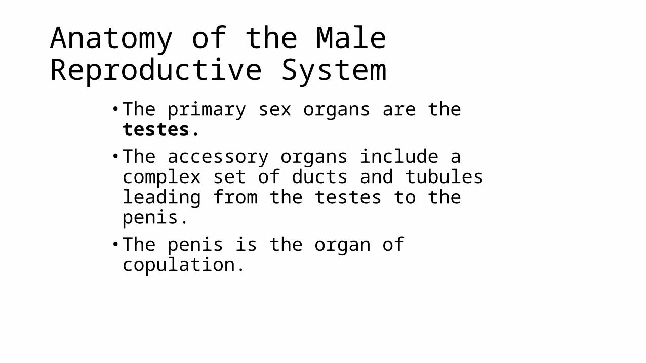
Anatomy of the Male Reproductive System
• The primary sex organs are the testes.• The accessory organs include a complex set of ducts
and tubules leading from the testes to the penis.• The penis is the organ of copulation.

Anatomy of the Male Reproductive System
Figure 28.11

Scrotum
• The scrotum is a skin-covered sac that provides the testes with a cooler environment than body temperature.
• Sperm develop successfully at approximately 3 Celsius below normal body temperature.
• The scrotum is homologous to the labia majora in the female.• A raphe separates the two scrotal sacs.• The dartos muscle is a layer of smooth muscle that is part of the wall
of the scrotum.

Scrotum
Figure 28.12

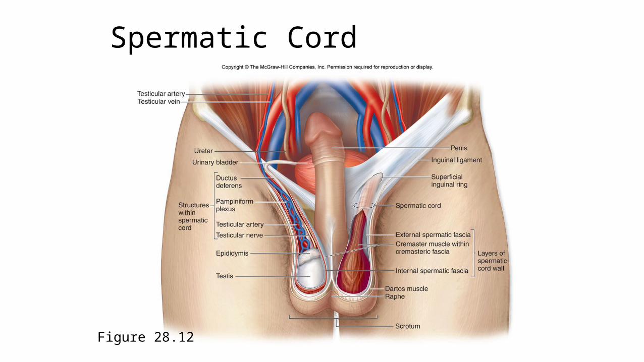
Spermatic Cord
The spermatic cord originates at the inguinal canal. It consists of the following:• Testicular artery—branch of the abdominal aorta• Pampiniform plexus—a network of veins surrounding the testicular artery• Cremaster muscle and fascia—formed from muscle fiber extensions of the internal oblique muscle• Autonomic nerves— travel with plexus and connect to the testes
Spermatic Cord
Figure 28.12
Testes
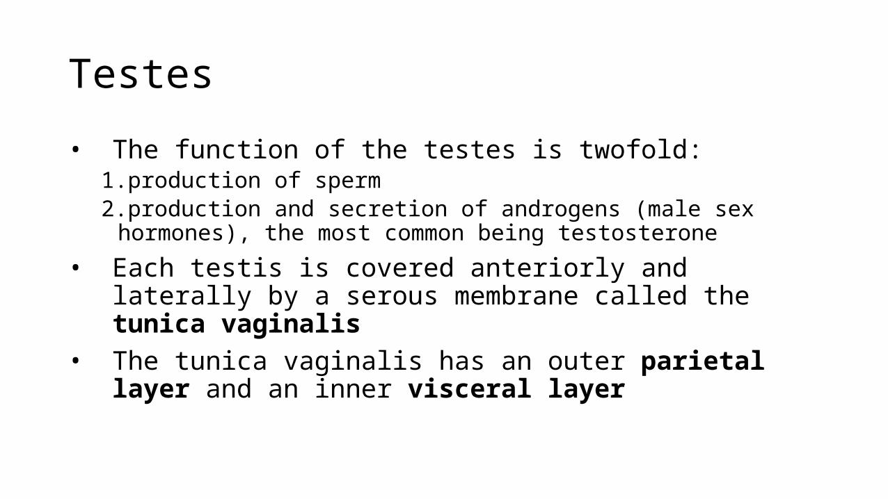
• The function of the testes is twofold:1.production of sperm2.production and secretion of androgens (male sex hormones), the most
common being testosterone
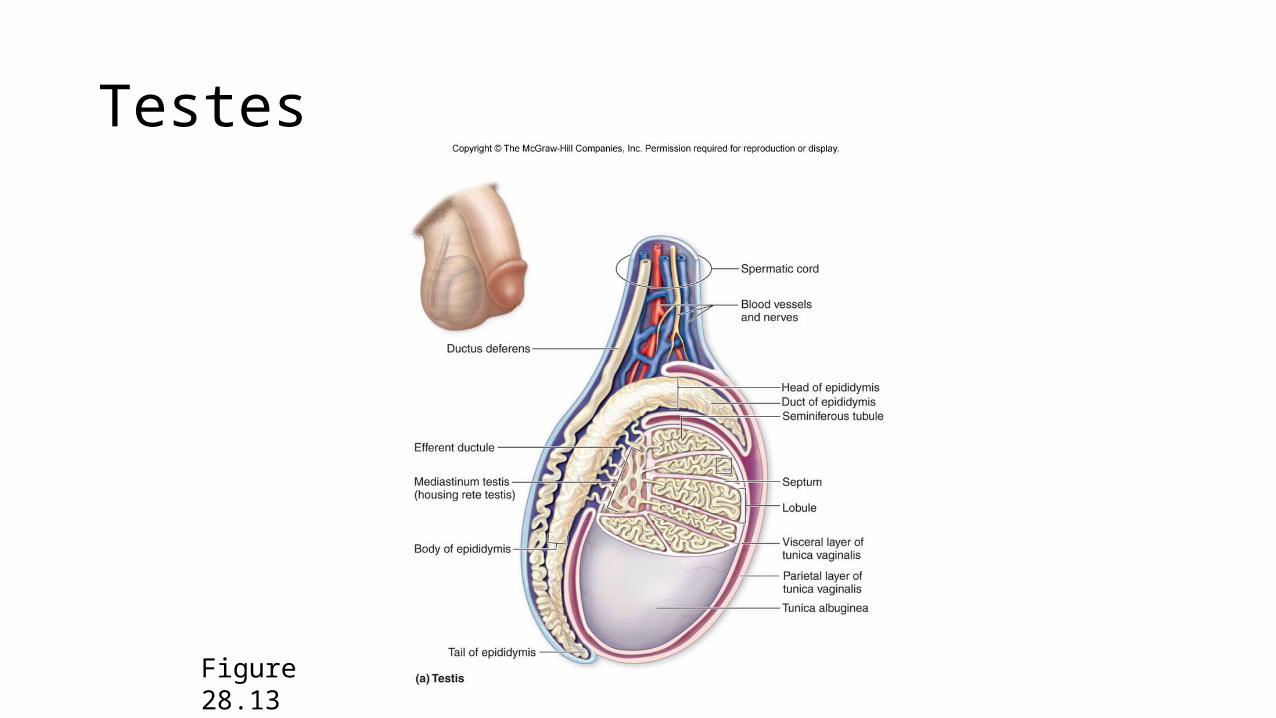
• Each testis is covered anteriorly and laterally by a serous membrane called the tunica vaginalis
• The tunica vaginalis has an outer parietal layer and an inner visceral layer
Testes
Figure 28.13
Testes
• Located just deep to the visceral layer and in contact with the testis is a thick, whitish fibrous capsule covering the testis called the tunica albuginea.
• The tunica albuginea projects into the interior of the testis as the mediastinum testis through which blood vessels, lymphatic vessels, and some nerves enter and leave the testis.
Testes
• The tunica albuginea projects internally into the testes to form septa.• The septa subdivide to form about 250 lobules.• Each lobule contains four convoluted seminiferous tubules.
Seminiferous Tubules
Seminiferous tubules contain two types of cells:1. Sustentacular cells—nondividing support cells that assist with
sperm development; connected to each other by tight junctions and form the blood-testis barrier
2. A population of dividing germ cells that continuously produce sperm beginning at puberty
Interstitial Space
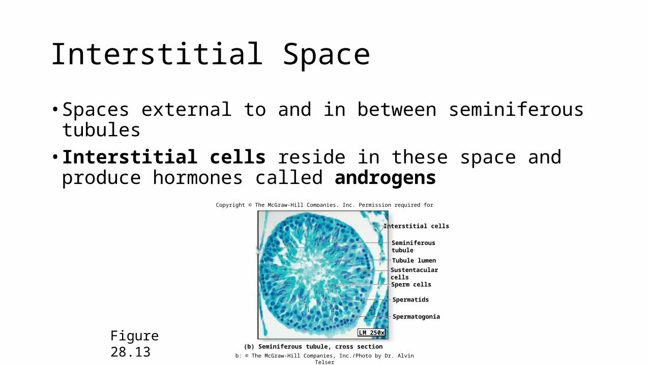
• Spaces external to and in between seminiferous tubules• Interstitial cells reside in these space and produce hormones called
androgens
Figure 28.13
Copyright © The McGraw-Hill Companies, Inc. Permission required for reproduction or display.
Spermatids
Sperm cells
Spermatogonia
Interstitial cells
LM 250x
(b) Seminiferous tubule, cross section
Seminiferoustubule
Tubule lumen
Sustentacularcells
b: © The McGraw-Hill Companies, Inc./Photo by Dr. Alvin Telser
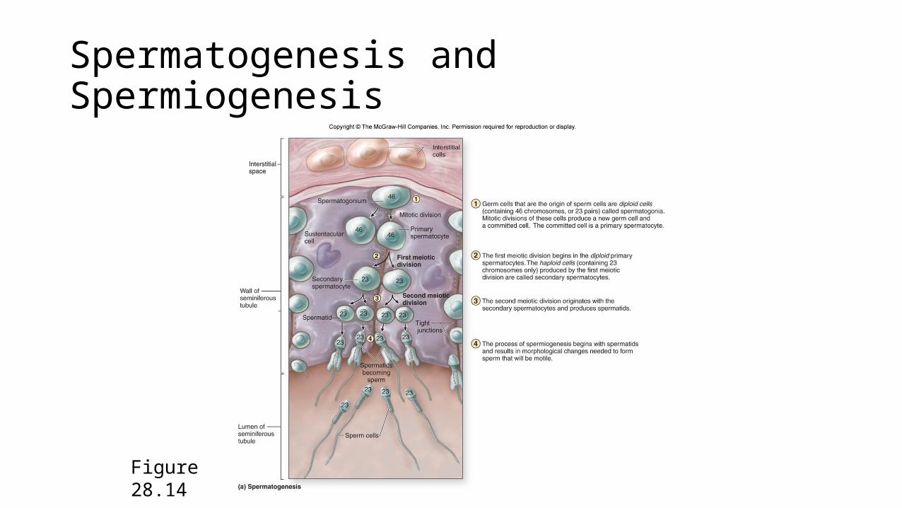
Spermatogenesis and Spermiogenesis
Figure 28.14
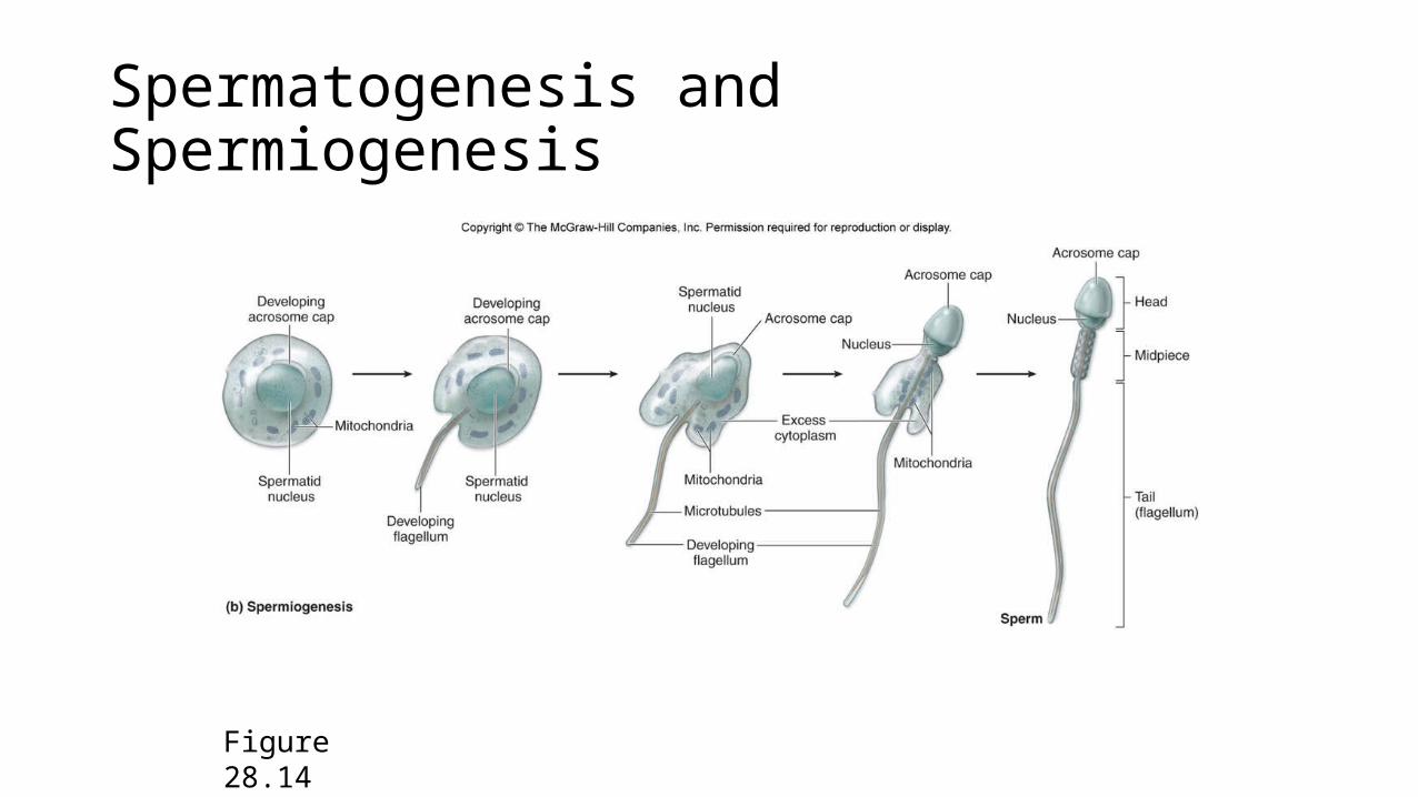
Spermatogenesis and Spermiogenesis
Figure 28.14
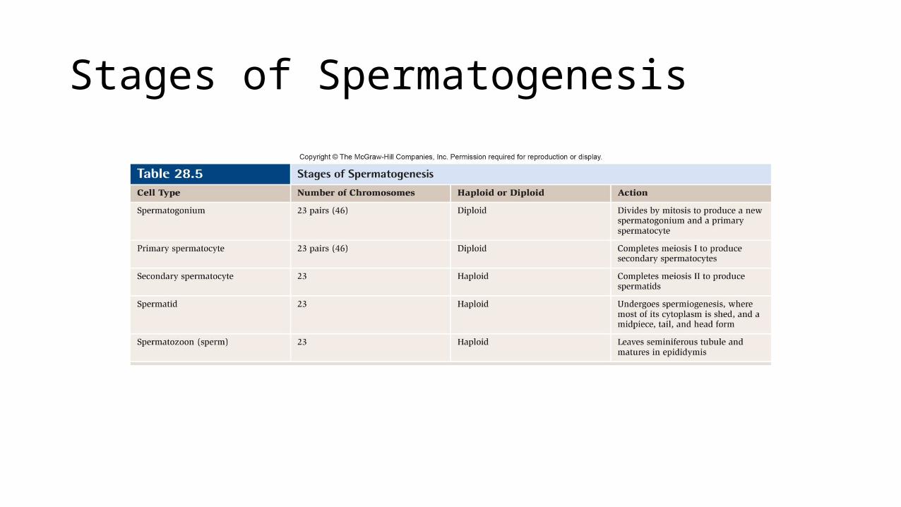
Stages of Spermatogenesis

Ducts in the Male Reproductive System
Beginning at the testis and extending through the penis, the ducts are:• Rete testis• Efferent ductules• Epididymis• Ductus deferens• Ejaculatory duct• Urethra
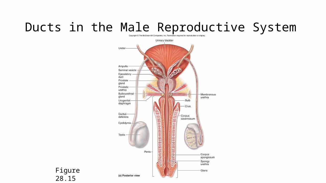
Ducts in the Male Reproductive System
Figure 28.15

Rete Testis• Receive sperm from seminiferous tubules
Figure 28.13
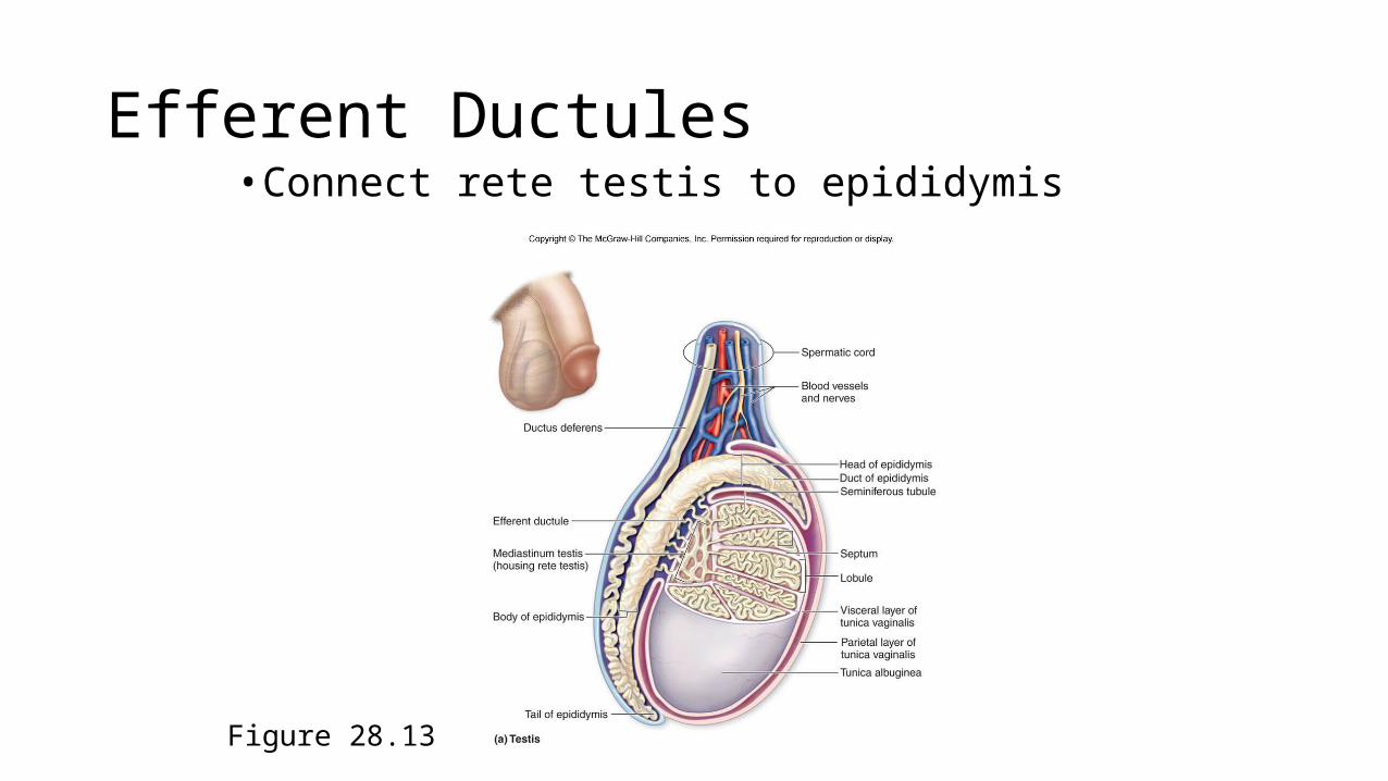
Efferent Ductules• Connect rete testis to epididymis
Figure 28.13

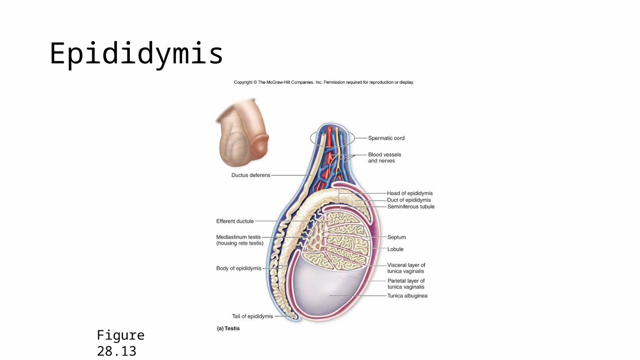
Epididymis
• Situated on the posterosuperior surface of the testes, the epididymis has three regions:- head- body- tail
• Internally, the epididymis contains a long convoluted duct of the epididymis
• The epididymis stores sperm and serves in the maturation process of sperm
Epididymis
Figure 28.13
Ductus Deferens
• Sperm leaving the epididymis enter the ductus deferens (vas deferens).
• This tube travels within the spermatic cord and enters the pelvic cavity through the inguinal canal.
• As the ductus deferens approaches the prostate gland, it enlarges to form the ampulla.
• The ampulla unites with the proximal portion of the seminal vesicle to form the ejaculatory duct.
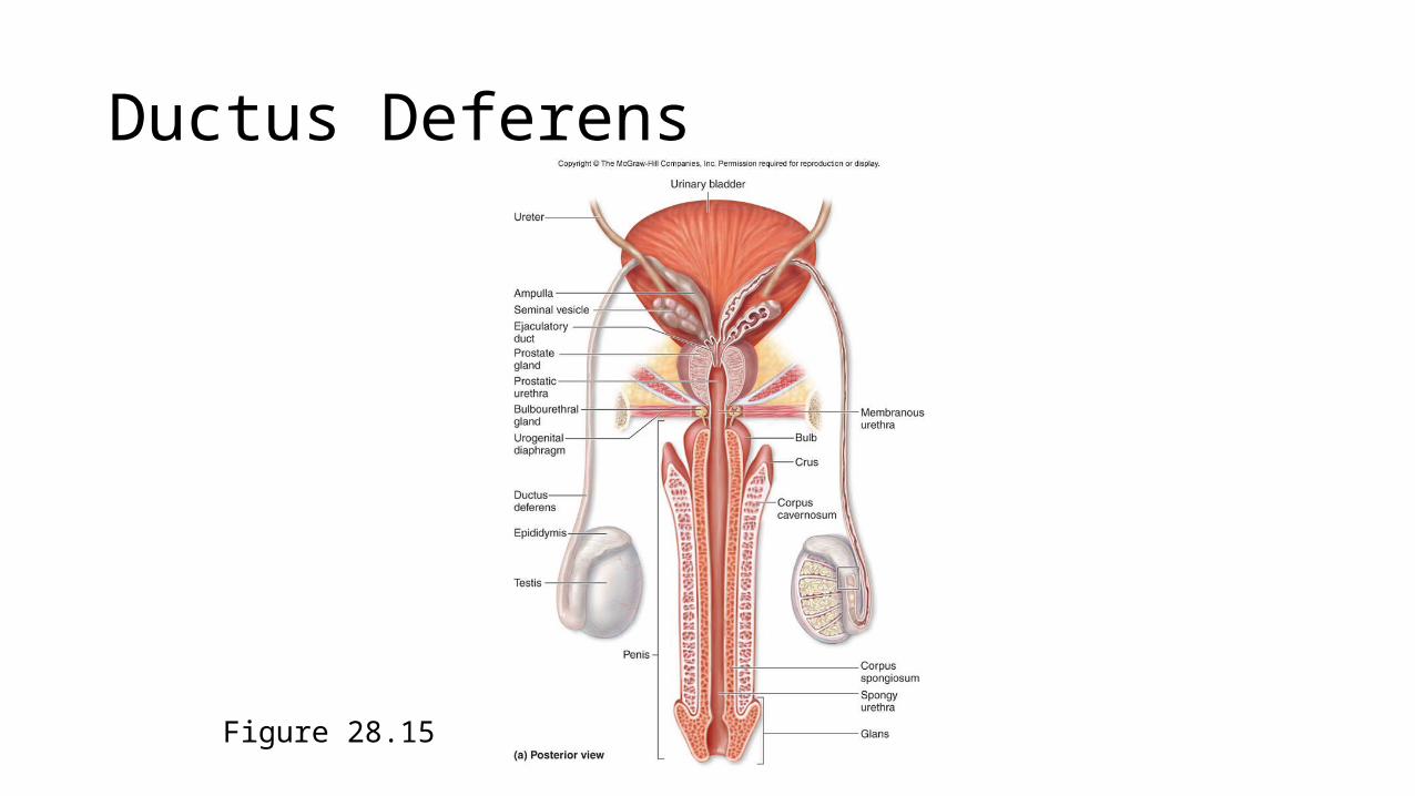
Ductus Deferens
Figure 28.15
Ejaculatory Duct
• Formed by a uniting of the ductus deferens and the seminal vesicle• Located within the substance of the prostate gland between 1–2 cm
long and conducts sperm from the ductus deferens to the prostatic urethra

Ejaculatory Duct
Figure 28.15
Urethra
• Transports semen from both ejaculatory ducts to the outside of the body
• The male urethra is subdivided into three regions:1. prostatic urethra2. membranous urethra3. spongy urethra

Urethra
Figure 28.15
Accessory Glands
• There are three glands that secrete fluids to mix with sperm to create seminal fluid.
• These secretions nourish the sperm and neutralize the acidity of the vagina.
• The three glands are as follows:1. seminal vesicles2. prostate gland3. bulbourethral glands
Seminal Vesicles
• The paired seminal vesicles are located on the posterior surface of the urinary bladder lateral to the ampulla of the ductus deferens.
• They secrete a viscous, whitish-yellow, alkaline fluid containing fructose and prostaglandins.

Seminal Vesicles
Figure 28.15
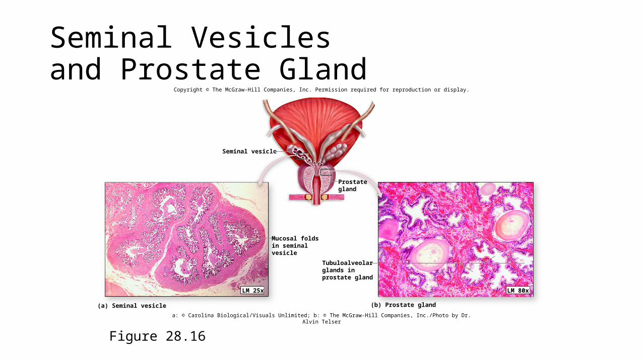
Seminal Vesicles and Prostate Gland
Figure 28.16
Copyright © The McGraw-Hill Companies, Inc. Permission required for reproduction or display.
Seminal vesicle
LM 80xLM 25x
(b) Prostate gland
Tubuloalveolarglands inprostate gland
Mucosal foldsin seminalvesicle
(a) Seminal vesicle
Prostategland
a: © Carolina Biological/Visuals Unlimited; b: © The McGraw-Hill Companies, Inc./Photo by Dr. Alvin Telser
Prostate Gland
• The prostate gland is located immediately inferior to the urinary bladder.
• It produces substances that are secreted directly into the prostatic urethra.
• Prostatic secretion is slightly acidic and contains mucin, citric acid (nutrient for sperm), seminalplasmin (antibacterial),and prostatic-specific antigen (PSA, an enzyme that helps liquify semen).
Prostate Gland
Figure 28.15
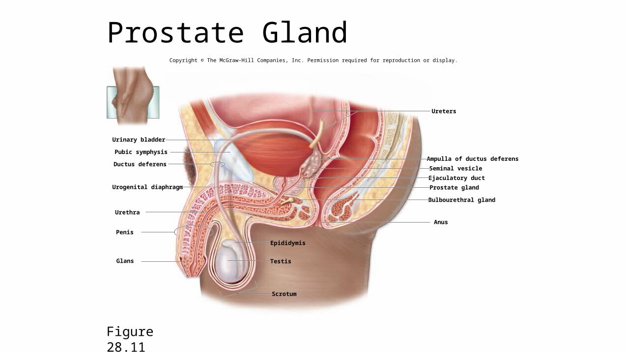
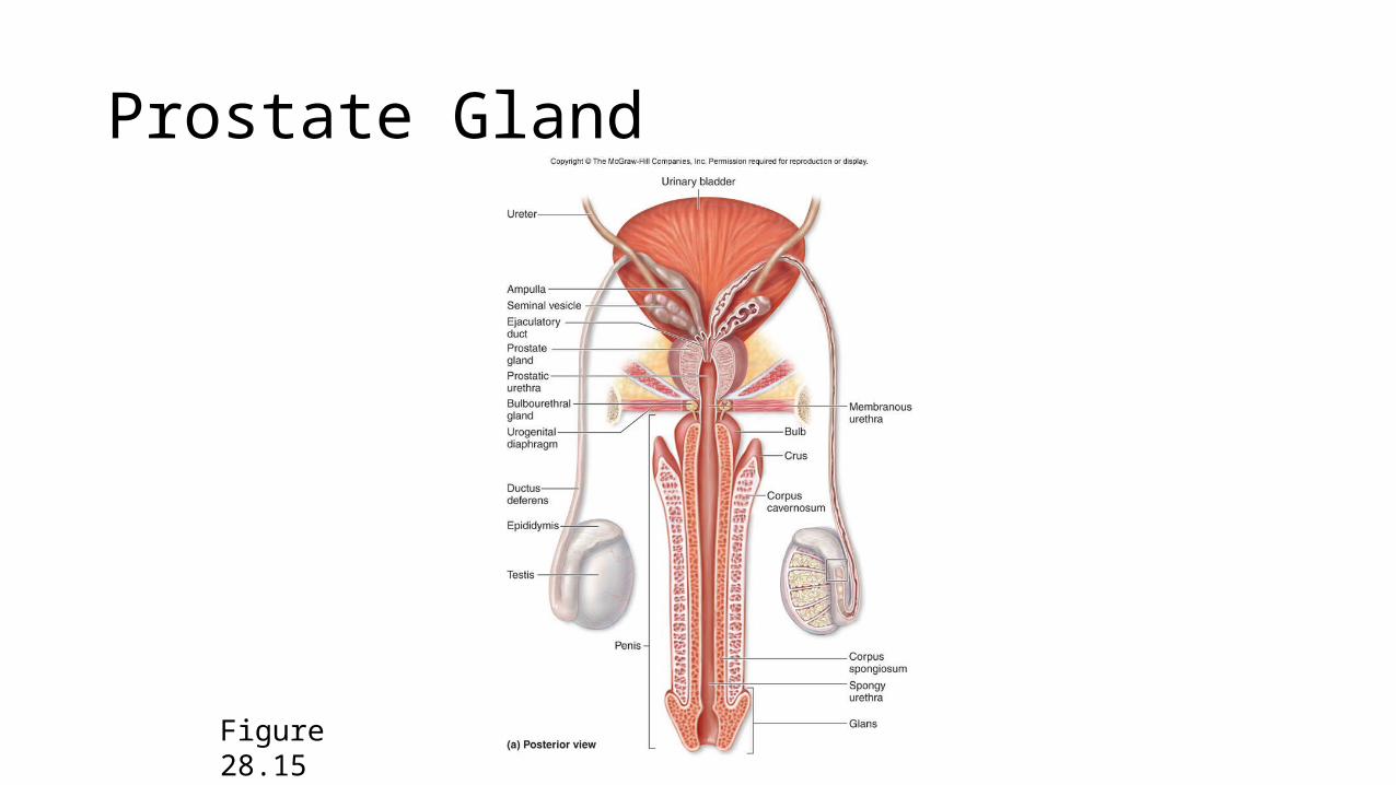
Prostate Gland
Figure 28.11
Copyright © The McGraw-Hill Companies, Inc. Permission required for reproduction or display.
Urinary bladder
Pubic symphysis
Ductus deferens
Urethra
Urogenital diaphragm
Penis
Glans
Ureters
Ampulla of ductus deferens
Seminal vesicle
Ejaculatory duct
Prostate gland
Bulbourethral gland
Anus
Epididymis
Scrotum
Testis
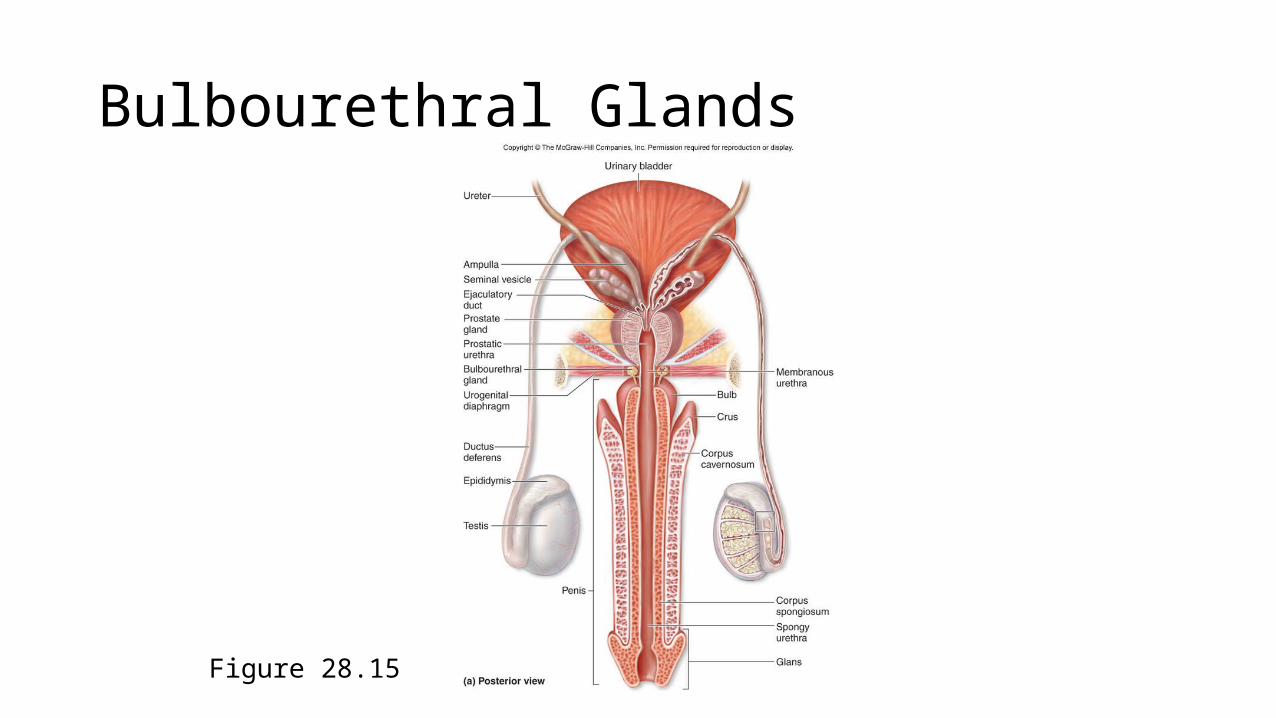
Bulbourethral Glands
• The bulbourethral glands are located in the urogenital diaphragm on either side of the membranous urethra.
• Mucin is secreted from these glands and enters the spongy urethra at the base of the penis.
Bulbourethral Glands
Figure 28.15
Semen
• Seminal fluid from the three accessory glands combines with sperm from the testes to make up semen.
• When released during intercourse, semen is called ejaculate.• Semen normally measures about 3–5 ml in volume and contains 200–
500 million sperm.
Penis
• The penis and the scrotum form the external genitalia in males.• Internally, the attached portion of the penis is the root forming both
the bulb and the crura of the penis.• The body (shaft) is the elongated portion of the penis.• The tip of the penis is the glans, which surrounds the external
urethral orifice.
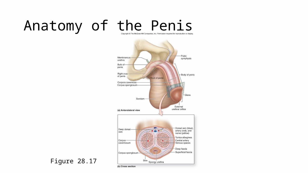
Anatomy of the Penis
Figure 28.17
Penis
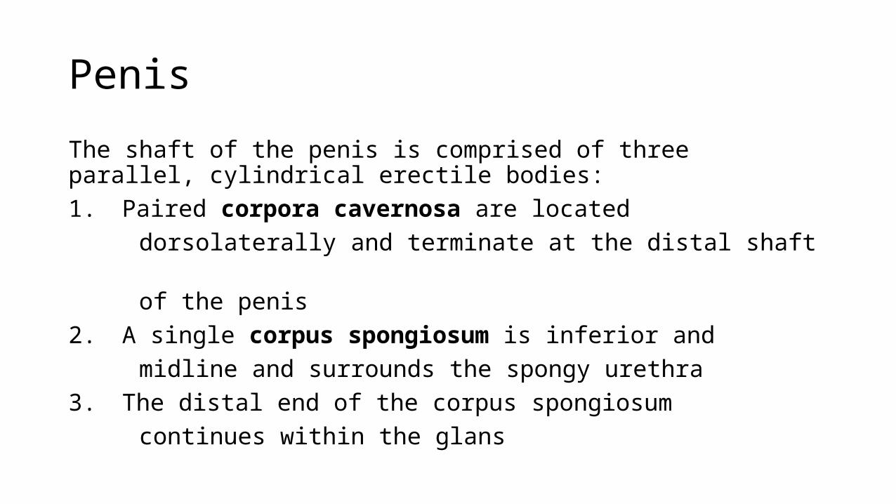
The shaft of the penis is comprised of three parallel, cylindrical erectile bodies:1. Paired corpora cavernosa are located dorsolaterally and terminate at the distal shaft of the penis2. A single corpus spongiosum is inferior and midline and surrounds the spongy urethra3. The distal end of the corpus spongiosum continues within the glans
Anatomy of the Penis
Figure 28.17
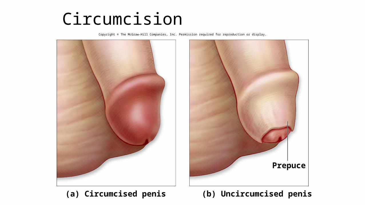
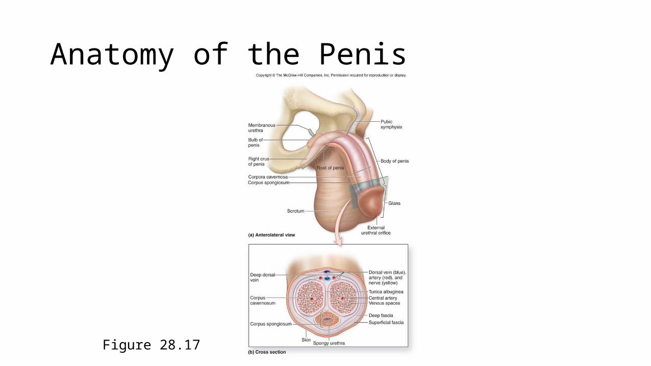
CircumcisionCopyright © The McGraw-Hill Companies, Inc. Permission required for reproduction or display.
Prepuce
(b) Uncircumcised penis(a) Circumcised penis
Erection and Ejaculation
• The erectile bodies are composed of venous spaces, which fill with blood from a central artery to produce an erection.
• Parasympathetic in nervation is responsible for penile erection.• Ejaculation is the expelling of semen from the penis, in part, by the
rhythmic contraction of the smooth muscle of the urethra.• Sympathetic in nervation promotes ejaculation.

Development of the Reproductive Systems
Figure 28.18
Copyright © The McGraw-Hill Companies, Inc. Permission required for reproduction or display.
Mesonephros
Genital ridge
Kidney
Mesonephric duct
Cloaca
MaleFemale
Sexually Indifferent Stage
Testes
Efferent ductules
Epididymis
Urinary bladder
Seminal vesicle
Urinary bladder
Seminal vesicle
Prostate gland
Bulbourethral gland
Ductus deferens
Epididymis
Efferent ductules
Testis
Urethra
At birth
Ovaries
Ovary
Uterus
Vagina
UrethraHymen
At birth
Paramesonephricduct
Weeks 5–6
Paramesonephricduct forming theuterine tube
Mesonephricduct(degenerating)
Paramesonephricduct (degenerating)
Mesonephric ductforming the ductusdeferens
Urogenital sinusforming the urethra
Weeks 10–12Weeks 10–12
Urogenital sinus formingthe urethra and inferior vagina
Fused paramesonephricducts forming the uterus
Uterinetube
Urinary bladder(moved aside)
Urinary bladder(moved aside)

Development of the Reproductive Systems
Figure 28.19


