39
Malignant Melanoma Residents Bonus Conference January 18, 2012 Kristy Kummerow, PGY 2

Malignant Melanoma Residents Bonus Conference
January 18, 2012
Kristy Kummerow, PGY 2
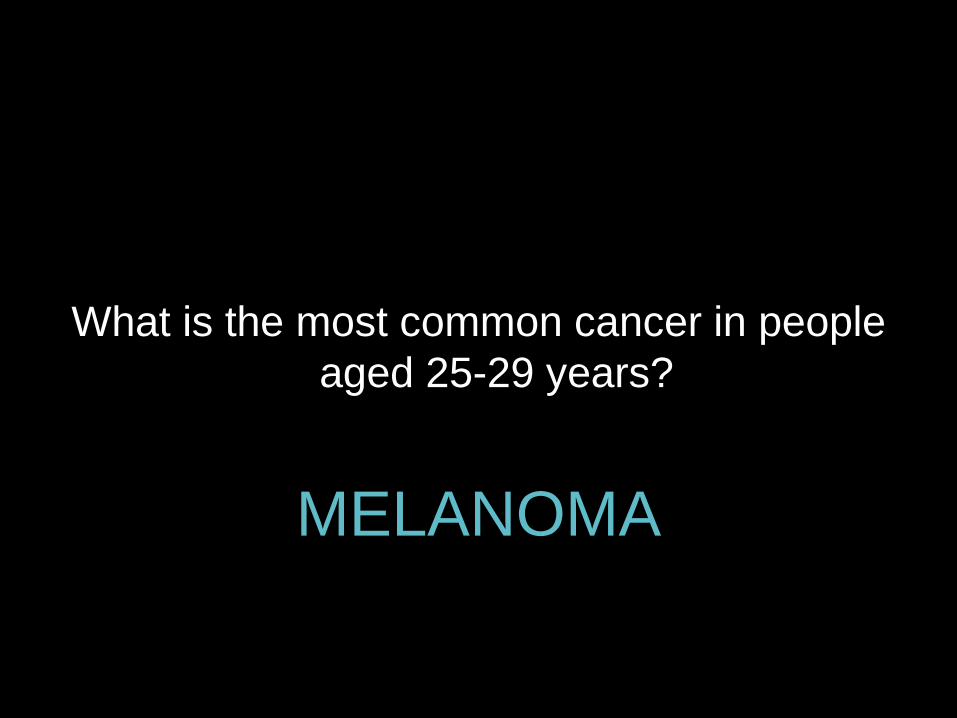
What is the most common cancer in people
aged 25-29 years?
MELANOMA
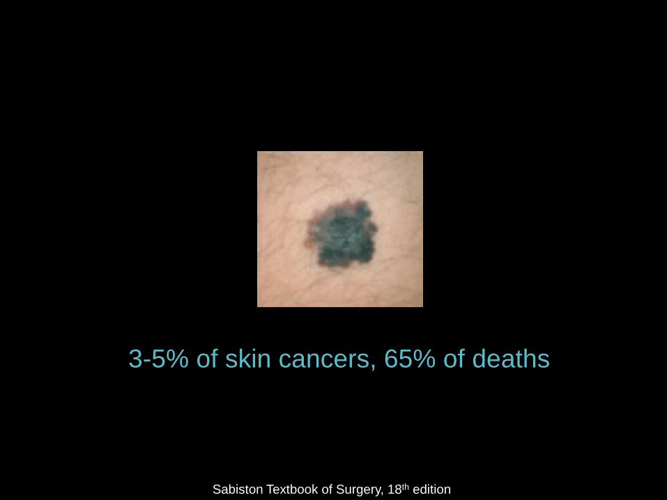
Sabiston Textbook of Surgery, 18th edition
3-5% of skin cancers, 65% of deaths
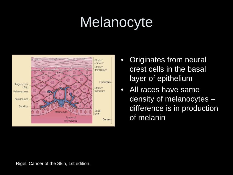
Melanocyte
• Originates from neural
crest cells in the basal layer of epithelium
• All races have same density of melanocytes – difference is in production of melanin
Rigel, Cancer of the Skin, 1st edition.
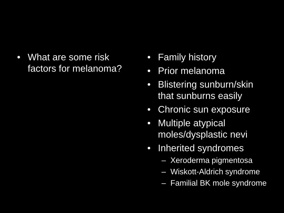
• What are some risk factors for melanoma?
• Family history • Prior melanoma • Blistering sunburn/skin
that sunburns easily • Chronic sun exposure • Multiple atypical
moles/dysplastic nevi • Inherited syndromes
– Xeroderma pigmentosa – Wiskott-Aldrich syndrome – Familial BK mole syndrome
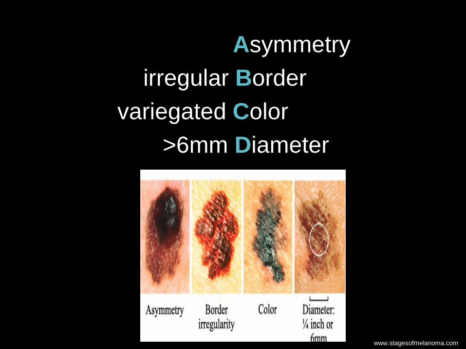
Asymmetry irregular Border variegated Color >6mm Diameter
www.stagesofmelanoma.com
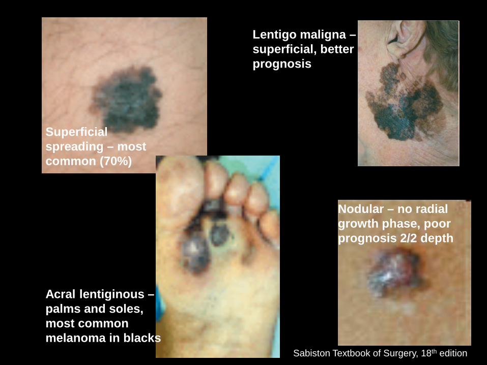
Superficial spreading – most common (70%)
Acral lentiginous – palms and soles, most common melanoma in blacks
Nodular – no radial growth phase, poor prognosis 2/2 depth
Lentigo maligna – superficial, better prognosis
Sabiston Textbook of Surgery, 18th edition
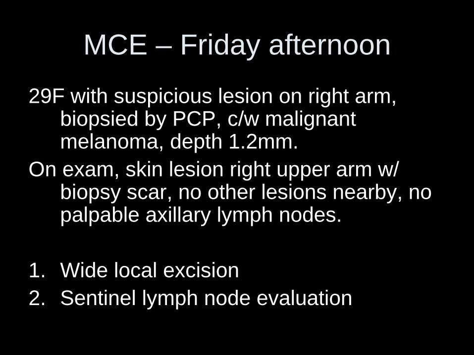
MCE – Friday afternoon
29F with suspicious lesion on right arm, biopsied by PCP, c/w malignant melanoma, depth 1.2mm.
On exam, skin lesion right upper arm w/ biopsy scar, no other lesions nearby, no palpable axillary lymph nodes.
1. Wide local excision 2. Sentinel lymph node evaluation
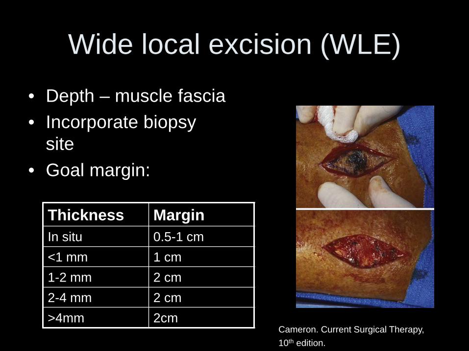
Wide local excision (WLE)
• Depth – muscle fascia • Incorporate biopsy
site • Goal margin:
Thickness Margin In situ 0.5-1 cm <1 mm 1 cm 1-2 mm 2 cm 2-4 mm 2 cm >4mm 2cm
Cameron. Current Surgical Therapy, 10th edition.
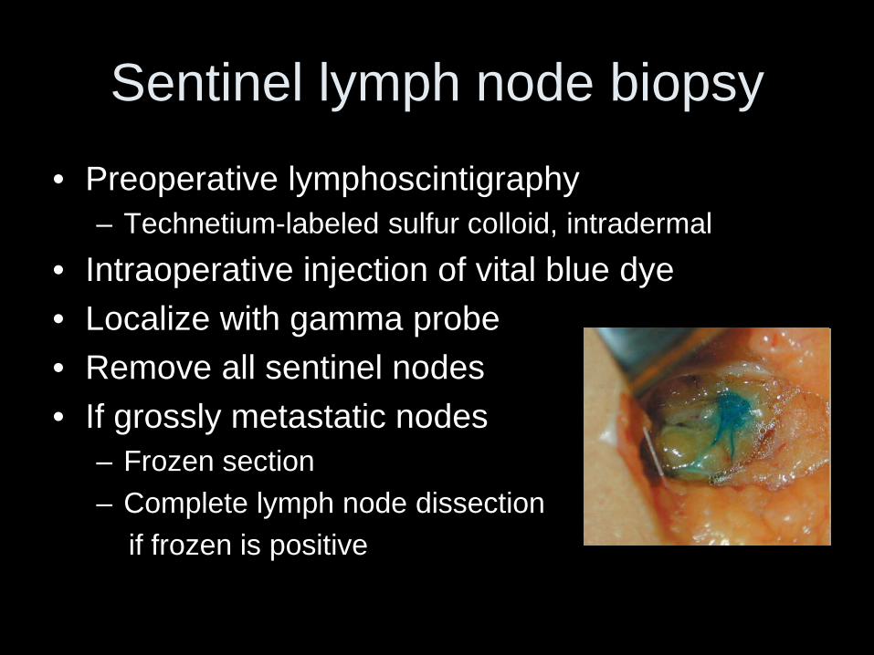
Sentinel lymph node biopsy
• Preoperative lymphoscintigraphy – Technetium-labeled sulfur colloid, intradermal
• Intraoperative injection of vital blue dye • Localize with gamma probe • Remove all sentinel nodes • If grossly metastatic nodes
– Frozen section – Complete lymph node dissection if frozen is positive
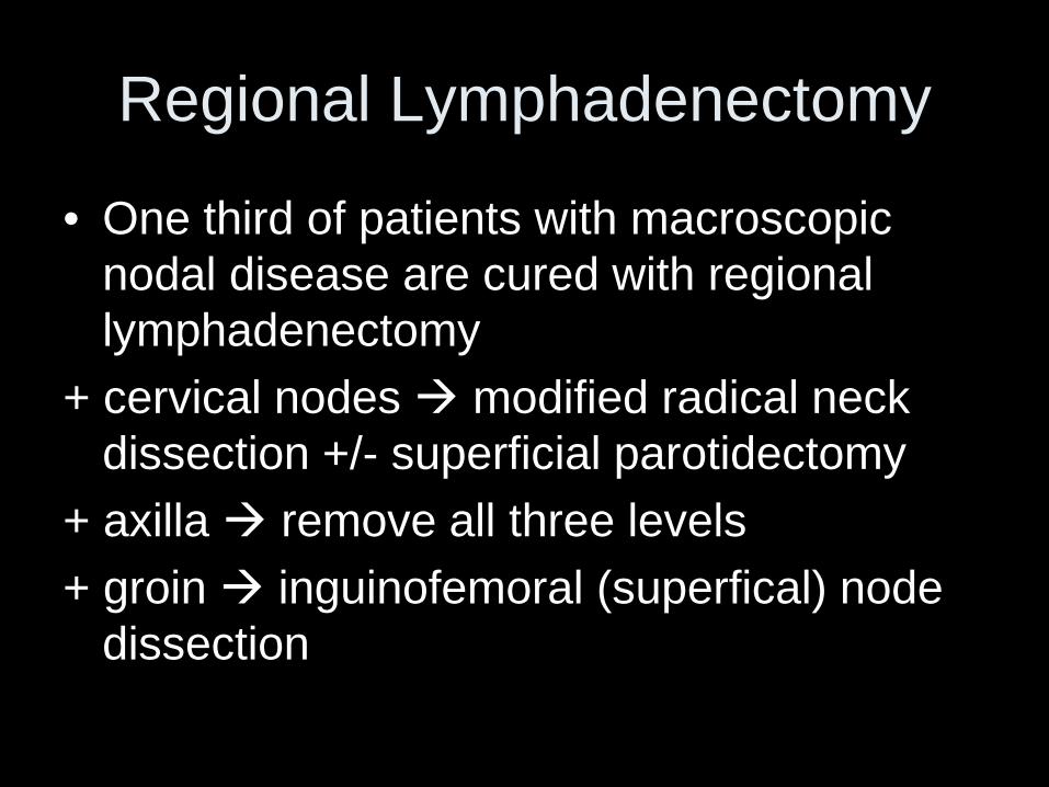
Regional Lymphadenectomy
• One third of patients with macroscopic nodal disease are cured with regional lymphadenectomy
+ cervical nodes modified radical neck dissection +/- superficial parotidectomy
+ axilla remove all three levels + groin inguinofemoral (superfical) node
dissection
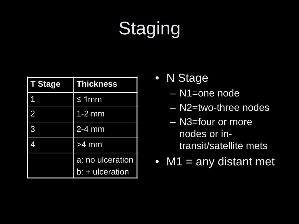
Staging
• N Stage
– N1=one node – N2=two-three nodes – N3=four or more
nodes or in-transit/satellite mets
• M1 = any distant met
T Stage Thickness
1 ≤ 1mm
2 1-2 mm
3 2-4 mm
4 >4 mm
a: no ulceration b: + ulceration
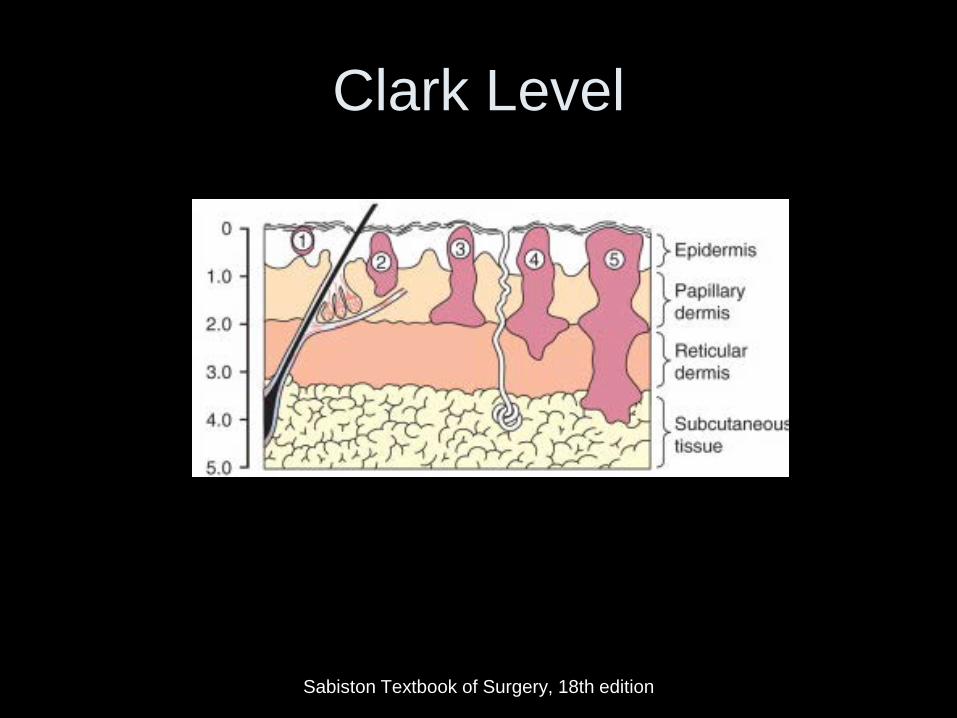
Clark Level
Sabiston Textbook of Surgery, 18th edition
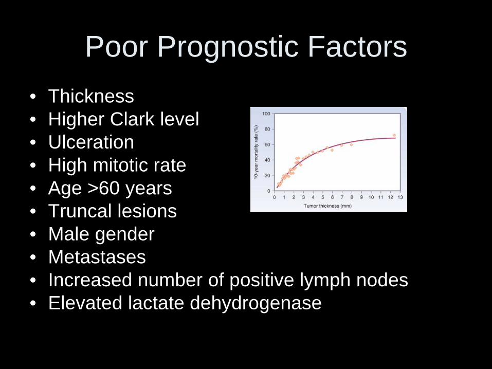
Poor Prognostic Factors • Thickness • Higher Clark level • Ulceration • High mitotic rate • Age >60 years • Truncal lesions • Male gender • Metastases • Increased number of positive lymph nodes • Elevated lactate dehydrogenase
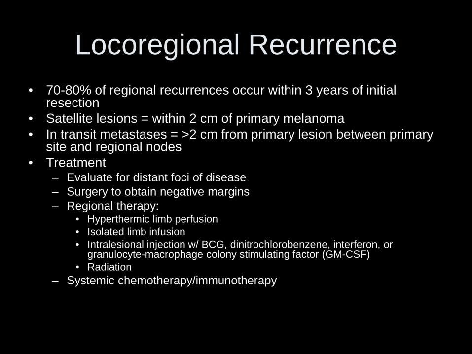
Locoregional Recurrence • 70-80% of regional recurrences occur within 3 years of initial
resection • Satellite lesions = within 2 cm of primary melanoma • In transit metastases = >2 cm from primary lesion between primary
site and regional nodes • Treatment
– Evaluate for distant foci of disease – Surgery to obtain negative margins – Regional therapy:
• Hyperthermic limb perfusion • Isolated limb infusion • Intralesional injection w/ BCG, dinitrochlorobenzene, interferon, or
granulocyte-macrophage colony stimulating factor (GM-CSF) • Radiation
– Systemic chemotherapy/immunotherapy
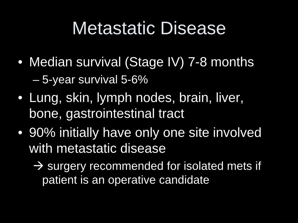
Metastatic Disease
• Median survival (Stage IV) 7-8 months – 5-year survival 5-6%
• Lung, skin, lymph nodes, brain, liver, bone, gastrointestinal tract
• 90% initially have only one site involved with metastatic disease surgery recommended for isolated mets if
patient is an operative candidate
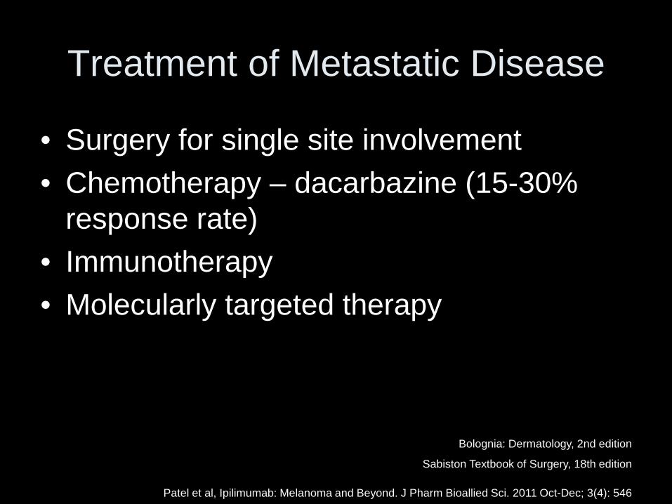
Treatment of Metastatic Disease
• Surgery for single site involvement • Chemotherapy – dacarbazine (15-30%
response rate) • Immunotherapy • Molecularly targeted therapy
Bolognia: Dermatology, 2nd edition
Sabiston Textbook of Surgery, 18th edition
Patel et al, Ipilimumab: Melanoma and Beyond. J Pharm Bioallied Sci. 2011 Oct-Dec; 3(4): 546

Immunotherapy
• IL-2 15% response • Interferon α2b 10-15% response • Monoclonal antibodies against T-cell and
melanoma cell surface antigens – Ipilimumab (Yervoy) 30% response
• Adoptive cell transfer • Melanoma vaccines
Bolognia: Dermatology, 2nd edition
Sabiston Textbook of Surgery, 18th edition
Patel et al, Ipilimumab: Melanoma and Beyond. J Pharm Bioallied Sci. 2011 Oct-Dec; 3(4): 546

Molecularly Targeted Therapy
• Vemurafenib – enzymatic inhibitor of BRAF (protein kinase involved in RAF/MEK/ERK cell signaling pathway) – 60% of pts have mutation in BRAF – BRIM3 trial (phase III) vs. dacarbazine
• Death ↓63% • Disease progression ↓74%
• Antiangiogenesis agents (eg. bevacizumab)
Bollag et al. Clinical efficacy of a RAF inhibitor needs broad target blockade in BRAF-mutant melanoma. Nature. 2010 September 30; 467(7315): 596–599.
Bolognia: Dermatology, 2nd edition.

Study Questions
When performing a sentinel lymph node biopsy in the axilla, you locate a node that is blue and hot. Your next step is:
• Close up and go home • Examine the area for additional positive
nodes • Send the node for frozen section • Proceed with three level axillary node
dissection
When performing a sentinel lymph node biopsy in the axilla, you locate a node that is blue and hot. Your next step is:
• Close up and go home • Examine the area for additional positive
nodes • Send the node for frozen section • Proceed with three level axillary node
dissection
75% of recurrences will occur within what time interval after primary excision of a melanoma skin lesion?
• 3 months • 1 year • 3 years • 5 years
75% of recurrences will occur within what time interval after primary excision of a melanoma skin lesion?
• 3 months • 1 year • 3 years • 5 years

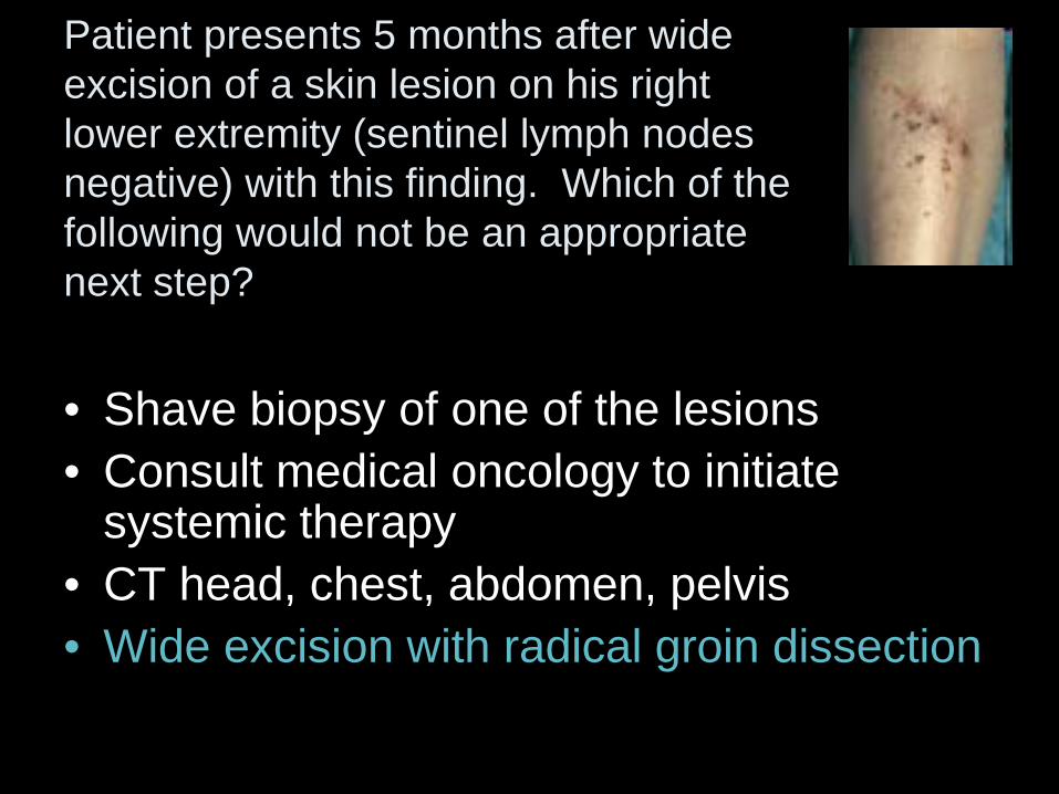
Patient presents 5 months after wide excision of a skin lesion on his right lower extremity (sentinel lymph nodes negative) with this finding. Which of the following would not be an appropriate next step?
• Shave biopsy of one of the lesions • Consult medical oncology to initiate
systemic therapy • CT head, chest, abdomen, pelvis • Wide excision with radical groin dissection
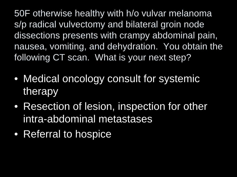
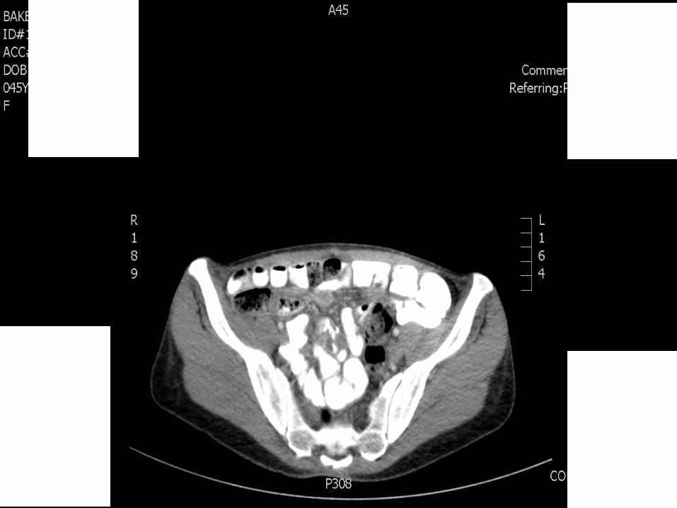
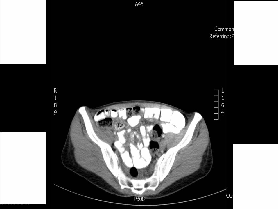
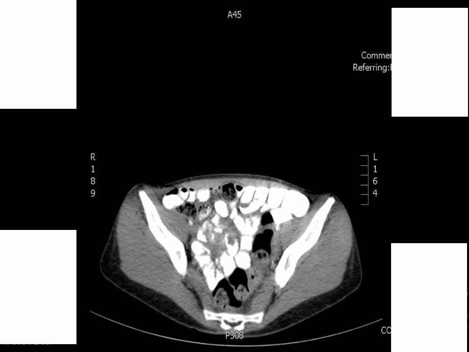
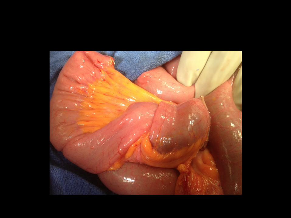
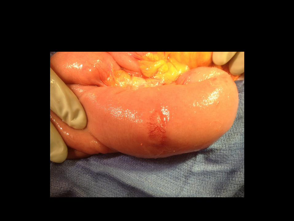
50F otherwise healthy with h/o vulvar melanoma s/p radical vulvectomy and bilateral groin node dissections presents with crampy abdominal pain, nausea, vomiting, and dehydration. You obtain the following CT scan. What is your next step?
• Medical oncology consult for systemic therapy
• Resection of lesion, inspection for other intra-abdominal metastases
• Referral to hospice
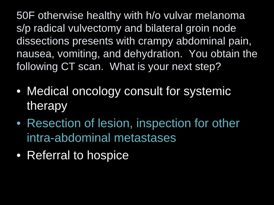


A New Era Approaches: Anti-CTLA-4 Monoclonal Antibodies for the Treatment of Malignant Melanoma
• Jeffrey S. Weber, MD, PhD