27
Malpresentation & malposition Dr. K ak ali Saha Associ ate professor MBBS,FCPS,MS (OBS&GYNAE) Medic al college for women & hospit al

Malpresentation & malpositionDr. Kakali Saha
Associate professor MBBS,FCPS,MS (OBS&GYNAE)
Medical college for women & hospital

Definition of Malpresentation
• It is defined as any presentation of foetus other than vertex.
• What is presentation? ✴The part of the foetus which occupies the lower pole of uterus (pelvic brim) is called presentation of the
foetus. Presentation like cephalic 96.5%, podalic 3% or shoulder and others 0.5%.
• What is presenting part? ✴The presenting part is defined as the part of the presentation which overlies the internal os & felt by
examining finger through the cervical opening. Thus, in cephalic presentation, presenting part may be vertex, brow or face depending on degree of flexion or head.
Types
• Name some Mal presentation: ✤ Breech presentation. Most commonest malpresentation. ✤ Brow presentation ✤ Face presentation ✤ Shoulder presentation ✤ Dorsal presentation ✤ Cord presentation

Breech Presentation
• When buttock of the foetus occupies the lower segment or pole of uterus as well as overlies the internal os.

Breech presentation Types
• Varieties—there are two varieties ✤ Complete or flexed breech ✤Incomplete or varying degree
of extension ➡ Frank breech ➡Footling presentation ➡Knee presentation
Types
• Clinical Varieties—there are two varieties ✤ Uncomplicated breech -it is defined as where there is no other associated
obstetric complications apart from the breech, prematurity being excluded. ✤ Complicated breech - when the breech is associated with conditions which
adversely affected the prognosis such as prematurity, twins, contracted pelvis, placenta Praevia, etc. ❖ Extended breech, Frank breech,Footling presentation,Knee presentation,
extended arm, cord prolapse or difficulty encounter during breech delivery should not be called complicated breech but are called complicated breech delivery.
Clinical varieties
Incidence
• Incidence is about 20% at 28th week & drops to 5% at 34th week & to 3-4% at term.
• Thus, in 3 out of 4 , spontaneous correction into vertex occurs by 34th week.

Etiology
• Prematurity - is the commonest cause
• Factors preventing spontaneous version (a) Breech with extended legs (b) Twins (c) Oligohydramnios (d) Congenital malformation of uterus such as bicornuated or septated uterus. (e) Short cord relative or absolute (f ) IUD
In a significant number of case cause remain obscure

• Favourable adaptation:
(a) Hydrocephalus -big head well occupied in the wide fungus
(b) Placenta Praevia
(c) Contracted pelvis
(d) Cornufundal attachment of the placenta -which minimised the space of fundus where smaller head well occupied
• Undue mobility of foetus: (a)Hydramnios, (b)Multipara with lax abdominal wall
• Foetal abnormality: congenital anomalies of foetus such as, trisomies 13,18,21, anencephaly, myotonic dystrophy due to alteration of foetal muscle tone & mobility.
Cont

Diagnosis• Clinical : mother felt foetal movement more in her lower abdomen
• Per abdominal findings & pervaginal findings see chart below

• USG- is most informative. ✴It confirmed the diagnosis specially in primi with extended breech ✴It can detect foetal congenital abnormalities & also anomalies of uterus ✴Type of breech complete or incomplete ✴It measures BPD, gestational age & foetal weight ✴It also localises the placenta ✴Assessment of volume of liquor ✴Attitude of the foetal head flexion or hyperextension
• some time CT & MRI can be used to asses pelvic capacity in addition to all above.
Investigation

Position
• The sacrum is the denominator of breech and there are four position of breech ✴First position- left sacro-anterior is the commonest ✴Second position -right sacro-anterior ✴Third position-right sacro-posterior ✴Fourth position- left sacro-posterior

Mechanism of labour in breech presentation
• Principle of movement occur at three place ➡Buttock ➡Shoulder ➡Head
๏The first two successive parts to be are bigger but more compressible while the head because of non moulding due to rapid descent, presents difficulties.
๏Each of the three components undergo cardinal movements as those of normal mechanism.
Sacro-anterior position

Buttock delivery
• The diameter of the buttock is one of the oblique diameters of inlet.
• Engaging diameter is bi-trochanteric (10 cm) with the sacrum directed towards the ilio-pubic eminence. When the diameter passes through the pelvic brim, the breech is engaged.
• Descent of the buttocks occurs until ant. buttock touches the pelvic floor.
• Internal rotation of ant buttock occurs through 1/8 of the circle placing it behind the symphysis pubis.
• Farther descent with lateral flexion of the trunk occur until the ant. hip hinges under the symphysis pubis which is released 1st followed by posterior hip.
• Delivery of trunk & lower limbs follow.
• Restitution occurs so that the buttocks occupy the original position as during engagement in oblique diameter

Shoulder delivery
• The diameter of the shoulder is same oblique diameters of inlet as that occupied by the buttock soon after delivery of the breech.
• Engaging diameter is bisacromial (12cm).
• Descent of the shoulder occurs until ant. shoulder touches the pelvic floor.
• Internal rotation of the shoulders bringing the shoulders to lie in the anteroposterior diameter of pelvic outlet. The trunk simultaneously rotate externally through 1/8th of a circle.
• Delivery of posterior shoulder followed by the anterior one is completed by anterior flexion of the delivered truck
• Restitution & external rotation : untwisting of the trunk occurs putting the anterior shoulder towards the right thigh in LSA & left thing in RSA.
• External rotation of the shoulders occurs to the same direction because of internal rotation of the occiput through 1/8th circle anteriorly.
• Foetal truck is now positioned as dorso-anterior.

After coming head delivery
• The engaging diameter of the head is suboccipitofrotal diameters (10cm).
• Engagement occurs either through the opposite oblique diameter as that occupied by buttocks or through the transverse diameter.
• Descent with increase flexion of the head occurs until it touches the pelvic floor.
• Internal rotation of the occiput occurs anteriorly, through 1/8th or 2/8th of a circle placing the occiput behind the symphysis pubis.
• Farther descent occurs until the subocciput hinges under the symphysis pubis.
• Head is born by flexion- the chin, mouth, nose, forehead, vertex & occiput.
• The expulsion of the head from the pelvic cavity depends entirely upon the bearing down efforts & not at all on uterine contractions.
• Sacro-posterior position, head has to rotate through 3/8th of a circle to bring the occiput behind the symphysis pubis.

Prognosis
• Maternal : increased frequency of operative delivery including C/S, the morbidity increased.
• foetal: risk in terms of perinatal mortality ranges from 5-35 per 1000 births.
• Overall perinatal mortality in breech still remain 9-25% compare with 1-2% for non breech delivery.

Foetal danger
• IUD
• ICH- intracranial haemorrhage- tear of tentorium cerebelli & haemorrhage in sub arachnoid space.
• Birth asphyxia- due to cord compression- period >10 min will produce asphyxia of varying degree.
• Birth injuries(7%)- happened during manipulative deliveries. It is 13 times more the vertex deliveries. ✤Haematoma ✤Fracturs- spcially femur, humerus, ribs, clavicle& odontoid process. Dislocation of the hip joint ✤Visceral injuries - liver, kidneys, suprarenal gland, lungs & haemorrhage in the testicles. ✤Nerve- medullary coning, spinal cord inj, stretching of brachial plexus lead to Erb’s palsy & Klumpke’s
palsy ✤Long term neurological damage.
Prevention
• External cephalic version
• Elective caesarean section
• Vaginal breech delivery by experts
• Vaginal manipulative delivery by skilled person specially during delivery of head.

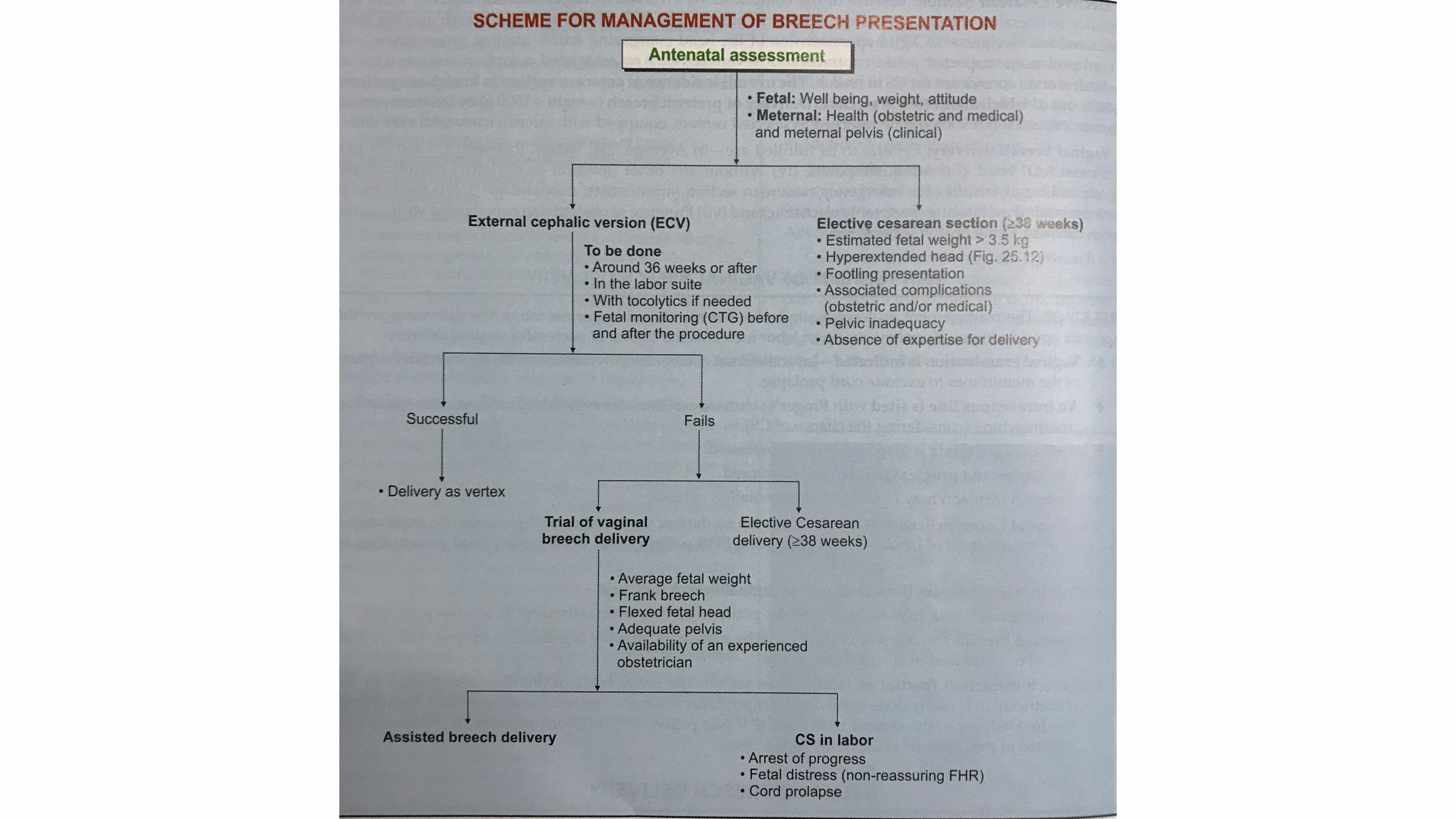
Management
• Identification of complicating factors related with breech
• External cephalic version, if not contraindicated - considered from 36 weeks onwards.
• Formulation of the line of management.
Antenatal management

Intrapartum management
• First stage: protocol similar normal labour. sp. onset successful outcome
• At the onset of labour pain vaginal exam for pelvic assessment
• I/V line with Ringer’s sol, oral intake avoid, blood ready for cross matches
• Adequate analgesia, epidural prefer
• Fotal status & progress of labour monitored
• Oxytocin infusion may be used for augmentation
Management of vaginal breech delivery

• Second stage: there are three methods of vaginal breech delivery: ✦Sponteneous 10% expulsion occurs with very little assistance. This is not preferrd. ✦Assisted breech delivery- assistance from beginning to end of delivery. It should be done
in all cases. ✦Breech extraction ( partial or total): when part or entire body of vthe foetus is extracted
by the obstetrician. Chace of injuries or trauma to ythe foetus & mother. Indictions (a)delivery of 2nd twin after IPV. (b)Cord prolapse (c)Extended legs arrested at the cavity or at the outlet.


The EndThank you all


