Proc. Natl. Acad. Sci. USA Vol. 92, pp. 6532-6536, July 1995 Cell Biology Mammalian karyopherin alj3 and a2f3 heterodimers: a1 or a2 subunit binds nuclear localization signal and f3 subunit interacts with peptide repeat-containing nucleoporins (digitonin-permeabilized cells/recombinant transport factors/docking and import reaction/immunofluorescence localization/nuclear envelope blot) JUNONA MOROIANU, MAKOTO HIJIKATA, GUNTER BLOBEL*, AND AURELIAN RADU Laboratory of Cell Biology, The Rockefeller University, Howard Hughes Medical Institute, 1230 York Avenue, New York, NY 10021 Contributed by Gunter Blobel, April 19, 1995 ABSTRACT Although only 44% identical to human karyopherin a,, human karyopherin a2 (Rchl protein) sub- stituted for human karyopherin a, (hSRP-1/NPI-1) in rec- ognizing a standard nuclear localization sequence and karyo- pherin (3-dependent targeting to the nuclear envelope of digitonin-permeabilized cells. By immunofluorescence mi- croscopy of methanol-fixed cells, karyopherin ( was localized, to the cytoplasm and the nuclear envelope and was absent from the nuclear interior. Digitonin permeabilization of buf- falo rat liver cells depleted their endogenous karyopherin (. Recombinant karyopherin ( can bind directly to the nuclear envelope of digitonin-permeabilized cells at 0°C (docking reaction). In contrast, recombinant karyopherin al or ci2 did not bind unless karyopherin ( was present. Likewise, in an import reaction (at 20°C) with all recombinant transport factors (karyopherin oil or ca2, karyopherin ,B, Ran, and plO) import depended on karyopherin (3. Localization of the ex- ogenously added transport factors after a 30-min import reaction showed karyopherin ,( at the nuclear envelope and karyopherin a1 or a2, Ran, and plO in the nuclear interior. In an overlay assay with SDS/PAGE-resolved and nitrocel- lulose-transferred proteins of the nuclear envelope, 35S- labeled karyopherin (3 bound to at least four peptide repeat-containing nucleoporins-Nup358, Nup214, Nupl53, and Nup98. Bidirectional transport of proteins, ribonucleoproteins (RNPs), and deoxyribonucleoproteins into and out of the nucleus proceeds through the nuclear pore complex (NPC). Using digitonin-permeabilized mammalian cells and a nuclear localization sequence (NLS)-containing protein, a number of transport factors have been isolated from cytosol and shown to be required for import into nuclei. So far these factors are a heterodimeric protein complex (1-4), termed karyopherin (3), which is required for targeting NLS substrate to NPCs, and two proteins, the small GTPase Ran (5, 6) and plO (7), which are required for transport into the nucleus. Considerable progress has been- made in identifying and characterizing NPC proteins (collectively referred to as nucleoporins) (for review, see ref. 8). Of particular interest is a subgroup of nucleoporins that share a variety of peptide repeats. Some of these repeat-containing nucleoporins are situated exclusively on the cytoplasmic side, such as Nup358 (9) and Nup214 (10), most likely as constituents of 50-nm-long fibers emanating from the NPC into the cytoplasm. Other repeat-containing nucleoporins, such as Nupl53 (11) and Nup98 (12), are located exclusively on the nucleoplasmic side, as components of the nuclear basket structure. Still another repeat-containing nucleoporin, p62, appears to be located in The publication costs of this article were defrayed in part by page charge payment. This article must therefore be hereby marked "advertisement" in accordance with 18 U.S.C. §1734 solely to indicate this fact. the center of the NPC (for review, see ref. 8). Some of these nucleoporins have previously been implicated in transport (import and export across the NPC) by showing that a variety of monoclonal antibodies or wheat germ agglutinin inhibit transport (for review, see ref. 8), although the possibility that the observed inhibition was due to nonspecific steric effects cannot be ruled out by this type of experiment. Recently, however, a direct biochemical interaction between the isolated transport factors and peptide repeat-containing nucleoporins has been demonstrated. Thus, Nup358, located at or near the tip of the cytoplasmic fibers, has been shown to contain four Ran-binding sites (9). Moreover, docking of NLS substrate to repeat-containing nucleoporins was found to be mediated by a cytosolic subfraction A (13) that contains the targeting activity and whose active component is karyopherin (3, 4). Indeed, mapping of the docking site(s) for Nup98 showed it (them) to be located in the repeat-containing N-terminal half of Nup98 (12). Finally, the repeat-containing nucleoporins also contain binding sites for plO (M. Matunis, G.B., and M.H., unpublished data). It has been proposed that the repeat- containing nucleoporins serve as a stationary phase and the transport factors as the mobile phase in transport across the NPC (12). We showed previously that the a subunit serves as an NLS receptor and proposed that the X3 subunit serves as an adaptor that binds to the a subunit-NLS substrate complex and to the repeat-containing nucleoporins (4). Here we show that the previously used karyopherin a subunit [corresponding to hSRP-1/NPI-1 (14, 15)], now termed karyopherin a1, can be replaced by karyopherin a2 [corresponding to the hSRP-1- related Rchl (16)]. Although karyopherin a, and a2 are only 44% identical, we did not detect any functional difference. We also show that karyopherin 3 is localized in the cytoplasm and at the nuclear rim and is largely lost after digitonin perme- abilization of cells. Unlike karyopherin a, which alone cannot bind to the nuclear envelope of digitonin-permeabilized cells, karyopherin , can bind without karyopherin a being present. In an import reaction with all recombinant transport factors, the karyopherin a subunits, Ran, and plO are shown to enter the nucleus, whereas the ,B subunit remains at the nuclear envelope. This result suggests that the heterodimeric karyo- pherin complex is dissociated during transport. Overlay bind- ing assays showed that the ,B subunit binds to the repeat- containing nucleoporins. These data are consistent with the previously proposed function of karyopherin J3 as an adaptor between karyopherin a/NLS substrate complex and repeat- containing nucleoporins. Abbreviations: HSA, human serum albumin; NLS, nuclear localization sequence; NPC, nuclear pore complex; FITC, fluorescein isothiocya- nate. *To whom reprint requests should be addressed. 6532
Transcript
Proc. Natl. Acad. Sci. USAVol. 92, pp. 6532-6536, July 1995Cell Biology
Mammalian karyopherin alj3 and a2f3 heterodimers: a1 or a2subunit binds nuclear localization signal and f3 subunitinteracts with peptide repeat-containing nucleoporins
(digitonin-permeabilized cells/recombinant transport factors/docking and import reaction/immunofluorescence localization/nuclearenvelope blot)
JUNONA MOROIANU, MAKOTO HIJIKATA, GUNTER BLOBEL*, AND AURELIAN RADULaboratory of Cell Biology, The Rockefeller University, Howard Hughes Medical Institute, 1230 York Avenue, New York, NY 10021
Contributed by Gunter Blobel, April 19, 1995
ABSTRACT Although only 44% identical to humankaryopherin a,, human karyopherin a2 (Rchl protein) sub-stituted for human karyopherin a, (hSRP-1/NPI-1) in rec-ognizing a standard nuclear localization sequence and karyo-pherin (3-dependent targeting to the nuclear envelope ofdigitonin-permeabilized cells. By immunofluorescence mi-croscopy of methanol-fixed cells, karyopherin ( was localized,to the cytoplasm and the nuclear envelope and was absentfrom the nuclear interior. Digitonin permeabilization of buf-falo rat liver cells depleted their endogenous karyopherin (.Recombinant karyopherin ( can bind directly to the nuclearenvelope of digitonin-permeabilized cells at 0°C (dockingreaction). In contrast, recombinant karyopherin al or ci2 didnot bind unless karyopherin ( was present. Likewise, in animport reaction (at 20°C) with all recombinant transportfactors (karyopherin oil or ca2, karyopherin ,B, Ran, and plO)import depended on karyopherin (3. Localization of the ex-ogenously added transport factors after a 30-min importreaction showed karyopherin ,( at the nuclear envelope andkaryopherin a1 or a2, Ran, and plO in the nuclear interior.In an overlay assay with SDS/PAGE-resolved and nitrocel-lulose-transferred proteins of the nuclear envelope, 35S-labeled karyopherin (3 bound to at least four peptiderepeat-containing nucleoporins-Nup358, Nup214, Nupl53,and Nup98.
Bidirectional transport of proteins, ribonucleoproteins(RNPs), and deoxyribonucleoproteins into and out of thenucleus proceeds through the nuclear pore complex (NPC).Using digitonin-permeabilized mammalian cells and a nuclearlocalization sequence (NLS)-containing protein, a number oftransport factors have been isolated from cytosol and shown tobe required for import into nuclei. So far these factors are aheterodimeric protein complex (1-4), termed karyopherin (3),which is required for targeting NLS substrate to NPCs, and twoproteins, the small GTPase Ran (5, 6) and plO (7), which arerequired for transport into the nucleus.
Considerable progress has been- made in identifying andcharacterizing NPC proteins (collectively referred to asnucleoporins) (for review, see ref. 8). Of particular interest isa subgroup of nucleoporins that share a variety of peptiderepeats. Some of these repeat-containing nucleoporins aresituated exclusively on the cytoplasmic side, such as Nup358(9) and Nup214 (10), most likely as constituents of 50-nm-longfibers emanating from the NPC into the cytoplasm. Otherrepeat-containing nucleoporins, such as Nupl53 (11) andNup98 (12), are located exclusively on the nucleoplasmic side,as components of the nuclear basket structure. Still anotherrepeat-containing nucleoporin, p62, appears to be located in
The publication costs of this article were defrayed in part by page chargepayment. This article must therefore be hereby marked "advertisement" inaccordance with 18 U.S.C. §1734 solely to indicate this fact.
the center of the NPC (for review, see ref. 8). Some of thesenucleoporins have previously been implicated in transport(import and export across the NPC) by showing that a varietyof monoclonal antibodies or wheat germ agglutinin inhibittransport (for review, see ref. 8), although the possibility thatthe observed inhibition was due to nonspecific steric effectscannot be ruled out by this type of experiment. Recently,however, a direct biochemical interaction between the isolatedtransport factors and peptide repeat-containing nucleoporinshas been demonstrated. Thus, Nup358, located at or near thetip of the cytoplasmic fibers, has been shown to contain fourRan-binding sites (9). Moreover, docking of NLS substrate torepeat-containing nucleoporins was found to be mediated bya cytosolic subfraction A (13) that contains the targetingactivity and whose active component is karyopherin (3, 4).Indeed, mapping of the docking site(s) for Nup98 showed it(them) to be located in the repeat-containing N-terminal halfof Nup98 (12). Finally, the repeat-containing nucleoporinsalso contain binding sites for plO (M. Matunis, G.B., and M.H.,unpublished data). It has been proposed that the repeat-containing nucleoporins serve as a stationary phase and thetransport factors as the mobile phase in transport across theNPC (12).We showed previously that the a subunit serves as an NLS
receptor and proposed that the X3 subunit serves as an adaptorthat binds to the a subunit-NLS substrate complex and to therepeat-containing nucleoporins (4). Here we show that thepreviously used karyopherin a subunit [corresponding tohSRP-1/NPI-1 (14, 15)], now termed karyopherin a1, can bereplaced by karyopherin a2 [corresponding to the hSRP-1-related Rchl (16)]. Although karyopherin a, and a2 are only44% identical, we did not detect any functional difference. Wealso show that karyopherin 3 is localized in the cytoplasm andat the nuclear rim and is largely lost after digitonin perme-abilization of cells. Unlike karyopherin a, which alone cannotbind to the nuclear envelope of digitonin-permeabilized cells,karyopherin , can bind without karyopherin a being present.In an import reaction with all recombinant transport factors,the karyopherin a subunits, Ran, and plO are shown to enterthe nucleus, whereas the ,B subunit remains at the nuclearenvelope. This result suggests that the heterodimeric karyo-pherin complex is dissociated during transport. Overlay bind-ing assays showed that the ,B subunit binds to the repeat-containing nucleoporins. These data are consistent with thepreviously proposed function of karyopherin J3 as an adaptorbetween karyopherin a/NLS substrate complex and repeat-containing nucleoporins.
Abbreviations: HSA, human serum albumin; NLS, nuclear localizationsequence; NPC, nuclear pore complex; FITC, fluorescein isothiocya-nate.*To whom reprint requests should be addressed.
6532
Proc. Natl. Acad. Sci. USA 92 (1995) 6533
MATERIALS AND METHODSThe nuclear docking or import assay using digitonin-permeabi-lized buffalo rat liver (BRL) cells was by described procedures (4,13).Recombinant Proteins. Preparation of recombinant karyo-
pherin , was as described (3). Recombinant human Ran wasprepared and loaded with GTP as described (17). The cDNAopen reading frame of human plO was obtained by PCR usingas template the pTacT7L-PP15 clone (18) (from U. Grund-mann, Behringwerke AG SGE Therapeutika, Marburg, Ger-many) and primers containing Nde I andAva I restriction sites.The PCR product was subcloned in the pET21 vector (Nova-gen) containing a C-terminal histidine tag, expressed in Esch-erichia coli strain BL21(DE3)pLysE and purified on a Ni-nitrilo-triacetic acid (NTA) column. A functional active dimerof plO was isolated by fast protein liquid chromatography(FPLC) on a Mono Q column followed by FPLC Superose 75gel filtration; details of the procedure will be described else-where. Purification of recombinant karyopherin ai/hSRP andkaryopherin a2/Rchl was as follows. A DNA segment con-taining the full coding sequence for hSRP (NPI-1) was ob-tained by PCR with hSRP cDNA as template and primerscontaining Bal I and EcoNI restriction sites. The PCR productwas subcloned in the pSE420 vector containing a 6-histidineC-terminal tag. A DNA segment coding for human Rchl(minus 33 amino acid residues from the N-terminal end) wasobtained by PCR with Rchl cDNA as template and primerscontaining Nco I and BamHI restriction sites. The PCRproduct was digested and ligated in the pQE-60 vector con-taining a C-terminal histidine tag (Qiagen, Chatsworth, CA).Expression was induced with 1 mM isopropyl ,B-D-thiogalac-toside fox 3 hr at 30°C in E. coli DH5a for karyopherin a, andin E. coli M15 (pREP4) for karyopherin a2. The cells wereharvested and resuspended in 50 mM sodium phosphate, pH8.0/300 mM NaCl/1 mM phenylmethylsulfonyl fluoride/pepstatin (10 ,ug/ml)/leupeptin (10 ,tg/ml)/aprotinin (10 ,tg/ml) (Boehringer Mannheim). Cells were subjected to twofreeze-thaw cycles and finally disrupted by ultrasonic sound.The insoluble material was pelleted by centrifugation at 20,000x g for 20 min at 4°C. The supernatant was adjusted to 10 mMimidazole/10% glycerol and incubated with 1 ml of Ni-NTASepharose (Qiagen) for 2 hr at 4°C. The mixture was placedinto a column, washed with 30 mM imidazole in the samebuffer, and eluted with 500 mM imidazole. The eluates weredialyzed overnight at 4°C against buffer A (3). 35S-labeledkaryopherin 3was prepared by growing E. coli at 30°C in 100ml of M9 medium. When cells reached an OD600 value of 0.9,isopropyl P3-D-thiogalactoside was added to 0.1 mM; then after15 min, rifampicin (Boehringer Mannheim) was added to 0.25mg/ml, and after another 15 min, 10 mCi (1 Ci = 37 GBq) ofthe labeling mix Expre35S35S (NEN) was added to the culture.After 3 hr bacteria were harvested, and karyopherin 3 waspurified as described (3).
Raising Antibodies. Purified recombinant rat karyopherin (3was subjected to SDS/PAGE; the karyopherin (3band was cutfrom the dried gel, rehydrated, homogenized, and used toimmunize rabbits. A Rchl peptide containing amino acidresidues 499-515 was synthesized and conjugated to keyholelimpet hemagglutinin (Genosys Biotechnologies, The Wood-lands, TX) and injected into rabbits.
Immunoblotting. Recombinant purified karyopherin (3, re-combinant karyopherin a2, digitonin-extracted BRL cells, andthe digitonin extract were separated by SDS/PAGE andtransferred to nitrocellulose. After blocking with 2% nonfat drymilk in phosphate-buffered saline the blots were incubated withanti-karyopherin (3 antibodies (1:1000) or anti-karyopherin a2antibodies (1:500) and with 1251-labeled protein A.Immunofluorescence. BRL cells in culture were fixed with
cold methanol (6 min at -20°C) before or after digitonin
permeabilization (digitonin at 50 gg/ml, 5 min at room temper-ature) or after completion of the docking or import reaction. Cellswere incubated with nonfat dry milk as a blocking agent andfurther probed with different antibodies: anti-karyopherin ((1:100), anti-karyopherin ai (1:100) (15), anti-karyopherin a2(1:100), and anti-human Ran (1:100) (7). The bound antibodieswere visualized by fluorescein isothiocyanate (FITC)-labeledgoat anti-rabbit IgG. Anti-human karyopherin a,, anti-humankaryopherin a2, and anti-human Ran do not cross-react with theircorresponding antigens in BRL cells. Recombinant plO waslabeled with FITC as described (19).
Blot Overlays. Proteins of rat liver nuclear envelopes (3)were subjected to SDS/PAGE, transferred to nitrocellulose,and incubated as described (3) with the import substrateNLS-human serum albumin (HSA) in the presence ofXenopusfraction A. The substrate was detected by anti-HSA antibodiesfollowed by 125I-labeled protein A (3). For the karyopherin(-binding assay, the nitrocellulose blot was blocked for 1 hr atroom temperature in buffer A/2% nonfat dry milk/0.2%Tween 20, incubated 1 hr at room temperature with 35S-labeledrecombinant karyopherin ( (2 jg/ml) in blocking buffer,washed three times for 10 min in the same blocking buffer and3 min in buffer A, then dried, and exposed for radioautogra-phy.
RESULTS
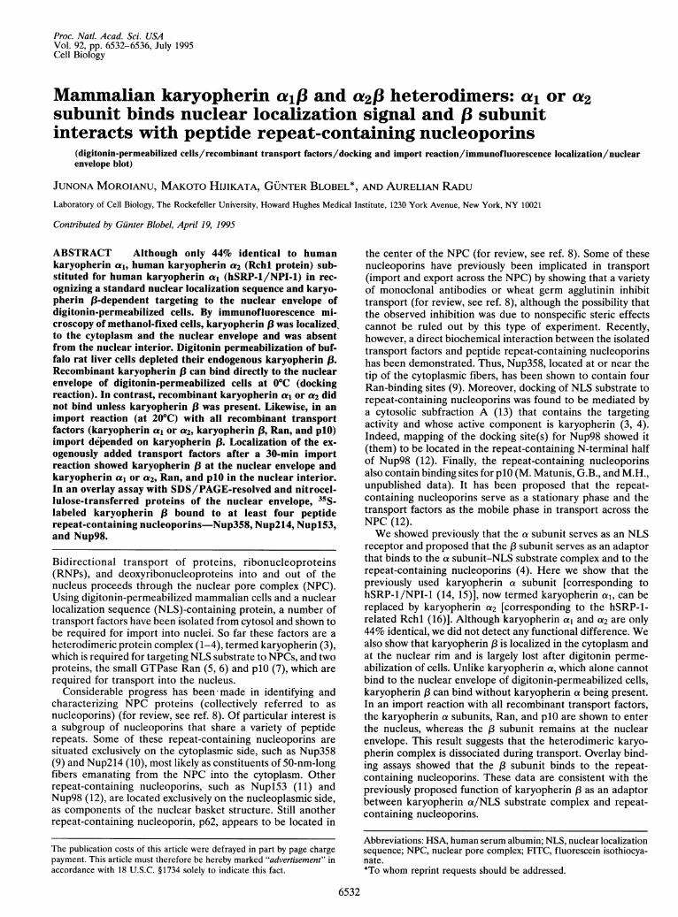
Karyopherin a2 Substitutes for Karyopherin ae. To testwhether human karyopherin a2 (Rchl) can substitute forkaryopherin a1 (hSRP-1/NPI-1) in NLS substrate binding, aswell as in the targeting and import reactions in digitonin-permeabilized cells, we purified recombinant karyopherin a2(lacking 33 N-terminal residues and containing a C-terminalhistidine tag) from E. coli (Fig. 1, lane 1). We also raised rabbitantibodies against a synthetic peptide corresponding to theC-terminal region of human karyopherin a2. The antipeptideantiserum reacted with recombinant human karyopherin a2(Fig. 1, lane 2) but did not react with its rat counterpart in BRLcells (data not shown). As previously demonstrated for recom-binant karyopherin a1 (4), the recombinant karyopherin a2bound import substrate containing a wild-type NLS (Fig. 1,lane 3) but not a mutant NLS-containing substrate (Fig. 1, lane4).As reported (4) for recombinant karyopherin ai in a tar-
geting reaction in digitonin-permeabilized cells, the recombi-nant karyopherin a2 subunit by itself does not target NLSsubstrate to the nuclear envelope (Fig. 24, a) but does so onlyin the presence of recombinant karyopherin ( (Fig. 24, b).
A B C
...... ::...... .. ....
.w......... ..
31- .. ..
:R .... ::...:..1-,. ........
1 2 3 4
FIG. 1. Karyopherin a2 binds the NLS-HSA import substrate. (A)Coomassie blue staining of recombinant karyopherin a2. (B) Immu-noblotting with anti-karyopherin a2 antibody of recombinant karyo-pherin a2. (C) Karyopherin a2 (200 ng) was subjected to SDS/PAGEand transferred to nitrocellulose; the strips were then incubated withwild-type NLS-HSA (lane 3) or mutant NLS-HSA (lane 4) (13).Ligand binding was detected by incubation with anti-HSA antibodies,125I-labeled protein A, and autoradiography (4). Mr markers (X 10-3)are indicated at left.
Cell Biology: Moroianu et al.
6534 Cell Biology: Moroianu et al.
A (x) + [ aniti-[
B (X2 + Riii + pO c2+ 13+ Ran + pl()
FIG. 2. Karyopherin a2 substitutes for karyopherin a, in karyo-pherin 13-dependent docking and import. (A) Docking. Digitonin-permeabilized cells were incubated for 30 min on ice with importsubstrate in the presence of 100 nM karyopherin a2 (a) or 100 nMkaryopherin a2 plus 100 nM karyopherin 13 (b). (Bar = 10 ,um.) (B)Nuclear import. Digitonin-permeabilized cells were incubated for 30min at room temperature in the presence of Ran at 100 ,ug/ml, plO at3 ,ug/ml (7), and either 100 nM karyopherin a2 alone (a) or togetherwith 100 nM karyopherin 13 (b). (Bar = 10 ,xm.)
Likewise, recombinant karyopherin a2 can substitute for re-combinant karyopherin a1 in an import reaction into digitonin-permeabilized BRL cells using recombinant 1, Ran, and plO(Fig. 2B, b). When recombinant 1 was omitted, there was noimport (Fig. 2B, a). We conclude that recombinant karyo-pherin a2 can substitute for karyopherin a1 in NLS substratebinding; karyopherin 13-dependent targeting; and karyopherin13-, Ran-, and p1O-dependent import.Karyopherin .3 Binds to the Nuclear Envelope. We immu-
nized rabbits with recombinant rat karyopherin 13 and ob-tained an antiserum that reacted with the antigen (Fig. 3, lane1). In indirect immunofluorescence of cold methanol-fixedBRL cells with anti-karyopherin 13 antibodies we observedsome staining in the cytoplasm and strong staining at thenuclear envelope (Fig. 4, a). When the BRL cells were firstpermeabilized by digitonin, the cytoplasmic and nuclear rimstaining was lost (Fig. 4b), suggesting that digitonin-permeabilized cells retain little, if any, of the endogenouskaryopherin 1. This result was confirmed when blots ofSDS/PAGE-resolved proteins of digitonin-permeabilized cells(Fig. 3, lane 2) and of the digitonin extract (Fig. 3, lane 3) were
anti-1.. ..... ......
97- !S.................
66- ... i.
45-
31-
21-14-
1 2 3
FIG. 3. Karyopherin 1 is extracted from BRL cells by digitoninpermeabilization. Recombinant rat karyopherin 13 (lane 1), digitonin-extracted BRL cells (lane 2), and the digitonin extract (lane 3) wereseparated by SDS/PAGE, transferred to nitrocellulose, and incubatedwith anti-karyopherin 13 antibodies (1:100 dilution) followed by 1251-labeled protein A. Molecular mass markers in kDa are indicated at left.
FIG. 4. Karyopherin 13 localized at the nuclear envelope and in thecytoplasm is lost during digitonin permeabilization. BRL cells inculture were fixed with methanol before (a) or after digitonin per-meabilization (b) and immunostained with anti-karyopherin 13 anti-bodies followed by FITC-labeled goat anti-rabbit antibodies. (Bar = 10ILm.)
probed with anti-karyopherin 13 antibodies. The extraction bydigitonin of endogenous karyopherin 13 explains the require-ment for exogenous karyopherin 13 in both the docking andimport reaction in digitonin-permeabilized cells. It should benoted here that digitonin permeabilization of BRL cells alsoleads to extraction of karyopherin a1 and a2 (data not shown)from the cytoplasm, consistent with the finding that each ofthese subunits forms a heterodimeric complex with karyo-pherin ,1. However, nuclear karyopherin a1 (19) and a2 (datanot shown) are not extracted by digitonin.We had previously proposed that the 03 subunit of the
karyopherin heterodimer functions as an adaptor, binding tokaryopherin a and to the NPC (4). To test this hypothesis weincubated digitonin-permeabilized cells with either recombi-
a'
anti- ai
anti- (X
al + D
CO
a1 + [
anti- P
FIG. 5. Karyopherin 13 functions as an adaptor between karyo-pherin a/import substrate complex and the NPC. Digitonin-permeabilized cells were incubated with the import substrate in thepresence of either karyopherin a, alone (a), karyopherin a2 alone (c),karyopherin 13 alone (e), karyopherin a, and 13 (b and f), or karyo-pherin a2 and 13 (d). After the docking reaction the cells were washedin transport buffer, fixed with methanol, and immunostained withanti-karyopherin at antibodies (a and b), anti-karyopherin a2 anti-bodies (c and d), or anti-karyopherin 13 antibodies (e and f). (Bar =10 ,um.)
Proc. Natl. Acad. Sci. USA 92 (1995)
I
Proc. Natl. Acad. Sci. USA 92 (1995) 6535
_- Nup358-- Nup214
_ -2(H)-Nup153
-116
+ Nup98
-66
-45
1 2
FIG. 6. Karyopherin ,B binds to peptide repeat-containing nucleo-porins. SDS/PAGE-resolved proteins of rat liver nuclear envelopeswere transferred to nitrocellulose and incubated with either 35S-labeled recombinant karyopherin f3 (detected by radioautography)(lane 1) or the import substrate NLS-HSA in the presence ofXenopuscytosolic fraction A (12) (lane 2). The import substrate was detectedby anti-HSA as described (3). The upper band in lane 2, previouslyreferred to as p270 (3), was characterized as nucleoporin Nup358 (9).Arrows point to nucleoporins labeled in both lanes. Molecular massmarkers in kDa are indicated at right.
nant karyopherin a,, a2, or ( subunit alone or with one of thetwo a subunits and the (3 subunit and then used indirectimmunofluorescence with monospecific antibodies to detectbinding (Fig. 5). Consistent with our proposal we found thatneither karyopherin a, (Fig. Sa) nor karyopherin a2 (Fig. Sc)bound by itself to the nuclear envelope. However, both karyo-pherin a, (Fig. Sb) and karyopherin a2 (Fig. Sd) bound whenincubated in the presence of karyopherin (3. Most importantly,karyopherin (3 could bind by itself (Fig. Se).Karyopherin f3 Binds to Peptide Repeat-Containing
Nucleoporins. We have previously shown in a blot assay that
anti-(x I
an1t -(X-
anti-Ran
the cytosolic subfraction A mediates binding of NLS substrateto peptide repeat-containing nucleoporins present in a rat livernuclear envelope fraction (3, 12) (Fig. 6, lane 2). When anidentical blot was probed with 35S-labeled recombinant karyo-pherin (3, we found that similar nucleoporins (Nup358,Nup214, Nupl53, Nup98, and perhaps p62) were decorated(Fig. 6, lane 1). We conclude that karyopherin 83 binds topeptide repeat-containing nucleoporins on both the cytoplas-mic (Nup358, Nup214) and nucleoplasmic side (Nupl53,Nup98).Karyopherin 13 Does Not Enter Nucleoplasm, Whereas
Karyopherin a, Ran, and plO Do. We performed an importreaction into nuclei of digitonin-permeabilized BRL cells withan NLS substrate and recombinant transport factors and thenlocalized each of the exogenously added transport factors byfluorescence microscopy (Fig. 7). Both karyopherin al and a2entered the nucleus (Fig. 7 a and c). So did Ran (Fig. 7e) andFITC-labeled plO (Fig. 7f) and, of course, the import substrate(Fig. 7d). The only transport factor that did not enter thenucleus was karyopherin (3 (Fig. 7b). This result suggested thatduring import the karyopherin heterodimer was dissociated.
DISCUSSIONOur data here indicate that recombinant karyopherin a2[corresponding to Rchl (16)] can functionally substitute forrecombinant karyopherin a1 [corresponding to hSRP/NPI-1(14, 15)]. Either of these two proteins bound an NLS substrate(ref. 4 and Fig. 1); either of them, together with karyopherin(3, docked the NLS substrate to the nuclear envelope ofdigitonin-permeabilized cells; and either of them imported theNLS substrate into nuclei of digitonin-permeabilized cells inthe presence of recombinant karyopherin (3, recombinant Ran,and recombinant plO. Thus, although human karyopherin a,and a2 are only 44% identical, they appear functionally indis-tinguishable. However, there are probably differences betweenkaryopherin a, and a2-e.g., in the recognition of NLSs and
anti-5
importsubstrate
FITC-plIO
FIG. 7. Karyopherin al/a2, Ran, and plO enter the nucleus, whereas karyopherin 13 remains at the nuclear envelope during nuclear import ofthe substrate. The import assay was done in digitonin-permeabilized cells in the presence of karyopherin atl (100 nM), karyopherin (3 (100 nM),Ran (100 ,ug/ml), and either plO, at 3 ,ug/ml (a, b, c, d, and e) or FITC-plO (f). In c, karyopherin a, was substituted by karyopherin a2. Afterthe import reaction the cells were washed, fixed with methanol, and probed either with anti-karyopherin a, antibodies (a), with anti-karyopherin(3 antibodies (b), with anti-karyopherin a2 antibodies (c), or with anti-Ran antibodies (e) and stained with FITC-labeled goat anti-rabbit IgG ordirectly examined for rhodamine-labeled import substrate (d) or FITC-labeled plO (f). (Bar = 10 ,um.)
Cell Biology: Moroianu et al.
6536 Cell Biology: Moroianu et al.
surrounding structures or in other parameters that our assayshave not detected. An important result is that karyopherin ,Bbinds directly to a subgroup of nucleoporins that share variouspeptide repeats and that are located on the cytoplasmic side(Nup358, Nup214) and the nucleoplasmic side (Nup98,Nupl53). This result indicates a division of labor; the a subunitof the karyopherin heterodimer serves in NLS recognition,and the ,B subunit mediates docking to the peptide repeat-containing nucleoporins. Most interesting is the finding thatthe karyopherin heterodimer dissociates at some point duringtransport; karyopherin a enters the nucleus and is retainedthere, whereas karyopherin ,3 either does not enter the nucle-oplasm or enters but is not retained there, being rapidlyexported back into the cytoplasm (19). Immunofluorescencelocalization with anti-karyopherin ,3 antibodies showed pri-marily cytoplasmic staining and a strong nuclear rim stainingbut no significant intranuclear staining, arguing against anintranuclear presence of karyopherin ,B. However, this cellulardistribution may simply result from kinetic partitioning ofkaryopherin (3between a short intranuclear transport phase, alonger transport phase across the NPC (in both directions),and a cytoplasmic pool.The retention of either karyopherin ai or a2 in the nucleus,
even after digitonin permeabilization (ref. 19; data not shownfor a2), is another point of interest. Is intranuclear karyopherina1 or a2 still docked to intranuclear sites (20), and is dockingmediated by a karyopherin P-like protein? There are severalproteins in GenBank that show some homology to karyopherin(3. Although the function and location of these proteins areunknown, they might function in nuclear retention of variouskaryopherin a subunits and/or transport out of the nucleus.
We are greatly indebted to Robert E. O'Neill and Peter Palese fortheir generous gift of antibodies against karyopherin al, karyopherin
al-expressing vector pSE420, and the cDNA for Rchl and to PhilipBernstein for recombinant human Ran.
1. Adam, E. J. H. & Adam, S. A. (1994) J. Cell Bio. 125, 547-555.2. Gorlich, D., Prehn, S., Laskey, R. A. & Hartman, E. (1994) Cell
79, 767-778.3. Radu, A., Blobel, G. & Moore, M. S. (1995) Proc. Natl. Acad. Sci.
USA 92, 1769-1773.4. Moroianu, J., Blobel, G. & Radu, A. (1995) Proc. Natl. Acad. Sci.
USA 92, 2008-2011.5. Moore, M. S. & Blobel, G. (1993) Nature (London) 365,661-663.6. Melchior, F. B., Paschal, J., Evans, J. & Gerace, L. (1993) J. Cell
Biol. 123, 1649-1659.7. Moore, M. S. & Blobel, G. (1994) Proc. Natl. Acad. Sci. USA 91,
10212-10216.8. Rout, M. P. & Wente, S. R. (1994) Trends Biochem. Sci. 4,
357-365.9. Wu, J., Matunis, M., Kraemer, D., Blobel, G. & Coutavas, E.
(1995) J. Biol. Chem., in press.10. Kraemer, D., Wozniak, R. W., Blobel, G. & Radu, A. (1994)
Proc. Natl. Acad. Sci. USA 91, 1519-1523.11. Sukegawa, J. & Blobel, G. (1993) Cell 72, 29-38.12. Radu, A., Moore, M. S. & Blobel, G. (1995) Cell 81, 1-8.13. Moore, M. S. & Blobel, G. (1992) Cell 69, 939-950.14. Cortes, P., Ye, Z. S. & Baltimore, D. (1994) Proc. Natl. Acad. Sci.
USA 91, 7633-7637.15. O'Neill, R. E. & Palese, P. (1994) Virology 206, 116-125.16. Cuomo, C. A., Kirch, S. A., Gyuris, J., Brent, R. & Oettinger,
M. A. (1994) Proc. Natl. Acad. Sci. USA 91, 6156-6160.17. Coutavas, E., Ren, M., Oppenheim, J. D., D'Eustachio, P. &
Rush, M. G. (1993) Nature (London) 366, 585-587.18. Grundmann, U., Nerlich, C., Rein, T., Lottspeich, F. & Kupper,
H. A. (1988) Nucleic Acids Res. 16, 4721.19. Moroianu, J. & Blobel, G. (1995) Proc. Natl. Acad. Sci. USA 92,
4318-4322.20. Meier, U. T. & Blobel, G. (1992) Cell 70, 127-138.