Manadosterols A and B, Sulfonated Sterol Dimers Inhibiting theUbc13−Uev1A Interaction, Isolated from the Marine SpongeLissodendryx f ibrosaShuntaro Ushiyama,† Hideharu Umaoka,† Hikaru Kato,† Yoshiaki Suwa,† Hiroshi Morioka,†
Henki Rotinsulu,‡,§ Fitje Losung,⊥ Remy E. P. Mangindaan,⊥ Nicole J. de Voogd,∥ Hideyoshi Yokosawa,∇
and Sachiko Tsukamoto*,†
†Graduate School of Pharmaceutical Sciences, Kumamoto University, Kumamoto 862-0973, Japan‡Tohoku Pharmaceutical University, Aoba-ku, Sendai 981-8558, Japan§Faculty of Agriculture, Universitas Pembangunan Indonesia, Manado 95361, Indonesia⊥Faculty of Fisheries and Marine Science, Sam Ratulangi University, Kampus Bahu, Manado 95115, Indonesia∥Netherlands Centre for Biodiversity Naturalis, PO Box 9517, 2300 RA Leiden, The Netherlands∇School of Pharmacy, Aichi Gakuin University, Chikusa-ku, Nagoya 464-8650, Japan
*S Supporting Information
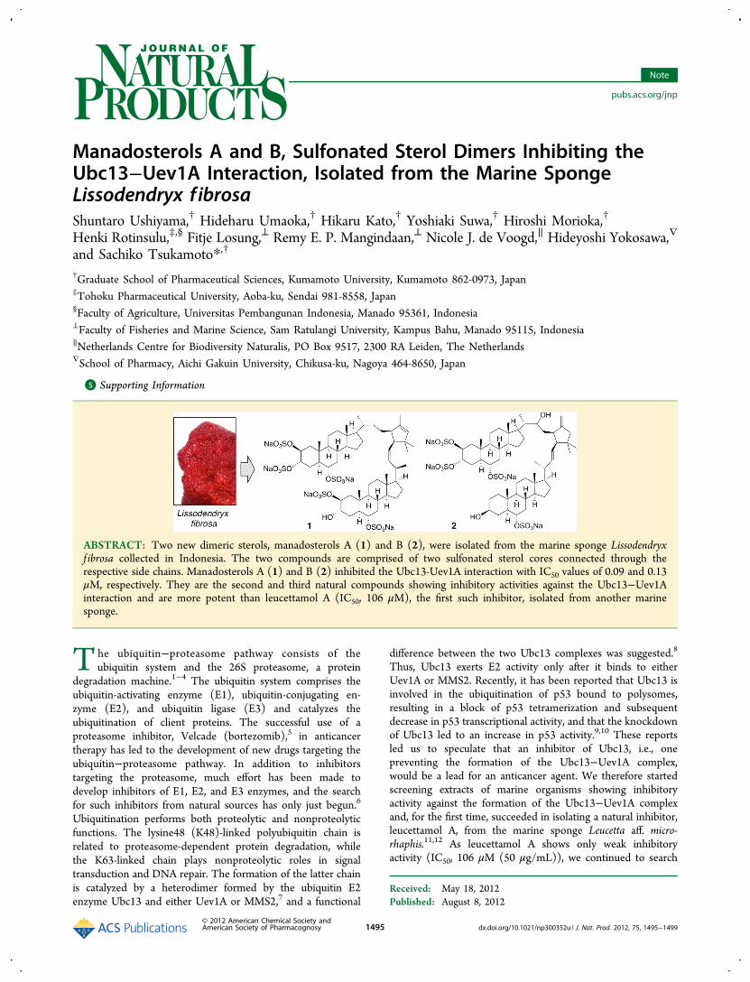
ABSTRACT: Two new dimeric sterols, manadosterols A (1) and B (2), were isolated from the marine sponge Lissodendryxf ibrosa collected in Indonesia. The two compounds are comprised of two sulfonated sterol cores connected through therespective side chains. Manadosterols A (1) and B (2) inhibited the Ubc13-Uev1A interaction with IC50 values of 0.09 and 0.13μM, respectively. They are the second and third natural compounds showing inhibitory activities against the Ubc13−Uev1Ainteraction and are more potent than leucettamol A (IC50, 106 μM), the first such inhibitor, isolated from another marinesponge.
The ubiquitin−proteasome pathway consists of theubiquitin system and the 26S proteasome, a protein
degradation machine.1−4 The ubiquitin system comprises theubiquitin-activating enzyme (E1), ubiquitin-conjugating en-zyme (E2), and ubiquitin ligase (E3) and catalyzes theubiquitination of client proteins. The successful use of aproteasome inhibitor, Velcade (bortezomib),5 in anticancertherapy has led to the development of new drugs targeting theubiquitin−proteasome pathway. In addition to inhibitorstargeting the proteasome, much effort has been made todevelop inhibitors of E1, E2, and E3 enzymes, and the searchfor such inhibitors from natural sources has only just begun.6
Ubiquitination performs both proteolytic and nonproteolyticfunctions. The lysine48 (K48)-linked polyubiquitin chain isrelated to proteasome-dependent protein degradation, whilethe K63-linked chain plays nonproteolytic roles in signaltransduction and DNA repair. The formation of the latter chainis catalyzed by a heterodimer formed by the ubiquitin E2enzyme Ubc13 and either Uev1A or MMS2,7 and a functional
difference between the two Ubc13 complexes was suggested.8
Thus, Ubc13 exerts E2 activity only after it binds to eitherUev1A or MMS2. Recently, it has been reported that Ubc13 isinvolved in the ubiquitination of p53 bound to polysomes,resulting in a block of p53 tetramerization and subsequentdecrease in p53 transcriptional activity, and that the knockdownof Ubc13 led to an increase in p53 activity.9,10 These reportsled us to speculate that an inhibitor of Ubc13, i.e., onepreventing the formation of the Ubc13−Uev1A complex,would be a lead for an anticancer agent. We therefore startedscreening extracts of marine organisms showing inhibitoryactivity against the formation of the Ubc13−Uev1A complexand, for the first time, succeeded in isolating a natural inhibitor,leucettamol A, from the marine sponge Leucetta aff. micro-rhaphis.11,12 As leucettamol A shows only weak inhibitoryactivity (IC50, 106 μM (50 μg/mL)), we continued to search
for more potent inhibitors and encountered an active extract ofthe marine sponge Lissodendryx f ibrosa collected in Indonesia.Here, we report the isolation and structure determination oftwo new dimeric sterols, manadosterols A (1) and B (2), alongwith inhibitory activities against the formation of the Ubc13−Uev1A complex.
Specimens of L. f ibrosa were collected in Indonesia. TheEtOH extract (7.3 g) of the sponge was evaporated, and theaqueous residue was extracted with EtOAc and then n-BuOH.The residual H2O and n-BuOH fractions, which showedinhibitory activity against the formation of the Ubc13−Uev1Acomplex, were subjected to ODS column chromatography andODS HPLC to afford manadosterols A (1) (1.4 mg, 0.0047%,wet weight) and B (2) (7.2 mg, 0.024%), respectively.ESIMS of manadosterol A (1) showed a quasi molecular ion
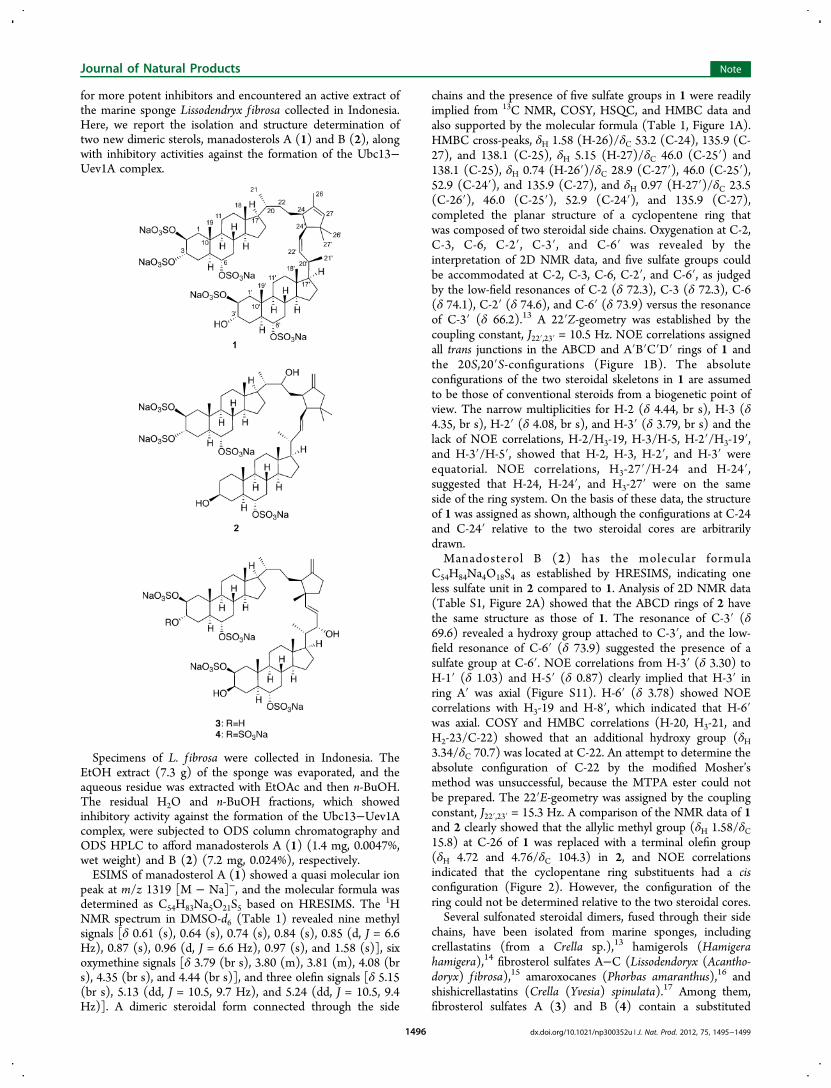
peak at m/z 1319 [M − Na]−, and the molecular formula wasdetermined as C54H83Na5O21S5 based on HRESIMS. The 1HNMR spectrum in DMSO-d6 (Table 1) revealed nine methylsignals [δ 0.61 (s), 0.64 (s), 0.74 (s), 0.84 (s), 0.85 (d, J = 6.6Hz), 0.87 (s), 0.96 (d, J = 6.6 Hz), 0.97 (s), and 1.58 (s)], sixoxymethine signals [δ 3.79 (br s), 3.80 (m), 3.81 (m), 4.08 (brs), 4.35 (br s), and 4.44 (br s)], and three olefin signals [δ 5.15(br s), 5.13 (dd, J = 10.5, 9.7 Hz), and 5.24 (dd, J = 10.5, 9.4Hz)]. A dimeric steroidal form connected through the side
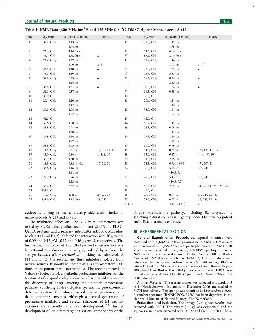
chains and the presence of five sulfate groups in 1 were readilyimplied from 13C NMR, COSY, HSQC, and HMBC data andalso supported by the molecular formula (Table 1, Figure 1A).HMBC cross-peaks, δH 1.58 (H-26)/δC 53.2 (C-24), 135.9 (C-27), and 138.1 (C-25), δH 5.15 (H-27)/δC 46.0 (C-25′) and138.1 (C-25), δH 0.74 (H-26′)/δC 28.9 (C-27′), 46.0 (C-25′),52.9 (C-24′), and 135.9 (C-27), and δH 0.97 (H-27′)/δC 23.5(C-26′), 46.0 (C-25′), 52.9 (C-24′), and 135.9 (C-27),completed the planar structure of a cyclopentene ring thatwas composed of two steroidal side chains. Oxygenation at C-2,C-3, C-6, C-2′, C-3′, and C-6′ was revealed by theinterpretation of 2D NMR data, and five sulfate groups couldbe accommodated at C-2, C-3, C-6, C-2′, and C-6′, as judgedby the low-field resonances of C-2 (δ 72.3), C-3 (δ 72.3), C-6(δ 74.1), C-2′ (δ 74.6), and C-6′ (δ 73.9) versus the resonanceof C-3′ (δ 66.2).13 A 22′Z-geometry was established by thecoupling constant, J22′,23′ = 10.5 Hz. NOE correlations assignedall trans junctions in the ABCD and A′B′C′D′ rings of 1 andthe 20S,20′S-configurations (Figure 1B). The absoluteconfigurations of the two steroidal skeletons in 1 are assumedto be those of conventional steroids from a biogenetic point ofview. The narrow multiplicities for H-2 (δ 4.44, br s), H-3 (δ4.35, br s), H-2′ (δ 4.08, br s), and H-3′ (δ 3.79, br s) and thelack of NOE correlations, H-2/H3-19, H-3/H-5, H-2′/H3-19′,and H-3′/H-5′, showed that H-2, H-3, H-2′, and H-3′ wereequatorial. NOE correlations, H3-27′/H-24 and H-24′,suggested that H-24, H-24′, and H3-27′ were on the sameside of the ring system. On the basis of these data, the structureof 1 was assigned as shown, although the configurations at C-24and C-24′ relative to the two steroidal cores are arbitrarilydrawn.Manadosterol B (2) has the molecular formula
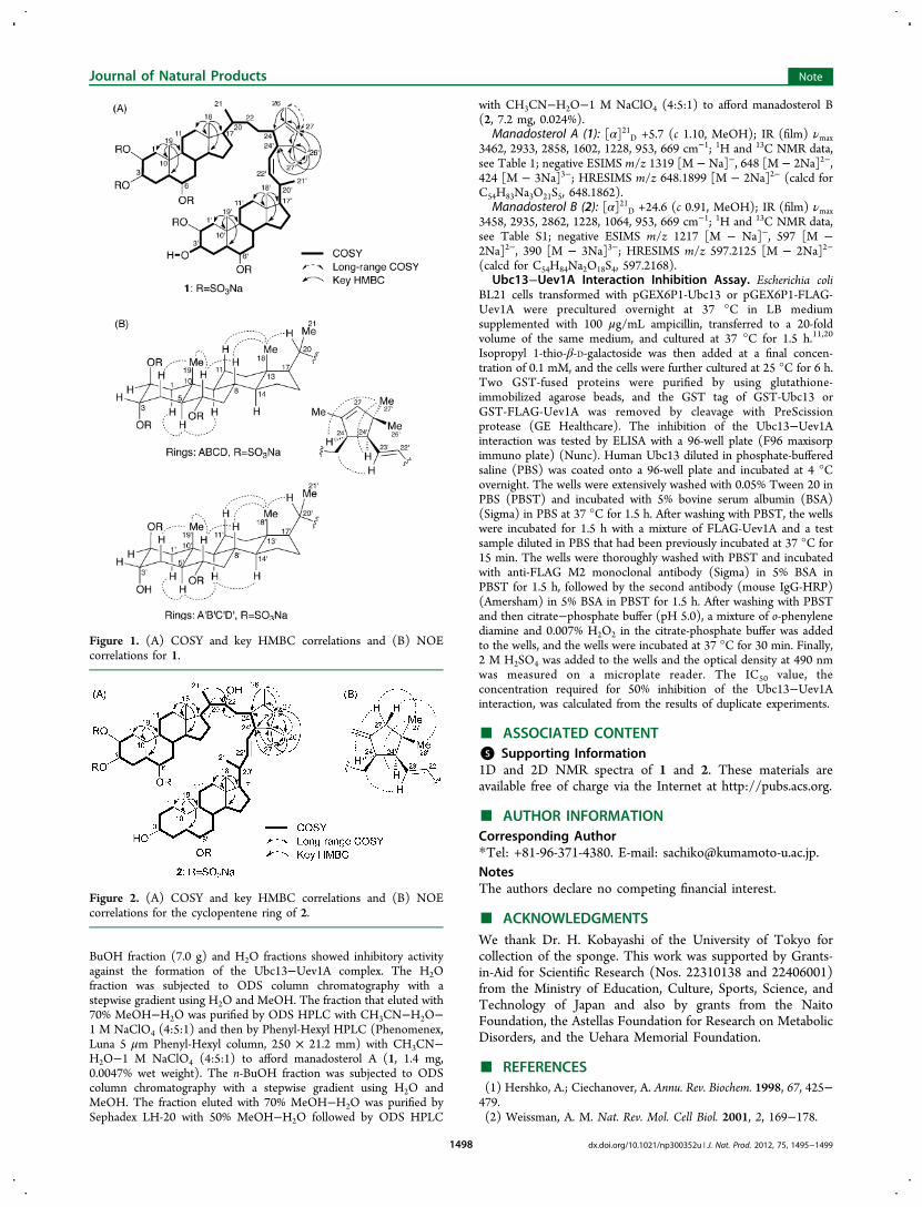
C54H84Na4O18S4 as established by HRESIMS, indicating oneless sulfate unit in 2 compared to 1. Analysis of 2D NMR data(Table S1, Figure 2A) showed that the ABCD rings of 2 havethe same structure as those of 1. The resonance of C-3′ (δ69.6) revealed a hydroxy group attached to C-3′, and the low-field resonance of C-6′ (δ 73.9) suggested the presence of asulfate group at C-6′. NOE correlations from H-3′ (δ 3.30) toH-1′ (δ 1.03) and H-5′ (δ 0.87) clearly implied that H-3′ inring A′ was axial (Figure S11). H-6′ (δ 3.78) showed NOEcorrelations with H3-19 and H-8′, which indicated that H-6′was axial. COSY and HMBC correlations (H-20, H3-21, andH2-23/C-22) showed that an additional hydroxy group (δH3.34/δC 70.7) was located at C-22. An attempt to determine theabsolute configuration of C-22 by the modified Mosher’smethod was unsuccessful, because the MTPA ester could notbe prepared. The 22′E-geometry was assigned by the couplingconstant, J22′,23′ = 15.3 Hz. A comparison of the NMR data of 1and 2 clearly showed that the allylic methyl group (δH 1.58/δC15.8) at C-26 of 1 was replaced with a terminal olefin group(δH 4.72 and 4.76/δC 104.3) in 2, and NOE correlationsindicated that the cyclopentane ring substituents had a cisconfiguration (Figure 2). However, the configuration of thering could not be determined relative to the two steroidal cores.Several sulfonated steroidal dimers, fused through their side
chains, have been isolated from marine sponges, includingcrellastatins (from a Crella sp.),13 hamigerols (Hamigerahamigera),14 fibrosterol sulfates A−C (Lissodendoryx (Acantho-doryx) f ibrosa),15 amaroxocanes (Phorbas amaranthus),16 andshishicrellastatins (Crella (Yvesia) spinulata).17 Among them,fibrosterol sulfates A (3) and B (4) contain a substituted
Journal of Natural Products Note
dx.doi.org/10.1021/np300352u | J. Nat. Prod. 2012, 75, 1495−14991496
cyclopentane ring in the connecting side chain similar tomanadosterols A (1) and B (2).The inhibitory effect on Ubc13−Uev1A interactions was
tested by ELISA using purified recombinant Ubc13 and FLAG-Uev1A proteins and a primary anti-FLAG antibody. Manados-terols A (1) and B (2) inhibited the interaction with IC50 valuesof 0.09 and 0.13 μM (0.12 and 0.16 μg/mL), respectively. Thefirst natural inhibitor of the Ubc13−Uev1A interaction wasleucettamol A, a dimeric sphingolipid, isolated by us from thesponge Leucetta aff. microrhaphis,11 making manadosterols A(1) and B (2) the second and third inhibitors isolated fromnatural sources. It should be noted that 1 is approximately 1000times more potent than leucettamol A. The recent approval ofVelcade (bortezomib), a synthetic proteasome inhibitor, for thetreatment of relapsed multiple myeloma has opened the way tothe discovery of drugs targeting the ubiquitin−proteasomepathway, consisting of the ubiquitin system, the proteasome, adelivery system for ubiquitinated client proteins, anddeubiquitinating enzymes. Although a second generation ofproteasome inhibitors and several inhibitors of E1 and E3enzymes are currently in clinical development,18,19 furtherdevelopment of inhibitors targeting various components of the
ubiquitin−proteasome pathway, including E2 enzymes, bysearching natural sources is urgently needed to develop potentand efficient anticancer drugs.
■ EXPERIMENTAL SECTIONGeneral Experimental Procedures. Optical rotations were
measured with a JASCO P-1020 polarimeter in MeOH. UV spectrawere measured on a JASCO V-550 spectrophotometer in MeOH. IRspectra were measured on a JEOL JIR-6500W spectrophotometer.NMR spectra were recorded on a Bruker Avance 500 or BrukerAvance 600 NMR spectrometer in DMSO-d6. Chemical shifts werereferenced to the residual solvent peaks (δH 2.49 and δC 39.5) asinternal standards. Mass spectra were measured on a Bruker Esquire3000plus-K1 or Bruker BioTOF-Q mass spectrometer. HPLC wascarried out on a Waters 515 HPLC pump and a Waters 2489 UV/visible detector.
Animal Material. The marine sponge was collected at a depth of 5m in North Sulawesi, Indonesia, in December 2006 and soaked inEtOH immediately. The sponge was identified as Lissodendryx f ibrosa.A voucher specimen (RMNH POR. 3988) has been deposited in theNational Museum of Natural History, The Netherlands.
Extraction and Isolation. The sponge (300 g, wet weight) wasextracted with EtOH. The extract (7.3 g) was evaporated, and theaqueous residue was extracted with EtOAc and then n-BuOH. The n-
Table 1. NMR Data (500 MHz for 1H and 125 MHz for 13C, DMSO-d6) for Manadosterol A (1)
no. δC, mult. δH, mult. (J in Hz) HMBC no. δC, mult. δH, mult. (J in Hz) HMBC
1 39.2, CH2 1.13, m 1′ 37.3, CH2 1.15, m1.74, m 1.88, m
2 72.3, CH 4.44, br s 2′ 74.6, CH 4.08, br s3 72.3, CH 4.35, br s 2 3′ 66.2, CH 3.79, br s4 23.9, CH2 1.37, m 4′ 27.9, CH2 1.44, m
1.90, m 2, 3 1.77, m 2′, 3′5 43.1, CH 1.40, m 6 5′ 43.8, CH 1.33, m 6′6 74.1, CH 3.80, m 6′ 73.9, CH 3.81, m7 39.2, CH2 0.73, m 6 7′ 39.2, CH2 0.76, m 6′
2.14, m 2.10, m8 33.5, CH 1.31, m 6 8′ 33.5, CH 1.32, m 6′9 54.1, CH 0.57, m 9′ 54.3, CH 0.58, m10 38.0, C 10′ 36.0, C11 20.4, CH2 1.20, m 11′ 20.4, CH2 1.14, m
1.42, m 1.40, m12 39.1, CH2 1.04, m 12′ 38.9, CH2 1.04, m
1.92, m 1.92, m13 42.1, C 13′ 42.0, C14 55.6, CH 1.08, m 14′ 55.7, CH 1.15, m15 23.8, CH2 0.98, m 15′ 23.8, CH2 0.98, m
1.38, m 1.41, m16 27.9, CH2 1.24, m 16′ 27.9, CH2 1.24, m
1.77, m 1.77, m17 55.9, CH 1.03, m 17′ 56.0, CH 0.99, m18 11.8, CH3 0.61, s 12, 13, 14, 17 18′ 11.2, CH3 0.64, s 12′, 13′, 14′, 17′19 15.0, CH3 0.84, s 1, 5, 9, 10 19′ 15.0, CH3 0.87, s 1′, 5′, 9′, 10′20 35.9, CH 1.30, m 20′ 34.0, CH 2.36, m21 18.3, CH3 0.85, d (6.6) 17, 20, 22 21′ 21.5, CH3 0.96, d (6.6) 17′, 20′, 22′22 32.6, CH2 1.16, m 22′ 138.0, CH 5.24, dd 20′, 24′
1.41, m (10.5, 9.4)23 29.9, CH2 0.96, m 23′ 127.8, CH 5.13, dd 20′, 24′
1.23, m (10.5, 9.7)24 53.2, CH 2.27, m 24′ 52.9, CH 2.38, m 24, 25, 23′, 25′, 26′, 27′25 138.1, C 25′ 46.0, C26 15.0, CH3 1.58, s 24, 25, 27 26′ 23.5, CH3 0.74, s 27, 24′, 25′, 27′27 135.9, CH 5.15, br s 25, 25′ 27′ 28.9, CH3 0.97, s 27, 24′, 25′, 26′
3′-OH 4.47, d (3.6) 2′
Journal of Natural Products Note
dx.doi.org/10.1021/np300352u | J. Nat. Prod. 2012, 75, 1495−14991497
BuOH fraction (7.0 g) and H2O fractions showed inhibitory activityagainst the formation of the Ubc13−Uev1A complex. The H2Ofraction was subjected to ODS column chromatography with astepwise gradient using H2O and MeOH. The fraction that eluted with70% MeOH−H2O was purified by ODS HPLC with CH3CN−H2O−1 M NaClO4 (4:5:1) and then by Phenyl-Hexyl HPLC (Phenomenex,Luna 5 μm Phenyl-Hexyl column, 250 × 21.2 mm) with CH3CN−H2O−1 M NaClO4 (4:5:1) to afford manadosterol A (1, 1.4 mg,0.0047% wet weight). The n-BuOH fraction was subjected to ODScolumn chromatography with a stepwise gradient using H2O andMeOH. The fraction eluted with 70% MeOH−H2O was purified bySephadex LH-20 with 50% MeOH−H2O followed by ODS HPLC
with CH3CN−H2O−1 M NaClO4 (4:5:1) to afford manadosterol B(2, 7.2 mg, 0.024%).
(calcd for C54H84Na2O18S4, 597.2168).Ubc13−Uev1A Interaction Inhibition Assay. Escherichia coli
BL21 cells transformed with pGEX6P1-Ubc13 or pGEX6P1-FLAG-Uev1A were precultured overnight at 37 °C in LB mediumsupplemented with 100 μg/mL ampicillin, transferred to a 20-foldvolume of the same medium, and cultured at 37 °C for 1.5 h.11,20
Isopropyl 1-thio-β-D-galactoside was then added at a final concen-tration of 0.1 mM, and the cells were further cultured at 25 °C for 6 h.Two GST-fused proteins were purified by using glutathione-immobilized agarose beads, and the GST tag of GST-Ubc13 orGST-FLAG-Uev1A was removed by cleavage with PreScissionprotease (GE Healthcare). The inhibition of the Ubc13−Uev1Ainteraction was tested by ELISA with a 96-well plate (F96 maxisorpimmuno plate) (Nunc). Human Ubc13 diluted in phosphate-bufferedsaline (PBS) was coated onto a 96-well plate and incubated at 4 °Covernight. The wells were extensively washed with 0.05% Tween 20 inPBS (PBST) and incubated with 5% bovine serum albumin (BSA)(Sigma) in PBS at 37 °C for 1.5 h. After washing with PBST, the wellswere incubated for 1.5 h with a mixture of FLAG-Uev1A and a testsample diluted in PBS that had been previously incubated at 37 °C for15 min. The wells were thoroughly washed with PBST and incubatedwith anti-FLAG M2 monoclonal antibody (Sigma) in 5% BSA inPBST for 1.5 h, followed by the second antibody (mouse IgG-HRP)(Amersham) in 5% BSA in PBST for 1.5 h. After washing with PBSTand then citrate−phosphate buffer (pH 5.0), a mixture of o-phenylenediamine and 0.007% H2O2 in the citrate-phosphate buffer was addedto the wells, and the wells were incubated at 37 °C for 30 min. Finally,2 M H2SO4 was added to the wells and the optical density at 490 nmwas measured on a microplate reader. The IC50 value, theconcentration required for 50% inhibition of the Ubc13−Uev1Ainteraction, was calculated from the results of duplicate experiments.
■ ASSOCIATED CONTENT*S Supporting Information1D and 2D NMR spectra of 1 and 2. These materials areavailable free of charge via the Internet at http://pubs.acs.org.
■ ACKNOWLEDGMENTSWe thank Dr. H. Kobayashi of the University of Tokyo forcollection of the sponge. This work was supported by Grants-in-Aid for Scientific Research (Nos. 22310138 and 22406001)from the Ministry of Education, Culture, Sports, Science, andTechnology of Japan and also by grants from the NaitoFoundation, the Astellas Foundation for Research on MetabolicDisorders, and the Uehara Memorial Foundation.
■ REFERENCES(1) Hershko, A.; Ciechanover, A. Annu. Rev. Biochem. 1998, 67, 425−479.(2) Weissman, A. M. Nat. Rev. Mol. Cell Biol. 2001, 2, 169−178.
Figure 1. (A) COSY and key HMBC correlations and (B) NOEcorrelations for 1.
Figure 2. (A) COSY and key HMBC correlations and (B) NOEcorrelations for the cyclopentene ring of 2.
Journal of Natural Products Note
dx.doi.org/10.1021/np300352u | J. Nat. Prod. 2012, 75, 1495−14991498
(3) Pickart, C. M. Annu. Rev. Biochem. 2001, 70, 503−533.(4) Glickman, M. H.; Ciechanover, A. Physiol. Rev. 2002, 82, 373−428.(5) Adams, J. Drug Discovery Today 2003, 8, 307−315.(6) Tsukamoto, S.; Yokosawa, H. Planta Med. 2010, 76, 1064−1074.(7) Li, W.; Ye, Y. Cell. Mol. Life Sci. 2008, 65, 2397−2406.(8) Andersen, P. L.; Zhou, H.; Pastushock, L.; Moraes, T.; McKenna,S.; Ziola, B.; Ellison, M. J.; Dixit, V. M.; Xiao, W. J. Cell Biol. 2005, 170,745−755.(9) Laine, A.; Topisirovic, I.; Zhai, D.; Reed, J. C.; Borden, K. L. B.;Ronai, Z. Mol. Cell. Biol. 2006, 26, 8901−8913.(10) Topisirovic, I.; Gutierrez, G. J.; Chen, M.; Appella, E.; Borden,K. L. B.; Ronai, Z. A. Proc. Natl. Acad. Sci. U. S. A. 2009, 106, 12676−12681.(11) Tsukamoto, S.; Takeuchi, T.; Rotinsulu, H.; Mangindaan, R. E.P.; van Soest, R. W. M.; Ukai, K.; Kobayashi, H.; Namikoshi, M.; Ohta,T.; Yokosawa, H. Bioorg. Med. Chem. Lett. 2008, 18, 6319−6320.(12) Dalisay, D. S.; Tsukamoto, S.; Molinski, T. F. J. Nat. Prod. 2009,72, 353−359.(13) Giannini, C.; Zampella, A.; Debitus, C.; Menou, J. L.; Roussakis,C.; D’Auria, M. V. Tetrahedron 1999, 55, 13749−13756.(14) Cheng, J. P.; Lee, J. S.; Sun, F.; Jares-Erijman, E. A.; Cross, S.;Rinehart, K. L. J. Nat. Prod. 2007, 70, 1195−1199.(15) Sun, H.; Reinscheid, U. M.; Whitson, E. L.; d’Auvergne, E. J.;Ireland, C. M.; Navarro-Vazquez, A.; Griesinger, C. J. Am. Chem. Soc.2011, 133, 14629−14636.(16) Morinaka, B. I.; Pawlik, J. R.; Molinski, T. F. J. Nat. Prod. 2009,72, 259−264.(17) Murayama, S.; Imae, Y.; Takada, K.; Kikuchi, J.; Nakao, Y.; vanSoest, R. W. M.; Okada, S.; Matsunaga, S. Bioorg. Med. Chem. 2011, 19,6594−6598.(18) Cohen, P.; Tcherpakov, M. Cell 2010, 143, 686−693.(19) Bedford, L.; Lowe, J.; Dick, L. R.; Mayer, R. J.; Brownell, J. E.Nat. Rev. Drug Discovery 2011, 10, 29−46.(20) Takeuchi, T.; Yokosawa, H. Biochem. Biophys. Res. Commun.2005, 336, 9−13.
Journal of Natural Products Note
dx.doi.org/10.1021/np300352u | J. Nat. Prod. 2012, 75, 1495−14991499