Page 1
8/14/2019 Management Acute Stroke
http://slidepdf.com/reader/full/management-acute-stroke 1/108
Management AcuteIschemic Stroke
Prof Dr. dr. Rusdi Lamsudin MMedScNeurologist
Department of Neurology
YARSIS HOSPITAL
Department of Neurology
Faculty of Medicine
Indonesian Islamic University
Page 2
8/14/2019 Management Acute Stroke
http://slidepdf.com/reader/full/management-acute-stroke 2/108
Prof.Dr.dr. H. Rusdi Lamsudin, M.Med.Sc
Neurologist
Medical Doctor, Faculty of Medicine, UGM, 1971
Neurologist, Unair-UGM, 1978
Master of Medical Sciences, New Castle Univ, Australia, 1986
Head of Executive Board Muhammadiyah Hospital, Yogyakarta, 1993-1999
Vice Dean, Faculty of Medicine Muhammadiyah Yogyaakarta University,1993-1999
PhD, UGM, 1996
Short-course, Unit Stroke & Neuro-Intensive, Insburck, Austria,1997
Head of Stroke Unit, Sardjito Hospital, Yogyakarta, 2001-2005
Head of Neurology Department Faculty of Medicine, UGM, 2001-2005Dean of Faculty Medicine, Indonesia Islamic University, Yogyakarta, 2001-2006, 2006-2010
Head of Neurology Department YARSIS Hospital, Surakarta
President IIMA, 2006-2012
Page 3
8/14/2019 Management Acute Stroke
http://slidepdf.com/reader/full/management-acute-stroke 3/108
References
1. Guidelines for stroke management; ESO Guideline2009
2. Clinical Guidelines for acute stroke; StrokeManagement Suppl. National Stroke Foundation,
Australia 2008
3. Clinical Guidelines; the diagnosis and acute strokemanagement of stroke and transient ischemic attack.NICE 2008
4. Guidelines for early management adult with ischemicstroke. AHA/ASA 2007
5. Canadian Best Practice. Recommendation for strokecare. Recommendation 4, 2006; acute strokemanagement
Page 4
8/14/2019 Management Acute Stroke
http://slidepdf.com/reader/full/management-acute-stroke 4/108
Outlines
1. Education, Referral and Emergency
room
2. Stroke Unit
3. Imaging and Diagnostics
4. Prevention
5. General Treatment
6. Acute Treatment7. Management of Complications
8. Rehabilitation
Page 5
8/14/2019 Management Acute Stroke
http://slidepdf.com/reader/full/management-acute-stroke 5/108
Stroke as an Emergency
Background
– Stroke is the most important cause of morbidity and long term
disability in the world1
– Demographic changes are likely to result in an increase in both
incidence and prevalence
– Stroke is also the second most common cause of dementia, the
most frequent cause of epilepsy in the elderly, and a frequent
cause of depression2,3
1: Lopez AD et al. Lancet (2006) 367:1747-1757
2: Rothwell PM et al. Lancet (2005) 366:1773-1783
3: O'Brien JT et al. Lancet Neurol (2003) 2:89-98
Page 6
8/14/2019 Management Acute Stroke
http://slidepdf.com/reader/full/management-acute-stroke 6/108
Stroke as an Emergency
Background
– Stroke is a medical and occasionally a surgical
emergency
– The majority of ischaemic stroke patients do notreach the hospital quickly enough
– The delay between stroke onset and hospital
admission is;
reduced if the Emergency Medical Systems (EMS) are used
increased if doctors outside the hospital are consulted first
Page 7
8/14/2019 Management Acute Stroke
http://slidepdf.com/reader/full/management-acute-stroke 7/108
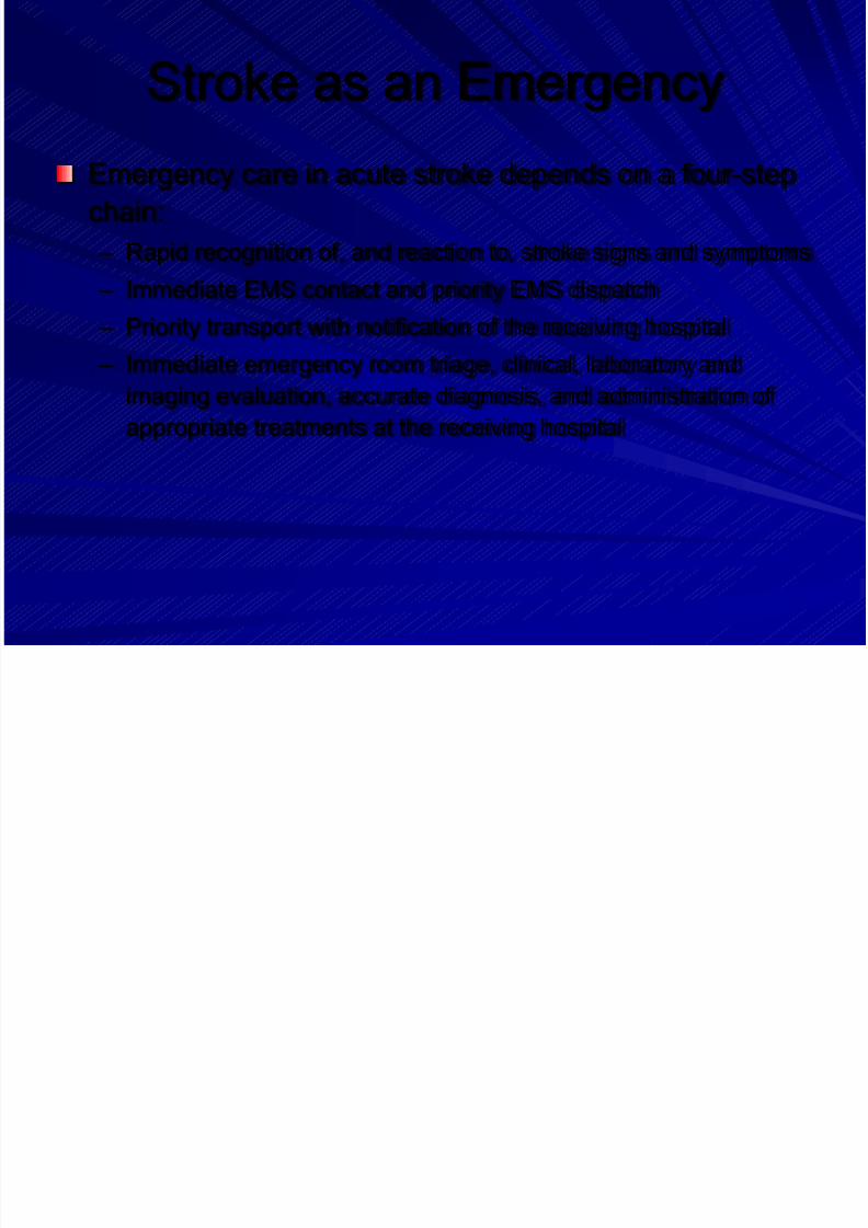
Stroke as an Emergency
Emergency care in acute stroke depends on a four-step
chain:
– Rapid recognition of, and reaction to, stroke signs and symptoms
– Immediate EMS contact and priority EMS dispatch
– Priority transport with notification of the receiving hospital
– Immediate emergency room triage, clinical, laboratory and
imaging evaluation, accurate diagnosis, and administration of
appropriate treatments at the receiving hospital
Page 8
8/14/2019 Management Acute Stroke
http://slidepdf.com/reader/full/management-acute-stroke 8/108
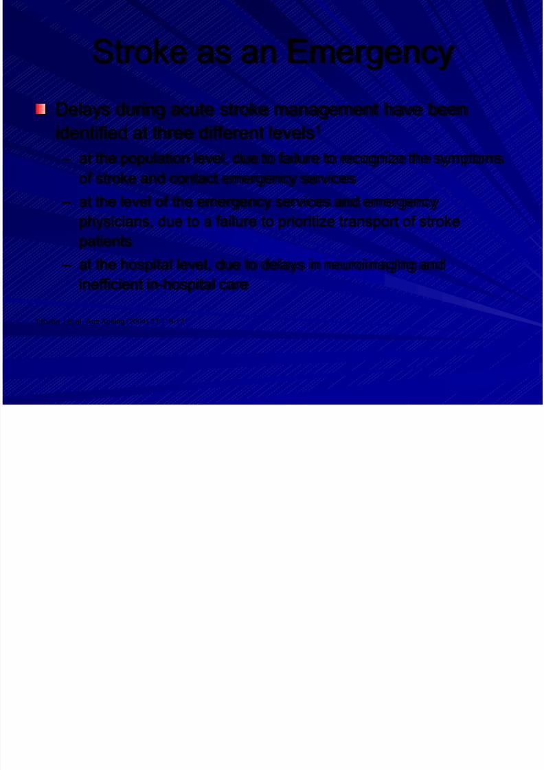
Stroke as an Emergency
Delays during acute stroke management have been
identified at three different levels1
– at the population level, due to failure to recognize the symptoms
of stroke and contact emergency services
– at the level of the emergency services and emergency
physicians, due to a failure to prioritize transport of stroke
patients
– at the hospital level, due to delays in neuroimaging and
inefficient in-hospital care
1:Kwan J et al. Age Ageing (2004) 33:116-121
Page 9
8/14/2019 Management Acute Stroke
http://slidepdf.com/reader/full/management-acute-stroke 9/108
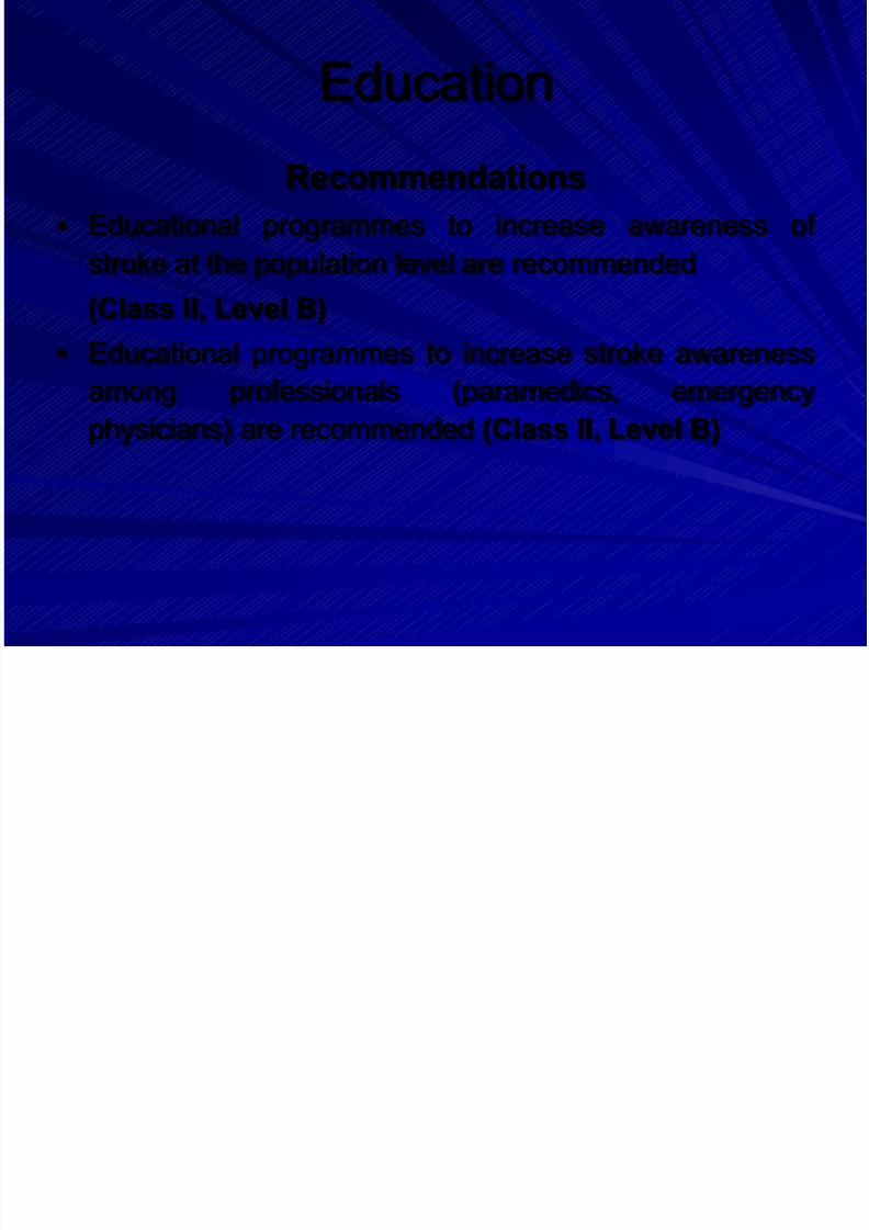
Education
Recommendations
Educational programmes to increase awareness of
stroke at the population level are recommended
(Class II, Level B)
Educational programmes to increase stroke awareness
among professionals (paramedics, emergency
physicians) are recommended (Class II, Level B)
Page 10
8/14/2019 Management Acute Stroke
http://slidepdf.com/reader/full/management-acute-stroke 10/108
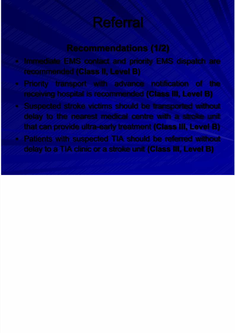
Referral
Recommendations (1/2)
Immediate EMS contact and priority EMS dispatch are
recommended (Class II, Level B)
Priority transport with advance notification of thereceiving hospital is recommended (Class III, Level B)
Suspected stroke victims should be transported without
delay to the nearest medical centre with a stroke unit
that can provide ultra-early treatment (Class III, Level B)
Patients with suspected TIA should be referred without
delay to a TIA clinic or a stroke unit (Class III, Level B)
Page 11
8/14/2019 Management Acute Stroke
http://slidepdf.com/reader/full/management-acute-stroke 11/108
Referral
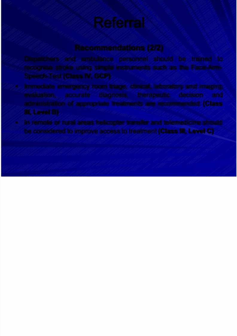
Recommendations (2/2)
Dispatchers and ambulance personnel should be trained to
recognise stroke using simple instruments such as the Face-Arm-
Speech-Test (Class IV, GCP)
Immediate emergency room triage, clinical, laboratory and imaging
evaluation, accurate diagnosis, therapeutic decision and
administration of appropriate treatments are recommended (Class
III, Level B)
In remote or rural areas helicopter transfer and telemedicine shouldbe considered to improve access to treatment (Class III, Level C)
Page 12
8/14/2019 Management Acute Stroke
http://slidepdf.com/reader/full/management-acute-stroke 12/108
Emergency Management
The time window for treatment ofpatients with acute stroke is narrow – Acute emergency management of stroke
requires parallel processes operating atdifferent levels of patient management
– Acute assessment of neurological and vital
functions parallels the treatment of acutelylife-threatening conditions
Time is the most important factor
Page 13
8/14/2019 Management Acute Stroke
http://slidepdf.com/reader/full/management-acute-stroke 13/108
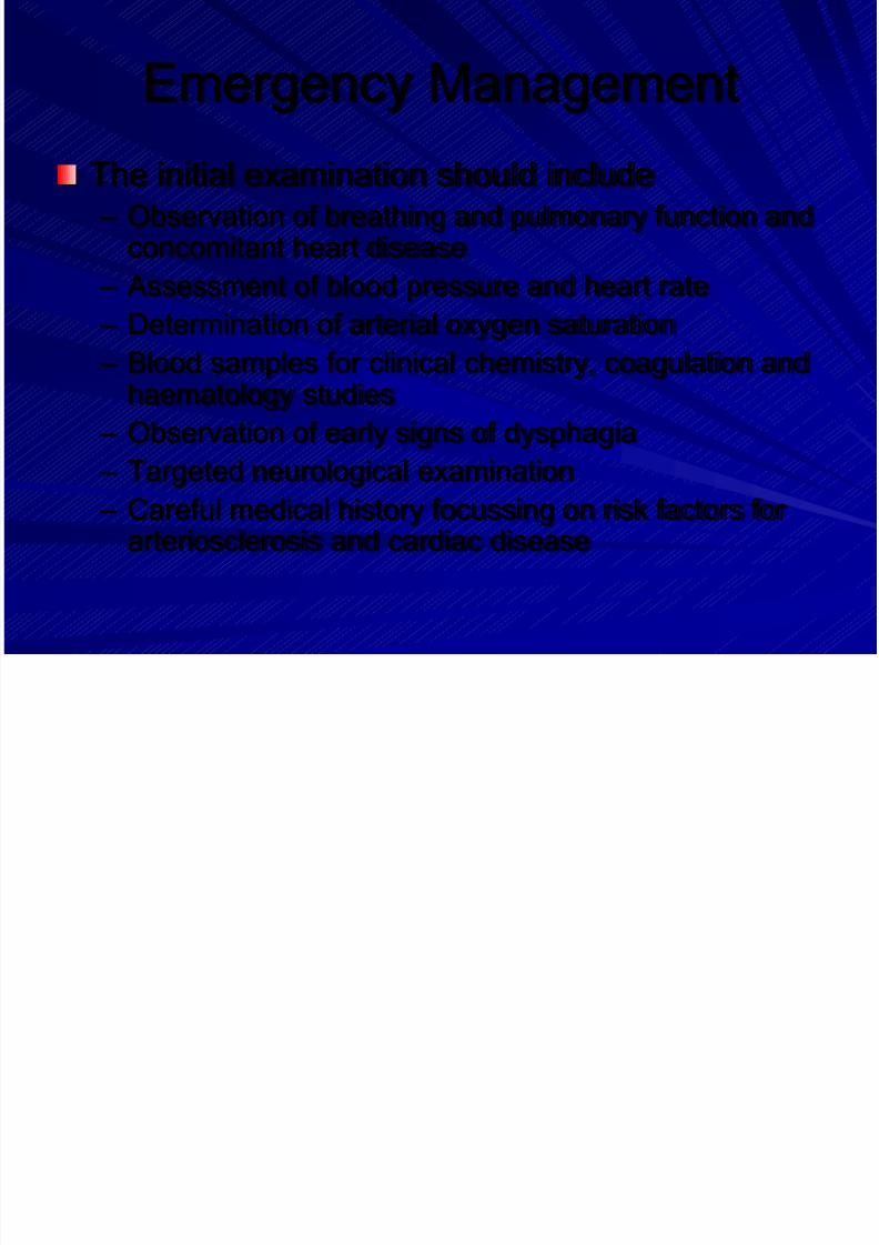
Emergency Management
The initial examination should include – Observation of breathing and pulmonary function and
concomitant heart disease
– Assessment of blood pressure and heart rate
– Determination of arterial oxygen saturation
– Blood samples for clinical chemistry, coagulation andhaematology studies
– Observation of early signs of dysphagia
– Targeted neurological examination
– Careful medical history focussing on risk factors forarteriosclerosis and cardiac disease
Page 14
8/14/2019 Management Acute Stroke
http://slidepdf.com/reader/full/management-acute-stroke 14/108
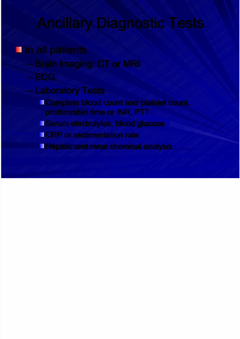
Ancillary Diagnostic Tests
In all patients
– Brain Imaging: CT or MRI
– ECG
– Laboratory Tests
Complete blood count and platelet count,
prothrombin time or INR, PTT
Serum electrolytes, blood glucoseCRP or sedimentation rate
Hepatic and renal chemical analysis
Page 15
8/14/2019 Management Acute Stroke
http://slidepdf.com/reader/full/management-acute-stroke 15/108
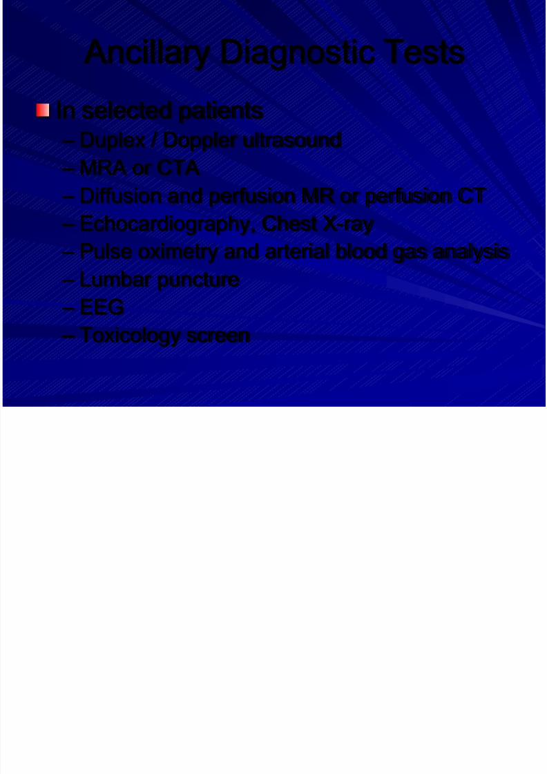
Ancillary Diagnostic Tests
In selected patients
– Duplex / Doppler ultrasound
– MRA or CTA
– Diffusion and perfusion MR or perfusion CT – Echocardiography, Chest X-ray
– Pulse oximetry and arterial blood gas analysis
– Lumbar puncture – EEG
– Toxicology screen
Page 16
8/14/2019 Management Acute Stroke
http://slidepdf.com/reader/full/management-acute-stroke 16/108
Emergency Management
Recommendations
Organization of pre-hospital and in-hospital
pathways and systems for acute stroke patients
is recommended (Class III, Level C)
All patients should receive brain imaging, ECG,
and laboratory tests. Additional diagnostic
examinations are necessary in selected patients
(Class IV, GCP)
Page 17
8/14/2019 Management Acute Stroke
http://slidepdf.com/reader/full/management-acute-stroke 17/108
Outlines
1. Education, Referral and Emergency
room
2. Stroke Unit
3. Imaging and Diagnostics
4. Prevention
5. General Treatment
6. Acute Treatment7. Management of Complications
8. Rehabilitation
Page 18
8/14/2019 Management Acute Stroke
http://slidepdf.com/reader/full/management-acute-stroke 18/108
Stroke Unit
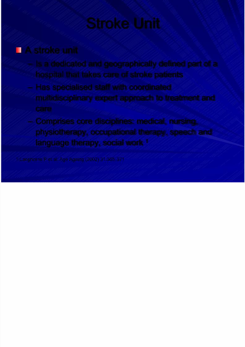
A stroke unit
– Is a dedicated and geographically defined part of a
hospital that takes care of stroke patients
– Has specialised staff with coordinatedmultidisciplinary expert approach to treatment and
care
– Comprises core disciplines: medical, nursing,
physiotherapy, occupational therapy, speech and
language therapy, social work 1
1:Langhorne P et al. Age Ageing (2002) 31:365-371
Page 19
8/14/2019 Management Acute Stroke
http://slidepdf.com/reader/full/management-acute-stroke 19/108
Stroke Unit
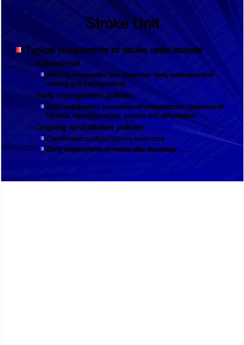
Typical components of stroke units include
– Assessment
Medical assessment and diagnosis, early assessment of
nursing and therapy needs – Early management policies
Early mobilisation, prevention of complications, treatment of
hypoxia, hyperglycaemia, pyrexia and dehydration
– Ongoing rehabilitation policiesCoordinated multidisciplinary team care
Early assessments of needs after discharge
Page 20
8/14/2019 Management Acute Stroke
http://slidepdf.com/reader/full/management-acute-stroke 20/108
Stroke Unit
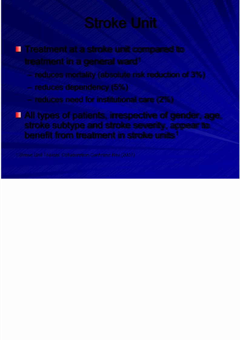
Treatment at a stroke unit compared to
treatment in a general ward1
– reduces mortality (absolute risk reduction of 3%)
– reduces dependency (5%)
– reduces need for institutional care (2%)
All types of patients, irrespective of gender, age,
stroke subtype and stroke severity, appear tobenefit from treatment in stroke units1
1:Stroke Unit Trialists' Collaboration Cochrane Rev (2007)
Page 21
8/14/2019 Management Acute Stroke
http://slidepdf.com/reader/full/management-acute-stroke 21/108
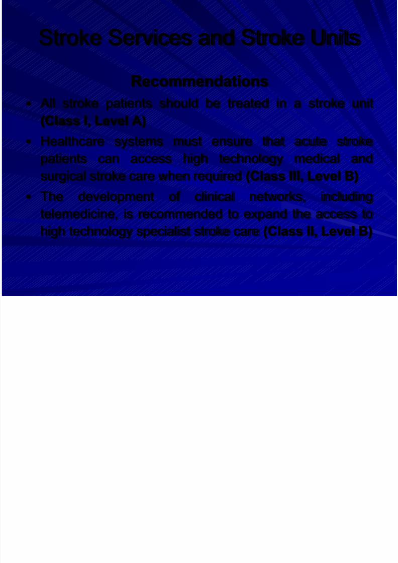
Stroke Services and Stroke Units
Recommendations
All stroke patients should be treated in a stroke unit
(Class I, Level A)
Healthcare systems must ensure that acute strokepatients can access high technology medical and
surgical stroke care when required (Class III, Level B)
The development of clinical networks, including
telemedicine, is recommended to expand the access to
high technology specialist stroke care (Class II, Level B)
Page 22
8/14/2019 Management Acute Stroke
http://slidepdf.com/reader/full/management-acute-stroke 22/108
Outlines
1. Education, Referral and Emergency
room
2. Stroke Unit
3. Imaging and Diagnostics
4. Prevention
5. General Treatment
6. Acute Treatment7. Management of Complications
8. Rehabilitation
Page 23
8/14/2019 Management Acute Stroke
http://slidepdf.com/reader/full/management-acute-stroke 23/108
Page 24
8/14/2019 Management Acute Stroke
http://slidepdf.com/reader/full/management-acute-stroke 24/108
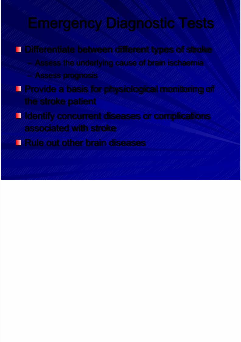
Emergency Diagnostic Tests
Cranial Computed Tomography (CT)
– Immediate plain CT scanning distinguishes reliably
between haemorrhagic and ischaemic stroke
– Detects signs of ischaemia as early as 2 h after strokeonset1
– Helps to identify other neurological diseases (e.g.
neoplasms)
– Most cost-effective strategy for imaging acute strokepatients2
1: von Kummer R et al. Radiology (2001) 219:95-100
2: Wardlaw J et al. Stroke (2004) 35:2477-2483
Page 25
8/14/2019 Management Acute Stroke
http://slidepdf.com/reader/full/management-acute-stroke 25/108
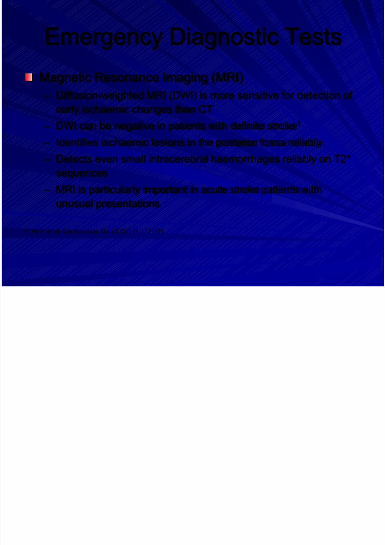
Emergency Diagnostic Tests
Magnetic Resonance Imaging (MRI)
– Diffusion-weighted MRI (DWI) is more sensitive for detection of
early ischaemic changes than CT
– DWI can be negative in patients with definite stroke1
– Identifies ischaemic lesions in the posterior fossa reliably
– Detects even small intracerebral haemorrhages reliably on T2*
sequences
– MRI is particularly important in acute stroke patients with
unusual presentations
1: Ay H et al. Cerebrovasc Dis (2002) 14:177-186
Page 26
8/14/2019 Management Acute Stroke
http://slidepdf.com/reader/full/management-acute-stroke 26/108
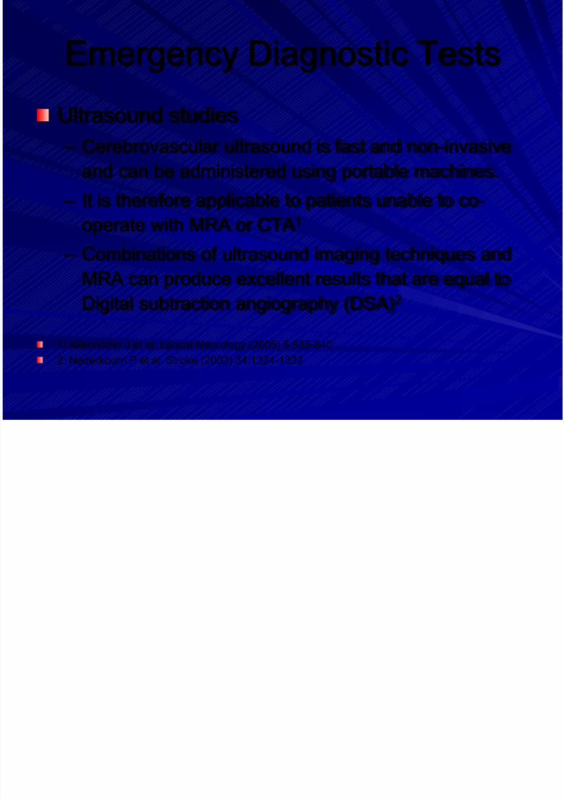
Emergency Diagnostic Tests
Ultrasound studies
– Cerebrovascular ultrasound is fast and non-invasive
and can be administered using portable machines.
– It is therefore applicable to patients unable to co-operate with MRA or CTA1
– Combinations of ultrasound imaging techniques and
MRA can produce excellent results that are equal to
Digital subtraction angiography (DSA)2
1: Allendörfer J et al. Lancet Neurology (2005) 5:835-840
2: Nederkoorn P et al. Stroke (2003) 34:1324-1332
Page 27
8/14/2019 Management Acute Stroke
http://slidepdf.com/reader/full/management-acute-stroke 27/108
Emergency Diagnostic Tests
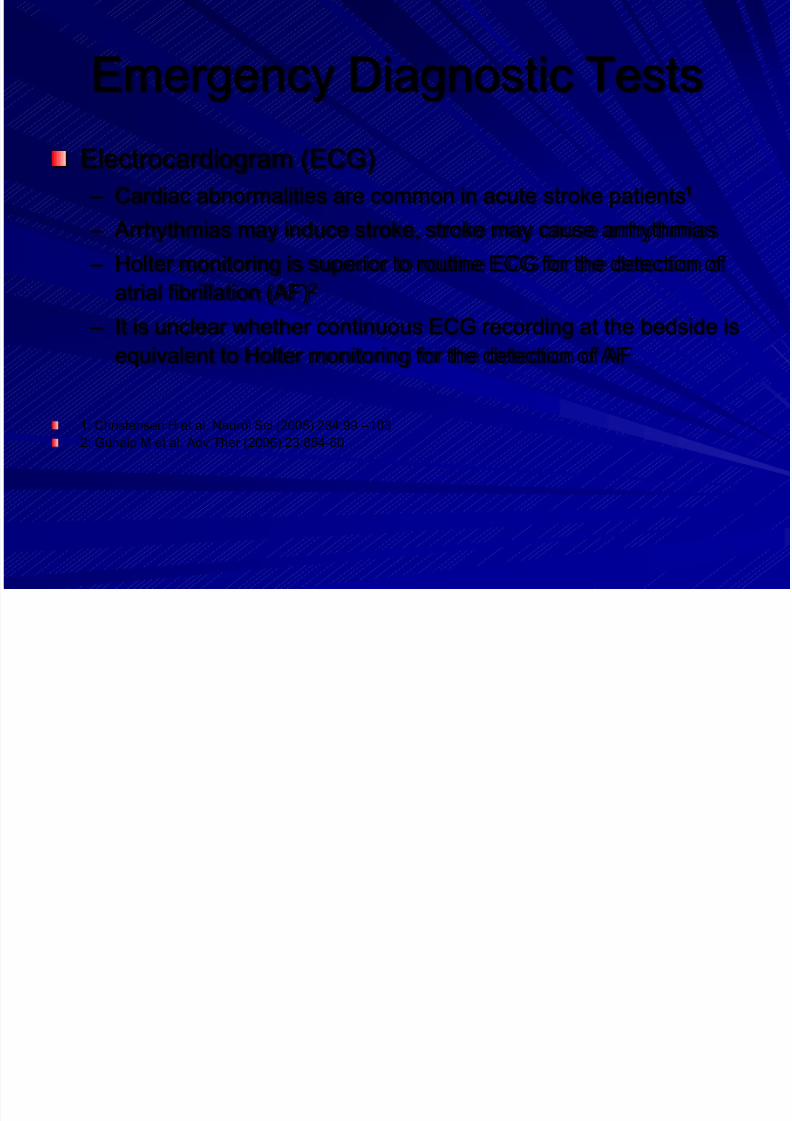
Electrocardiogram (ECG)
– Cardiac abnormalities are common in acute stroke patients1
– Arrhythmias may induce stroke, stroke may cause arrhythmias
– Holter monitoring is superior to routine ECG for the detection of
atrial fibrillation (AF)2
– It is unclear whether continuous ECG recording at the bedside is
equivalent to Holter monitoring for the detection of AF
1: Christensen H et al. Neurol Sci (2005) 234:99 –103
2: Gunalp M et al. Adv Ther (2006) 23:854-60
Page 28
8/14/2019 Management Acute Stroke
http://slidepdf.com/reader/full/management-acute-stroke 28/108
Emergency Diagnostic Tests
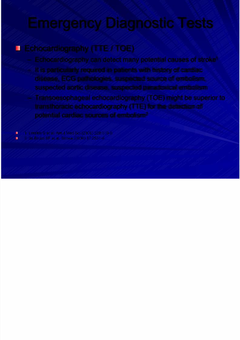
Echocardiography (TTE / TOE)
– Echocardiography can detect many potential causes of stroke1
– It is particularly required in patients with history of cardiac
disease, ECG pathologies, suspected source of embolism,
suspected aortic disease, suspected paradoxical embolism
– Transoesophageal echocardiography (TOE) might be superior to
transthoracic echocardiography (TTE) for the detection of
potential cardiac sources of embolism2
1: Lerakis S et al. Am J Med Sci (2005) 329:310-6
2: de Bruijn SF et al. Stroke (2006) 37:2531-4
Page 29
8/14/2019 Management Acute Stroke
http://slidepdf.com/reader/full/management-acute-stroke 29/108
Emergency Diagnostic Tests
Laboratory tests
– Haematology (RBC, WBC, platelet count)
– Basic clotting parameters
– Electrolytes
– Renal and hepatic chemistry
– Blood Glucose – CRP, sedimentation rate
Page 30
8/14/2019 Management Acute Stroke
http://slidepdf.com/reader/full/management-acute-stroke 30/108
Diagnostic Imaging
Recommendations
In patients with suspected TIA or stroke, urgent cranial CT (Class I),
or alternatively MRI (Class II), is recommended (Level A)
If MRI is used, the inclusion of diffusion weighted imaging (DWI) and
T2*-weighted gradient echo sequences is recommended (Class II,Level A)
In patients with TIA, minor stroke, or early spontaneous recovery
immediate diagnostic work-up, including urgent vascular imaging
(ultrasound, CT-angiography, or MR angiography) is recommended
(Class I, Level A)
Page 31
8/14/2019 Management Acute Stroke
http://slidepdf.com/reader/full/management-acute-stroke 31/108
Other Diagnostics
Recommendations (1/2)
In patients with acute stroke and TIA, early evaluation of
physiological parameters, routine blood tests, and
electrocardiography (ECG) is recommended (Class I,Level A)
All acute stroke and TIA patients should have a 12-
channel ECG. Continuous ECG recording is
recommended for ischaemic stroke and TIA patients(Class I, Level A)
Page 32
8/14/2019 Management Acute Stroke
http://slidepdf.com/reader/full/management-acute-stroke 32/108
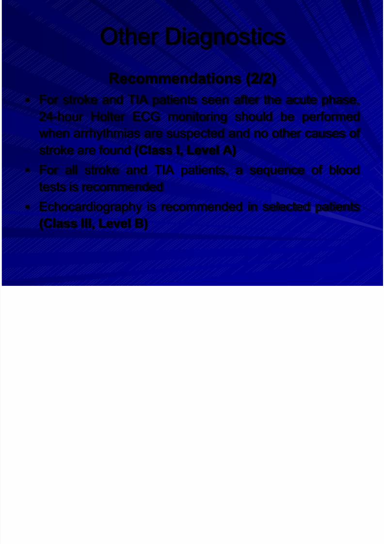
Other Diagnostics
Recommendations (2/2)
For stroke and TIA patients seen after the acute phase,
24-hour Holter ECG monitoring should be performed
when arrhythmias are suspected and no other causes ofstroke are found (Class I, Level A)
For all stroke and TIA patients, a sequence of blood
tests is recommended
Echocardiography is recommended in selected patients(Class III, Level B)
Page 33
8/14/2019 Management Acute Stroke
http://slidepdf.com/reader/full/management-acute-stroke 33/108
Outlines
1. Education, Referral and Emergency
room
2. Stroke Unit
3. Imaging and Diagnostics
4. Prevention
5. General Treatment
6. Acute Treatment7. Management of Complications
8. Rehabilitation
Page 34
8/14/2019 Management Acute Stroke
http://slidepdf.com/reader/full/management-acute-stroke 34/108
Primary Prevention
Content
– Management of vascular risk factors
– Antithrombotic therapy
– Carotid surgery and angioplasty
Page 35
8/14/2019 Management Acute Stroke
http://slidepdf.com/reader/full/management-acute-stroke 35/108
Content
– Management of vascular risk factors
– Antithrombotic therapy
– Surgery and angioplasty
Secondary Prevention
Page 36
8/14/2019 Management Acute Stroke
http://slidepdf.com/reader/full/management-acute-stroke 36/108
Blood pressure control
Background
– Antihypertensive drugs reduce stroke
recurrence risk after stroke or TIA (RR 0.76;
95%CI 0.63-0.92)1
– Target BP level and reduction should be
individualized
– The reduction in stroke occurs regardless ofbaseline BP and type of stroke2
1: Rashid P et al.: Stroke (2003) 34:2741-8
2: PROGRESS group: Lancet (2001) 358:1033-41
Page 37
8/14/2019 Management Acute Stroke
http://slidepdf.com/reader/full/management-acute-stroke 37/108
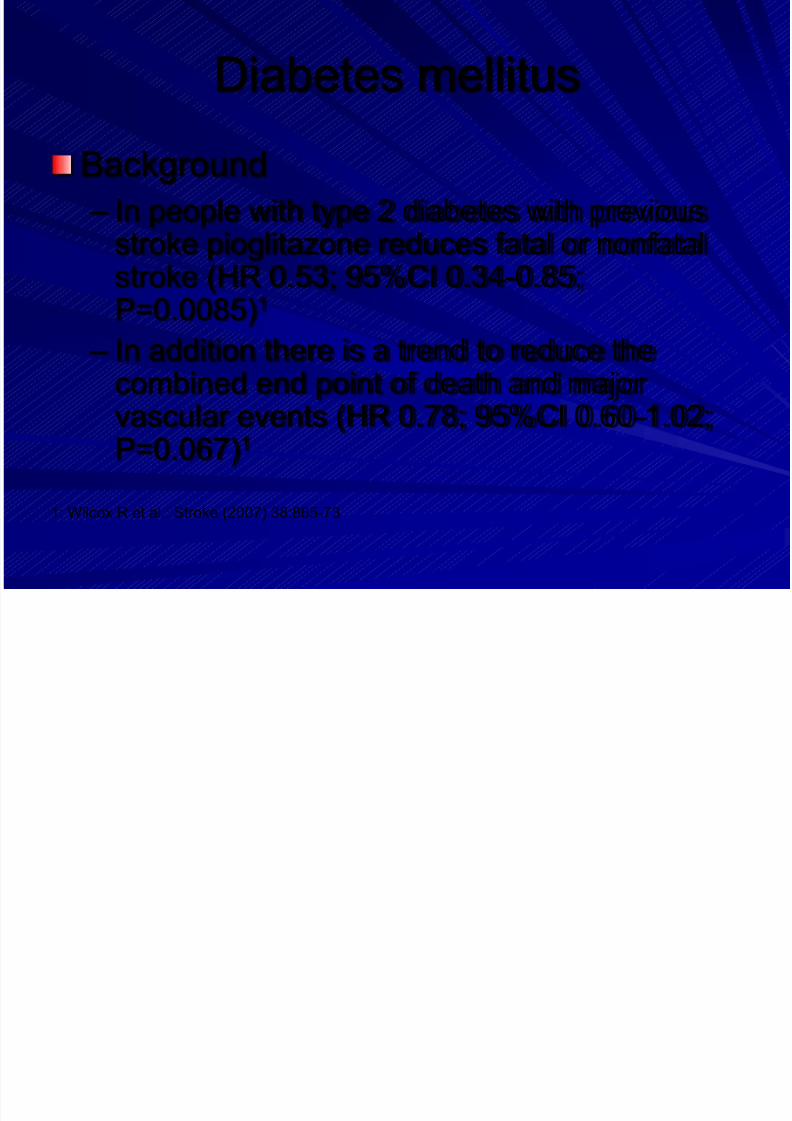
Diabetes mellitus
Background
– In people with type 2 diabetes with previousstroke pioglitazone reduces fatal or nonfatal
stroke (HR 0.53; 95%CI 0.34-0.85;P=0.0085)1
– In addition there is a trend to reduce thecombined end point of death and major
vascular events (HR 0.78; 95%CI 0.60-1.02;P=0.067)1
1: Wilcox R et al.: Stroke (2007) 38:865-73
Page 38
8/14/2019 Management Acute Stroke
http://slidepdf.com/reader/full/management-acute-stroke 38/108
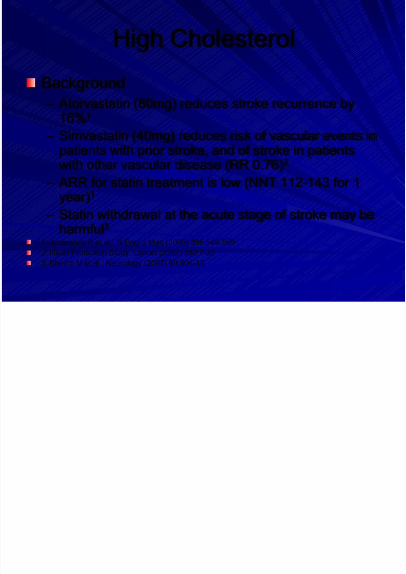
Page 39
8/14/2019 Management Acute Stroke
http://slidepdf.com/reader/full/management-acute-stroke 39/108
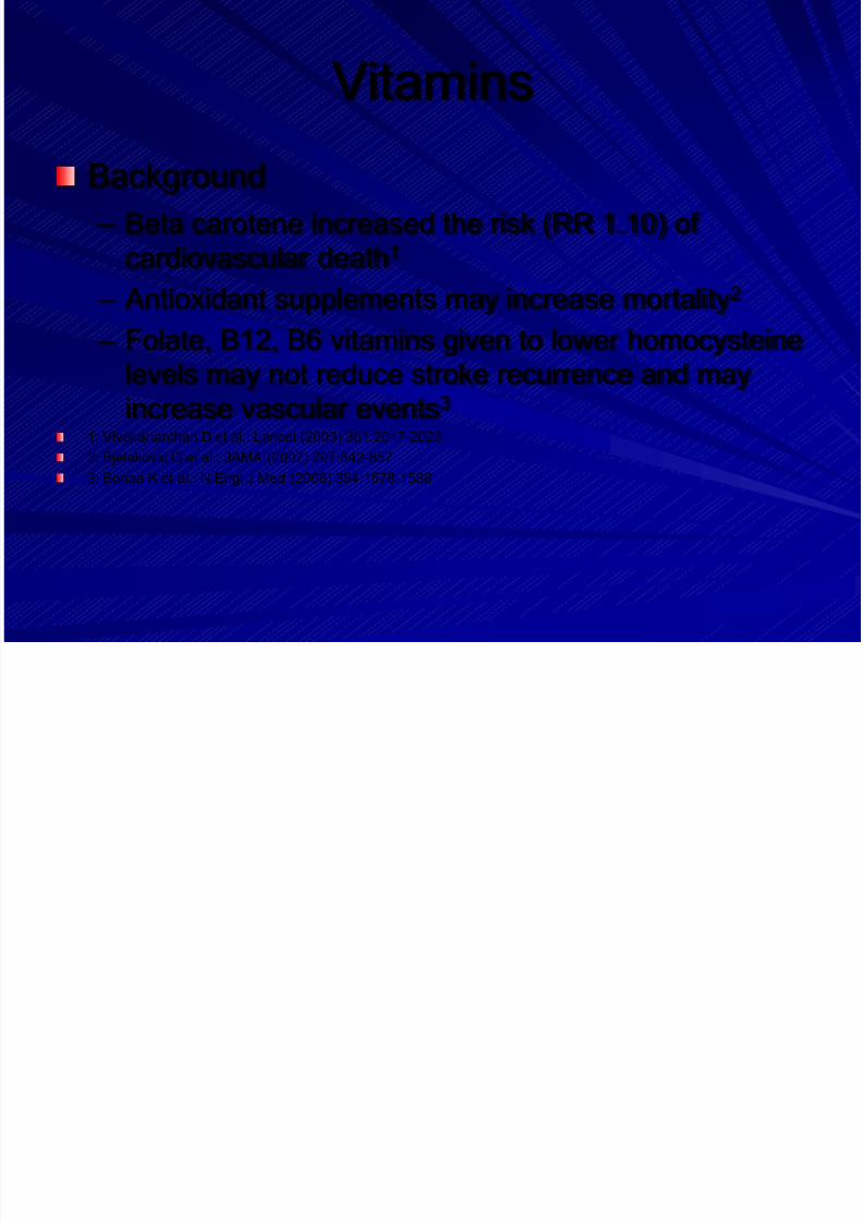
Vitamins
Background
– Beta carotene increased the risk (RR 1.10) of
cardiovascular death1
– Antioxidant supplements may increase mortality2
– Folate, B12, B6 vitamins given to lower homocysteine
levels may not reduce stroke recurrence and may
increase vascular events3
1: Vivekananthan D et al.: Lancet (2003) 361:2017-2023
2: Bjelakovic G et al.: JAMA (2007) 297:842-8573: Bonaa K et al.: N Engl J Med (2006) 354:1578-1588
Page 40
8/14/2019 Management Acute Stroke
http://slidepdf.com/reader/full/management-acute-stroke 40/108
Hormone Replacement Therapy
Background
– Oestrogen therapy is not effective in
secondary prevention after TIA or stroke and
may increase stroke severity1
1: Viscoli CM et al.: N Engl J Med (2001) 345:1243-9.
Page 41
8/14/2019 Management Acute Stroke
http://slidepdf.com/reader/full/management-acute-stroke 41/108
Sleep-disordered Breathing
Background
– Sleep-disordered breathing (SDB) is both a risk factor
and a consequence of stroke
– More than 50% of stroke patients have SDB, mostly inthe form of obstructive sleep apnoea (OSA).
– SDB is linked with poorer long-term outcome and
increased long-term stroke mortality1
– Continuous positive airway pressure is the treatmentof choice for OSA.
1: Bassetti CL: Semin Neurol (2005) 25:19-32
Page 42
8/14/2019 Management Acute Stroke
http://slidepdf.com/reader/full/management-acute-stroke 42/108
Risk Factor Management
Recommendations (1/3)
Blood pressure should be checked regularly. Blood pressure
lowering is recommended after the acute phase, including in
patients with normal blood pressure (Class I, Level A)
Blood glucose should be checked regularly. Diabetes should bemanaged with lifestyle modification and individualized
pharmacological therapy (Class IV, GCP)
In patients with type 2 diabetes who do not need insulin, treatment
with pioglitazone is recommended after stroke (Class III, Level B)
Page 43
8/14/2019 Management Acute Stroke
http://slidepdf.com/reader/full/management-acute-stroke 43/108
Risk Factor Management
Recommendations (2/3)
Statin therapy is recommended (Class I, Level A)
Cigarette smoking should be stopped (Class III, Level C)
Heavy use of alcohol should be discouraged (Class IV, GCP)
Regular physical activity is recommended (Class IV, GCP)
A diet low in salt and saturated fat, high in fruit and vege-tables, and
rich in fibre is recommended (Class IV, GCP)
Page 44
8/14/2019 Management Acute Stroke
http://slidepdf.com/reader/full/management-acute-stroke 44/108
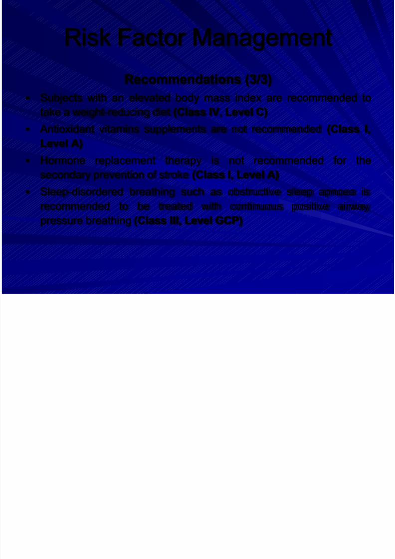
Risk Factor Management
Recommendations (3/3)
Subjects with an elevated body mass index are recommended to
take a weight-reducing diet (Class IV, Level C)
Antioxidant vitamins supplements are not recommended (Class I,
Level A)
Hormone replacement therapy is not recommended for the
secondary prevention of stroke (Class I, Level A)
Sleep-disordered breathing such as obstructive sleep apnoea is
recommended to be treated with continuous positive airwaypressure breathing (Class III, Level GCP)
Page 45
8/14/2019 Management Acute Stroke
http://slidepdf.com/reader/full/management-acute-stroke 45/108
Antithrombotic Therapy
Background: Aspirin
– 13% relative risk reduction for stroke after TIA or
stroke1
– Most widely studied dosages of aspirin are 50-150mg – The incidence of GI-disturbances with aspirin is dose
dependent
– No difference in effectiveness amongst low (<
160mg), medium (160 – 325mg) or high (500 -1500mg) dose aspirin1: Antithrombotic Trialists' Collaboration: BMJ (2002) 324:71-86
Page 46
8/14/2019 Management Acute Stroke
http://slidepdf.com/reader/full/management-acute-stroke 46/108
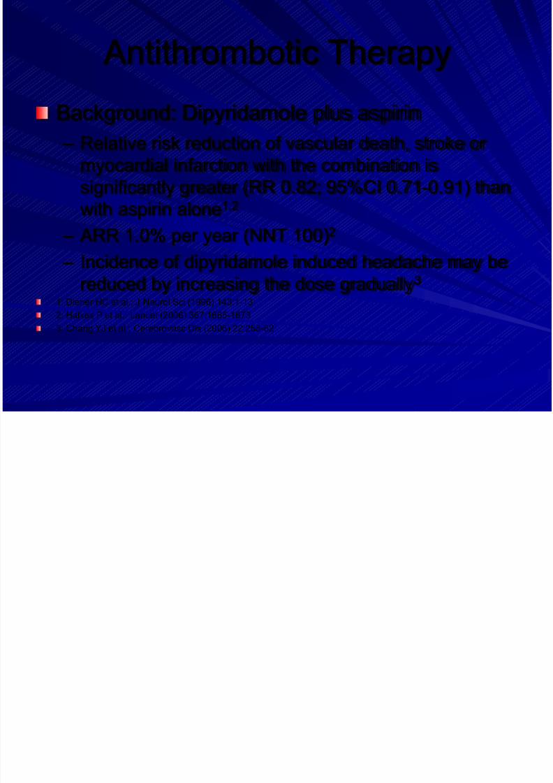
Antithrombotic Therapy
Background: Dipyridamole plus aspirin
– Relative risk reduction of vascular death, stroke or
myocardial infarction with the combination is
significantly greater (RR 0.82; 95%CI 0.71-0.91) than
with aspirin alone1,2
– ARR 1.0% per year (NNT 100)2
– Incidence of dipyridamole induced headache may be
reduced by increasing the dose gradually3
1: Diener HC et al.: J Neurol Sci (1996) 143:1-13
2: Halkes P et al.: Lancet (2006) 367:1665-1673
3: Chang YJ et al.: Cerebrovasc Dis (2006) 22:258-62
Page 47
8/14/2019 Management Acute Stroke
http://slidepdf.com/reader/full/management-acute-stroke 47/108
Antithrombotic Therapy
Background: Clopidogrel:
– Clopidogrel is slightly but significantly more
effective than medium-dose aspirin (RRR
8.7%, ARR 0,5%) in preventing vascularevents in patients with previous stroke, MI or
PAD1
1: CAPRIE Steering Committee: Lancet (1996) 348:1329-1339
Page 48
8/14/2019 Management Acute Stroke
http://slidepdf.com/reader/full/management-acute-stroke 48/108
Antithrombotic Therapy
Background: Clopidogrel plus aspirin
– Compared with clopidogrel the combination of aspirinand clopidogrel does not reduce the risk of ischaemicstroke, myocardial infarction, vascular death, or re-
hospitalisation1
– Compared with aspirin alone the combination doesnot reduce the risk of myocardial infarction, stroke, orcardiovascular death2
– Risk of life-threatening or major bleeding isincreased1,2
1: Diener H et al.: Lancet (2004) 364:331-337
2: Bhatt D et al.: N Engl J Med (2006) 354:1706-1717
Page 49
8/14/2019 Management Acute Stroke
http://slidepdf.com/reader/full/management-acute-stroke 49/108
Antithrombotic Therapy
Recommendations (1/4)
Patients should receive antithrombotic therapy (Class I,
Level A)
Patients not requiring anticoagulation should receiveantiplatelet therapy (Class I, Level A). Where possible,
combined aspirin and dipyridamole, or clopidogrel alone,
should be given. Alternatively, aspirin alone, or triflusal
alone, may be used (Class I, Level A)
Page 50
8/14/2019 Management Acute Stroke
http://slidepdf.com/reader/full/management-acute-stroke 50/108
Antithrombotic Therapy
Recommendations (2/4)
The combination of aspirin and clopidogrel is not
recommended in patients with recent ischaemic stroke,
except in patients with specific indications (e.g. unstableangina or non-Q-wave MI during the last 12 months, or
recent stenting); treatment should be given for up to 9
months after the event (Class I, Level A)
Patients who have a stroke on antiplatelet therapyshould be re-evaluated for pathophysiology and risk
factors (Class IV, GCP)
Page 51
8/14/2019 Management Acute Stroke
http://slidepdf.com/reader/full/management-acute-stroke 51/108
Anticoagulation
Background
– Oral antiocoagulation (target INR 2.0 – 3.0) reduces
the risk of recurrent stroke in patients with AF1
– Oral anticoagulation is well established for othercauses of embolism such as mechanical prosthetic
valve replacement, rheumatic valvular heart disease,
ventricular aneurysm and cardiomyopathy
– There is no indication for oral anticoagulation in
patients with non-cardiac cause of ischaemic stroke2
1: EAFT Study Group: Lancet (1993) 342:1255-1262
2: Mohr JP et al.: N Engl J Med (2001) 345:1444-1451
Page 52
8/14/2019 Management Acute Stroke
http://slidepdf.com/reader/full/management-acute-stroke 52/108
Anticoagulation
Specific issues – In patients with AF and stable coronary disease,
aspirin should not be added to oral anticoagulation1
– Some retrospective studies suggest that anticoagu-
lation may be beneficial in aortic atheroma2, fusiformbasilar artery aneurysms3, or arterial dissection4
– It is unclear if patients with patent foramen ovale(PFO) benefit from oral anticoagulation5
1: Flaker GC et al.: Am Heart J (2006) 152:967-73
2: Dressler FA et al.: J Am Coll Cardiol (1998) 31:134-83: Echiverri HC et al.: Stroke (1989) 20:1741-7
4: Engelter ST et al.: Stroke (2007) 38:2605-11
5: Mas JL et al.: N Engl J Med (2001) 345:1740-6
Page 53
8/14/2019 Management Acute Stroke
http://slidepdf.com/reader/full/management-acute-stroke 53/108
Antithrombotic Therapy
Recommendations (3/4)
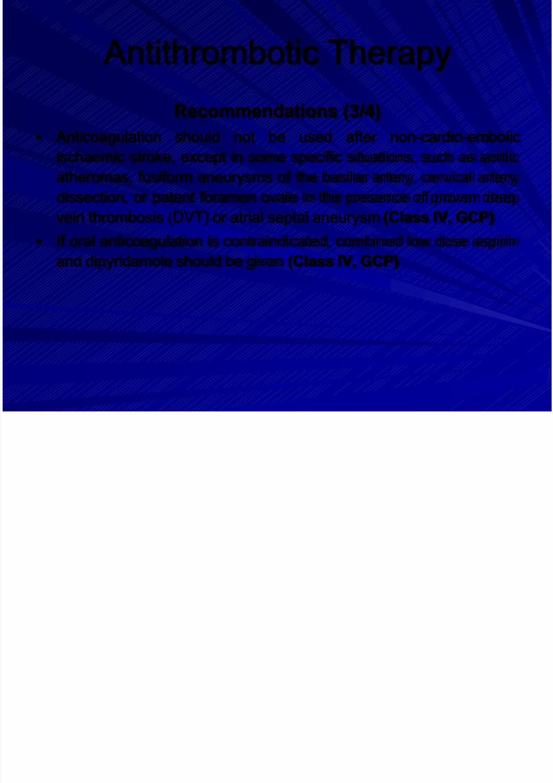
Anticoagulation should not be used after non-cardio-embolic
ischaemic stroke, except in some specific situations, such as aortic
atheromas, fusiform aneurysms of the basilar artery, cervical artery
dissection, or patent foramen ovale in the presence of proven deepvein thrombosis (DVT) or atrial septal aneurysm (Class IV, GCP)
If oral anticoagulation is contraindicated, combined low dose aspirin
and dipyridamole should be given (Class IV, GCP)
Page 54
8/14/2019 Management Acute Stroke
http://slidepdf.com/reader/full/management-acute-stroke 54/108
Antithrombotic Therapy
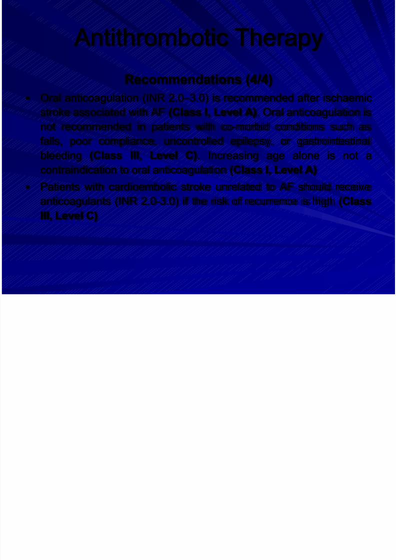
Recommendations (4/4)
Oral anticoagulation (INR 2.0 –3.0) is recommended after ischaemic
stroke associated with AF (Class I, Level A). Oral anticoagulation is
not recommended in patients with co-morbid conditions such as
falls, poor compliance, uncontrolled epilepsy, or gastrointestinalbleeding (Class III, Level C). Increasing age alone is not a
contraindication to oral anticoagulation (Class I, Level A)
Patients with cardioembolic stroke unrelated to AF should receive
anticoagulants (INR 2.0-3.0) if the risk of recurrence is high (Class
III, Level C)
Page 55
8/14/2019 Management Acute Stroke
http://slidepdf.com/reader/full/management-acute-stroke 55/108
Carotid Endarterectomy (CEA)
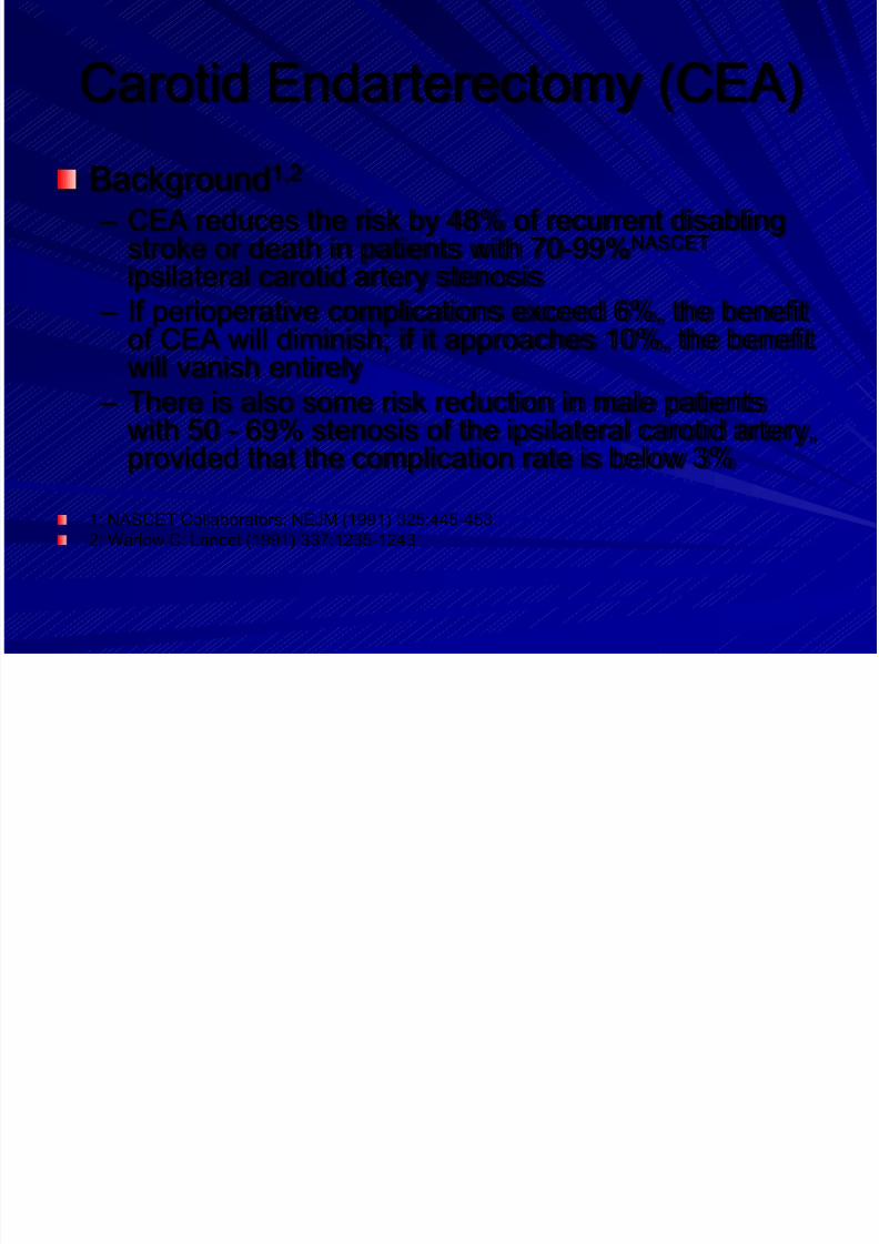
Background1,2
– CEA reduces the risk by 48% of recurrent disablingstroke or death in patients with 70-99%NASCET
ipsilateral carotid artery stenosis
– If perioperative complications exceed 6%, the benefitof CEA will diminish; if it approaches 10%, the benefitwill vanish entirely
– There is also some risk reduction in male patientswith 50 - 69% stenosis of the ipsilateral carotid artery,
provided that the complication rate is below 3%
1: NASCET Collaborators: NEJM (1991) 325:445-453
2: Warlow C: Lancet (1991) 337:1235-1243
Page 56
8/14/2019 Management Acute Stroke
http://slidepdf.com/reader/full/management-acute-stroke 56/108
Page 57
8/14/2019 Management Acute Stroke
http://slidepdf.com/reader/full/management-acute-stroke 57/108
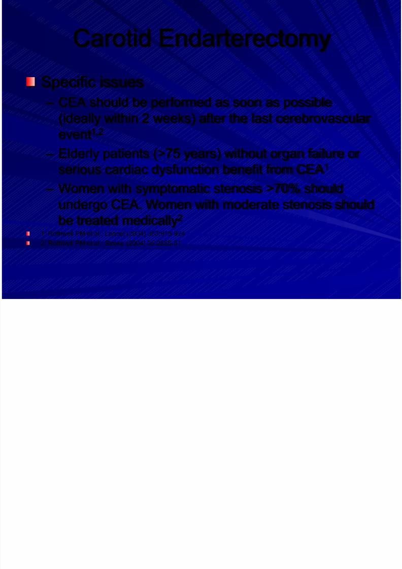
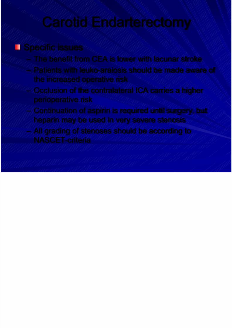
Carotid Endarterectomy
Specific issues
– The benefit from CEA is lower with lacunar stroke
– Patients with leuko-araiosis should be made aware of
the increased operative risk
– Occlusion of the contralateral ICA carries a higher
perioperative risk
– Continuation of aspirin is required until surgery, but
heparin may be used in very severe stenosis
– All grading of stenoses should be according to
NASCET-criteria
Page 58
8/14/2019 Management Acute Stroke
http://slidepdf.com/reader/full/management-acute-stroke 58/108
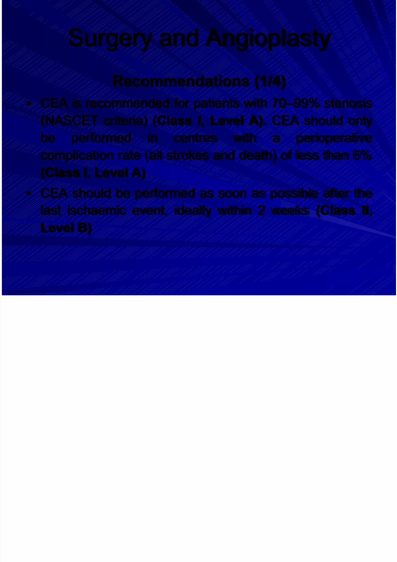
Surgery and Angioplasty
Recommendations (1/4)
CEA is recommended for patients with 70 –99% stenosis
(NASCET criteria) (Class I, Level A). CEA should only
be performed in centres with a perioperativecomplication rate (all strokes and death) of less than 6%
(Class I, Level A)
CEA should be performed as soon as possible after the
last ischaemic event, ideally within 2 weeks (Class II,Level B)
Page 59
8/14/2019 Management Acute Stroke
http://slidepdf.com/reader/full/management-acute-stroke 59/108
Page 60
8/14/2019 Management Acute Stroke
http://slidepdf.com/reader/full/management-acute-stroke 60/108
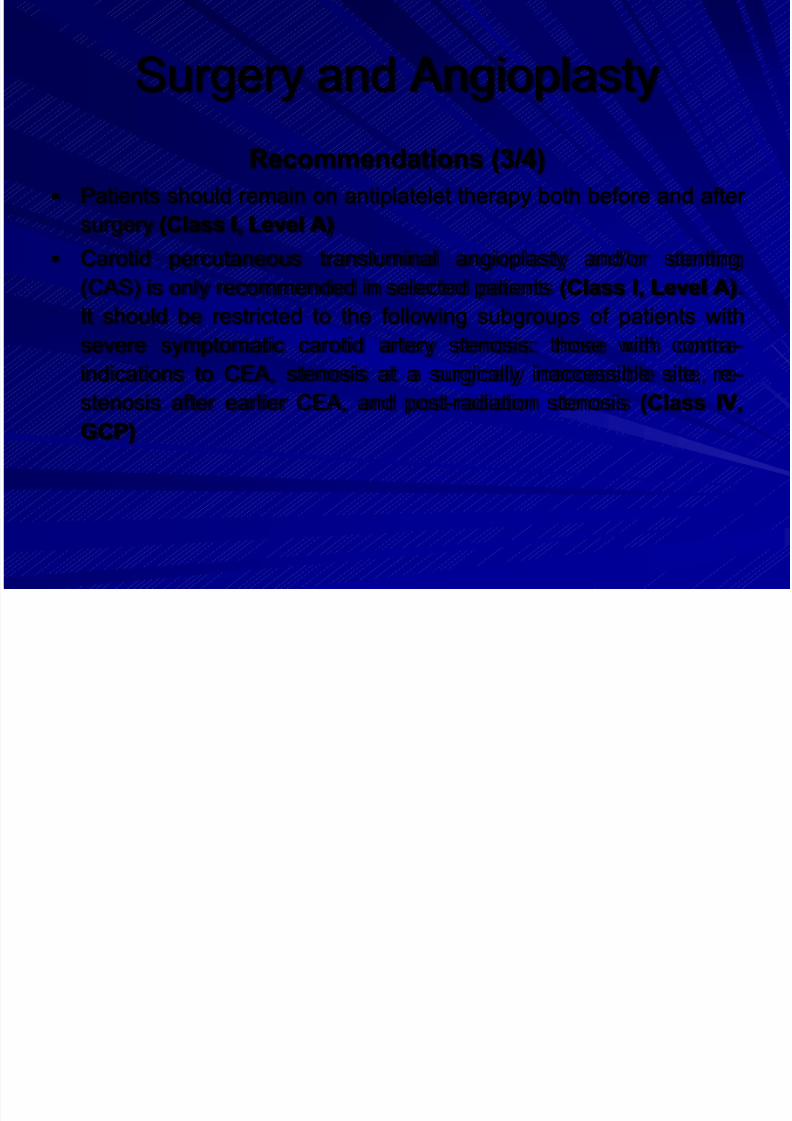
Surgery and Angioplasty
Recommendations (3/4)
Patients should remain on antiplatelet therapy both before and after
surgery (Class I, Level A)
Carotid percutaneous transluminal angioplasty and/or stenting
(CAS) is only recommended in selected patients (Class I, Level A).It should be restricted to the following subgroups of patients with
severe symptomatic carotid artery stenosis: those with contra-
indications to CEA, stenosis at a surgically inaccessible site, re-
stenosis after earlier CEA, and post-radiation stenosis (Class IV,
GCP)
Page 61
8/14/2019 Management Acute Stroke
http://slidepdf.com/reader/full/management-acute-stroke 61/108
Surgery and Angioplasty
Recommendations (4/4)
Patients should receive a combination of
clopidogrel and aspirin immediately before and
for at least 1 months after stenting (Class IV,
GCP)
Endovascular treatment may be considered in
patients with symptomatic intracranial stenosis(Class IV, GPC)
Page 62
8/14/2019 Management Acute Stroke
http://slidepdf.com/reader/full/management-acute-stroke 62/108
Outlines
1. Education, Referral and Emergency
room
2. Stroke Unit3. Imaging and Diagnostics
4. Prevention
5. General Treatment
6. Acute Treatment7. Management of Complications
8. Rehabilitation
Page 63
8/14/2019 Management Acute Stroke
http://slidepdf.com/reader/full/management-acute-stroke 63/108
General Stroke Treatment
Content
– Monitoring
– Pulmonary and airway care
– Fluid balance
– Blood pressure
– Glucose metabolism
– Body temperature
Page 64
8/14/2019 Management Acute Stroke
http://slidepdf.com/reader/full/management-acute-stroke 64/108
Monitoring
Continuous monitoring
– Heart rate
– Breathing rate
– O2 saturationDiscontinuous monitoring
– Blood pressure
– Blood glucose
– Vigilance (GCS), pupils
– Neurological status (e.g. NIH stroke scale or
Scandinavian stroke scale)
Page 65
8/14/2019 Management Acute Stroke
http://slidepdf.com/reader/full/management-acute-stroke 65/108
Pulmonary function
Background
– Adequate oxygenation is important
– Improve blood oxygenation by administration of > 2 l
O2 – Risk for aspiration in patients with side positioning
– Hypoventilation may be caused by pathological
respiration pattern
– Risk of airway obstruction (vomiting, oropharyngealmuscular hypotonia): mechanical airway protection
Page 66
8/14/2019 Management Acute Stroke
http://slidepdf.com/reader/full/management-acute-stroke 66/108
Blood pressure
Background
– Elevated in most patients with acute stroke
– BP drops spontaneously during the first days
after stroke
– Blood flow in the critical penumbra passively
dependent on the mean arterial pressure
– There are no adequately sized randomised,controlled studies guiding BP management
Page 67
8/14/2019 Management Acute Stroke
http://slidepdf.com/reader/full/management-acute-stroke 67/108
Blood pressure
Specific issues
– Elevated BP (e.g. up to 200mmHg systolic or
110mmHg diastolic) may be tolerated in the acute
phase of ischaemic stroke without intervention
– BP may be lowered if this is required by cardiac
conditions
– Upper level of systolic BP in patients undergoing
thrombolytic therapy is 180mmHg
– Avoid and treat hypotension
– Avoid drastic reduction in BP
Page 68
8/14/2019 Management Acute Stroke
http://slidepdf.com/reader/full/management-acute-stroke 68/108
Glucose metabolism
Background
– High glucose levels in acute stroke may increase the
size of the infarction and reduce functional outcome
– Hypoglycemia can mimic acute ischaemic infarction – Routine use of glucose potassium insulin (GKI)
infusion regimes in patients with mild to moderate
hyperglycaemia did not improve outcome1
It is common practise to treat hyperglycemia with insulinwhen blood glucose exceeds 180mg/dl2 (10mmol/l)1: Gray CS et al.: Lancet Neurol (2007) 6:397-406
2: Langhorne P et al.: Age Ageing (2002) 31:365-71.
Page 69
8/14/2019 Management Acute Stroke
http://slidepdf.com/reader/full/management-acute-stroke 69/108
Body temperature
Background
– Fever is associated with poorer neurological outcome
after stroke
– Fever increases infarct size in experimental stroke – Many patients with acute stroke develop a febrile
infection
There are no adequately sized trials guiding temperature
management after strokeIt is common practice treat fever (and its cause) when
the temperature reaches 37.5°C
Page 70
8/14/2019 Management Acute Stroke
http://slidepdf.com/reader/full/management-acute-stroke 70/108
General Stroke Treatment
Recommendations (1/4)
Intermittent monitoring of neurological status, pulse,
blood pressure, temperature and oxygen saturation is
recommended for 72 hours in patients with significantpersisting neurological deficits (Class IV, GCP)
Oxygen should be administered if sPO2 falls below 95%
(Class IV, GCP)
Regular monitoring of fluid balance and electrolytes isrecommended in patients with severe stroke or
swallowing problems (Class IV, GCP)
Page 71
8/14/2019 Management Acute Stroke
http://slidepdf.com/reader/full/management-acute-stroke 71/108
General Stroke Treatment
Recommendations (2/4)
Normal saline (0.9%) is recommended for fluid replacement during
the first 24 hours after stroke (Class IV, GCP)
Routine blood pressure lowering is not recommended following
acute stroke (Class IV, GCP) Cautious blood pressure lowering is recommended in patients with
any of the following; extremely high blood pressures
(>220/120 mmHg) on repeated measurements, or severe cardiac
failure, aortic dissection, or hyper-tensive encephalopathy (Class IV,
GCP)
Page 72
8/14/2019 Management Acute Stroke
http://slidepdf.com/reader/full/management-acute-stroke 72/108
General Stroke Treatment
Recommendations (3/4)
Abrupt blood pressure lowering should be avoided (Class II, Level
C)
Low blood pressure secondary to hypovolaemia or associated with
neurological deterioration in acute stroke should be treated withvolume expanders (Class IV GCP)
Monitoring serum glucose levels is recommended (Class IV, GCP)
Treatment of serum glucose levels >180mg/dl (>10mmol/l) with
insulin titration is recommended (Class IV, GCP)
Page 73
8/14/2019 Management Acute Stroke
http://slidepdf.com/reader/full/management-acute-stroke 73/108
General Stroke Treatment
Recommendations (4/4)
Severe hypoglycaemia (<50 mg/dl [<2.8 mmol/l]) should
be treated with intravenous dextrose or infusion of 10 –
20% glucose (Class IV, GCP points) The presence of pyrexia (temperature >37.5°C) should
prompt a search for concurrent infection (Class IV, GCP)
Treatment of pyrexia (>37.5°C) with paracetamol and
fanning is recommended (Class III, Level C) Antibiotic prophylaxis is not recommended in
immunocompetent patients (Class II, Level B)
Page 74
8/14/2019 Management Acute Stroke
http://slidepdf.com/reader/full/management-acute-stroke 74/108
Outlines
1. Education, Referral and Emergency
room
2. Stroke Unit3. Imaging and Diagnostics
4. Prevention
5. General Treatment
6. Acute Treatment7. Management of Complications
8. Rehabilitation
Page 75
8/14/2019 Management Acute Stroke
http://slidepdf.com/reader/full/management-acute-stroke 75/108
Specific Stroke Treatment
Content
– Thrombolytic therapy
– Early antithrombotic treatment
– Treatment of elevated intracranial pressure
– Prevention and management of complications
Page 76
8/14/2019 Management Acute Stroke
http://slidepdf.com/reader/full/management-acute-stroke 76/108
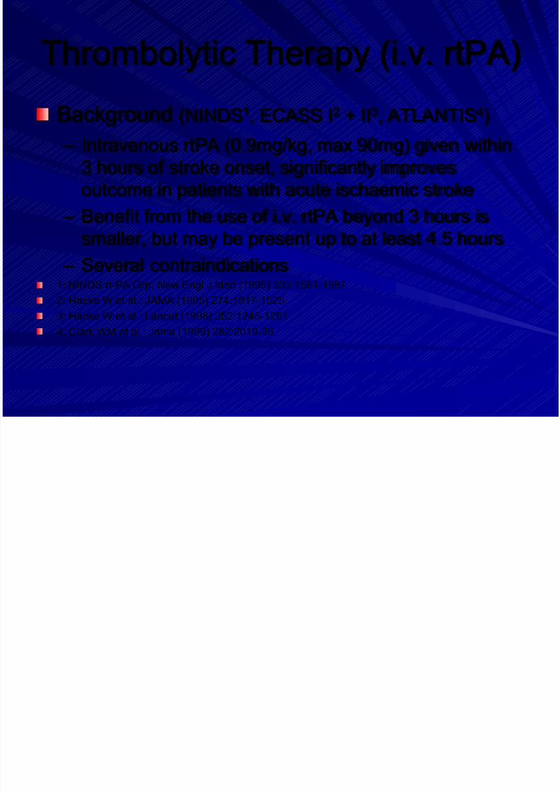
Thrombolytic Therapy (i.v. rtPA)
Background (NINDS1, ECASS I2 + II3, ATLANTIS4)
– Intravenous rtPA (0.9mg/kg, max 90mg) given within
3 hours of stroke onset, significantly improves
outcome in patients with acute ischaemic stroke
– Benefit from the use of i.v. rtPA beyond 3 hours is
smaller, but may be present up to at least 4.5 hours
– Several contraindications1: NINDS rt-PA Grp: New Engl J Med (1995) 333:1581-1587
2: Hacke W et al.: JAMA (1995) 274:1017-10253: Hacke W et al.: Lancet (1998) 352:1245-1251
4: Clark WM et al.: Jama (1999) 282:2019-26.
Page 77
8/14/2019 Management Acute Stroke
http://slidepdf.com/reader/full/management-acute-stroke 77/108
Specific issues
– A pooled analysis of the 6 i.v. rtPA trials confirms that
i.v. thrombolysis may work up to 4.5 hours1
– Caution is advised when considering i.v. rtPA in
persons with severe stroke (NIHSSS>25), or if the CT
demonstrates extended early infarcts signs
– Thrombolytic therapy must be given by an
experienced stroke physician after the imaging of the
brain is assessed by physicians experienced inreading this imaging study2
1: Hacke W et al.: Lancet (2004) 363:768-74
2: Wahlgren N et al.: Lancet (2007) 369:275-82
Page 78
8/14/2019 Management Acute Stroke
http://slidepdf.com/reader/full/management-acute-stroke 78/108
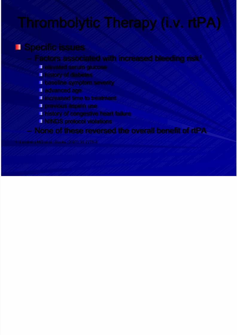
Thrombolytic Therapy (i.v. rtPA)
Specific issues – Factors associated with increased bleeding risk1
elevated serum glucose
history of diabetes
baseline symptom severity
advanced age
increased time to treatment
previous aspirin use
history of congestive heart failure
NINDS protocol violations
– None of these reversed the overall benefit of rtPA
1: Lansberg MG et al.: Stroke (2007) 38:2275-8
S ifi T
Page 79
8/14/2019 Management Acute Stroke
http://slidepdf.com/reader/full/management-acute-stroke 79/108
Specific Treatment
Recommendations (1/5)
Intravenous rtPA (0.9 mg/kg BW, maximum 90 mg), with
10% of the dose given as a bolus followed by a 60-
minute infusion, is recommended within 3 hours of onset
of ischaemic stroke (Class I, Level A)
Intravenous rtPA may be of benefit also for acute
ischaemic stroke beyond 3 hours after onset (Class I,
Level B) but is not recommended for routine clinicalpractice. The use of multimodal imaging criteria may be
useful for patient selection (Class III, Level C)
S ifi T t t
Page 80
8/14/2019 Management Acute Stroke
http://slidepdf.com/reader/full/management-acute-stroke 80/108
Specific Treatment
Recommendations (2/5)
Blood pressures of 185/110 mmHg or higher must be
lowered before thrombolysis (Class IV, GCP)
Intravenous rtPA may be used in patients with seizuresat stroke onset, if the neurological deficit is related to
acute cerebral ischaemia (Class IV, GCP)
Intravenous rtPA may also be administered in selected
patients over 80 years of age, although this is outsidethe current European labelling (Class III, Level C)
S ifi T t t
Page 81
8/14/2019 Management Acute Stroke
http://slidepdf.com/reader/full/management-acute-stroke 81/108
Specific Treatment
Recommendations (3/5)
Intra-arterial treatment of acute MCA occlusion within a
6-hour time window is recommended as an option
(Class II, Level B)
Intra-arterial thrombolysis is recommended for acute
basilar occlusion in selected patients (Class III, Level B)
Intravenous thrombolysis for basilar occlusion is an
acceptable alternative even after 3 hours (Class III,Level B)
A ti l t l t th
Page 82
8/14/2019 Management Acute Stroke
http://slidepdf.com/reader/full/management-acute-stroke 82/108
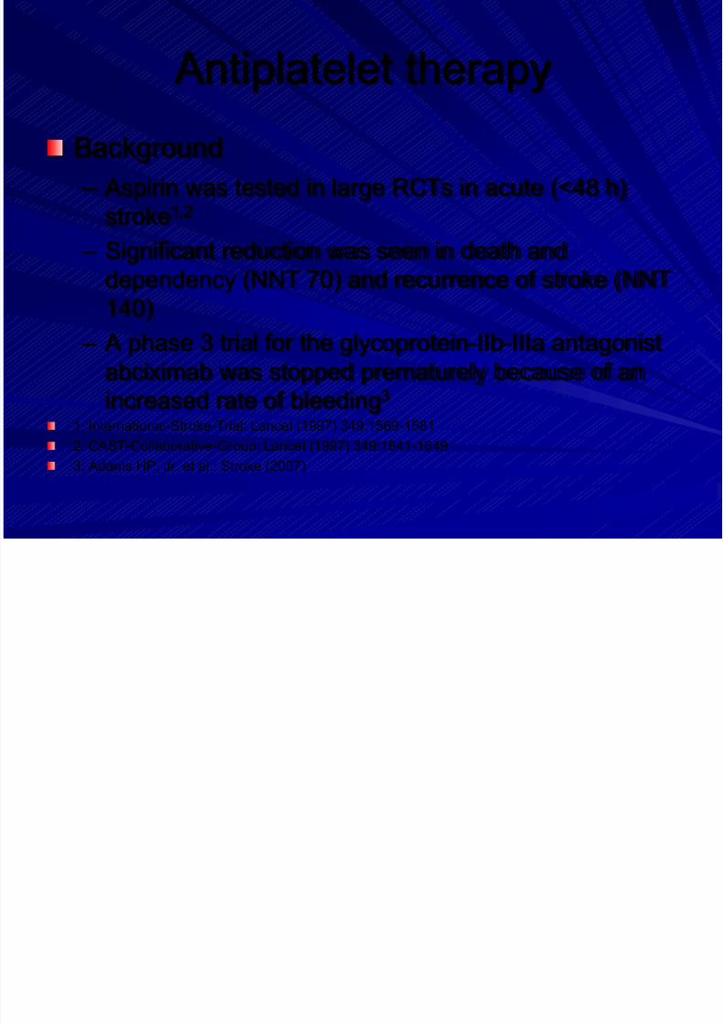
Antiplatelet therapy
Background
– Aspirin was tested in large RCTs in acute (<48 h)
stroke1,2
– Significant reduction was seen in death and
dependency (NNT 70) and recurrence of stroke (NNT
140)
– A phase 3 trial for the glycoprotein-IIb-IIIa antagonist
abciximab was stopped prematurely because of an
increased rate of bleeding3
1: International-Stroke-Trial: Lancet (1997) 349:1569-1581
2: CAST-Collaborative-Group: Lancet (1997) 349:1641-1649
3: Adams HP, Jr. et al.: Stroke (2007)
A ti l ti
Page 83
8/14/2019 Management Acute Stroke
http://slidepdf.com/reader/full/management-acute-stroke 83/108
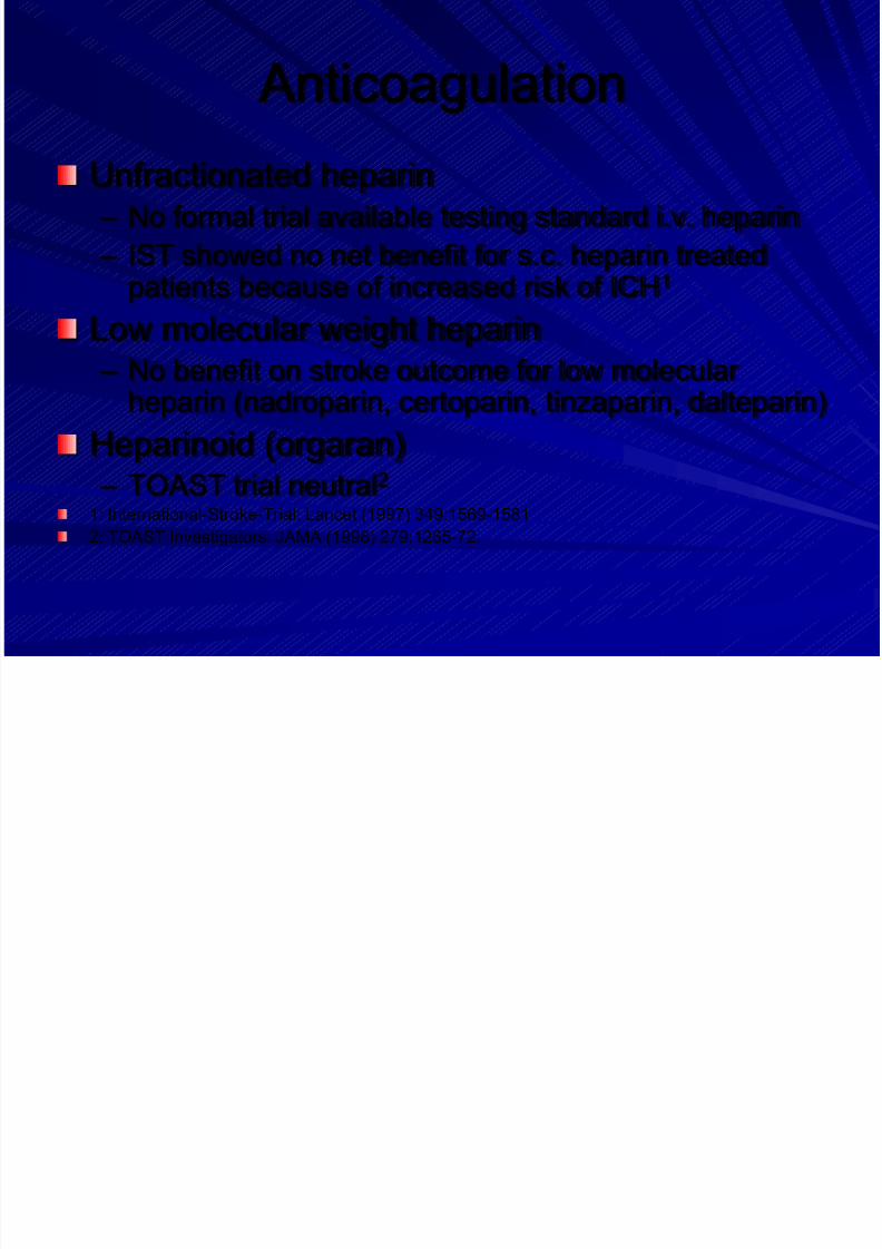
Anticoagulation
Unfractionated heparin – No formal trial available testing standard i.v. heparin
– IST showed no net benefit for s.c. heparin treatedpatients because of increased risk of ICH1
Low molecular weight heparin – No benefit on stroke outcome for low molecular
heparin (nadroparin, certoparin, tinzaparin, dalteparin)
Heparinoid (orgaran)
– TOAST trial neutral2 1: International-Stroke-Trial: Lancet (1997) 349:1569-1581
2: TOAST Investigators: JAMA (1998) 279:1265-72.
N t ti
Page 84
8/14/2019 Management Acute Stroke
http://slidepdf.com/reader/full/management-acute-stroke 84/108
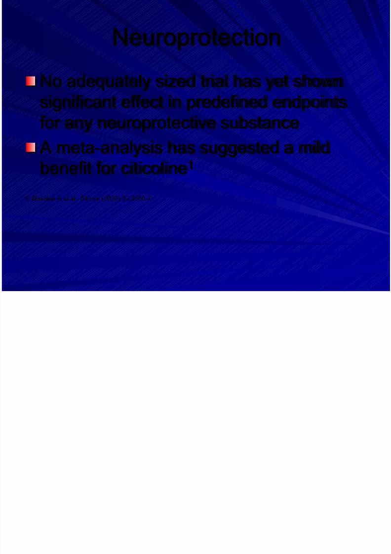
Neuroprotection
No adequately sized trial has yet shownsignificant effect in predefined endpoints
for any neuroprotective substance
A meta-analysis has suggested a mildbenefit for citicoline1
1: Davalos A et al.: Stroke (2002) 33:2850-7
S ifi T t t
Page 85
8/14/2019 Management Acute Stroke
http://slidepdf.com/reader/full/management-acute-stroke 85/108
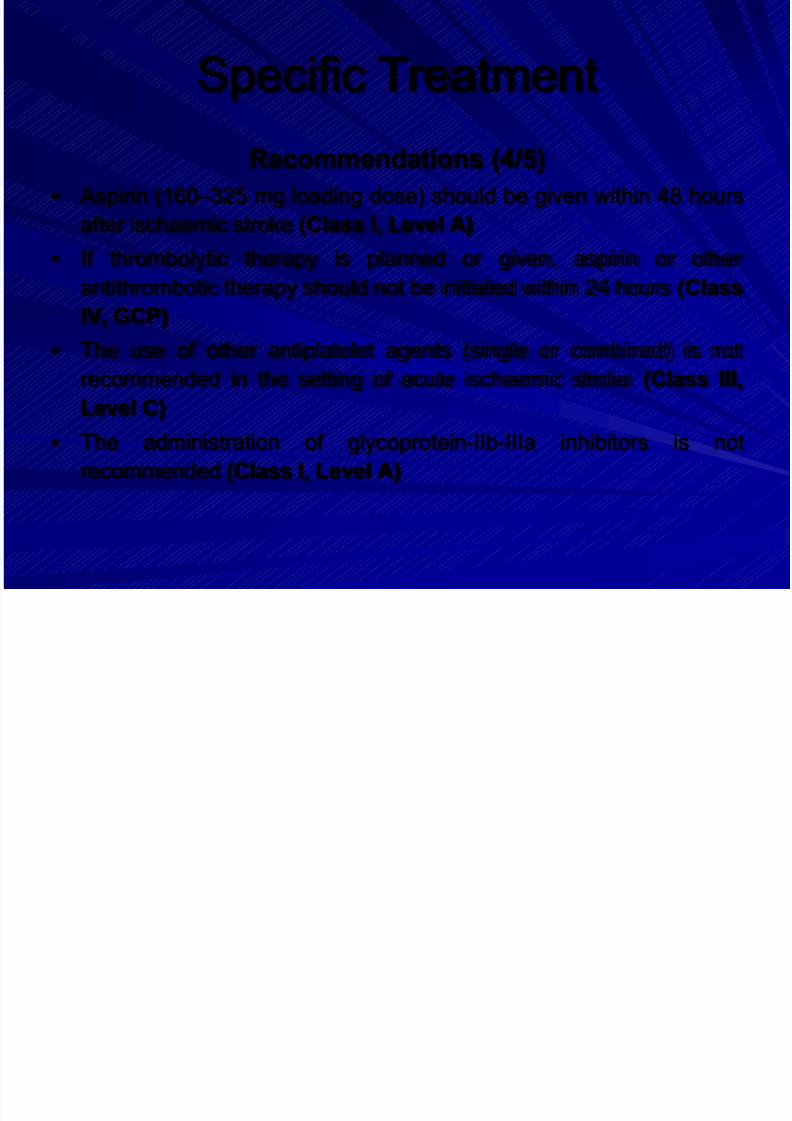
Specific Treatment
Recommendations (4/5)
Aspirin (160 –325 mg loading dose) should be given within 48 hours
after ischaemic stroke (Class I, Level A)
If thrombolytic therapy is planned or given, aspirin or other
antithrombotic therapy should not be initiated within 24 hours (ClassIV, GCP)
The use of other antiplatelet agents (single or combined) is not
recommended in the setting of acute ischaemic stroke (Class III,
Level C)
The administration of glycoprotein-IIb-IIIa inhibitors is not
recommended (Class I, Level A)
S ifi T t t
Page 86
8/14/2019 Management Acute Stroke
http://slidepdf.com/reader/full/management-acute-stroke 86/108
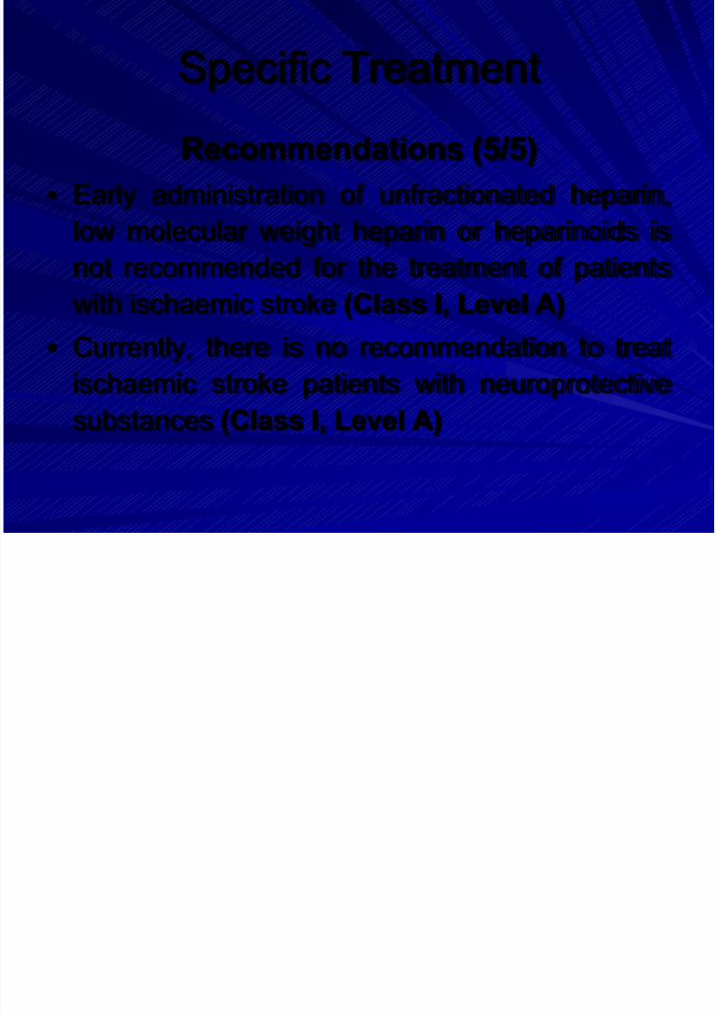
Specific Treatment
Recommendations (5/5)
Early administration of unfractionated heparin,
low molecular weight heparin or heparinoids is
not recommended for the treatment of patientswith ischaemic stroke (Class I, Level A)
Currently, there is no recommendation to treat
ischaemic stroke patients with neuroprotectivesubstances (Class I, Level A)
El t d I t i l P
Page 87
8/14/2019 Management Acute Stroke
http://slidepdf.com/reader/full/management-acute-stroke 87/108
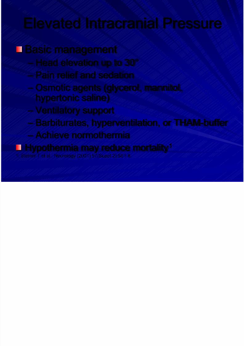
Elevated Intracranial Pressure
Basic management – Head elevation up to 30°
– Pain relief and sedation
– Osmotic agents (glycerol, mannitol,hypertonic saline)
– Ventilatory support
– Barbiturates, hyperventilation, or THAM-buffer
– Achieve normothermia
Hypothermia may reduce mortality1
1: Steiner T et al.: Neurology (2001) 57(Suppl 2):S61-8.
Page 88
8/14/2019 Management Acute Stroke
http://slidepdf.com/reader/full/management-acute-stroke 88/108
El t d I t i l P
Page 89
8/14/2019 Management Acute Stroke
http://slidepdf.com/reader/full/management-acute-stroke 89/108
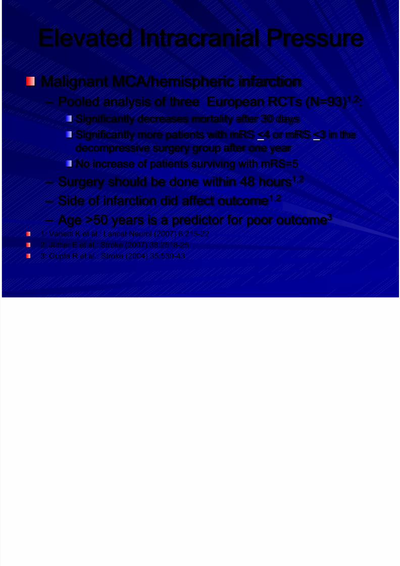
Elevated Intracranial Pressure
Recommendations (1/2)
Surgical decompressive therapy within 48 hours
after symptom onset is recommended in patients
up to 60 years of age with evolving malignantMCA infarcts (Class I, Level A)
Osmotherapy can be used to treat elevated
intracranial pressure prior to surgery if this isconsidered (Class III, Level C)
El t d I t i l P
Page 90
8/14/2019 Management Acute Stroke
http://slidepdf.com/reader/full/management-acute-stroke 90/108
Elevated Intracranial Pressure
Recommendations (2/2)
No recommendation can be given regarding
hypothermic therapy in patients with space-
occupying infarctions (Class IV, GCP)
Ventriculostomy or surgical decompression can
be considered for treatment of large cerebellar
infarctions that compress the brainstem (ClassIII, Level C)
O tlines
Page 91
8/14/2019 Management Acute Stroke
http://slidepdf.com/reader/full/management-acute-stroke 91/108
Outlines
1. Education, Referral and Emergency
room
2. Stroke Unit3. Imaging and Diagnostics
4. Prevention
5. General Treatment
6. Acute Treatment7. Management of Complications
8. Rehabilitation
Management of Complications
Page 92
8/14/2019 Management Acute Stroke
http://slidepdf.com/reader/full/management-acute-stroke 92/108
Management of Complications
Aspiration and pneumonia – Bacterial pneumonia is one of the most
important complications in stroke patients1
– Preventive strategies
Withhold oral feeding until demonstration of intactswallowing, preferable using a standardized test
Nasogastric (NG) or percutaneous enteral gastrostomy(PEG)
Frequent changes of the patient’s position in bed andpulmonary physical therapy
– Prophylactic administration of levofloxacin isnot superior to optimal care2
1: Weimar C et al.: Eur Neurol (2002) 48:133-40
2: Chamorro A et al.: Stroke (2005) 36:1495-500
Management of Complications
Page 93
8/14/2019 Management Acute Stroke
http://slidepdf.com/reader/full/management-acute-stroke 93/108
Management of Complications
Urinary tract infections – Most hospital-acquired urinary tract infections
are associated with the use of indwelling
catheters1
– Intermittent catheterization does not reduce
the risk of infection
– If urinary infection is diagnosed, appropriate
antibiotics should be chosen following basic
medical principles1: Gerberding JL: Ann Intern Med (2002) 137:665-70c
Management of Complications
Page 94
8/14/2019 Management Acute Stroke
http://slidepdf.com/reader/full/management-acute-stroke 94/108
Management of Complications
Deep vein thrombosis and pulmonaryembolism
– Risk might be reduced by good hydration andearly mobilization
– Low-dose LMWH reduces the incidence ofboth DVT (OR 0.34) and pulmonary embolism(OR 0.36), without a significantly increasedrisk of intracerebral (OR 1.39) or extracerebralhaemorrhage (OR 1.44)1,2
1: Diener HC et al.: Stroke (2006) 37:139-44
2: Sherman DG et al.: Lancet (2007) 369:1347-55
Management of Complications
Page 95
8/14/2019 Management Acute Stroke
http://slidepdf.com/reader/full/management-acute-stroke 95/108
Management of Complications
Pressure ulcer – Use of support surfaces, frequent repositioning,
optimizing nutritional status, and moisturizing sacral
skin are appropriate preventive strategies1
Seizures
– Prophylactic anticonvulsive treatment is not beneficial
Agitation
– Causal treatment must precede any type of sedation
or antipsychotic treatment1: Reddy M et al.: JAMA (2006) 296:974-84
Management of Complications
Page 96
8/14/2019 Management Acute Stroke
http://slidepdf.com/reader/full/management-acute-stroke 96/108
Management of Complications
Falls – Are common in every stage of stroke treatment
– Risk factors include cognitive impairment, depression,polypharmacy and sensory impairment1
– A multidisciplinary package focusing on personal andenvironmental factors might be preventive2
– Exercise, calcium supplements and bisphosphonatesimprove bone strength and decrease fracture rates instroke patients3,4
1: Aizen E et al.: Arch Gerontol Geriatr (2007) 44:1-12
2: Oliver D et al.: BMJ (2007) 334:82
3: Pang MY et al.: Clin Rehabil (2006) 20:97-111
4: Sato Y et al.: Cerebrovasc Dis (2005) 20:187-92
Management of Complications
Page 97
8/14/2019 Management Acute Stroke
http://slidepdf.com/reader/full/management-acute-stroke 97/108
Management of Complications
Dysphagia and feeding – Dysphagia occurs in up to 50% of patients with
unilateral hemiplegic stroke and is an independent
risk-factor for poor outcome1
– For patients with continuing dysphagia, options forenteral nutrition include NG or PEG feeding
– PEG does not provide better nutritional status or
improved clinical outcome, compared to NG2,3
1: Martino R et al.: Stroke (2005) 36:2756-632: Dennis MS et al.: Lancet (2005) 365:764-72
3: Callahan CM et al.: J Am Geriatr Soc (2000) 48:1048-54
Management of Complications
Page 98
8/14/2019 Management Acute Stroke
http://slidepdf.com/reader/full/management-acute-stroke 98/108
Management of Complications
Recommendations (1/4) Infections after stroke should be treated with appropriate antibiotics
(Class IV, GCP)
Prophylactic administration of antibiotics is not recommended, and
levofloxacin can be detrimental in acute stroke patients (Class II,Level B)
Early rehydration and graded compression stockings are
recommended to reduce the incidence of venous thromboembolism
(Class IV, GCP)
Early mobilization is recommended to prevent compli-cations such
as aspiration pneumonia, DVT and pressure ulcers (Class IV, GCP)
Management of Complications
Page 99
8/14/2019 Management Acute Stroke
http://slidepdf.com/reader/full/management-acute-stroke 99/108
Management of Complications
Recommendations (2/4) Low-dose s.c. heparin or low molecular weight heparins should be
considered for patients at high risk of DVT or pulmonary embolism
(Class I, Level A)
Administration of anticonvulsants is recommended to preventrecurrent seizures (Class I, Level A)
Prophylactic administration of anticonvulsants to patients with recent
stroke who have not had seizures is not recommended (Class IV,
GCP)
An assessment of falls risk is recommended for every stroke patient
(Class IV, GCP)
Page 100
8/14/2019 Management Acute Stroke
http://slidepdf.com/reader/full/management-acute-stroke 100/108
Management of Complications
Page 101
8/14/2019 Management Acute Stroke
http://slidepdf.com/reader/full/management-acute-stroke 101/108
Management of Complications
Recommendations (4/4)
Oral dietary supplements are only recommended for
non-dysphagic stroke patients who are malnourished
(Class II, Level B)
Early commencement of nasogastric (NG) feeding
(within 48 hours) is recommended in stroke patients with
impaired swallowing (Class II, Level B)
Percutaneous enteral gastrostomy (PEG) feeding shouldnot be considered in stroke patients in the first 2 weeks
(Class II, Level B)
Rehabilitation
Page 102
8/14/2019 Management Acute Stroke
http://slidepdf.com/reader/full/management-acute-stroke 102/108
Rehabilitation
Early rehabilitation – More than 40 % of stroke patients need active
rehabilitation
– Active rehabilitation should start early,providing the patient is clinically stable
– Passive rehabilitation should be given if the
patient is unconscious or paralysed
– Rehabilitation should be continued as long as
perceptable recovery is taking place
Rehabilitation
Page 103
8/14/2019 Management Acute Stroke
http://slidepdf.com/reader/full/management-acute-stroke 103/108
Rehabilitation
Multidisciplinary stroke team for rehabilitation – Stroke physician
– Nurses experienced in stroke management
– Physiotherapist trained in stroke rehabilitation
– Occupational therapist skilled in stroke – Speech therapist familiar with speech problems in
stroke patients
– Neuropsychologist accustomed to stroke
rehabilitation – Social worker familiar with the problems of stroke
patients
Setting of Rehabilitation
Page 104
8/14/2019 Management Acute Stroke
http://slidepdf.com/reader/full/management-acute-stroke 104/108
Setting of Rehabilitation
Recommendations (1/2)
Admission to a stroke unit is recommended for acute
stroke patients to receive coordinated multidisciplinary
rehabilitation (Class I, Level A)
Early discharge from stroke unit care is possible in
medically stable patients with mild or moderate
impairment providing that rehabilitation is delivered in the
community by a multidisciplinary team with strokeexpertise (Class I, Level A)
Setting of Rehabilitation
Page 105
8/14/2019 Management Acute Stroke
http://slidepdf.com/reader/full/management-acute-stroke 105/108
Setting of Rehabilitation
Recommendations (2/2)
Rehabilitation should be continued after
discharge during the first year after stroke
(Class II, Level A)
Early initiation of rehabilitation is recommended
(Class III, Level C)
It is recommended that the duration and intensityof rehabilitation is increased (Class II, Level B)
Elements of Rehabilitation
Page 106
8/14/2019 Management Acute Stroke
http://slidepdf.com/reader/full/management-acute-stroke 106/108
Elements of Rehabilitation
Recommendations (1/3) Physiotherapy is recommended, but the optimal mode of delivery is
unclear (Class I, Level A)
Occupational therapy is recommended, but the optimal mode of
delivery is unclear (Class I, Level A) While assessment for communication deficits is recommended,
there are insufficient data to recommend specific treatments (Class
III, GCP)
Information should be provided to patient and carers but evidence
does not support use of a dedicated stroke liaison service for all
patients (Class II, Level B)
Elements of Rehabilitation
Page 107
8/14/2019 Management Acute Stroke
http://slidepdf.com/reader/full/management-acute-stroke 107/108
Elements of Rehabilitation
Recommendations (2/3) Rehabilitation must be considered for all stroke patients, but there is
limited evidence to guide appropriate treatment for the most
severely disabled (Class II, Level B)
While assessment for cognitive deficits appears desirable, there areinsufficient data to recommend specific treatments (Class I, Level
A)
Patients should be monitored for depression during hospital stay
and throughout follow up (Class IV, Level B)
Page 108
8/14/2019 Management Acute Stroke
http://slidepdf.com/reader/full/management-acute-stroke 108/108