The prevalence of cardiovascular disease is on theincrease worldwide. In the United States alone, about2.2 million patients have atrial tachyarrhythmias(ATa), and its prevalence will almost double in the next20 years [1]. The two major clinical consequences of
ATa are an increase in risk of stroke and poor quality of life.About 15% of strokes occur in the context of ATa [2], and, fre-quently, they can be lethal. ATa can also significantly degradethe quality of life of patients as it is associated with symptomsof tiredness, palpitations, dizziness, and shortness of breath.These symptoms are most likely the result of the high andirregular ventricular rates that occur during ATa as well as thedecreased cardiac output due to loss of the atrial contractility.The ventricular response during ATa depends on the conduc-tion properties of the atrioventricular (AV) node, the rate ofATa, and whether the atrial rhythm is irregular (atrial fibrilla-tion) or regular (atrial flutter and tachycardia). During atrial fib-rillation, the atrial rate is fast and irregular while the ventricularrate tends to be slower although irregular. During atrial flutterand tachycardia, the atrium is activated at a high regular ratewhile the ventricular response is typically slower, but it can beregular or irregular. Some patients may only have one type ofATa, but frequently, irregular and regular ATa are bothobserved in the same patient.
The long-term prevention of ATa will require significantchanges in lifestyle habits that can impact risk factors such ashypertension and heart disease. However, in the short term,the goal is to improve clinical outcomes such as hospitaliza-tion rate, symptoms, and the risk of stroke by reducing theamount of ATa the patients have (i.e., rhythm control) and bycontrolling the high ventricular rate due to ATa (i.e., rate con-trol). In the presence of ATa and risk factors such as hyperten-sion, diabetes, heart failure, and previous thromboembolism,patients need to be well anticoagulated to prevent strokes.
Rhythm control is defined as maintenance of sinus rhythm[3]. One measure of rhythm control is the percent of timespent in ATa, commonly referred to as ATa burden. Rhythmcontrol can be attempted with antiarrhythmic drugs, atrialablation, or implantable devices. Antiarrhythmic drugs aremarginally effective, and some of them do not have a goodsafety profile (i.e., they have been shown to cause ventriculartachyarrhythmias and other side effects). Research over the
last few years has shown that atrial fibrillation is frequentlyinitiated by focal firing in the left pulmonary veins, suggestingthat ablating it or blocking its conduction to the atrium wouldbe effective in its suppression [4]. The preliminary data withpulmonary vein ablation looks promising in subsets of patientswith ATa, but well-designed, randomized studies still need tobe conducted. Greater success has been obtained with ablationof regular tachyarrhythmias such as atrial flutter.
The definition of good rate control is “a ventricular ratebetween 60–80 beats/min during rest and 90–115 during mod-erate exercise” [3]. Since the goal of rate control is to mini-mize symptoms and tachycardia-induced cardiomyopathy, notonly should the ventricular rate during ATa be controlled, butthe percent of time spent at elevated rates should be mini-mized. Therefore, rate control may be defined as the percentof time a patient spends at ventricular rates greater than a cer-tain threshold during rest or exercise as a result of their ATa.This implies that doing rhythm control (i.e., minimizing ATaburden) can also affect the measures of rate control, and thetwo are not mutually exclusive. Rate control is typically donewith drugs and, if they are ineffective, by ablating the AVnode and implanting a pacemaker that consistently paces theventricles (i.e., ablate and pace strategy). Although very effec-tive in improving symptoms, ablating the AV node is irre-versible, and the patient typically needs to be on anticoagulantdrug therapy for the reminder of his life. Due to these reasons,it is considered the last approach if all other strategies havebeen unsuccessful and is typically done in elderly patients.
Research on the management of ATa with implantabledevices began more than a decade ago. Such devices mustdetect ATa accurately and either prevent them from recurringor terminate them by using electrical stimulation techniques.This will be discussed in the following four sections: detec-tion, monitoring, termination, and prevention of ATa.
Detection of ATaThe present generation of implantable medical devices acquiresignals using leads placed inside the chambers of the heart.Devices used for the management of ATa are typically dual-chamber devices, with one lead placed in the right atrium andthe other in the right ventricle. These leads record intra-cardiac electrograms (EGMs) from the atrium and ventricle,
IEEE ENGINEERING IN MEDICINE AND BIOLOGY MAGAZINE NOVEMBER/DECEMBER 2006
which are used to sense P-waves and R-waves, respectively. P-waves and R-waves are sensed by filtering the raw signaland comparing the filtered signal with an autoadjusting thresh-old. The filters are tuned differently for the atrial and ventricu-lar channels. From these sensed signals, the P-P, P-R, and R-Rintervals are computed for detection of ATa [5].
ATa detection algorithms are designed based on the applica-tion for which they will be used. For a dual-chamber pacemak-er, ATa needs to be detected for switching the mode of pacingto prevent ventricular tracking of the rapidly activating atrium,which would result in pacing of the ventricle at very rapidrates. For such applications, ATa detection needs to be veryfast and sensitive. For devices that provide atrial therapies, ATadetection should be highly sensitive to longer episodes and spe-cific such that inappropriate therapies are not delivered.
ATa Detection for Therapeutic DevicesIn implantable devices, three sequences make up the overallATa detection process: detecting the onset or initiation of theATa, confirming detection (i.e., ATa of sufficient rate andduration has occurred such that it needs to be treated), and fol-lowing delivery of therapy to evaluate if ATa has been termi-nated or redetected (Figure 1). Criteria for onset, detection, andtermination are based on a combination of rules that utilize thesensed P-P, P-R, and R-R intervals by the device. Detection ofonset is very similar to the automatic mode switching (AMS)criteria that will be discussed later. To confirm detection ofATa, two criteria must be met: the atrial sensed intervals mustbe shorter than a certain cycle length (atrial tachycardia detec-tion interval), and there must be a certain number of these fastatrial beats. One key challenge has been to avoid inappropriate
sensing of far-field R-waves (i.e., signals sensed by the atriallead that are originating from the ventricle). Far-field R-wavesensing can be minimized by appropriate lead positioning,bandpass characteristics of the atrial amplifier, and short inter-electrode spacing of the atrial bipole. Despite these efforts, far-field R-wave senses can still occur, and their detection isfurther minimized by algorithms that utilize the sequence ofatrial senses and the timing between the atrial and ventricularsenses. It is also important that a fast sinus tachycardia is notdetected as an ATa. This is avoided by evaluating if there isonly one P-wave (excluding far-field R-waves) within an R-Rinterval. Using these strategies, high accuracy of detection canbe obtained [6]. Termination of ATa is determined by slowingof atrial rate in association with appropriate ventricular detec-tion. Various manufacturers have developed algorithms that aresimilar in concept but different in details for detection and ter-mination of ATa.
Regular and Irregular ATaIt is clinically desirable to terminate ATa with painless thera-pies. Regular atrial arrhythmias have certain characteristicsthat make them amenable to a painless termination usingatrial antitachycardia pacing (ATP), whereas irregular tach-yarrhythmias frequently need to be cardioverted with a high-voltage shock, which can often be painful. Therefore,classification of ATa into regular and irregular tachyarrhyth-mias is a significant component of the detection process.Atrial cycle length and its variation is used to differentiatesuch arrhythmias. Again, manufacturers have incorporatedalgorithms and detection zones that are similar in concept butdiffer in detailed implementation.
Fig. 1. An illustration of how atrial cycle lengths are used for ATa detection and application of atrial antitachycardia pacing(ATP) therapy to terminate the arrhythmia. The interval plot shows atrial and ventricular cycle lengths before and after theonset of ATa. The programmable rate zones for atrial tachycardias and atrial fibrillation are indicated by the two top white hori-zontal lines. The blue dots indicate each A-A interval, and the magenta dots indicate the V-V intervals. Note that the V-V inter-vals are more than twice the A-A cycle length after onset, which is a required criterion for detection. First, the onset criteria ismet, and after 11 s, the atrial tachyarrhythmia meets detection criteria. The ATP therapy is delivered after 1 min, which termi-nates the tachyarrhythmia.
1,500
1,200
900
600
400
1,800Interval (ms)
200
−15 −10 −10−5 −50 0 −5 0
Plot:
V-V
A-A
Interval (ms)
AT = 360
AF = 270
Min AT = 220
Time (s)
Onset Detection First Fix Termina11 s 1 min - 4 s -
53
ATa Detection for ModeswitchDual chamber pacemakers and implantable cardioverterdefibrillators (ICDs) that do not deliver atrial therapies havehistorically employed AMS detection algorithms. Sinceventricular pacing in dual-chamber devices typically tracksatrial activity, the goal of these algorithms is to preventrapid ventricular pacing by switching from a tracking to anontracking mode when an ATa at a rapid rate is detected.A simple AMS algorithm looks for a small number of atrialcycle lengths shorter than a programmed detection interval.This can result in a high sensitivity of ATa onset detectionand an increased activation of the AMS algorithm, leadingto large and undesirable variation of the ventricular pacingrate during ATa. To reduce this, AMS algorithms have beenimproved so they take longer to switch their mode. Sensingof far-field R-waves is also a challenge in such devices andis alleviated by increasing atrial blanking and refractoryintervals. During these periods, either sensing does notoccur, or the sensed activity is ignored. This can furtherlimit the ability of AMS algorithms to detect ATa accurate-ly. These problems have been significantly alleviated by
reducing the blanking periods in devices that treat ATa.Consequently, the accuracy of such devices to estimate bur-den and frequency of ATa is significantly higher than indevices that only use AMS algorithms.
Future PossibilitiesThe present generation of detection and classification algo-rithms in dual chamber devices is based on P-P intervals, theirregularity, and P:R ratios. These are all dependent on accuratesensing of P-waves, which sometimes can be a challengebecause of blanking periods, far-field R-waves, and the lowamplitude of atrial signals. The rigid atrial rate and regularity-based detection zones can cause false classification for episodesthat have cycle lengths near the boundary of the detection zones.In the future, the atrial rate and regularity criteria may be sup-plemented by the use of morphologic features that quantify thenarrowness of the atrial activations, the presence of an isoelec-tric baseline between events, and morphologic similaritybetween atrial activations. Narrow monomorphic atrial activa-tions with a cycle length >200 ms and isoelectric baselinebetween activations has been shown to have higher efficacy of
termination by atrial ATP [7]. A typi-cal example of an ATa that is moreamenable to termination by atrial ATPis shown in Figure 2(b), and one that isless likely to be terminated with ATPtherapy is shown in Figure 2(a).Characteristics of atrial EGM morphol-ogy could be incorporated into thesensing process such that each atrialactivation can be labeled as normalsinus rhythm, far-field R-wave, atrialfibrillation, atrial tachycardia, or atrialactivation due to retrograde conductionfrom the ventricles.
Identifying ATa that can be termi-nated by pacing techniques may alsorequire more information than from asingle intra-atrial lead. In patientswith intact AV conduction, the ven-tricular intervals are irregular anduncorrelated during atrial fibrillation(Figure 2). However, during orga-nized ATa (including atrial flutter),ventricular activations can be irregu-lar, regular, or beat in groups [Figure2(b)]. The irregular ventricular beatsduring an organized ATa are morecorrelated than during atrial fibrilla-tion. Therefore, the regularity andirregularity patterns of ventricularactivations can provide a surrogatemeasurement for the global nature ofthe arrhythmia in both atria and thushelp in identifying arrhythmias thatcan be pace terminated. Presently,several external event monitors detectATa based on R-R interval irregulari-ty [8]–[9]. ATa detection based on R-R intervals can be used as a diagnosticfeature in single-chamber ventriculardevices as well as in implantable
IEEE ENGINEERING IN MEDICINE AND BIOLOGY MAGAZINE NOVEMBER/DECEMBER 2006
Fig. 2. Examples of recorded signals during (a) atrial fibrillation and (b) atrial flutter. Thetop trace in each plot shows intracradiac electrograms (EGMs) from a lead placed inthe right atrial appendage (RAA), while the next trace is an ECG signal. The sensed atri-al (A) and ventricular (V) markers for the corresponding signals are also shown. The atri-al activations are sensed from the RAA lead and the ventricular activations are sensedfrom the ventricle. Note the differences in atrial cycle length, atrial EGM morphologicfeatures, and V-V interval regularity/irregularity between these two examples.
ECG
RAA EGM
A
V
Sense Markers
(a)
ECG
A
V
RAA EGM
Sense Markers
(b)
54
IEEE ENGINEERING IN MEDICINE AND BIOLOGY MAGAZINE NOVEMBER/DECEMBER 2006
diagnostic devices without intracardiac leads (e.g., Reveal,Medtronic Inc., Minneapolis, Minnesota).
Monitoring of ATaOnce an ATa has been detected accurately, information can bedisplayed 1) to evaluate if the patient symptoms are related toan arrhythmic cause and 2) to optimize/monitor efficacy of ther-apies and clinical outcomes. As mentioned previously, the two
key strategies for management of ATa are rhythm and ratecontrol. Rhythm control is best measured by ATa burden,although the frequency and duration of the episodes also pro-vide additional clinically useful information. An example ofthis is illustrated in Figure 3, which is the cardiac compass dis-play from a patient with the AT500 pacemaker. The data indi-cates that the patient has had many ATa episodes since thedevice was implanted. The device provides the cumulative
Fig. 3. Diagnostic data from a patient implanted with the AT500 pacemaker. Various physiological parameters are plotted dailyfor up to 14 months. P indicates programming changes. For AT/AF patient check, the marks indicate when the patient markedan ATa episode as symptomatic. Daily burden (AT/AF total hours per day) and frequency (AT/AF episodes per day) of ATa are-plotted along with treated episodes per day, percent ATP efficacy, and percent atrial and ventricular pacing per day. Notethat ATP is enabled in September, and there is significant reduction in ATa burden in following months.
Medtronic AT500
Program/Interrogate
Drug Change
CV/Ablation/Other
AT/AF PatientCheck
AT/AF Total hr/Day
AT/AF Episodes/Day
ATP Change
Treated AT/AFEpisodes/Day
% ATP Success
% Pacing/Day
% V Pacing/Day
A. TotalA. ARS/APP
Antici-Pace Change
Serial Number IJF200776SFull Summary Report
Date of Visit 07 Jun 2001
Cardiac Compass
May 2000 July 2000 Sept. 2000 Nov. 2000 Jan. 2001 Mar. 2001 May 2001
P P I
I
I I
24201612840
>2520151050
>2520151050
1007550250
1007550250
100
May 2000 July 2000 Sept. 2000 Nov. 2000 Jan. 2001 Mar. 2001 May 2001
7550250
55
56
ATa burden and frequency between patient follow-up visits aswell as the daily burden and frequency of the ATa over a 14-month period. In Figure 3, ATP was enabled in September,and the burden was reduced significantly thereafter. This wasassociated with a high atrial ATP success, which variedbetween 50–100% on a daily basis. The long-term trendsallow the physician to evaluate how rhythm control is chang-ing over time as electrical or pharmacologic therapies are
being altered. Based on this information, appropriate rhythmcontrol and anticoagulation strategies can be implemented.These devices also store a histogram of the duration of thetachyarrhythmia episodes. The duration histogram is impor-tant to determine the need for anticoagulation if the patient hassymptomatic ATa that needs to be defibrillated [3].
Several implantable devices also facilitate monitoring of symp-tomatic events with an external pager-sized device. The patient
presses a button on this external device to indicate thetime and date of the symptoms. For example, in Figure 3,the patient pressed the symptom marker several timesbetween July and September (indicated by marks in therow labeled AT/AF Patient Check) but not later. Fromother diagnostic information stored in these devices, onecan also evaluate whether the symptom marker waspressed when the patient was not in ATa as well as theventricular rate at the time of the symptomatic episode.This helps answer several clinically relevant questions:Did the patient have any symptomatic events and whatwas their frequency? Were these events during ATa, andwhat was the ventricular rate during these events? Whattime of day did these symptoms occur?
Opportunities for rate control with drugs can bebased on the frequency of symptoms associated withATa as well as the ventricular rate during the tach-yarrhythmia. Most devices also display the ventricularrates during ATa as a measure of rate control (Figure4). If the patient’s heart rate is high during ATa, then
IEEE ENGINEERING IN MEDICINE AND BIOLOGY MAGAZINE NOVEMBER/DECEMBER 2006
Fig. 5. Atrial ATP ramp terminates ATa. Signals are shown from (a) a dual-chamber system at the detection and (b) the termina-tion of an ATa. At detection, the atrial electrogram and the atrial (TD) and ventricular senses (VS) are shown, while at termina-tion, just the sense markers are shown. The atrial sensed intervals are shown in both traces.
Fig. 4. A typical histogram of percent of ventricular beats at variousventricular rates during an ATa from a patient with AT500 pacemaker.The white bars indicate the sensed ventricular beats (VS), and blackbars indicate the paced ventricular beats (VP). Note that most of theventricular beats are at rates less than 100 beats/min, and only feware at greater rates.
IEEE ENGINEERING IN MEDICINE AND BIOLOGY MAGAZINE NOVEMBER/DECEMBER 2006
this is important information for management of their symp-toms. If the heart rate cannot be controlled with rate control-ling medications or the patient cannot comply with the drugregimen, AV nodal ablation and implant of a pacemaker maybe indicated. The potential clinical value of all this informa-tion to improve management of patients with ATa still needsto be tested prospectively with well-conducted trials.
Electrical Therapies for Termination of ATaSome implantable devices have features that attempt to preventATa with pacing algorithms or terminate it with ATP or high-voltage shocks. All of these therapies are designed to facilitaterhythm control (i.e., reducing the burden and frequency of theATa episodes) with the goal of improving clinical outcomes.
Antitachycardia PacingMost regular ATa arise due to a reentrant circuit within theatrium. By pacing a regular arrhythmia with a burst of pulsesfor a few seconds (typically 2–20) at rates faster than thearrhythmia cycle length, the reentrant circuit can be capturedand terminated [10]. Extra stimuli with shortened couplingintervals can be added at the end of the burst to increase the
efficacy of the termination therapy [11]. Detection of ATa anddelivery of atrial ATP can occur either in a pacemaker(Medtronic AT500 and EnRhythm, Medtronic Inc.,Minneapolis, Minnesota), or in combination with atrial shocksin an ICD (Biotronik Tachos DR, Biotronik, Berlin, Germany;Guidant Ventak 3 AVT, Guidant Inc., St. Paul, Minnesota;and the Medtronic Gem III AT and EnTrust ICDs, MedtronicInc., Minneapolis, Minnesota). These devices detect and clas-sify the atrial arrhythmias based on rate and regularity but waitfor a programmed duration before deployment of the first ATPtherapy. The device then evaluates whether termination hasoccurred and, if the ATa is redetected before sinus rhythm isobserved, it continues to deploy additional therapies until alltherapies have been expended. In ICDs, shocks can also bedeployed (see “Atrial Defibrillation”). These are deliveredindependently of the atrial ATP and have a separately pro-grammable delay before they are delivered. Figure 5 shows anexample of a ramp therapy that successfully terminated anatrial flutter episode. Sinus rhythm was detected after the ATPwas delivered, so this ATP attempt was classified as a success-ful termination by the device. In very fast ATa, it is frequentlydifficult to enter the reentrant circuit with ATP as the excitable
Fig. 6. Termination with 50-Hz burst stimulation in the atrium. The stable atrial rhythm (180–200 ms cycle length) temporarily accel-erates into an unstable rhythm (110–180 ms), which terminates 3 s after burst delivery. (a) Signals from a dual-chamber ICD at thedetection and (b) the termination of an ATa. At detection, the electrogram between an electrode in the atrium and ventricle isshown along with the atrial (FD) and ventricular sense markers. At termination, just the atrial and ventricular sense markers aredisplayed. The atrial sensed intervals are shown in both traces. The 50-Hz burst therapy is seen at the beginning of (b).
AP
AP
AP
AR
AR
AS
AR
AR
AS
AR
AR
AR
AS
AR
AR
AR
AS
AR
AS
AP
AP
AP
VS
VS
VS
VS
VS
VP
VP
VP
VP
2:2 2:20:00:0
250
160
150
130
130
110
110
140
180
160
150
150
150
170
250
360
450
660
670
VS
VS
VS
VS
VP
VP
VP
VP
200
190
190
200
190
180
200
190
190
210
190
180
190
200
190
180
180
200
200
160
180
180
200
200
190
FDI F
DI FDI F
DI FDI F
DI FDI F
DI FDI F
DI FDI F
DI FDI F
DI FDI F
DI FDI F
DI FDI F
DI FDI F
DI FDI F
DI FDI
(a)
(b)
57
58
gap is very short. For such fast episodes, a high-frequencyburst therapy was developed (e.g., 50 Hz). These high-fre-quency bursts have been shown to terminate very fast and reg-ular ATa, but they are not effective for the irregular rhythms.Following a high- frequency burst, the regular ATa typicallyaccelerates in rate and becomes unstable prior to termination(Figure 6). Therefore, the painless termination therapies (ATPand high-frequency burst stimulation) can be effective for reg-ular ATa while shocks are the effective option for terminationof irregular rhythms like atrial fibrillation.
The efficacy of device-based ATP has been evaluated in sev-eral studies. The largest of these was the ATTEST study [12],which evaluated the combination of atrial ATP and preventionalgorithms in 368 pacemaker-indicated patients. It was foundthat ATP treated 29% of all episodes and terminated 54% ofthose. Therefore, only 16% of all episodes were terminated,and, consequently, no reduction in ATa burden was observed.This is partially because of the large number of atrial fibrilla-tion episodes, which could not be treated by ATP. In an analy-sis of a subgroup of patients where ATP efficacy was greaterthan 60%, a significant reduction in burden was observed (2.4h/day versus 0.68 h/day; p < 0.001) [13]. In another study, thepercent of episodes terminated within 15 min was analyzed forregular and irregular ATa [14]. For irregular ATa episodes,
there was no difference in the percentage of episodes terminat-ed with or without ATP. However, for the regular rhythms, thepercentage of episodes terminating within 15 min was signifi-cantly greater with ATP. This suggests that a reduction in bur-den may occur in patients with frequent regular ATa as thesepatients are more likely to demonstrate high ATP efficacy. Thisneeds to be evaluated in prospective studies. A retrospectiveanalysis of the “Low Energy in Atrial Fibrillation” (LEAF)study [15] also showed a trend towards burden reduction (p =0.05) with ATP and prevention therapies activated in patientswith at least one ATa episode in the one month period of thestudy. This suggests that patients with a high number ofepisodes are more likely to show a reduction in burden.
There has been one limitation in how ATP has been deliv-ered by these devices. These devices were programmed todeliver a predetermined set of ATP therapies for an ATa, andonce these were depleted within a short period of time, no further ATP could be delivered. Clinical ATa, however, canlast for a long duration, and frequent transitions between regu-lar and irregular rhythms can occur within the same episode.An example is shown in Figure 7, where the arrhythmia start-ed as atrial fibrillation and later regularized. As discussed pre-viously, ATP is much more successful when the arrhythmia isregular and slow. Oral antiarrhythmic drugs are often used to
IEEE ENGINEERING IN MEDICINE AND BIOLOGY MAGAZINE NOVEMBER/DECEMBER 2006
Fig. 7. Episode starts as atrial fibrillation, later regularizes into an atrial tachycardia, and is terminated by an ATP burst train 1 h 38min after the onset of the arrhythmia. (a) Signals from a dual-chamber pacemaker at detection. The atrial fibrillation electro-gram is irregular with variable cycle length 100–310 ms. The atrial and ventricular electrograms and the corresponding sensemarkers are shown. (b) At termination: after 1 h 38 min, the atrial rate has regularized and is much slower (TS markers at 220–230ms). ATP is delivered at a faster rate (TP markers), and the tachyarrhythmia terminates.
230
220
220
220
180
180
180
180
180
180
180
180
180
180
180
860
670
710
690
650
180
180
180
180
160
150
300 1060
10001380
980
980
960
960
230
A AR
TS
TS
VP
VS
VS
VP
VP
VP
VP
VP
TS
T TS S P
TP
TP
TP
TP
TP
TP
TP
TP
TP
TP
TP
TP
TP
TP
TP
TP
TS
AP
AP
AP
MVS
150
500
760
460
490
460
470
470
630
17
Ts
Ts
Ts
Ts
Ts
Ts
Ts
Ts T
sTs
Ts
Ts
Ts
Ts
Ts
Ts T
sTs
Ts
Ts
Ts
A
A
p
vs
vs
vs
vs
vs
vs
vs
vs
vs
0
220
170
200
170
200
140
230
280
100
130
250
200
170
220
170
170
260
280
3
E
f10
(a)
(b)
−
IEEE ENGINEERING IN MEDICINE AND BIOLOGY MAGAZINE NOVEMBER/DECEMBER 2006
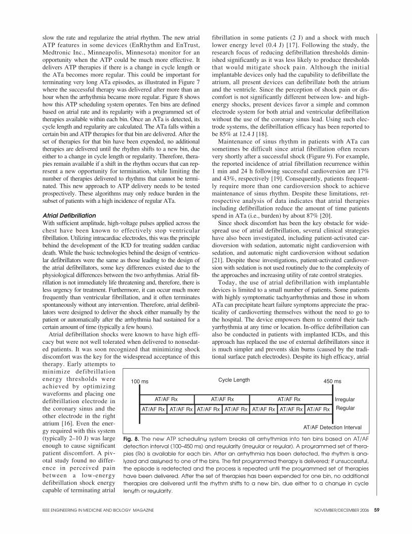
slow the rate and regularize the atrial rhythm. The new atrialATP features in some devices (EnRhythm and EnTrust,Medtronic Inc., Minneapolis, Minnesota) monitor for anopportunity when the ATP could be much more effective. Itdelivers ATP therapies if there is a change in cycle length orthe ATa becomes more regular. This could be important forterminating very long ATa episodes, as illustrated in Figure 7where the successful therapy was delivered after more than anhour when the arrhythmia became more regular. Figure 8 showshow this ATP scheduling system operates. Ten bins are definedbased on atrial rate and its regularity with a programmed set oftherapies available within each bin. Once an ATa is detected, itscycle length and regularity are calculated. The ATa falls within acertain bin and ATP therapies for that bin are delivered. After theset of therapies for that bin have been expended, no additionaltherapies are delivered until the rhythm shifts to a new bin, dueeither to a change in cycle length or regularity. Therefore, thera-pies remain available if a shift in the rhythm occurs that can rep-resent a new opportunity for termination, while limiting thenumber of therapies delivered to rhythms that cannot be termi-nated. This new approach to ATP delivery needs to be testedprospectively. These algorithms may only reduce burden in thesubset of patients with a high incidence of regular ATa.
Atrial DefibrillationWith sufficient amplitude, high-voltage pulses applied across thechest have been known to effectively stop ventricular fibrillation. Utilizing intracardiac electrodes, this was the principlebehind the development of the ICD for treating sudden cardiacdeath. While the basic technologies behind the design of ventricu-lar defibrillators were the same as those leading to the design ofthe atrial defibrillators, some key differences existed due to thephysiological differences between the two arrhythmias. Atrial fib-rillation is not immediately life threatening and, therefore, there isless urgency for treatment. Furthermore, it can occur much morefrequently than ventricular fibrillation, and it often terminatesspontaneously without any intervention. Therefore, atrial defibril-lators were designed to deliver the shock either manually by thepatient or automatically after the arrhythmia had sustained for acertain amount of time (typically a few hours).
Atrial defibrillation shocks were known to have high effi-cacy but were not well tolerated when delivered to nonsedat-ed patients. It was soon recognized that minimizing shockdiscomfort was the key for the widespread acceptance of thistherapy. Early attempts tominimize defibrillationenergy thresholds wereachieved by optimizingwaveforms and placing onedefibrillation electrode inthe coronary sinus and theother electrode in the rightatrium [16]. Even the ener-gy required with this system(typically 2–10 J) was largeenough to cause significantpatient discomfort. A piv-otal study found no differ-ence in perceived painbetween a low-energydefibrillation shock energycapable of terminating atrial
fibrillation in some patients (2 J) and a shock with muchlower energy level (0.4 J) [17]. Following the study, theresearch focus of reducing defibrillation thresholds dimin-ished significantly as it was less likely to produce thresholdsthat would mitigate shock pain. Although the initialimplantable devices only had the capability to defibrillate theatrium, all present devices can defibrillate both the atriumand the ventricle. Since the perception of shock pain or dis-comfort is not significantly different between low- and high-energy shocks, present devices favor a simple and commonelectrode system for both atrial and ventricular defibrillationwithout the use of the coronary sinus lead. Using such elec-trode systems, the defibrillation efficacy has been reported tobe 85% at 12.4 J [18].
Maintenance of sinus rhythm in patients with ATa cansometimes be difficult since atrial fibrillation often recursvery shortly after a successful shock (Figure 9). For example,the reported incidence of atrial fibrillation recurrence within1 min and 24 h following successful cardioversion are 17%and 43%, respectively [19]. Consequently, patients frequent-ly require more than one cardioversion shock to achievemaintenance of sinus rhythm. Despite these limitations, ret-rospective analysis of data indicates that atrial therapiesincluding defibrillation reduce the amount of time patientsspend in ATa (i.e., burden) by about 87% [20].
Since shock discomfort has been the key obstacle for wide-spread use of atrial defibrillation, several clinical strategieshave also been investigated, including patient-activated car-dioversion with sedation, automatic night cardioversion withsedation, and automatic night cardioversion without sedation[21]. Despite these investigations, patient-activated cardiover-sion with sedation is not used routinely due to the complexity ofthe approaches and increasing utility of rate control strategies.
Today, the use of atrial defibrillation with implantabledevices is limited to a small number of patients. Some patientswith highly symptomatic tachyarrhythmias and those in whomATa can precipitate heart failure symptoms appreciate the prac-ticality of cardioverting themselves without the need to go tothe hospital. The device empowers them to control their tach-yarrhythmia at any time or location. In-office defibrillation canalso be conducted in patients with implanted ICDs, and thisapproach has replaced the use of external defibrillators since itis much simpler and prevents skin burns (caused by the tradi-tional surface patch electrodes). Despite its high efficacy, atrial
Fig. 8. The new ATP scheduling system breaks all arrhythmias into ten bins based on AT/AFdetection interval (100–450 ms) and regularity (irregular or regular). A programmed set of thera-pies (Rx) is available for each bin. After an arrhythmia has been detected, the rhythm is ana-lyzed and assigned to one of the bins. The first programmed therapy is delivered; if unsuccessful,the episode is redetected and the process is repeated until the programmed set of therapieshave been delivered. After the set of therapies has been expended for one bin, no additionaltherapies are delivered until the rhythm shifts to a new bin, due either to a change in cyclelength or regularity.
defibrillation has not been widely accepted. Additionalresearch is needed to discover novel methods that minimizepatient discomfort while maintaining safety and effectiveness.
Prevention of ATa A variety of different strategies aimed at the prevention of ATaby pacing techniques have been pursued. While the electrophysi-ological mechanisms responsible for initiation and sustenance ofATa are still being investigated, preliminary research suggeststhat several mechanisms may be responsible. Various pacingstrategies designed to address these different mechanismsinclude 1) consistent atrial pacing, 2) atrial rate stabilization, 3)pacing from optimal sites, and 4) minimizing ventricular pacing.
Consistent Atrial PacingSlow cardiac rhythms can induce dispersion in the repolariza-tion of atrial cells, which can initiate ATa. Consistently pacingthe atrium may prevent this dispersion and, consequently, theinitiation of ATa. Early attempts at providing a high percentageof atrial pacing were achieved simply by increasing the pacingrate to 70, 80, or even 90 beats/min. Although this did result inincreased atrial pacing, it also resulted in significant increasesin heart rate, which patients frequently found to be sympto-matic. To address this problem, consistent atrial pacing algo-rithms were designed to provide a high percentage of atrialpacing with only a very modest increase in heart rate. Thesealgorithms operate by increasing the pacing rate by a pre-scribed amount when an intrinsic atrial event is sensed by thedevice. This elevated rate is maintained for a specified numberof beats and then gradually reduced until another intrinsic atrial
event is sensed. As a result, the paced atrial rate is maintainedjust slightly faster than the intrinsic heart rate. These algorithmsgenerally result in greater than 90% atrial pacing with modestincreases in heart rate of approximately 5 beats/min. High rateatrial pacing has also been used to prevent the early recurrenceof another ATa episode shortly after the termination of anepisode. These algorithms increase the atrial pacing rate for aspecified period of time immediately following the end of oneepisode when the likelihood of ATa recurrence is at its peak.Once this vulnerable period has subsided, the pacing rate isallowed to return to the normal lower rate.
Atrial Rate StabilizationPremature atrial contractions (PACs) can cause alternatingshort-long sequences that can serve as a trigger of ATa. If thisis the underlying initiation mechanism, stabilizing the variabilityin atrial cycle lengths may be effective in preventing ATa.Consequently, algorithms have been developed to eliminatethe compensatory pause that commonly follows a PAC. Whena PAC is sensed, the device automatically extends the intervalbetween the PAC and the next paced beat by a prescribedinterval. This gradual extension of the atrial interval continuesuntil the lower rate or intrinsic rate is reached.
Effect from Optimal Pacing SitesAnother strategy for the prevention of AF focuses not on howor when to pace but rather on where to pace. Traditionally, pac-ing leads have been placed in the appendage of the right atriumand the apex of the right ventricle because it is easy and reli-able, with a low risk of dislodgement. However, recent studies
IEEE ENGINEERING IN MEDICINE AND BIOLOGY MAGAZINE NOVEMBER/DECEMBER 2006
Fig. 9. Real-time telemetered signals from an implanted device. (a) Surface ECG. (b) Atrial electrogram. (c) Device markers. Anatrial cardioversion shock is delivered (see arrow). The shock is successful in terminating atrial fibrillation and restoring sinusrhythm. However, atrial fibrillation recurs within a few beats.
AS
AS
AS
AS
AS
AS
AS
AS
AR
AS
AR
AS
AR
AS
AR
AS
AR
AS
AR
AR
AS
AR
AS
AR
AS
AR
AS
SSV V VS
VS
VP
VS
VS
VS
VS
VS
VS
VS
VS
VS
(a)
(b)
(c)
IEEE ENGINEERING IN MEDICINE AND BIOLOGY MAGAZINE NOVEMBER/DECEMBER 2006 61
have shown that there are several nontraditional pacing sites(e.g., the atrial septum) that result in better hemodynamicperformance [22], [23] and fewer atrial arrhythmias [24], [25].
Clinical Results with Prevention AlgorithmsDespite the efforts to prevent ATa by various atrial pacingalgorithms, clinical trials have only shown a very moderatereduction in the number of symptomatic episodes experiencedby the patient. The Medtronic ASPECT study [26] reported a44% reduction in symptomatic AT/AF episodes in patientswith septal lead placement when consistent atrial pacing andatrial rate stabilization algorithms were activated. Similarly,the St. Jude ADOPT study [27] demonstrated a 25% reductionin the number of symptomatic AF days when their consistentatrial pacing algorithm was enabled.
No large prospective trials have been able to demonstrate areduction in the total number (asymptomatic and symptomatic)of episodes (ATa frequency) or the amount of time spent in atri-al arrhythmias (ATa burden). The increase in atrial pacing andheart rate produced by such algorithms may create additionaltachyarrhythmia detection challenges for some pacemakers and,therefore, objective evaluation of algorithm efficacy must bedone carefully. In the future, prevention algorithms may need tobe customized to individual patients. Hemodynamic and electri-cal sensors could facilitate placement of the lead at an optimalsite and provide feedback to allow implantable devices to auto-matically optimize algorithm performance.
Minimizing Ventricular PacingHistorically, pacemakers have been implanted in patients withsymptomatic bradycardia where ATa is a comorbidity. Even inpatients with an intact AV node receiving dual-chamber pace-makers, there is a high percentage of right ventricular pacingbecause these devices are programmed to pace the ventricleshortly after contraction of the atria (typically within 120–150ms). These short AV delays often do not allow sufficient time forintrinsic conduction to occur in the ventricle. As a result, pacingfrom the right ventricular apex causes less physiologic contrac-tion of the ventricle, which leads to dysynchrony of ventricularcontraction, valvular regurgitation, stretch-induced changes inatrial repolarization, and atrial dysfunction. Numerous clinicaltrials have now demonstrated that unnecessary pacing of the ven-tricle from certain locations results in an increased risk of deathor heart failure related hospitalization [28] and an increasedprevalence of AF [29], [30]. Therefore, an important componentto preventing ATa with implantable devices is to prevent ventric-ular dysfunction. Subsequent generations of pacemakers includefeatures that automatically extend the AV delay to allow forintrinsic conduction whenever possible and adjust the AV delayin response to changes in heart rate. More recently, several com-panies have introduced entirely new pacing modes that only pacethe ventricle when absolutely necessary.
SummaryPresently, most devices with atrial diagnostic and therapeuticfeatures are implanted in patients for electrical treatment ofbradyarrhythmias and ventricular tachyarrhythmias. The pain-less electrical strategies for prevention and termination of ATahave not demonstrated significant clinical effectiveness in thegeneral population with ATa. The effectiveness of ATP inreducing burden may be significantly higher in a subgroup ofpatients with a high incidence of stable ATa, but this needs to be
evaluated prospectively. Smart sensing and detection schemeswill also help provide accurate information and determine whenATa can be terminated with ATP. Although electrical defibrilla-tion is effective, the discomfort associated with atrial shocks haslimited the widespread use of this technology. Recent techno-logical advances have increased the capabilities of implantabledevices to store large amounts of diagnostic information. In thenear future, implantable devices without transvenous leads maybe implanted to monitor a variety of physiologic signals. Thiscould help improve clinical outcomes and determine whichtherapies (pharmacologic, ablative, or electrical) would be mosteffective as well as monitor their safety and efficacy. Frequentmonitoring from home and the availability of this data to thephysician/nurse on the Internet can potentially improve themanagement of patients’ ATa at a much lower cost.
Rahul Mehra received his B.S. in electricalengineering from the Indian Institute ofTechnology, Kharagpur, India, and his Ph.D.in biomedical engineering from PolytechnicInstitute of New York in 1975. Currently, heis the senior director of monitoring andcomorbidities research at Medtronic Inc,Minneapolis, Minnesota. He has been in the
research division at Medtronic for the last 22 years. During thisperiod, he was involved in research leading to the developmentof implantable defibrillators for management of sudden cardiacdeath as well as therapeutic devices for the treatment of atrialfibrillation. Before that, he was the director of computerizedelectrophysiology at the Brooklyn VA Medical Center, assistantprofessor of medicine at the Downstate Medical Center, andresearch associate at Montefiore Hospital in New York. He hasauthored or coauthored more than 70 manuscripts and 20 bookchapters, and he has more than 50 issued patents. His currentresearch interests include the development of implantable moni-toring technologies as well as therapeutic devices for manage-ment of atrial fibrillation and heart failure.
Paul Ziegler received his B.S. in mechani-cal engineering from Bradley University in1987 and his M.S. in biomedical engineer-ing with a minor in chemical engineeringfrom the University of Minnesota in 1993.From 1988–1992, he designed test equip-ment and analyzed nuclear reactor coolantpump seals for Westinghouse Corp. In
1992, he joined the heart valve division of Medtronic, Inc. todesign, develop, and test artificial mechanical heart valves andannuloplasty products used in the repair of mitral and tricuspidvalves. Since 1999, he has been a scientist with the cardiacrhythm management division of Medtronic and is responsiblefor research in the area of therapies for atrial fibrillation.
Shantanu Sarkar received his B. Tech.(Honors) degree in electronics and electri-cal communication engineering in 1993and M.Tech. degree in biomedical engi-neering in 1997 from the Indian Institute ofTechnology, Kharagpur and Bombay,respectively. In 2002, he was awardedM.E.E. and Ph.D. in biomedical engineer-
ing degrees from the University of Minnesota. From
62
1997–2002, he was a research assistant at the Center forMagnetic Resonance Research, working on developing dataacquisition and processing techniques for magnetic resonancespectroscopic imaging and functional MRI. He is presentlyworking as a scientist in the research department at MedtronicInc., Minneapolis, Minnesota, designing arrhythmia-detectionalgorithms for implantable medical devices that are used formonitoring and treating arrhythmias of the heart.
David Ritscher received his B.S.E.E. fromthe University of Massachusetts and hisM.S.E.E. from Rensselaer PolytechnicInstitute in electrical engineering, focusingon electrocardiology. He was a researcherat the University of Ulm, Germany, work-ing in cardiac diagnostics and biomedicalsignal processing, including late potential
analysis and biomagnetism. Then he was at the University ofAlabama in Birmingham with a focus on electrical mapping offibrillation. He is currently a principal scientist at Medtronic,Inc., where he is responsible for atrial therapies in pacemakersand ICDs and for implantable monitoring devices for thedetection, diagnosis, and monitoring of cardiac arrhythmias.He has more than seven patents pending or issued.
Eduardo Warman received his M.S. inphysics from the University of BuenosAires, Argentina in 1985 and his Ph.D. inbiomedical engineering from CaseWestern Reserve University in 1992. He iscurrently a senior principal scientist atMedtronic, Inc. The focus of his researchhas been the development and testing of
novel device-based atrial therapies. He has been elected atechnical fellow in Medtronic. He has more than 25 patentsissued or pending. His current research interests include thedevelopment of implantable devices for the management ofcardiac arrhythmias and hypertension.
Address for Correspondence: Rahul Mehra, Ph.D.,Medtronic Inc., 7000 Central Avenue NE, Minneapolis, MN55432 USA. Phone: +1 763 514 4500. Fax +1 763 514 2701. E-mail: [email protected].
References[1] W.M. Feinberg, J.L. Blackshear, A. Laupacis, R. Kronmal, and R.G. Hart,“Prevalence, age distribution and gender of patients with atrial fibrillation: Analysisand implications,” Arch. Internal Med., vol. 155, no. 5, pp. 469–473, 1995.[2] J. Gajewski, and R.B. Singer, “Mortality in an insured population with atrialfibrillation,” JAMA, vol. 245, no. 15, pp. 1540–1544, 1981.[3] V. Fuster, L.E. Ryden, R.W. Asinger, D.S. Cannom, H.J. Crijns, R.L. Frye, J.L.Halperin, G.N. Kay, W.W. Klein, S. Levy, R.L, McNamar, E.N. Prystowsky, L.S. Wann,and G.G. Wyse, “ACC/AHA/ESC guidelines for the management of patients with atrialfibrillation: Executive summary,” Circulation, vol. 104, no. 17, pp. 2118–2150, 2001.[4] M. Haissaguerre, D.C. Shah, P. Jais, M. Hocini, T. Yamane, I. Deisenhofer, M.Chauvin, S. Garrigue, and J. Clementy, “Electrophysiological breakthroughs fromthe left atrium to the pulmonary veins,” Circulation, vol. 102, no. 20, pp.2463–2465, 2000.[5] C.D. Swerdlow, “Detection of atrial fibrillation and flutter by a dual chamberimplantable cardioverter defibrillator. For the Worldwide Jewel AF investigators,”Circulation, vol. 101, no. 8, pp. 878–885, 2000.[6] C.W. Israel, B. Hugl, C. Unterberg, T. Lawo, I. Kennis, D. Hettrick, and S.H.Hohnloser, “The AT500 verification study investigators. Pace-termination andpacing for prevention of atrial tachyarrhythmias: Results from a multicenter studywith an implantable device for atrial therapy,” J. Cardiovasc. Electrophysiol., vol.12, no. 10, pp. 1121–1128, 2001.[7] C.W. Israel, J.R. Ehrlich, G. Gronefeld, A. Klesius, T. Lawo, B. Lemke, and S.H.Hohnloser, “Prevalence characteristics and clinical implications of regular atrial tach-
yarrhythmias in patients with atrial fibrillation: Insights from a study using a newimplantable device,” J. Amer. Coll. Cardiol., vol. 38, no. 2, pp. 355–363, 2001.[8] A. Shalaby, et al. “Can novel digital signal processing technology enable suc-cessful device classification of atrial tachyarrhythmias?” Heart Rhythm, vol. 2, no. 5, pg. S124, 2005.[9] A.K. Joshi, et al. “First experience with a mobile cardiac outpatient telemetry(MCOT) system for the diagnosis and management of cardiac arrhythmia,” Amer.J. Cardiol., vol. 95, no. 7, pp. 878–881, 2005.[10] A.L. Waldo, W.A. MacLean, R.B. Karp, N.T. Kouchoukos, and T.N. James,“Entrainment and interruption of atrial flutter with atrial pacing: Studies in manfollowing open heart surgery,” Circulation, vol. 56, no. 5, pp. 737-745, 1977. [11] J.T. Hii, L.B. Mitchell, H.J. Duff, D.G. Wyse, and AM. Gillis, “Comparisonof atrial overdrive pacing with and without extrastimuli for termination of atrialflutter,” Amer. J. Cardiol., vol. 70, no. 4, pp. 463–467, 1992.[12] M.A. Lee, R. Weachter, S. Pollak, M.S. Kremers, A.M. Naik, R. Silverman, J.Tuzi, W. Wang, L.J. Johnson, and D.E. Euler, ATTEST Investigators. “The effectof atrial pacing therapies on atrial tachyarrhythmia burden and frequency results ofa randomized trial in patients with bradycardia and atrial tachyarrhythmias,” J.Amer. Coll. Cardiol., vol. 41, no. 11, pp. 926–932, 2003.[13] A.M. Gillis, J. Koehler, R. Mehra, D.A. Hettrick, “High atrial ATP therapyefficacy is associated with a reduction in atrial tachyarrhythmia burden,” HeartRhythm, vol. 1, no. 8, p. S66, 2004.[14] S. Sarkar, J. Koehler, D. Ritscher, and R. Mehra, “Reduction in atrial tachy-cardia duration in episodes treated with atrial ATP therapy,” Heart Rhythm, vol. 2,no. 5, p. S320, 2005.[15] P. Mabo, et. al. “The LEAF (low energy in atrial fibrillation) study results:Evaluation of device based therapies for atrial tachyarrhythmia prevention and ter-mination,” Heart Rhythm, vol. 2, no. 5, p. S17, 2005.[16] A.R. Mitchell, P.A. Spurrell, K. Kamalvand, M. Higson, R. Shanmuganathan,N.R. Patel, and N. Sulke, “What is the optimal electrode configuration for atrialdefibrillators in man?” Europace, vol. 4, no.1, pp. 41–44, 2002.[17] D.M. Steinhaus, D.S. Cardinal, L. Mongeon, S.K. Musley, L. Foley, and S.Corrigan, “Internal defibrillation: Pain perception of low energy shocks,” PacingClin. Electrophysiol., vol. 25, no. 7, pp. 1090–1093, 2002.[18] C.D. Swerdlow, D. Schwartzman, R. Hoyt, S.J. Bailin, J.L. Koehler, and E.N.Warman, “Determinants of first-shock success for atrial implantable cardioverterdefibrillators,” J. Cardiovasc. Electrophysiol., vol. 13, no. 4, pp. 347–354, 2002. [19] D. Schwartzman, S.K. Musley, C. Swerdlow, R.H. Hoyt, and E.N. Warman,“Early recurrence of atrial fibrillation after ambulatory shock conversion,” J.Amer. Coll. Cardiol., vol. 40, no. 1, pp. 93–99, 2002.[20] P.A. Friedman, B. Dijkman, E.N. Warman, H.A. Xia, R. Mehra, M.S.Stanton, and S.C. Hammill, “Atrial therapies reduce atrial arrhythmia burden indefibrillator patients,” Circulation, vol. 104, no. 9, pp. 1023–1028, 2001.[21] L. Boodhoo, A. Mitchell, M. Ujhelyi, and N. Sulke, “Improving the accept-ability of the atrial defibrillator: Patient-activated cardioversion versus automaticnight cardioversion with and without sedation (ADSAS 2),” Pacing Clin.Electrophysiol., vol. 27, no. 7, pp. 910–917, 2004.[22] D.A. Hettrick, D.E. Euler, P.S. Pagel, S.K. Musley, E.N. Warman, P.D.Ziegler, and R. Mehra, “Atrial pacing lead location alters the effects of atrioven-tricular delay on atrial and ventricular hemodynamics,” Pacing Clin.Electrophysiol., vol. 25, no. 6, pp. 888–896, 2002.[23] A. Prakash, S. Saksena, P.D. Ziegler, T. Lokhandwala, D.A. Hettrick, P.Delfaut, N.C. Nanda, and D.G. Wyse, “Dual site right atrial pacing can improvethe impact of standard dual chamber pacing on atrial and ventricular mechanicalfunction in patients with symptomatic atrial fibrillation: Further observations fromthe dual site atrial pacing for prevention of atrial fibrillation trial,” J. Interv. Card.Electrophysiol., vol. 12, no. 3, pp. 177–187, 2005.[24] S.J. Bailin, S. Adler, and M. Giudici, “Prevention of chronic atrial fibrillationby pacing in the region of Bachmann’s bundle: Results of a multicenter random-ized trial,” J. Cardiovasc. Electrophysiol., vol. 12, no. 8, pp. 912–917, 2001.[25] S. Saksena, A. Prakash, P. Ziegler, J.D. Hummel, P. Friedman, V.J. Plumb,D.G. Wyse, E. Johnson, S. Fitts, and R. Mehra, “Improved suppression of recur-rent atrial fibrillation with dual-site right atrial pacing and antiarrhythmic drugtherapy,” J. Amer. Coll. Cardiol., vol. 40, no. 6, pp. 1140–1150, 2002.[26] L. Padeletti, H. Purerfellner, S.W. Adler, T.J. Waller, M. Harvey, L. Horvitz, R.Holbrook, K. Kempen, A. Mugglin, and D.A. Hettrick, “Combined efficacy of atrialseptal lead placement and atrial pacing algorithms for prevention of paroxysmal atrialtachyarrhythmia,” J. Cardiovasc. Electrophysiol., vol. 14, no. 11, pp. 1189–1195, 2003.[27] M.D. Carlson, J. Ip, J. Messenger, S. Beau, S. Kalbfleisch, P. Gervais, D.A.Cameron, A. Duran, J. Val-Mejias, J. Mackall, and M. Gold, “A new pacemaker algo-rithm for the treatment of atrial fibrillation: Results of the atrial dynamic overdrivepacing trial (ADOPT),” J. Amer. Coll. Cardiol., vol. 42, no. 4, pp. 627–633, 2003.[28] B.L. Wilkoff, J.R. Cook, A.E. Epstein, H.L. Greene, A.P. Hallstrom, H. Hsia,S.P. Kutalek, and A. Sharma, “Dual-chamber pacing or ventricular backup pacing inpatients with an implantable defibrillator: The dual chamber and VVI implantabledefibrillator (DAVID) trial,” JAMA, vol. 288, no. 24, pp. 3115–3123, 2002.[29] C.R. Kerr, S.J. Connolly, H. Abdollah, R.S. Roberts, M. Gent, S. Yusuf, A.M.Gillis, A.S. Tang, M. Talajic, G.J. Klein, and D.M. Newman, “Canadian trial ofphysiological pacing: Effects of physiological pacing during long-term follow-up,”Circulation, vol. 109, no. 3, pp. 357–362, 2004.[30] G.A. Lamas, K.L. Lee, M.O. Sweeney, R. Silverman, A. Leon, R. Yee, R.A.Marinchak, G. Flaker, E. Schron, E.J. Orav, A.S. Hellkamp, S. Greer, J. McAnulty,K. Ellenbogen, F. Ehlert, R.A. Freedman, N.A. Estes 3rd, A. Greenspon, and L.Goldman, “Ventricular pacing or dual-chamber pacing for sinus-node dysfunction,”New Eng. J. Med., vol. 346, no. 24, pp. 1854–1862, 2002.
IEEE ENGINEERING IN MEDICINE AND BIOLOGY MAGAZINE NOVEMBER/DECEMBER 2006