66
Management of Cervical Cancer Lilie Lin, MD Assistant Professor March 23, 2013 University of Pennsylvania
Management of Cervical Cancer Lilie Lin, MD
Assistant Professor March 23, 2013
University of Pennsylvania

Learning Objectives
• Discuss the state of the art treatments for cervical cancer
• Identify basic competencies needed to perform gynecologic IMRT
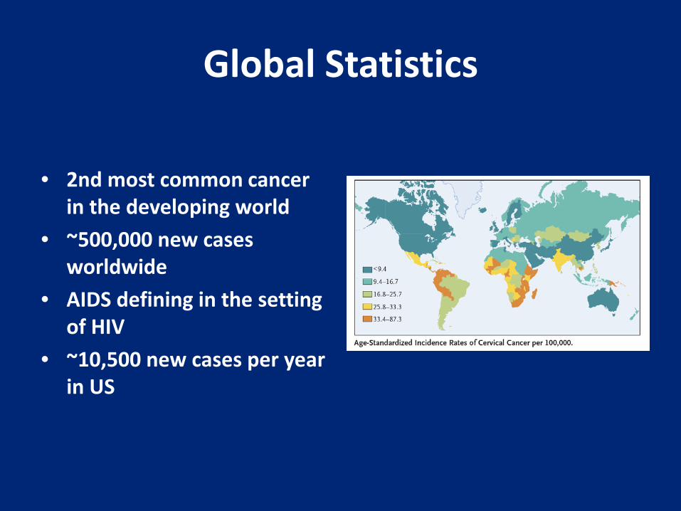
Global Statistics
• 2nd most common cancer in the developing world
• ~500,000 new cases worldwide
• AIDS defining in the setting of HIV
• ~10,500 new cases per year in US
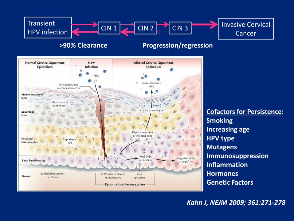
Kahn J, NEJM 2009; 361:271-278
Transient HPV infection CIN 1 CIN 2 CIN 3
>90% Clearance
Invasive Cervical Cancer
Progression/regression
Cofactors for Persistence: Smoking Increasing age HPV type Mutagens Immunosuppression Inflammation Hormones Genetic Factors
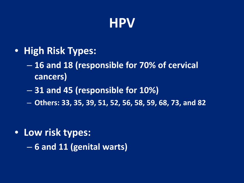
HPV
• High Risk Types: – 16 and 18 (responsible for 70% of cervical
cancers) – 31 and 45 (responsible for 10%) – Others: 33, 35, 39, 51, 52, 56, 58, 59, 68, 73, and 82
• Low risk types:
– 6 and 11 (genital warts)
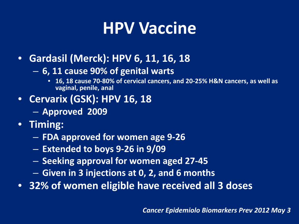
HPV Vaccine • Gardasil (Merck): HPV 6, 11, 16, 18
– 6, 11 cause 90% of genital warts • 16, 18 cause 70-80% of cervical cancers, and 20-25% H&N cancers, as well as
vaginal, penile, anal
• Cervarix (GSK): HPV 16, 18 – Approved 2009
• Timing: – FDA approved for women age 9-26 – Extended to boys 9-26 in 9/09 – Seeking approval for women aged 27-45 – Given in 3 injections at 0, 2, and 6 months
• 32% of women eligible have received all 3 doses
Cancer Epidemiolo Biomarkers Prev 2012 May 3

Histologic Subtypes
• Squamous cell carcinoma (85%) • Adenocarcinoma (10%) • Clear cell carcinoma (associated with in utero
DES exposure) 1% • Other (rare)
– Sarcoma – Lymphoma – Small cell carcinoma – Neuroendocrine carcinoma
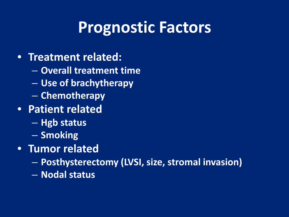
Prognostic Factors
• Treatment related: – Overall treatment time – Use of brachytherapy – Chemotherapy
• Patient related – Hgb status – Smoking
• Tumor related – Posthysterectomy (LVSI, size, stromal invasion) – Nodal status

Staging and Workup

FIGO Clinical Staging
• Pelvic exam-under anesthesia • Cystoscopy • Proctoscopy
• Chest Xray • Intravenous Pyelogram • Skeletal Survey

FIGO Clinical Staging-Not allowed
• CT
• MRI
• PET/CT
• Exploratory Surgery

FIGO Staging
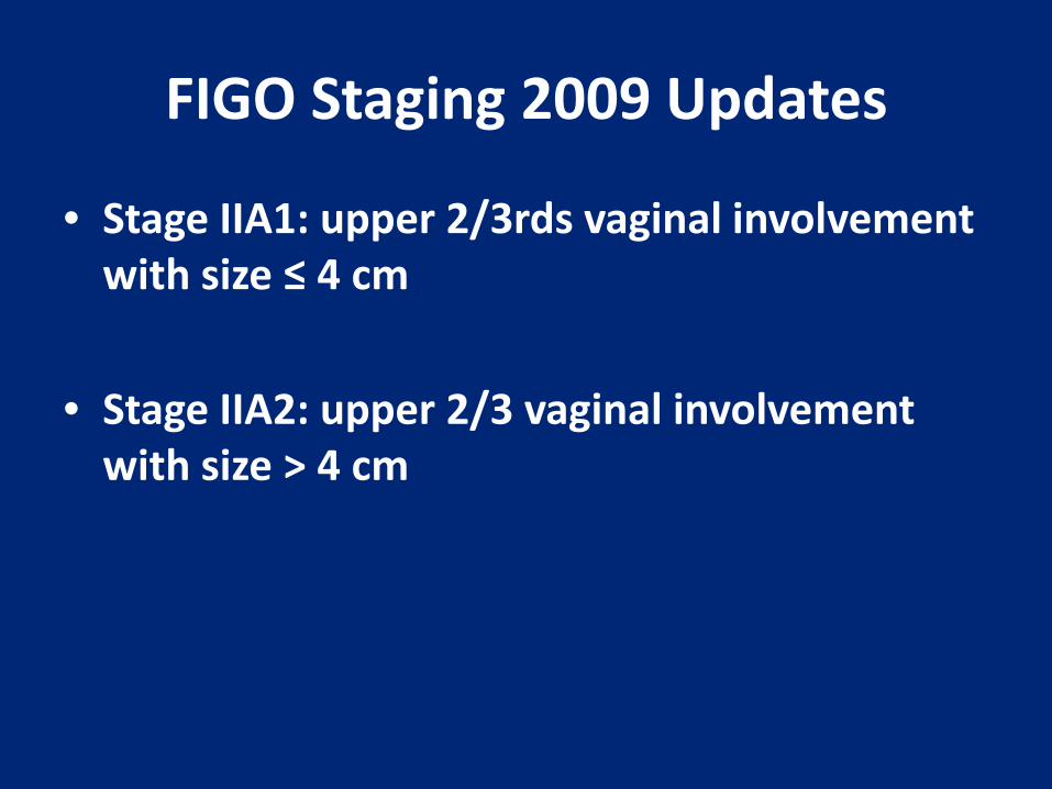
FIGO Staging 2009 Updates
• Stage IIA1: upper 2/3rds vaginal involvement with size ≤ 4 cm
• Stage IIA2: upper 2/3 vaginal involvement with size > 4 cm

Patterns of Spread
• Direct Extension – Vaginal mucosa – Myometrium of lower
uterine segment and corpus (endocervical lesions)
– Parametria • Lymph Nodes:
– Obturator LNs, Int/Ext/ common Iliacs, Para-aortic LNs, and inguinals (vaginal disease)
• Late disease – Distant metastases to bone, lung or liver,
supradiaphragmatic LN disease

Determining Extent of Primary Tumor
• Pelvic examination – Staging Accuracy: 47%
• Bipat et al, Gyn Onc 2003
• MRI vs CT
– Staging Accuracy: 86% – MR is superior to CT for detecting uterine body
involvement/PM invasion (ACRIN 6651/GOG 183) • JCO 2006
– MR superior in detecting vaginal extension

Determining Extent of LN/Distant Metastases: Role of FDG-PET/CT

Risk of Lymph Node Metastases
Stage Pelvic LN PA LN IA1 <1% IA2 6-7% <3% IB 15% 10% IIB 30% 20% III 45% 30%

FDG PET/CT Nodal Status
Kidd et al, JCO 2010
RFS ALL DSS ALL
DSS Stage I
DSS Stage III
DSS Stage II
Treatment
• Surgery
• Radiation Therapy
• Chemotherapy

Surgical Management: Early stage IA
• LEEP (Loop electrosurgical excision procedure)
• Conization • Cryotherapy • Radical trachelectomy
– 3-6% recurrence rate when limited to <2 cm (-nodes, -LVSI)
• Radical hysterectomy: IA2

Surgery • Radical Hysterectomy: Class II hysterectomy generally sufficient
Lymphadenectomy:
-Stage IA2 or small IB1: -low risk of node + (2-8%) -pelvic LN resection only (no -Exception: pelvic + paraaortic LN resection if PET/CT data that they may be positive.
Partial mobilization of the ureters
Uterosacral ligaments ligated midway b/t uterus and sacrum.
Medial half of cardinal ligament excised
Upper third of vagina excised
Uterine vessels ligated medial to ureters.

Treatment for Stage IB
• Radical hysterectomy and pelvic +/- PA LND
• Radiotherapy – EBRT and brachytherapy: 80 Gy to point A

Primary radiotherapy vs Radical Hysterectomy
469 Pts 1986-1991
Stages IB-IIA
Radical surgery Class III rad hyst
RT 40-53 Gy 1.8-2 Gy
ICI x1 Pt A dose 70-90 Gy
Adj XRT 50.4 Gy to pelvis
PA to 45 Gy if PA nodes +
Stage pT2b/+LN/+SM
Landoni et al, Lancet 1997
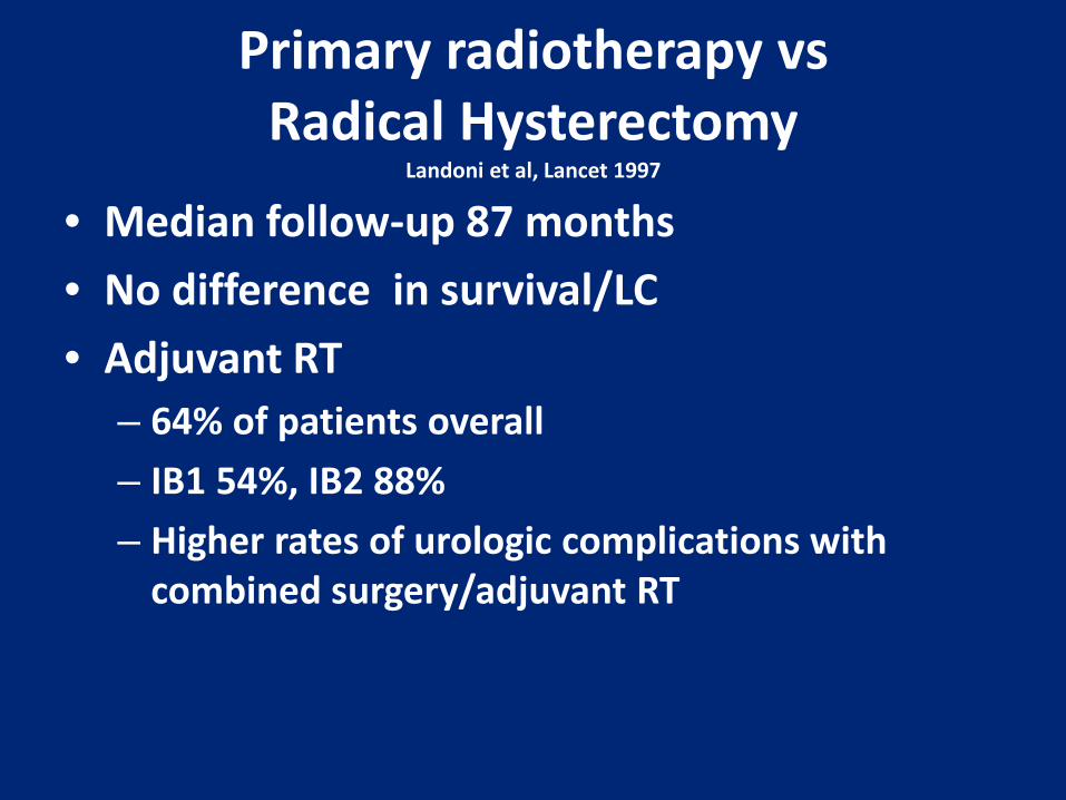
• Median follow-up 87 months • No difference in survival/LC • Adjuvant RT
– 64% of patients overall – IB1 54%, IB2 88% – Higher rates of urologic complications with
combined surgery/adjuvant RT
Primary radiotherapy vs Radical Hysterectomy
Landoni et al, Lancet 1997
Role of Adjuvant Radiotherapy

GOG 92: LN Negative Role of adjuvant post-operative radiotherapy
277 Pts Stage IB 1988-1995
Rad Hyst & LND >1/3 stromal invasion/LVSI/LTD*
RT 46-50.4 Gy pelvis 23-28 fx 1.8-2.0 Gy
(No brachy) No Further Therapy
Sedlis et al, Gyn Onc 1999
* Pts with LN not eligible
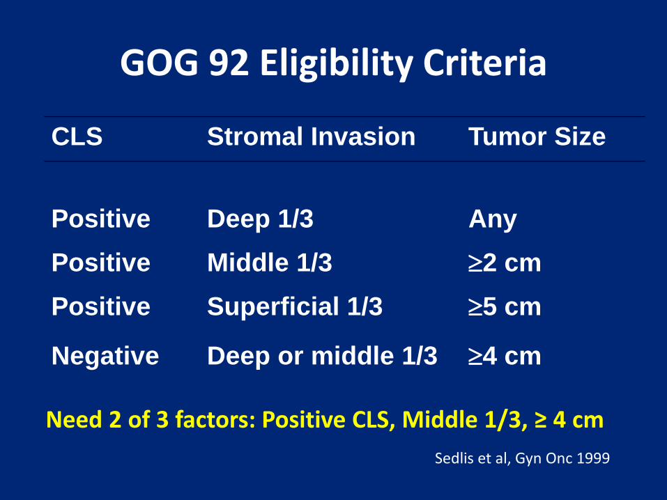
GOG 92 Eligibility Criteria
CLS Stromal Invasion Tumor Size
Positive Deep 1/3 Any Positive Middle 1/3 ≥2 cm Positive Superficial 1/3 ≥5 cm
Negative Deep or middle 1/3 ≥4 cm
Sedlis et al, Gyn Onc 1999
Need 2 of 3 factors: Positive CLS, Middle 1/3, ≥ 4 cm

GOG 92: Sites of Failure
Radiotherapy No Radiotherapy
Local
13% 19%
Distant
2% 7%
Sedlis et al, Gyn Onc 1999

GOG 92: Update • Median f/u: 10 years • PFS: 46% reduction in HR • Overall survival:
– 30% improvement (p=0.074)
• Grade 3/4 toxicity: – 6.6% (RT) vs 2.1% (obs)
• Adenocarcinoma and adenosquamous recurrence rate: – 8.8% (RT) vs 44% (obs),
p=0.019
Rotman et al, IJROPB 2006

Radiation vs Radiation and Concurrent and Adjuvant
Cisplatin/5FU after Hysterectomy
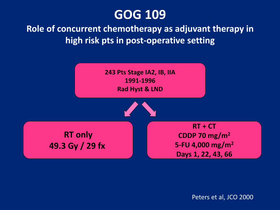
GOG 109 Role of concurrent chemotherapy as adjuvant therapy in
high risk pts in post-operative setting
243 Pts Stage IA2, IB, IIA 1991-1996
Rad Hyst & LND
RT only 49.3 Gy / 29 fx
RT + CT CDDP 70 mg/m2
5-FU 4,000 mg/m2
Days 1, 22, 43, 66
Peters et al, JCO 2000

Eligibility Criteria
• Positive Pelvic Lymph Nodes
• Positive Parametrial Involvement
• Positive Surgical Margins
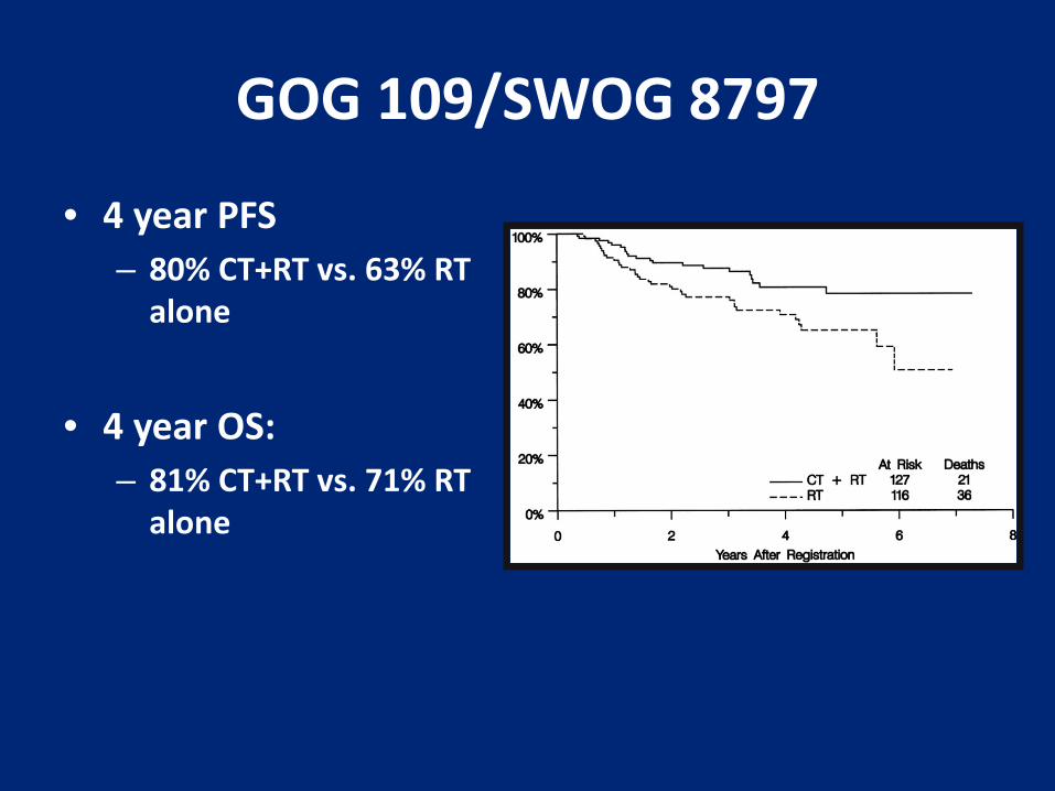
GOG 109/SWOG 8797
• 4 year PFS – 80% CT+RT vs. 63% RT
alone
• 4 year OS:
– 81% CT+RT vs. 71% RT alone

Treatment of Locally Advanced Cervical Cancer

Cervical Cancer Definitive Chemoradiation

Concurrent weekly CDDP/RT
GOG 120 Rose, 1999
NCIC Pearcey, 2002
GOG 123 Keys, 1999
Stage IIB-IVA IA – IIA, > 5cm IIB
IB2
Arms WPRT/B/HU WPRT/B/cis/5FU/HU WPRT/B/weekly cis
WPRT/B WPRT/B + wkly cis
WPRT/B + SH WPRT/B/wkly cis + SH
OS 47% 65% (3 year)
62% 58% (5 year), p = NS
74% 83% (3 year)
LR 21% 37%
Notes ↓toxicity with cis or HU alone
Non-surgical staging of nodes
↑pCR with chemo (52 vs 41%)

Concurrent CDDP/5FU and RT
RTOG 90-01 Morris, 1999
SWOG 8797 Peters, 2000
GOG 85 Whitney, 1999
Stage IIB – IVA IB-IIA > 5cm LN +
IA2-IIA (posthys) IIB-IVA
Arms EFRT/B WPRT/B + cis5FU
WPRT WPRT + cis/5FU x 4 cycles
WPRT/B /HU WPRT/B/cis/5FU
OS 41% 67% (8 year)
71% 81% (4 year)
43% 55% (3 year)
LR 35% 18%
Notes NS ↑ PAN failures in CRT arm
Postop (+LN, +PM, +margins)
Late complications 16% (equivalent)
What was the 5 year overall survival benefit of concurrent chemo observed in the meta-analysis published in 2008 (JCO) by the
Chemoradiotherapy for Cervical Cancer Meta-analysis collaboration?
1. 20% 2. 15% 3. 12% 4. 6%

Chemo-radiotherapy Meta-analysis
• 15 randomized trials of CT+RT vs RT – 11 Platinum based – 3 Nonplatinum
• 3452 pts • CT+RT vs RT
– 8% absolute improvement in DFS (50% to 58%)
• Also for locoregional DFS and distant metastases free survival
– Overall survival benefit of 6% (60% to 66%) for CT+RT vs RT
• CT+RTCT vs RT alone – Two trials – 19% absolute OS improvement
(60% to 79%)
JCO 2010
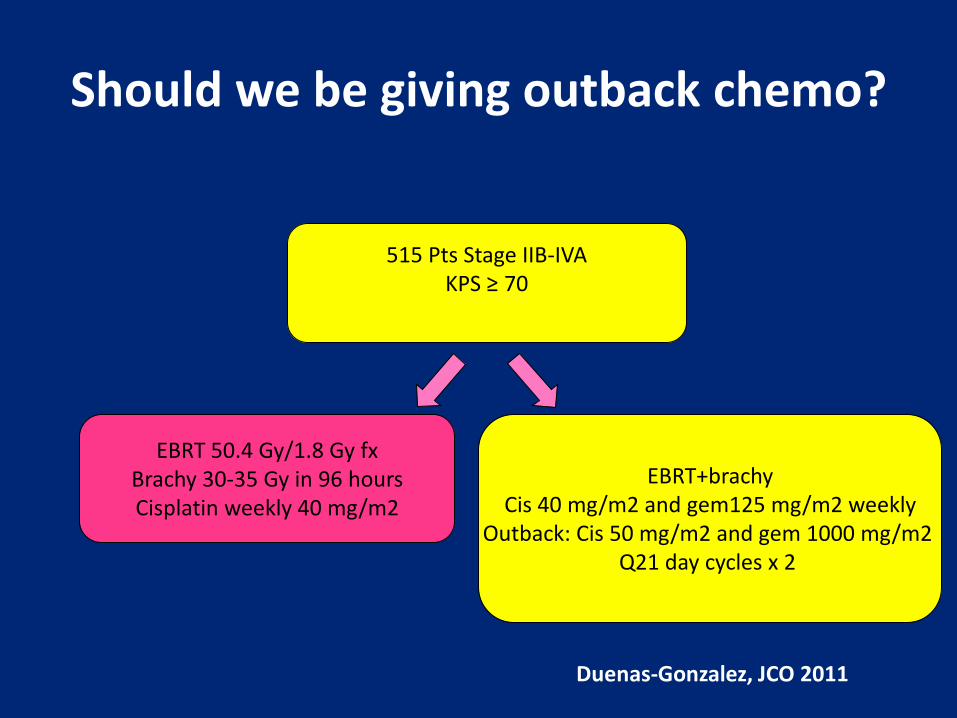
Should we be giving outback chemo?
515 Pts Stage IIB-IVA KPS ≥ 70
EBRT 50.4 Gy/1.8 Gy fx Brachy 30-35 Gy in 96 hours Cisplatin weekly 40 mg/m2
EBRT+brachy Cis 40 mg/m2 and gem125 mg/m2 weekly
Outback: Cis 50 mg/m2 and gem 1000 mg/m2 Q21 day cycles x 2
Duenas-Gonzalez, JCO 2011

Should we be giving outback chemo?
• Use of cisplatin plus gemcitabine resulted in • An improvement in progression free survival
compared to cisplatin alone (3 year PFS 74% versus 65%)
• An improvement in overall survival • Significantly more serious (grade ¾) toxicities
(87% versus 46%) and rate of hospitalizations (30% versus 11%)
Duenas-Gonzalez A et al, JCO 2011
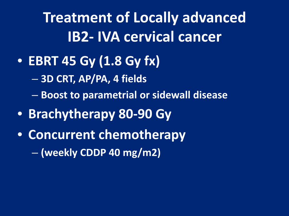
Treatment of Locally advanced IB2- IVA cervical cancer
• EBRT 45 Gy (1.8 Gy fx) – 3D CRT, AP/PA, 4 fields – Boost to parametrial or sidewall disease
• Brachytherapy 80-90 Gy • Concurrent chemotherapy
– (weekly CDDP 40 mg/m2)

Radiation Therapy
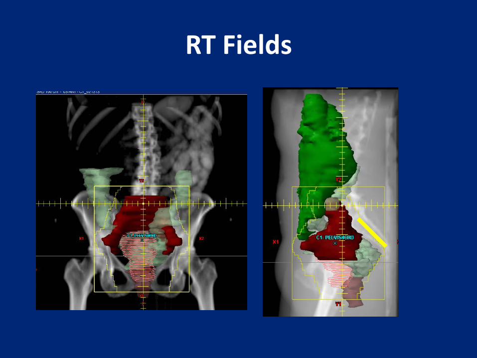
RT Fields

FDG PET/CT and MR simulation

FDG PET/CT Simulation

MR Simulation

Use of IMRT for Intact Cervix: Controversies
• Contouring
• Organ Motion
• Simulation/Setup/IGRT

• http://www.rtog.org/CoreLab/ContouringAtlases/GYN.aspx

IMRT for Intact Cervix Cancer
Lim et al, IJROBP 2011
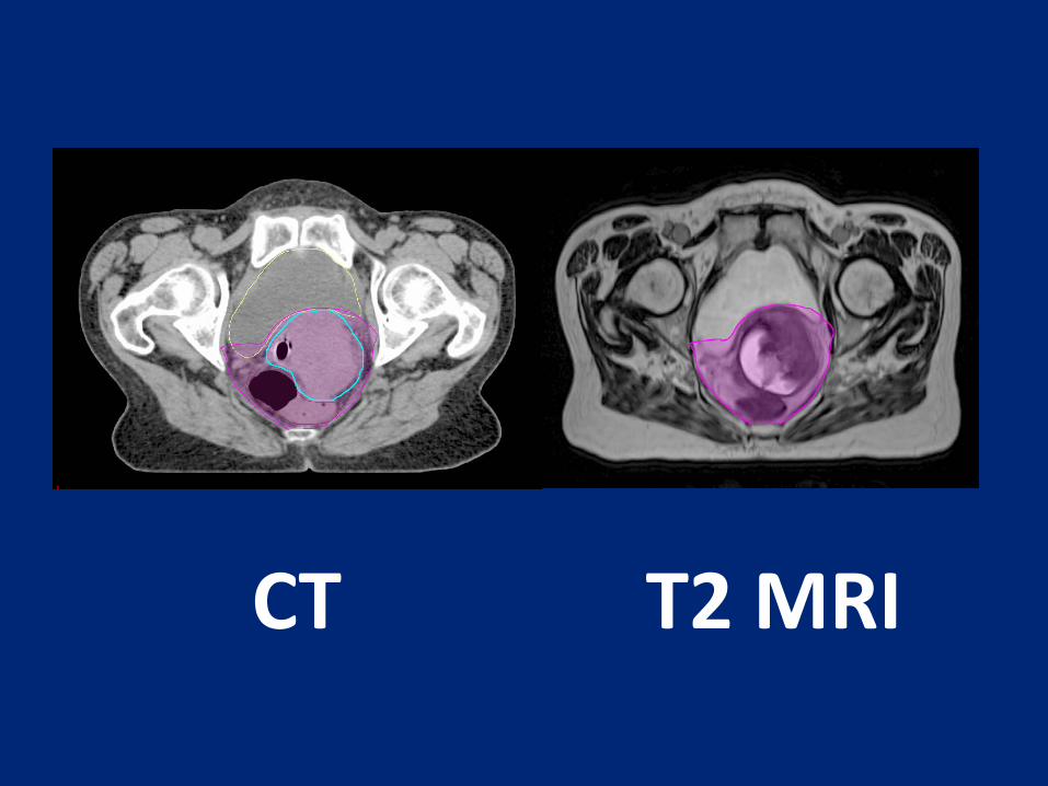
CT T2 MRI

How much margin is needed?
Van den Bunt et al, Radio and Onc 2008

Every Patient is Unique…
Van den Bunt et al, Radio and Onc 2008

IMRT Considerations for Intact Cervix Cancer
• Simulation: bladder full and bladder empty • MRI pretreatment or at the time of
simulation • Margins:
– CTV PTV margins for primary CTV: 1.5-2 cm – CTV PTV margins for nodal CTV: 7 mm
• Daily soft tissue IGRT
Uses for IMRT
• PA nodes
• Inguinal nodes
• Boost – Nodal boost – Sidewall boost

Contouring PA Nodes
Takiar et al, IJROBP 2013
Upper 1/3 4%
Middle 1/3 36%
Lower 1/3 60%
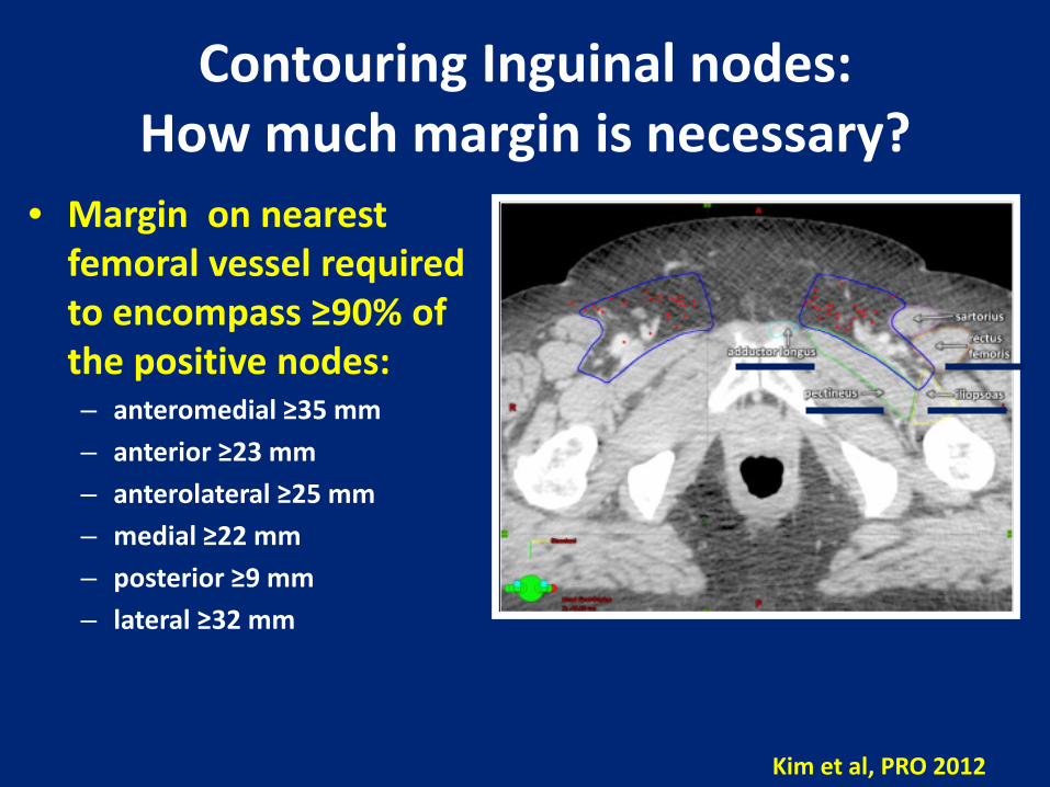
Contouring Inguinal nodes: How much margin is necessary?
• Margin on nearest femoral vessel required to encompass ≥90% of the positive nodes: – anteromedial ≥35 mm – anterior ≥23 mm – anterolateral ≥25 mm – medial ≥22 mm – posterior ≥9 mm – lateral ≥32 mm
Kim et al, PRO 2012

PET Defined Nodal Boost

Brachytherapy
Brachytherapy
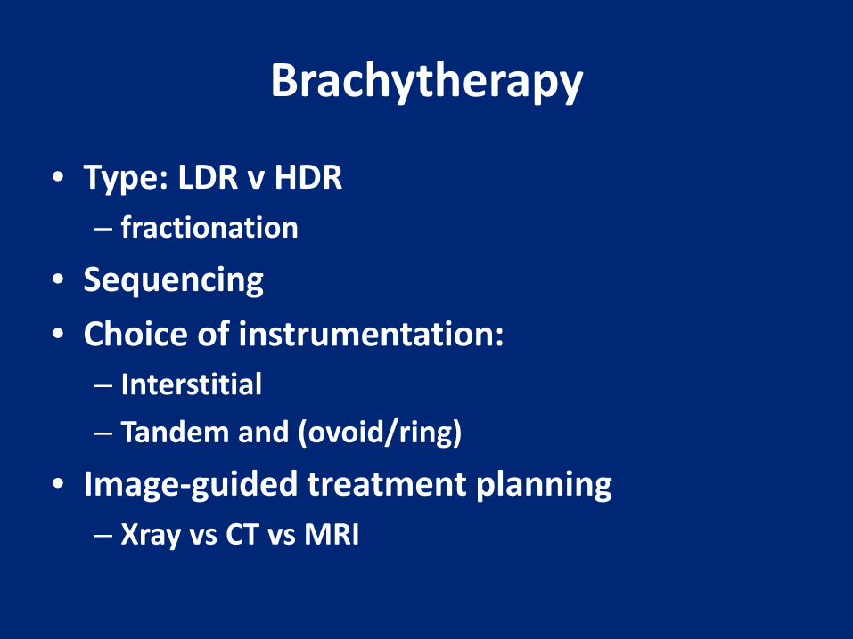
• Type: LDR v HDR – fractionation
• Sequencing • Choice of instrumentation:
– Interstitial – Tandem and (ovoid/ring)
• Image-guided treatment planning – Xray vs CT vs MRI
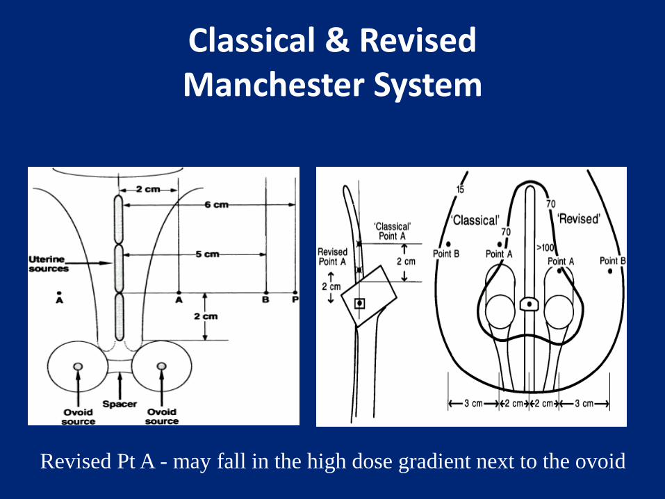
Classical & Revised Manchester System
Revised Pt A - may fall in the high dose gradient next to the ovoid
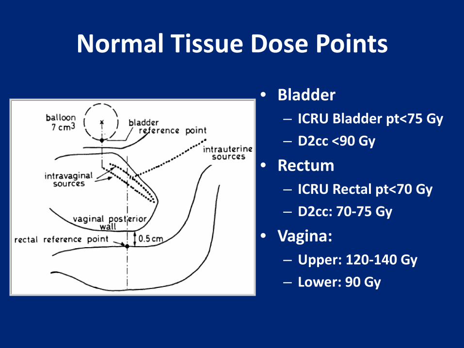
Normal Tissue Dose Points
• Bladder – ICRU Bladder pt<75 Gy – D2cc <90 Gy
• Rectum – ICRU Rectal pt<70 Gy – D2cc: 70-75 Gy
• Vagina: – Upper: 120-140 Gy – Lower: 90 Gy

MRI for Image Guided Brachytherapy

Treatment Schema
EBRT (40-45 Gy)
Chemo (CDDP)
LDR Brachy
HDR Brachy
Week 1 2 3 4 5 6 7 8
Boost

45 Gy Pelvis EQD2
Fx # Dose EQD2 Tumor
EQD2 Normal tissue (90% PD)
EQD2 Normal tissue (70% of PD)
4 7 Gy 83.9 Gy 90.1 Gy 74.2 Gy
5 6 Gy 84.3 Gy 88.6 Gy 73.4 Gy
6 5 Gy 81.8 Gy 83.7 Gy 70.5 Gy
5 5.5 Gy 79.8 Gy 82.6 Gy 69.6 Gy

Summary
• Concurrent chemoradiotherapy for locally advanced cervical cancer
• Role of outback chemotherapy in the locally advanced setting is an open question
• IMRT may be reasonable, but must consider: – Contouring – Organ motion/Margins – Image guidance/daily IGRT


