44
PETRA KLINGE ASSOCIATE PROFESSOR OF NEUROSURGERY WARREN ALPERT MEDICAL SCHOOL BROWN UNIVERSITY Management of children with spina bifida and hydrocephalus

PETRA KLINGE ASSOCIATE PROFESSOR OF NEUROSURGERY
WARREN ALPERT MEDICAL SCHOOL BROWN UNIVERSITY
Management of children with spina bifida and hydrocephalus

Management of children with Spina bifida and Hydrocephalus
www.revolutionhealth.com
Spina Bifida and Neural Tube Defects
Epidemiology One of the most common birth defects: 1-2 cases/1,000 births
Certain populations have a greater risk: Highest incidence in Ireland and Wales More common in girls U.S.: 0.7/1,000 live births Higher on the East Coast than on the West Coast Higher in whites (1/1,000 births) Lower in African-Americans (0.1-0.4/1,000 births)

Spina Bifida and Neural Tube Defects
Epidemiology Risk factors:
Race and ethnicity Family history of neural tube defects Folate deficiency Medication/teratogenic effect: valproic acid Maternal age Diabetes Obesity Increased body temperature
Hol FA et al, Clinical Genetics, 2008

Embryology of spina bifida Weeks 3-4 of gestation
“Primary Neurulation” Canalization
Weeks 6 – 10 of gestation “ Secondary Neurulation: Retrogressive differentiation
Management of children with spina bifida in the age of fetal intervention
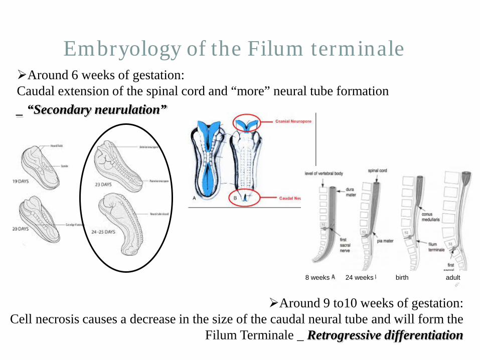
Embryology of the Filum terminale Around 6 weeks of gestation: Caudal extension of the spinal cord and “more” neural tube formation _ “Secondary neurulation”
Around 9 to10 weeks of gestation: Cell necrosis causes a decrease in the size of the caudal neural tube and will form the
Filum Terminale _ Retrogressive differentiation
8 weeks 24 weeks birth adult

Spina Bifida and Neural Tube Defects
Definitions and Classification Open spina bifida (Aperta)
Meningocele in 5% Myelomeningocele (cord and cauda equina exposed) in 95%
Closed spina bifida (Occulta) 50% have cutaneous stigmata Cord is tethered through abnormal FILUM

Can it be diagnosed in utero? Magnetic Resonance Imaging
Management of children with spina bifida and hydrocephalus

Surgical Aspects MMC closure
N E U R O S U R G E R Y & P L A S T I C S U R G E R Y

Spina Bifida and Neural Tube Defects
Definitive repair of the open neural tube defect Closure within 24 hours No evidence that immediate/urgent closure improves function But: early closure reduces risk of infection
Wound colonization after 36 hours
Surgical technique: (neurosurgeon + plastic surgeon team) Placode dissected off arachnoid Allowed to drop into spinal canal Dura dissected off skin and lumbodorsal fascia Dura closed Muscular fascia closed Skin closed
CSF Placode
Meninges
SKIN
FASCIA

Spina Bifida and Neural Tube Defects
Definitive repair of the open neural tube defect No Repair of posterior vertebral defect Thecal sac Cord extruded into the sac (placode)
Plate of embryonic epithelial cells: spinal cord


„Formal repair of MMC“
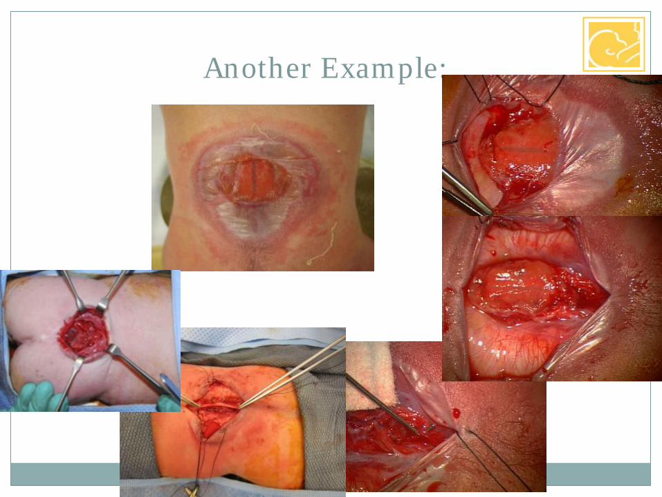
Another Example:

Spina Bifida and Neural Tube Defects
Pathophysiology and associated disorders Hydrocephalus 80-95% incidence in myelomeningocele
100% of 35 thoracic lesions 88% of 114 lumbar lesions 68% of 40 sacral lesions
Significant in 20% at birth
Rintoul et al, Pediatrics 2002

Spina Bifida and Neural Tube Defects
Management of hydrocephalus Serial head ultrasounds in the newborn:

Treatment of Hydrocephalus
Acute: External ventricular drain
Treatment of Hydrocephalus
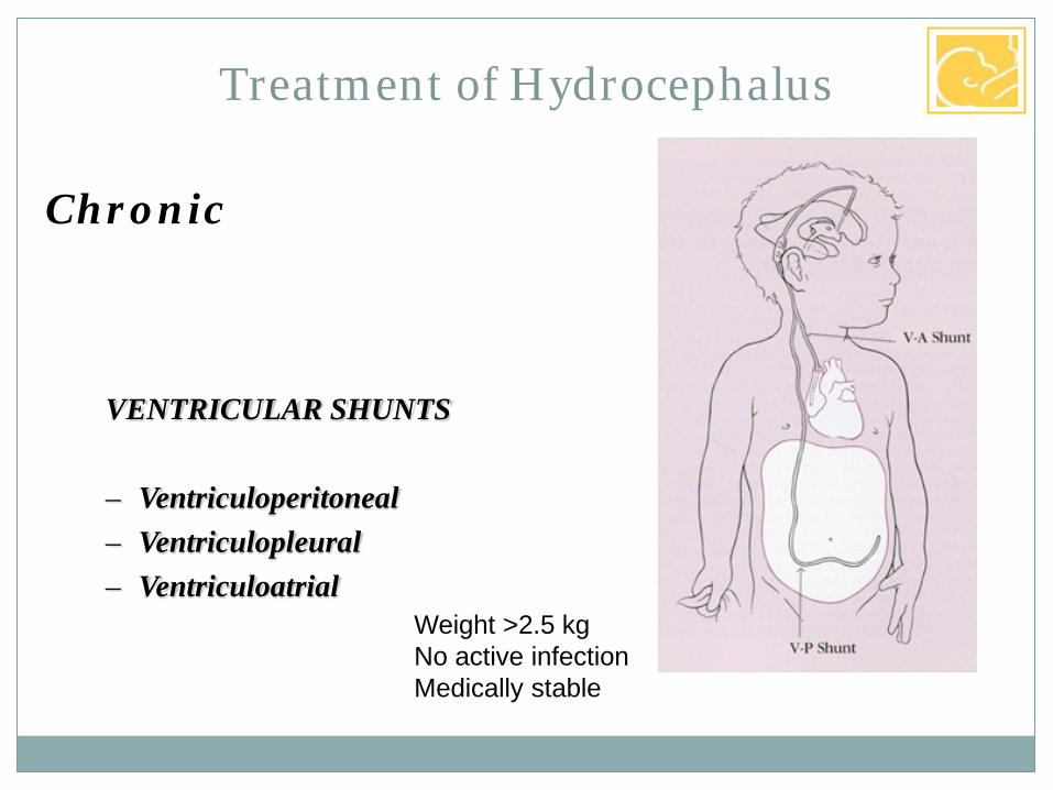
Chronic
VENTRICULAR SHUNTS
– Ventriculoperitoneal – Ventriculopleural – Ventriculoatrial
Weight >2.5 kg No active infection Medically stable

What is a shunt made of?
5cm

Spina Bifida and Neural Tube Defects
Management of hydrocephalus Types of shunts:
Adjustable valves

Endoscopic 3rd ventriculoscopy for obstructive Hydrocephalus
* C
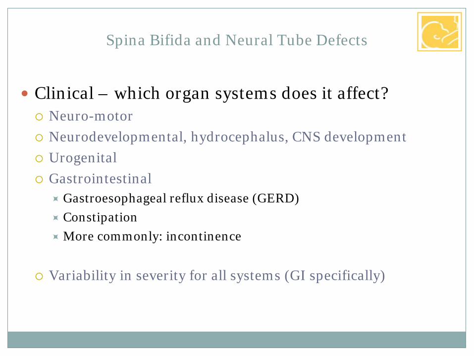
Clinical – which organ systems does it affect? Neuro-motor Neurodevelopmental, hydrocephalus, CNS development Urogenital Gastrointestinal
Gastroesophageal reflux disease (GERD) Constipation More commonly: incontinence
Variability in severity for all systems (GI specifically)
Spina Bifida and Neural Tube Defects

Current management of spina bifida: Spina bifida clinic
Relatively recent: now that these children survive long-term The most difficult – chronic vigilance CNS monitoring: VP shunt management and Management of tethered cord
(10%) Physical therapy evaluation/motor function of lower extremities Preventive medicine – insensate lower body Psychological support Gastroesophageal reflux disease (GERD) Incontinence (urine and stool) Rectum and bladder share parasympathetic (S2-S4) and
sympathetic (L1-L3) nerve roots Dysfunctional Elimination Syndrome (DES)
Spina Bifida and Neural Tube Defects

Spina Bifida and Neural Tube Defects
Current management of spina bifida: SURGICAL
Management of tethered cord: Second Detethering surgery for decline in function and/or before correction of scoliosis
Tethering at the MMC closure site
after surgery

Spina Bifida and Neural Tube Defects
Pathophysiology and associated disorders Chiari II malformation 99% of myelomeningocele have radiographic Chiari II Only symptomatic ones require treatment (30% at 5 years) Responsible for 15-20% of deaths in children with MMC
Respiratory failure/arrest Syringomyelia

Peripheral effects of open neural tube defect Exposed spinal cord during gestation (Progressive?) damage to the exposed neural
tube Variable paresis, urine & stool incontinence CSF leak into amniotic cavity
Basis for prenatal testing: leakage of alpha-fetoprotein (AFP)
Spina Bifida and Neural Tube Defects

Can it be prevented? Progressive development theory
Is only one theory – and the most simplistic one Prolonged in utero exposure of the neural tube leads to Chronic leakage of CSF Gradual siphoning and hindbrain herniation Increased risk of hydrocephalus Progressive damage to the neural placode Progressive peripheral nerve damage
• Lower extremity function • Sphincter function
Management of children with spina bifida in the age of fetal intervention

Animal experiments – Fetal sheep Creation of a neural tube defect in a mid-gestation lamb:
Leads to phenotype resembling clinical spina bifida Causes hind limb paralysis Causes hydrocephalus
Management of children with spina bifida in the age of fetal intervention
Normal Spina bifida Repaired Spina bifida
Meuli M et al, Nature Medicine 1995

Animal experiments – Fetal sheep Creation of a neural tube defect in a mid-gestation lamb:
Leads to phenotype resembling clinical spina bifida Causes hind limb paralysis Causes hydrocephalus
Closure of the defect in utero: Corrects all these problems
Caveat: because this is a surgical created, then corrected defect, it may not be the same as the clinical syndrome
Management of children with spina bifida in the age of fetal intervention
Meuli M et al, Nature Medicine 1995

Fetal surgery for spina bifida: from sheep to man Proof of concept in animal model Progress in fetal surgery for other indications
Endoscopic fetal surgery for Twin-to-twin Transfusion Syndrome 1998: Vanderbilt reports on endoscopic repair of MMC
2/4 survivors – technique abandoned
Management of children with spina bifida in the age of fetal intervention
Bruner JP et al, Am J Obstet Gynecol 1998

Fetal surgery for spina bifida: from sheep to man Early 2000: anecdotal, then non-randomized series
Vanderbilt, CHOP, UCSF In utero repair is feasible Possible improvement over postnatal repair? Less hydrocephalus? Final conclusion: it does NOT improve motor function
Management of children with spina bifida in the age of fetal intervention

Started in 2003 Randomized to 3 prenatal centers or postnatal R/ Goal: 100 patients/arm Prenatal closure at 19-25 weeks All deliveries in a MOMS center
Vanderbilt, Nashville University of California San Francisco Children’s Hospital of Philadelphia
Hypothesis: Fetal repair delays hydrocephalus, prevents Chiari II Not: Better chance of walking!
Management Of Myelomeningocele Study: The MOMS trial

Started in 2003 Was supposed to take only 3 years By 2010: Still only 140 patients recruited (of 200 needed) Late 2011: Study suddenly stopped at 85% recruitment
Why? Because of better-than-expected results!
Management Of Myelomeningocele Study: The MOMS trial
New York Times 2011

Results (%)
Fetal Control P
• Shunt criteria met 65 92 <0.01 • Shunt placed 40 82 <0.01 • Hindbrain herniation 64 96 <0.01
Moderate or severe 25 67 • Baylor Psychomotor 64.0 58.3 0.03 • Walking unassisted 42 21 0.03
Management Of Myelomeningocele Study: The MOMS trial
Adzick NS et al, New Engl J Med 2011
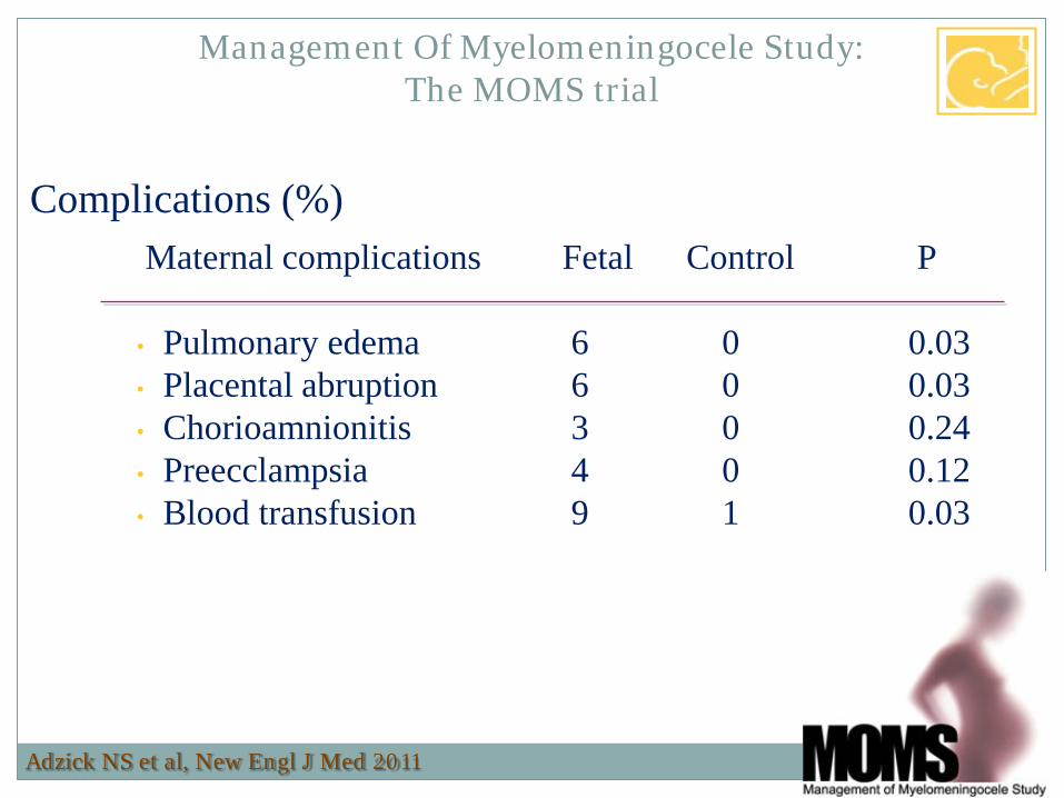
Complications (%)
Maternal complications Fetal Control P
• Pulmonary edema 6 0 0.03 • Placental abruption 6 0 0.03 • Chorioamnionitis 3 0 0.24 • Preecclampsia 4 0 0.12 • Blood transfusion 9 1 0.03
Management Of Myelomeningocele Study: The MOMS trial
Adzick NS et al, New Engl J Med 2011
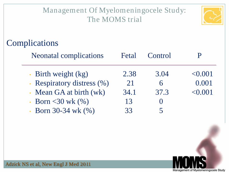
Complications
Neonatal complications Fetal Control P
• Birth weight (kg) 2.38 3.04 <0.001 • Respiratory distress (%) 21 6 0.001 • Mean GA at birth (wk) 34.1 37.3 <0.001 • Born <30 wk (%) 13 0 • Born 30-34 wk (%) 33 5
Management Of Myelomeningocele Study: The MOMS trial
Adzick NS et al, New Engl J Med 2011

Complications (%)
Pregnancy complications Fetal Control P
• Oligohydramnios 21 4 0.001 • PROM 46 8 <0.001 • Uterine wound:
• Intact and healed 64 • Very thin 25 • Some dehiscence 10
Management Of Myelomeningocele Study: The MOMS trial
Adzick NS et al, New Engl J Med 2011

In utero repair of spina bifida: how is it done? Multidisciplinary team approach Maternal Anesthesia Maternal-Fetal Medicine Pediatric Surgery
Pediatric Neurosurgery Pediatric Plastic Surgery Neonatalogy
Management of children with spina bifida in the age of fetal intervention

In utero repair of spina bifida: how is it done? Wide maternal laparotomy Full exposure of the uterus
Management of children with spina bifida in the age of fetal intervention
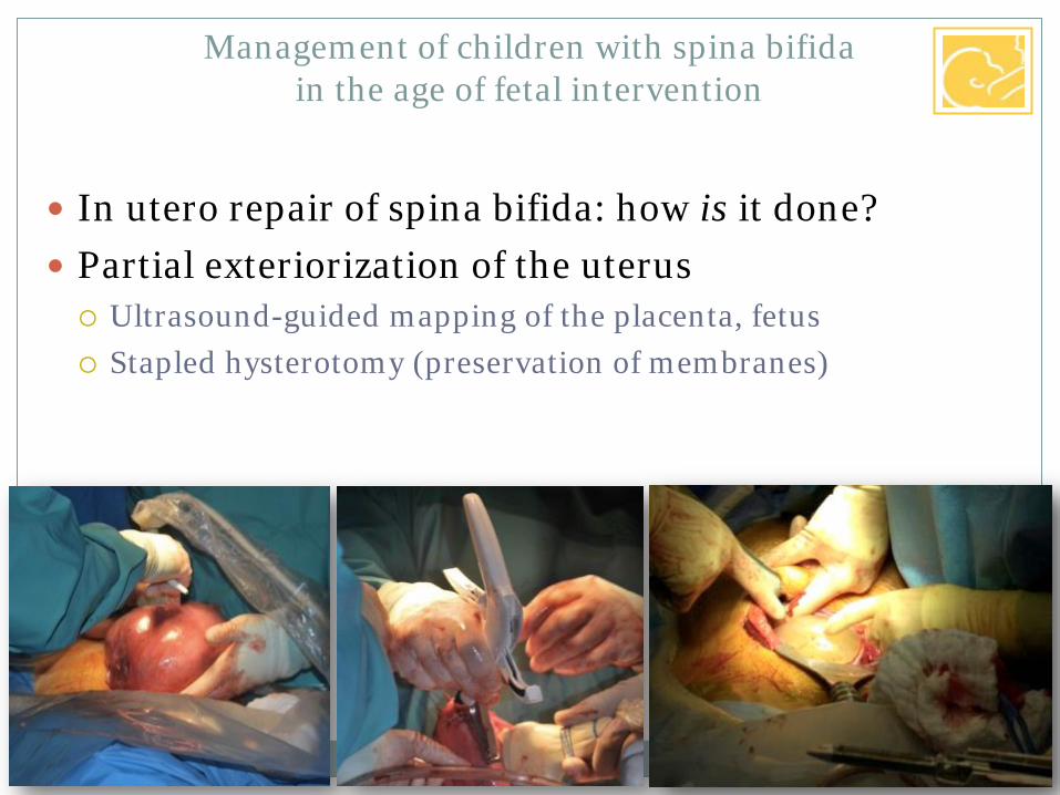
In utero repair of spina bifida: how is it done? Partial exteriorization of the uterus Ultrasound-guided mapping of the placenta, fetus Stapled hysterotomy (preservation of membranes)
Management of children with spina bifida in the age of fetal intervention

In utero repair of spina bifida: how is it done? Exposure of the neural tube defect
Management of children with spina bifida in the age of fetal intervention

In utero repair of spina bifida: how is it done? Exposure of the neural tube defect Rapid closure
Management of children with spina bifida in the age of fetal intervention

Post- vs. Prenatal repair POSTNATAL challenges and conditions: A) Separation of placode from
Epithelium: -„trimming of the placode“ - Use of surgical microscope
B) Preservation of the placode and vascular supply
- „meticulous“ hemostasis and microdissection
- Use of surgical microscope C) Anatomical reconstruction: Prevention of re-tethering, ischemia, CSF leak and infection!
- Sufficient dissection of dural layer to prevent ischemia - Myofascial skin/subcutaneous fat dissection, preparation and closure important!
PRENATAL challenges and conditions: A) YES,
BUT much faster healthy spinal cord without epithelium, inflammation and infarction of placode, no trimming of the placode
B) YES, But NO significant dural vasular supply of placode („bloodless“) No use of surgical microscope! C) NO! Only attempt to approximate DURA and SKIN (occasionally; Dural substitute and Skin substitute)
Counsel parents:Fetal repair is NOT formal and anatomical repair: Second Repair at birth or soon after necessary (e.g. skin breakdown etc. ) and close watch for tethering
3 -4 hours! 0.5 hours!

MOMS II Further analysis of the results in the initial cohort
It improves motor function Does it improve GERD?
No real evidence (25% if shunted, v. 8% if not shunted) Does it improve continence?
No word yet – but the answer appears to be “no” Does it improve cognitive outcome?
No word yet – but the answer appears to be “no” Does it prevent/ Improve Tethering?
No word yet – but appears to be the opposite
Management of children with spina bifida in the age of fetal intervention
Danzer E et al, Neuropediatrics 2008


