MANAGEMENT OF ENDOCRINE DISEASEThe role of rhTSH in the management of differentiated thyroid cancer: pros and consLuca Giovanella1 and Leonidas H Duntas2
1Clinic for Nuclear Medicine and Competence Center for Thyroid Diseases, Imaging Institute of Southern Switzerland, Bellinzona, Switzerland and 2Unit of Endocrinology Diabetes and Metabolism, Thyroid Section, Evgenideion Hospital, University of Athens, Athens, Greece
Abstract
The use of recombinant human thyrotropin (rhTSH) testing in the diagnosis and therapy of differentiated thyroid cancer (DTC) has been adopted over the last two decades as an alternative to the classical thyroid hormone withdrawal avoiding the threat of hypothyroidism. Serum thyroglobulin (Tg) measurement is crucial for monitoring DTC patients over time. Until about a decade ago, optimal sensitivity of Tg assays for the detection of smaller disease foci required Tg measurement after thyrotropin (TSH) stimulation, carried out following thyroid hormone withdrawal or rhTSH administration. In very recent years, significant improvements in assay technology have resulted in highly sensitive Tg (hsTg) assays, sufficiently sensitive to obviate the need for rhTSH stimulation in most DTC patients. The aim of this paper is to review and discuss, via a ‘pros and cons’ approach, the current clinical role of rhTSH to stimulate radioiodine (RAI) uptake for treatment and/or imaging purposes and to increase the clinical sensitivity of Tg measurement for monitoring DTC patients when high-sensitive Tg assays are available.
Introduction
The mainstays of treatment for DTC are surgery, risk-adapted postoperative administration of radioiodine (RAI) and thyroid hormone therapy tailored on individual risk to relapse. Overall, such approach results in excellent
response for more than 80% of subjects (1). Despite its good prognosis, however, DTC is characterized by a significant risk of disease persistence and recurrence over time making essential long-term active surveillance (2). Serum thyroglobulin (Tg) is a large glycoprotein that in
European Journal of Endocrinology (2019) 181, R133–R145
-19-0149
Review
1814
Invited Author’s profile
Prof. Dr med. Luca Giovanella is the Medical and Scientific Director of the Imaging Institute of Southern Switzerland (IIMSI), Director of the Clinic of Nuclear Medicine and Competence Centre for Thyroid Diseases and Consultant of the Department of Laboratory Medicine at Ente Ospedaliero Cantonale, Bellinzona and Lugano (Switzerland). He serves as Titular Professor at University Hospital and University of Zurich, Zurich (Switzerland). His clinical and scientific activities are mainly focused on the diagnosis and treatment of thyroid diseases with an emphasis on molecular imaging, radioiodine theragnostics, circulating biomarkers of thyroid cancers and thyroid function in vivo and in vitro tests. He serves as Deputy Chairman of Thyroid Committee-European Association of Nuclear Medicine and Chairman of Thyroid Committee-Swiss Society of Nuclear Medicine.
Downloaded from Bioscientifica.com at 02/06/2022 03:32:09PMvia free access
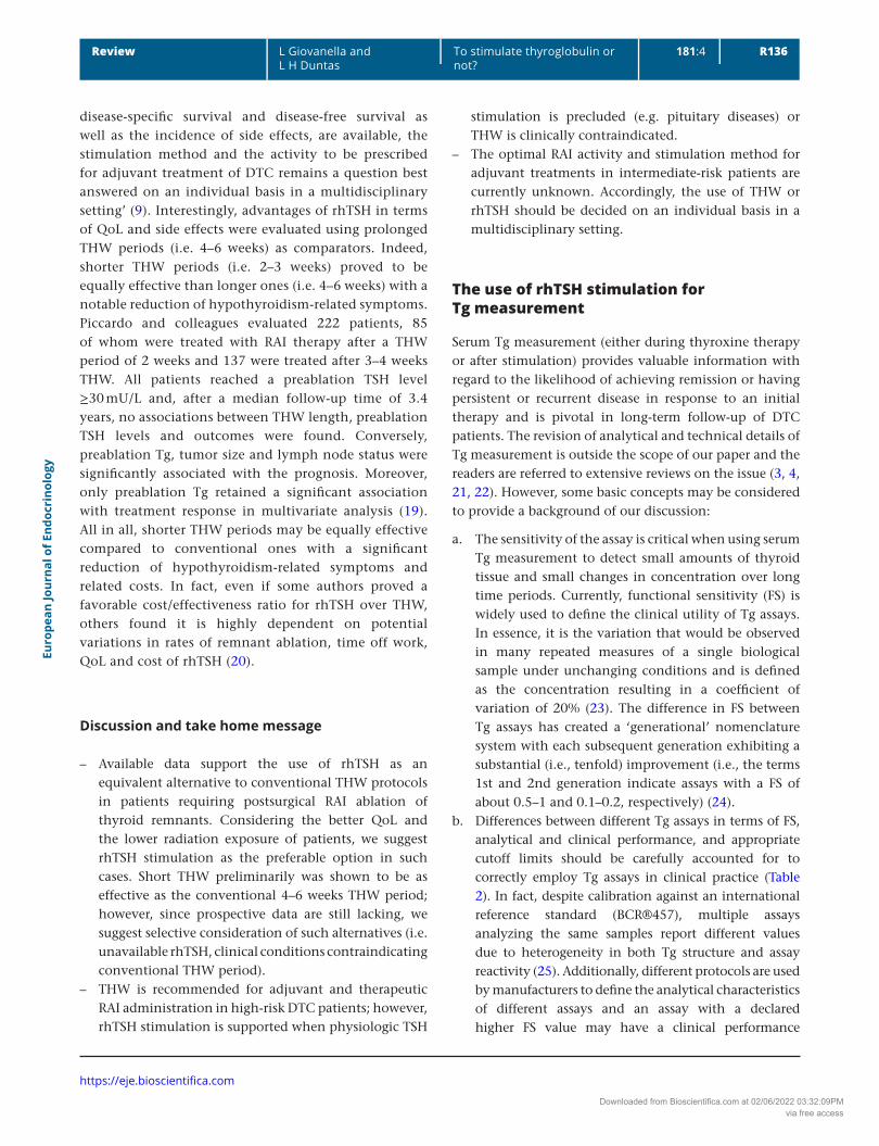
healthy thyroid tissue acts as a substrate for the synthesis of thyroid hormones and is released into the bloodstream together with thyroid hormones (Table 1). Its thyroid tissue-specific origin makes it extremely useful as a DTC tumor marker (3). Since iodine uptake and Tg secretion depend on thyrotropin (TSH) levels and follicular-derived tumor cells mostly preserve thyrotropin (TSH) receptors, TSH stimulation is required when RAI administration is indicated for treatment and/or imaging and/or a stimulated Tg value is required. Tg concentrations measured after stimulation of TSH receptor exceed Tg values under TSH suppression (i.e. Tg-on) by one order of magnitude (4). The TSH receptor stimulation was traditionally carried out following thyroid hormone withdrawal (THW) for 4–6 weeks. However, after its introduction in late 1990s, the recombinant human thyrotropin-stimulating hormone (rhTSH) gained momentum as a safe and effective alternative to THW in diagnosis, treatment and follow-up of DTC patients (5, 6). rhTSH is typically administered intramuscularly over 2 consecutive days: Tg is generally determined on day 5 following the first injection, while 131I, used for treatment or diagnostic purposes, is administered on day 3, and imaging with whole-body scan (WBS) is done at least on day 5 (7) (Fig. 1). Currently, divergences still exist in different clinical guidelines on the preferential use of rhTSH or TWH before RAI administration and significant improvements in assay technology have resulted in new, highly sensitive Tg (hsTg) assays which appeared sufficiently sensitive to obviate the need for TSH stimulation in most DTC patients (8). The aim of this paper is therefore to review, via a ‘pros (L D) and cons (L G) approach’, the available literature data on the use of rhTSH stimulation in DTC patients management, to discuss existing divergences and to provide some practical suggestions.
The use of rhTSH stimulation before RAI therapy
The adoption of a nomenclature that uses ‘I-131 therapy’ as the broad term encompassing the three primary goals associated with an administered activity of I-131 was recently proposed (9).
a. Remnant ablation refers to the use of I-131 to destroy post-operatively residual, presumably benign thyroid tissue to facilitate initial staging and follow-up studies (such as serum Tg and RAI imaging).
b. Adjuvant treatment can be defined as I-131 administered in an effort to destroy subclinical tumor deposits that may or may not be present after surgical resection of all known primary tumor tissue and metastatic foci. The goals of adjuvant treatment are to improve disease-specific survival and decrease recurrence rates, as well as to improve progression-free survival.
c. Treatment of known biochemical or structural disease refers to the goal of destroying persistent or recurrent DTC foci with I-131 in order to improve progression-free, disease-specific and overall survival. It can be given either with curative or palliative intent.
Anyway, TSH stimulation, with either THW or rhTSH, is recommended before diagnostic and therapeutic
Table 1 Causes of increased Tg levels in the bloodstream.
TSH receptor stimulation by TSH, TSH receptor antibodies and human chorionic gonadotropic hormone (hCG)
Increased proliferation (e.g. benign and malignant nodules, goiter)
Iodine deficiencyDestructive thyroiditis (e.g. subacute, postpartum and silent
thyroiditis, Hashitoxicosis)Thyroid surgery Radioactive iodine therapy Fine-needle aspiration cytology and core-needle biopsyThermal ablation of thyroid nodulesSerious manipulation of the thyroid gland (e.g. anterior neck
trauma, strangulation)
Figure 1rhTSH is administered intramuscularly over 2 consecutive days: thyroglobulin (Tg) is determined on day 5 (96 h) following the first injection (if needed radioiodine (RAI), used for treatment (yellow circle) or diagnostic purposes (green circle), is applied orally on day 3 (48 h)). Imaging with whole-body scan (WBS) is performed at least on day 5 (96 h), following the RAI administration. Note: rhTSH stimulation is not indicated in patients with positive thyroglobulin antibodies (TgAb) and/or detectable Tg-on.
Downloaded from Bioscientifica.com at 02/06/2022 03:32:09PMvia free access
RAI administrations (5, 7). In the following paragraphs pros and cons of the use of rhTSH before RAI therapy administration are discussed.
PROS: rhTSH-stimulated RAI therapy/imaging
The use of rhTSH during levothyroxine treatment instead of THW has far-reaching implications for DTC patients. The exogenous stimulation not only conserves adequate thyroid hormone availability, thus avoiding the deleterious effects of short-term hypothyroidism, preserving quality of life (QoL) and normal renal iodine clearance and lowering bone marrow and whole-body exposure to the radiation dose, thereby reducing potential radiation-related side effects (10, 11). Two recent multicenter trials studying long-term outcome (ESTIMABL1) and recurrence rates (Hi-Lo) following RAI remnant ablation, in which the patients were randomly assigned to 1.1 GBq or 3.7 GBq, did not observe any difference between the two modalities (12, 13). The ESTIMABL1 study was a randomized phase 3 trial of RAI administration in patients with low-risk thyroid cancer following total thyroidectomy, which was conducted in 24 centers in France over a follow-up period of 5 years. A total of 92% of the patients had complete thyroid ablation at 6–10 months, defined as a rhTSH-Tg concentration of 1 μg/L or less and normal neck ultrasonography. Of note, the outcome was similar between low-activity (1.1 GBq) and high-activity (3.7 GBq) RAI and also between the use of rhTSH administration and THW. Hi-Lo was a parallel, open-label, randomized controlled factorial trial carried out at 29 centers in the United Kingdom. Eligible patients were aged 16–80 years, with histological confirmation of DTC requiring RAI (tumor stage T1–T3), with the possibility of N1a lymph node involvement but neither N1b lymph node and/or distant metastasis nor microscopic residual disease. The recurrence rate was equivalent among patients who had 1.1 GBq and 3.7 GBq RAI. In addition, a recent French multicenter retrospective study, carried out in a real-life setting of DTC patients being treated for nodal metastases (N1), evaluated the use of rhTSH vs THW in preparation to RAI and demonstrated their equivalence (14). The patients were matched according to age (<45 vs ≥45 years), number of N1 (≤5 vs >5 lymph nodes) and stage of disease (pT1-T2 vs pT3). Disease-free rate was not inferior in the rhTSH group (75.1%) compared to the THW group (71.9%), suggesting that rhTSH is equivalent to THW and that rhTSH appears to be an effective stimulation method for RAI therapy in DTC, this including in patients with N1 metastases. Stimulation with rhTSH is not currently
approved by the FDA in patients with high-risk and metastatic DTC; however, data on its compassionate use suggest that it is equally effective as THW for preparation for dosimetry-based RAI treatment in such patients subset too (15). In 56 patients (31 women and 25 men) with RAI-avid distant metastases of DTC treated with either rhTSH-aided (n = 15) or THW-aided RAI (n = 41) and followed for 72 ± 36.2 months, no statistical significant differences were found regarding the mean size of target lesions, mean baseline Tg level (6995 μg/L vs 5544 μg/L, P = 0.83), distribution of micronodular and macronodular pulmonary, osseous, liver/kidney metastases or side effect profile of rhTSH (16). When adjusted for age, rates of response and progression-free survival (PFS) were not different between the two groups, the only independent risk factor being age. In a retrospective chart review at nine centers in Europe, 144 patients with T4 tumors were recruited who had received adjuvant RAI therapy with rhTSH or with THW. No significant differences in successful ablation rate were observed and no serious adverse events were registered (17). Additionally, one long-term study showed that the use of rhTSH for ablation was more likely to be associated with excellent response to therapy though the final outcomes were not significantly different with respect to recurrence rate in patients with intermediate to high risk without known distant metastases. Finally, as concerns total Medicare payments in the USA, these were higher among outpatients, lower among inpatients and similar to those not receiving rhTSH in conjunction with RAI. Thus, from at least the US payer perspective, the use of rhTSH seems to be a cost-neutral treatment option (18).
CONS: rhTSH-stimulated RAI therapy/imaging
As reported above, rhTSH is not recommended before adjuvant and therapeutic RAI administration in high-risk DTC patients, while the available literature allows its use before RAI ablation (mostly performed, selectively, in low-risk DTC patients) as a reliable alternative to THW. No evidences exist, however, on optimal RAI activities and stimulation methods for adjuvant RAI therapy in intermediate-risk DTC patients. Admittedly, previous literature is severely hampered by the retrospective design of most studies, the inclusion of patients treated with mixed ablation and adjuvant intents and the use of different stimulation protocols and RAI-administered activities. Accordingly, an international multidiciplinary panel recently stated that ‘until the results of prospective multicenter studies centering on relevant outcomes of adjuvant postoperative RAI treatment, including
Downloaded from Bioscientifica.com at 02/06/2022 03:32:09PMvia free access
disease-specific survival and disease-free survival as well as the incidence of side effects, are available, the stimulation method and the activity to be prescribed for adjuvant treatment of DTC remains a question best answered on an individual basis in a multidisciplinary setting’ (9). Interestingly, advantages of rhTSH in terms of QoL and side effects were evaluated using prolonged THW periods (i.e. 4–6 weeks) as comparators. Indeed, shorter THW periods (i.e. 2–3 weeks) proved to be equally effective than longer ones (i.e. 4–6 weeks) with a notable reduction of hypothyroidism-related symptoms. Piccardo and colleagues evaluated 222 patients, 85 of whom were treated with RAI therapy after a THW period of 2 weeks and 137 were treated after 3–4 weeks THW. All patients reached a preablation TSH level ≥30 mU/L and, after a median follow-up time of 3.4 years, no associations between THW length, preablation TSH levels and outcomes were found. Conversely, preablation Tg, tumor size and lymph node status were significantly associated with the prognosis. Moreover, only preablation Tg retained a significant association with treatment response in multivariate analysis (19). All in all, shorter THW periods may be equally effective compared to conventional ones with a significant reduction of hypothyroidism-related symptoms and related costs. In fact, even if some authors proved a favorable cost/effectiveness ratio for rhTSH over THW, others found it is highly dependent on potential variations in rates of remnant ablation, time off work, QoL and cost of rhTSH (20).
Discussion and take home message
– Available data support the use of rhTSH as an equivalent alternative to conventional THW protocols in patients requiring postsurgical RAI ablation of thyroid remnants. Considering the better QoL and the lower radiation exposure of patients, we suggest rhTSH stimulation as the preferable option in such cases. Short THW preliminarily was shown to be as effective as the conventional 4–6 weeks THW period; however, since prospective data are still lacking, we suggest selective consideration of such alternatives (i.e. unavailable rhTSH, clinical conditions contraindicating conventional THW period).
– THW is recommended for adjuvant and therapeutic RAI administration in high-risk DTC patients; however, rhTSH stimulation is supported when physiologic TSH
stimulation is precluded (e.g. pituitary diseases) or THW is clinically contraindicated.
– The optimal RAI activity and stimulation method for adjuvant treatments in intermediate-risk patients are currently unknown. Accordingly, the use of THW or rhTSH should be decided on an individual basis in a multidisciplinary setting.
The use of rhTSH stimulation for Tg measurement
Serum Tg measurement (either during thyroxine therapy or after stimulation) provides valuable information with regard to the likelihood of achieving remission or having persistent or recurrent disease in response to an initial therapy and is pivotal in long-term follow-up of DTC patients. The revision of analytical and technical details of Tg measurement is outside the scope of our paper and the readers are referred to extensive reviews on the issue (3, 4, 21, 22). However, some basic concepts may be considered to provide a background of our discussion:
a. The sensitivity of the assay is critical when using serum Tg measurement to detect small amounts of thyroid tissue and small changes in concentration over long time periods. Currently, functional sensitivity (FS) is widely used to define the clinical utility of Tg assays. In essence, it is the variation that would be observed in many repeated measures of a single biological sample under unchanging conditions and is defined as the concentration resulting in a coefficient of variation of 20% (23). The difference in FS between Tg assays has created a ‘generational’ nomenclature system with each subsequent generation exhibiting a substantial (i.e., tenfold) improvement (i.e., the terms 1st and 2nd generation indicate assays with a FS of about 0.5–1 and 0.1–0.2, respectively) (24).
b. Differences between different Tg assays in terms of FS, analytical and clinical performance, and appropriate cutoff limits should be carefully accounted for to correctly employ Tg assays in clinical practice (Table 2). In fact, despite calibration against an international reference standard (BCR®457), multiple assays analyzing the same samples report different values due to heterogeneity in both Tg structure and assay reactivity (25). Additionally, different protocols are used by manufacturers to define the analytical characteristics of different assays and an assay with a declared higher FS value may have a clinical performance
Downloaded from Bioscientifica.com at 02/06/2022 03:32:09PMvia free access
equal to or better than one with a lower declared FS. Accordingly, for longitudinal consistency of clinical care, consecutive measurements of Tg concentrations should be performed in the same laboratory using the same assay each time. If an assay change is unavoidable, a new baseline should be established through parallel Tg measurements using both the old and the new assay (26). Finally, clinical thyroidologists and laboratory specialists are strongly advised to carefully evaluate the analytical and clinical performance of any Tg assay, including a comparison between basal and stimulated values in the same assay, and to confirm cutoff and decision limits in their own DTC patient populations.
c. Serum Tg measurement is primarily limited by the presence of thyroglobulin autoantibodies (TgAb), which occur in up to 15–30% of DTC patients. In this case, Tg measurement is unreliable because of interference causing underestimation. Thus, any sample with a positive TgAb result should be considered as unreliable for measuring serum Tg, and TgAb concentration becomes the primary (surrogate) tumor-marker. Notably, serum Tg response to TSH stimulation is blunted in the presence of TgAb and, consequently, our discussion exclusively refers to TgAb-negative patients. More extensive information on the management of TgAb-positive DTC patients can be found in recent reviews and position papers (21, 22, 24, 27).
Tg measurement after surgery/before RAI therapy (preablation Tg)
Several studies have suggested that a high serum Tg level measured after thyroidectomy and just before RAI therapy (preablation Tg) could be a prognostic marker for predicting DTC recurrence or metastases (28, 29). A meta-analysis of 3947 patients across a broad spectrum of disease demonstrated that the preablation Tg level is
a useful negative predictor of persistent and recurrent DTC. Especially, THW postoperative Tg values of 10 μg/L achieve the best balance of sensitivity and specificity for predicting recurrent or persistent disease over time and poorer survival with a negative predictive value (NPV) of 94% (30). On the basis of the above data , some authors have proposed postoperative serum Tg testing (during thyroid hormone therapy or after TSH stimulation) to help in assessing persistence of disease and to assist in RAI decision making (20, 31).
PROS: rhTSH-stimulated Tg measurement after surgery/before RAI therapy (preablation Tg)
Stimulated Tg levels measured at the time of RAI ablation following rhTSH (rhTSH-Tg) were shown to have prognostic value in predicting disease-free status in a study including 293 consecutive patients, with no initial evidence of distant metastasis who were submitted to total or near-total thyroidectomy followed by ablation either under rhTSH (n = 151) or via THW (n = 142) (32). Meanwhile, based on the hypothesis that preablative stimulated Tg (Abl-Tg) could have value as a predictor of recurrent and persistent DTC, a study was performed to establish the potential correlations between Abl-Tg and therapeutic response and to calculate a threshold for prediction of poor response (33). In total, 452 patients with DTC were monitored for a median of 38 months and were grouped according to Abl-Tg level: group 1, less than 1 μg/L (n = 82), group 2, 1–9.9 μg/L (n = 173) and group 3 at least 10 μg/L (n = 197). According to different Abl-Tg strata, it was possible to distinguish the therapeutic response as a structurally incomplete response (SIR) in none of group 1, in 1.73% of group 2 and in 42.74% of group 3 (P < 0.001). It is therefore evident that Abl-Tg had independent predictive value for disease-free status 1 year after RAI ablation, indicating that a low Tg under rhTSH-aided ablation may be validated as a favorable prognostic
Table 2 Analytical sensitivity of some commercially available Tg assays as reported in the literature and/or quoted by manufacturers.
parameter. Nevertheless, despite this postulated potential of Abl-Tg for predicting prognosis in patients with DTC, its levels can be affected by TSH (34). The value of Tg levels at the time of rhTSH-aided remnant ablation for predicting disease status 1 year later in patients with DTC who underwent total thyroidectomy with central neck dissection (CND) was estimated in a prospective observational study of 253 consecutive patients with DTC (35). The results showed that those patients who had persistent or recurrent disease were more likely to be ≥45 years of age, as well as to have N1b stage and TNM stage III or IV. Moreover, rhTSH-Tg levels <1.79 μg/L at ablation had a NPV of 99·5% suggesting the role of Tg levels at rhTSH-aided RAI ablation as useful prognostic marker in such patients. To ascertain the need for RAI remnant ablation, the ATA recommendations have to date mainly been based on American Joint Committee Cancer (AJCC/TNM) staging. Given that Tg has a half-life of approximately 65 h subsequent to thyroidectomy, Tg levels may not be undetectable until several weeks have passed. However, Tg levels after this period reflects the presence or absence of residual disease and can also be used to predict the likelihood of identifying RAI-avid metastatic thyroid cancer outside the thyroid bed on the post-therapy scan at the time of RAI ablation. On this basis, some authors proposed postoperative Tg testing to enable a tailor-made decision for RAI treatment (36). The optimal cutoff value for postoperative serum Tg or state in which it is measured (on thyroid hormone therapy or after TSH stimulation) to guide decision making regarding RAI administration, however, is not known (20).
CONS: rhTSH-stimulated Tg measurement after surgery/before RAI therapy (preablation Tg)
Although postoperative serum Tg measurement can provide valuable information with regard to the likelihood of achieving remission or having persistent or recurrent disease in response to an initial therapy, its predictive value is significantly influenced by a wide variety of factors, including the time elapsed since total thyroidectomy, the amount of thyroid remnant, the individual’s risk of having loco-regional and distant metastasis, the Tg cutoff used for analysis and TSH level at the time of Tg measurement. The latter point is crucial when different stimulation methods are used and, consequently, when rhTSH is used instead of THW before ablation lower preablation Tg levels are expected. In fact, even if a significant correlation exists between peaks of THW-Tg and rhTSH-Tg levels, the latter are usually and
significantly (~4-fold) lower (37). Notably, when rhTSH is used before RAI therapy, Tg is traditionally measured 72 h after the second injection of rhTSH corresponding to 48 h after RAI administration. Taieb and colleagues assessed the changes in serum Tg 48 h after RAI ablative therapy performed in the hypothyroid state in order to evaluate if this parameter could be used in rhTSH-treated patients (38). They performed a prospective evaluation of 26 consecutive patients with low-risk DTC treated during hypothyroidism with 3.7 GBq of RAI. Baseline Tg values were compared to post-therapy Tg values (at 24 and 48 h). The authors found that Tg increased after RAI therapy because of the acute radiation effects on residual thyroid cells. Median values at each of the three time points were 1.8 (baseline), 3 (Tg-24) and 11.3 μg/L (Tg-48), respectively. Tg-48 values were not statistically correlated with baseline and Tg-24 values. In summary, the first rhTSH-stimulated Tg cannot be used as a corresponding value for preablative THW-Tg. The Tg value obtained just before RAI administration (i.e. 24 h after the second rhTSH injection) should be alternatively used in such cases providing appropriate cutoff levels are settled. Postsurgical Tg measurement (either TSH-stimulated or nonstimulated) may also help in assessing the persistence of disease or thyroid remnant and predicting potential future disease recurrence. Then, it was recently proposed as a yard-stick for adjuvant RAI therapy decision making. Vaisman and colleagues proposed to skip radioiodine ablation in patients with TgAb-negative low-risk DTC treated with thyroidectomy plus selective central compartment neck dissection (CND) having postsurgical rhTSH-stimulated Tg levels from <1 to 5 μg/L and negative neck ultrasound (31). However, CND is not currently recommended in low-risk DTC patients due to its not negligible morbidity and, in addition, problems of various kinds may occur with Tg measurement at the time of ablation in up to 15% of patients (39). In particular, thyroid remnants and recurrences/metastases on the posttreatment RAI WBS were proved in up to 20 and 8.5%, respectively, of patients with an undetectable preablative Tg value (40, 41). Most importantly, RAI administration likely contributed to good patient outcome observed in previous studies on the role of preablative Tg, while similar results in non-ablated patients remain largely unproved and no precise Tg value cutoffs are available to define what is an excellent response after surgery and justify the omission of RAI therapy (20, 42). Therefore, it appears that the postoperative serum Tg value will be more helpful in identifying patients that will benefit from RAI ablation rather than in identifying patients that do not require ablation (20) and, as a
Downloaded from Bioscientifica.com at 02/06/2022 03:32:09PMvia free access
consequence, a benchmark between RAI imaging and Tg measurement remains of pivotal value by authenticating the use of serum Tg during further follow-up of DTC patients (43, 44, 45). Additionally, if postsurgery Tg measurement and RAI imaging are stimulated by rhTSH, a significant increase of costs should be considered. In fact, a second stimulation cycle will be required in case of subsequent RAI therapy.
Discussion and take home message
– The measurement of either TWH- and rhTSH-stimulated serum Tg before RAI therapy constitutes a powerful prognostic marker providing valuable information with regard to the likelihood of achieving remission or having persistent or recurrent disease in response to an initial therapy. Thought THW and rhTSH are reliable and equivalent methods in selective RAI ablation (i.e. low risk DTC) rhTSH-aided RAI administration and, consequently, preablative rhTSH-Tg measurement are preferable in this setting due to the resulting superior QoL and decreased hypothyroidism-related morbidity.
– Preablation THW-Tg and rhTSH-Tg values of ~10 μg/L and ~1 μg/L, respectively, achieve the best balance of sensitivity and specificity for predicting recurrent or persistent disease over time and poorer survival. Notably, cutoff levels reported in the literature should be considered with caution and their transferability to local settings carefully evaluated.
– When rhTSH is employed we recommend to obtain preablative Tg levels before RAI administration to avoid the jatrogenic Tg increase related to the acute RAI-induced cytolysis.
– No reliable criteria for either THW- or rhTSH-stimulated Tg are currently available to guide RAI therapy decision making. However, integrating conventional risk-stratification criteria (i.e. histopathology) with postoperative Tg measurement and morphological/functional imaging may provide relevant support in tailoring a patient-based postoperative management and follow-up.
Tg measurement during DTC follow-up
Until about a decade ago, optimal sensitivity of Tg assays for the detection of smaller disease foci required stimulation of endogenous Tg production. In very recent years, significant improvements in assay technology have,
however, resulted in new, highly sensitive Tg (hsTg) assays, which, in fact, appeared sufficiently sensitive to obviate the need for TSH stimulation in most DTC patients (3, 4, 20, 21, 46). Notably, in early days most comparative studies adopted stimulated Tg values, instead of structural evidence of disease, as comparator for unstimulated hsTg. That means patients with undetectable basal Tg but indeterminate or incomplete stimulated Tg response (i.e. 1 to 10 μg/L) were classified as having DTC recurrence and vice versa. The ultimate evaluation of assay performance for detecting persistent or recurrent disease is, however, the occurrence of clinically detectable foci of disease during the course of DTC follow-up (i.e. structural recurrence) as well as their biochemical detection. Accordingly, recent studies proved that slightly detectable basal and/or stimulated Tg at 6–12 months after therapy spontaneously normalize over time in most cases. In addition, higher cutoff values for basal and/or stimulated Tg were found using ROC curve analysis with structural evidence of disease as comparator. Clearly, the increasing adoption of these assays in clinical practice has considerable implications, such as avoidance of hypothyroidism or rhTSH injections with increased patient comfort and a significant reduction of costs of DTC follow-up (21). In the following paragraphs pros and cons of the use of stimulated (with either THW or rhTSH) and unstimulated hsTg to follow-up DTC patients are discussed.
PROS: rhTSH-stimulated Tg testing during DTC follow-up
Patients who have not had RAI ablation following surgery are excluded from TSH stimulation during follow-up as Tg will be detectable due to remaining healthy thyroid tissue and will obscure any possible tumor-related Tg level rise. Hence, the recommendation for TSH stimulation to achieve optimal Tg measurement sensitivity traditionally interested any patient with DTC who has low- or high-risk stages of disease and received at least one course of ablative radioiodine therapy. As stated by the current guidelines, stimulated serum Tg should be measured in all patients who have had remnant ablation and negative cervical US and undetectable TSH-suppressed Tg within the first year after treatment. It should be measured after T4 withdrawal or recombinant human TSH stimulation within 6–12 months after ablation (20). An additional radioiodine whole-body scan is suggested in the follow-up of patients with high or intermediate risk of persistent disease, positive TgAb and/or RAI uptake outside the thyroid bed at posttreatment WBS. In this context, considering advantages of rhTSH
Downloaded from Bioscientifica.com at 02/06/2022 03:32:09PMvia free access
in terms of patients ‘comfort and QoL’, rhTSH is favored to stimulate Tg testing, and undetectable rhTSH-Tg levels provide the main evidence of the patient’s remission or cure. Notably, different stimulation methods may result in different Tg values and cutoff thresholds should be adopted. Overall, rhTSH-Tg values >2 μg/L are capable to predict residual tumor, while rhTSH-Tg values <0.5 μg/L render a 98% NPV, thus signaling a disease-free state (26, 47). Spencer et al. (48) have pointed out that there may be a directly proportional relationship between the basal and stimulated Tg levels measured by a highly sensitive assay. Additionally, pooled results of a meta-analysis of nine studies including 3178 low-to-intermediate-risk DTC patients confirmed the very high NPV (98–100%) of an undetectable basal highly sensitive Tg (FS 0.1 μg/L) (49). However, factors such as tumor differentiation and biological behavior might play an independent role in this relationship and, notably, few high-risk DTC patients were enrolled in the available studies. Accordingly, stimulated Tg testing, with a concurrent diagnostic RAI WBS when indicated, is recommended in high-risk DTC at 6–12 months after RAI therapy (20, 21, 29).
Finally, even if hsTg values <0.1 μg/L are reassuring, slightly detectable values (i.e. 0.1–1.0 μg/L) represent a ‘gray zone’. Malandrino et al. (50) found recurrent disease in 12.5% of patients with low-risk DTC when basal highly sensitive Tg levels exceeded 0.15 μg/L, while a very low risk of recurrence was found when basal highly sensitive
Tg was <0.15 μg/L. Castagna et al. (51) also evaluated the clinical performance of two high-sensitive assays using a 0.1 μg/L cutoff for basal highly sensitive Tg and found that nearly 15% of disease-free patients had detectable Tg in the range of 0.1–1.0 μg/L but a stimulated Tg values <1.0 μg/L. Thus, although both assays displayed a good sensitivity and an excellent NPV, they showed poor specificity and positive predictive value. Accordingly, TSH-stimulated Tg testing should therefore be considered for patients with slightly detectable basal highly sensitive Tg levels (i.e. between 0.1 and 1 μg/L).
CONS: rhTSH-stimulated Tg testing during DTC follow-up
In 2002 a group of prominent experts released a ‘Consensus Report’ whose final statement was that ‘an undetectable serum Tg measured during thyroid hormone suppression is misleading in a large proportion of patients with residual DTC’ (52). They based such a strong statement on the retrospective analysis of eight selected studies comprising a total of 1028 low-risk DTC patients thought to be clinically free of disease. Briefly, of the 784 patients with a Tg-on level below 1 μg/L, the number with a rise in Tg over 2 μg/L with rhTSH administration was 168 of 784 (21.4%). Fifty-three (6.8%) of the 784 patients were found to have metastases, making debatable the concept of ‘large proportion’, and, additionally, 731 patients (93.2%)
Thyroidectomy
day 1 and 2: rhTSH injections
day 3: blood sampling for Tg measurement & RAI administration
day 6-12: negative post-treatment RAI whole body scan
subjected to the process and expense of testing had no change in clinical management. In subsequent years, Giovanella and colleagues proved a significant increase of detection rate of basal Tg measured with an assay having higher sensitivity: briefly, detection rate improved from 66 to 92% using the FS 0.4 μg/L as cutoff instead of the conventional threshold (i.e. 1 μg/L), respectively (53). When Tg-on was measured in 117 low-risk DTC patients, the NPV of a Tg-on level <0.4 µg/L was 96% and increased to 99% when combined with a negative neck US. rhTSH-stimulated Tg measurement only detected one additional lymph node recurrence in 104 patients with an undetectable Tg-on (54). When novel hsTg assays (FS ≤ 0.1 μg/L) become commercially available (Table 2) a number of studies consistently proved their ability to obviate the need of TSH stimulation in DTC patients with basal Tg levels ≤0.1 μg/L (49, 55, 56, 57, 58). In addition, although the low frequency of DTC recurrences impacts the ability to study positive predictive values (PPVs) (59), the PPV of an rhTSH-stimulated thyroglobulin above 1–2 μg/L appears comparable to a basal hsTg above 0.10–0.20 μg/L (24). Moreover, no structural recurrences were detected over time in patients with basal hsTg values below ~0.3–0.4 μg/L in newly available highly sensitive assays and basal hsTg emerged as the only independent predictor of cancer relapse in multivariate analysis conferring hazard ratios for reduced disease-free survival of 67.94 and 81.61, depending on the assay employed (60, 61). By considering
the principle factors influencing serum thyroglobulin concentrations (thyroid tissue mass, injury and TSH), it is evident that the trend in basal hsTg, measured when TSH is stable, should reflect changes in thyroid tissue mass and thus provide a more sensitive parameter for disease than rhTSH-stimulated thyroglobulin testing. This is supported by a growing number of studies showing the prognostic utility of monitoring the basal hsTg trend and thyroglobulin doubling time (62, 63, 64). Well in line with this background, Verburg et al. recently demonstrated in a retrospective study on 773 DTC patients that true positive US results (i.e. structural recurrence) are exceedingly rare in front of a basal hsTg <1 µg/L, while these patients are much more likely to be classified as false positive than false negative at US examination, potentially causing a large number of additional follow-up visits as well as more additional investigations. Thus, reserving periodic US examination primarily to patients with basal hsTg exceeding 1 µg/L seems reasonable while tracking the hsTg trend suffices in patients with lower Tg values (65).
Discussion and take home messages
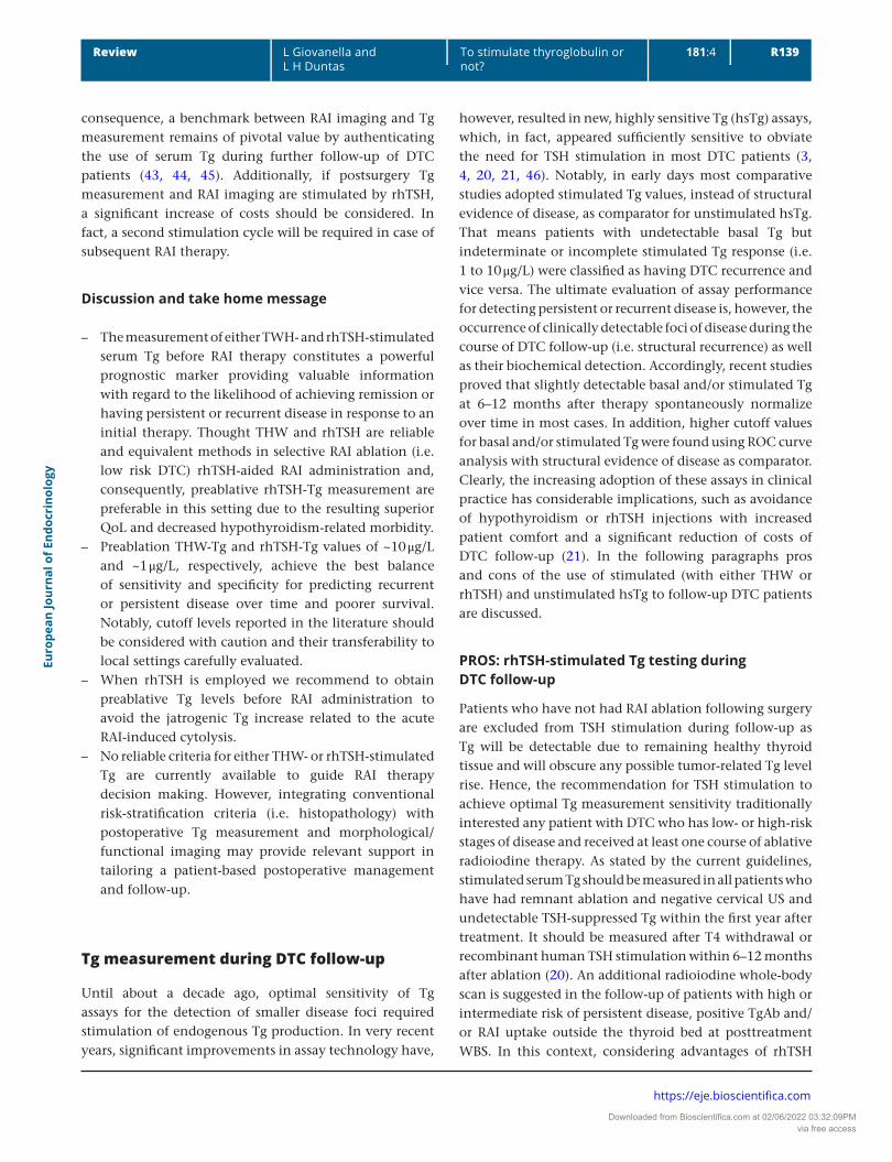
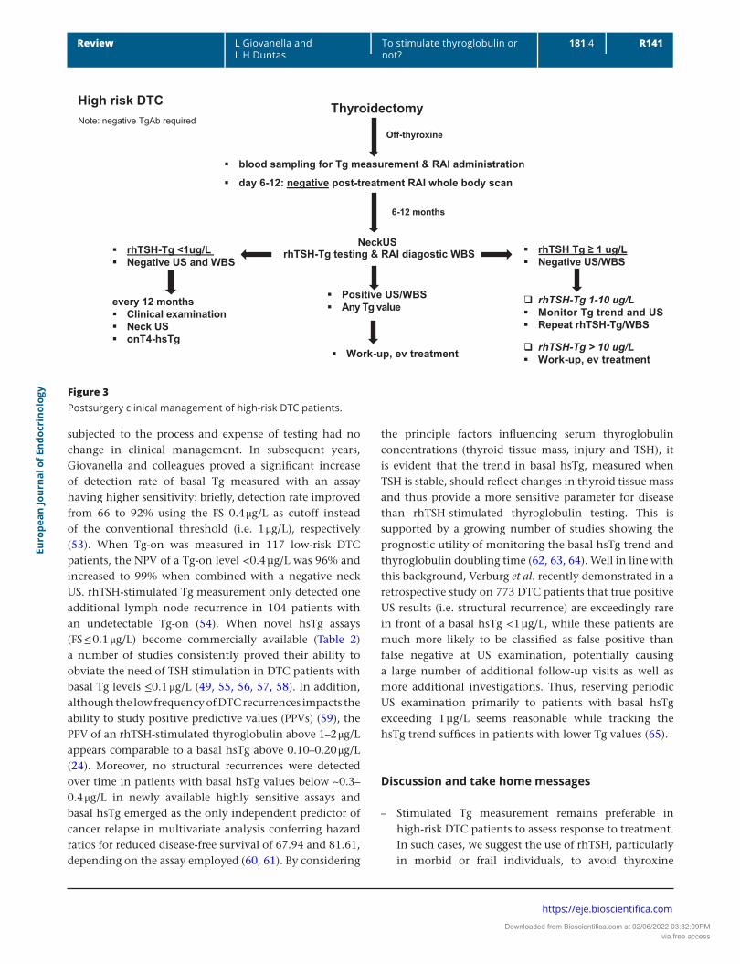
– Stimulated Tg measurement remains preferable in high-risk DTC patients to assess response to treatment. In such cases, we suggest the use of rhTSH, particularly in morbid or frail individuals, to avoid thyroxine
Thyroidectomy
blood sampling for Tg measurement & RAI administration
day 6-12: negative post-treatment RAI whole body scan
Off-thyroxine
6-12 months
NeckUSrhTSH-Tg testing & RAI diagostic WBS
Note: negative TgAb required
rhTSH-Tg <1ug/LNegative US and WBS
every 12 monthsClinical examinationNeck USonT4-hsTg
rhTSH Tg ≥ 1 ug/LNegative US/WBS
rhTSH-Tg 1-10 ug/LMonitor Tg trend and USRepeat rhTSH-Tg/WBS
rhTSH-Tg > 10 ug/LWork-up, ev treatment
Positive US/WBSAny Tg value
High risk DTC
Work-up, ev treatment
Figure 3Postsurgery clinical management of high-risk DTC patients.
Downloaded from Bioscientifica.com at 02/06/2022 03:32:09PMvia free access
discontinuation. A diagnostic RAI WBS imaging, if needed, can easily be incorporated within the same TSH stimulation course.
– Unstimulated hsTg should be preferred to monitor remaining DTC patients treated by thyroidectomy and RAI administration. Stable TSH levels are required to properly compare Tg levels over time.
– Cured patients with slightly detectable hsTg generally demonstrate further decrease over time while progressive increase of serum Tg early detect recurrences. Then, tracking basal hsTg is recommended to manage slightly detectable hsTg Tg . Alternatively, a rhTSH-Tg testing may also be considered in selected cases, especially if a RAI imaging study is planned also.
Conclusions
Stimulation of TSH is required to perform ablative, adjuvant and therapeutic RAI administration. Based on both current evidence and the present discussion, we conclude that rhTSH may be preferentially adopted for RAI ablation in low-risk DTC patients, when required. Vice versa, THW remains the recommended stimulation procedure in high-risk patients. The management of adjuvant RAI administration, including stimulation method, should be personalized basing on a careful, multidisciplinary, assessment of clinical, pathological, biochemical and imaging data.
The chief aim of follow-up of DTC patients after primary treatment is the early identification of the small proportion of patients with residual disease or who will develop recurrences. In TgAb-negative patients, it is based on Tg measurement as the reference standard. Robust data suggest that an undetectable Tg value using a highly sensitive assay is associated with adequate sensitivity and NPV to obviate the need for measuring TSH-stimulated Tg concentrations in most cases (Fig. 2). Stimulated Tg measurement remains preferable, however, to assess the early response to treatment in high-risk DTC patients (Fig. 3). Finally, more research is awaited on the role of postoperative Tg measurement to drive decisions on ablative/adjuvant RAI administration in DTC patients: for the moment, we advise caution in this setting, as robust data are not yet available.
Declaration of interestL G is a member of Roche Diagnostics Advisory Board and received research grants from Roche Diagnostics and speaker honoraria from Roche Diagnostics, BRAHMS GmbH, EISAI, Siemens Healthcare and Sanofi-Genzyme. L D has nothing to disclose related to this work.
FundingThis research did not receive any specific grant from any funding agency in the public, commercial or not-for-profit sector.
References 1 Mazzaferri EL & Jhiang SM. Long-term impact of initial surgical and
medical therapy on papillary and follicular thyroid cancer. American Journal of Medicine 1994 97 418–428. (https://doi.org/10.1016/0002-9343(94)90321-2)
2 Meier CA, Braverman LE, Ebner SA, Veronikis I, Daniels GH, Ross DS, Deraska DJ, Davies TF, Valentine M & DeGroot LJ. Diagnostic use of recombinant human thyrotropin in patients with thyroid carcinoma (phase I=II study). Journal of Clinical Endocrinology and Metabolism 1994 78 188–196. (https://doi.org/10.1210/jcem.78.1.8288703)
3 Grebe SKG. Diagnosis and management of thyroid carcinoma: focus on serum thyroglobulin. Expert Review of Endocrinology and Metabolism 2009 4 25–43. (https://doi.org/10.1586/17446651.4.1.25)
4 Giovanella L, D’Aurizio F, Tozzoli R, Schalin-Jantti C & Feldt-Rasmussen U. Thyroglobulin and thyroglobulin antibodies in. In Atlas of Thyroid and Neuroendocrine Tumor Markers, pp 65–91. Ed L Giovanella. Springer International Publishing AG, 2018.
5 Ladenson PW, Braverman LE, Mazzaferri EL, Brucker-Davis F, Cooper DS, Garber JR, Wondisford FE, Davies TF, DeGroot LJ, Daniels GH et al. Comparison of administration of recombinant human thyrotropin with withdrawal of thyroid hormone for radioactive iodine scanning in patients with thyroid carcinoma. New England Journal of Medicine 1997 337 888–896. (https://doi.org/10.1056/NEJM199709253371304)
6 Schlumberger MJ. Papillary and follicular thyroid carcinoma. New England Journal of Medicine 1998 338 297–306. (https://doi.org/10.1056/NEJM199801293380506)
7 Duntas LH & Cooper DS. Review on the occasion of a decade of recombinant human TSH: prospects and novel uses. Thyroid 2008 18 509–516. (https://doi.org/10.1089/thy.2007.0331)
8 Zöphel K, Wunderlich G & Smith BR. Serum thyroglobulin measurements with a high sensitivity enzyme linked immunosorbent assay: is there a clinical benefit in patients with differentiated thyroid carcinoma? Thyroid 2003 13 861–865. (https://doi.org/10.1089/105072503322401050)
9 Tuttle RM, Ahuja S, Avram AM, Bernet VJ, Bourguet P, Daniels GH, Dillehay G, Draganescu C, Flux G, Fuhrer D et al. Controversies, consensus, and collaboration in the use of 131I therapy in differentiated thyroid cancer: a joint statement from the American Thyroid Association, the European Association of Nuclear Medicine, the Society of Nuclear Medicine and Molecular Imaging, and the European Thyroid Association. Thyroid 2019 29 461–470. (https://doi.org/10.1089/thy.2018.0597)
10 Duntas LH & Biondi B. Short-term hypothyroidism after levothyroxine-withdrawal in patients with differentiated thyroid cancer: clinical and quality of life consequences. European Journal of Endocrinology 2007 156 13–19. (https://doi.org/10.1530/eje.1.02310)
11 Schroeder PR, Haugen BR, Pacini F, Reiners C, Schlumberger M, Sherman SI, Cooper DS, Schuff KG, Braverman LE, Skarulis MC et al. A comparison of short-term changes in health-related quality of life in thyroid carcinoma patients undergoing diagnostic evaluation with recombinant human thyrotropin compared with thyroid hormone withdrawal. Journal of Clinical Endocrinology and Metabolism 2006 91 878–884. (https://doi.org/10.1210/jc.2005-2064)
12 Schlumberger M, Leboulleux S, Catargi B, Deandreis D, Zerdoud S, Bardet S, Rusu D, Godbert Y, Buffet C, Schvartz C et al. Outcome after ablation in patients with low-risk thyroid cancer (ESTIMABL1): 5-year follow-up results of a randomised, phase 3, equivalence trial. Lancet: Diabetes and Endocrinology 2018 6 618–626. (https://doi.org/10.1016/S2213-8587(18)30113-X)
Downloaded from Bioscientifica.com at 02/06/2022 03:32:09PMvia free access
13 Dehbi HM, Mallick U, Wadsley J, Newbold K, Harmer C & Hackshaw A. Recurrence after low-dose radioiodine ablation and recombinant human thyroid-stimulating hormone for differentiated thyroid cancer (HiLo): long-term results of an open-label, non-inferiority randomised controlled trial. Lancet: Diabetes and Endocrinology 2019 7 44–51. (https://doi.org/10.1016/S2213-8587(18)30306-1)
14 Leenhardt L, Leboulleux S, Bournaud C, Zerdoud S, Schvartz C, Ciappuccini R, Kelly A, Morel O, Dygai-Cochet I, Rusu D et al. Recombinant thyrotropin vs levothyroxine withdrawal in 131I therapy of N1 thyroid cancer: a large matched cohort study (ThyrNod). Journal of Clinical Endocrinology and Metabolism 2019 104 1020–1028. (https://doi.org/10.1210/jc.2018-01589)
15 Klubo-Gwiezdzinska J, Burman KD, Van Nostrand D, Mete M, Jonklaas J & Wartofsky L. Potential use of recombinant human thyrotropin in the treatment of distant metastases in patients with differentiated thyroid cancer. Endocrine Practice 2013 19 139–148. (https://doi.org/10.4158/EP12244.RA)
16 Klubo-Gwiezdzinska J, Burman KD, Van Nostrand D, Mete M, Jonklaas J & Wartofsky L. Radioiodine treatment of metastatic thyroid cancer: relative efficacy and side effect profile of preparation by thyroid hormone withdrawal versus recombinant human thyrotropin. Thyroid 2012 22 310–317. (https://doi.org/10.1089/thy.2011.0235)
17 Bartenstein P, Calabuig EC, Maini CL, Mazzarotto R, Muros de Fuentes MA, Petrich T, Rodrigues FJC, Vallejo Casas JA, Vianello F, Basso M et al. High-risk patients with differentiated thyroid cancer T4 primary tumors achieve remnant ablation equally well using rhTSH or thyroid hormone withdrawal. Thyroid 2014 24 480–487. (https://doi.org/10.1089/thy.2013.0157)
18 Dinan MA, Li Y, Reed SD & Sosa JA. Initial adoption of recombinant human thyroid stimulating hormone following thyroidectomy in the medicare thyroid cancer patient population. Endocrine Practice 2019 25 31–42. (https://doi.org/10.4158/EP-2018-0253)
19 Piccardo A, Puntoni M, Ferrarazzo G, Foppiani L, Bottoni G, Altrinetti V, Treglia G, Naseri M, Dib B, Cabria M et al. Could short thyroid hormone withdrawal be an effective strategyfor radioiodine remnant ablation in differentiated thyroid cancerpatients? European Journal of Nuclear Medicine and Molecular Imaging 2018 45 1218–1223. (https://doi.org/10.1007/s00259-018-3955-x)
20 Haugen BR, Alexander EK, Bible KC, Doherty GM, Mandel SJ, Nikiforov YE, Pacini F, Randolph GW, Sawka AM, Schlumberger M et al. 2015 American Thyroid Association Management Guidelines for adult patients with thyroid nodules and differentiated thyroid cancer: the American Thyroid Association Guidelines task force on thyroid nodules and differentiated thyroid cancer. Thyroid 2016 26 1–133. (https://doi.org/10.1089/thy.2015.0020)
21 Giovanella L, Clark PM, Chiovato L, Duntas LH, Elisei R, Feldt-Rasmussen U, Leenhardt L, Luster M, Schalin-Jantti C, Schott M et al. Thyroglobulin measurement using highly sensitive assays in patients with differentiated thyroid cancer: a clinical position paper. European Journal of Endocrinology 2014 171 R33–R46. (https://doi.org/10.1530/EJE-14-0148)
22 Giovanella L, Feldt-Rasmussen U, Verburg FA, Grebe SKG, Plebani M & Clark PM. Thyroglobulin measurement by highly sensitive assays: focus on laboratory challenges. Clinical Chemistry and Laboratory Medicine 2015 53 1301–1314. (https://doi.org/10.1515/cclm-2014-0813)
23 Baloch Z, Carayon P, Conte-Devolx B, Demers LM, Feldt-Rasmussen U, Henry JF, LiVolsi VA, Niccoli-Sire P, John R, Ruf J et al. Laboratory medicine practice guidelines. Laboratory support for the diagnosis and monitoring of thyroid diseases. Thyroid 2003 13 3–126. (https://doi.org/10.1089/105072503321086962)
24 Spencer CA, LoPresti J & Fatemi S. How sensitive (second-generation) thyroglobulin measurement is changing paradigms for monitoring patients with differentiated thyroid cancer, in the absence or
presence of thyroglobulin autoantibodies. Current Opinion in Endocrinology, Diabetes, and Obesity 2014 21 394–404. (https://doi.org/10.1097/MED.0000000000000092)
25 Kim M, Jeon MJ, Kim WG, Lee JJ, Ryu JS, Cho EJ, Ko DH, Lee W, Chun S, Min WK et al. Comparison of thyroglobulin measurements using three different immunoassay kits: a BRAMHS Tg-Plus RIA Kit, a BRAMHS hTg Sensitive Kryptor Kit, and a Beckman Coulter ACCESS Immunoassay Kit. Endocrinology and Metabolism 2016 31 462–468. (https://doi.org/10.3803/EnM.2016.31.3.462)
26 Giovanella L. Highly sensitive thyroglobulin measurements in differentiated thyroid carcinoma management. Clinical Chemistry and Laboratory Medicine 2008 46 1067–1073. (https://doi.org/10.1515/CCLM.2008.212)
27 Verburg FA, Luster M, Cupini C, Chiovato L, Duntas L, Elisei R, Feldt-Rasmussen U, Rimmele H, Seregni E, Smit JW et al. Implications of thyroglobulin antibody positivity in patients with differentiated thyroid cancer: a clinical position statement. Thyroid 2013 23 1211–1225. (https://doi.org/10.1089/thy.2012.0606)
28 Heemstra KA, Liu YY, Stokkel M, Kievit J, Corssmit E, Pereira AM, Romijn JA & Smit JW. Serum thyroglobulin concentrations predict disease-free remission and death in differentiated thyroid carcinoma. Clinical Endocrinology 2007 66 58–64. (https://doi.org/10.1111/j.1365-2265.2006.02685.x)
29 Piccardo A, Arecco F, Puntoni M, Foppiani L, Cabria M, Corvisieri S, Arlandini A, Altrinetti V, Bandelloni R & Orlandi F. Focus on high-risk DTC patients: high postoperative serum thyroglobulin level is a strong predictor of disease persistence and is associated to progression-free survival and overall survival. Clinical Nuclear Medicine 2013 38 18–24. (https://doi.org/10.1097/RLU.0b013e318266d4d8)
30 Webb RC, Howard RS, Stojadinovic A, Gaitonde DY, Wallace MK, Ahmed J & Burch HB. The utility of serum thyroglobulin measurement at the time of remnant ablation for predicting disease-free status in patients with differentiated thyroid cancer: a meta-analysis involving 3947 patients. Journal of Clinical Endocrinology and Metabolism 2012 97 2754–2763. (https://doi.org/10.1210/jc.2012-1533)
31 Vaisman A, Orlov S, Yip J, Hu C, Lim T, Dowar M, Freeman JL & Walfish PG. Application of postsurgical stimulated thyroglobulin for radioiodine remnant ablation selection in low-risk papillary thyroid carcinoma. Head and Neck 2010 32 689–698. (https://doi.org/10.1002/hed.21371)
32 Melo M, Costa G, Ribeiro C, Carrilho F, Martins MJ, da Rocha AG, Sobrinho-Simões M, Carvalheiro M & Soares P. Stimulated thyroglobulin at recombinant human TSH-aided ablation predicts disease-free status one year later. Journal of Clinical Endocrinology and Metabolism 2013 98 4364–4372. (https://doi.org/10.1210/jc.2013-2267)
33 Yang X, Liang J, Li T, Zhao T & Lin Y. Preablative stimulated thyroglobulin correlates to new therapy response system in differentiated thyroid cancer. Journal of Clinical Endocrinology and Metabolism 2016 101 1307–1313. (https://doi.org/10.1210/jc.2015-4016)
34 Zhao T, Liang J, Guo Z, Li J & Lin Y. Serum thyrotropin level of 30 μIU/mL is inadequate for preablative thyroglobulin to serve as a prognostic marker for differentiated thyroid cancer. Endocrine 2016 53 166–173. (https://doi.org/10.1007/s12020-015-0842-0)
35 Moon JH, Choi JY, Jeong WJ, Ahn SH, Lee WW, Kim KM, Choi SH, Lim S, Park YJ, Yi KH et al. Recombinant human thyrotropin-stimulated thyroglobulin level at the time of radioactive iodine ablation is an independent prognostic marker of differentiated thyroid carcinoma in the setting of prophylactic central neck dissection. Clinical Endocrinology 2016 85 459–465. (https://doi.org/10.1111/cen.13029)
36 Twining CL, Lupo MA & Tuttle RM. Implementing key changes in the American Thyroid Association 2015 thyroid nodules/
Downloaded from Bioscientifica.com at 02/06/2022 03:32:09PMvia free access
differentiated thyroid cancer guidelines across practice types. Endocrine Practice 2018 24 833–840. (https://doi.org/10.4158/EP-2018-0130)
37 Kowalska A, Pałyga I , Gąsior-Perczak D, Walczyk A, Trybek T , Słuszniak A, Mezyk R & Gozdz S. The cut-off level of recombinant human TSH-stimulated thyroglobulin in the follow-up of patients with differentiated thyroid cancer. PLoS ONE 2015 10 e0133852. (https://doi.org/10.1371/journal.pone.0133852)
38 Taieb D, Lussato D, Guedj E, Roux F & Mundler O. Early sequential changes in serum thyroglobulin after radioiodine ablation for thyroid cancer: possible clinical implications for recombinant human thyrotropin-aided therapy. Thyroid 2006 16 177–179. (https://doi.org/10.1089/thy.2006.16.177)
39 Giovanella L, Suriano S, Ceriani L & Verburg FA. Undetectable thyroglobulin in patients with differentiated thyroid carcinoma and residual radioiodine uptake on a postablation whole-body scan. Clinical Nuclear Medicine 2011 36 109–112. (https://doi.org/10.1097/RLU.0b013e318203bb84)
40 Park EK, Chung JK, Lim IH, Park DJ, Lee DS, Lee MC & Cho BY. Recurrent/metastatic thyroid carcinomas false negative for serum thyroglobulin but positive by posttherapy I-131 whole body scans. European Journal of Nuclear Medicine and Molecular Imaging 2009 36 172–179. (https://doi.org/10.1007/s00259-008-0912-0)
41 Phan HTT, Jager PL, van der Wall JE, Sluiter WJ, Plukker JT, Dierckx RA, Wolffenbuttel BH & Links TP. The follow-up of patients with differentiated thyroid cancer and undetectable thyroglobulin (Tg) and Tg antibodies during ablation. European Journal of Endocrinology 2008 158 77–83. (https://doi.org/10.1530/EJE-07-0399)
42 Giovanella L. Thyroglobulin-guided (131)I ablation in low-risk differentiated thyroid carcinoma: is the yardstick accurate enough? Head and Neck 2011 33 1379–1380; author reply 1380. (https://doi.org/10.1002/hed.21788)
43 Giovanella L & Verburg FA. Ruling out 131I ablation in low-risk differentiated thyroid carcinoma basing on thyroglobulin measurement. Thyroid 2011 21 809–810; author reply 811. (https://doi.org/10.1089/thy.2010.0435)
44 Giovanella L, Avram AM, Clerc J, Hindie E, Taieb D & Verburg FA. Postoperative serum thyroglobulin and neck ultrasound to drive decisions about iodine-131 therapy in patients with differentiated thyroid carcinoma: an evidence-based strategy? European Journal of Nuclear Medicine and Molecular Imaging 2018 45 2155–2158. (https://doi.org/10.1007/s00259-018-4110-4)
45 Clerc J, Verburg FA, Avram AM, Giovanella L, Hindie E & Taieb D. Radioiodine after surgery for differentiated thyroid cancer: a reasonable option. European Journal of Nuclear Medicine and Molecular Imaging 2017 44 918–925. (https://doi.org/10.1007/s00259-017-3654-z)
46 Persoon AC, Jager PL, Sluiter WJ, Plukker JT, Wolffenbuttel BH & Links TP. A sensitive Tg assay or rhTSH stimulated Tg: what’s the best in the long-term follow-up of patients with differentiated thyroid carcinoma? PLoS ONE 2007 2 e816. Accessed May 3, 2019. (https://doi.org/10.1371/journal.pone.0000816)
47 Kloos RT & Mazzaferri EL. A single recombinant human thyrotropin-stimulated serum thyroglobulin measurement predicts differentiated thyroid carcinoma metastases three to five years later. Journal of Clinical Endocrinology and Metabolism 2005 90 5047–5057. (https://doi.org/10.1210/jc.2005-0492)
48 Spencer C, Fatemi S, Singer P, Nicoloff J & Lopresti J. Serum basal thyroglobulin measured by a second-generation assay correlates with the recombinant human thyrotropin-stimulated thyroglobulin response in patients treated for differentiated thyroid cancer. Thyroid 2010 20 587–595. (https://doi.org/10.1089/thy.2009.0338)
49 Giovanella L, Treglia G, Sadeghi R, Trimboli P, Ceriani L & Verburg FA. Unstimulated high sensitive thyroglobulin in follow-up of differentiated thyroid cancer patients: a meta-analysis. Journal of
Clinical Endocrinology and Metabolism 2014 99 440–447. (https://doi.org/10.1210/jc.2013-3156)
50 Castagna MG, Tala Jury HP, Cipri C, Belardini V, Fioravanti C, Pasqui L, Sestini F, Theodoropoulou A & Pacini F. The use of ultrasensitive thyroglobulin assays reduces but does not abolish the need for TSH stimulation in patients with differentiated thyroid carcinoma. Journal of Endocrinological Investigation 2011 34 e219–e223. (https://doi.org/10.3275/7571)
51 Malandrino P, Latina A, Marescalco S, Spadaro A, Regalbuto C, Fulco RA, Scollo C, Vigneri R & Pellegriti G. Risk-adapted management of differentiated thyroid cancer assessed by a sensitive measurement of basal serum thyroglobulin. Journal of Clinical Endocrinology and Metabolism 2011 96 1703–1709. (https://doi.org/10.1210/jc.2010-2695)
52 Mazzaferri EL, Robbins RJ, Spencer CA, Braverman LE, Pacini F, Wartofsky L, Haugen BR, Sherman SI, Cooper DS, Braunstein GD et al. A consensus report of the role of serum thyroglobulin as a monitoring method for low-risk patients with papillary thyroid carcinoma. Journal of Clinical Endocrinology and Metabolism 2003 88 1433–1441. (https://doi.org/10.1210/jc.2002-021702)
53 Giovanella L & Ceriani L. High-sensitivity human thyroglobulin (hTG) immunoradiometric assay in the follow-up of patients with differentiated thyroid cancer. Clinical Chemistry and Laboratory Medicine 2002 40 480–484. (https://doi.org/10.1515/CCLM.2002.083)
54 Giovanella L, Ceriani L, Ghelfo A, Maffioli M, Keller F & Spriano G. Thyroglobulin assay during thyroxine treatment in low-risk differentiated thyroid cancer management: comparison with recombinant thyrotropin stimulated assay and imaging procedures. Clinical Chemistry and Laboratory Medicine 2006 44 648–652. (https://doi.org/10.1515/CCLM.2006.107)
55 Iervasi A, Iervasi G, Ferdeghini M, Solimeo C, Bottoni A, Rossi L, Colato C & Zucchelli GC. Clinical relevance of highly sensitive Tg assay in monitoring patients treated for differentiated thyroid cancer. Clinical Endocrinology 2007 67 434–441. (https://doi.org/10.1111/j.1365-2265.2007.02907.x)
56 Smallridge RC, Meek SE, Morgan MA, Gates GS, Fox TP, Grebe S & Fatourechi V. Monitoring thyroglobulin in a sensitive immunoassay has comparable sensitivity to recombinant human TSH-stimulated thyroglobulin in follow-up of thyroid cancer patients. Journal of Clinical Endocrinology and Metabolism 2007 92 82–87. (https://doi.org/10.1210/jc.2006-0993)
57 Rosario PW & Purisch S. Does a highly sensitive thyroglobulin (Tg) assay change the clinical management of low-risk patients with thyroid cancer with Tg on T4 <1 ng/ml determined by traditional assays?. Clinical Endocrinology 2008 68 338–342. (https://doi.org/10.1111/j.1365-2265.2007.03043.x)
58 Groen AH, Klein Hesselink MS, Plukker JT, Sluiter WJ, van der Horst-Schrivers AN, Brouwers AH, Lentjes EG, Muller Kobold AC & Links TP. Additional value of a high sensitive thyroglobulin assay in the follow-up of patients with differentiated thyroid carcinoma. Clinical Endocrinology 2017 86 419–424. (https://doi.org/10.1111/cen.13180)
59 Schlumberger M, Hitzel A, Toubert ME, Corone C, Troalen F, Schlageter MH, Claustrat F, Koscielny S, Taieb D, Toubeau M et al. Comparison of seven serum thyroglobulin assays in the follow-up of papillary and follicular thyroid cancer patients. Journal of Clinical Endocrinology and Metabolism 2007 92 2487–2495. (https://doi.org/10.1210/jc.2006-0723)
60 Trimboli P, Imperiali M, Piccardo A, Campenni’ A, Giordani I, Ruggeri RM, Baldari S, Orlandi F & Giovanella L. Multicentre clinical evaluation of the new highly sensitive Elecsys® thyroglobulin II assay in patients with differentiated thyroid carcinoma. Clinical Endocrinology 2018 88 295–302. (https://doi.org/10.1111/cen.13487)
61 Trimboli P, Zilioli V, Imperiali M, Ceriani L & Giovanella L. High-sensitive basal serum thyroglobulin 6–12 months after thyroid ablation is strongly associated with early response to therapy
Downloaded from Bioscientifica.com at 02/06/2022 03:32:09PMvia free access
and event-free survival in patients with low-to-intermediate risk differentiated thyroid carcinomas. European Journal of Endocrinology 2017 176 497–504. (https://doi.org/10.1530/EJE-16-1011)
62 Miyauchi A, Kudo T, Miya A, Kobayashi K, Ito Y, Takamura Y, Higashiyama T, Fukushima M, Kihara M, Inoue H et al. Prognostic impact of serum thyroglobulin doubling-time under thyrotropin suppression in patients with papillary thyroid carcinoma who underwent total thyroidectomy. Thyroid 2011 21 707–716. (https://doi.org/10.1089/thy.2010.0355)
63 Wong H, Wong KP, Yau T, Tang V, Leung R, Chiu J & Lang BH. Is there a role for unstimulated thyroglobulin velocity in predicting recurrence in papillary thyroid carcinoma patients with detectable thyroglobulin after radioiodine ablation? Annals of Surgical Oncology 2012 19 3479–3485. (https://doi.org/10.1245/s10434-012-2391-6)
64 Giovanella L, Trimboli P, Verburg FA, Treglia G, Piccardo A, Foppiani L & Ceriani L. Thyroglobulin levels and thyroglobulin
doubling time independently predict a positive 18F-FDG PET/CT scan in patients with biochemical recurrence of differentiated thyroid carcinoma. European Journal of Nuclear Medicine and Molecular Imaging 2013 40 874–880. (https://doi.org/10.1007/s00259-013-2370-6)
65 Verburg FA, Mäder U, Giovanella L, Luster M & Reiners C. Low or undetectable basal thyroglobulin levels obviate the need for neck ultrasound in differentiated thyroid cancer patients after total thyroidectomy and 131I ablation. Thyroid 2018 28 722–728. (https://doi.org/10.1089/thy.2017.0352)
66 Netzel BC, Grebe SK, Carranza Leon BG, Castro MR, Clark PM, Hoofnagle AN, Spencer CA, Turcu AF & Algeciras-Schimnich A. Thyroglobulin (Tg) Testing Revisited: Tg Assays, TgAb Assays, and Correlation of Results With Clinical Outcomes. Journal of Clinical Endocrinology & Metabolism 2015 100 E1074–E1083. (https://doi.org/10.1210/jc.2015-1967)
Received 7 March 2019Revised version received 9 June 2019Accepted 27 June 2019
Downloaded from Bioscientifica.com at 02/06/2022 03:32:09PMvia free access