Case report 6 Journal of Lymphoedema, 2015, Vol 10, No 1 Carol Brailsford Management of obesity-related chronic oedema C hronic oedema is a broad term used to describe the presence of excess fluid within the interstitial spaces that has been present for more than 3 months and is not relieved by elevation or bed rest (Williams and Craig, 2007). If untreated, fluid build-up will trigger changes such as tissue fibrosis, hyperkeratosis, papillomatosis, lymphangiomas, increased subcutaneous fat deposition and limb distortion (Beldon, 2012). Chronic oedema and its associated complications can have a devastating effect on an individual: increased limb size can interfere with mobility and adversely affect body image (Tobin el al, 1993); sufferers frequently report discomfort and pain (Carroll and Rose, 1992); and changes to skin and soſt tissue structure and function can lead to repeated episodes of infection (Moffa, 2007). ese changes not only seriously affect physical health but also reduce individuals capacity to self-care and work and can place strain on relationships. Management of the condition can also pose challenges for healthcare professionals. As a chronic, oſten progressive disorder, obesity, where it is estimated that 80% of individuals will suffer chronic oedema or lymphoedema (Fife and Carter, 2008). As weight increases, there is an increase in resistance within the venous and lymphatic systems preventing transport of fluid and proteins from the lower limbs. is is exacerbated by the compression caused by folds of excess skin and fat (Beldon, 2012). Obesity also reduces mobility. Mr M was referred to a specialist lymphoedema unit in March 2010 (Figure 1) as advised in the Best Practice for the Management of Lymphoedema, International Consensus (Lymphoedema Framework, 2006). In line with these guidelines, he was advised to undertake a weight-loss programme and commence multi-layer lymphoedema bandaging, part of phase 1 of decongestive lymphatic therapy. However, as the clinic was some distance from Mr M’s home, in-clinic care could not be undertaken. Mr M undertook 10 months of self-care. Deterioration and reassessment Between December 6, 2010 and January 12, Abstract Background: A collaborative, multidisciplinary and long-term treatment approach, with the implementation of an effective therapy and management regimen, based on an agreed treatment pathway and goals, can bring about significant health and psychological benefits for those with obesity-related chronic oedema. Aim: To illustrate the role of compression bandaging as part of decongestive lymphatic therapy (phase I) and layering of custom-fit, flat-knit, L compression garments in the maintenance phase (phase II), can have positive treatment outcomes, as well as improvement patient mobility and quality of life. Method: Daily, bilateral, toe-to-thigh, short-stretch bandaging over a 3-week period followed by layering of custom-fit, flat-knit, L compression garments. Results: A 36% limb volume reduction (average of both limbs) was achieved in the presented case. Within 8 weeks of therapy commencement, ulceration and lymphorrhoea resolved. Conclusion: A long-term treatment approach, along with continued patient support, can provide positive treatment outcomes and tangible benefits to the patient’s quality of life. Key words Cellulitis; chronic oedema; flat-knit, L compression garments; garment layering; lymphoedema; multilayer lymphoedema bandaging; obesity Carol Brailsford is Macmillan Lymphoedema Clinical Nurse Specialist, Lincolnshire Community Health Service NHS Trust Declaration of interest: None. interventional therapy needs to be continued for prolonged periods and may require on-going support from appropriate healthcare professionals. Patients need to be motivated to make lifestyle changes and continue treatment. e case study presented here outlines how a collaborative, multidisciplinary approach and the implementation of an effective therapy and management regimen, based on an agreed treatment pathway, brought about significant health and psychological benefits for an obese individual with intractable lower-limb lymphoedema. Case study Presentation Mr M, a 40-year-old morbidly-obese man, presented with obesity-related chronic oedema. He had been treated in hospital for deep vein thrombosis 10 years earlier. He had since developed chronic oedema to both legs and experienced frequent episodes of cellulitis, leg ulceration and a “leaky leg”. Obesity is a known cause of chronic oedema, particularly in those with morbid
Transcript
Case report
6 Journal of Lymphoedema, 2015, Vol 10, No 1
Carol Brailsford
Management of obesity-related chronic oedema
Chronic oedema is a broad term used to describe the presence of excess fluid within the interstitial
spaces that has been present for more than 3 months and is not relieved by elevation or bed rest (Williams and Craig, 2007). If untreated, fluid build-up will trigger changes such as tissue fibrosis, hyperkeratosis, papillomatosis, lymphangiomas, increased subcutaneous fat deposition and limb distortion (Beldon, 2012).
Chronic oedema and its associated complications can have a devastating effect on an individual: increased limb size can interfere with mobility and adversely affect body image (Tobin el al, 1993); sufferers frequently report discomfort and pain (Carroll and Rose, 1992); and changes to skin and soft tissue structure and function can lead to repeated episodes of infection (Moffatt, 2007). These changes not only seriously affect physical health but also reduce individuals capacity to self-care and work and can place strain on relationships.
Management of the condition can also pose challenges for healthcare professionals. As a chronic, often progressive disorder,
obesity, where it is estimated that 80% of individuals will suffer chronic oedema or lymphoedema (Fife and Carter, 2008). As weight increases, there is an increase in resistance within the venous and lymphatic systems preventing transport of fluid and proteins from the lower limbs. This is exacerbated by the compression caused by folds of excess skin and fat (Beldon, 2012). Obesity also reduces mobility.
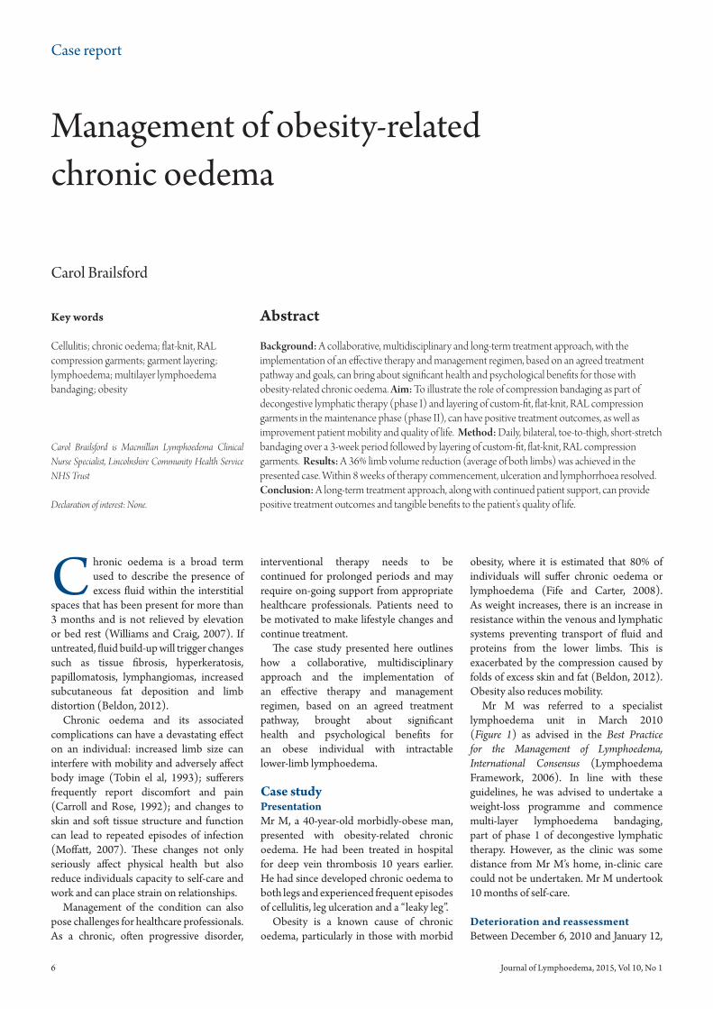
Mr M was referred to a specialist lymphoedema unit in March 2010 (Figure 1) as advised in the Best Practice for the Management of Lymphoedema, International Consensus (Lymphoedema Framework, 2006). In line with these guidelines, he was advised to undertake a weight-loss programme and commence multi-layer lymphoedema bandaging, part of phase 1 of decongestive lymphatic therapy. However, as the clinic was some distance from Mr M’s home, in-clinic care could not be undertaken. Mr M undertook 10 months of self-care.
Deterioration and reassessmentBetween December 6, 2010 and January 12,
Abstract
Background: A collaborative, multidisciplinary and long-term treatment approach, with the implementation of an effective therapy and management regimen, based on an agreed treatment pathway and goals, can bring about significant health and psychological benefits for those with obesity-related chronic oedema. Aim: To illustrate the role of compression bandaging as part of decongestive lymphatic therapy (phase I) and layering of custom-fit, flat-knit, RAL compression garments in the maintenance phase (phase II), can have positive treatment outcomes, as well as improvement patient mobility and quality of life. Method: Daily, bilateral, toe-to-thigh, short-stretch bandaging over a 3-week period followed by layering of custom-fit, flat-knit, RAL compression garments. Results: A 36% limb volume reduction (average of both limbs) was achieved in the presented case. Within 8 weeks of therapy commencement, ulceration and lymphorrhoea resolved. Conclusion: A long-term treatment approach, along with continued patient support, can provide positive treatment outcomes and tangible benefits to the patient’s quality of life.
Carol Brailsford is Macmillan Lymphoedema Clinical Nurse Specialist, Lincolnshire Community Health Service NHS Trust
Declaration of interest: None.
interventional therapy needs to be continued for prolonged periods and may require on-going support from appropriate healthcare professionals. Patients need to be motivated to make lifestyle changes and continue treatment.
The case study presented here outlines how a collaborative, multidisciplinary approach and the implementation of an effective therapy and management regimen, based on an agreed treatment pathway, brought about significant health and psychological benefits for an obese individual with intractable lower-limb lymphoedema.
Case studyPresentationMr M, a 40-year-old morbidly-obese man, presented with obesity-related chronic oedema. He had been treated in hospital for deep vein thrombosis 10 years earlier. He had since developed chronic oedema to both legs and experienced frequent episodes of cellulitis, leg ulceration and a “leaky leg”.
Obesity is a known cause of chronic oedema, particularly in those with morbid
Case report
8 Journal of Lymphoedema, 2015, Vol 10, No 1
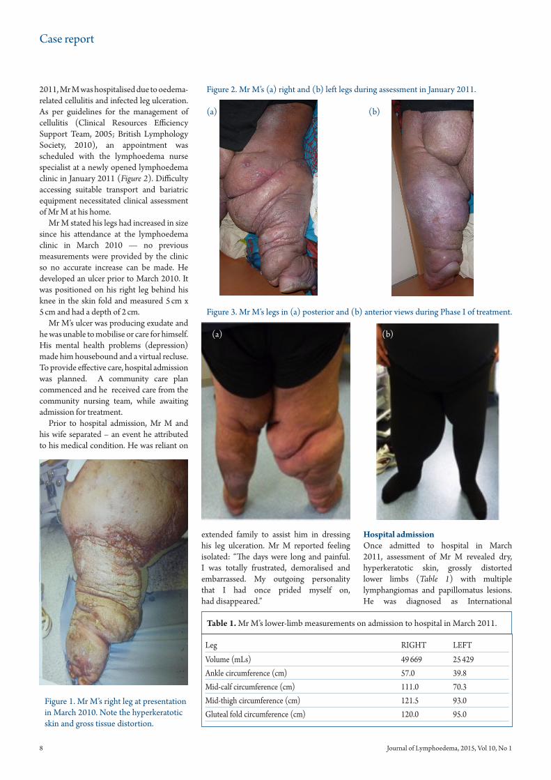
2011, Mr M was hospitalised due to oedema-related cellulitis and infected leg ulceration. As per guidelines for the management of cellulitis (Clinical Resources Efficiency Support Team, 2005; British Lymphology Society, 2010), an appointment was scheduled with the lymphoedema nurse specialist at a newly opened lymphoedema clinic in January 2011 (Figure 2). Difficulty accessing suitable transport and bariatric equipment necessitated clinical assessment of Mr M at his home.
Mr M stated his legs had increased in size since his attendance at the lymphoedema clinic in March 2010 — no previous measurements were provided by the clinic so no accurate increase can be made. He developed an ulcer prior to March 2010. It was positioned on his right leg behind his knee in the skin fold and measured 5 cm x 5 cm and had a depth of 2 cm.
Mr M’s ulcer was producing exudate and he was unable to mobilise or care for himself. His mental health problems (depression) made him housebound and a virtual recluse. To provide effective care, hospital admission was planned. A community care plan commenced and he received care from the community nursing team, while awaiting admission for treatment.
Prior to hospital admission, Mr M and his wife separated – an event he attributed to his medical condition. He was reliant on
Hospital admissionOnce admitted to hospital in March 2011, assessment of Mr M revealed dry, hyperkeratotic skin, grossly distorted lower limbs (Table 1) with multiple lymphangiomas and papillomatus lesions. He was diagnosed as International
extended family to assist him in dressing his leg ulceration. Mr M reported feeling isolated: “The days were long and painful. I was totally frustrated, demoralised and embarrassed. My outgoing personality that I had once prided myself on, had disappeared.”
Table 1. Mr M’s lower-limb measurements on admission to hospital in March 2011.
Figure 1. Mr M’s right leg at presentation in March 2010. Note the hyperkeratotic skin and gross tissue distortion.
Figure 2. Mr M’s (a) right and (b) left legs during assessment in January 2011.
(a) (b)
Figure 3. Mr M’s legs in (a) posterior and (b) anterior views during Phase I of treatment.
(a) (b)
Case report
Journal of Lymphoedema, 2015, Vol 10, No 1 9
Society of Lymphology Stage III (International Society of Lymphology, 2003).
An ulcer (5 cm × 5 cm × 2 cm) to the posterior aspect of his right leg was leaking profuse volumes of lymphorrhoea, which necessitated redressing with absorbent material at least twice per day. The periwound skin was macerated, however there were no signs of infection and no malodour. The wound bed was estimated to comprise 50% slough, 30% epithelial tissue (deep pink to pearly pink, light purple from edges in full-thickness wounds or this may form islands in superficial wounds)and 20% granulation tissue.
Vascular assessment of both limbs with a Doppler ultrasound indicated good arterial blood flow and suitability for sustained, graduated compression therapy.
Mr M appeared depressed on admission: he was withdrawn, made no eye contact and avoided conversation. Treatment goals were identified as being to reduce limb volume by 30% by use of compression bandaging and improve skin and wound condition. Treatment commenced with a robust skin cleansing and emollient regimen and daily application of toe-to-thigh, short-stretch, compression bandaging to both legs. This regimen forms part of the four cornerstones of care of phase 1 of decongestive lymphatic therapy (Lymphoedema Framework, 2006).
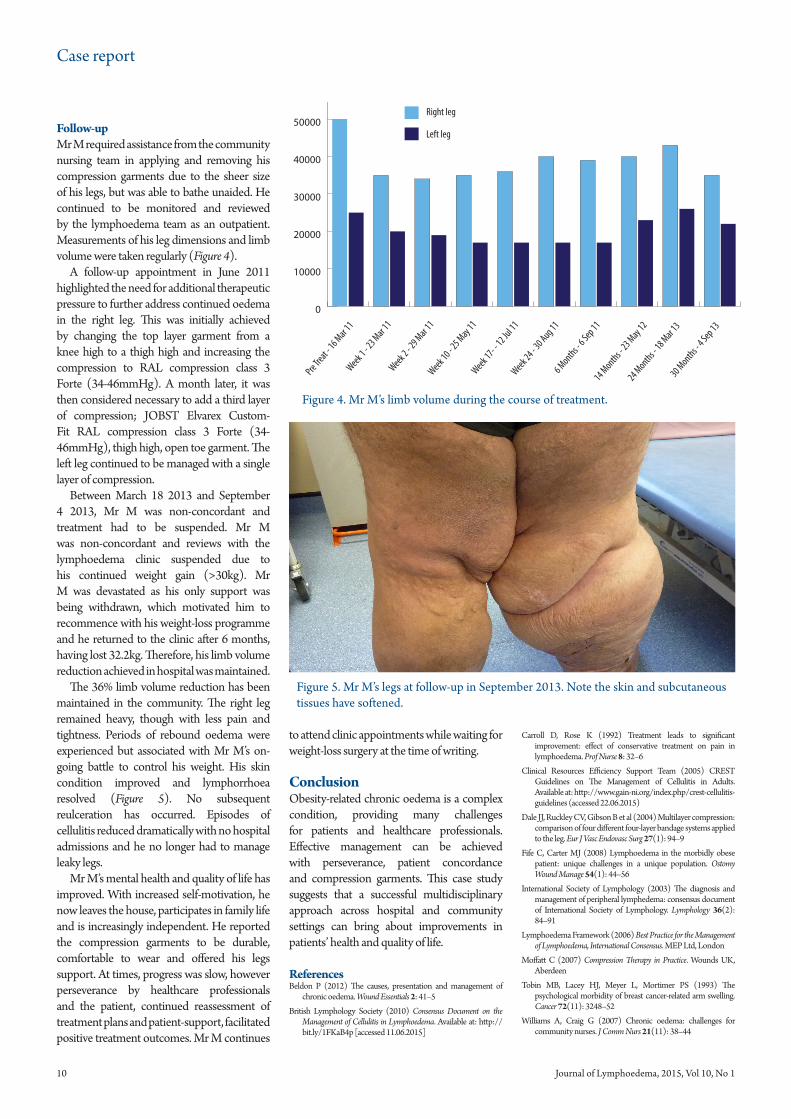
Completion of intensive treatment and discharge planningDuring his inpatient stay (admission was March 22 2011 and he was discharged April 8 2011), Mr M’s limb volume reduced by 36% (average of both), skin condition improved and lymphorrhoea resolved (Figure 3).
Within 2 weeks of the commencement of compression therapy, granulation tissue was observed on the ulcer and by week 8 it had healed. During the course of his inpatient stay, Mr M’s mood improved: “As the days went on, my confidence built, my legs improved and my mobility improved with it. Before I knew it I was able to walk, in a fashion, to have my treatment.”
Prior to discharge, Mr M was measured for compression garments as per the maintenance phase II of care (Lymphoedema Framework, 2006). He was provided with a JOBST® (Hamburg) Elvarex® Custom-Fit, RAL compression class 3 (34–46 mmHg), thigh-high, closed-toe garment with silicone band and T-heel for the left leg. A RAL CCL 3 Forte (34–46 mmHg) was provided for the right leg. A JOBST Elvarex Custom-Fit RAL compression class 2 (23–32 mmHg) below-knee, open-toe garment to be worn as an additional compression layer on the right leg (Figure 3b)
Due to the extensive, stubborn oedema in Mr M’s right leg, layering of compression garments was considered the best clinical option. Two layers of garment produce a higher pressure on the limb and are stiffer than one. The second layer is likely to add about 70% of the pressure it would when applied alone (Dale et al, 2004).
Mr M stated he was happy to wear his compression garments at home and felt they were comfortable. He chose black as, when worn under trousers, they resembled a man’s sock.
3D KNITTING
JOBST ELVAREX MATERIAL
CONFIDENCE IN TREATMENT OUTCOMES JOBST Elvarex Plus is part of a robust family of products which through innovative product development, continues the JOBST Elvarex success story in the management of lymphoedema and chronic oedema.
AVAILABLE ON DRUG TARIFF.Contact your BSN medical Vascular and Wound Care Account Manager, for more information.
JOBST® ELVAREX® PLUSA NEW GENERATION IN FLAT-KNIT COMPRESSION GARMENTS
Follow-upMr M required assistance from the community nursing team in applying and removing his compression garments due to the sheer size of his legs, but was able to bathe unaided. He continued to be monitored and reviewed by the lymphoedema team as an outpatient. Measurements of his leg dimensions and limb volume were taken regularly (Figure 4).
A follow-up appointment in June 2011 highlighted the need for additional therapeutic pressure to further address continued oedema in the right leg. This was initially achieved by changing the top layer garment from a knee high to a thigh high and increasing the compression to RAL compression class 3 Forte (34-46mmHg). A month later, it was then considered necessary to add a third layer of compression; JOBST Elvarex Custom-Fit RAL compression class 3 Forte (34-46mmHg), thigh high, open toe garment. The left leg continued to be managed with a single layer of compression.
Between March 18 2013 and September 4 2013, Mr M was non-concordant and treatment had to be suspended. Mr M was non-concordant and reviews with the lymphoedema clinic suspended due to his continued weight gain (>30kg). Mr M was devastated as his only support was being withdrawn, which motivated him to recommence with his weight-loss programme and he returned to the clinic after 6 months, having lost 32.2kg. Therefore, his limb volume reduction achieved in hospital was maintained.
The 36% limb volume reduction has been maintained in the community. The right leg remained heavy, though with less pain and tightness. Periods of rebound oedema were experienced but associated with Mr M’s on-going battle to control his weight. His skin condition improved and lymphorrhoea resolved (Figure 5). No subsequent reulceration has occurred. Episodes of cellulitis reduced dramatically with no hospital admissions and he no longer had to manage leaky legs.
Mr M’s mental health and quality of life has improved. With increased self-motivation, he now leaves the house, participates in family life and is increasingly independent. He reported the compression garments to be durable, comfortable to wear and offered his legs support. At times, progress was slow, however perseverance by healthcare professionals and the patient, continued reassessment of treatment plans and patient-support, facilitated positive treatment outcomes. Mr M continues
Carroll D, Rose K (1992) Treatment leads to significant improvement: effect of conservative treatment on pain in lymphoedema. Prof Nurse 8: 32–6
Clinical Resources Efficiency Support Team (2005) CREST Guidelines on The Management of Cellulitis in Adults. Available at: http://www.gain-ni.org/index.php/crest-cellulitis-guidelines (accessed 22.06.2015)
Dale JJ, Ruckley CV, Gibson B et al (2004) Multilayer compression: comparison of four different four-layer bandage systems applied to the leg. Eur J Vasc Endovasc Surg 27(1): 94–9
Fife C, Carter MJ (2008) Lymphoedema in the morbidly obese patient: unique challenges in a unique population. Ostomy Wound Manage 54(1): 44–56
International Society of Lymphology (2003) The diagnosis and management of peripheral lymphedema: consensus document of International Society of Lymphology. Lymphology 36(2): 84–91
Lymphoedema Framework (2006) Best Practice for the Management of Lymphoedema, International Consensus. MEP Ltd, London
Moffatt C (2007) Compression Therapy in Practice. Wounds UK, Aberdeen
Tobin MB, Lacey HJ, Meyer L, Mortimer PS (1993) The psychological morbidity of breast cancer-related arm swelling. Cancer 72(11): 3248–52
Williams A, Craig G (2007) Chronic oedema: challenges for community nurses. J Comm Nurs 21(11): 38–44
to attend clinic appointments while waiting for weight-loss surgery at the time of writing.
ConclusionObesity-related chronic oedema is a complex condition, providing many challenges for patients and healthcare professionals. Effective management can be achieved with perseverance, patient concordance and compression garments. This case study suggests that a successful multidisciplinary approach across hospital and community settings can bring about improvements in patients’ health and quality of life.
ReferencesBeldon P (2012) The causes, presentation and management of
chronic oedema. Wound Essentials 2: 41–5
British Lymphology Society (2010) Consensus Document on the Management of Cellulitis in Lymphoedema. Available at: http://bit.ly/1FKaB4p [accessed 11.06.2015]
Figure 4. Mr M’s limb volume during the course of treatment.
0
10000
20000
30000
40000
50000
60000
Pre Treat -
16 Mar 11
Week 1 - 2
3 Mar 11
Right leg
Left leg
Week 2 - 2
9 Mar 11
Week 10 - 2
5 May 11
Week 17- -
12 Jul 11
Week 24 - 3
0 Aug 116 Months -
6 Sep 1114 Months -
23 May 12
24 Months - 18 Mar 1
330 Months -
4 Sep 13
Figure 5. Mr M’s legs at follow-up in September 2013. Note the skin and subcutaneous tissues have softened.