Clin Oral Impl Res 2000: 11 (Suppl.): 108–125 Copyright C Munksgaard 2000 Printed in Denmark ¡ All rights reserved ISSN 0905-7161 Chapter 7 Management of the edentulous patient Mericske-Stern RD, Taylor TD, Belser U. Management of the edentulous Regina D. Mericske-Stern 1 , patient. Thomas D. Taylor 2 , Urs Belser 3 Clin Oral Impl Res 2000: 11 (Suppl.): 108–125. C Munksgaard 2000. 1 Department of Prosthodontics, Being edentulous is a handicap, and the main objective of implant place- University of Bern, Bern, Switzerland; ment is to provide support of fixed prostheses or to stabilize complete 2 Department of Prosthodontics, University of Farmington, CT, USA; dentures in the edentulous jaw. Clinical experience and clinical studies 3 Department of Prosthodontics, have demonstrated the broad application of non-submerged ITI im- University of Geneva, Geneva, plants in prosthetic therapy in standard sites and in situations of advanced Switzerland atrophy or substantial loss of tissue. The ITI implant was developed for universal use in partially and completely edentulous patients and for re- placement of single missing teeth. The abutment system offers the choice of both removable and fixed prostheses with identical secondary parts. The present article describes the use of ITI implants for prosthodontic re- Key words: ITI implants – edentulous habilitation in the completely edentulous jaw. Indications and various jaw – treatment planning – number of types of fixed or removable prostheses, alternatives and variations of implants – overdentures – fixed design are discussed. Prosthetic design is dependent on the number and prosthesis – occlusion location of implants, and conversely, the number of implants that can be placed will determine the choice of prosthesis. Treatment planning in PD Dr. R. Mericske-Stern, University general and with respect to individual anatomic-morphologic conditions of Bern, Freiburgstrasse 7, is described for the upper and lower jaw. Details of clinical procedures 3010 Bern, Switzerland with ITI implants related to the specific design of prostheses are presented. Tel.: π41 31 632 25 39 Biomechanical aspects of fixation and stabilization of prostheses and as- Fax: π41 31 632 49 33 pects of occlusion to be built up complete the overview. e-mail: regina.mericske/zmk.unibe.ch For many patients, being edentulous must be re- garded as a handicap with respect to oral function and psychosocial impact on quality of life. As a result, restoration of oral function through oral surgery and placement of implants is often wel- come. Long-term studies have demonstrated that the edentulous jaw can be restored successfully with implant-supported fixed prostheses (Zarb & Schmitt 1989, 1990a, 1990b; Adell et al. 1990; Qui- rynen et al. 1992). The success rate has been de- fined at various times by various authors (e.g. Al- brektsson et al. 1986; Smith & Zarb 1989; Buser et al. 1991; Albrektsson & Zarb 1993; Roos et al. 1997), and different limits have been set for the upper and lower jaw: Ø95% and Ø90% after 5 and 10 years for the mandible and Ø90% and Ø85% for the maxilla, respectively. This definition of success has been first applied to fixed implant prostheses in the edentulous jaw. Nowadays it appears that mandibular implants supporting overdentures are particularly successful 108 (Batenburg et al. 1998), whereas for maxillary im- plants an increased failure rate is reported. This increased failure rate has mainly been observed with Brånemark fixtures; for ITI implants the re- sults of only a small number of patients are avail- able. Recently, multicenter studies of ITI implants have reported on a small number of edentulous maxillary jaws that were restored and maintained successfully during a still-restricted observation period (Buser et al. 1997, 1999). Nowadays the use of implants has a great im- pact on the prosthodontic treatment of the edentu- lous patient. A variety of prosthetic designs associ- ated with implant prostheses can be observed, and some new designs have emerged in response to the specific clinical conditions of the edentulous jaw provided with implants. Valid clinical methods and treatment strategies – involving the implant tech- nology – exist to ensure the quality of prosthetic reconstruction. Hence, with regard to some special implant indications, specific prosthodontic prin- ciples are needed.
Transcript
Clin Oral Impl Res 2000: 11 (Suppl.): 108–125 Copyright C Munksgaard 2000Printed in Denmark ¡ All rights reserved
ISSN 0905-7161
Chapter 7
Management of the edentulous patient
Mericske-Stern RD, Taylor TD, Belser U. Management of the edentulous Regina D. Mericske-Stern 1,patient. Thomas D. Taylor 2, Urs Belser 3
Clin Oral Impl Res 2000: 11 (Suppl.): 108–125. C Munksgaard 2000.1Department of Prosthodontics,
Being edentulous is a handicap, and the main objective of implant place- University of Bern, Bern, Switzerland;ment is to provide support of fixed prostheses or to stabilize complete 2Department of Prosthodontics,
University of Farmington, CT, USA;dentures in the edentulous jaw. Clinical experience and clinical studies3Department of Prosthodontics,have demonstrated the broad application of non-submerged ITI im-University of Geneva, Geneva,plants in prosthetic therapy in standard sites and in situations of advancedSwitzerlandatrophy or substantial loss of tissue. The ITI implant was developed for
universal use in partially and completely edentulous patients and for re-placement of single missing teeth. The abutment system offers the choiceof both removable and fixed prostheses with identical secondary parts.The present article describes the use of ITI implants for prosthodontic re-
Key words: ITI implants – edentuloushabilitation in the completely edentulous jaw. Indications and variousjaw – treatment planning – number oftypes of fixed or removable prostheses, alternatives and variations ofimplants – overdentures – fixeddesign are discussed. Prosthetic design is dependent on the number andprosthesis – occlusionlocation of implants, and conversely, the number of implants that can
be placed will determine the choice of prosthesis. Treatment planning inPD Dr. R. Mericske-Stern, Universitygeneral and with respect to individual anatomic-morphologic conditions of Bern, Freiburgstrasse 7,
is described for the upper and lower jaw. Details of clinical procedures 3010 Bern, Switzerlandwith ITI implants related to the specific design of prostheses are presented. Tel.: π41 31 632 25 39Biomechanical aspects of fixation and stabilization of prostheses and as- Fax: π41 31 632 49 33pects of occlusion to be built up complete the overview. e-mail: regina.mericske/zmk.unibe.ch
For many patients, being edentulous must be re-garded as a handicap with respect to oral functionand psychosocial impact on quality of life. As aresult, restoration of oral function through oralsurgery and placement of implants is often wel-come.
Long-term studies have demonstrated that theedentulous jaw can be restored successfully withimplant-supported fixed prostheses (Zarb &Schmitt 1989, 1990a, 1990b; Adell et al. 1990; Qui-rynen et al. 1992). The success rate has been de-fined at various times by various authors (e.g. Al-brektsson et al. 1986; Smith & Zarb 1989; Buser etal. 1991; Albrektsson & Zarb 1993; Roos et al.1997), and different limits have been set for theupper and lower jaw: Ø95% and Ø90% after 5 and10 years for the mandible and Ø90% and Ø85%for the maxilla, respectively.
This definition of success has been first appliedto fixed implant prostheses in the edentulous jaw.Nowadays it appears that mandibular implantssupporting overdentures are particularly successful
108
(Batenburg et al. 1998), whereas for maxillary im-plants an increased failure rate is reported. Thisincreased failure rate has mainly been observedwith Brånemark fixtures; for ITI implants the re-sults of only a small number of patients are avail-able. Recently, multicenter studies of ITI implantshave reported on a small number of edentulousmaxillary jaws that were restored and maintainedsuccessfully during a still-restricted observationperiod (Buser et al. 1997, 1999).
Nowadays the use of implants has a great im-pact on the prosthodontic treatment of the edentu-lous patient. A variety of prosthetic designs associ-ated with implant prostheses can be observed, andsome new designs have emerged in response to thespecific clinical conditions of the edentulous jawprovided with implants. Valid clinical methods andtreatment strategies – involving the implant tech-nology – exist to ensure the quality of prostheticreconstruction. Hence, with regard to some specialimplant indications, specific prosthodontic prin-ciples are needed.
Administrator
Highlight
Administrator
Sticky Note
Administrator
Sticky Note
Cancelled set by Administrator
Administrator
Highlight
Management of the edentulous patient
In many cases the treatment of the edentulousmaxilla will require more elective procedures thanare necessary for the mandible, particularly withrespect to the following criteria:O degree of atrophy of the residual jaw;O prospective location of the implants and incli-
nation of the implant axis;O tissue volume dimensions;O facial morphology;O esthetics;O function and phonetics.In general, the following criteria will determine thetreatment planning of the edentulous jaw:O the prosthetic design will depend on the distri-
bution of the implants over the arch, their loca-tion and their number;
O the natural dentition or type of prosthesis in theopposing jaw will influence the implant-pros-thodontic design;
O the intermaxillary relationship has to be con-sidered;
O the occlusal scheme is influenced by all thesefactors;
O esthetic considerations have to be involved.In the context of these criteria, implant prosthesesmust be planned, designed and managed for theedentulous jaw.
Indications for implants in completely edentulousjawsThe main objective of implants in the edentulousjaw is either 1) to avoid removable complete den-tures by placement of implant-supported fixedprostheses or 2) to stabilize complete dentures byplacement of implant-retained overdentures.
Local anatomic/morphologic conditions andgeneral patient-related factors determine thechoice of prosthesis. In general, more implants arerequired for support of fixed prostheses than foroverdenture retention. Therefore, in many cases theindication for fixed prostheses will be limited dueto inadequate structure of the bone, unless ad-ditional surgical procedures such as bone augmen-tation by graft procedures are used. This is par-ticularly true for the maxilla, and implies a morespecific patient selection than is necessary forsimple implant-prosthodontic procedures of themandible. Here, as well as in the case of advancedatrophy, a standard surgical and prosthodonticprotocol can often be utilized.
Indications for overdenturesThe indications may be different for the upper andlower jaw.
109
Mandibular overdenturesMandibular overdentures supported by only a fewintraforaminal implants are regarded today as ageriatric treatment modality. The indication com-prises a large segment of older patients who willprofit from implant-retained complete dentures ifthey lose their teeth in advanced age. In addition,mandibular overdentures may benefit older pa-tients who, having had complete dentures for manyyears, lose their motor skills and no longer feelable to wear complete dentures.
This problem is observed much more often forthe edentulous mandible than maxilla. Even withadvanced atrophy, standard surgical implant pro-cedures can be applied for mandibular overden-tures. Reduced treatment goals – e.g. the placementof only two implants – will minimize the risk topatients and tissues.
The recent literature (for review see Batenburget al. 1998) exhibits a high success rate for man-dibular overdentures, with the use of different im-plant systems and a varying number of implants(Batenburg et al. 1994; Wismeyer et al. 1995; Spie-kermann et al. 1995). The success of using fewer(generally two) implants has been clearly demon-strated (Mericske-Stern et al. 1994; Mericske-Stern & Zarb 1993; Mericske-Stern 1990; Mom-belli & Mericske-Stern 1990), but has not entirelybecome the standard clinical protocol in dailypractice. Age itself is no longer regarded as acontraindication (Bryant & Zarb 1998), andstudies with ITI implants have demonstrated thatmandibular overdentures are highly successful inolder patient groups (Mericske-Stern & Zarb 1993;Cune et al. 1994; Zarb & Schmitt 1994, 1995).Thus, mandibuar overdentures are a true alterna-tive to fixed prostheses in terms of economics andtime-saving procedures.
Maxillary overdenturesWhile most patients asking for mandibular over-dentures are completely edentulous in both jaws,the maxillary overdenture is indicated for patientswho have natural teeth in the opposing mandibleor fixed or removable prostheses supported by im-plants and teeth.
Studies published in the last 5 years exhibit asurprisingly high failure rate for maxillary over-dentures, i.e. over 20% (Jemt 1991; Jemt 1993; Jemtet al. 1996; Hutton et al. 1995). This failure rateis significantly increased in comparison with fixedprostheses or mandibular implants. A criticalanalysis of the treatment outcomes revealed thatthe indication for overdentures was often given inan emergency situation (Palmqvist et al. 1994),meaning that overdentures were a substitute forfailing fixed prostheses and were prescribed if ade-
Mericske-Stern et al.
quate placement of implants to support fixed pros-theses was not possible (Jemt 1991). Otherwise, inproperly planned overdentures, an increased sur-vival of implants was found (Bergendal & Enquist1998; Widmark et al. 1998). The marginal bonesurrounding the implants was maintained at thesame level as with fixed prostheses (Palmqvist et al.1994, 1996), also in ridges with advanced atrophy.
One advantage of overdentures is that their util-ization may be more consistent, with optimumplacement of the implants with regard to the re-maining bone structures. Full congruence of toothposition on the prosthesis and implant location isnot necessary for overdentures. Fewer implants areneeded than with fixed prostheses. Furthermore,requirements of extraoral esthetics such as facialsupport can be fulfilled and problems with phonet-ics and oral hygiene are often better resolved withoverdentures. In fact, hygiene procedures are most-ly facilitated with removable prostheses; however,under maxillary overdentures soft tissue hyper-plasia may develop.
Indications for fixed prosthesesFor the mandible, in many situations a fixed pros-thesis or an overdenture can be suggested, accord-ing to the individual needs of the patient. Even inthe case of advanced atrophy a screw-retainedcantilever prosthesis may be mounted on 4 to 6intraforaminal implants. For this type of recon-struction a full congruence of implant and toothposition is not required. If bone structure and bonequality are adequate, a fixed prosthesis with acrown-and-bridge design can be fabricated, sup-ported by intraforaminal and posterior implants.Esthetic or speech problems are rarely encounteredwith any type of mandibular reconstruction.
While for the edentulous mandible both op-tions – i.e. fixed and removable prostheses – canusually be offered, the anatomic-morphologicproblems of the maxilla and esthetic requirementsmust be underscored and will determine thechoice of the prosthetic design. A younger seg-ment among patients with edentulous maxilla willask for fixed bridgework. Patients asking for fixedimplant-prostheses in the edentulous upper jawoften present with a full complement of naturalteeth or fixed reconstruction in the opposing jaw.The inability to adapt to removable prostheses isbased on psychosocial aspects and/or on adversemorphological conditions of the oral cavitywhich would hinder wearing complete dentures.A screw-retained cantilever prosthesis is not fre-quently recommended for the maxilla, due to es-thetic problems and impaired hygienic pro-cedures. In most cases a combination of several
110
of the following aspects will determine the re-spective treatment plan:
O anatomic and morphologic structure of themaxilla;
O bone quantity;O esthetic considerations: facial support, tooth
length, soft tissue management;O ease of repair;O economics.
Favorable conditions related to bone quantity andquality for elective placement of multiple implantsare required for fixed bridgework. Clinical experi-ence today shows that the soft tissue may be man-aged successfully in case of one single-tooth re-placement (Belser et al. 1998; Salama et al. 1995).Recreating a well-contoured soft tissue borderaround implants over an entire dental arch has notyet been documented to be practicable. With fixedprostheses, in contrast to overdentures, phoneticproblems have been reported (Jemt 1991; Jemt1994; Lundqvist et al. 1992). In addition, compen-sation for lost hard and soft tissue becomes diffi-cult (Albora 1997) and is a problem that requiresspecial attention in the planning phase. The inter-maxillary distance between the incisal edge of thelower teeth and the contour of the maxillary jawshould not exceed 15 mm, otherwise the teeth willbecome too long and an overdenture would be abetter indication. A low lip-line (no gummy smile)is advantageous for fixed prostheses with regard toesthetic and cosmetic demands for the upper jaw.Complex skeletal, alveolar and occlusal conditionssuch as skeletal class 3 may determine the choiceof a removable prosthesis. With respect to variousextraoral and intraoral diagnostic criteria, over-dentures may often become the preferred option ifthe maxillary jaw is restored with implants.
Table 7.1 gives an overview of the intra- andextraoral diagnostic criteria for the choice of fixedor removable implant-supported prostheses in theedentulous maxilla.
Number of implants and choice of prosthesisThe number of implants to be placed depends onthe type of prosthesis and the choice of prostheticdesign. Conversely, the number of implants thatcan be placed with respect to anatomic-morpho-logic conditions will determine to a certain degreethe type and design of prosthesis. Additionally, thesize, curvature and shape of the ridges determinethe distribution of the implants over the arch.
Mandibular overdenturesFor the placement of mandibular overdentures inedentulous older patients wearing complete den-
Management of the edentulous patient
Table 7.1. Summary: diagnostic criteria for the maxilla
Lip-line Low High Ridge (shape) Vertical Buccal inclinationTooth display Little Distinct Convex Buccal concavityFacial support, lip support No need Necessary Intermax. dist. Æ10 mm ±15 mm
tures, two to four interforaminal implants willserve the purpose and satisfy the patients’ de-mands. There is no scientific evidence that failuresoccur more often with a small number of implants,namely two for overdenture retention. Singleattachments or bars can be mounted. If, due toadvanced atrophy, the implant length becomes ∞8
Fig. 7.1. The distribution of intraforaminal implants dependson the shape of the ridge. a) Bar connector would interfere withspace for tongue. Ball anchors are suggested; however, this willresult in a hinging movement. Implants located in more an-terior position: this may result in inadequate length of the bar.b/c) Three or four implants with a connecting bar are in betterharmony with the shape of the ridge. Four implants allow forfixed prosthesis. d) Two anterior implants with a connectingbar of adequate length. e) U-shaped mandibular jaw with largecurvature will allow for placement of four implants and a con-necting bar. f) This configuration is also favorable for mountingof a fixed screw-retained cantilever prosthesis. g) Alignment ofthe implants in a rather straight line does not favor fixed pros-theses.
111
mm, or if narrow, thin ridges require a reduced im-plant diameter (3.3 mm), the use of three or fourimplants is recommended. In the presence of largeor V-shaped anterior ridges, three to four implantswill provide for a more favorable design of the barand the prosthesis.
It is not necessary to recommend four intrafora-minal implants for mandibular overdentures as astandard procedure. It must be taken into accountthat bar segments may become rather short, andshort female bar retainers are subject to frequentloosening or loss. The length of the bar segmentsshould not be less than 15 mm, and can range from15 to 25 mm. If four intraforaminal implants areplaced they must be well spaced, or as an alterna-tive a cantilever-fixed prosthesis can be mounted.
O 2 implants for mandibular overdentures⇒ge-riatric treatment conception;
O 3 or 4 intraforaminal implants: if reduced di-ameter or length of 6 mm;
O 3 or 4 intraforaminal implants: length of barsegments must be adequate;
O 4 intraforaminal implants: fixed cantilever-prostheses may be recommended as an alter-native.
Fig. 7.1 gives an overview of the distribution ofmandibular implants for overdenture connection.A clinical situation with two intraforaminal man-dibular implants is shown in Fig. 7.2.
Fig. 7.2. Two mandibular implants with ball anchors.
Mericske-Stern et al.
Maxillary overdenturesThe actual state of the art for maxillary overden-tures is to adopt the biomechanical and technicalconcept of fixed prostheses–namely, multiple im-plants and a rigid connection of the prostheses tothe implants. In the maxilla most often bone qual-ity and quantity are not favorable: i.e. accordingto the criteria of Albrektsson et al. (1986), degreeof atrophy corresponds to class C and D. Four tosix well-spaced implants, evenly distributed overthe arch and connected by a bar, will enhance thestability of the overdenture. The implants aremostly located in the anterior part of the upperjaw, between the first premolars. Thus, additionalsurgery such as sinus floor elevation can beavoided in many cases. The implant length shouldpreferably be Ø10 mm, and a standard diameterof 4.1 mm is suggested. The literature provides evi-
Fig. 7.3. Distribution of maxillary implants for overdentureconnection. a) Use of two implants is not the standard pro-cedure. In this situation only ball anchors are suggested; a barwould interfere with the space of the tongue. b) Four implants,well distributed, with a sufficient length of bar segments. c) Abar cannot be recommended. It would result in a hinging move-ment. d) Four implants, often located in an anterior positiondue to the extension of the sinus. e) Depending on the specificanatomic situation, the bar may be divided into segments. Anirregular number of implants can also be used. f) In rare cases,more bone is available in the posterior part of the maxilla. Par-allel placement of two separate bars might be recommended.
112
Fig. 7.4. Clinical situation with four well-spaced implants in theanterior part of the maxilla and a bar connector.
dence of an increased failure rate for short im-plants. Therefore, the use of implants which are‘‘non-standard’’ in length or diameter must becompensated for by a sufficient number of stan-dard-sized implants. A connecting bar cannot bemounted to two implants in the maxilla, due to theanatomic-morphologic conditions. Thus, the useof two maxillary implants for overdenture supportis rarely recommended. The use of two ball an-chors results in a hinging movement of the denturethat may cause discomfort.O the minimum number of implants is preferably
not less than four;O using two implants is not a standard procedure;O the implants should be evenly distributed over
the arch;O implants of 6 mm length should be avoided;O implants with a reduced diameter (3.3 mm) have
to be combined with implants of standard diam-eter.
Fig. 7.3 gives an overview of the distribution ofmaxillary implants for overdenture connection. Aclinical situation with maxillary implants for over-denture connection is shown in Fig. 7.4.
Fixed prostheses in the mandible and maxillaA typical feature of the mandibular fixed cantileverprosthesis is 4 to 6 interforaminal implants.Congruence of implant location and tooth posi-tion is not necessary. This type of prosthesis isfavored by a distinct anterior curvature of the jaw.If the implants are aligned on a straight anteriorline this will result in a biomechanically unfavor-able situation for loading and designing of theprostheses. The material and characteristics re-semble removable partial dentures. The prosthesisoften has shortened dental arches.
For placement of full-arch bridgework the mostimportant prerequisite is the congruence of im-
Management of the edentulous patient
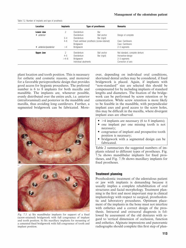
Table 7.2. Number of implants and type of prosthesis
Upper Jaw 2 Overdenture Ball anchor Not standard, complete denture4–5 Overdenture Bar (rigid) Horseshoe-design
±4–8 Bridgework 2–3 segmentsIndividual abutments Correction of axis
plant location and tooth position. This is necessaryfor esthetic and cosmetic reasons, and moreoverfor a favorable perioprosthetic design that providesgood access for hygienic procedures. The preferrednumber is 6 to 8 implants for both maxilla andmandible. The implants are, whenever possible,evenly distributed over the entire arch, i.e. anterior(intraforaminal) and posterior in the mandible andmaxilla, thus avoiding long cantilevers. Further, asegmented bridgework can be fabricated. More-
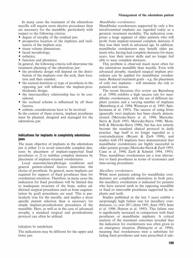
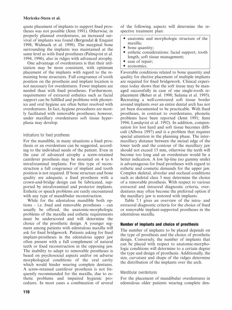
Fig. 7.5. a) Six mandibular implants for support of a fixed(screw-retained) bridgework with full congruence of implantand tooth position. b) Six maxillary implants for mounting ofa cemented fixed bridgework with full congruence of tooth andimplant position.
113
over, depending on individual oral conditions,shortened dental arches may be considered, if fixedbridgework is placed. Again, if implants with‘‘non-standard’’ size are selected this should becompensated for by including implants of standardlengths and diameters. The fixation of the bridge-work can be performed by screw retention or bycementation. While screw retention is more likelyto be feasible in the mandible, with perpendicularimplant axes and good access to the screw holes,this may be difficult in the maxilla, where divergentimplant axes are observed.
O ±4 implants are necessary (6 to 8 implants);O one implant per one missing tooth is not
necessary;O congruence of implant and prospective tooth
position is necessary;O bridgework with a segmented design can be
fabricated.
Table 2 summarizes the suggested numbers of im-plants related to different types of prostheses. Fig.7.5a shows mandibular implants for fixed pros-theses, and Fig. 7.5b shows maxillary implants forfixed prostheses.
Treatment planningProsthodontic treatment of the edentulous patientor jaw with implants is demanding because itusually implies a complete rehabilitation of oralstructures and facial morphology. Treatment plan-ning is the first and most important step in clinicalimplantology with respect to surgical, prosthodon-tic and laboratory procedures. Optimum place-ment of the implants in the bone must not interferewith esthetics and a correct design of the pros-thesis. Intraoral and extraoral diagnosis is fol-lowed by assessment of the old dentures with re-gard to vertical dimension of occlusion, functionand esthetics. Alginate impressions and panoramicradiographs should complete this first step of plan-
Mericske-Stern et al.
Fig. 7.6. Panoramic radiograph with metallic markers in inter-foraminal position.
Fig. 7.7. Radiographic template is used as surgical guide.
ning. Metallic markers are helpful landmarks forassessment of the prospective implant location andfor diagnosis of anatomic structures of the edentu-lous jaw. During the non-submerged healing phase,provisional dentures must be carefully adapted fortemporary use to protect the implants from inad-vertent loading. Clinical studies give evidence ofa highly successful non-submerged healing period(Mericske-Stern et al. 1995; Behneke et al. 1997;Buser et al. 1997, 1999). Only well-fitting old den-tures may be used. This preliminary clinical andradiographic diagnosis determines whether the en-visaged therapeutic measures can be performed.More detailed information about individual oralconditions is necessary if extended implant over-dentures of the maxilla and/or fixed bridgeworkare planned.
Mandibular overdenturesLateral radiographs are not necessary but mayprovide useful information about the shape andlingual profile of the mandibular bone. If radio-graphic templates with markers are fabricated forpanoramic radiographs these can be adapted andused as surgical templates. Existing old denturesmay be used as surgical templates as well. Fig. 7.6
114
shows a panoramic radiograph with metallicmarkers for quantitative analysis of bone. In Fig.7.7, a template is used as the surgical guide.
Overdentures in the maxilla and fixed prostheses in themaxilla or mandibleThese types of reconstruction usually require amore specific treatment plan (Mericske-Stern1998). The prosthodontic treatment of the maxillawith implants is challenging and determined by in-herent problems that have been presented in anec-dotal case reports (Hallmann & Carlsson 1996).These are: divergent implant axis, long teeth, wideinterdental spaces, missing congruence of implantlocation and tooth position, and buccal access tothe occlusal screw. Such adverse morphological ef-fects can be more easily eliminated with the utiliza-tion of overdentures instead of fixed prostheses.Nonetheless, treatment planning, particularly forthe maxilla, usually has to consider both treatmentoptions – fixed and removable prostheses – becausepatients often ask first of all for fixed prostheses.A tooth setup in wax should be performed forplanning of fixed prostheses, and will ultimatelydetermine whether the edentulous jaw can be re-stored with bridgework or overdentures should beused.
Mounted casts and the tooth setup are used toconsider aspects of design, and to assess function,occlusion and esthetic aspects of the prospectivereconstruction, such as vertical dimension of oc-clusion, relationship of maxillary to mandibularjaw, occlusion, phonetics, facial support and cos-metics. The orientation index obtained from thetooth setup is utilized to analyze dimensions oftissue volume and to compare the prospective im-plant and tooth axis. The tooth setup is furtherused to fabricate templates for radiographs with
– not necessary. π useful. ππ recommended or necessary.
Management of the edentulous patient
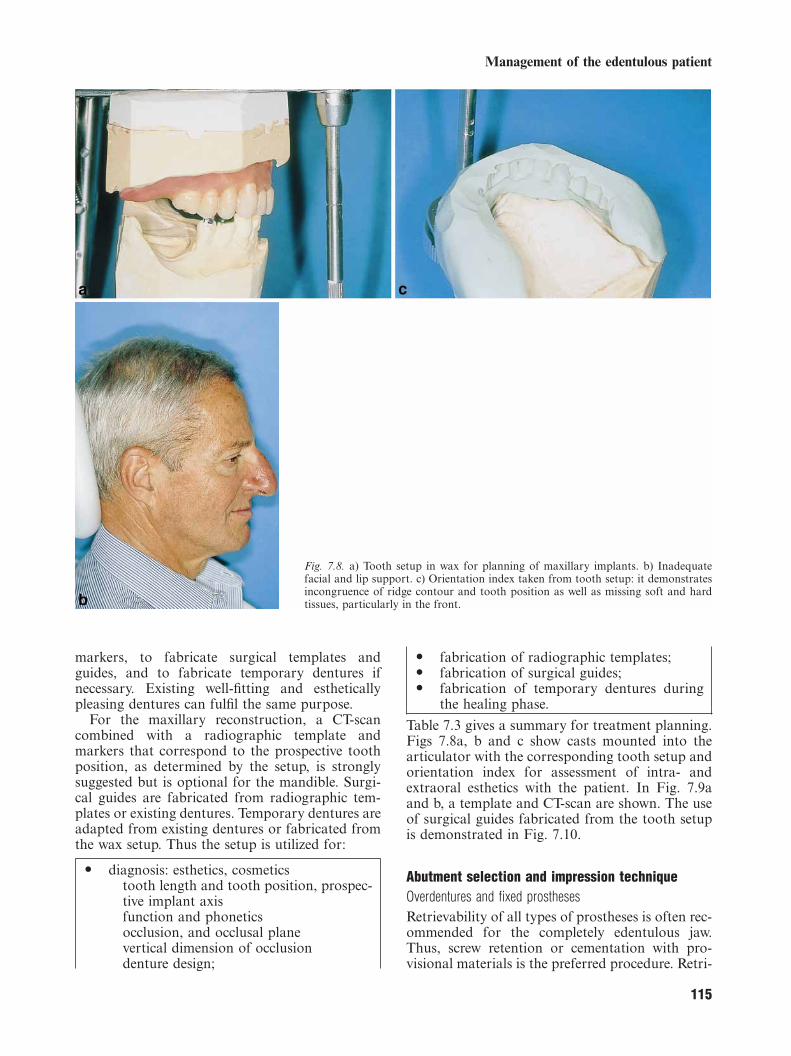
Fig. 7.8. a) Tooth setup in wax for planning of maxillary implants. b) Inadequatefacial and lip support. c) Orientation index taken from tooth setup: it demonstratesincongruence of ridge contour and tooth position as well as missing soft and hardtissues, particularly in the front.
markers, to fabricate surgical templates andguides, and to fabricate temporary dentures ifnecessary. Existing well-fitting and estheticallypleasing dentures can fulfil the same purpose.
For the maxillary reconstruction, a CT-scancombined with a radiographic template andmarkers that correspond to the prospective toothposition, as determined by the setup, is stronglysuggested but is optional for the mandible. Surgi-cal guides are fabricated from radiographic tem-plates or existing dentures. Temporary dentures areadapted from existing dentures or fabricated fromthe wax setup. Thus the setup is utilized for:
O diagnosis: esthetics, cosmeticstooth length and tooth position, prospec-tive implant axisfunction and phoneticsocclusion, and occlusal planevertical dimension of occlusiondenture design;
115
O fabrication of radiographic templates;O fabrication of surgical guides;O fabrication of temporary dentures during
the healing phase.
Table 7.3 gives a summary for treatment planning.Figs 7.8a, b and c show casts mounted into thearticulator with the corresponding tooth setup andorientation index for assessment of intra- andextraoral esthetics with the patient. In Fig. 7.9aand b, a template and CT-scan are shown. The useof surgical guides fabricated from the tooth setupis demonstrated in Fig. 7.10.
Abutment selection and impression techniqueOverdentures and fixed prosthesesRetrievability of all types of prostheses is often rec-ommended for the completely edentulous jaw.Thus, screw retention or cementation with pro-visional materials is the preferred procedure. Retri-
Mericske-Stern et al.
Fig. 7.9. a) Radiographic template: duplication of tooth setupor of a well-fitting existing denture, with titanium pins, indi-cating tooth position and tooth length. b) CT-scan: pins allowfor detailed analysis and measurements.
Fig. 7.10. Radiographic template is modified and used as surgi-cal guide for placement of six maxillary implants.
116
evability is advantageous in case of any compli-cation. In the presence of multiple implants, theremoval of one implant will not necessarily lead tochanges in the prosthetic superstructures. All re-tention devices used for removable prostheses withthe ITI system, such as bars or retentive anchorsof overdentures, are retrievable.
The octa abutment and the corresponding set ofprefabricated, secondary parts for clinical and lab-oratory procedures can be used in all indications,i.e. overdentures and fixed prostheses. This abut-ment ensures optimum precision in clinical andlaboratory procedures, be it in conjunction withthe standard implant type or the new SynoctaA
system. The octa abutment’s design, in combi-nation with prefabricated gold copings for the bar,is likely to facilitate good oral hygiene and to avoidplaque accumulation. Full-arch fixed prosthesesmay be screw-retained. However, the octa abut-ment also allows for fabrication of individual castabutments with optimum parallel alignment andcorrection of non-parallel implant axes. As aconsequence, such individual abutments requirethe cementation of the reconstruction.
Use of the double impression technique is ad-
Fig. 7.11. a) Cast with axis indicator of four implants. Individ-ual cast tray with corresponding access holes. A bar is planned.(Same case as Fig. 4). b) Individual tray in situ. Transfer copingsof octa system are visible.
Administrator
Highlight
Administrator
Highlight
Administrator
Highlight
Administrator
Highlight
Administrator
Highlight
Management of the edentulous patient
Fig. 7.12. a) Impression with Zn-eugeno paste is taken. Tray isrepositioned in mouth with transfer copings in situ. b) Fixationof transfer copings with Impregum impression material.
vised for overdentures retained by only two im-plants. This type of overdenture has characteristicsresembling those of a complete denture, with acombination of tissue support and implant reten-tion. For all other indications a one-step techniqueis indicated. Taking an impression with ball an-chors achieves better precision without the use oftransfer copings. Individually cast trays are oftenrecommended in the completely edentulous jaw.For optimum design of the individually cast tray,the alginate impression is taken with some direc-tion indicator mounted onto the implants. This isto ensure proper size of the tray above themounted transfer copings. Ease of access to theocclusal screw through the holes of the individualtray is provided if a screw-retained abutment sys-tem is utilized.
O Octa abutment fulfills all indications of theedentulous jaw: bars, screw retention of fixedprostheses, and individually cast abutmentsfor fixed prostheses;
O Retentive anchors: mandibular overdentures,geriatric conception;
O Double impression technique: overdenturessupported by two implants;
117
Fig. 7.13. a) Screw-retained transfer copings mounted to man-dibular implants. b) Impression is removed, with transfercopings in situ. c) Simple transfer system in combination withSynoctaA system: click-fit copings in situ.
O One-step impression technique: all other in-dications;
O Individually cast tray with space for thetransfer copings is recommended;
O Access to the occlusal screws of the transfercopings is mandatory if screw-retained trans-fer copings are used.
Figs 7.11a and b demonstrate optimum fabricationof the individual tray for impression with thescrew-retained transfer copings. Figs 7.12a and bdemonstrate the step-by-step procedure for a
Mericske-Stern et al.
double impression technique with two mandibularimplants. Figs 7.13a, b, c show the impressiontechnique with the standard octa abutment andwith the SynoctaA system.
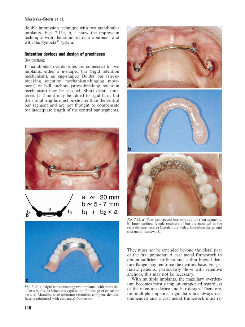
Retention devices and design of prosthesesOverdenturesIf mandibular overdentures are connected to twoimplants, either a u-shaped bar (rigid retentionmechanism), an egg-shaped Dolder bar (stress-breaking retention mechanismΩhinging move-ment) or ball anchors (stress-breaking retentionmechanism) may be selected. Short distal canti-levers (5–7 mm) may be added to rigid bars, buttheir total lengths must be shorter than the centralbar segment and are not thought to compensatefor inadequate length of the central bar segments.
Fig. 7.14. a) Rigid bar connecting two implants, with short dis-tal extensions. b) Schematic explanation for design of extensionbars. c) Mandibular overdenture resembles complete denture.Base is reinforced with cast metal framework.
118
Fig. 7.15. a) Four well-spaced implants and long bar segments.b) Inner surface: female retainers of bar are mounted in theresin denture base. c) Overdenture with a horseshoe design andcast metal framework.
They must not be extended beyond the distal partof the first premolar. A cast metal framework toobtain sufficient stiffness and a thin lingual den-ture flange may reinforce the denture base. For ge-riatric patients, particularly those with retentiveanchors, this may not be necessary.
With multiple implants, the maxillary overden-ture becomes mostly implant-supported regardlessof the retention device and bar design. Therefore,for multiple implants, rigid bars are always rec-ommended and a cast metal framework must re-
Management of the edentulous patient
inforce the denture base to ensure stability andstiffness (Mericske-Stern 1998a). Four to six im-plants connected by a rigid bar provide high sta-bility and a maximum of support. The overdentureitself has a horseshoe design. This is well toleratedby patients, also from a psychological point ofview, because the feeling of wearing complete den-tures is absent. An individually cast metal frame-work is mandatory for a horseshoe design of themaxillary overdenture and provides for adequatestiffness and rigidity of the overdenture. The pala-tal seal is cast in metal. Female bar retainers arenot soldered to the cast metal framework, butrather are mounted in the acrylic denture base.This facilitates prosthetic services like tighteningor renewal of retainers. The use of ball anchorswith maxillary overdentures is not standard andhas the character of a long-term provisional res-toration. Full coverage of the palate may becomenecessary. The connection of overdentures to ballanchors is more favorable if parallel alignment ofthe implant axes is achieved. With pronounced di-vergence of the axes, stable fit is not achievedthrough the female parts.
Based on biomechanical studies with mandibu-lar implants, it has been concluded that rigid barsprovide the best distribution of forces in a verticaldirection onto the implants (Mericske-Stern et al.1996a, 1996b; Mericske-Stern 1998b). It seemsthat short distal cantilevers do not negatively in-fluence the force pattern (Mericske-Stern 1997).With three implants, force magnitudes measuredon the central implant are lower in a vertical direc-tion than in a transverse direction (Burgin et al.1998). In vivo force measurements with maxillaryimplants connected by a rigid bar or supportingfull-arch bridgework have been shown to result inmostly identical force patterns and force magni-tudes for both types of prosthesis (Mericske-Sternet al. 1998). From this study the authors concludedthat the splinting effect of bars connecting multipleimplants might resemble fixed prostheses.
Figs 7.14a–c show mandibular overdentures andexplain the design of extension bars connectingmandibular implants. Figs 7.15 a–c show maxillaryimplants for overdenture connection and details oftechnical aspects.
Fig. 7.16. Planning of screw-retained cantilever prosthesis: de-sign and biomechanical considerations.
119
Fig. 7.17. a) Extreme skeletal class 3: cast metal framework ofscrew-retained cantilever prosthesis in situ (same case as Fig.13a, b). b) Full congruence of tooth and implant position is notnecessary. Access to three occlusal screws is buccally located.
Screw-retained cantilever prosthesesA screw-retained fixed cantilever prosthesis is afavorable alternative to overdentures in the man-dible, especially for a younger segment of the olderpopulation. It is less expensive than a ceramicbridgework. High precision of fit is achieved byfixation of the titanium copings into the frame-work. The cast framework can either be a gold al-loy or a non-precious alloy. The design is consist-ent with perioprosthetic requirements and the es-thetic appearance is not impaired. However,greater manual skills are necessary for daily hy-giene procedures than with removable overden-tures. The design cannot be recommended for themaxilla for esthetical and functional reasons.Speech problems may arise and a buccal flange be-comes necessary to hide the metal structures of thedenture or the implant shoulder. The selection ofthis prosthetic design is based on the morphologiccondition of the mandible and the distribution ofthe implants over the arch.
Fig. 7.16 is a schematic picture explaining thedesigning of cantilever prostheses. Figs 17.7a andb show a screw-retained cantilever prosthesis.
Mericske-Stern et al.
Fig. 7.18. a) Individually cast abutments fitting on octa abut-ment: optimum parallel alignment of abutments is achieved(same case as Fig. 5b). b) Orientation index for mounting theabutments. c) Framework (gold alloy) in situ with occlusal stops(Dura-lay). Framework is cast in two segments. Optimumcongruence of tooth position and implant location is achieved.d) Completed bridgework (ceramic) in situ: no access holes forocclusal screws necessary. This improves design of occlusal con-tacts and esthetic appearance. e) Low lip-line: no gingival bor-der visible.
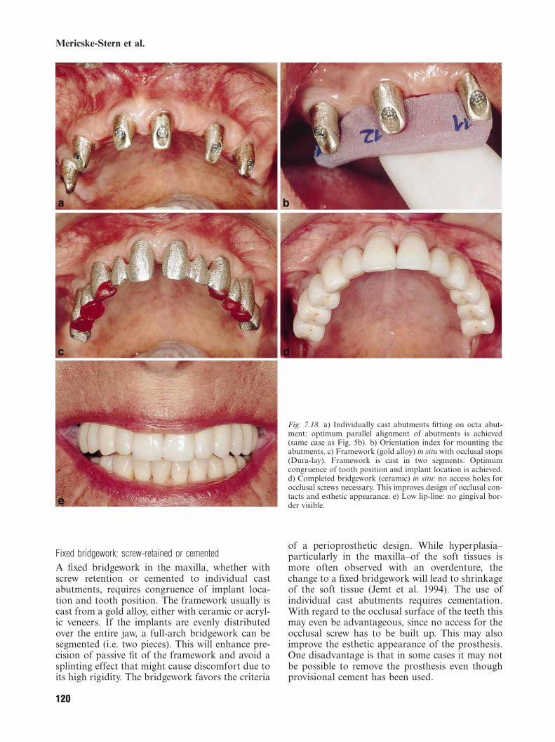
Fixed bridgework: screw-retained or cementedA fixed bridgework in the maxilla, whether withscrew retention or cemented to individual castabutments, requires congruence of implant loca-tion and tooth position. The framework usually iscast from a gold alloy, either with ceramic or acryl-ic veneers. If the implants are evenly distributedover the entire jaw, a full-arch bridgework can besegmented (i.e. two pieces). This will enhance pre-cision of passive fit of the framework and avoid asplinting effect that might cause discomfort due toits high rigidity. The bridgework favors the criteria
120
of a perioprosthetic design. While hyperplasia–particularly in the maxilla–of the soft tissues ismore often observed with an overdenture, thechange to a fixed bridgework will lead to shrinkageof the soft tissue (Jemt et al. 1994). The use ofindividual cast abutments requires cementation.With regard to the occlusal surface of the teeth thismay even be advantageous, since no access for theocclusal screw has to be built up. This may alsoimprove the esthetic appearance of the prosthesis.One disadvantage is that in some cases it may notbe possible to remove the prosthesis even thoughprovisional cement has been used.
Management of the edentulous patient
Fig. 7.19. a) Porcelain-to-gold fused bridgework, screw-retained(same case as Fig. 5a). Access holes for screw retention arevisible. Bridgework shows optimum congruence of tooth andimplant position. b) Bridgework is cast in two pieces and con-nected by a precision attachment, which provides resilience invertical direction.
Fig. 7.20. Tightening of screws.
O 2 implants and mandibular overdentures:bars or single anchors for overdenture reten-tion;
O ±2 mandibular implants: rigid bar connectorand overdenture;
O Ø4 mandibular implants: rigid bar or canti-lever fixed prostheses;
O 4 to 6 maxillary implants: rigid bars withoverdentures;
O 6 to 8 maxillary implants: full-arch fixedbridgework, segmentation possible.
121
Fig. 7.21. Esthetic results. Maxillary overdenture supported byfive implants matches esthetic appearance of fixed bridgeworksupported by six mandibular implants (same case as Figs 5a,19a and b).
In Figs 7.18a–e, individually cast abutmentsmounted on six maxillary implants and cementedfixed bridgework are shown.
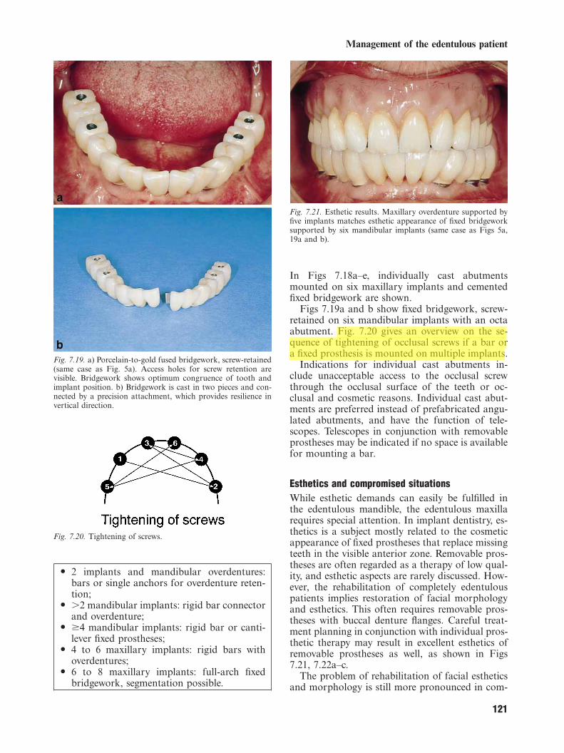
Figs 7.19a and b show fixed bridgework, screw-retained on six mandibular implants with an octaabutment. Fig. 7.20 gives an overview on the se-quence of tightening of occlusal screws if a bar ora fixed prosthesis is mounted on multiple implants.
Indications for individual cast abutments in-clude unacceptable access to the occlusal screwthrough the occlusal surface of the teeth or oc-clusal and cosmetic reasons. Individual cast abut-ments are preferred instead of prefabricated angu-lated abutments, and have the function of tele-scopes. Telescopes in conjunction with removableprostheses may be indicated if no space is availablefor mounting a bar.
Esthetics and compromised situationsWhile esthetic demands can easily be fulfilled inthe edentulous mandible, the edentulous maxillarequires special attention. In implant dentistry, es-thetics is a subject mostly related to the cosmeticappearance of fixed prostheses that replace missingteeth in the visible anterior zone. Removable pros-theses are often regarded as a therapy of low qual-ity, and esthetic aspects are rarely discussed. How-ever, the rehabilitation of completely edentulouspatients implies restoration of facial morphologyand esthetics. This often requires removable pros-theses with buccal denture flanges. Careful treat-ment planning in conjunction with individual pros-thetic therapy may result in excellent esthetics ofremovable prostheses as well, as shown in Figs7.21, 7.22a–c.
The problem of rehabilitation of facial estheticsand morphology is still more pronounced in com-
Administrator
Highlight
Mericske-Stern et al.
Fig. 7.22. Esthetic results. a) Maxillary overdenture with horse-shoe design (same case as Figs 4, 11a and b). b) Excellent es-thetics of front teeth and resin denture base. c) Patient displays‘‘mucosa’’ when laughing.
promised situations. Compromised oral conditionssuch as acquired defects (trauma, malignant tu-mors) or congenital defects (cleft palate) may leadto full disability and the impossibility of wearingcomplete dentures (Mericske-Stern et al. 1999).This includes impairment of phonetics, chewingfunction and extra- and intraoral esthetics. Thus,placement of implants to support individually castprostheses becomes highly important. In thesesituations, removable prostheses are often thebetter or only solution. Since size, extension and
122
Fig. 7.23. Acquired defect. a) Patient who underwent multiplesurgeries and is left with a completely flat maxillary jaw. Threeimplants are placed with individually cast telescopes. Distanceis not adequate for a connecting bar. b) Complete denture withgold-galvano retainers.
morphology of defects are highly individual, eachprosthesis is a masterpiece. Figs 7.23a and b, and7.24a–c illustrate the esthetically demanding im-plant prosthodontic treatment in situations ofcompromised oral conditions.
OcclusionThere exist two basic principles of occlusion thatapply to an occlusal scheme of either completedentures (i.e. bilateral guidance and lingualized oc-clusion) or fixed prostheses (i.e. freedom in centric,with lateral guidance on the working side and nobalancing contacts; the lateral guidance is a can-ine-protected guidance or a group function). Theseempirical occlusal schemes were originally de-veloped for rehabilitation of complete edentu-lousness with complete dentures or natural teethwith fixed prostheses. They are adopted with minormodifications for implant prostheses.
In implant prosthodontics a specific evidence-based occlusal philosophy has not yet been de-veloped. However, there are a few specific rules,which may favor optimum load distribution onto
Administrator
Highlight
Administrator
Highlight
Administrator
Highlight
Administrator
Highlight
Management of the edentulous patient
Fig. 7.24. Congenital defect. a) Patient with compromised situ-ation of maxilla due to a cleft palate. Six implants with threebar segments are placed to support an overdenture. b) Innersurface of the maxillary overdenture with a bulky buccal flangeis shown. c) Cosmetic appearance is highly satisfactory andmatches characteristics of natural dentition in mandible.
the implants and ensure stability of the dentures.The greater the number of implants placed and thegreater the rigidity of the prosthetic connectionachieved, the more the occlusal scheme may re-semble freedom in centric. From a biomechanicalpoint of view, however, a balanced occlusal guid-ance as utilized with complete dentures might fav-or equilibration of occlusal loads due to simul-taneous contacts on the working and non-workingsides.
123
Fig. 7.25. 1) Bilaterally balanced occlusion. 2) Bilaterally bal-anced occlusion, biting on front teeth is possible. 3) Canine-protected guidance is possible, combination with front teethguidance recommended. However, balancing contacts mightcontribute to higher stability of max. overdenture. 4) Bilaterallybalanced occlusion is necessary for stabilizing max. complexdenture. However, balancing contacts on cantilevers must beavoided. 5) Max. overdenture with long bars: balanced contactsmay not be necessary. However, if balancing contacts areplanned, these must be avoided on cantilevers. Group functionmay be recommended (combination of canine and premolar orfront teeth). 6) Canine-protected guidance is possible. However,group function may be recommended to avoid overload of im-plant located in position 13 and 23.
Occlusal conception of complete denturesIndications for a bilaterally balanced occlusion area combination of mandibular overdentures sup-ported by a few implants occluding with a com-plete denture in the opposing jaw, or mandibularoverdentures occluding with maxillary overden-tures. This type of balanced occlusion provides forprimary stability of the dentures during functionalloading. It also permits an even distribution ofload between implants and denture-bearing tissues.The characteristics are:
O cusp-to-fossa contacts in centric occlusion;O lingualized occlusion;O bilateral guidance, i.e. simultaneous guidance
on working and non-working sides.
Administrator
Highlight
Administrator
Highlight
Administrator
Highlight
Administrator
Highlight
Administrator
Highlight
Administrator
Highlight
Mericske-Stern et al.
Freedom in centricIndications are multiple implants supportingbridgework and occluding with fixed prostheses ornatural teeth. With fixed prostheses rigidly sup-ported by multiple implants, the concept of free-dom in centric can be applied unless a completedenture is worn in the opposing jaw. It is stronglysuggested that the lateral guidance of the workingside should not be exclusively on a single tooth orimplant. While a canine-protected lateral guidanceis easy for the technician to build up, a group func-tion may have a better protective function for theimplants and may distribute loading forces equallyto the suprastructure. Balancing contacts as builtup with complete dentures, although not pre-scribed with freedom in centric, may also contrib-ute to load distribution, but they must be avoidedon cantilevers. Examples are shown in Fig. 7.25.
O Canine-protected lateral guidance or groupfunction;
O no balancing contacts on cantilevers;O no guidance on single implants.
ReferencesAdell, R., Eriksson, B., Lekholm, U., Brånemark, P.I. & Jemt,
T. (1990) A long-term follow-up of tissue-integrated im-plants in the treatment of the totally edentulous jaw. Interna-tional Journal of Oral & Maxillofacial Implants 5: 347–359.
Albora, P. (1997) Tissue volume considerations in implant pros-thodontics. Journal of Prosthetic Dentistry 77: 492–496.
Albrektsson, T. & Zarb, G.A. (1993) Current interpretations ofthe osseointegrated response. Clinical significance. Interna-tional Journal of Prosthodontics 6: 95–105.
Albrektsson, T., Zarb, G., Worthington, P. & Eriksson, A.R.(1986) The long-term efficacy of currently used dental im-plants: a review and proposed criteria of success. Interna-tional Journal of Oral & Maxillofacial Implants 1: 11–25.
Batenburg, R.H.K., van Oort, R.P., Reintsema, H., Brouwer,T.J., Raghoebar, G.M. & Boering, G. (1994) Overdenturessupported by two IMZ implants in the lower jaw. A retro-spective study of periimplant tissues. Clinical Oral ImplantsResearch 5: 207–212.
Batenburg, R.H.K., Meijer, H.J.A., Raghoebar, G.M. & Vis-sink, A. (1998) Treatment concept for mandibular overden-tures supported by endosseous implants: a literature review.International Journal of Oral & Maxillofacial Implants 13:539–545.
Behneke, A., Behneke, N., d’Hoedt, B. & Wagner, W. (1997)Hard and soft tissue reactions to ITI screw implants: 3-yearlongitudinal results of a prospective study. InternationalJournal of Oral & Maxillofacial Implants 12: 749–757.
Belser, U.C., Buser, D., Hess, D., Schmid, B., Bernard, J.P. &Lang, N.P. (1998) Aesthetic implant restorations in partiallyedentulous patients–a critical appraisal. Periodontology2000 17: 132–150.
Bergendal, T. & Enquist, B. (1998) Implant-suppported over-dentures, a longitudinal prospective study. InternationalJournal of Oral & Maxillofacial Implants 13: 253–262.
Bryant, S.R. & Zarb, G.A. (1998) Osseointegration of oral im-
124
plants in older and younger adults. International Journal ofOral & Maxillofacial Implants 13: 492–499.
Burgin, W., Mericske-Stern, R. & Radics, P. (1998) 3-D in vivoforce measurements on three mandibular implants support-ing an overdenture. Journal of Dental Research 77: 711,Abstr. No. 635, Special Issue B, 76th General Session ofIADR.
Buser, D., Weber, H.P., Bragger, U. & Balsiger, C.H. (1991)Tissue integration of one-stage ITI implants: 3-year resultsof a longitudinal study with hollow-cylinder and hollow-screw implants. International Journal of Oral & Maxillo-facial Implants 6: 405–412.
Buser, D., Mericske-Stern, R., Bernard, J.P., Behneke, A.,Behneke, N., Hirt, H.P., Belser, U.C. & Lang, N.P. (1997)Long-term evaluation of non-submerged ITI implants. Part1: 8-year life table analysis of a prospective multi-centerstudy with 2359 implants. Clinical Oral Implants Research8: 161–172.
Buser, D., Mericske-Stern, R., Dula, K. & Lang, N.P. (1999)Clinical experience with one-stage, non-submerged dentalimplants. Advances in Dental Research 13: 153–161.
Cune, M.S., de Putter, C. & Hoogstraten, J. (1994) Treatmentoutcome with implant-retained overdentures. Part 1: Clin-ical findings and predictability of clinical treatment out-come. Journal of Prosthetic Dentistry 72: 152–158.
Hallmann, M. & Carlsson, B. (1996) Surgical correction of mal-positioned implants. A case report. Clinical Oral ImplantsResearch 7: 316–319.
Hutton, J.E., Heath, R., Chai, J.Y., Harnett, J., Jemt, T., Johns,R.B., McKenna, S., McNamara, D.C., van Steenberghe, D.,Taylor, R., Watson, R. & Herrmann, I. (1995) Factors re-lated to success and failure rates at 3-year follow-up in amulticenter study of overdentures supported by Brånemarkimplants. International Journal of Oral & Maxillofacial Im-plants 10: 33–42.
Jemt, T. (1991) Failures and complications in 391 consecutivelyinserted fixed prostheses supported by Brånemark implantsin edentulous jaws: a study of treatment from the time ofprostheses placement to the first annual checkup. Interna-tional Journal of Oral & Maxillofacial Implants 6: 270–276.
Jemt, T. (1993) Implant treatment in resorbed edentulous upperjaws. Clinical Oral Implants Research 4: 187–194.
Jemt, T. (1994) Fixed implant-supported prostheses in the eden-tulous maxilla. A five-year follow-up report. Clinical OralImplants Research 5: 142–147.
Jemt, T., Book, K., Lie, A. & Borjesson, T. (1994) Mucosaltopography around implants in edentulous upper jaws. Pho-togrammetric three-dimensional measurements of the effectof replacement of a removable prosthesis with a fixed pros-thesis. Clinical Oral Implants Research 5: 220–228.
Jemt, T., Chai, J., Harnett, J., Heath, M.R., Hutton, J.E.,Johns, R.B., McKenna, S., McNamara, D., van Steenberghe,D., Taylor, R., Watson, R.M. & Herrmann, I. (1996) A 5-year prospective multicenter follow-up report on overden-tures supported by osseointegrated implants. InternationalJournal of Oral & Maxillofacial Implants 11: 291–298.
Lundqvist, S., Lohmander-Agerskov, A. & Haraldson, T. (1992)Speech before and after treatment with bridges on osseointe-grated implants in the edentulous upper jaw. Clinical OralImplants Research 3: 57–62.
Mericske-Stern, R. (1990) Clinical evaluation of overdenturerestorations supported by osseointegrated titanium im-plants. A retrospective study. International Journal of Oral &Maxillofacial Implants 5: 375–383.
Mericske-Stern, R. (1997) Force distribution on implants sup-porting overdentures: the effect of distal bar extensions. A 3-D in vivo study. Clinical Oral Implants Research 8: 142–151.
Mericske-Stern, R. (1998a) Treatment outcomes with implant-supported overdentures: clinical considerations. Journal ofProsthetic Dentistry 79: 66–73.
Administrator
Highlight
Administrator
Highlight
Administrator
Highlight
Administrator
Highlight
Management of the edentulous patient
Mericske-Stern, R. (1998b) Three-dimensional force measure-ments with mandibular overdentures connected to implantsby ball-shaped retentive anchors. A clinical study. Interna-tional Journal of Oral & Maxillofacial Implants 13: 36–43.
Mericske-Stern, R. & Zarb, G.A. (1993) Overdentures: an alter-native implant methodology for edentulous patients. Inter-national Journal of Prosthodontics 6: 203–208.
Mericske-Stern, R., Steinlin Schaffner, T., Marti, P. & Geering,A.H. (1994) Peri-implant mucosal aspects of ITI implantssupporting overdentures. A five-year longitudinal study.Clinical Oral Implants Research 5: 9–18.
Mericske-Stern, R., Milani, O., Mericske, E. & Olah, A. (1995)Periotest measurements and osseointegration of mandibularITI-implants supporting overdentures. A one-year longitudi-nal study. Clinical Oral Implants Research 6: 73–82.
Mericske-Stern, R., Assal, P. & Burgin, W. (1996a) Simul-taneous force measurements in 3 dimensions on oral endos-seous implants in vitro and in vivo. A methodological study.Clinical Oral Implants Research 7: 378–386.
Mericske-Stern, R., Piotti, M. & Sirtes, G. (1996b) 3-D in vivoforce measurements on mandibular implants supportingoverdentures. A comparative study. Clinical Oral ImplantsResearch 7: 387–396.
Mericske-Stern, R., Fahrlander, F., Venetz, E., Geering, A.H. &Burgin, W. (1998) 3-D force measurements in vivo on maxil-lary implants: comparison of a fixed prosthesis and a barretained overdenture. Journal of Dental Research 77: 1025,Abstr. No. 3145, Special Issue B, 76th General Session ofIADR.
Mericske-Stern, R., Perren, R. & Raveh, J. (1999) Life tableanalysis and clinical evaluation of oral implants supportingprostheses after resection of malignant tumors. InternationalJournal of Oral & Maxillofacial Implants 14: 673–680.
Mombelli, A. & Mericske-Stern, R. (1990) Microbiological fea-tures of stable osseointegrated implants used as abutmentsfor overdentures. Clinical Oral Implants Research 1: 1–7.
Palmqvist, S., Sondell, K. & Swartz, B. (1994) Implant-sup-ported maxillary overdentures: outcome in planned andemergency cases. International Journal of Oral & Maxillo-facial Implants 9: 184–190.
Palmqvist, S., Sondell, K., Swartz, B. & Svenson, B. (1996)Marginal bone levels around maxillary implants supportingoverdentures or fixed prostheses: a comparative study usingdetailed narrow-beam radiographs. International Journal ofOral & Maxillofacial Implants 11: 223–227.
Quirynen, M., Naert, I., van Steenberghe, D. & Nys, L. (1992)
125
A study of 589 consecutive implants supporting completefixed prostheses. Part I: Periodontal aspects. Journal of Pros-thetic Dentistry 68: 655–663.
Roos, J., Sennerby, L., Lekholm, U., Jemt, T., Grondahl, K.,Albrektsson, T. (1997) A qualitative and quantitativemethod for evaluating implant success: a 5-year retrospec-tive analysis of the Brånemark implant. InternationalJournal of Oral & Maxillofacial Implants 12: 504–514.
Salama, H., Salama, M. & Garber, D.A. (1995) Techniques fordeveloping optimal peri-implant papillae within the estheticzone. I. Guided soft tissue augmentation: the three-stage ap-proach. Journal of Esthetic Dentistry 7: 3–9.
Smith, D. & Zarb, G.A. (1989) Criteria for success of osseointe-grated endosseous implants. Journal of Prosthetic Dentistry62: 567–572.
Spiekermann, H., Jansen, V.K. & Richter, E.J. (1995) A 10-yearfollow-up study of IMZ and TPS implants in the edentulousmandible using bar-retained overdentures. InternationalJournal of Oral & Maxillofacial Implants 10: 231–243.
Widmark, G., Andersson, B., Andrup, B., Carlsson, G.E., Ivan-off, C.J. & Lindvall, A.M. (1998) Rehabilitation of patientswith severely resorbed maxillae by means of implants withor without bone grafts. A 1-year follow-up study. Interna-tional Journal of Oral & Maxillofacial Implants 13: 474–482.
Wismeyer, D., van Waas, A.J. & Vermeeren, J.I.J.F. (1995) Over-dentures supported by ITI implants: a 6.5-year evaluationof patient satisfaction and prosthetic aftercare. InternationalJournal of Oral & Maxillofacial Implants 10: 744–749.
Zarb, G.A. & Schmitt, A. (1989) The longitudinal clinical effec-tiveness of osseointegrated implants. The Toronto study.Part I: Surgical results. Journal of Prosthetic Dentistry 62:451–470.
Zarb, G.A. & Schmitt, A. (1990a) The longitudinal clinical ef-fectiveness of osseointegrated dental implants: The Torontostudy. Part II: The prosthetic results. Journal of ProstheticDentistry 64: 53–61.
Zarb, G.A. & Schmitt, A. (1990b) The longitudinal clinical ef-fectiveness of osseointegrated implants. The Toronto study.Part III: Problems and complications encountered. Journalof Prosthetic Dentistry 64: 185–194.
Zarb, G.A. & Schmitt, A. (1994) Osseointegration for elderlypatients: The Toronto study. Journal of Prosthetic Dentistry72: 559–568.
Zarb, G.A. & Schmitt, A. (1995) Implant prosthodontic treat-ment options for the edentulous patients. Journal of OralRehabilitation 22: 661–671.