Mannheimia haemolytica and Its Leukotoxin Cause MacrophageExtracellular Trap Formation by Bovine Macrophages
Nicole A. Aulik,a,c Katrina M. Hellenbrand,a and Charles J. Czuprynskia,b
Department of Pathobiological Sciencesa and Food Research Institute,b University of Wisconsin—Madison, Madison, Wisconsin, USA, and Department of Biology, WinonaState University, Winona, Minnesota, USAc
Human and bovine neutrophils release neutrophil extracellular traps (NETs), which are protein-studded DNA matrices capableof extracellular trapping and killing of pathogens. Recently, we reported that bovine neutrophils release NETs in response to theimportant respiratory pathogen Mannheimia haemolytica and its leukotoxin (LKT). Here, we demonstrate macrophage extra-cellular trap (MET) formation by bovine monocyte-derived macrophages exposed to M. haemolytica or its LKT. Both nativefully active LKT and noncytolytic pro-LKT (produced by an lktC mutant of M. haemolytica) stimulated MET formation. Confo-cal and scanning electron microscopy revealed a network of DNA fibrils with colocalized histones in extracellular traps releasedfrom bovine macrophages. Formation of METs required NADPH oxidase activity, as previously demonstrated for NET forma-tion. METs formed in response to LKT trapped and killed a portion of the M. haemolytica cells. Bovine alveolar macrophages,but not peripheral blood monocytes, also formed METs in response to M. haemolytica cells. MET formation was not restrictedto bovine macrophages. We also observed MET formation by the mouse macrophage cell line RAW 264.7 and by human THP-1cell-derived macrophages, in response to Escherichia coli hemolysin. The latter is a member of the repeats-in-toxin (RTX) toxinfamily related to the M. haemolytica leukotoxin. This study demonstrates that macrophages, like neutrophils, can form extracel-lular traps in response to bacterial pathogens and their exotoxins.
Mannheimia haemolytica is the most important bacterialpathogen of the bovine respiratory disease complex. In its
most severe form it causes a severe fibrinous pleuropneumoniacharacterized by intense leukocyte infiltration in alveoli, intra-alveolar hemorrhage, fibrin deposition, and consolidation of thelungs (38). The most important virulence factor for M. haemo-lytica is its leukotoxin (LKT), a 104-kDa exotoxin released duringlogarithmic-phase growth (16, 22). LKT is a member of the re-peats-in-toxin (RTX) toxin family of exoproteins produced by awide variety of Gram-negative bacteria, including Escherichia coli,Actinobacillus pleuropneumoniae, and Aggregatibacter actinomyce-temcomitans (47). Activation of pro-LKT requires acylation by thetransacylase encoded by lktC (39). The acylated LKT then bindsamino acids 5 to 17 of the signal sequence of bovine CD18 onruminant leukocytes (29), leading to cell death. This restricts cy-totoxicity to ruminant leukocytes, because the signal sequence forCD18 is not present on mature leukocytes from other mammalianspecies (19, 37, 40). A similar RTX toxin, the hemolysin producedby uropathogenic E. coli, is neither species nor cell type restricted(37).
Activated neutrophils undergo a form of cell death, calledNETosis, in which nuclear DNA is released into the extracellularenvironment with little concomitant release of lactate dehydroge-nase (LDH) (10, 11, 21, 34, 35, 46). The resulting network ofextracellular DNA and associated proteins (e.g., histones andgranule constituents) are called neutrophil extracellular traps(NETs) (10). The extracellular DNA within NETs is studded withantimicrobial proteins. These include histones and primary, sec-ondary, and tertiary granular components such as neutrophil elas-tase, myeloperoxidase, lactoferrin, and gelatinase (10). Activationof human neutrophils with interleukin-8, phorbol 12-myristate13-acetate (PMA), or lipopolysaccharide (LPS) alone causes NETformation (34, 46). NET formation also occurs in response toprokaryotic and eukaryotic pathogens (10, 21, 46). Extracellular
trap formation has also been demonstrated for other types ofgranulocytes, such as eosinophils and mast cells (44, 48).
NETs are capable of trapping and killing a variety of Gram-negative and Gram-positive bacteria, fungi, and protozoa (6, 7, 10,11, 13, 18, 21, 23, 24, 34, 41, 46). Recently published data demon-strate that NETs are formed in response to M. haemolytica and itsleukotoxin and that some of the M. haemolytica cells are killedduring this process (4). In this report, we present evidence thatbovine macrophages also form extracellular traps (i.e., macro-phage extracellular traps [METs]) that are capable of snaring andkilling M. haemolytica cells in vitro. Furthermore, we present evi-dence that MET formation is also a property of murine and hu-man macrophage cell lines. These findings are consistent withrecent reports that murine macrophages form METs in responseto Staphylococcus aureus, resulting in death of a portion of thebacterial cells (17). Similarly, human monocytes formed METs inresponse to certain types of gold nanoparticles in vitro (5).
MATERIALS AND METHODSCell lines and primary cell preparation. RAW 264.7 (mouse macro-phage) and THP-1 (human monocyte) cell lines were grown in RPMI1640 (Cellgro, Manassas, VA) supplemented with 10% (vol/vol) fetal bo-vine serum (FBS; Atlanta Biologicals, Lawrenceville, GA), 100 U/ml pen-icillin, and 100 �g/ml streptomycin (Cellgro). All cells were grown at 37°C
Received 24 October 2011 Returned for modification 30 November 2011Accepted 10 February 2012
with 5% CO2 in a humidified incubator. Differentiation of the THP-1 cellsinto macrophage-like cells was performed by incubation with 100 nMPMA in culture medium for 7 days at 37°C with 5% CO2 (36). Differen-tiated THP-1 cells were deemed acceptable when �95% of the THP-1 cellswere adherent (36).
Whole blood was collected by venipuncture from healthy Holsteincows housed at the University of Wisconsin—Madison Dairy Cattle Cen-ter using 0.38% (vol/vol) sodium citrate as anticoagulant. Blood was cen-trifuged at 1,000 � g for 15 min, and the buffy coat was removed. Thebuffy coat, containing mononuclear cells, was suspended in Hanks’ bal-anced salt solution (HBSS; Cellgro) with 4 mM EDTA (without calciumor magnesium), layered onto Histopaque-1083 (Sigma-Aldrich, St. Louis,MO), and centrifuged at 1,000 � g for 30 min at room temperature.Mononuclear cells were removed, and contaminating red blood cells(RBCs) were lysed in a 1:10 dilution of lysis buffer (150 mM ammoniumchloride, 10 mM Tris [pH 7.5]) while rotating at 8 rpm for 10 min. Cellswere pelleted at 1,000 � g and washed 3 times with HBSS with 4 mMEDTA. Mononuclear cells were resuspended in RPMI 1640 with 1% (vol/vol) FBS and incubated at 37°C with 5% CO2 for 2 h on 100-mm carboxyl-coated dishes (Becton, Dickinson and Company, Franklin Lakes, NJ).Nonadherent cells were removed by repeated washing. Adherent mono-cytes were allowed to differentiate into monocyte-derived macrophagesby incubating them in RPMI 1640 with 10% FBS, 100 U/ml penicillin, and100 �g/ml streptomycin for 7 days at 37°C. The medium was exchangedtwice during this period. Monolayers with greater than 99% viability, asdetermined by trypan blue staining and light microscopy, were deemedacceptable for further use.
Neutrophils were isolated by lysis of the red blood cell pellet using a 1:3dilution in lysis buffer while rotating at 8 rpm for 10 min. Bovine neutro-phils (bovine polymorphonuclear leukocytes [bPMNs]) were pelleted at1,000 � g and washed 4 times with HBSS. Cells were resuspended inserum- and phenol red-free RPMI 1640 medium and examined by lightmicroscopy. Cell suspensions found to be �98% bPMNs, as determinedby cell morphology, and to have �99% viability, as determined by trypanblue staining, were deemed acceptable for use in experiments.
Bovine alveolar macrophages were isolated from bronchoalveolar la-vage (BAL) fluid obtained from donor cows at the University of Wiscon-sin—Madison Veterinary Medicine Teaching Hospital. Lavage fluid wasfiltered through a 0.2-�m-pore-size nylon filter, washed 3 times, and re-suspended in RPMI 1640 at a concentration of 106 macrophages/ml. Bo-vine alveolar macrophages were allowed to adhere for 1 h, washed 2 timeswith phosphate-buffered saline (PBS), and used immediately in RPMI1640. Cultures with greater than 90% viability, as determined by trypanblue staining and light microscopy, were used in experiments.
Bacteria. Mannheimia haemolytica A1 (obtained from a bovine pneu-monic lung), M. haemolytica �lktC (SH1562; a gift from S. Highlander),and M. haemolytica �lktA (a gift from R. Briggs) were grown in brain heartinfusion (BHI) broth without shaking at 37°C for 10 h. Bacterial cells werepelleted at 3,750 � g, washed 3 times in PBS, and resuspended in RPMI toan optical density of 0.7, which corresponds to 5 � 109 CFU/ml. Thenumber of CFU in each broth culture was extrapolated from growthcurves performed in our laboratory and confirmed by dilution plating ontryptic soy agar (TSA) with 5% sheep red blood cells (Becton, Dickinsonand Company) to enumerate the CFU.
Production and purification of toxins. LKT, pro-LKT, and �lktALKT were prepared from Mannheimia haemolytica A1, M. haemolytica�lktC strain SH1562 (20), and the M. haemolytica �lktA strain, respec-tively (39). Hemolysin (HLY) was prepared from Escherichia coli strainWAM1824 (kindly provided by R. A. Welch, Madison, WI) (33) grown tologarithmic growth phase in RPMI 1640 medium supplemented with 10�g/ml chloramphenicol. Toxins were produced and purified as describedpreviously (2). Briefly, bacteria were pelleted from 12-h broth cultures bycentrifugation at 3,750 � g for 10 min. The pelleted cells were resus-pended in RPMI 1640 medium and incubated for 6 h at 37°C until theyreached logarithmic growth phase. Bacterial cells were centrifuged at
7,500 � g for 30 min at 4°C. The supernatant was removed, filtered (poresize, 0.2 �m), and concentrated using an Amicon filtration system with a100-kDa-molecular-mass-cutoff membrane (Millipore, Billerica, MA).The LKT or HLY was stored at �70°C with 20% (vol/vol) glycerol untilused in an experiment. One unit of LKT or HLY is defined as the greatestdilution of toxin that killed 50% of target cells (106 bovine lymphoblastoid[BL-3] cells and human kidney epithelial [A498] cells, respectively) in 2 hat 37°C, as determined by the CellTiter 96 AQueous One Solution cellproliferation assay (Promega, Madison, WI). lktC and lktA deletion mu-tant LKT preparations did not exhibit any cytotoxicity in this assay. Pro-tein concentrations were determined using a bicinchoninic acid (BCA)assay (Pierce, Rockford, IL).
Quantification of extracellular DNA. Macrophage extracellular DNAwas quantified using a modified technique described by Fuchs et al. (21).Briefly, macrophages were incubated for the indicated times with variousstimuli and then pelleted at 500 � g for 3 min. The supernatant wasremoved, micrococcal nuclease buffer with 0.1 U/�l micrococcal nucleasewas added (New England BioLabs, Ipswich, MA), and the mixture wasincubated for 15 min at 37°C (as described by the manufacturer). A 1:200dilution of PicoGreen reagent (Invitrogen, Carlsbad, CA) in 10 mM Trisbase buffered with 1 mM EDTA was added to an equal volume of thenuclease-treated macrophage mixture. Fluorescence was determined atan excitation wavelength of 488 nm and an emission wavelength of 520nm using an automated plate reader (DTX 800 multimode detector; Beck-man Coulter, Brea, CA). Extracellular trap production was quantified asthe fold increase in the number of fluorescence units for LKT-treatedmacrophages divided by the number of fluorescence units for untreatedcontrol macrophages.
Reagents. DNase I (source, bovine pancreas), PMA, and cytochalasin D(Cyto D) were purchased from Sigma-Aldrich. The NADPH oxidase inhibi-tor diphenyleneiodonium chloride (DPI) was purchased from Calbiochem/EMD (Darmstadt, Germany). The pancaspase inhibitor fluoromethylketone(Z-VAD-FMK) was purchased from Alexis Biochemicals (Plymouth Meet-ing, PA). The anti-CD18 antibody (BAQ20A) was purchased from VMRD(Pullman, WA). M. haemolytica A1 LPS was kindly provided by D. McClena-han (Cedar Falls, IA) (32). The anti-LKT antibody (MM601) was kindly pro-vided by S. Srikumaran (Pullman, WA). The antihistone antibody was pur-chased from AbCam (Cambridge, MA), and the Alexa Fluor 488 anti-rabbitIgG antibody and Alexa Fluor 488 anti-mouse IgG antibody were purchasedfrom Invitrogen. LDH release was determined using the CytoTox 96 nonra-dioactive cytotoxicity assay as described by the manufacturer (Promega).
Immunofluorescence. To perform immunofluorescence microscopy,macrophages (2.5 � 105) were grown on 12-mm glass coverslips for 16 hat 37°C (Fisher Scientific, Hanover Park, IL). Slides were washed, resus-pended in RPMI 1640, and incubated for 30 min at 37°C with 1 U LKT,5 � 107 fluorescein-labeled M. haemolytica, 250 nM LPS, 100 �m PMA, or1 U LKT and 180 U DNase. Slides were washed 3 times with PBS and fixedfor 10 min with 4% paraformaldehyde. The slides were then washed,permeabilized with cold acetone for 5 min, washed again, and blockedwith 1% bovine serum albumin (BSA; Pierce) in PBS for 20 min at roomtemperature. Slides were incubated for 1 h with 1 �M TOPRO stain. Insome experiments, an antihistone monoclonal antibody (AbCam, Cam-bridge, MA) was added, and slides were washed and then incubated for 1h at room temperature with an Alexa Fluor 488-conjugated anti-mouseIgG antibody (Molecular Probes, Eugene, OR) in 1% BSA (in PBS). Slideswere washed and examined by confocal microscopy (Eclipse TE2000-Umicroscope; Nikon Corporation, Tokyo, Japan).
SEM. Bovine macrophages (5 � 106) were incubated on glass slidesovernight at 37°C, washed twice with PBS, and then incubated with 5 �107 M. haemolytica cells for 5 min. Slides were washed and fixed using 4%paraformaldehyde in PBS, postfixed with 2.5% glutaraldehyde, and pre-pared as previously described (9). Images were taken at Winona StateUniversity using a Feico Phenom scanning electron microscope (SEM;FEI Company, Hillsboro, OR).
Bacterial trapping and killing. M. haemolytica cells were grown to logphase as described earlier, washed 3 times in PBS, and resuspended for 15min on ice in 0.5 mg/ml fluorescein isothiocyanate (FITC; Sigma-Al-drich) in 50 mM sodium carbonate buffer. The M. haemolytica cells werethen washed 3 times with PBS and resuspended at a concentration of 5 �109 CFU/ml in serum-free RPMI 1640. FITC-labeled M. haemolytica cellswere serially diluted and plated on TSA agar with 5% sheep RBCs, andcolonies were enumerated to confirm viability.
Macrophages (105) were incubated with FITC-labeled or unlabeled M.haemolytica cells (107) for various times and analyzed for trapping andkilling of bacterial cells. As a control, 180 U DNase I was added to somecultures to cleave extracellular DNA and free the bacteria. In a similarexperiment, 0.5 U LKT was incubated with macrophages (105) for 30 minat 37°C prior to the addition of M. haemolytica. DNase-treated and LKT-treated macrophages were washed and then incubated with FITC-labeledor unlabeled M. haemolytica cells (107) in RPMI 1640 for various times at37°C. Some macrophages were incubated with 180 U DNase I and M.haemolytica cells for the entire length of the experiment.
To quantify the bacterial cells trapped within the METs, samples wereremoved and washed 3 times with PBS and fluorescence was determinedusing an automated plate reader at 488 nm. To determine bacterial killing
by METs, macrophages incubated with M. haemolytica suspensions wereserially diluted in PBS and plated on TSA with 5% sheep RBCs. At eachtime point, serial dilutions of M. haemolytica cells that had been incubatedin RPMI (without macrophages) were plated to quantify the total num-bers of CFU. The percent bactericidal activity of METs was determined asdescribed previously (4).
Statistical analysis. Group means were compared by analysis of vari-ance, followed by the Tukey-Kramer pairwise comparison test, as per-formed by the Instat statistical package (GraphPad, San Diego, CA). Thelevel of significance was set at a P value of �0.05.
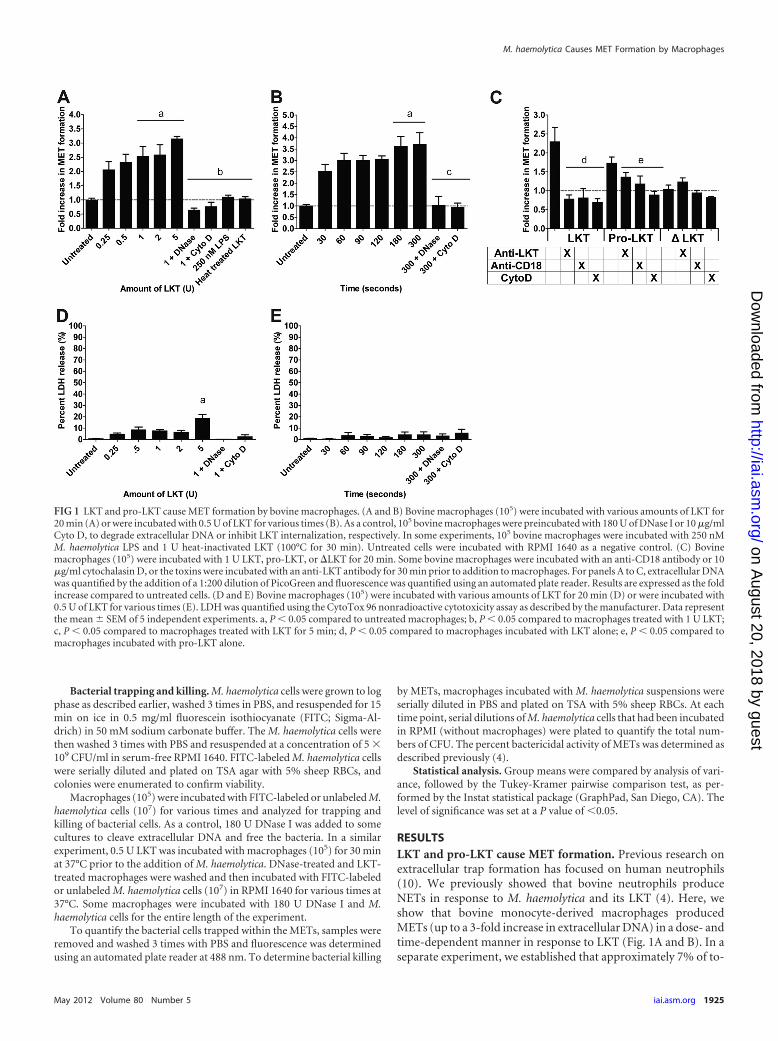
RESULTSLKT and pro-LKT cause MET formation. Previous research onextracellular trap formation has focused on human neutrophils(10). We previously showed that bovine neutrophils produceNETs in response to M. haemolytica and its LKT (4). Here, weshow that bovine monocyte-derived macrophages producedMETs (up to a 3-fold increase in extracellular DNA) in a dose- andtime-dependent manner in response to LKT (Fig. 1A and B). In aseparate experiment, we established that approximately 7% of to-
FIG 1 LKT and pro-LKT cause MET formation by bovine macrophages. (A and B) Bovine macrophages (105) were incubated with various amounts of LKT for20 min (A) or were incubated with 0.5 U of LKT for various times (B). As a control, 105 bovine macrophages were preincubated with 180 U of DNase I or 10 �g/mlCyto D, to degrade extracellular DNA or inhibit LKT internalization, respectively. In some experiments, 105 bovine macrophages were incubated with 250 nMM. haemolytica LPS and 1 U heat-inactivated LKT (100°C for 30 min). Untreated cells were incubated with RPMI 1640 as a negative control. (C) Bovinemacrophages (105) were incubated with 1 U LKT, pro-LKT, or �LKT for 20 min. Some bovine macrophages were incubated with an anti-CD18 antibody or 10�g/ml cytochalasin D, or the toxins were incubated with an anti-LKT antibody for 30 min prior to addition to macrophages. For panels A to C, extracellular DNAwas quantified by the addition of a 1:200 dilution of PicoGreen and fluorescence was quantified using an automated plate reader. Results are expressed as the foldincrease compared to untreated cells. (D and E) Bovine macrophages (105) were incubated with various amounts of LKT for 20 min (D) or were incubated with0.5 U of LKT for various times (E). LDH was quantified using the CytoTox 96 nonradioactive cytotoxicity assay as described by the manufacturer. Data representthe mean � SEM of 5 independent experiments. a, P � 0.05 compared to untreated macrophages; b, P � 0.05 compared to macrophages treated with 1 U LKT;c, P � 0.05 compared to macrophages treated with LKT for 5 min; d, P � 0.05 compared to macrophages incubated with LKT alone; e, P � 0.05 compared tomacrophages incubated with pro-LKT alone.
M. haemolytica Causes MET Formation by Macrophages
tal macrophage DNA (i.e., cells lysed with 0.5% SDS) was releasedduring a 20-min incubation with 2 U LKT (data not shown). Wedid not observe release of extracellular DNA by peripheral bloodmonocytes incubated with LKT under the same conditions (datanot shown). MET formation was reduced when DNase I or Cyto Dwas added to cleave extracellular DNA or inhibit intracellulartransport of LKT, respectively (Fig. 1A). Neither purified M. hae-molytica LPS nor heat-inactivated LKT (100°C for 1 h) inducedMET formation (Fig. 1A). Taken together these observations in-dicate that contaminating LPS alone does not cause MET forma-tion. We quantified LDH release as an indicator of necrosis (21,35) and observed no significant increase in LDH except whenlarger amounts (e.g., 5 U) of LKT were incubated with bovinemacrophages (Fig. 1D and E).
MET formation in response to LKT was further examined us-ing two LKT variants. The first is a pro-LKT purified from a �lktCstrain of M. haemolytica (a kind gift from S. K. Highlander, Hous-ton, TX), and the second is a truncated leukotoxin protein(�LKT) purified from a �lktA M. haemolytica strain (kind giftfrom R. E. Briggs, Ames, IA). The �lktC strain of M. haemolyticadoes not produce an active LKTC protein; therefore, acylation ofthe LKTA protein does not occur and the resulting pro-LKT isnoncytolytic in conventional assays, despite being capable ofbinding to CD18 (4). The �lktA strain produces a �LKT that lacksamino acids 34 to 378, rendering it incapable of binding CD18 orcausing cytotoxicity (39). Incubation of bovine macrophages withpro-LKT caused MET formation, although it was somewhat re-duced compared to that achieved with native LKT (Fig. 1C). METformation in response to LKT and pro-LKT was reduced whenbovine macrophages were preincubated with an anti-CD18 recep-tor antibody or cytochalasin D or when the toxins were incubatedwith a neutralizing anti-LKT antibody (MM601) before beingadded to the macrophages (Fig. 1C). As expected, the truncated�LKT did not cause MET formation (Fig. 1C). These data confirmthat MET formation requires LKT binding to its receptor, CD18.
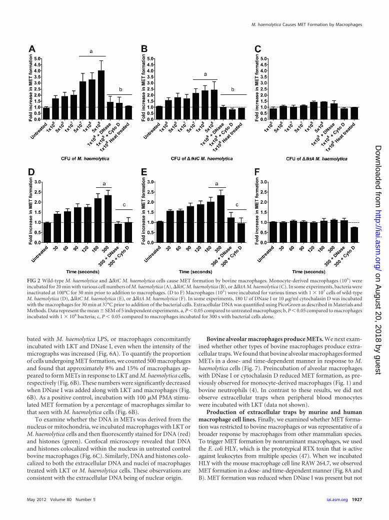
Bovine macrophages form METs in response to M. haemo-lytica cells that produce LKT or pro-LKT. We next tested if M.haemolytica cells stimulated MET formation. Bovine macro-phages produced METs in a time- and dose-dependent mannerwhen incubated with wild-type M. haemolytica cells that produceactive LKT or �lktC M. haemolytica cells that produce a nonacy-lated pro-LKT (Fig. 2). MET formation did not occur in responseto �lktA M. haemolytica cells that produce a truncated noncyto-lytic LKT, nor were METs observed when DNase I or cytochalasinD was added to macrophages incubated with M. haemolytica cellsthat produce LKT (Fig. 2). MET formation was not due to mac-rophage lysis. Little LDH release was observed in medium frommacrophages incubated with wild-type or �lktC M. haemolyticacells (data not shown), suggesting that DNA release did not simplyreflect necrosis.
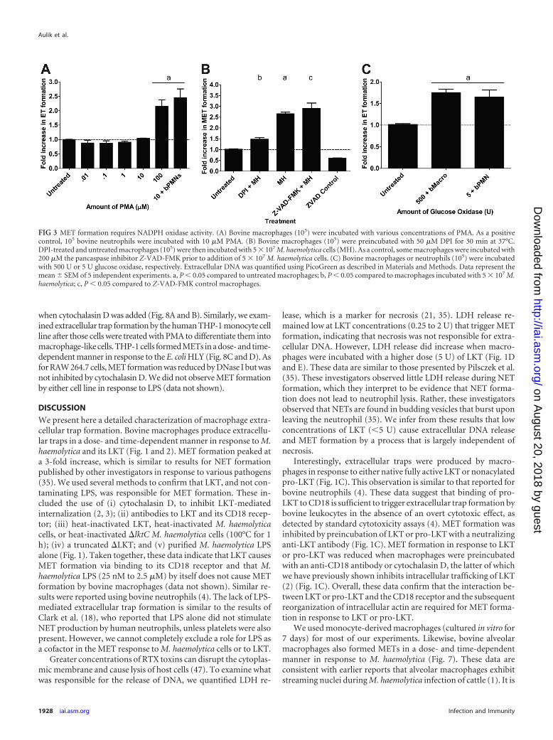
MET production is dependent on NADPH oxidase activity.NET formation by human neutrophils is dependent on the gen-eration of reactive oxygen species (ROS) by NADPH oxidase (21).Neutrophils from chronic granulomatous disease patients or frompatients who have a mutation in the NADPH oxidase gene, whichrenders them susceptible to recurrent infections, do not formNETs (7, 21, 28). We tested if incubation with PMA, which acti-vates the assembly and production of NADPH oxidase in phago-cytes (21), stimulates MET production. We found that bovinemacrophages formed METs in response to PMA, although this
required concentrations higher than those needed to stimulateNET formation by bovine neutrophils (100 �M versus 10 �M,respectively) (Fig. 3A). Addition of the NADPH oxidase inhibitorDPI significantly decreased M. haemolytica-induced MET forma-tion (Fig. 3B), as described previously for neutrophils. METs alsoformed when bovine macrophages were incubated with glucoseoxidase (GO), which produces hydrogen peroxide and activatesROS production within macrophages (Fig. 3C). It should be notedthat the concentrations of PMA, GO, and DPI required to exerttheir effects were greater than those reported for trap formation byneutrophils (21). MET formation in response to LKT was notinhibited by the pancaspase inhibitor Z-VAD-FMK, indicatingthat MET formation is not the result of apoptosis (Fig. 3B).
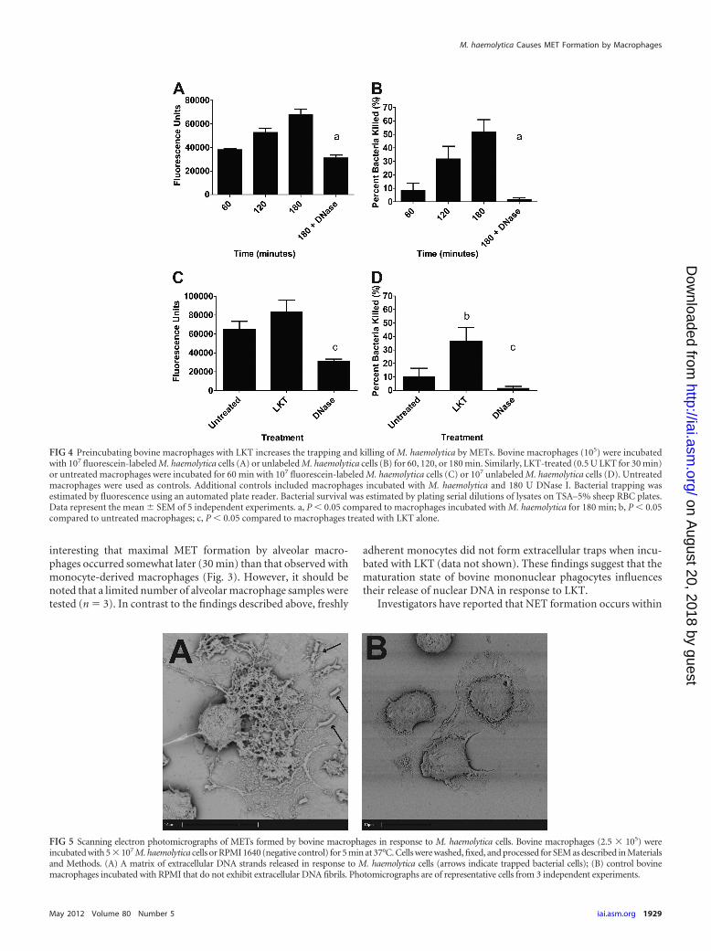
METs trap and kill M. haemolytica cells. Previous investiga-tions have shown that NETs and METs are capable of trapping andkilling extracellular bacteria (17, 21). Using fluorescein-labeled M.haemolytica cells, we found that bacterial cells were trapped, in atime-dependent manner, by METs formed in response to M. hae-molytica cells (Fig. 4A). Furthermore, approximately 50% of theM. haemolytica cells were killed by METs during a 180-min incu-bation (Fig. 4B). To confirm that the bacteria were trapped inMETs, we added DNase I to cleave extracellular DNA and freetrapped bacterial cells. DNase I treatment significantly reducedthe number of M. haemolytica cells trapped and killed by bovinemacrophages (Fig. 4A and B). We infer that the remaining smallproportion of bacterial cells that were killed by DNase I-treatedmacrophages (�2%) represents bacterial cells that were phagocy-tosed and killed intracellularly.
During pulmonary infection with M. haemolytica, bovine leu-kocytes are likely exposed to LKT before they encounter the bac-terial cells. We therefore examined whether prior exposure to LKTaffects the ability of METs to trap and kill M. haemolytica cells. Todo so, we incubated bovine macrophages with a small amount ofLKT (0.5 U) for 30 min before M. haemolytica cells (10 bacterialcells per macrophage) were added. Prior incubation with LKT hadlittle effect on the numbers of M. haemolytica cells trapped withinMETs (Fig. 4C) but significantly increased the percentage of bac-terial cells killed (Fig. 4D). DNase I treatment reduced the num-bers of bacteria that were trapped and killed (Fig. 4C and D),implicating DNA-containing METs in bacterial killing. We alsoused a Syto 9 and propidium iodide staining kit (Live/DeadBacLight bacterial viability kit; Invitrogen) to confirm that a pro-portion of the M. haemolytica cells trapped within METs werekilled and to exclude the possibility that the reduced numbers ofCFU simply reflect clumped bacteria (data not shown).
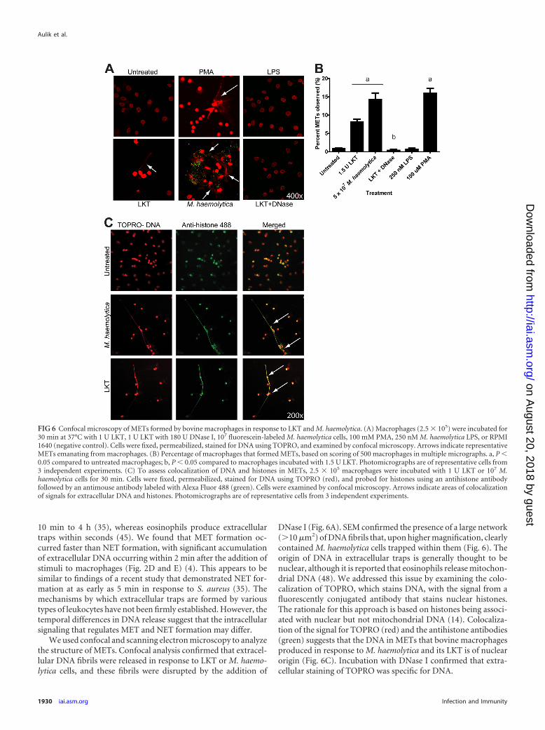
Scanning electron and confocal microscopy examination ofMETs. Using SEM, we observed web-like structures in which M.haemolytica cells were trapped, protruding from some macro-phages (Fig. 5A). The extracellular network appeared to exceed 10�m in length (Fig. 5A). These structures were not seen when bo-vine macrophages were incubated with medium that did not con-tain M. haemolytica cells (Fig. 5B).
To further examine the structure of the METs, we incubatedbovine macrophages with LKT or fluorescein-labeled M. haemo-lytica cells and then fluorescently stained nucleic acids withTOPRO (red). Confocal microscopy analysis confirmed the pres-ence of large extracellular strands and clumps of DNA releasedfrom macrophages incubated with LKT, M. haemolytica cells(green), or 100 �M PMA (Fig. 6A). Extracellular DNA was notobserved for untreated bovine macrophages, macrophages incu-
bated with M. haemolytica LPS, or macrophages concomitantlyincubated with LKT and DNase I, even when the intensity of themicrographs was increased (Fig. 6A). To quantify the proportionof cells undergoing MET formation, we counted 500 macrophagesand found that approximately 8% and 15% of macrophages ap-peared to form METs in response to LKT and M. haemolytica cells,respectively (Fig. 6B). These numbers were significantly decreasedwhen DNase I was added along with LKT and macrophages (Fig.6B). As a positive control, incubation with 100 �M PMA stimu-lated MET formation by a percentage of macrophages similar tothat seen with M. haemolytica cells (Fig. 6B).
To examine whether the DNA in METs was derived from thenucleus or mitochondria, we incubated macrophages with LKT orM. haemolytica cells and then fluorescently stained for DNA (red)and histones (green). Confocal microscopy revealed that DNAand histones colocalized within the nucleus in untreated controlbovine macrophages (Fig. 6C). Similarly, DNA and histones colo-calized to both the extracellular DNA and nuclei of macrophagestreated with LKT or M. haemolytica cells. These observations areconsistent with the extracellular DNA being of nuclear origin.
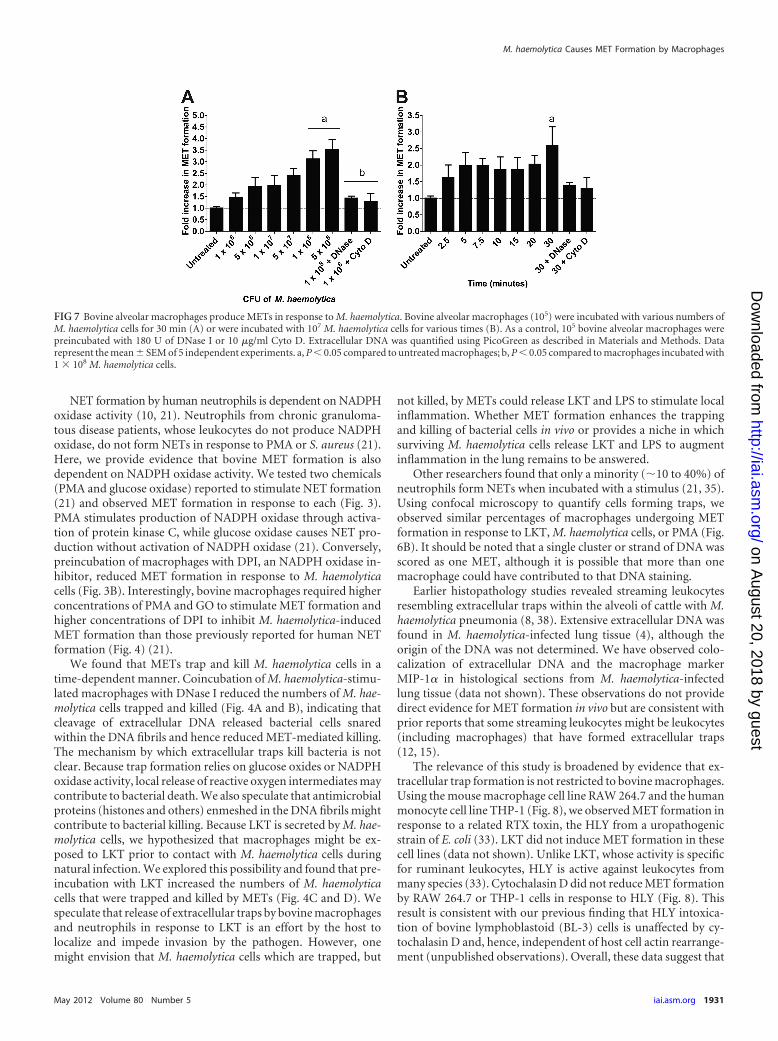
Bovine alveolar macrophages produce METs. We next exam-ined whether other types of bovine macrophages produce extra-cellular traps. We found that bovine alveolar macrophages formedMETs in a dose- and time-dependent manner in response to M.haemolytica cells (Fig. 7). Preincubation of alveolar macrophageswith DNase I or cytochalasin D reduced MET formation, as pre-viously observed for monocyte-derived macrophages (Fig. 1) andbovine neutrophils (4). In contrast to these results, we did notobserve extracellular traps when peripheral blood monocyteswere incubated with LKT (data not shown).
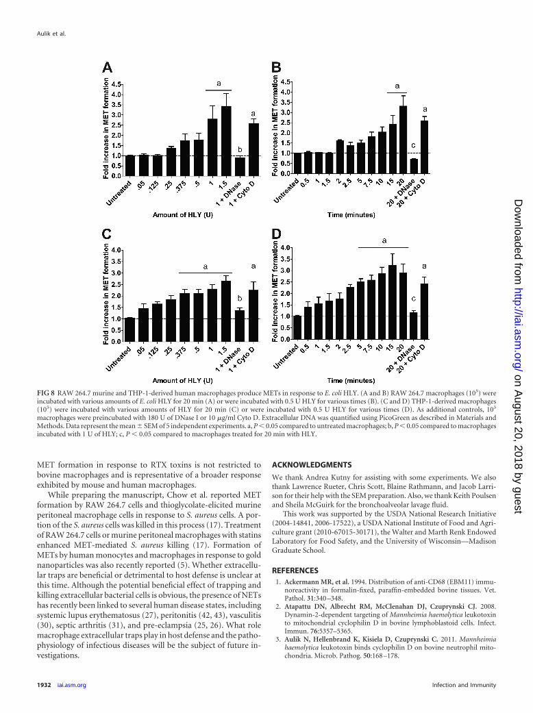
Production of extracellular traps by murine and humanmacrophage cell lines. Finally, we examined whether MET forma-tion was restricted to bovine macrophages or was representative of abroader response by macrophages from other mammalian species.To trigger MET formation by nonruminant macrophages, we usedthe E. coli HLY, which is the prototypical RTX toxin that is activeagainst leukocytes from multiple species (47). When we incubatedHLY with the mouse macrophage cell line RAW 264.7, we observedMET formation in a dose- and time-dependent manner (Fig. 8A andB). MET formation was reduced when DNase I was present but not
FIG 2 Wild-type M. haemolytica and �lktC M. haemolytica cells cause MET formation by bovine macrophages. Monocyte-derived macrophages (105) wereincubated for 20 min with various cell numbers of M. haemolytica (A), �lktC M. haemolytica (B), or �lktA M. haemolytica (C). In some experiments, bacteria wereinactivated at 100°C for 30 min prior to addition to macrophages. (D to F) Macrophages (105) were incubated for various times with 1 � 107 cells of wild-typeM. haemolytica (D), �lktC M. haemolytica (E), or �lktA M. haemolytica (F). In some experiments, 180 U of DNase I or 10 �g/ml cytochalasin D was incubatedwith the macrophages for 30 min at 37°C prior to addition of the bacterial cells. Extracellular DNA was quantified using PicoGreen as described in Materials andMethods. Data represent the mean � SEM of 5 independent experiments. a, P � 0.05 compared to untreated macrophages; b, P � 0.05 compared to macrophagesincubated with 1 � 108 bacteria; c, P � 0.05 compared to macrophages incubated for 300 s with bacterial cells alone.
M. haemolytica Causes MET Formation by Macrophages
when cytochalasin D was added (Fig. 8A and B). Similarly, we exam-ined extracellular trap formation by the human THP-1 monocyte cellline after those cells were treated with PMA to differentiate them intomacrophage-like cells. THP-1 cells formed METs in a dose- and time-dependent manner in response to the E. coli HLY (Fig. 8C and D). Asfor RAW 264.7 cells, MET formation was reduced by DNase I but wasnot inhibited by cytochalasin D. We did not observe MET formationby either cell line in response to LPS (data not shown).
DISCUSSION
We present here a detailed characterization of macrophage extra-cellular trap formation. Bovine macrophages produce extracellu-lar traps in a dose- and time-dependent manner in response to M.haemolytica and its LKT (Fig. 1 and 2). MET formation peaked ata 3-fold increase, which is similar to results for NET formationpublished by other investigators in response to various pathogens(35). We used several methods to confirm that LKT, and not con-taminating LPS, was responsible for MET formation. These in-cluded the use of (i) cytochalasin D, to inhibit LKT-mediatedinternalization (2, 3); (ii) antibodies to LKT and its CD18 recep-tor; (iii) heat-inactivated LKT, heat-inactivated M. haemolyticacells, or heat-inactivated �lktC M. haemolytica cells (100°C for 1h); (iv) a truncated �LKT; and (v) purified M. haemolytica LPSalone (Fig. 1). Taken together, these data indicate that LKT causesMET formation via binding to its CD18 receptor and that M.haemolytica LPS (25 nM to 2.5 �M) by itself does not cause METformation by bovine macrophages (data not shown). Similar re-sults were reported using bovine neutrophils (4). The lack of LPS-mediated extracellular trap formation is similar to the results ofClark et al. (18), who reported that LPS alone did not stimulateNET production by human neutrophils, unless platelets were alsopresent. However, we cannot completely exclude a role for LPS asa cofactor in the MET response to M. haemolytica cells or to LKT.
Greater concentrations of RTX toxins can disrupt the cytoplas-mic membrane and cause lysis of host cells (47). To examine whatwas responsible for the release of DNA, we quantified LDH re-
lease, which is a marker for necrosis (21, 35). LDH release re-mained low at LKT concentrations (0.25 to 2 U) that trigger METformation, indicating that necrosis was not responsible for extra-cellular DNA. However, LDH release did increase when macro-phages were incubated with a higher dose (5 U) of LKT (Fig. 1Dand E). These data are similar to those presented by Pilsczek et al.(35). These investigators observed little LDH release during NETformation, which they interpret to be evidence that NET forma-tion does not lead to neutrophil lysis. Rather, these investigatorsobserved that NETs are found in budding vesicles that burst uponleaving the neutrophil (35). We infer from these results that lowconcentrations of LKT (�5 U) cause extracellular DNA releaseand MET formation by a process that is largely independent ofnecrosis.
Interestingly, extracellular traps were produced by macro-phages in response to either native fully active LKT or nonacylatedpro-LKT (Fig. 1C). This observation is similar to that reported forbovine neutrophils (4). These data suggest that binding of pro-LKT to CD18 is sufficient to trigger extracellular trap formation bybovine leukocytes in the absence of an overt cytotoxic effect, asdetected by standard cytotoxicity assays (4). MET formation wasinhibited by preincubation of LKT or pro-LKT with a neutralizinganti-LKT antibody (Fig. 1C). MET formation in response to LKTor pro-LKT was reduced when macrophages were preincubatedwith an anti-CD18 antibody or cytochalasin D, the latter of whichwe have previously shown inhibits intracellular trafficking of LKT(2) (Fig. 1C). Overall, these data confirm that the interaction be-tween LKT or pro-LKT and the CD18 receptor and the subsequentreorganization of intracellular actin are required for MET forma-tion in response to LKT or pro-LKT.
We used monocyte-derived macrophages (cultured in vitro for7 days) for most of our experiments. Likewise, bovine alveolarmacrophages also formed METs in a dose- and time-dependentmanner in response to M. haemolytica (Fig. 7). These data areconsistent with earlier reports that alveolar macrophages exhibitstreaming nuclei during M. haemolytica infection of cattle (1). It is
FIG 3 MET formation requires NADPH oxidase activity. (A) Bovine macrophages (105) were incubated with various concentrations of PMA. As a positivecontrol, 105 bovine neutrophils were incubated with 10 �M PMA. (B) Bovine macrophages (105) were preincubated with 50 �M DPI for 30 min at 37°C.DPI-treated and untreated macrophages (105) were then incubated with 5 � 107 M. haemolytica cells (MH). As a control, some macrophages were incubated with200 �M the pancaspase inhibitor Z-VAD-FMK prior to addition of 5 � 107 M. haemolytica cells. (C) Bovine macrophages or neutrophils (105) were incubatedwith 500 U or 5 U glucose oxidase, respectively. Extracellular DNA was quantified using PicoGreen as described in Materials and Methods. Data represent themean � SEM of 5 independent experiments. a, P � 0.05 compared to untreated macrophages; b, P � 0.05 compared to macrophages incubated with 5 � 107 M.haemolytica; c, P � 0.05 compared to Z-VAD-FMK control macrophages.
interesting that maximal MET formation by alveolar macro-phages occurred somewhat later (30 min) than that observed withmonocyte-derived macrophages (Fig. 3). However, it should benoted that a limited number of alveolar macrophage samples weretested (n � 3). In contrast to the findings described above, freshly
adherent monocytes did not form extracellular traps when incu-bated with LKT (data not shown). These findings suggest that thematuration state of bovine mononuclear phagocytes influencestheir release of nuclear DNA in response to LKT.
Investigators have reported that NET formation occurs within
FIG 4 Preincubating bovine macrophages with LKT increases the trapping and killing of M. haemolytica by METs. Bovine macrophages (105) were incubatedwith 107 fluorescein-labeled M. haemolytica cells (A) or unlabeled M. haemolytica cells (B) for 60, 120, or 180 min. Similarly, LKT-treated (0.5 U LKT for 30 min)or untreated macrophages were incubated for 60 min with 107 fluorescein-labeled M. haemolytica cells (C) or 107 unlabeled M. haemolytica cells (D). Untreatedmacrophages were used as controls. Additional controls included macrophages incubated with M. haemolytica and 180 U DNase I. Bacterial trapping wasestimated by fluorescence using an automated plate reader. Bacterial survival was estimated by plating serial dilutions of lysates on TSA–5% sheep RBC plates.Data represent the mean � SEM of 5 independent experiments. a, P � 0.05 compared to macrophages incubated with M. haemolytica for 180 min; b, P � 0.05compared to untreated macrophages; c, P � 0.05 compared to macrophages treated with LKT alone.
FIG 5 Scanning electron photomicrographs of METs formed by bovine macrophages in response to M. haemolytica cells. Bovine macrophages (2.5 � 105) wereincubated with 5 � 107 M. haemolytica cells or RPMI 1640 (negative control) for 5 min at 37°C. Cells were washed, fixed, and processed for SEM as described in Materialsand Methods. (A) A matrix of extracellular DNA strands released in response to M. haemolytica cells (arrows indicate trapped bacterial cells); (B) control bovinemacrophages incubated with RPMI that do not exhibit extracellular DNA fibrils. Photomicrographs are of representative cells from 3 independent experiments.
M. haemolytica Causes MET Formation by Macrophages
10 min to 4 h (35), whereas eosinophils produce extracellulartraps within seconds (45). We found that MET formation oc-curred faster than NET formation, with significant accumulationof extracellular DNA occurring within 2 min after the addition ofstimuli to macrophages (Fig. 2D and E) (4). This appears to besimilar to findings of a recent study that demonstrated NET for-mation at as early as 5 min in response to S. aureus (35). Themechanisms by which extracellular traps are formed by varioustypes of leukocytes have not been firmly established. However, thetemporal differences in DNA release suggest that the intracellularsignaling that regulates MET and NET formation may differ.
We used confocal and scanning electron microscopy to analyzethe structure of METs. Confocal analysis confirmed that extracel-lular DNA fibrils were released in response to LKT or M. haemo-lytica cells, and these fibrils were disrupted by the addition of
DNase I (Fig. 6A). SEM confirmed the presence of a large network(�10 �m2) of DNA fibrils that, upon higher magnification, clearlycontained M. haemolytica cells trapped within them (Fig. 6). Theorigin of DNA in extracellular traps is generally thought to benuclear, although it is reported that eosinophils release mitochon-drial DNA (48). We addressed this issue by examining the colo-calization of TOPRO, which stains DNA, with the signal from afluorescently conjugated antibody that stains nuclear histones.The rationale for this approach is based on histones being associ-ated with nuclear but not mitochondrial DNA (14). Colocaliza-tion of the signal for TOPRO (red) and the antihistone antibodies(green) suggests that the DNA in METs that bovine macrophagesproduced in response to M. haemolytica and its LKT is of nuclearorigin (Fig. 6C). Incubation with DNase I confirmed that extra-cellular staining of TOPRO was specific for DNA.
FIG 6 Confocal microscopy of METs formed by bovine macrophages in response to LKT and M. haemolytica. (A) Macrophages (2.5 � 105) were incubated for30 min at 37°C with 1 U LKT, 1 U LKT with 180 U DNase I, 107 fluorescein-labeled M. haemolytica cells, 100 mM PMA, 250 nM M. haemolytica LPS, or RPMI1640 (negative control). Cells were fixed, permeabilized, stained for DNA using TOPRO, and examined by confocal microscopy. Arrows indicate representativeMETs emanating from macrophages. (B) Percentage of macrophages that formed METs, based on scoring of 500 macrophages in multiple micrographs. a, P �0.05 compared to untreated macrophages; b, P � 0.05 compared to macrophages incubated with 1.5 U LKT. Photomicrographs are of representative cells from3 independent experiments. (C) To assess colocalization of DNA and histones in METs, 2.5 � 105 macrophages were incubated with 1 U LKT or 107 M.haemolytica cells for 30 min. Cells were fixed, permeabilized, stained for DNA using TOPRO (red), and probed for histones using an antihistone antibodyfollowed by an antimouse antibody labeled with Alexa Fluor 488 (green). Cells were examined by confocal microscopy. Arrows indicate areas of colocalizationof signals for extracellular DNA and histones. Photomicrographs are of representative cells from 3 independent experiments.
NET formation by human neutrophils is dependent on NADPHoxidase activity (10, 21). Neutrophils from chronic granuloma-tous disease patients, whose leukocytes do not produce NADPHoxidase, do not form NETs in response to PMA or S. aureus (21).Here, we provide evidence that bovine MET formation is alsodependent on NADPH oxidase activity. We tested two chemicals(PMA and glucose oxidase) reported to stimulate NET formation(21) and observed MET formation in response to each (Fig. 3).PMA stimulates production of NADPH oxidase through activa-tion of protein kinase C, while glucose oxidase causes NET pro-duction without activation of NADPH oxidase (21). Conversely,preincubation of macrophages with DPI, an NADPH oxidase in-hibitor, reduced MET formation in response to M. haemolyticacells (Fig. 3B). Interestingly, bovine macrophages required higherconcentrations of PMA and GO to stimulate MET formation andhigher concentrations of DPI to inhibit M. haemolytica-inducedMET formation than those previously reported for human NETformation (Fig. 4) (21).
We found that METs trap and kill M. haemolytica cells in atime-dependent manner. Coincubation of M. haemolytica-stimu-lated macrophages with DNase I reduced the numbers of M. hae-molytica cells trapped and killed (Fig. 4A and B), indicating thatcleavage of extracellular DNA released bacterial cells snaredwithin the DNA fibrils and hence reduced MET-mediated killing.The mechanism by which extracellular traps kill bacteria is notclear. Because trap formation relies on glucose oxides or NADPHoxidase activity, local release of reactive oxygen intermediates maycontribute to bacterial death. We also speculate that antimicrobialproteins (histones and others) enmeshed in the DNA fibrils mightcontribute to bacterial killing. Because LKT is secreted by M. hae-molytica cells, we hypothesized that macrophages might be ex-posed to LKT prior to contact with M. haemolytica cells duringnatural infection. We explored this possibility and found that pre-incubation with LKT increased the numbers of M. haemolyticacells that were trapped and killed by METs (Fig. 4C and D). Wespeculate that release of extracellular traps by bovine macrophagesand neutrophils in response to LKT is an effort by the host tolocalize and impede invasion by the pathogen. However, onemight envision that M. haemolytica cells which are trapped, but
not killed, by METs could release LKT and LPS to stimulate localinflammation. Whether MET formation enhances the trappingand killing of bacterial cells in vivo or provides a niche in whichsurviving M. haemolytica cells release LKT and LPS to augmentinflammation in the lung remains to be answered.
Other researchers found that only a minority (�10 to 40%) ofneutrophils form NETs when incubated with a stimulus (21, 35).Using confocal microscopy to quantify cells forming traps, weobserved similar percentages of macrophages undergoing METformation in response to LKT, M. haemolytica cells, or PMA (Fig.6B). It should be noted that a single cluster or strand of DNA wasscored as one MET, although it is possible that more than onemacrophage could have contributed to that DNA staining.
Earlier histopathology studies revealed streaming leukocytesresembling extracellular traps within the alveoli of cattle with M.haemolytica pneumonia (8, 38). Extensive extracellular DNA wasfound in M. haemolytica-infected lung tissue (4), although theorigin of the DNA was not determined. We have observed colo-calization of extracellular DNA and the macrophage markerMIP-1� in histological sections from M. haemolytica-infectedlung tissue (data not shown). These observations do not providedirect evidence for MET formation in vivo but are consistent withprior reports that some streaming leukocytes might be leukocytes(including macrophages) that have formed extracellular traps(12, 15).
The relevance of this study is broadened by evidence that ex-tracellular trap formation is not restricted to bovine macrophages.Using the mouse macrophage cell line RAW 264.7 and the humanmonocyte cell line THP-1 (Fig. 8), we observed MET formation inresponse to a related RTX toxin, the HLY from a uropathogenicstrain of E. coli (33). LKT did not induce MET formation in thesecell lines (data not shown). Unlike LKT, whose activity is specificfor ruminant leukocytes, HLY is active against leukocytes frommany species (33). Cytochalasin D did not reduce MET formationby RAW 264.7 or THP-1 cells in response to HLY (Fig. 8). Thisresult is consistent with our previous finding that HLY intoxica-tion of bovine lymphoblastoid (BL-3) cells is unaffected by cy-tochalasin D and, hence, independent of host cell actin rearrange-ment (unpublished observations). Overall, these data suggest that
FIG 7 Bovine alveolar macrophages produce METs in response to M. haemolytica. Bovine alveolar macrophages (105) were incubated with various numbers ofM. haemolytica cells for 30 min (A) or were incubated with 107 M. haemolytica cells for various times (B). As a control, 105 bovine alveolar macrophages werepreincubated with 180 U of DNase I or 10 �g/ml Cyto D. Extracellular DNA was quantified using PicoGreen as described in Materials and Methods. Datarepresent the mean � SEM of 5 independent experiments. a, P � 0.05 compared to untreated macrophages; b, P � 0.05 compared to macrophages incubated with1 � 108 M. haemolytica cells.
M. haemolytica Causes MET Formation by Macrophages
MET formation in response to RTX toxins is not restricted tobovine macrophages and is representative of a broader responseexhibited by mouse and human macrophages.
While preparing the manuscript, Chow et al. reported METformation by RAW 264.7 cells and thioglycolate-elicited murineperitoneal macrophage cells in response to S. aureus cells. A por-tion of the S. aureus cells was killed in this process (17). Treatmentof RAW 264.7 cells or murine peritoneal macrophages with statinsenhanced MET-mediated S. aureus killing (17). Formation ofMETs by human monocytes and macrophages in response to goldnanoparticles was also recently reported (5). Whether extracellu-lar traps are beneficial or detrimental to host defense is unclear atthis time. Although the potential beneficial effect of trapping andkilling extracellular bacterial cells is obvious, the presence of NETshas recently been linked to several human disease states, includingsystemic lupus erythematosus (27), peritonitis (42, 43), vasculitis(30), septic arthritis (31), and pre-eclampsia (25, 26). What rolemacrophage extracellular traps play in host defense and the patho-physiology of infectious diseases will be the subject of future in-vestigations.
ACKNOWLEDGMENTS
We thank Andrea Kutny for assisting with some experiments. We alsothank Lawrence Rueter, Chris Scott, Blaine Rathmann, and Jacob Larri-son for their help with the SEM preparation. Also, we thank Keith Poulsenand Sheila McGuirk for the bronchoalveolar lavage fluid.
This work was supported by the USDA National Research Initiative(2004-14841, 2006-17522), a USDA National Institute of Food and Agri-culture grant (2010-67015-30171), the Walter and Marth Renk EndowedLaboratory for Food Safety, and the University of Wisconsin—MadisonGraduate School.
REFERENCES1. Ackermann MR, et al. 1994. Distribution of anti-CD68 (EBM11) immu-
noreactivity in formalin-fixed, paraffin-embedded bovine tissues. Vet.Pathol. 31:340 –348.
2. Atapattu DN, Albrecht RM, McClenahan DJ, Czuprynski CJ. 2008.Dynamin-2-dependent targeting of Mannheimia haemolytica leukotoxinto mitochondrial cyclophilin D in bovine lymphoblastoid cells. Infect.Immun. 76:5357–5365.
3. Aulik N, Hellenbrand K, Kisiela D, Czuprynski C. 2011. Mannheimiahaemolytica leukotoxin binds cyclophilin D on bovine neutrophil mito-chondria. Microb. Pathog. 50:168 –178.
FIG 8 RAW 264.7 murine and THP-1-derived human macrophages produce METs in response to E. coli HLY. (A and B) RAW 264.7 macrophages (105) wereincubated with various amounts of E. coli HLY for 20 min (A) or were incubated with 0.5 U HLY for various times (B). (C and D) THP-1-derived macrophages(105) were incubated with various amounts of HLY for 20 min (C) or were incubated with 0.5 U HLY for various times (D). As additional controls, 105
macrophages were preincubated with 180 U of DNase I or 10 �g/ml Cyto D. Extracellular DNA was quantified using PicoGreen as described in Materials andMethods. Data represent the mean � SEM of 5 independent experiments. a, P � 0.05 compared to untreated macrophages; b, P � 0.05 compared to macrophagesincubated with 1 U of HLY; c, P � 0.05 compared to macrophages treated for 20 min with HLY.
4. Aulik N, Hellenbrand K, Klos H, Czuprynski C. 2010. Mannheimiahaemolytica and its leukotoxin causes neutrophil extracellular trap (NET)formation by bovine neutrophils. Infect. Immun. 78:4454 – 4466.
5. Bartneck M, Keul H, Zwadlo-Klarwasser G, Groll J. 2010. Phagocytosisindependent extracellular nanoparticle clearance by human immune cells.Nano Lett. 10:59 – 63.
6. Beiter K, et al. 2006. An endonuclease allows Streptococcus pneumoniae toescape from neutrophil extracellular traps. Curr. Biol. 16:401– 407.
7. Bianchi M, et al. 2009. Restoration of NET formation by gene therapy inCGD controls aspergillosis. Blood 114:2619 –2622.
8. Breider MA, Walker RD, Hopkins FM, Schultz TW, Bowersock TL.1988. Pulmonary lesions induced by Pasteurella haemolytica in neutrophilsufficient and neutrophil deficient calves. Can. J. Vet. Res. 52:205–209.
9. Brinkmann V, Laube B, Abu Abed U, Goosmann C, Zychlinsky A.2010. Neutrophil extracellular traps: how to generate and visualize them.J. Vis. Exp. 36:pii�1724.
11. Brinkmann V, Zychlinsky A. 2007. Beneficial suicide: why neutrophilsdie to make NETs. Nat. Rev. Microbiol. 5:577–582.
12. Brogden KA, DeBey B, Audibert F, Lehmkuhl H, Chedid L. 1995.Protection of ruminants by Pasteurella haemolytica A1 capsular polysac-charide vaccines containing muramyl dipeptide analogs. Vaccine 13:1677–1684.
13. Buchanan JT, et al. 2006. DNase expression allows the pathogen group AStreptococcus to escape killing in neutrophil extracellular traps. Curr.Biol. 16:396 – 400.
14. Caron F, Jacq C, Rouviere-Yaniv J. 1979. Characterization of a histones-like protein extracted from yeast mitochondria. Proc. Natl. Acad. Sci.U. S. A. 76:4265– 4269.
15. Caswell JL, Middleton DM, Sorden SD, Gordon JR. 1998. Expression ofthe neutrophil chemoattractant interleukin-8 in the lesions of bovinepneumonic pasteurellosis. Vet. Pathol. 35:124 –131.
16. Chang YF, Young R, Post D, Struck DK. 1987. Identification andcharacterization of the Pasteurella haemolytica leukotoxin. Infect. Immun.55:2348 –2354.
17. Chow O, et al. 2010. Statins enhance formation of phagocyte extracellulartraps. Cell Host Microbe 8:445– 454.
18. Clark SR, et al. 2007. Platelet TLR4 activates neutrophil extracellulartraps to ensnare bacteria in septic blood. Nat. Med. 13:463– 469.
19. Deshpande MS, Ambagala TC, Ambagala AP, Kehrli ME, Jr, Srikuma-ran S. 2002. Bovine CD18 is necessary and sufficient to mediateMannheimia (Pasteurella) haemolytica leukotoxin-induced cytolysis. In-fect. Immun. 70:5058 –5064.
20. Fedorova ND, Highlander SK. 1997. Generation of targeted nonpolargene insertions and operon fusions in Pasteurella haemolytica and creationof a strain that produces and secretes inactive leukotoxin. Infect. Immun.65:2593–2598.
21. Fuchs TA, et al. 2007. Novel cell death program leads to neutrophilextracellular traps. J. Cell Biol. 176:231–241.
22. Gentry MJ, Srikumaran S. 1991. Neutralizing monoclonal antibodies toPasteurella haemolytica leukotoxin affinity-purify the toxin from crudeculture supernatants. Microb. Pathog. 10:411– 417.
23. Grinberg N, Elazar S, Rosenshine I, Shpigel NY. 2008. Beta-hydroxybutyrate abrogates formation of bovine neutrophil extracellulartraps and bactericidal activity against mammary pathogenic Escherichiacoli. Infect. Immun. 76:2802–2807.
24. Guimaraes-Costa AB, et al. 2009. Leishmania amazonensis promastigotesinduce and are killed by neutrophil extracellular traps. Proc. Natl. Acad.Sci. U. S. A. 106:6748 – 6753.
25. Gupta A, Hasler P, Gebhardt S, Holzgreve W, Hahn S. 2006. Occur-rence of neutrophil extracellular DNA traps (NETs) in pre-eclampsia: alink with elevated levels of cell-free DNA? Ann. N. Y. Acad. Sci. 1075:118 –122.
26. Gupta AK, Hasler P, Holzgreve W, Hahn S. 2007. Neutrophil NETs: a
novel contributor to preeclampsia-associated placental hypoxia? Semin.Immunopathol. 29:163–167.
27. Hakkim A, et al. 2010. Impairment of neutrophil extracellular trap deg-radation is associated with lupus nephritis. Proc. Natl. Acad. Sci. U. S. A.107:9813–9818.
29. Highlander SK, Engler MJ, Weinstock GM. 1990. Secretion and expres-sion of the Pasteurella haemolytica leukotoxin. J. Bacteriol. 172:2343–2350.
30. Kessenbrock K, et al. 2009. Netting neutrophils in autoimmune small-vessel vasculitis. Nat. Med. 15:623– 625.
31. Logters T, et al. 2009. Diagnostic accuracy of neutrophil-derived circu-lating free DNA (cf-DNA/NETs) for septic arthritis. J. Orthop. Res. 27:1401–1407.
32. McClenahan D, et al. 2008. Effects of lipopolysaccharide andMannheimia haemolytica leukotoxin on bovine lung microvascular endo-thelial cells and alveolar epithelial cells. Clin. Vaccine Immunol. 15:338 –347.
33. Moayeri M, Welch RA. 1997. Prelytic and lytic conformations of eryth-rocyte-associated Escherichia coli hemolysin. Infect. Immun. 65:2233–2239.
34. Papayannopoulos V, Zychlinsky A. 2009. NETs: a new strategy for usingold weapons. Trends Immunol. 30:513–521.
35. Pilsczek FH, et al. 2010. A novel mechanism of rapid nuclear neutrophilextracellular trap formation in response to Staphylococcus aureus. J.Immunol. 185:7413–7425.
36. Schwende H, Fitzke E, Ambs P, Dieter P. 1996. Differences in the stateof differentiation of THP-1 cells induced by phorbol ester and 1,25-dihydroxyvitamin D3. J. Leukoc. Biol. 59:555–561.
37. Shanthalingam S, Srikumaran S. 2009. Intact signal peptide of CD18, thebeta-subunit of beta2-integrins, renders ruminants susceptible toMannheimia haemolytica leukotoxin. Proc. Natl. Acad. Sci. U. S. A. 106:15448 –15453.
38. Slocombe RF, Malark J, Ingersoll R, Derksen FJ, Robinson NE. 1985.Importance of neutrophils in the pathogenesis of acute pneumonic pas-teurellosis in calves. Am. J. Vet. Res. 46:2253–2258.
39. Thumbikat P, Briggs RE, Kannan MS, Maheswaran SK. 2003. Biologicaleffects of two genetically defined leukotoxin mutants of Mannheimia hae-molytica. Microb. Pathog. 34:217–226.
41. Urban CF, Reichard U, Brinkmann V, Zychlinsky A. 2006. Neutrophilextracellular traps capture and kill Candida albicans yeast and hyphalforms. Cell. Microbiol. 8:668 – 676.
42. Vitkov L, Klappacher M, Hannig M, Krautgartner WD. 2009. Extracel-lular neutrophil traps in periodontitis. J. Periodontal Res. 44:664 – 672.
43. Vitkov L, Klappacher M, Hannig M, Krautgartner WD. 2010. Neutro-phil fate in gingival crevicular fluid. Ultrastruct. Pathol. 34:25–30.
44. von Kockritz-Blickwede M, et al. 2008. Phagocytosis-independent anti-microbial activity of mast cells by means of extracellular trap formation.Blood 111:3070 –3080.
45. von Kockritz-Blickwede M, Nizet V. 2009. Innate immunity turnedinside-out: antimicrobial defense by phagocyte extracellular traps. J. Mol.Med. 87:775–783.
46. Wartha F, Beiter K, Normark S, Henriques-Normark B. 2007. Neutro-phil extracellular traps: casting the NET over pathogenesis. Curr. Opin.Microbiol. 10:52–56.
47. Welch RA. 2001. RTX toxin structure and function: a story of numerousanomalies and few analogies in toxin biology. Curr. Top. Microbiol. Im-munol. 257:85–111.
48. Yousefi S, et al. 2008. Catapult-like release of mitochondrial DNA byeosinophils contributes to antibacterial defense. Nat. Med. 14:949 –953.
M. haemolytica Causes MET Formation by Macrophages


![static-curis.ku.dk · [Pasteurella ] haemolytica or [ P.] anatis by DNA-DNA hybridization, 16S rRNA and 16S-23S ITS sequencing, AFLP, PFGE, plasmid profiling and phenotypic characterization.](https://static.documents.pub/doc/80x56/5ceed30088c99330508cabea/static-curiskudk-pasteurella-haemolytica-or-p-anatis-by-dna-dna-hybridization.jpg)


