CUN. CHEM. 21/11, 1575-1581 (1975) CLINICAL CHEMISTRY, Vol. 21, No. 11, 1975 1575 Manual and Semi-Automated Procedures for Measurement of Triglycerides in Serum Joseph L. Glegel, Anna Bell Ham, and William Clema We describe manual and semi-automated procedures for serum triglyceride determinations, in which lipids are partitioned between a water/isopropanol phase and a nonane phase. More than 99% of the triglyceride is ex- tracted into the nonane phase, as determined by recov- ery of 3H-labeled triolein. Studies with “C-labeled leci- thin demonstrate that less than 1.3% is extracted into the nonane phase at concentrations up to 2.5 g/Iiter. A novel feature of the method is that glycerol can be liber- ated from triglyceride by sodium hydroxide at room tem- perature in less than 5 mm. Glycerol is oxidized by per- iodate in 1-2 mm at 25 #{176}C; the formaldehyde produced is reacted with 2,4-pentanedione to yield 3,5-diacetyl- 1,3-dihydrolutidine. The manual procedure requires less than 20 mm; the semi-automated method requires 7 mm from sampling to readout. The procedure may be run at 30-40 samples/h, with stable baseline and less than 2.0% carryover. Both methods are linear to 0.50 g (5.6 mol) of triolein per liter. Analytical recoveries at several concentrations ranged from 97-101% (mean, 100%). Additional Keyphrases: solvent partition method #{149} normal (reference) values #{149} continuous-flow analysis #{149} stability of triglycerides in serum #{149} hyperlipemia Triglyceride (tniacylglycerol) determinations have assumed increasing importance since triglycerides have been identified as an important risk factor in coronary artery disease. Accurate determination of both serum cholesterol and serum triglycerides is now required for adequate diagnosis of hyperlipi- demia (1). In contrast to serum cholesterol determi- nations, measurement of triglyceride is somewhat complicated, which has hindered its widespread use. Here, we present a simplified method for fast and ac- curate triglyceride analysis, in which phase partition of serum lipids is used. Triglyceride methods can be broadly grouped into three categories: (a) methods that involve extraction with an organic solvent followed by adsorption on solid phases such as zeolite or silicic acid to remove interfering phospholipids (2, 3); (b) enzymatic meth- ods, which do not require separation of phospholipids Clinical Chemistry Research and Development Laboratory, DADE Division American Hospital Supply Corp., Box 520672, Miami, Fla. 33152. Received Mar. 31, 1975; accepted July 2, 1975. because of the enzymatic specificities involved (4); and (c) methods based on the partition of serum lip- ids between two liquid phases. Methods a and b gen- erally require serum blank corrections if results are to be accurate. Method c is highly selective for tri- glycerides and the results do not require blank cor- rection except for highly icteric specimens. Solvent partition of serum lipids was first used for triglycer- ide analysis by Royer and Ko (5, 6), who used a modi- fied Dole extraction technique (7) to develop a semi- automated triglyceride procedure. Others (8-10) have published modifications of the same extraction technique. The methods presented here represent ex- tensions and modifications of these procedures to provide compatible manual and semi-automated pro- cedures. A novel feature of the method is that the ex- tracted triglyceride can be converted to glycerol in less than 5 mm at room temperature. Materials Reagents Extraction reagent: n-nonane/isopropanol (2.0/3.5 by vol). Caution: flammable. Triolein standard: triolein (99% purity) 1000 mg/ liter in extraction reagent (1.13 mmol/liter). Caution: flammable. Sulfuric acid, 40 mmol/liter. Transesterifying reagent: NaOH in isopropanol, 100 mmol/liter. A fine precipitate may be present in the reagent, primarily as a result of carbonate forma- tion. It will settle on standing, or it can be removed by filtration. A slight yellow color may develop with age but will not affect test results. Oxidizing reagent: sodium periodate, 18 mmol/ liter, in 2.0 mol/liter acetic acid. Color buffer for manual method: ammonium ace- tate (6.0 mol/liter, pH 6.0 at 25 #{176}C). Color buffer for semi-automated method: ammo- nium acetate (3.0 mol/liter, pH 6.0). Acetylacetone (2,4-pentanedione), reagent grade. n-Nonane, Philips “pure” grade. The above reagents are all stable for at least a year at room temperature. Store them in tightly stoppered vessels.
Transcript
CUN. CHEM. 21/11, 1575-1581 (1975)
CLINICAL CHEMISTRY, Vol. 21, No. 11, 1975 1575
Manual and Semi-Automated Procedures for
Measurement of Triglycerides in Serum
Joseph L. Glegel, Anna Bell Ham, and William Clema
We describe manual and semi-automated proceduresfor serum triglyceride determinations, in which lipids arepartitioned between a water/isopropanol phase and anonane phase. More than 99% of the triglyceride is ex-tracted into the nonane phase, as determined by recov-
ery of 3H-labeled triolein. Studies with “C-labeled leci-thin demonstrate that less than 1.3% is extracted intothe nonane phase at concentrations up to 2.5 g/Iiter. Anovel feature of the method is that glycerol can be liber-ated from triglyceride by sodium hydroxide at room tem-perature in less than 5 mm. Glycerol is oxidized by per-iodate in 1-2 mm at 25 #{176}C;the formaldehyde producedis reacted with 2,4-pentanedione to yield 3,5-diacetyl-1,3-dihydrolutidine. The manual procedure requires lessthan 20 mm; the semi-automated method requires 7 mmfrom sampling to readout. The procedure may be run at30-40 samples/h, with stable baseline and less than2.0% carryover. Both methods are linear to 0.50 g (5.6mol) of triolein per liter. Analytical recoveries at severalconcentrations ranged from 97-101% (mean, 100%).
Triglyceride (tniacylglycerol) determinations have
assumed increasing importance since triglycerideshave been identified as an important risk factor in
coronary artery disease. Accurate determination ofboth serum cholesterol and serum triglycerides isnow required for adequate diagnosis of hyperlipi-demia (1). In contrast to serum cholesterol determi-nations, measurement of triglyceride is somewhatcomplicated, which has hindered its widespread use.Here, we present a simplified method for fast and ac-curate triglyceride analysis, in which phase partitionof serum lipids is used.
Triglyceride methods can be broadly grouped into
three categories: (a) methods that involve extractionwith an organic solvent followed by adsorption onsolid phases such as zeolite or silicic acid to removeinterfering phospholipids (2, 3); (b) enzymatic meth-
ods, which do not require separation of phospholipids
Clinical Chemistry Research and Development Laboratory,DADE Division American Hospital Supply Corp., Box 520672,Miami, Fla. 33152.
Received Mar. 31, 1975; accepted July 2, 1975.
because of the enzymatic specificities involved (4);
and (c) methods based on the partition of serum lip-ids between two liquid phases. Methods a and b gen-
erally require serum blank corrections if results areto be accurate. Method c is highly selective for tri-glycerides and the results do not require blank cor-rection except for highly icteric specimens. Solvent
partition of serum lipids was first used for triglycer-ide analysis by Royer and Ko (5, 6), who used a modi-fied Dole extraction technique (7) to develop a semi-
automated triglyceride procedure. Others (8-10)have published modifications of the same extractiontechnique. The methods presented here represent ex-
tensions and modifications of these procedures toprovide compatible manual and semi-automated pro-cedures. A novel feature of the method is that the ex-tracted triglyceride can be converted to glycerol inless than 5 mm at room temperature.
Materials
Reagents
Extraction reagent: n-nonane/isopropanol (2.0/3.5
by vol). Caution: flammable.Triolein standard: triolein (99% purity) 1000 mg/
liter in extraction reagent (1.13 mmol/liter). Caution:flammable.
Sulfuric acid, 40 mmol/liter.Transesterifying reagent: NaOH in isopropanol,
100 mmol/liter. A fine precipitate may be present inthe reagent, primarily as a result of carbonate forma-tion. It will settle on standing, or it can be removedby filtration. A slight yellow color may develop withage but will not affect test results.
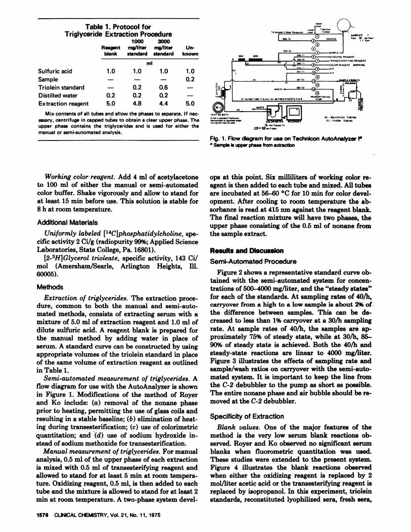
FIg. 1. Flow diagram for use on Technicon AutoAnalyzer iaa Sample is upper phase from extraction
1578 CUNICALCHEMISTRY, Vol. 21. No. 11, 1975
Sulfuric acid
SampleTrioleiri standard
Distilled waterExtraction reagent
Mix contents of all tubes and allow the phases to separate. If nec-essary. centrifuge in capped tubes to obtain a clear upper phase. Theupper phase contains the triglycerides and is used for either themanual or semi-automated analysis.
Working color reagent. Add 4 ml of acetylacetoneto 100 ml of either the manual or semi-automatedcolor buffer. Shake vigorously and allow to stand forat least 15 mm before use. This solution is stable for8 h at room temperature.
[2-3H]Glycerol trioleate, specific activity, 143 Ci/mol (Amersham/Searle, Arlington Heights, ill.60005).
Methods
Extraction of triglycerides. The extraction proce-dure, common to both the manual and semi-auto-mated methods, consists of extracting serum with amixture of 5.0 ml of extraction reagent and 1.0 ml of
dilute sulfuric acid. A reagent blank is prepared forthe manual method by adding water in place ofserum. A standard curve can be constructed by usingappropriate volumes of the triolein standard in placeof the same volume of extraction reagent as outlinedin Table 1.
Semi-automated measurement of triglycerides. Aflow diagram for use with the AutoAnalyzer is shownin Figure 1. Modifications of the method of Royerand Ko include: (a) removal of the nonane phaseprior to heating, permitting the use of glass coils andresulting in a stable baseline; (b) elimination of heat-ing during transesterification; (c) use of colorirnetric
quantitation; and (d) use of sodium hydroxide in-stead of sodium methoxide for transesterification.
Manual measurement of triglycerides. For manualanalysis, 0.5 ml of the upper phase of each extractionis mixed with 0.5 ml of transesterifying reagent andallowed to stand for at least 5 mm at room tempera-ture. Oxidizing reagent, 0.5 ml, is then added to eachtube and the mixture is allowed to stand for at least 2mm at room temperature. A two-phase system devel-
ops at this point. Six milliliters of working color re-agent is then added to each tube and mixed. All tubesare incubated at 56-60 #{176}Cfor 10 mm for color devel-opment. After cooling to room temperature the ab-sorbance is read at 415 nm against the reagent blank.The final reaction mixture will have two phases, theupper phase consisting of the 0.5 ml of nonane fromthe sample extract.
Results and DiscussionSemi-Automated Procedure
Figure 2 shows a representative standard curve ob-tained with the semi-automated system for concen-trations of 500-4000 mg/liter, and the “steady states”for each of the standards. At sampling rates of 40/h,carryover from a high to a low sample is about 2% of.the difference between samples. This can be de-creased to less than 1% carryover at a 30/h samplingrate. At sample rates of 40/h, the samples are ap-proximately 75% of steady state, while at 30/h, 85-90% of steady state is achieved. Both the 40/h andsteady-state reactions are linear to 4000 mg/liter.Figure 3 illustrates the effects of sampling rate andsample/wash ratios on carryover with the semi-auto-mated system. It is important to keep the line fromthe C-2 debubbler to the pump as short as possible.The entire nonane phase and air bubble should be re-moved at the C-2 debubbler.
Specificity of Extraction
Blank values. One of the major features of themethod is the very low serum blank reactions ob-served. Royer and Ko observed no significant serumblanks when fluorometric quantitation was used.These studies were extended to the present system.Figure 4 illustrates the blank reactions observedwhen either the oxidizing reagent is replaced by 2mol/liter acetic acid or the transesterifying reagent isreplaced by isopropanol. In this experiment, trioleinstandards, reconstituted lyophilized sera, fresh sera,
Recoverya
0
I
aIaaI-
%
9810099
101100102
Mean 100%
98100101100100101
Mean 100%
Fig. 4. Serum blank reactions in the semi-automated system
CUNICAL CHEMISTRY, Vol. 21, No. 11. 1975 1577
FIg. 2. Representative standard curves and “steady state”levels for the semi-automated systemTrioleIn standards
Fig. 3. Effect of sampling rate and sample-to-wash ratio onsensitivity and sample Interaction with the semi-automatedsystem
and a 200 mg/liter solution of unconjugated bilirubinin human albumin were analyzed. As shown in Figure4, only biirubin gave a detectable blank reaction.This interference is negligible, because only unconju-gated bilirubin is extracted into the upper phase;conjugated bilirubin remains in the lower phase (11).Analyses of icteric specimens with total bilirubin
values as high as 160 mg/liter have shown serumblanks of less than 30 mg/liter as triolein. The blankreaction caused by bilirubin in the absence of per-iodate is significantly lower than in the absence ofNaOH and is comparable to the reaction of bmlirubinin the complete test system. This indicates that somebilirubin is destroyed in the presence of NaOH, andtherefore the appropriate blank isobtained in the ab-sence of periodate.
We further studied the effect of glucose and glycer-ol by reconstituting lyophilized control materialswith either water or a solution of 3000 mg/liter glu-cose or glycerol, and saw no significant differences invalues between these solutions. In addition, direct ex-traction of glucose or glycerol solutions gave no reac-tion.
Analytical recovery. Because it is difficult to addtriglycerides in significant amounts directly to serum,these experiments were performed by co-extracting aserum sample along with aliquots of triolein standardin extraction reagent. Analytical recovery of trioleinin various concentrations averaged 100% (Table 2).Recovery was also studied by adding tritium-labeledtriolein directly to serum, followed by extraction. Re-
Table 2. Recovery of TriglycerideAdded to Two Sera
serum, mg/liter
Serum x
mg/liter mg/liter
50010001500200025003000
330820
13301820234028403400
49010001490201025103070
Serum y
5001000150020002500
380870
1380189023802870
4901000151020002490
Co-extracted with the indicated quantities of triolein in extrac-tion reagent.
amt. found of addedaRecovery%= X 100amt. added
Fig. 5. Phospholipid contamination in the extraction of triglyc-eride
1578 CLINICALCHEMISTRY,Vol. 21, No. 11, 1975
coveries averaged 99.5%, as determined by counting aportion of the upper phase and comparing this to thecounts originally added to the serum.
Royer and Ko reported that less than 1% of serumphospholipids were extracted along with triglycer-ides. Figure 5 shows results of an experiment inwhich [14C]phosphatidylcholine was co-extractedwith serum samples and the recovery of counts in theupper layer determined. With this technique, 1.3% ofthe added counts were extracted into the upperphase. As indicated in Figure 5, the upper phase ofthis extraction was successively washed with unla-beled lower phase to determine if the radioactivitywould partition as it did in the original extractions.The radioactivity in the upper phase did decreaseslowly with successive washings and decreased tozero when silicic acid was added to the extract, indi-cating that the counts were in a polar material thatcould be adsorbed onto silicic acid. The reason forthe extensive washings of the upper phase in this ex-periment was to determine that the counts observedwere actually phospholipid and did not represent aminor contaminant of the radioactive material. Be-cause the counts did not partition as in the originalextraction, at least a portion of them was not phos-pholipid but possibly fatty acids derived from thebreakdown of the phospholipid molecule. These re-sults indicate that at least part of the 1.3% of thecounts in the upper phase is phospholipid. It appearsthat the upper limit of phospholipid contamination isless than 1%, data that are in agreement with the re-sults of Royer and Ko. Attempts to add radioactively
labeled phospholipid directly to serum, followed byextraction, invariably led to the conversion of a por-tion of the counts in phospholipid to cholesterol es-ters, presumably by the action of lecithin:cholesterolacyltransferase (EC 2.3.1.43). Royer and Ko have fur-ther confirmed the insignificance of phospholipidcontamination by comparing normal extracts withextracts that had been treated with silicic acid to re-move phospholipid. We repeated these studies andalso find no significant difference between untreatedand silicic acid-treated extracts in the value for thetriglyceride measured.
Optimum Reaction Conditions
Liberation of glycerol. Perhaps the most inter-esting result of these studies was the discovery thatglycerol is very efficiently liberated from triglycerideat room temperature. In our initial experiments sodi-um ethoxide in isopropanol was used as a transester-ifying agent. A study of the optimum temperature ofthis reaction indicated that the reaction proceeded aswell at room temperature as at 56 #{176}C.This promptedfurther investigation. Traditionally, triglycerideshave been hydrolyzed by using alcoholic solutions ofsodium (or potassium) hydroxide, or of sodium meth-oxide or sodium ethoxide (5, 8). We found all three ofthese reagents to be equally effective at room tem-perature. Thin-layer chromatographic analysis of theproducts of these three reaction mixtures indicatedthat both saponification and transesterification hadoccurred and that identical reaction products wereproduced. The esters formed with triolein in these re-action mixtures move slightly further on thin-layerchromatograms than does methyl oleate. This prod-uct is probably isopropyl oleate.
Glycerol liberation proceeds rapidly at room tem-perature; it is complete in 2 to 3 mm. The relativeamounts of free acid and ester produced depend onthe water content of the sodium hydroxide/isopropa-nol solution. Figure 6 illustrates the efficiency of theoverall reaction in the semi-automated procedure asa function of water content of the transesterifying re-agent. At concentrations exceeding 2 ml of water perdeciliter, the efficiency decreases rapidly, and the hy-drolysis reverts to classical saponification kinetics,requiring increased temperatures and prolonged in-cubation times for complete reaction.
Either sodium methoxide, sodium ethoxide, or so-dium hydroxide dissolved in isopropanol is equallyefficient in the procedure. Isopropanol solutions ofsodium hydroxide were selected for routine use be-cause blank values were lowest. Figure 7 illustratesthat concentrations of sodium hydroxide as low as 25mmol/liter are sufficient for quantitative hydrolysisin the manual procedure. However, 100 mmol/literwas selected to assure complete reaction in the semi-automated procedure.
Oxidation. Figure 8 shows the effect of periodateconcentration in the manual method. Periodate con-centrations as low as 12 mmol/liter will completely
% WAifS (“)
z4
a2
174
1
z4174
4
1)24
004
Fig. 6. Effect of water content of transesterifying reagent onthe efficiency of transesterification
0.I
0.7
0.3
0.2
I
0.4
0.5(74
C 0.4“1
4
C.,z4
I004
0.1
Fig. 7. Effect of concentration of NaOH in the transesterifyingreagent
I
5 10 20 30 40 SO 40
NaOH ool/Iiter)
0.6
0.7
0.6
0.3
0.4
0.3
0.2
0.1
2UiI-4
SC-
2
4(74
EC
C-4
UiC-,z4
60(C,04
I (CCl TRIO’-’
‘I/
DELTA ISTD.BLK)
//
///
I,1/
//I,
//Ii
- -
REAGENT BLANK
#{163}2.5 150 17.5 -SODIUM PERIODATE Imol, hter)
Fig. 8. Effect of concentration of periodate in the oxidizing re-agent
CLINICAL CHEMISTRY, Vol. 21, No. 11, 1975 1579
Fig. 9. Variation of sample volume to change sensitivity of theprocedure
oxidize the glycerol produced from samples contain-ing 3000 mg of triolein per liter. We selected a con-
______________ centration of 18 mmol/liter for routine use because70 10 50 100 the final reaction mixture is clear.
Color development. We also investigated the con-centration of acetylacetone used in the working colorreagent. Extremely low concentrations of acetylace-
tone are effective. We selected a concentration of 4ml/dl to accelerate the reaction in the semi-auto-mated procedure.
Ammonium acetate (6 mol/liter) was selected forthe manual procedure to eliminate turbidity in thefinal reaction mixture. In the semi-automated proce-dure, 3 mol/liter ammonium acetate suffices becausethe final reaction mixture reaches the colorimeterquite rapidly after heating and turbidity is not aproblem. In the manual procedure, color develop-ment is complete in 10 mm at 56 #{176}C.Lower tempera-tures for longer times (e.g., 37 #{176}Cfor 30 to 45 mm)also will produce a stable color reaction. The colorproduced is stable for at least 24 h, but we recom-mend that samples be read within 2 h after the reac-
-o tion is complete.Sensitivity and reaction linearity. The sensitivity
of both the manual and semi-automated procedures
can be adjusted to suit the individual needs of thelaboratory. Figure 9 indicates the linear response tochanges in the sample volume in the extraction. Al-though 200 jl of sample is usually used, smaller sam-
Table 3. Effect of Storage at Room Temperatureon Triglyceride Values
served in samples stored at room temperature. Serumsamples stored at 4 #{176}Care stable for at least 24 h.Frozen storage extends the stability of triglycerides,
in serum to several weeks if the tubes are carefully
Serum 7 + EDTA 970 930 920 830 850 stoppered to avoid evaporation. Additional studiesat(10 mg/liter) refrigerator temperature and room temperature (not
Serum 7 + neo- 990 900 930 910 880 shown) support these data. In general, it appears thatmycin + chloro-phenol
Serum 7 + EDTA 880 870plasma
840 - 820
triglycerides in serum are quite stable at refrigeratorand room temperatures. Samples may be collected ei-ther as serum or ethylenediaminetetraacetate-antico-
agulated plasma. Citrated and oxalated plasma haveEDTA, ethylenediaminetetraacetate. also been examined and the results compare well
with values obtained on serum from the same pa-tients.
1580 CUNICAL CHEMISTRY,Vol. 21. No. 11, 1975
ple volumes can be used to extend the range of valuesmeasured, and larger sample sizes (up to 500 gil) canbe used to increase the sensitivity in the normalrange. The nonlinearity exhibited by Serum 1 is dueprimarily to the nonlinearity of the colorimeter. Witha high-performance spectrophotometer the reactionhas been found to be linear to at least an absorbanceof 2.0 at 415 nm; on less-sensitive instruments the re-lationship may not be quite linear.
Stability of Triglycerides in Serum
Some controversy exists about the stability of tri-glyceride in serum, and we examined this. Table 3summarizes data on five freshly drawn sera and threefreshly reconstituted lyophilized control materials.The samples were stored in clean, nonsterile tubesfor the period indicated and were analyzed over afour-day period for triglycerides. These samples ex-hibited generally good stability during this period.Some samples show decreasing values after two daysof storage at room temperature. We studied the ef-fect of the addition of ethylenediaminetetraacetateor the antibiotics neomycin and chlorophenol withtwo other specimens (Table 3, samples 6 and 7). Insample 6 the antibiotics seemed to effectively pre-serve the triglycerides during four days at room tem-perature. This was not the case in sample 7. Bacterialgrowth may have caused the loss in triglycerides ob-
Normal Ranges for Triglycerides
Fredrickson (1) has suggested an upper limit ofnormal for triglycerides of 1400 mg/liter at age 30,with values progressively increasing with age. Otherinvestigators have suggested other ranges, but theseare often method-dependent. In an effort to establisha normal range, we examined 40 Dade employeesafter an overnight fast of at least 12 h. Figure 10shows the results. The mean for the total populationwas 826 mg/liter (SD, 385 mg/liter). Two values wereoutside the 2 SD range and the results of the remain-ing 38 samples were recalculated, giving a mean of776 mg/liter (SD, 326 mg/liter). The resulting ±2 SDrange is 120 to 1430 mg/liter. Because the curve issomewhat skewed, the application of gaussian statis-tics is not fully appropriate. Excluding the two highvalues, results by this method that exceed 1400 mg/liter should be regarded as indicative of hyperlipi-demia, in accordance with the proposals of Fredrick-son.
Comparison of Methods
Results for 25 samples were compared by the semi-automated and manual procedures. They comparedwell over a range of 540-2480 mg/liter. Student’s t-
test for paired observations indicated no significantdifference between methods at the 95% confidencelevel. Least-squares analysis of the data gave a slope
Fig. 11. Correlation between results of the semi-automatedprocedure and an enzymatic procedure
mg/liter
Portions of this work were presented at the 8th InternationalCongress on Clinical Chemistry, Copenhagen, June 1972, and atthe 24th national meeting of the AACC, August 1972.
References1. Fredrickson, D. S., A physician’s guide to hyperlipidemia. Mod.Concepts Cardiovasc. Dis. 41, 31 (1972).
2. van Handel, E., and Zilversmit, D. B., Micromethod for the di-rect determination of serum triglycerides. J. Lab. Clin. Med. 50,152 (1957).
3. Carlson, L. A., and Wadstrom, L. B., Determination of glycer-ides in blood serum. Clin. Chim. Acta 4, 197 (1959).
4. Eggstein, M., and Kreutz, F. M., A new determination of neu-tral fats in blood serum and tissues. Kim. Wochenschr. 44, 267(1966).
5. Royer, M. fi., and Ko, H., A simplified semi-automated assayfor plasma triglycerides. Anal. Biochem. 29,405 (1969).
6. Ko, H., and flayer, M. B., Automatic extraction and analysis ofserum triglycerides. Biochem. Med. 6, 144 (1972).
7. Dole, V. P., A relation between non-esterified fatty acids inplasma and the metabolism of glucose. J. Ciin. Invest. 35, 150(1956).
9. Levy, A. L., and Keyloun, C., An automated colorimetric (non-fluorimetric) assay for triglycerides. In Advances in AutomatedAnalysis, Techni con International Congress 1970, 1, C. E. Bartonet al., Eds., Thurman Associates, Miami, Fla., 1971, p 497.
10. Holub, W. R., Rapid, simplified continuous-flow colorimetryof serum triglycerides. Clin. Chem. 19, 1391 (1973).
11. Ham, A. B., Extraction of unconjugated bilirubin from serum.Ciin. Chem. 18, 1547 (1972).
of the regression line of 1.02, with a y-intercept of-22.3 mg/liter and a standard error of 31.3 mg/liter.With 40 patients’ sera, the manual procedure wascompared to the procedure of Soloni (8); no signifi-cant difference was observed. Samples obtained fromthe Surveillance Phase of the CDC Cooperative Tri-glyceride Survey were also examined by the proposedmethod, and the results were within the limits of ac-ceptability established by CDC. An additional studywas conducted at the University Hospitals of theUniversity of Wisconsin (10) in which the semi-auto-mated procedure was compared to an enzymatic pro-
cedure (Figure 11). Results with the enzymatic proce-dure were corrected for free glycerol blanks and reac-tion drift. Least-squares analysis of the data gave aslope of the regression line of 0.990, with a y-inter-cept of 16 mg/liter and a standard error of 114 mg/liter. The correlation coefficient was 0.990. Student’st-test of the data indicated no significant differencebetween methods at the 95% confidence level.
Precision
Precision with the manual procedure has beenfound to be quite good. Values for a normal concen-tration of triglycerides (in a lyophifized serum pool)obtained on 11 occasions during a six-month periodwere 760, 770, 770, 770, 730, 750, 750, 760, 720, 750,and 750 mg/liter (i#{241}ean,752.7; SD, 16.2, CV 2.14%).Table 4 shows within-day precision (CV, -1%) withfresh serum (10 replicate determinations, manualprocedure). The reagent blank and the standard ab-sorbance values were found to be quite reproducible
from day to day by either the manual or the semi-automated procedure.