371
The Manual
| Date post: | 28-Oct-2014 |
| Category: |
Documents |
| Upload: | lucas-zabala |
| View: | 509 times |
| Download: | 33 times |

The Manual

The
OXOID MANUAL8th Edition 1998
Compiled by E. Y. Bridson
(former Technical Director of Oxoid)
Price: £10


The
OXOID MANUAL8th Edition 1998Compiled by E. Y. Bridson
(former Technical Director of Oxoid)
Eighth Edition 1998
Published by OXOID Limited, Wade Road, Basingstoke,Hampshire, RG24 8PW, England
Telephone National: 01256 841144 International: +44 1256 841144Telex: 858793 OXOID G Telegrams: OXOID BasingstokeFacsimile National: 01256 463388 International: +44 1256 463388Website http://www.oxoid.co.uk
OXOID SUBSIDIARIES AROUND THE WORLD
AustraliaOxoid Australia Pty Ltd104 Northern RoadWest HeidelbergMelbourneVictoria 3081AustraliaTel: 61 39 458 1311Fax: 61 39 458 4759
BelgiumOxoid N.V./S.A.Industriepark, 4EB-9031 DrongenBelgiumTel: 32 9 2811220Fax: 32 9 2811223
BrazilOxoid Brasil Ltdarua Arizona 1349-88andar ± Conjunto 01Brooklin NovoSao Paulo ± SP04567-003BrasilTel: 55 11 5505 0014Fax: 55 11 5505 0014
North AmericaOxoid Inc217 Colonnade RoadNepeanOntario K2E 7K3CanadaTel: 613 226 1318Fax: 613 226 3728
DenmarkOxoid A/STempovej 42-442750 BallerupDenmarkTel: 45 44 97 97 35Fax: 45 44 97 97 45
FranceOxoid s.a.6 Route de Paisy BP1369571 Dardilly CedexFranceTel: 33 4 78 35 1731Fax: 33 4 78 66 0376
GermanyOxoid GmbHPostfach 10 07 53D-46467WeselGermanyTel: 49 281 1520Fax: 49 281 1521
NetherlandsOxoid B.V.Pieter Goedkoopweg 382031 EL HaarlemPostbus 4902000 AL HaarlemHollandTel: 31 2353 19173Fax: 31 2353 10543
ItalyOxoid S.p.A.Via Montenero 18020024 Garbagnate Mil.sc (MI)ItalyTel: 39 02 994 721Fax: 39 02 995 8260
SpainOxoid S.AVia de los Poblados 10Nave 3-13Madrid 28033SpainTel: 34 1 382 2021Fax: 34 1 763 7662
SwedenOxoid ABSjoÈaÈngsvaÈgen 7S-192 72 SollentunaSwedenTel: 46 8 626 6050Fax: 46 8 626 6059
SwitzerlandOxoid AGReichensteinerstrasse 14Postfach CH-4002 BaselSwitzerlandTel: 41 61 271 6660Fax: 41 61 271 6608
UKOxoid LimitedWade RoadBasingstokeHantsRG24 8PWEnglandTel: 44 (0) 1256 841144Fax: 44 (0) 1256 463388Telex: 858793 Oxoid GE-Mail:[email protected]
Head Office: Wade Road, Basingstoke,Hampshire, RG24 8PW, England
The words `Oxoid', `Lab-Lemco', `Signal', `Oxoid Signal',and `Staphylase' are Trade Marks.
Copyright#1998 by Oxoid Ltd.All rights reserved. No part of this publication may be reproduced,stored in a retrieval system, or transmitted, in any form or by anymeans, electronic, mechanical, photocopying, recording orotherwise, without the prior written permission of the publisher.
Printed in the United Kingdom.
CONTENTS
1 INTRODUCTION (History of Company).
The Oxoid Quality Policy.
Storage of Oxoid Microbiological Products.
Precautions in Microbiology.
2 CULTURE MEDIA.
Culture Media Quality Assurance.
Formulation of culture media.
General guide to the use of culture media.
Preparation of dehydrated media.
Reconstitution of dehydrated media.
Sterilisation of media.
Preparation of sterilised media.
Storage of prepared media.
Precautions in the use and disposal of inoculated prepared media.
Hazard data and First Aid Procedures.
User-laboratory quality control tests on prepared media.
Special fields of culture media application.
Examination of clinical and veterinary samples.
Examination of food and dairy products.
Applications in sterility and pharmaceutical products.
Applications in the brewing industry.
Applications in water and sewage samples.
Culture media for specific groups of organisms.
Culture media product descriptions.
Quality Control Organisms.
3 PEPTONES, HYDROLYSATES, AGARS, CONSTITUENTS.
Peptones, hydrolysates and biological extracts.
Agars.
Bile, bile salts and derivatives.
Chemicals for culture media.
4 SELECTIVE SUPPLEMENTS, STERILE REAGENTS,PREPARED MEDIA & DIP SLIDES.
5 BIOCHEMICAL REAGENTS, DIAGNOSTIC DISCS.
6 ANTIMICROBIAL SUSCEPTIBILITY TESTING.
7 ANAEROBIC SYSTEMS.
8 BLOOD CULTURE SYSTEMS.
9 DIAGNOSTIC KITS AND REAGENTS, RAPID FOOD TESTS.
10 TOXIN DETECTION KITS.
11 PRODUCT INDEX.


1INTRODUCTION

November 1998
INTRODUCTIONThe origins of OXOID Ltd go back to the beginningsof the science of microbiology.
Justus von Liebig (a famous chemist who clashedwith Louis Pasteur about the microbiological cause offermentation) had long been concerned aboutmalnutrition in the poor of Europe. In 1860 hedevised a concentrated meat extract which could bestored at room temperature without risk of spoilage.He called it ``Extractum carnis'' and he hoped it couldbe made available to everyone. This hope could notbe achieved in Europe because of the high price ofmeat. However, in 1861, George Christian Giebert, aBelgian engineer working in Uruguay, read of thiswork and of Liebig's promise to help anyone whocould produce the Extract to the same high standards.Both men knew that in South America, cattle werebeing slaughtered in thousands, solely for their hidesand fat, the meat being abandoned to rot. Giebertvisited Liebig in his Munich factory, learned theprocess and raised money in Antwerp to create ameat extract factory at Fray Bentos in Uruguay.Liebig approved of Giebert's product and allowed itto be called Liebig Extract of Meat. By 1865,production was so successful that the company wasrunning out of money. This problem was solvedwhen the Liebig Extract of Meat Company wasformed and registered in London that same year. Bothscientist and engineer had succeeded in their tasks.When Liebig died in 1873, he knew that his excellentextract was available to all in Europe. When Giebertdied, a year later, he knew that he had established asound industrial basis for the production of highquality products. Later more factories wereestablished in South America, with surroundingranches to breed cattle.
After Liebig's death, it was no longer possible toprotect the great man's name on the bottle of Extract.Inferior Liebig Extracts began to appear on themarket. To overcome this problem the Liebig Extractof Meat Company registered the trade mark LEMCO,from its initials.
Whilst sales of LEMCO and its by product CornedBeef continued to rise, the Company expanded itsproduct range. Another meat extract, OXO wasdeveloped for English taste which preferred its highsalt, low fat piquant flavour. It was this productwhich formed the penny OXO cube, a cheap andconvenient form of nourishment. The commencementof the First World War in 1914 severed all links withBelgium and the Liebig marketing company OxoLimited was formed in London that same year to sellproducts in the UK.
In 1924 Oxo Limited formed a Medical Division to sellglandular products to doctors under the trade nameOXOID. About this time, LAB-LEMCO wasdeveloped for use in culture media. It was formulatedfrom pale-coloured, low fat meat extracts which weremore appropriate for the growth of micro-organisms.This was also the period when Liebig-Oxo increasedinvestigation into enzymic- and acid-hydrolysis ofmeat and vegetable proteins to increase flavour andamino-nitrogen content of OXO. This work would
eventually lead to the familiar peptones, such asBacteriological Peptone L37.
The Second World War in 1939 presented newchallenges and opportunities for change. Theformation of the Emergency Pathology Service (EPS)to combat epidemics and the threat of biologicalwarfare, meant that bacteriological investigationsincreased greatly. The development of penicillin inthe 1940s gave further impetus to this activity. TheEPS was transformed into the Public HealthLaboratory Service and penicillin was followed bymany other antibiotics. The Medical Division of OxoLtd., began to turn its attention to this rapidlygrowing market where infectious disease diagnosisand the industrial production of allied biologicalswere of increasing importance. When, in 1957, it wasdecided to stop sales of pharmaceutical products, thereplacement products (Oxoid Culture Media) werealready being developed. Experience in the War hadshown the value of dehydrated media and it wasdecided that this would be the main activity of theOxoid Division. So successful was this decision that in1965, Oxoid Limited was created as a separatesubsidiary company of Liebig Extract of MeatCompany.
In 1968 Liebig Extract of Meat Company merged withBrooke Bond Limited. The merged company wasgiven the name Brooke-Bond Oxo and trade in culturemedia continued under Oxoid Limited.
In 1984 Brooke-Bond Oxo was purchased by UnileverPlc and for the first time in its history Oxoid wasseparated from Oxo. This prepared the way for allUnilever's medical products companies to berelaunched under a single international corporateidentity, UNIPATH.
Finally, in 1996 Unilever decided to concentrate moreon its core businesses and as a result Oxoid becamean independent company for the first time in itshistory.
During the entire period of the Company'sdevelopment outlined above the science ofbacteriology was itself evolving with considerablespeed. Early observers of microscopic life formsincluding Needham (1745) had recognised the needfor the preparation of suitable nutrient fluids for theirgrowth but there was a lack of uniformity in themethods followed.
The study of nutrient media possessing more exactcomposition was initiated by Pasteur in 1860. Cohndeveloped this work and published the formula forhis ``normal bacterial liquid'' in 1870. Klebs notedNeedham's early observations that saprophytic andputrefactive bacteria grew particularly well in awatery extract of meat and used a culture fluid of thisnature when he commenced study of pathogenicbacteria in 1871. Nageli first described ``peptone'' in1880. He has been credited as the first bacteriologist todiscover that chemo-organotrophic organisms growbest in culture media containing a partially digestedprotein. Robert Koch quickly took up thisdevelopment and in 1881 described his culturemedium which was made from an aqueous meatextract to which was added peptone and sodium
Introduction
November 1998 1-1

chloride. To this day this simple formula is the basicone for culture media.
In the following year (1882) Heuppe described thelabour saving convenience of substituting commercialmeat extract for Koch's watery extract of fresh meat.By 1902 an American text book of bacteriology wasrecommending the use of LEMCO for this purpose.This is quite possibly the first record of exportingculture media ingredients by the company.
It will be seen that the business of manufacturingdehydrated culture media was a natural consequenceof the converging commercial activities of Oxoid andthe development of the science of microbiology. Theearly history explains why OXOID is one of the veryfew producers of culture media that actuallymanufactures its own extracts and hydrolysates.
The OXOID Quality PolicyIt is the policy of OXOID, Basingstoke to manufactureand sell OXOID products which are fit for thepurpose for which they are intended and are safe inuse.
OXOID Ltd (Basingstoke) is registered with the BSI toBS EN ISO 9001 (Reg No. FM 09914) with extendedscope to include BS EN 46001: 1997. This standardendorses our quality management system forproducts manufactured at the Basingstoke site andincludes: Dehydrated Culture Media, SelectiveSupplements, Sterile Reagents, Biochemical Reagents,Laboratory Preparations, Signal Blood Culture Systembottles, Susceptibility Discs in cartridges, DiagnosticReagents, Salmonella Rapid Test and Listeria RapidTest.
Ready Prepared Media and Lab Ready Media aremanufactured by G. M. Procter and are coveredunder BS EN ISO 9002 Reg No. FM 27644.
The essential elements of the Oxoid Qualitymanagement System include:
- product lot testing according to a defined protocol
- documented complaints and technical enquiriesprocedure
- policy for raw material procurement
- good manufacturing practice combined with in-process control
- comprehensive training for staff at all levels
- conformance to statutory Health and Safety andEnvironmental requirements
The Oxoid Quality Policy functions through allprocedures described above and maintains thecompany's high reputation for the performance of itsproducts.
Introduction
1-2 November 1998
Storage of OXOID MicrobiologicalProductsTo ensure optimum performance from OXOIDproducts they must be stored under specifiedconditions and for no longer than the allocated shelf-life. The storage conditions and expiry date of eachproduct are shown on the label, container or productinsert. Products should be used in order of their lot/batch numbers.
LightAll prepared culture media should be stored awayfrom light and exposure to direct sunlight avoided atall times.
HumiditySealed glass and plastic containers are unaffected bynormal laboratory humidity. Opened containers ofdehydrated powders will be affected by highhumidity. Hot, steamy media preparation rooms areunsuitable environments to store containers of culturemedia; particularly containers which are frequentlyopened and closed. An adjacent cooler room or anadequate storage cupboard are preferable storageareas.
Temperature and timeThe temperature storage conditions of culture mediaand their components vary widely. The followingproduct groupings will help to differentiate thevarious requirements.
Prepared Agar and Broth Media (PM, R products)Store at 2±88C. do not allow the products to freeze.Shelf life 3 months to 2 years.
Biochemical Reagents (BR products)Store at 2±88C.Shelf life 1 to 5 years.
Gas Generating KitsStore at 2±258C. in a dry place. Do not store these kitsat a higher temperature for long periods.Shelf life 3 years.20 months 20 months 20 monthsAnaerogen2 Campygen2 CO2Gen2
Selective and Sterile Reagents (SR products,Selective supplements)Store at 2±88C.
exceptHorse Serum SR35
store at ±20 to +88C.Nitrocefin SR112
Reconstitution fluid SR112Astore at ±20 to +88C.
Penase SR129store at ±208C.
Shelf life 1 week to 2 years.
Culti LoopsStore at 2±88C. or frozen for Campylobacter sp.Shelf life 6±10 months (exceptCampylobacter jejuni ± 4±6 months).
Toxin Detection KitsStore at 2±88C.Shelf life 1 year.
Salmonella Rapid TestStore at room temperature 15±258C.Shelf life 1 year to 15 months.
Listeria Rapid TestStore at 2±88C.Shelf life maximum 18 months.
Dip SlidesStore at 10±158C.Shelf life 6±9 months.
QuanticultStore at 2±88C.Shelf life 6±10 months.
Diagnostic Reagents (DR products)Store at 2±88C, do not freeze.Shelf life 9 months to 2 years.
Diagnostic Discs (DD range)Store at ±208C but keep working stock at 2±88C.Shelf life 1 to 2 years.
DRYSPOTStore at room temperature 15±258C.Shelf life 2 years.
Susceptibility DiscsStore at ±208C but keep working stock at 2±88C.Shelf life 1 to 3 years.
Dehydrated Culture Media (CM, L products)Sealed, unopened containers should be stored at roomtemperature 15±208C.
Opened containers should have the cap carefully andsecurely replaced. It is important that openedcontainers are stored in a dry atmosphere at roomtemperature.
Shelf life 1 to 5 years.
Prepared Plates Of Culture MediaPoured plates of agar media are especially vulnerableto infection, dehydration and chemical degradation.Aseptic preparation and storage are essential toprotect plates from microbial infection.
Water losses on storage can be minimised byimpermeable wrapping and/or storage at 2±88C.
Chemical degradation e.g. oxidation or antimicrobialloss, can be retarded by protection from light, heatand dehydration.
Therefore storage of prepared plates at 2±88C (unlessotherwise stated) in the absence of light and protectedagainst moisture loss will minimise agar mediadeterioration from these defects.
It is important, however, to monitor the storage ofprepared plates by quality control tests so that anydeterioration can be detected and the storage periodaccurately determined. Simple weighing tests of freshand stored plates will determine the rate of moistureloss. Greater than 5% loss of weight will indicate asignificant loss of water.
Introduction
November 1998 1-3
PRECAUTIONS IN MICROBIOLOGYManipulations with micro-organisms may releasesome of them into the environment and lead tolaboratory-acquired infections. Such release may beentirely accidental or it may be intrinsic in thetechnique or equipment used. Even the most carefulworker, using the best methods and the correctequipment, is not immune from accidents and errors.Over 4500 such infections have been reported so farthis century1.
Accidents that release micro-organisms includespillage and breakage. Activities that frequentlyrelease micro-organisms include opening cultures,using inoculating needles and loops, usinghypodermic needles, pipetting, mixing,homogenising, and centrifuging1.
Micro-organisms released into the environment mayenter the bodies of workers and other people in andaround the laboratory and initiate infections. Thosemost at risk are clinical laboratory and research staff.Even in industry, e.g. in food testing laboratories,pathogens that are present in small numbers insamples submitted for examination may beconcentrated by culture into infectious doses.
ROUTES OF INFECTION
Micro-organisms may enter the human body by anyof several routes: through the respiratory tract, thealimentary tract, the skin, and the conjunctivae.
The Respiratory Tract ± InhalationVery small droplets of liquids ± aerosols ± that maycontain micro-organisms are generated when films ofliquids are broken, e.g. when cultures are opened orbroken, liquids are pipetted violently, burstingbubbles, splashes, falling drops impacting onsurfaces, and breakages in centrifuges. The smallest ofthese droplets, those less than 5mm in size, remainsuspended in the air and dry rapidly. The organismsthey contain then become ``droplet nuclei'' and aremoved around the room or to other parts of thebuilding by air currents. If they are inhaled they aresmall enough to reach the alveoli, where they mayinitiate an infection. Larger droplets sediment rapidlyunder the influence of gravity and may contaminate
benches, equipment or the hands. If they are inhaledthey are trapped and removed in the upper airpassages.
The Alimentary Tract ± IngestionWorkers' hands may be contaminated by spillage andby the larger aerosol droplets. The organisms maythen be transferred to the mouth by the fingers, or bycontaminated pencils, pipettes, food etc.
The SkinAlthough the intact skin is a good barrier againstmicro-organisms, the exposed parts, e.g. the handsand face, are frequently damaged by small cuts andabrasions, many of which may not be visible to thenaked eye. These are portals of entry for micro-organisms.
In addition, ``sharps'' injuries are not uncommon inlaboratories2. Pricks and cuts with needles, knives,broken glass, etc. will allow the entry of micro-organisms.
The ConjunctivaeThe very thin membranes surrounding the eyes arereadily penetrated by micro-organisms in splashes orfrom contaminated fingers. Some people touch theireyes several times an hour.
CLASSIFICATION OF MICRO-ORGANISMSON THE BASIS OF HAZARD
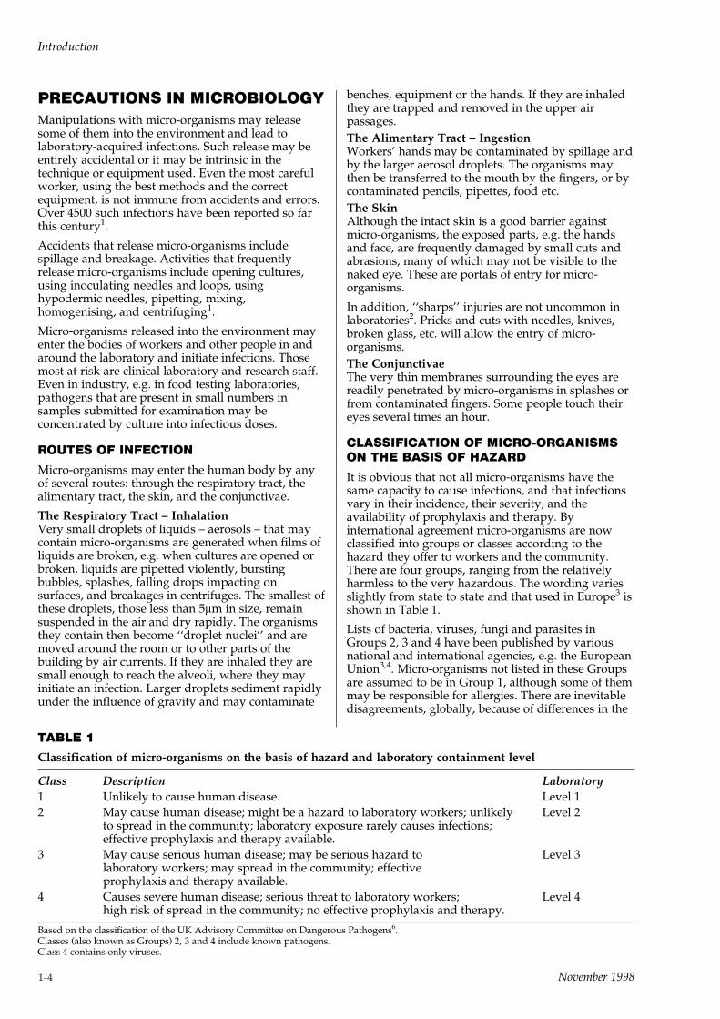
It is obvious that not all micro-organisms have thesame capacity to cause infections, and that infectionsvary in their incidence, their severity, and theavailability of prophylaxis and therapy. Byinternational agreement micro-organisms are nowclassified into groups or classes according to thehazard they offer to workers and the community.There are four groups, ranging from the relativelyharmless to the very hazardous. The wording variesslightly from state to state and that used in Europe3 isshown in Table 1.
Lists of bacteria, viruses, fungi and parasites inGroups 2, 3 and 4 have been published by variousnational and international agencies, e.g. the EuropeanUnion3,4. Micro-organisms not listed in these Groupsare assumed to be in Group 1, although some of themmay be responsible for allergies. There are inevitabledisagreements, globally, because of differences in the
TABLE 1
Classification of micro-organisms on the basis of hazard and laboratory containment level
Class Description Laboratory
1 Unlikely to cause human disease. Level 1
2 May cause human disease; might be a hazard to laboratory workers; unlikely Level 2to spread in the community; laboratory exposure rarely causes infections;effective prophylaxis and therapy available.
3 May cause serious human disease; may be serious hazard to Level 3laboratory workers; may spread in the community; effectiveprophylaxis and therapy available.
4 Causes severe human disease; serious threat to laboratory workers; Level 4high risk of spread in the community; no effective prophylaxis and therapy.
Based on the classification of the UK Advisory Committee on Dangerous Pathogens6.Classes (also known as Groups) 2, 3 and 4 include known pathogens.Class 4 contains only viruses.
Introduction
1-4 November 1998
geographical distribution, incidence, and localsignificance5.
CLASSIFICATION OF LABORATORIESACCORDING TO HAZARD GROUP
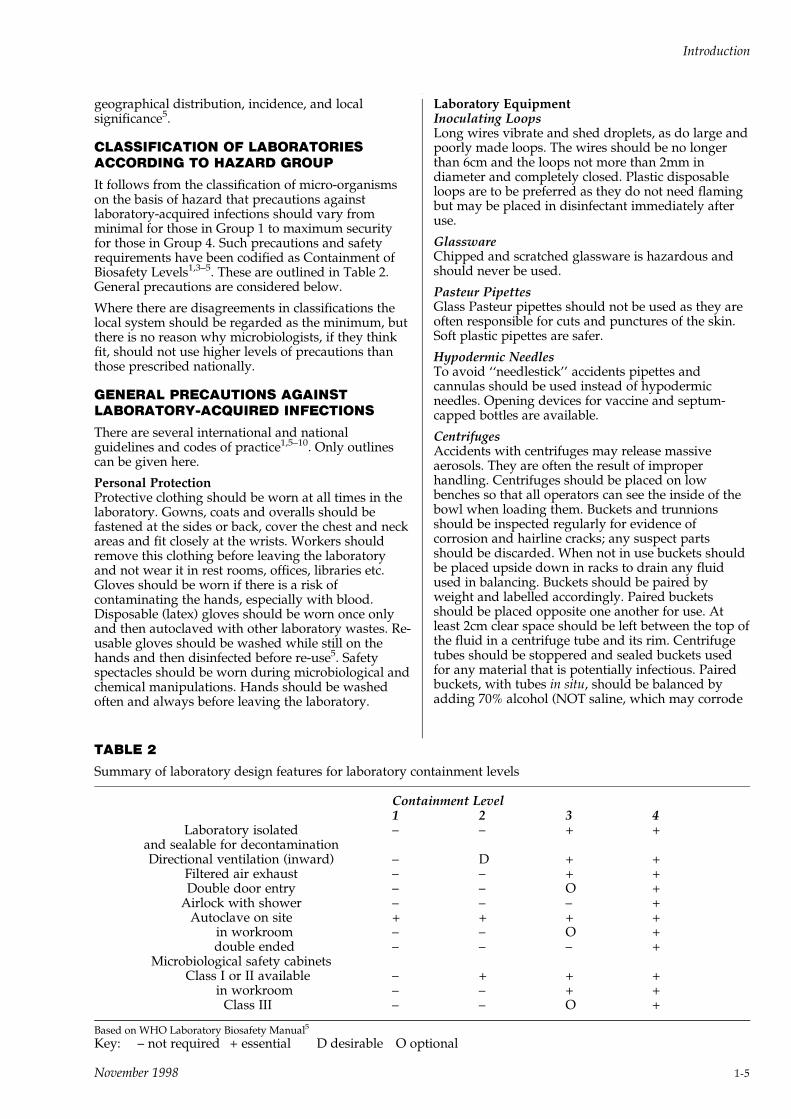
It follows from the classification of micro-organismson the basis of hazard that precautions againstlaboratory-acquired infections should vary fromminimal for those in Group 1 to maximum securityfor those in Group 4. Such precautions and safetyrequirements have been codified as Containment ofBiosafety Levels1,3±5. These are outlined in Table 2.General precautions are considered below.
Where there are disagreements in classifications thelocal system should be regarded as the minimum, butthere is no reason why microbiologists, if they thinkfit, should not use higher levels of precautions thanthose prescribed nationally.
GENERAL PRECAUTIONS AGAINSTLABORATORY-ACQUIRED INFECTIONS
There are several international and nationalguidelines and codes of practice1,5±10. Only outlinescan be given here.
Personal ProtectionProtective clothing should be worn at all times in thelaboratory. Gowns, coats and overalls should befastened at the sides or back, cover the chest and neckareas and fit closely at the wrists. Workers shouldremove this clothing before leaving the laboratoryand not wear it in rest rooms, offices, libraries etc.Gloves should be worn if there is a risk ofcontaminating the hands, especially with blood.Disposable (latex) gloves should be worn once onlyand then autoclaved with other laboratory wastes. Re-usable gloves should be washed while still on thehands and then disinfected before re-use5. Safetyspectacles should be worn during microbiological andchemical manipulations. Hands should be washedoften and always before leaving the laboratory.
TABLE 2
Summary of laboratory design features for laboratory containment levels
Containment Level1 2 3 4
Laboratory isolatedand sealable for decontamination
± ± + +
Directional ventilation (inward) ± D + +Filtered air exhaust ± ± + +Double door entry ± ± O +
Airlock with shower ± ± ± +Autoclave on site + + + +
in workroom ± ± O +double ended ± ± ± +
Microbiological safety cabinetsClass I or II available ± + + +
in workroom ± ± + +Class III ± ± O +
Based on WHO Laboratory Biosafety Manual5
Key: ± not required + essential D desirable O optional
Laboratory EquipmentInoculating LoopsLong wires vibrate and shed droplets, as do large andpoorly made loops. The wires should be no longerthan 6cm and the loops not more than 2mm indiameter and completely closed. Plastic disposableloops are to be preferred as they do not need flamingbut may be placed in disinfectant immediately afteruse.
GlasswareChipped and scratched glassware is hazardous andshould never be used.
Pasteur PipettesGlass Pasteur pipettes should not be used as they areoften responsible for cuts and punctures of the skin.Soft plastic pipettes are safer.
Hypodermic NeedlesTo avoid ``needlestick'' accidents pipettes andcannulas should be used instead of hypodermicneedles. Opening devices for vaccine and septum-capped bottles are available.
CentrifugesAccidents with centrifuges may release massiveaerosols. They are often the result of improperhandling. Centrifuges should be placed on lowbenches so that all operators can see the inside of thebowl when loading them. Buckets and trunnionsshould be inspected regularly for evidence ofcorrosion and hairline cracks; any suspect partsshould be discarded. When not in use buckets shouldbe placed upside down in racks to drain any fluidused in balancing. Buckets should be paired byweight and labelled accordingly. Paired bucketsshould be placed opposite one another for use. Atleast 2cm clear space should be left between the top ofthe fluid in a centrifuge tube and its rim. Centrifugetubes should be stoppered and sealed buckets usedfor any material that is potentially infectious. Pairedbuckets, with tubes in situ, should be balanced byadding 70% alcohol (NOT saline, which may corrode
Introduction
November 1998 1-5
metal, leading to mechanical failure) to the spacebetween the tube and the bucket. Instructions for useof centrifuges and action to be taken if a centrifugetube breaks, usually indicated by a sudden change insound and/or visible imbalance of the machine,should be posted adjacent to each machine.
Physical hazards associated with centrifuges arediscussed in detail by Kennedy11.
Water BathsThe water in water baths may become contaminatedfrom the outsides of culture tubes or the leakage oftheir contents. These baths, even those operated attemperatures >608C should be emptied when not inuse or a deposit may form in which micro-organismscan grow. A disinfectant that does not attack metalsmay be added to the water in baths that are incontinuous use (hypochlorites should not be used; seebelow).
Homogenisers and ShakersBench-mounted models may generate aerosols andshould be covered, (e.g. by clear plastic boxes) whenin use. These covers should be disinfected after use.Hand-held homogenisers should be held in a wad ofcotton wool in case they break. Homogenisers andcontainers from shakers should be opened inmicrobiological safety cabinets.
PipettingPipetting by mouth, even water, should be banned.Pipetting devices should be provided. Pipettes shouldnot be blown out vigorously, otherwise bubbles andaerosols may be formed.
Microbiological Safety CabinetsThese should conform to national standards andshould be tested regularly by independent engineersto ensure that their performance is in accordance withthe requirements of that standard. These cabinets aredesigned to protect the user from the inhalation ofinfectious aerosols and air-borne particles. They giveno protection against spillages of cultures or againstchemicals. Class II and Class III cabinets also protectthe test or product from external air-bornecontamination.
Microbiological safety cabinets should be used onlyby experienced personnel who have received properinstructions about their limitations. They must not beused as fume cupboards or for work with flammableor toxic substances.
They should be decontaminated at regular intervalsby qualified staff who follow manufacturers', or otherrecognised procedures1,5,10.
Laminar Outflow (clean air) CabinetsThese are NOT microbiological safety cabinets. Theyare designed to protect the work from external air-borne contamination and do not protect the worker,whose face and respiratory tract receive air that haspassed over the workpiece. (See Cell and Tissueculture, below).
Fume CupboardsFume cupboards are designed to protect workers andthe environment from toxic chemical fumes and
gases. They should not be used for micro-organismsor other living material.
SPILLAGE AND BREAKAGE
Spillage of cultures and chemicals and breakage ofvessels containing them must be reportedimmediately to the supervisor or local safety office. Ifthe spillage is considerable the room should bevacated pending decontamination by qualified staff(see below).
Instructions for dealing with small-scale spillages andbreakages should be posted in each laboratory, andshould include the following:
± wear heavy-duty gloves
± cover the spillage/breakage with absorbentmaterial, e.g. large paper towels
± pour disinfectant (see Table 3) over the papertowels and leave for at least 15 minutes
± scoop up the paper towels with a dust pan or stiffcardboard and place them along with the dust panor cardboard, along with any broken glass into alaboratory discard container
± pick up any residual broken glass with forceps andadd it to the discard container
± cover the area again with paper towels and pouron more disinfectant. Leave for 30 minutes beforeany further cleaning up
± autoclave the discard container.
PRECAUTIONS AGAINST BLOOD-BORNEINFECTIONS
In addition to the precautions listed above personnelwho handle blood specimens or blood-stainedmaterial should wear high quality disposable glovesand also plastic disposable aprons over their normalprotective clothing. Guidelines for the safe handlingin laboratories of materials that may contain hepatitisand/or the human immunodeficiency virus havebeen published1,7,8,12,13.
PRECAUTIONS WITH CELL AND TISSUECULTURE
Separate accommodations should be provided tominimise contamination of cultures.
Some cells and tissue cultures may containadventitious and unidentified micro-organisms orviruses from which the operator must be protected.All work with cells and cell lines should therefore beconducted in Class II microbiological safety cabinets.Laminar outflow cabinets (see above) must NOT beused.
STERILISATION, DISINFECTION ANDDECONTAMINATION
These terms are not interchangeable. In microbiology:
Sterilisation ± implies the complete destruction of allmicro-organisms.
Introduction
1-6 November 1998
Disinfection ± is the destruction or inactivation,usually by chemicals, of the vegetative forms ofmicro-organisms and the spores of some of them. Notall spores are inactivated. Not all spores areinactivated by chemical disinfectants.
Decontamination ± usually means makingequipment, materials and waste free from infectiousagents.
SterilisationHere, this is restricted to autoclaving. For othermethods, e.g. hot air, standard textbooks shouldbe consulted1,10.
The hazard most frequently encountered inautoclaving is failure to sterilise, i.e. to achieve andmaintain the temperature/time ratio that is known tokill micro-organisms. (The physical hazards ofautoclaving are described elsewhere11).
Autoclaves should be used only by personnelspecifically trained and employed for that purpose.Infected materials and ``clean'' articles should betreated in separate loads and preferably separateautoclaves. Autoclaves should not be tightly packed:space must be left between articles in the load toenable steam to circulate freely.
The ``Holding time at temperature'' (HTAT) for steamsterilisation is normally 20 minutes at 1218C. The timebegins when the temperature in the load has reached1218C as indicated by the recorder of thethermocouple in that load, NOT when the draintemperature reaches that temperature1,10.
Higher temperatures are required for the treatment ofmaterial containing ``unconventional agents'' (e.g.scrapie, CJD, etc.).
Control of SterilisationIn modern autoclaves this is achieved byinstrumentation (thermocouple probes and recorders).It is advisable, however, to include some form ofindicator, e.g. ``autoclave tape'' in each load, and tocheck the HTAT independently at regular intervals.Alternatively, or in addition, biological tests may beused in the form of strips that contain Bacillusstearothermophilus1,10.
TABLE 3
Properties of some disinfectants
Active against Inactivated by Toxicity
Fungi Bacteria Myco- Spores Viruses Protein Materials Hard Deter- Skin Eyes LungsG+ G± bacteria Lipid Non
lipidNatural Man-
madewater gent
Phenolics +++ +++ +++ ++ ± + v + ++ ++ + C + + ±Hypochlorites + +++ +++ ++ ++ + + +++ + + + C + + +Alcohols ± +++ +++ +++ ± + v + + + + ± ± + ±Formaldehyde +++ +++ +++ +++ +++a + + + + + + ± + + +Glutaraldehyde +++ +++ +++ +++ +++b + + NA + + + ± + + +Iodophors +++ +++ +++ +++ + + + +++ + + + A + + ±QAC + +++ ++ ± ± ± ± +++ +++ +++ +++ A(C) + + ±
+++ Good: ++ Fair: + Slight: ± Nil: V Depends on virus: a Above 408C: b Above 208C: C Catonic: A Anionic
From Collins, C.H. (1993) Laboratory Acquired Infections. 3rd.edn. by permission of the publishers Butterworth-Heinemann, Oxford
Chemical DisinfectionDisinfectants vary in the action against bacteria,spores, fungi and viruses and should be chosen inaccordance with the intended use. Most disinfectantsare toxic, in varying degrees, and precautions, e.g. eyeprotection, should be taken when stock solutions arediluted.
Table 3 summarises the properties of some commonlyused chemical disinfectants.
Disinfectants should be diluted according to themanufacturers' instructions. It is best to preparedilutions daily as some deteriorate if use-dilutions arestored. For most purposes hypochlorites are adequateand should be diluted to contain 1,000±2,500 ppmavailable chlorine for normal work and 10,000 ppmfor blood and high concentrations of protein.Industrial hypochlorite solutions usually contain100,000 ppm available chlorine and should be diluted1±2.5% or 10%.
Bench discard jars and containersA discard jar containing an appropriate disinfectantshould be provided at every work station to receivesmall items such as slides, Pasteur pipettes and plasticloops. Large jars, for pipettes are also needed. Plasticcontainers are safer than glass. Articles placed in thesecontainers should be completely submerged in thedisinfectant. Discard containers should be emptiedand replaced daily.
Containers for discarded cultures should also beprovided at each work station. These should not leak,be shallow ± not more than 25cm deep to facilitatesteam penetration during autoclaving, and preferablyof heat-resistant plastic. Plastic bags, usually blue ortransparent with blue lettering, are used in some(mostly UK) laboratories. They should be supportedin the containers described above.
Decontamination of Benches, Equipment andRoomsBenches should be wiped down with a suitabledisinfectant at the end of the working day (glovesshould be worn). The accessible parts of equipmentmay similarly be disinfected but not withhypochlorites as they may attack metals.
Introduction
November 1998 1-7

Equipment to be serviced must also bedecontaminated in this way and clearly labelled toindicate that this has been done and that it should notbe used until after servicing.
The working surfaces and inner walls ofmicrobiological safety cabinets should be swabbedwith a suitable disinfectant, and the cabinets befumigated with formaldehyde, as indicated abovebefore filters are changed or maintenance carried out.
Rooms rarely need disinfection unless a majoraccident has released massive aerosols. Formerly thiswas done by formaldehyde fumigation, but this isnow regarded as hazardous and uncertain. Sprayingor washing with disinfectant/detergent mixtures issafer and more effective.
DISPOSAL OF INFECTED WASTE
Infected laboratory waste is included in thedefinitions of clinical waste and must ultimately beincinerated. Table 4 lists the materials that should beregarded as infectious in microbiological and clinicallaboratories. As these are likely to be the most heavilyinfected of all such waste and may have to travelalong the public highway, often for long distances. Itis prudent to autoclave it first1,14,15.
CONCLUSIONSEvery microbiological laboratory should have writtensafety policy and instructions that describe in full thesafety precautions deemed necessary by the Directorand Safety Officer.
All members of the staff should be aware of theauthorised procedures for containing and destroyingmicro-organisms.
A schedule of regular microbiological safety cleaningshould be maintained for all working surfaces andadjacent areas.
References1 Collins, C.H. (1993) Laboratory Acquired Infections. 3rd edn.
Oxford: Butterworth-Heinemann.
2 Collins, C.H. and Kennedy, D.A. (1987) J. Appl. Bact. 62, 385±
402.
3 European Commission (1990/93) Council Directive on the
Protection of Workers from Risks Relating to Biological Agents at
Work. 90/679/EEC as modified 93/88/EEC.
4 Control of Substances Hazardous to Health Regulations (1994).
Biological Agents: Approved Code of Practice. London: Health and
Safety Executive.
5 World Health Organization (1993) Laboratory Biosafety Manual.
2nd edn. Geneva: WHO.
6 Advisory Committee on Dangerous Pathogens (1990)
Categorization of Pathogens on the Basis of Hazard and Categories of
Containment. London: HMSO.
7 National Research Council (1989) Biosafety in the Laboratory.
Washington DC: National Academy Press.
8 Health Services Advisory Committee (1991) Safe working and the
Prevention of Infection in Clinical Laboratories. London: HMSO.
9 Centers for Disease Control (1993) Biosafety in Microbiological and
Biomedical Laboratories. 3rd edn. HHS Publication No (CDC) 93±
8395. Washington: US Government Printing Office.
10 Collins, C.H., Lyne, P.M. and Grange, J.M. (1995) Collins and
Lyne's Microbiological Methods. 7th edn. Oxford: Butterworth-
Heinemann.
11 Kennedy, D.A. (1991) In Safety in Clinical and Biomedical
Laboratories ed. Collins C.H. London: Chapman and Hall.
12 Advisory Committee on Dangerous Pathogens. HIV ± the
Causative Agent of AIDS and Related Conditions. London: HMSO.
13 World Health Organization (1991) Biosafety Guidelines for
Diagnostic and Research Laboratories Working with HIV. Geneva:
WHO.
14 Collins, C.H. and Kennedy, D.A. (1993) The Treatment and
Disposal of Clinical Waste. Leeds: H & H Scientific.
15 Collins, C.H. (1994) Lett. Appl. Microbiol. 19, 61±62.
TABLE 4
Infected and potentially infected waste frommicrobiological laboratories
Disposables other than sharps± Specimens or their remains (in their containers)
submitted for tests containing blood, faeces,sputum, urine, secretions, exudates, transudates,other normal or morbid fluids but not tissues.
± All cultures made from these specimens, directlyor indirectly.
± All other stocks of micro-organisms that are nolonger required.
± Used diagnostic kits (which may contain glass,plastics, chemicals and biologicals).
± Used disposable transfer loops, rods, plasticPasteur pipettes.
± Disposable cuvettes and containers used inchemical analyses.
± Biologicals, standards and quality controlmaterials.
± Food samples submitted for examination inoutbreaks of food poisoning.
± Paper towels and tissues used to wipe benchesand equipment and to dry hands.
± Disposable gloves and gowns.
Sharps± Hypodermic needles (syringes attached if custom
so requires).± Disposable knives, scalpels, blades, scissors,
forceps, probes.± Glass Pasteur pipettes; slides and cover glasses.± Broken glass, ampoules and vials.
Tissues and animal carcasses
Bedding from animal cages
Adapted from Collins and Kennedy14 by permissionof the authors and publisher.
Introduction
1-8 November 1998

2CULTURE MEDIA

November 1998
CULTURE MEDIA QUALITYASSURANCEAll manufacturing operations are conductedaccording to protocols which describe suchprocedures as the monitoring, maintenance, cleaningand calibration of equipment; plant sanitation;warehouse control of in-coming materials andmaterials under test; labelling control and handling,storage and distribution of finished goods. The masterformula and accompanying documents for each lot/batch of product includes manufacturing control andpackaging information pertaining to the product.
Quality tests on raw materials include identity tests,tests for performance and compatibility with otheringredients in a pre-production laboratory mix of themedium components. Additional tests are performedwhere required. For example, peptones are examinedphysically, chemically and microbiologically. Agarsare tested for clarity, gel strength, diffusioncharacteristics etc.
Dehydrated media mixtures are examined forappearance, homogeneity and moisture content.Representative samples are reconstituted andexamined for colour, clarity, pH, gel strength (if agaris present), compatibility with post-sterilisationadditives and for microbiological performance. Themedium is challenged with small inocula of well-defined cultures to measure recovery of growth,colony size and morphology, colour reactions,differentiation and selectivity.
Special procedures such as antimicrobialsusceptibility tests are performed where appropriatefor the recommended use of the medium.
All tests are performed in parallel with a previouslyapproved reference batch of the medium. This use ofa control standard medium with each test ensuresuniformity in reading the results.
Samples of each manufactured lot/batch are retainedfor the total shelf-life of the product. Stability testsand lot-to-lot uniformity tests are carried out usingthese retained samples.
FORMULATION OF CULTUREMEDIA: DEVELOPMENT ANDMANUFACTUREThe formulation of all Oxoid culture media arepublished in Section 2.7 and the components can bedivided into different roles or functions:
1 Nutrients: proteins/peptides/amino-acids.
2 Energy: carbohydrates.
3 Essential metals and minerals: calcium,magnesium, iron, trace metals: phosphates,sulphates etc.
4 Buffering agents: phosphates, acetates etc.
5 Indicators for pH change: phenol red, bromo-cresol purple etc.
6 Selective agents: chemicals, antimicrobial agents.
7 Gelling agent: usually agar.
There is often an overlap of functions of some mediacomponents, thus protein hydrolysates will supplyamino-nitrogen, energy, some metals/minerals andact as buffering agents. Phosphate buffers areimportant suppliers of minerals and agar contributesmetals.
1 NutrientsNaegeli is credited with the earliest publications(1880/82) describing the requirements of micro-organisms for a protein component which he called`peptone'.
Later work showed that the group of bacteria, nowdefined as chemo-organotrophs, required amino-nitrogen compounds as essential growth factors intheir culture media.
Meat infusions contain water-soluble fractions ofprotein (amino-acids and small peptides) along withother water-soluble products such as vitamins, tracemetals, minerals and carbohydrates (glycogen). Suchinfusions or extracts may have been regarded as`peptones' but their amino-nitrogen content wasusually too low to sustain the growth of largenumbers of bacteria.
It was not until deliberate attempts were made tohydrolyse proteins with acids or enzymes thatsufficiently high concentrations of water-solubleprotein fractions (peptides) were made available forbacterial growth. Many nutrient media usuallycontain a mixture of protein hydrolysate (peptone)and meat infusion (meat extract/Lab-Lemco).
The difficulties associated with the production ofprotein hydrolysates were soon recognised andcommercial suppliers of peptones became establishedby the 1920s. The commercial supply of dried peptoneeventually led to complete culture media beingproduced in the form of dehydrated media.
Although meat was the first and most obviousprotein to hydrolyse, other proteins were tried laterand some showed specific advantages which ensuredtheir retention in culture media to this day. Caseinhydrolysate with its pale colour and high tryptophan
Culture Media
November 1998 2-1
content and soya peptone with its high energycarbohydrate content are popular examples of non-meat peptones.
A detailed description of these products is given inSection 3.1 `Peptones-Hydrolysates'.
The nutrient components of culture media arecarefully selected to recover the required spectrum oforganisms in the sample e.g. coliforms or anaerobes.General purpose media such as blood agar in itsvarious forms will often contain mixtures of peptonesto ensure that peptides of sufficient variety areavailable for the great majority of organisms likely tobe present. However, more demanding organismswill require supplemental growth factors to be addedand examples of such requirements can be seen inmedia for Legionella species.
Most of the components used for the nutrition ofmicro-organisms are undefined and require extensivetesting with careful selection to ensure a reasonabledegree of uniformity. Would it not be better to usewholly defined peptides and amino-acids to producea totally defined medium? Whilst such media wouldimprove uniformity, experience has shown that theylack good performance as general purpose media.They would also be very expensive compared withundefined media. The use of totally defined culturemedia is an understandable goal of mostmicrobiologists but defined media have yet to provethemselves equal in performance to currently usedcomplex mixtures of meat and plant proteinhydrolysates.
2 EnergyThe most common substance added to culture mediaas a source of energy to increase the rate of growth oforganisms is glucose. Other carbohydrates may beused as required.
Carbohydrates added to media at 5±10 grammes perlitre are usually present as biochemical substrates todetect the production of specific enzymes in theidentification of organisms. It is usual to add pHindicators to such formulations.
3 Essential Metals and MineralsThe inorganic essential components of culture mediaare many and can be divided on a semi-quantitativebasis:
Typical macro-components (gm/litre):Na, K, Cl , P, S, Ca, Mg, Fe.
Typical micro-components (mgm-microgm/litre):Zn, Mn, Br, B, Cu, Co, Mo, V, Sr, etc.
As previously mentioned, a formulation may nothave specific metals and minerals listed in itsformulation. In such cases it is assumed that all thefactors required are present in the hydrolysates,buffers and agar components.
4 Buffering AgentsIt is important that the pH of a culture medium ispoised around the optimum necessary for growth ofthe desired micro-organisms. The use of buffercompounds at specific pK values is especiallynecessary when fermentable carbohydrates are addedas energy sources.
Phosphates, acetates, citrates, zwitterion compoundsand specific amino-acids are examples of bufferingagents that may be added to culture media.
A side effect of such compounds is their ability tochelate (or bind) divalent cations (Ca++ and Mg++).Polyphosphate salts, sometimes present in sodiumphosphate, are compounds which can bind essentialcations so firmly that they are made inaccessible tothe micro-organisms.
The effect of these binding or chelating agents will beseen in diminished growth or failure to grow at all,unless care has been taken to supplement the essentialcations in the formulation. Opacity forming in amedium, after heating or on standing at 508C forseveral hours, is commonly caused by phosphateinteraction with metals. Such phosphate precipitatescan very effectively bind Fe and lower the availableamount of this essential metal in the medium.
5 Indicator SubstancesThe addition of coloured indicator substances is avery effective way of detecting fermentation ofspecific carbohydrates in a culture medium. Suchcompounds should change colour distinctly andrapidly at critical pH values.
Most of the compounds used e.g. phenol red, bromo-cresol purple, fuchsin, etc., are toxic and it is essentialto use low concentrations of pre-screened batches/lots. Known sensitive strains of micro-organisms areused in the screening tests.
6 Selective AgentsChemicals or antimicrobials are added to culturemedia to make them selective for certain micro-organisms. The selective agents are chosen and addedat specific concentrations to suppress the growth ofunwanted organisms in a polymicrobial sample. It is,of course, essential to have established that theselective agents, at the appropriate concentration, willallow uninhibited growth of the desired organisms.
Common chemical selective agents are: bile salts, dye-stuffs, selenite, tetrathionate, tellurite and azide.Antimicrobial agents are commonly used in mixtureswhen suppressing polymicrobial contaminating flora.Antimicrobials are more specific in their selectiveaction than the chemical agents shown above.However, the critical weighing and heat-lability ofmost antimicrobials demand special care and post-sterilisation addition.
The wide variety of organisms and their almostinfinite ability to adapt to changing conditions makesa truly selective medium unlikely. Selective media canbe said to suppress most of the unwanted organismsand allow most of the desired organisms to grow. Thefinal formulation is usually a compromise whichachieves the best of these criteria.
7 Gelling AgentsAlthough gelatin is still used for a few specific mediaand carrageenans, alginates, silica gel andpolyacrylamides are sometimes used as gellingagents, the outstanding gel-forming substance used inculture media is agar.
Culture Media
2-2 November 1998

Hesse, a worker in Robert Koch's laboratory, iscredited with its first use in culture media, althoughFrau Hesse gave him the idea from its use in table-jellies in hot climates.
Its inertness to microbial action, the unique settingand melting temperatures (388C and 848Crespectively) the high gel strength which allows lowconcentrations of agar to be used, its clarity and lowtoxicity have contributed to its wide popularity withmicrobiologists. Its ability to retain its gel structure at608C makes agar of special value to culture mediawhich have to be incubated at this temperature toisolate thermophilic organisms.
Agar is obtained from agarophyte sea-weeds mainlyGelidium, Gracilaria and Pterocladia species. It isextracted as an aqueous solution at greater than1008C, decolourised, filtered, dried and milled to apowder.
Agar is not an inert gelling agent; it contributesnutrients and/or toxic agents to culture media,depending on the chemical processing carried out bythe suppliers.
Microbiological agar is specially processed to yield alow toxicity, high clarity, low mineral and highdiffusion gel.
Other ComponentsThere are many other substances added to culturemedia for specific purposes e.g. growth factors forfastidious organisms, eH-reducing compounds foranaerobic organisms (thioglycollate and cysteine),whole blood to detect haemolytic enzymes andencourage the growth of organisms which arevulnerable to oxidation products.
Development and Manufacture of Culture MediaThe development of dehydrated culture media is aprocess leading to the large-scale manufacture of areproducible, stable product. The initial developmentof the formulation is usually carried out bymicrobiologists who wish to create a novel mediumwith specific characteristics or who wish to improvethe performance of an existing product. Such work isusually written up in microbiological journals, havingfirst been judged by some form of peer review andproved to be of special value by other workers in thefield.
Simple conversion of the published formula into amixture of dehydrated components is seldomachieved. Usually the peptone/hydrolysate base hasto be adapted and variations in concentration of othercomponents may be required. Laboratory mixes of themedium are prepared as R&D trials and after testingin the laboratory are sent to the originator forcomment. Opportunity may also be taken to get theviews of other experts in this field. Special strains oforganisms may be required to check the finer pointsof performance.
Subject to good report, a trial batch will bemanufactured and this will be used for larger trialsand wider-scale testing. During these trials QC testingand performance criteria will be established and thespecifications of the components will be determined.Bought-in components will have buying specificationsand in-house components will have manufacturingspecifications and standard-operating-processesproduced. Stability trials will begin if there isconfidence that the final formulation has beenachieved.
The reports on the larger and wider-spread trials arestudied and if the results are satisfactory preparationwill be made to manufacture a full production batch/lot. All the components of the medium, includingspecial protein hydrolysates which may have to bespecially manufactured, are assembled and alaboratory mix tested to see that it meets theperformance specification. Finally the components aremilled, mixed and blended to produce a finelydivided, homogeneous powder which is held in largecontainers for further testing before release.
All this work, plus literature, labels and productinserts is carried out under the aegis of R&D/Marketing. Subsequent production lots aremanufactured under surveillance which includesGMP monitoring and end-product testing by theQuality Department.
No product can be released without clearance fromThe Quality Department.
Culture Media
November 1998 2-3

GENERAL GUIDE TO THE USE OF OXOID CULTURE MEDIA
PREPARATION OF DEHYDRATED MEDIA
Dehydrated media are hygroscopic and are sensitiveto moisture, heat and light. They are adverselyaffected by drastic changes in temperature e.g. hot/cold cycling temperatures which may occur betweenday and night laboratory temperatures in winter.
Storage conditions are usually indicated on theproduct label and should be followed.
1 Write on the label the date of receipt in thelaboratory.
2 Store as directed on the label; usually below 258Cin a dry area, away from direct sunlight,autoclaves, drying ovens or other heat sources.Where indicated store at 2±88C.
3 Check expiry date on the label, some media havesignificantly shorter shelf-lives than others.
4 Use stock in lot/batch number order. Do not opena new bottle until the previous bottle has beenemptied. Note on the label the date the container isfirst opened. After use, make sure the container istightly closed and return it to the designatedstorage area.
5 Order the medium in an appropriate size ofcontainer and in a quantity which accords tonormal use requirements. A medium in a largecontainer which has been opened many times willdeteriorate on storage. Discard the medium if thepowder is not free flowing, if the colour haschanged or if it appears abnormal in any way.
RECONSTITUTION OF DEHYDRATED MEDIA
Complete instructions for the preparation of culturemedia are given on the label of each bottle. As ageneral rule it is wise to prepare one week'srequirement only.
1 Use water prepared by distillation, deionisation orreverse osmosis. Toxic metal ions such as coppermust be absent. Check the pH of the water, ifbelow 5.5, heat to drive off CO2 and re-check. Theconductivity of the water should ideally be below15 micro siemens (mS). Rinse glassware before use.
2 Prepare the medium in a vessel about twice thefinal volume of the medium to allow adequatemixing. Follow the instructions given on the labelof each product.
3 Open the culture medium container away fromdraughts and moisture. Avoid inhaling the powderand prolonged skin contact. Weigh the powderquickly, accurately and without creating `clouds ofdust'. Reclose the container as soon as possible.
4 Pour half the required volume of water in thevessel, then the weighed quantity of medium andagitate briskly for a few minutes. Pour the rest ofthe water down the sides of the vessel to wash anyadherent medium back into solution. This is animportant step because dry culture media powder
above the level of the water may not be sterilised inthe autoclave and may be a source ofcontamination.
Agar-free media will usually dissolve with gentleagitation.
Media containing agar should be heated to dissolvethe agar before autoclaving. Bring the medium to theboil without scorching or burning. Those mediawhich should not be autoclaved will be ready to pourinto dishes or other containers after this amount ofheating. Most culture media will require finalsterilisation in an autoclave at 1218C for 15 minutes.
The pH of the dehydrated medium has been adjustedso that the final pH of the prepared mediumconforms with the label specification when themedium has been cooled to 258C. Do not adjust thepH before sterilisation.
STERILISATION OF CULTURE MEDIA
Although sterilisation of culture media is best carriedout in a steam autoclave at temperatures around1218C it has to be recognised that damage is caused tothe medium by the heating process.
Heat-treatment of complex culture media whichcontain peptides, sugars, minerals and metals resultsin nutrient destruction, either by direct thermaldegradation or by reaction between the mediumcomponents. Toxic products caused by chemo-oxidation can also be formed during heat-treatment.
It is important, therefore, to optimise the heatingprocess so that a medium is sterile after heating butminimal damage is caused to the ingredients of themedium. As a general rule it is accepted that short-duration, high-temperature processes are more lethalto organisms and less chemically damaging than arelonger, lower temperature processes.
A general instruction for sterilising culture media involumes up to one litre at 1218C for 15 minutes isgiven on each label. Autoclaves vary in performance,however, and thermocouple tests using differentvolumes of media should be carried out to determinethe `heat-up' and `cool-down' times. It will beessential to do this when volumes of media greaterthan two litres are prepared. In order to avoidoverheating large volume units of media, the `heat-up' and `cool-down' periods are normally integratedinto the 1218C holding time.
Sterilisation CycleThe sterilisation cycle can be divided into its fourstages:
1 Chamber heat-up time2 Heat penetration time of the medium container3 Holding time at the prescribed temperature4 Cool-down time for the chamber to reach 808C.
Culture Media
2-4 November 1998
Sterilisation Cycle
Stage 1 208±1218C
Stage 2 <1008±1218C
Stage 3 1218±1218C
Stage 4 1218±808C
Stage 1 The chamber heat-up time depends on theefficiency of the autoclave (air discharge/steam input)and the size of the load in the chamber. The timerequired for this stage is measured with a recordingprobe located in the air-discharge valve located in thebase of the chamber.
Stage 2 The heat penetration time depends mainly onthe volume of the individual containers, although theshape and the heat-transfer properties of thecontainers may affect this stage. The time required forthe medium volume to reach 1218C is measured withthermocouples placed in the centre of the innermostcontainer.
Volume (ml) in glass bottles Time (mins)100 19500 18
1000 222000 275000 37
These times assume that agar media have beendissolved before autoclaving. It is also assumed thatmaximum exposure to steam is possible. Thusalthough the single 100ml bottle required 12 minutesto reach 1218C when placed in a crate with otherbottles it required 19 minutes and when placed in thecentre of stacked crates it required 30 minutes.
Stage 3 The holding time at 1218C depends on (i) thenumber of organisms originally present in themedium (ii) the fractional number of an organismpresumed present after heating e.g. N = 0.001equivalent to one bottle in every 1000 bottles heatedbecoming contaminated (iii) the thermal death rateconstant of the presumed organism present at 1218C.
Stage 4 The cool-down time depends on the size ofthe load in the chamber and the heat loss rate fromthe autoclave. Water-sprays are used to acceleratecooling in commercial sterilisers but very carefulcontrol is required to avoid bottle fracture and theingress of the cooling spray into the sterilisedmedium. The latter problem occurs when the vacuumformed in the head-space during cooling suckscontaminated cooling fluid up the thread of the capand into the bottle.
Culture media autoclaves should be unlagged and ofmoderate chamber capacity only. Thermal locks onthe doors should prevent them opening when thechamber temperature is above 808C but even in thesecircumstances care should be taken to avoid suddenthermal shock when removing glass bottles of hotliquid from the autoclave. When screw-cappedcontainers are placed in an autoclave the caps shouldbe a half-turn free to allow the escape of heated air.When removed from the autoclave the caps arescrewed down tightly after the contents have cooledto ambient temperature.
Sterilisation ChecksAll autoclaves should be calibrated and checked atfixed periods of time to ensure that they arefunctioning efficiently. Physical measurements shouldbe made on temperature and pressure readings, thequality of the steam should be checked, the efficiencyof the `near-to-steam' air traps in the base of theautoclave should be determined and the safety valveschecked. Mandatory inspections of autoclaves aspressure vessels are normally carried out annually byspecialists under instructions from insurers of suchapparatus.
Biological indicators of sterilisation will demonstratethe ability of the autoclave to destroy bacterial spores.Such tests may be compulsory in certain countries.
Chemical indicators will show the temperaturereached or exceeded and some will indicate the timeheld at the specified temperature. Under-autoclavingis usually self-evident because failure to destroy allthe bacterial spores naturally present in dehydratedmedia (the `bioburden') will allow growth to takeplace in the stored or incubated medium. Failure ofsterilisation should always be suspected whencontamination of prepared media occurs with sporingorganisms.
Overheating EffectsOverheating is a common cause of pH drift,darkening, precipitation, poor gel strength andreduced bacteriological performance. These effectscan also be produced if a concentrated `pool' ofingredients at the bottom of the container is heated.All culture media should be in solution beforesterilisation. This will reduce the occurrence ofMaillard-type reactions (non-enzymatic browning)taking place in the medium.
Overheating effects will occur if agar media areallowed to gel in bottles and later steamed to melt theagar. They will also occur if molten media are held at508C for more than 3 hours before use. Agar media,with pH values at or below 5.0, are very sensitive tooverheating in any form because the agar ishydrolysed and the gel strength fails.
Most of the difficulties in culture media sterilisationoccur when large unit volumes of media (>2 litres)must be processed. The best solution to this problemis the use of a culture medium preparator. Thesesemi-automatic processors, made by New Brunswickand other manufacturers overcome the problem ofpoor heat penetration of agar by a continuous stirringor agitation of the medium during the heating phase.Such preparators will significantly reduce the timerequired for sterilisation at 1218C or in some modelsat 1348C. They are strongly recommended because oftheir high efficiency and minimal damage to culturemedia.
Culture Media
November 1998 2-5

Table of Faults and Possible Causes in MediaSterilisation
FaultWrong pH value.
Possible CausespH test carried out above 258C.
Overheating through prolonged sterilisation,remelting or overlong period at 508C.
Incomplete solution of medium.
Poor quality water or containers.
Dehydrated medium stored incorrectly or beyondthe stated shelf-life.
FaultTurbidity, precipitation.
Possible CausesPoor quality water or containers.
Overheating or prolonged storage at 508C.
pH value incorrect.
Incomplete solution.
FaultDarkening.
Possible CausesOverheating, incomplete solution or pH drift.
FaultSoft gel.
Possible CausesAgar not in solution, poor mixing, prolongedstorage at 508C.
Overheating at low pH values.
Error in weighing or overdilution with inoculum ormedia supplements.
FaultPoor bacterial growth.
Possible CausesProlonged and excessive heating, incompletesolution.
Inhibitory substances in water or containers.
Darkening and pH drift.
PREPARATION OF STERILISED MEDIA
Liquid media which are sterilised in their finalcontainers should be cooled down to roomtemperature as rapidly as possible. Screw caps shouldthen be tightened.
Containers of agar media which have been sterilisedshould be placed in a 508C water bath and themedium dispensed as soon as it reaches thistemperature, or within a maximum of 3 hours in thebath. The medium should be mixed thoroughly,without bubble formation and aseptically dispensedinto sterile containers. Do not expose dishes of agarmedia to sunlight; it causes excessive condensation onthe lids and may cause the formation of inhibitorysubstances by photo-oxidation.
Heat-labile supplements should be added to themedium after it has cooled to 508C. Allow the sterilesupplement to come to room temperature beforeadding it to the agar medium. Cold liquids may causeagar to gel or form transparent flakes which caneasily be seen in blood-enriched agar. Mix allsupplements into the medium gently and thoroughly,then distribute into the final containers as quickly aspossible.
Blood used for the preparation of blood agar shouldbe as fresh as possible and should have been stored at2±88C (blood must not be frozen). Warm the blood inan incubator to about 35±378C before addition tosterile molten agar base, which has been cooled to 40±458C. Adequate mixing in a large head-space vessel isessential to ensure aeration of the blood. Poorlyoxygenated blood plates are purplish in colourwhereas properly aerated blood agar is cherry-red.Defibrinated blood is recommended for use ratherthan blood containing an anticoagulant.
STORAGE OF PREPARED MEDIA
The recommended shelf-life of prepared culturemedia varies considerably. Screw-capped bottles ofnutrient broth and agar can be stored for 6 months atlow ambient temperatures (12±168C). It is importantto store all media away from light.
Agar plates should be stored at 2±88C in sealedcontainers to avoid loss of moisture.
DO NOT FREEZE.
Fresh media are better than stored media thereforeavoid long storage times. Some very labile beta-lactam selective agents have very short active livesand media containing such substances should be usedwithin a few days of preparation.
It is good laboratory practice to establish shelf-livesfor all prepared media and date-stamp the containersor holders accordingly.
Loss of moisture from agar plates is a common causeof poor bacteriological performance. Do not pre-incubate all plates overnight as a sterility check. Onlyobviously wet plates require pre-inoculation drying.Ensure that all plates are incubated in a humidenvironment.
Examine prepared media before inoculation. Look forevidence of contamination, uneven filling or bubbleson surface of agar, colour changes, haemolysis andsigns of dehydration such as shrinking, cracking andloss of volume. Discard any defective plates or tubes.
PRECAUTIONS IN THE USE AND DISPOSALOF PREPARED MEDIA
It should be recognised that inoculation of culturemedia with bacteria, deliberately or accidentally,leads to very great numbers of organisms beingproduced. High concentrations of any organisms arepotentially hazardous and must be disposed of safelyby approved methods.
All infected specimens and inoculated culture mediashould be handled only by qualified personnel whohave been trained in microbiological procedures. Such
Culture Media
2-6 November 1998
staff should ensure that all specimens and culturesunder their care are properly handled and finallyautoclaved before disposal. Any apparatus used andcontaminated must be safely disinfected or sterilised;this is particularly important when such apparatusmust be serviced or passed out of the laboratory.
The environment in which microbiological culturesare handled must also be taken into account. Mostcountries have categories of organisms which aredivided into those which may be handled in thegeneral microbiological laboratory, those whichrequire special laboratory conditions and for the mostdangerous organisms a totally contained and highlyprotected environment. It may be a criminal offencenot to observe these rules and regulations. Whenusing culture media always label or identify thecontainer with the specimen details beforeinoculation.
Inoculate the medium using aseptic techniques andincubate under the appropriate conditions.
Examine the medium after incubation for evidence ofmicrobial growth and carry out the appropriateisolation and identification procedures.
PRECAUTIONS ± DEHYDRATED MEDIA
Most of the products supplied by OXOID Limitedhave no known risks except those usually associatedwith fine powders. However, to prevent the risk ofinhaling fine dust it is recommended that approvedmasks should be worn whilst handling dehydratedmedia.
Material Safety Data Sheets are available forindividual products.
Dehydrated culture media supplied as powders,granules or tablets should not be eaten. Powdersshould not be inhaled because irritation of the upperrespiratory tract may occur. To avoid mild skin rashesprevent prolonged contact with the powder andensure excessive dust is not produced.
Powdered products, if spilt, can be collected anddisposed of in the normal way. Any residue shouldbe washed away with ample cold water.
HAZARDOUS PRODUCTS
There are a number of substances which contain toxicsubstances. These must be used in accordance withthe product specific Material Safety Data Sheet. Allrelevant Risk and Safety Phrases are on the productlabel.
Media containing Sodium AzideThese products contain less than 1% sodium azideand have low toxicity. Some persons, however, haveenhanced sensitivity to azide and therefore couldreact to accidental exposure to the product.Precautions must be taken to prevent ingestion orinhalation of the dust. Sodium azide reacts with manymetals, especially copper, to produce explosive metalazides.
If Local or National legislation permits and disposalto sink is employed, use sufficient water to preventthe powder remaining in contact with pipework and
drains. The same precaution applies to any biologicalsolution which contains sodium azide as apreservative.
Products containing BarbitoneThese products are labelled POISON/TOXIC.
They are subject to the Misuse of Drugs Act 1973. Thesupply is controlled and in the United Kingdom suchproducts are available to bona fide laboratories only(orders must be signed by the head of department) orto establishments authorised to possess suchproducts. Certain establishments are exempt underthe regulations e.g. NHS hospital laboratories andother government departments but they will be askedto confirm this status.
Exports to some countries may require an importlicence for the country of destination.
CycloheximideThis compound, prepared in Supplement vials,reaches a concentration which is considered to betoxic and is labelled accordingly. However, whendiluted out into the culture medium its concentrationfalls below the minimum level considered to behazardous. It is important when reconstituting vialscontaining toxic levels of cycloheximide to ensure thatthe vial solution does not touch the skin and toprevent the creation of aerosols which would allowthe compound to be inhaled. Wear personalprotective equipment in accordance with theinformation in the Material Safety Data Sheet. Thefollowing First Aid procedures should be taken incases of accident with any of the products describedabove:
FIRST AID PROCEDURES
Skin ContactRemove all contaminated clothing; wash the affectedareas thoroughly with soap and water. If anysymptoms occur obtain medical advice.
Eye ContactIrrigate thoroughly with water. Obtain medicaladvice.
IngestionWash out mouth thoroughly with water. Give onepint of water to drink immediately. Obtain medicaladvice.
InhalationRemove person from area of exposure. Rest and keepwarm. Obtain medical advice.
SpillageLarge quantities Wear protective overalls, gloves, eyeprotection and face mask. Collect the material into asuitable container and seal. Dispose according to localregulations. Wash away the residue with plenty ofwater.
Small quantities Wash away with large volumes ofrunning water, using protective gloves.
Culture Media
November 1998 2-7
USER-LABORATORY QUALITY CONTROL TESTS ON PREPARED MEDIA
Quality control tests should be carried out by the end-user laboratory to ensure that the performancecharacteristics of the medium are within specificationand that the methodology of medium preparation issatisfactory.
Each lot/batch of prepared medium should besubjected to a minimal testing programme which willensure that it is acceptable and will demonstrate atypical bacterial performance.
1 pH value: check that the pH of the preparedmedium, when tested in final form at ambienttemperature (258C) lies within the range given onthe product label. The medium should bediscarded if the pH value lies outside the specifiedrange.
2 Sterility: a representative sample of each lot/batchof medium should be incubated for 2±5 days at 35±378C. As a general rule, for a lot of 100 or less unitsa 3±5% sample should be tested. For a larger lot, 10random plates or tubes are taken. There should beno evidence of microbial growth after incubation.Discard all sterility samples when the tests havebeen completed.
3 Growth performance: test the growth supportproperties of the product by inoculating themedium with appropriate stock cultures and/orfresh isolates. Use a standard inoculationprocedure and examine the quantitative andqualitative results obtained. If testing new lots/batches of media, inoculate old and new lots in onetest and compare the performance of the two lotsside by side.
4 Stability: periodically perform the aboveprocedures on stored prepared media in order todetermine whether the storage conditions will giveoptimal results.
NOTE: If a medium does not perform to expectationsand all the manufacturer's recommendations havebeen followed, then the following steps should betaken:
1 Record the nature of the problem and the methodof preparation of the medium.
2 Note the lot/batch number and the date it wasreceived.
3 Call the Technical Support department of thecompany.
Culture Media
2-8 November 1998
SPECIAL FIELDS OF CULTURE MEDIA APPLICATION
EXAMINATION OF CLINICAL ANDVETERINARY SAMPLESIn both clinical and veterinary microbiology thepurpose of examining samples of tissue, fluids orexcreta is to isolate and identify pathogenicorganisms.
Although both fields of investigation have commoninterests and common organisms, they are separatespecialist activities. Reference should be made to theappropriate specialist publications in either field toobtain specific guidance.
It should be stressed that every specimen must beevaluated, many laboratories cannot cover the wholemicrobiological field, the various infective agentsshould be taken into consideration and, if necessary,material referred to the appropriate referencelaboratory.
Poor specimen samples can only yield poor ormisleading results. It is important that personnelcollecting or taking samples are instructed by thelaboratory to prevent faulty collection procedures.
Satisfactory samples, collected without extraneouscontamination and before antimicrobial therapyshould be transferred to the laboratory with minimaldelay. If transportation is required then appropriatetransport media should be used to protect delicateorganisms. Where quantitative results are importante.g. urine cytology and bacteriology, or wherecommensal overgrowth should be prevented,refrigeration of samples at 2±88C is essential.
All samples should be clearly labelled and sent inleak-proof, satisfactory containers. Sealed, transparentplastic bags, containing the sample container and therequest form attached to but not inside the plasticbag, is the most acceptable method of sendingpathological samples to the laboratory.
BLOOD CULTURESA full description of the Oxoid Signal Blood CultureSystem and the Isolator Blood Culture System isgiven on pages 8±1 to 8±10.
Examination of blood for infectious agents is one ofthe most important and often most urgentexaminations requested. All the various systems ofblood culturing require blood samples to be collectedwith scrupulous care to avoid extravenouscontamination. The blood/broth medium should besubcultured to appropriate media either at fixed timeintervals or whenever changes in appearance of themedium are noted e.g. turbidity, darkening, lysis etc.Subculture after 24 hours incubation, regardless ofappearance, is recommended to detect early evidenceof bacteraemia. All subcultures must be made withgreat care to avoid contaminating the blood/brothmedium.
Associated pathogensStaphylococci (coagulase positive and negative)Streptococci (alpha/beta/non-haemolytic strains)
Coliform organisms (including other entericorganisms)Non-fermentative organisms (Pseudomonas andAcinetobacter species)Anaerobes (Clostridia, Bacteroides, Fusiformis speciesand anaerobic cocci)Neisseria speciesHaemophilus influenzaeBrucella speciesImmune-compromised patients are subject to blood-borne infections by any opportunistic organism:mycobacteria, fungi and rare/exotic organismsshould be anticipated.
Commensal organismsNone.
CEREBROSPINAL FLUID (CSF)It is very important that all samples of CSF areexamined with minimal delay. A description of theappearance of the sample must be made e.g. colour,clarity, clots etc., the cells, protein and sugar contentshould then be measured. The following results areindications of infection:
raised polymorphs/low sugar ± indicates bacterialinfection
raised lymphocytes/normal sugar ± indicates viralinfection
raised lymphocytes/high protein/low sugar ±indicates mycobacterial infection.
If a fibrin clot is present then particular attentionshould be paid to Mycobacterium tuberculosis.
Cell counts are of little validity when clots arepresent.
Centrifuge a portion of the CSF and make three filmsof sufficiently small area so that the whole may beexamined under the microscope. Stain one film byGram's stain, one by Leishmann or Giemsa stain andone by an acid-fast bacilli stain. In purulent samplesHaemophilus influenzae may be difficult to see underGram's stain. Carbol-thionin or a similar nucleic-acidstain may be helpful to see the bacteria in suchcircumstances.
Inoculate a portion of the centrifuged sample (takingsuitable aseptic precautions) on blood agar (incubateaerobically and anaerobically), `chocolate' ColumbiaAgar (incubate in a 5% CO2 atmosphere) examineafter 18±24 hours incubation at 358C. Carry outantimicrobial susceptibility tests on any organismsisolated. Select appropriate antimicrobials for blood/brain infections. Culture for Mycobacterium tuberculosisif the examination results indicate tuberculosis.
Direct tests to identify common bacterial antigens inCSF are available.
Associated pathogensHaemophilus influenzaeNeisseria meningitidisStreptococcus pneumoniaeMycobacterium tuberculosisListeria monocytogenes
Culture Media
November 1998 2-9
Nocardia species and Bacillus Cryptococcus neoformansColiform bacilli, Pseudomonas species and Group Bstreptococci occur in neonates.
Patients involved in surgical manipulations e.g.shunts, valves etc., can become infected withStaphylococcus epidermidis and micrococci.
Commensal organismsNone.
SPUTUMSamples of sputum are often the poorest samplesreceived. The ideal of obtaining discharge from thebronchial tree, without contamination from saliva, isseldom achieved.
Obvious samples of saliva should be rejected.Washing the sample with sterile saline to separatepurulent material may be necessary to reduce salivalcontamination. Homogenisation with Sputosol (SR89)will also help assess the significant flora which maybe localised in one small part of the sample. Makefilms for a Gram's stain and an acid-fast bacilli stain.Inoculate blood agar media and incubate in 5% CO2
atmosphere at 358C overnight.
A MacConkey Agar plate will help distinguish thecoliforms and streptococci frequently found insputum. If legionellosis is suspected inoculateLegionella Medium (CM655 + SR110 + SR111).
If mycetoma or other fungal diseases are suspectedinoculate Sabouraud Dextrose Agar (CM41) orDermasel Agar Base (CM539+ SR75).
Associated pathogensStaphylococcus aureusStreptococcus pneumoniaeHaemophilus influenzaeColiform bacilliKlebsiella pneumoniaePasteurella species/Yersinia speciesMycobacterium tuberculosisBranhamella catarrhalisMycoplasma speciesLegionella speciesCandida/Aspergillus/Histoplasma/Cryptococcus/Blastomyces species
Commensal organismsStaphylococcus epidermidis, micrococci, non-pathogenicneisseria, Streptococcus viridans, small numbers ofCandida and coliform bacilli.
EAR, NOSE AND THROAT SWABSThe ENT department will send good samples to thelaboratory but samples taken elsewhere may be lesssatisfactory and care should be taken that staff areinstructed on how to take satisfactory ENT swabs.
Ear swabs: make films and stain with Gram'ssolutions and with methylene blue if diphtheria issuspected. Inoculate blood agar plates and incubateaerobically and anaerobically for 18±24 hours at 358C.Inoculate tellurite medium if the swab is from a childof school age or if diphtheria is suspected for otherreasons.
Associated pathogensStaphylococcus aureusStreptococcus pyogenesHaemophilus speciesCorynebacterium diphtheriaePseudomonas aeruginosaColiform bacilliBacteroides/Fusiformis speciesFungi
Commensal organismsMicrococci, diphtheroids and Staphylococcusepidermidis.
Nose swabs: anterior nasal swabs or pernasal swabsmay be sent depending on the organisms suspected.Direct films are of little value. Inoculate blood agarand tellurite media, incubate 18±24 hours at 358C.Pernasal swabs for Bordetella pertussis should beinoculated on to Charcoal Agar (CM119 + SR82).
Associated pathogensStaphylococcus aureusStreptococcus pyogenesNeisseria meningitidisBordetella pertussisHaemophilus speciesCorynebacterium diphtheriae
Commensal organismsDiphtheroids, Stapylococcus epidermidis, non-pathogenic neisseria, Bacillus species, small numbersof coliform bacilli.
Throat swabs: make a film and stain with dilutecarbol-fuchsin, examine for Vincent's organisms,yeasts and mycelium.
Inoculate blood agar and incubate aerobically andanaerobically for 18±24 hours at 358C. Inoculatetellurite medium and incubate for 48 hours at 358C.
Associated pathogensStreptococcus pyogenesCorynebacterium diphtheriaeCorynebacterium ulceransStaphylococcus aureusNeisseria meningitidisCandida albicansBorrelia vincenti
Commensal organismsStreptococcus viridans, Staphylococcus epidermidis,diphtheroids, Streptococcus pneumoniae, Haemophilusinfluenzae non-type B, non-pathogenic neisseria.
URINESpecimens of urine for microbiological examinationare usually `mid-stream' samples, more rarelycatheter collected samples or supra-pubic aspirations.All samples should be delivered quickly to thelaboratory, or preserved for short periods at 2±88C, ora small amount of boric acid can be added.
Dip Slides have the advantage that they can beimmersed in fresh urine or the patient can micturatedirectly on to the agar surface of the Dip Slide. Thusthe bacterial colonies seen after transport andincubation reflect accurately the original microbialecology.
Culture Media
2-10 November 1998

Examination of urine includes counting white cells,red cells and urinary casts, estimating the number ofbacteria per ml and identifying the organisms grown.
Samples of urine can be inoculated on to MacConkeyAgar and CLED Agar using a calibrated loop(0.01ml) or filter paper inoculation. Incubateovernight at 358C, and count the number of coloniesdeveloped.
<20 colonies = <104 orgs/ml20±200 colonies = 104-105 orgs/ml
>200 colonies = >105 orgs/ml
The question of significance of growth depends on theflora grown and the clinical history of the patient. Thecriteria proposed by Kass (significance = >105 cfu/ml)for asymptomatic patients does not apply universallyto all patients.
Associated pathogensEscherichia coliEnterobacter and Proteus speciesStaphylococci (coagulase positive and negative)EnterococcusMycobacterium tuberculosis
Commensal organismsWhen in doubt contact the physician or repeat thesample.
PUS AND WOUND SWABS
Samples of pus or properly taken swabs of woundexudates should be sent quickly to the laboratory.
Pus samples should be diluted with sterile saline todetect the `sulphur granules' of Actinomyces israelii.Inoculate the granules on to blood agar and incubateaerobically and anaerobically. To avoid Proteusspecies spreading across the plates use chloralhydrate in one of the plates or take equivalentprecautions. Wilkins-Chalgren Anaerobic Agar(CM6l9 + SR107 or SR108) can be used to isolateanaerobes. Inoculation into thioglycollate broth ishelpful to enrich the growth of anaerobes andaerobes.
Examine Gram-stained films and acid-fast bacillistained films. Superficial wounds may be infectedwith atypical mycobacteria (Myco. marinum, Myco.ulcerans, Myco. chelonei), culture on Lowenstein-Jensenmedium and incubate at 308C. Wounds from burns,although infected with Staphylococcus aureus andStreptococcus pyogenes, may also be heavily colonisedwith Gram-negative organisms ± especiallyPseudomonas species. Examination of films andinoculation on to blood agar plates containing Staph/Strep Supplement SR70, chloral hydrate or phenethylalcohol should help separate the infecting organism.Incubate aerobically and anaerobically at 358C.
Examine the plates soon after removal from theincubator because Proteus species become more motileat room temperature.
Associated pathogensStaphylococcus aureusStreptococcus pyogenesAnaerobic cocciClostridia species
Bacteroides speciesPasteurella speciesYersinia speciesActinomyces speciesMycobacterium speciesBacillus anthracisListeria monocytogenesProteus and Pseudomonas speciesNocardia and other fungi
Commensal organismsPus ± noneWound swabs ± small numbers of skin commensalorganisms.
EYE SWABS (purulent discharges)Eye discharge swabs should arrive in transport mediabut preferably the eye discharge should be sampleddirectly on to culture media.
Examine smears for Neisseria gonorrhoeae andChlamydia trachomatis, using Gram's stain and Giemsastain or immuno-fluorescent reagents.
Inoculate blood agar plates and incubate aerobicallyand anaerobically at 358C overnight.
Inoculate a Columbia `chocolate' blood agar andincubate in a 5% CO2 atmosphere at 358C overnight.Prolong the incubation for 48 hours if the Gram filmis doubtful.
Associated pathogensStaphylococcus aureusStreptococcus pneumoniaeNeisseria gonorrhoeaeHaemophilus speciesChlamydia trachomatisMoraxella speciesCorynebacterium diphtheriaePseudomonas aeruginosaColiform organisms
Commensal organismsStaphylococcus epidermidisMicrococcus speciesDiphtheroids
FAECES, FAECAL AND RECTAL SWABSRectal swabs are of the least value compared withsamples of faeces or faecal swabs. All samples shouldbe sent to the laboratory quickly or placed intransport media.
There is a very wide range of culture media availableto cultivate the growing list of enteric pathogens. Itwould not be cost-effective to use themindiscriminately therefore the clinical history of thepatient is essential to focus attention on the mostlikely organisms.
Salmonellae and Shigellae: inoculate one or moreenrichment media (selenite/tetrathionate/RV broths)and at least two isolation media, one of which mustbe able to support the growth of shigella (DCLS,DCA, Hektoen, Modified SS, XLD). Incubate for18±24 hours at 358C although tetrathionate broth andRV broth can be incubated at 438C to increaseselectivity for salmonellae. Subculture on toappropriate media.
Culture Media
November 1998 2-11
Enterotoxigenic Escherichia coli (ETEC): inoculateMacConkey Agar and MacConkey Sorbitol AgarCM813. Look for non-sorbitol fermenting coloniesindicative of Escherichia coli O157:H7; confirm identitywith serological tests. Look for Staphylococcus aureusalso on MacConkey Agar in case the disease isstaphylococcal enterocolitis.
Campylobacter: inoculate Campylobacter Selectivemedia made with one of the various selectivesupplements available.
Vibrios: V. cholerae or V. parahaemolyticus may besuspected. Inoculate alkaline peptone water andTCBS Agar CM333.
Yersinia: Y. enterocolitica may be isolated on YersiniaSelective Agar Base (CM653 + SR109). Inoculate themedium and incubate for 18±24 hours at 328C.
Clostridium perfringens: inoculate blood agar andincubate anaerobically (and aerobically as a control).Inoculate two tubes of Cooked Meat Broth and heatone at 808C for 30 minutes to detect heat-resistantspores. Subculture to blood agar and incubateanaerobically and aerobically.
Aeromonas: A. hydrophila and A. sobria are associatedwith enteritis of children and adults. InoculateAeromonas Medium Base (Ryan) CM833 + SR136 orBlood Agar containing 20mgm per litre of Ampicillin.Incubate 18±24 hours at 358C.
Clostridium difficile: when this organism is isolatedfrom antimicrobial-associated-colitis it is consideredto be a pathogen. It can be found fairly commonly ininfant stools where it is usually non-toxigenic.Inoculate alcohol-treated faeces on ClostridiumDifficile Agar Base (CM601 + SR96) and incubateanaerobically at 358C for 18±24 hours.
Associated pathogensBacillus cereusPlesiomonas shigelloidesClostridium botulinum
Commensal organismsColiform bacilli, Proteus species, Pseudomonas species,Bacteroides species and many Clostridium species.
SEXUALLY TRANSMITTED DISEASE SWABSSTD samples may come from the eye, throat, rectum,cervix, vagina or urethra.
Eye swabs: look for Neisseria gonorrhoeae andChlamydia trachomatis as previously described.
Throat swabs: look specifically for N. gonorrhoeae.
Vaginal/cervical swabs: examine a Gram's stainedsmear for N. gonorrhoeae and a `wet' slide preparationfor Trichomonas vaginalis and for `clue cells' diagnosticfor Gardnerella vaginalis. Yeast cells may be seen ineither preparation. To isolate G. vaginalis inoculateColumbia Blood Agar Base containing 10% human,rabbit or horse blood plus G. vaginalis SelectiveSupplement (SR119). Incubate at 358C in a 7% CO2
atmosphere for 48 hours.
Urethral swabs: as well as N. gonorrhoeae, include C.trachomatis in the smear examination using Giemsastain or a specific immunofluorescent reagent.
Inoculate all swabs on Thayer-Martin Medium(CM367 + SR90 + SR91 or SR101) or on ModifiedNew York City Medium (CM367 + SR105+ SR95 orSR104). Incubate in a 5% CO2 atmosphere at 358C for24±48 hours. Sabouraud Dextrose Agar or DermaselAgar can be inoculated if Candida are suspected.
PUERPERAL INFECTIONSHigh vaginal swabs from such conditions should beexamined carefully for Clostridium perfringens. Non-sporing, square-ended Gram-positive rods whichappear to be capsulated may be seen in the Gramstained film and should reported to the physicianimmediately.
Inoculate blood agar plates and incubate aerobicallyand anaerobically at 358C for 18±24 hours.
EXAMINATION OF FOOD ANDDAIRY PRODUCTSThere is no general agreement on methods for thelaboratory examination of foods and dairy products.
The standard reference books used are:Compendium of Methods for the MicrobiologicalExamination of Foods by the American Public HealthAssociation. Washington D.C. 1976.
Bacteriological Analytical Manual 8th Edn. Revision A bythe Association of Official Analytical Chemists.Washington D.C. 1978.
Microorganisms in Foods Vols. 1 & 2. by theInternational Commission on MicrobiologicalSpecifications for Foods. Toronto University Press.1988 with Revision.
In Europe, the Codex Alimentarius Commission isconsidering standard methods, aided by publishedstandards from the International Organization forStandardization (ISO).
The bacteriological examination of food and dairyproducts falls into one or more of the following fourcategories:
1 Total Viable Count: this is an attempt to measurethe total number of bacteria, yeasts and moulds ina product by inoculating dilutions of suspensionsof the sample into various culture media andincubating them for fixed periods at temperaturesvarying from 228C to 558C. The resulting colonycounts are then calculated as organisms per gramof product. The results obtained are compared withexpected figures and the product is passed orfailed. It is not an accurate process and fairly grosschanges in numbers are looked for which indicateunsatisfactory raw materials, processing or storageconditions.
2 Indicator Organism Count: specific organisms aresought, most often coliforms (lactose-fermenters) orEnterobacteriaceae (glucose-fermenters) usingselective media. See section on Violet Red Bile Agarand Violet Red Bile Glucose Agar. These organismsindicate the standard of hygiene used in themanufacture of the food products.
3 Detection of Specific Spoilage organisms:spoilage organisms are usually associated with
Culture Media
2-12 November 1998

taints and off-flavours in stored products. They arethe major factor in determining the shelf-lives offood products and are now considered to be ofmore relevance than total viable counts. Mouldsand psychrotrophic Gram-negative rods arespecifically sought, using selective culture mediaand low temperature incubation.
4 Detection of Food Poisoning Organisms: Hazardanalysis critical control point technique (HACCP)is a systematic approach to hazard identification,
assessment and control. The hazards aredetermined, the critical control points of thosehazards are identified and procedures to monitorthe critical control points are established. AnHACCP audit is an essential stage in theimplementation of this process. [ICMSF (1989)``Micro-organisms in Foods, 4. Application ofhazard analysis critical control point (HACCP)system to ensure microbiological safety andquality''. Blackwell Scientific Publications, Oxford.]
Culture Media
November 1998 2-13
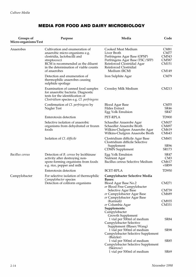
MEDIA FOR FOOD AND DAIRY MICROBIOLOGY
Groups of Purpose Media CodeMicro-organisms/Test
Anaerobes Cultivation and enumeration of Cooked Meat Medium CM81anaerobic micro-organisms e.g. Liver Broth CM77clostridia, lactobacilli and Perfringens Agar Base (OPSP) CM543streptococci Perfringens Agar Base (TSC/SFP) CM587RCM is recommended as the diluent Reinforced Clostridial Agar CM151in the determination of viable counts Reinforced Clostridialof anaerobes Medium (RCM) CM149
Detection and enumeration of Iron Sulphite Agar CM79thermophilic anaerobes causingsulphide spoilage
Examination of canned food samples Crossley Milk Medium CM213for anaerobic bacteria. Diagnostictests for the identification ofClostridium species e.g. Cl. perfringens
Confirmation of Cl. perfringens by Blood Agar Base CM55Nagler Test Fildes Extract SR46
Egg Yolk Emulsion SR47
Enterotoxin detection PET-RPLA TD900
Selective isolation of anaerobic Schaedler Anaerobe Agar CM437organisms from dehydrated or frozen Schaedler Anaerobe Broth CM497foods Wilkins-Chalgren Anaerobe Agar CM619
Wilkins-Chalgren Anaerobe Broth CM643
Isolation of Cl. difficile Clostridium difficile Agar Base CM601Clostridium difficile Selective
Supplement SR96CDMN Supplement SR173
Bacillus cereus Detection of B. cereus by lecithinase Egg Yolk Emulsion SR47activity after destroying non- Nutrient Agar CM3spore-forming organisms from foods Bacillus cereus Selective Medium CM617e.g. rice, pepper and milk +SR99
Enterotoxin detection BCET-RPLA TD950
Campylobacter For selective isolation of thermophilic Campylobacter Selective MediaCampylobacter species Bases:Detection of coliform organisms Blood Agar Base No.2 CM271
or Blood Free CampylobacterSelective Agar Base CM739
or Campylobacter Agar Base CM689or Campylobacter Agar Base
(Karmali) CM935or Columbia Agar CM331Supplements:Campylobacter
Growth Supplement1 vial per 500ml of medium SR84
Campylobacter SelectiveSupplement (Blaser/Wang)1 vial per 500ml of medium SR98
Campylobacter Selective Supplement(Butzler)1 vial per 500ml of medium SR85
Campylobacter Selective Supplement(Skirrow)1 vial per 500ml of medium SR69
Culture Media
2-14 November 1998
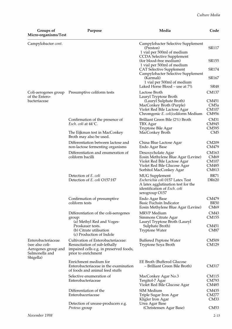
Groups of Purpose Media CodeMicro-organisms/Test
Campylobacter cont. Campylobacter Selective Supplement(Preston) SR117
1 vial per 500ml of mediumCCDA Selective Supplement(for blood-free medium) SR1551 vial per 500ml of medium
CAT Selective Supplement SR174Campylobacter Selective Supplement
(Karmali) SR1671 vial per 500ml of medium
Laked Horse Blood ± use at 7% SR48
Coli-aerogenes group Presumptive coliform tests Lactose Broth CM137of the Entero- Lauryl Tryptose Brothbacteriaceae (Lauryl Sulphate Broth) CM451
MacConkey Broth (Purple) CM5aViolet Red Bile Lactose Agar CM107Chromgenic E. coli/coliform Medium CM956
Confirmation of the presence of Brilliant Green Bile (2%) Broth CM31Esch. coli at 448C. TBX Agar CM945
Tryptone Bile Agar CM595The Eijkman test in MacConkey MacConkey Broth CM5Broth may also be used.
Differentiation between lactose and China Blue Lactose Agar CM209non-lactose fermenting organisms Endo Agar Base CM479
Differentiation and enumeration of Desoxycholate Agar CM163coliform bacilli Eosin Methylene Blue Agar (Levine) CM69
Violet Red Bile Lactose Agar CM107Violet Red Bile Glucose Agar CM485Sorbitol MacConkey Agar CM813
Detection of E. coli MUG Supplement BR71Detection of E. coli O157:H7 Escherichia coli 0157 Latex Test DR620
A latex agglutination test for theidentification of Esch. coliserogroup O157
Confirmation of presumptive Endo Agar Base CM479coliform tests Basic Fuchsin Indicator BR50
Eosin Methylene Blue Agar (Levine) CM69
Differentiation of the coli-aerogenes MRVP Medium CM43group: Simmons Citrate Agar CM155
(a) Methyl Red and Voges- Lauryl Tryptose Broth (LaurylProskauer tests. Sulphate Broth) CM451(b) Citrate utilisation Tryptone Water CM87(c) Production of Indole
Enterobacteriaceae Cultivation of Enterobacteriaceae: Buffered Peptone Water CM509(see also coli- Resuscitation of sub-lethally Tryptone Soya Broth CM129Aerogenes group and impaired cells e.g. in preserved foods,Salmonella and prior to enrichmentShigella)
Enrichment medium for EE Broth (Buffered GlucoseEnterobacteriaceae in the examination ± Brilliant Green Bile Broth) CM317of foods and animal feed stuffs
Selective enumeration of MacConkey Agar No.3 CM115Enterobacteriaceae Tergitol-7 Agar CM793
Violet Red Bile Glucose Agar CM485
Differentiation of the SIM Medium CM435Enterobaceriaceae Triple Sugar Iron Agar CM277
Kligler Iron Agar CM33Detection of urease-producers e.g. Urea Agar BaseProteus group (Christensen Agar Base) CM53
Culture Media
November 1998 2-15
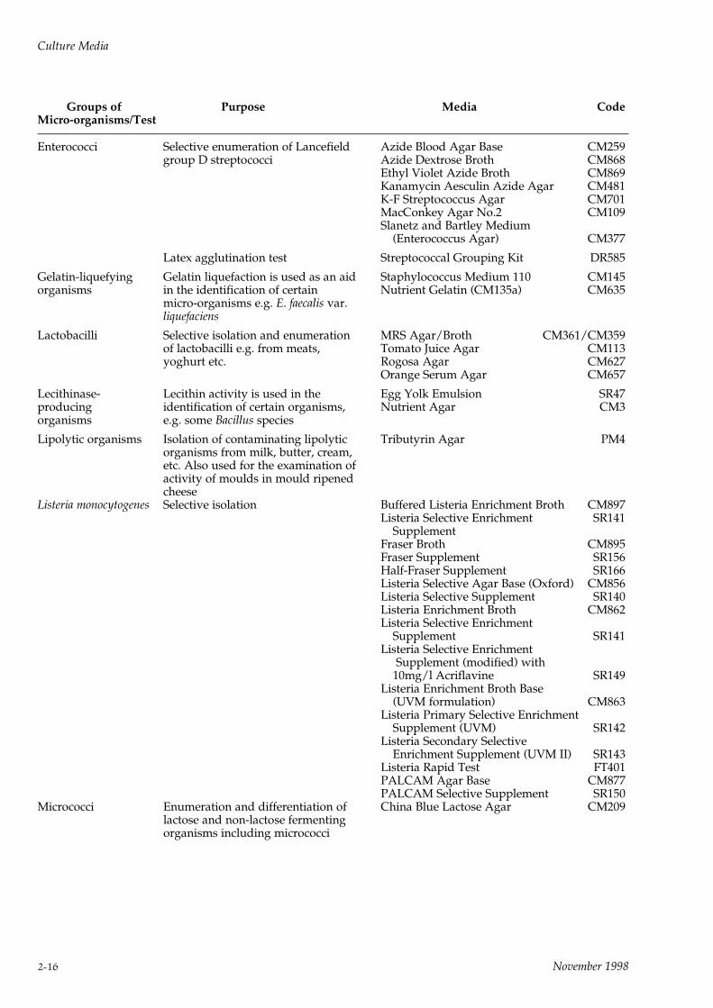
Groups of Purpose Media CodeMicro-organisms/Test
Enterococci Selective enumeration of Lancefield Azide Blood Agar Base CM259group D streptococci Azide Dextrose Broth CM868
Ethyl Violet Azide Broth CM869Kanamycin Aesculin Azide Agar CM481K-F Streptococcus Agar CM701MacConkey Agar No.2 CM109Slanetz and Bartley Medium
(Enterococcus Agar) CM377
Latex agglutination test Streptococcal Grouping Kit DR585
Gelatin-liquefying Gelatin liquefaction is used as an aid Staphylococcus Medium 110 CM145organisms in the identification of certain Nutrient Gelatin (CM135a) CM635
micro-organisms e.g. E. faecalis var.liquefaciens
Lactobacilli Selective isolation and enumeration MRS Agar/Broth CM361/CM359of lactobacilli e.g. from meats, Tomato Juice Agar CM113yoghurt etc. Rogosa Agar CM627
Orange Serum Agar CM657
Lecithinase- Lecithin activity is used in the Egg Yolk Emulsion SR47producing identification of certain organisms, Nutrient Agar CM3organisms e.g. some Bacillus species
Lipolytic organisms Isolation of contaminating lipolytic Tributyrin Agar PM4organisms from milk, butter, cream,etc. Also used for the examination ofactivity of moulds in mould ripenedcheese
Listeria monocytogenes Selective isolation Buffered Listeria Enrichment Broth CM897Listeria Selective Enrichment SR141
SupplementFraser Broth CM895Fraser Supplement SR156Half-Fraser Supplement SR166Listeria Selective Agar Base (Oxford) CM856Listeria Selective Supplement SR140Listeria Enrichment Broth CM862Listeria Selective Enrichment
Supplement SR141Listeria Selective Enrichment
Supplement (modified) with10mg/l Acriflavine SR149
Listeria Enrichment Broth Base(UVM formulation) CM863
Listeria Primary Selective EnrichmentSupplement (UVM) SR142
Listeria Secondary SelectiveEnrichment Supplement (UVM II) SR143
Listeria Rapid Test FT401PALCAM Agar Base CM877PALCAM Selective Supplement SR150
Micrococci Enumeration and differentiation of China Blue Lactose Agar CM209lactose and non-lactose fermentingorganisms including micrococci
Culture Media
2-16 November 1998

Groups of Purpose Media CodeMicro-organisms/Test
Plate Count Colony count for indicating the Nutrient Gelatin (CM135a) CM635efficiency of water treatment Tryptone Glucose Extract Agar CM127processes, or suitability of water for Tryptone Soya Agar CM131food preparation. Nutrient gelatin is Yeast Extract Agar CM19used for the plate count of Plate Count Agar CM325psychrophilic organisms e.g. Standard Plate Count Agar (APHA) CM463Pseudomonas species and for testing Milk Plate Count Agar withgelatinase activity antibiotic free skim milk CM681
Milk Agar CM21PPCT Selective Supplement SR159
Salmonellae and Pre-enrichment procedures prior to Buffered Peptone Water CM509Shigellae isolation Mannitol Selenite Broth Base CM399
Enrichment procedures prior to Muller-Kauffmann Tetrathionateisolation Broth Base CM343
Selenite Broth Base CM395Sodium Biselenite L121Tetrathionate Broth Base CM29
Isolation and identification of Rappaport-Vassiliadis (RV) ±Salmonella and Shigella species Enrichment Broth CM669
Rappaport Vassiliadis Soya (RVS)Peptone Broth CM866
Selenite Cystine Broth Base CM699Tetrathionate Broth USA CM671
Bismuth Sulphate Agar CM201Brilliant Green Agar CM263Brilliant Green Agar (Modified)
(Edel-Kampelmacher Medium) CM329Sulphamandelate Supplement SR87DCLS Agar CM393Desoxycholate Citrate Agar (Hynes) CM227Hektoen Enteric Agar CM419XLD Medium CM469MLCB Agar CM783
Identification of salmonella and Kligler Iron Agar CM33shigella. Lysine Iron Agar CM381
Triple Sugar Iron Agar CM277Salmonella Rapid Test Elective Medium CM857
Rapid Test FT201Latex Test FT203
Staphylococci Isolation and differentiation of Baird-Parker Agar Base CM275pathogenic staphylococci Egg Yolk Tellurite Emulsion SR54
Egg Yolk Emulsion SR47Giolitti-Cantoni Broth CM523Staphylococcus Medium No.110 CM145Staph/Strep. Supplement SR70Vogel-Johnson Agar CM641Mannitol Salt Agar
(Chapman Medium) CM85
Selective enumeration of the total Baird-Parker Agar Base CM275staphylococci count from foodstuffs Egg Yolk Emulsion SR47
RPF Supplement SR122
Differentiation of staphylococci:(a) DNase production DNase Agar CM321(b) Phosphatase production Blood Agar Base CM55(c) Coagulase production Staphylase Test
A test kit for the identification ofStaphylococcus aureus: DR595
Staphytect DR650
Culture Media
November 1998 2-17
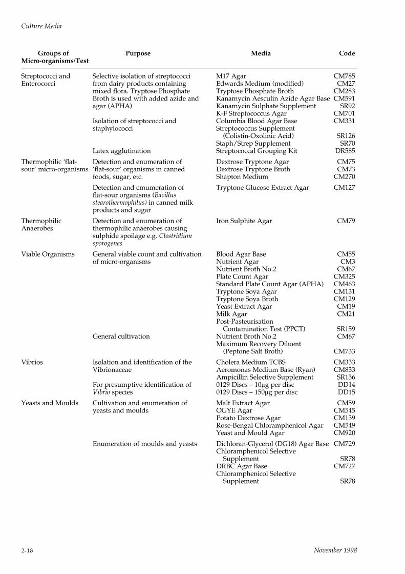
Groups of Purpose Media CodeMicro-organisms/Test
Streptococci and Selective isolation of streptococci M17 Agar CM785Enterococci from dairy products containing Edwards Medium (modified) CM27
mixed flora. Tryptose Phosphate Tryptose Phosphate Broth CM283Broth is used with added azide and Kanamycin Aesculin Azide Agar Base CM591agar (APHA) Kanamycin Sulphate Supplement SR92
K-F Streptococcus Agar CM701Isolation of streptococci and Columbia Blood Agar Base CM331staphylococci Streptococcus Supplement
(Colistin-Oxolinic Acid) SR126Staph/Strep Supplement SR70
Latex agglutination Streptococcal Grouping Kit DR585
Thermophilic `flat- Detection and enumeration of Dextrose Tryptone Agar CM75sour' micro-organisms 'flat-sour' organisms in canned Dextrose Tryptone Broth CM73
foods, sugar, etc. Shapton Medium CM270
Detection and emumeration of Tryptone Glucose Extract Agar CM127flat-sour organisms (Bacillusstearothermophilus) in canned milkproducts and sugar
Thermophilic Detection and enumeration of Iron Sulphite Agar CM79Anaerobes thermophilic anaerobes causing
sulphide spoilage e.g. Clostridiumsporogenes
Viable Organisms General viable count and cultivation Blood Agar Base CM55of micro-organisms Nutrient Agar CM3
Nutrient Broth No.2 CM67Plate Count Agar CM325Standard Plate Count Agar (APHA) CM463Tryptone Soya Agar CM131Tryptone Soya Broth CM129Yeast Extract Agar CM19Milk Agar CM21Post-Pasteurisation
Contamination Test (PPCT) SR159General cultivation Nutrient Broth No.2 CM67
Maximum Recovery Diluent(Peptone Salt Broth) CM733
Vibrios Isolation and identification of the Cholera Medium TCBS CM333Vibrionaceae Aeromonas Medium Base (Ryan) CM833
Ampicillin Selective Supplement SR136For presumptive identification of 0129 Discs ± 10mg per disc DD14Vibrio species 0129 Discs ± 150mg per disc DD15
Yeasts and Moulds Cultivation and enumeration of Malt Extract Agar CM59yeasts and moulds OGYE Agar CM545
Potato Dextrose Agar CM139Rose-Bengal Chloramphenicol Agar CM549Yeast and Mould Agar CM920
Enumeration of moulds and yeasts Dichloran-Glycerol (DG18) Agar Base CM729Chloramphenicol Selective
Supplement SR78DRBC Agar Base CM727Chloramphenicol Selective
Supplement SR78
Culture Media
2-18 November 1998

Groups of Purpose Media CodeMicro-organisms/Test
Plant Hygiene and Diluent or rinse in bacteriological Ringer Solution Tablets BR52Food Sample examination of food products, plantDilution and apparatus
Solvent diluent solution for calcium 'Calgon' Ringer Tablets BR49alginate swabs
Chlorine-neutralising Ringer Thiosulphate Ringer Tablets BR48Solution to counteract thebactericidal effect of hypochloritesor other chlorine sources
Culture Media
November 1998 2-19
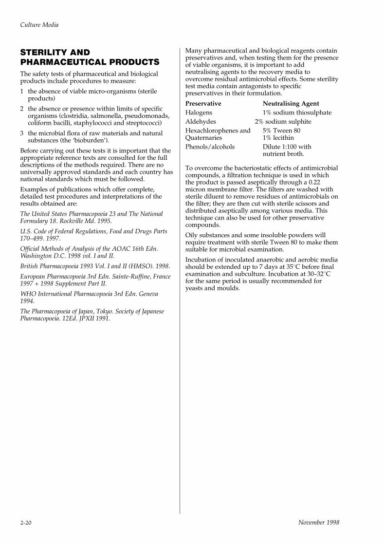
STERILITY ANDPHARMACEUTICAL PRODUCTSThe safety tests of pharmaceutical and biologicalproducts include procedures to measure:
1 the absence of viable micro-organisms (sterileproducts)
2 the absence or presence within limits of specificorganisms (clostridia, salmonella, pseudomonads,coliform bacilli, staphylococci and streptococci)
3 the microbial flora of raw materials and naturalsubstances (the `bioburden').
Before carrying out these tests it is important that theappropriate reference texts are consulted for the fulldescriptions of the methods required. There are nouniversally approved standards and each country hasnational standards which must be followed.
Examples of publications which offer complete,detailed test procedures and interpretations of theresults obtained are:
The United States Pharmacopoeia 23 and The NationalFormulary 18. Rockville Md. 1995.
U.S. Code of Federal Regulations, Food and Drugs Parts170±499. 1997.
Official Methods of Analysis of the AOAC 16th Edn.Washington D.C. 1998 vol. I and II.
British Pharmacopoeia 1993 Vol. I and II (HMSO). 1998.
European Pharmacopoeia 3rd Edn. Sainte-Ruffine, France1997 + 1998 Supplement Part II.
WHO International Pharmacopoeia 3rd Edn. Geneva1994.
The Pharmacopoeia of Japan, Tokyo. Society of JapanesePharmacopoeia. 12Ed. JPXII 1991.
Many pharmaceutical and biological reagents containpreservatives and, when testing them for the presenceof viable organisms, it is important to addneutralising agents to the recovery media toovercome residual antimicrobial effects. Some sterilitytest media contain antagonists to specificpreservatives in their formulation.
Preservative Neutralising Agent
Halogens 1% sodium thiosulphate
Aldehydes 2% sodium sulphite
Hexachlorophenes and 5% Tween 80Quaternaries 1% lecithin
Phenols/alcohols Dilute 1:100 withnutrient broth.
To overcome the bacteriostatic effects of antimicrobialcompounds, a filtration technique is used in whichthe product is passed aseptically through a 0.22micron membrane filter. The filters are washed withsterile diluent to remove residues of antimicrobials onthe filter; they are then cut with sterile scissors anddistributed aseptically among various media. Thistechnique can also be used for other preservativecompounds.
Oily substances and some insoluble powders willrequire treatment with sterile Tween 80 to make themsuitable for microbial examination.
Incubation of inoculated anaerobic and aerobic mediashould be extended up to 7 days at 358C before finalexamination and subculture. Incubation at 30±328Cfor the same period is usually recommended foryeasts and moulds.
Culture Media
2-20 November 1998
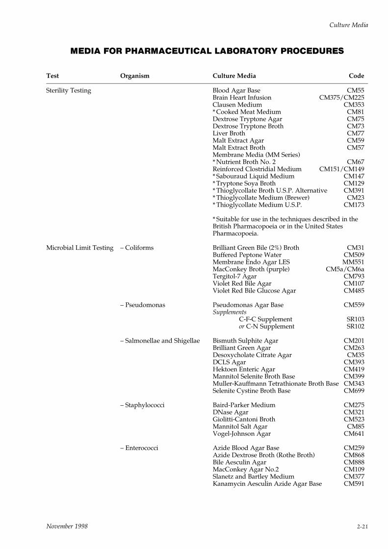
MEDIA FOR PHARMACEUTICAL LABORATORY PROCEDURES
Test Organism Culture Media Code
Sterility Testing Blood Agar Base CM55Brain Heart Infusion CM375/CM225Clausen Medium CM353* Cooked Meat Medium CM81Dextrose Tryptone Agar CM75Dextrose Tryptone Broth CM73Liver Broth CM77Malt Extract Agar CM59Malt Extract Broth CM57Membrane Media (MM Series)* Nutrient Broth No. 2 CM67Reinforced Clostridial Medium CM151/CM149* Sabouraud Liquid Medium CM147* Tryptone Soya Broth CM129* Thioglycollate Broth U.S.P. Alternative CM391* Thioglycollate Medium (Brewer) CM23* Thioglycollate Medium U.S.P. CM173
* Suitable for use in the techniques described in theBritish Pharmacopoeia or in the United StatesPharmacopoeia.
Microbial Limit Testing ± Coliforms Brilliant Green Bile (2%) Broth CM31Buffered Peptone Water CM509Membrane Endo Agar LES MM551MacConkey Broth (purple) CM5a/CM6aTergitol-7 Agar CM793Violet Red Bile Agar CM107Violet Red Bile Glucose Agar CM485
± Pseudomonas Pseudomonas Agar Base CM559Supplements
C-F-C Supplement SR103or C-N Supplement SR102
± Salmonellae and Shigellae Bismuth Sulphite Agar CM201Brilliant Green Agar CM263Desoxycholate Citrate Agar CM35DCLS Agar CM393Hektoen Enteric Agar CM419Mannitol Selenite Broth Base CM399Muller-Kauffmann Tetrathionate Broth Base CM343Selenite Cystine Broth Base CM699
± Staphylococci Baird-Parker Medium CM275DNase Agar CM321Giolitti-Cantoni Broth CM523Mannitol Salt Agar CM85Vogel-Johnson Agar CM641
± Enterococci Azide Blood Agar Base CM259Azide Dextrose Broth (Rothe Broth) CM868Bile Aesculin Agar CM888MacConkey Agar No.2 CM109Slanetz and Bartley Medium CM377Kanamycin Aesculin Azide Agar Base CM591
Culture Media
November 1998 2-21
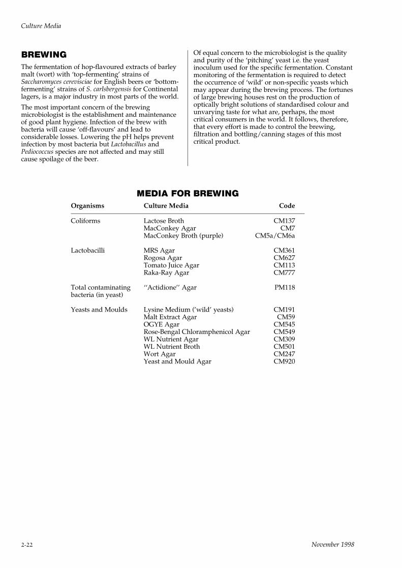
BREWINGThe fermentation of hop-flavoured extracts of barleymalt (wort) with `top-fermenting' strains ofSaccharomyces cerevisciae for English beers or `bottom-fermenting' strains of S. carlsbergensis for Continentallagers, is a major industry in most parts of the world.
The most important concern of the brewingmicrobiologist is the establishment and maintenanceof good plant hygiene. Infection of the brew withbacteria will cause `off-flavours' and lead toconsiderable losses. Lowering the pH helps preventinfection by most bacteria but Lactobacillus andPediococcus species are not affected and may stillcause spoilage of the beer.
Of equal concern to the microbiologist is the qualityand purity of the `pitching' yeast i.e. the yeastinoculum used for the specific fermentation. Constantmonitoring of the fermentation is required to detectthe occurrence of `wild' or non-specific yeasts whichmay appear during the brewing process. The fortunesof large brewing houses rest on the production ofoptically bright solutions of standardised colour andunvarying taste for what are, perhaps, the mostcritical consumers in the world. It follows, therefore,that every effort is made to control the brewing,filtration and bottling/canning stages of this mostcritical product.
MEDIA FOR BREWINGOrganisms Culture Media Code
Coliforms Lactose Broth CM137MacConkey Agar CM7MacConkey Broth (purple) CM5a/CM6a
Lactobacilli MRS Agar CM361Rogosa Agar CM627Tomato Juice Agar CM113Raka-Ray Agar CM777
Total contaminating ``Actidione'' Agar PM118bacteria (in yeast)
Yeasts and Moulds Lysine Medium ('wild' yeasts) CM191Malt Extract Agar CM59OGYE Agar CM545Rose-Bengal Chloramphenicol Agar CM549WL Nutrient Agar CM309WL Nutrient Broth CM501Wort Agar CM247Yeast and Mould Agar CM920
Culture Media
2-22 November 1998

WATER SUPPLY AND SEWAGEDISPOSALThe close connection between water fit for drinkingand sewage disposal is best illustrated by the largetowns which sit astride the major rivers in centralUSA. Each town draws water for consumption up-stream and discharges sewage effluent down-stream.The last town in such a chain may be drawing watercontaining the effluents of seven or eight largeconurbations.
Such practices, which operate in all major countries,are safe, providing great care is taken in filtering andchlorinating the in-coming water. Equally, theprocessing of sewage must be safely operated so thatpathogen-free and chemically clean effluent of lowbiological-oxygen-demand (BOD) is released backinto the river down-stream.
Drinking waterStored and river water may contain a wide variety oforganisms, mainly saprophytic bacteria with optimaltemperatures of growth around 228C. Filtration andchlorination of the water, before distribution to thepublic, removes most of these organisms.
Microbiological tests are carried out to make sure thatthe quality of the treated water meets thespecifications required by the Regulatory Authorities.[DHSS 1982. The Bacteriological Examination of DrinkingWater Supplies Report No.71. HMSO London. AmericanPublic Health Association. 1975. Standard Methods for theExamination of Water and Wastewater. 14th Edn.Washington D.C.]
Bacterial pollution of water may originate fromindividuals with clinical symptoms of disease or fromsymptomless carriers of enteric pathogens such asSalmonella typhi. Such pathogens are difficult to detectin a water supply because their numbers are oftenfew and their incidence sporadic. Therefore, indicatororganisms of intestinal contamination are looked forbecause they are present in much larger numbers andthey persist much longer than pathogens in pollutedwater.
From a public health point of view, the coliform test isthe most important as the presence of Escherichia coliat >5 bacilli per 100ml of unchlorinated waterindicates a less than satisfactory supply.
The quantitative assessment used is either a multipletube, most probable number (MPN) or a membranefiltration method. The exact techniques and mediaused are cited in the references mentioned or in othernational reference publications. Clostridium perfringensand Enterococcus faecalis can persist in water suppliesfor long periods. Their presence in water, whencoliform organisms are absent, indicates faecalcontamination at a more remote time.
Sewage disposalIn highly industrialised countries where largecommunities have developed, the disposal ofindustrial and domestic waste is an increasingproblem.
International opinion is against untreated sewagebeing discharged into coastal or estuarine waters andthe use of efficient treatment plants to process sewagebefore discharge is now recommended.
Untreated sewage consists mainly of water containingorganic and inorganic dissolved and suspendedsubstances, together with many micro-organisms.After preliminary screening to remove solid matter,the liquid is treated by one of three common methods:
(i) activated sludge process ± this involves vigorousstirring or aeration by other means to reduce theBOD and cause separation of the organic matter.
(ii) biological filtration ± in this process the liquid isfiltered through large beds of sand and themicro-organisms are trapped in the zooglealslime which forms during filtration.
(iii) oxidation ponds ± settled sewage is held inponds or lagoons for 30 days before thesupernatant fluid is released.
All three processes utilise living organisms to reducethe BOD of the effluent to levels where it can bedischarged into waterways without causing pollution.
LEGIONNAIRE'S DISEASEThis disease, caused by inhalation of large numbers ofLegionella species, is essentially a water-borneinfection. The infection starts in air-conditioningplants where large volumes of water are recirculatedand cooled by blowing air through the water. Suchwarm circulated water quickly grows large quantitiesof legionellae and the aerosol of organisms caused bythe air-cooling system spreads down-wind to infectpassers-by. Not every passer-by develops the diseaseof legionellosis and the characteristics of susceptiblevictims are still being determined but a major factor isthe quantity of organisms inhaled. A large inhaleddose of legionellae will inevitably lead to atypicalpneumonia.
The most severe form of the disease is caused byLegionella pneumophila SG1 and it can be rapidly fatalwithout prompt antimicrobial treatment.
The organisms can easily be isolated from the waterusing specific legionella media as described in thismanual. Isolation of the organism from the patient ismore difficult and most infections are diagnosed byimmunological tests.
It is now advised that all recirculating, air-cooledwater systems are treated at regular and frequentintervals with bactericidal compounds to prevent thebuild-up of large numbers of legionellae.
Culture Media
November 1998 2-23

MEDIA FOR WATER AND SEWAGE MICROBIOLOGY
Groups of Purpose Media CodeMicro-organisms/Test
Clostridium Detection of Clostridium perfringens Iron Sulphite Agar CM79perfringens indicating remote or intermittent Perfringens Agar (OPSP) CM543
pollution in water Supplement A SR76Supplement B SR77
Perfringens Agar Base TSC/SFP CM587TSC Supplement SR88SFP Supplement SR93
Reinforced Clostridial Medium CM149Reinforced Clostridial Agar CM151
Confirmation of Clostridium perfringens(a) by `stormy-clot' reaction Crossley Milk Medium CM213(b) by Nagler Test Blood Agar Base CM55(c) Reverse CAMP Test Egg Yolk Emulsion SR47
Fildes Extract SR46
Coliform Group Detection of coliforms or Esch. coli(including Esch. coli) (at 448C) as indicators of pollution
(a) Mean Probable Number Technique Lactose Broth CM137Lauryl Tryptose Broth CM451MacConkey Broth (Purple) CM5aMinerals Modified Medium Base CM607Sodium Glutamate L124
(b) Membrane Filtration Technique Membrane Media DehydratedM-Endo Agar LES MM551
Basic Fuchsin Indicator BR50Membrane Lauryl Sulphate Broth MM615Tryptone Bile Agar CM595
Confirmation of coliforms or Brilliant Green Bile (2%) Broth CM31Esch. coli Endo Agar Base CM479
Basic Fuchsin Indicator BR50MacConkey Agar CM7MacConkey Agar No.3 CM115MUG Supplement BR71Tergitol-7 Agar CM793Violet Red Bile Lactose Agar CM107Violet Red Bile Glucose Agar CM485
Detection of Esch. coli O157: H7 Sorbitol MacConkey Agar CM813Eosin Methylene Blue Agar CM69Escherichia coli O157 Latex Test DR620
A latex agglutination test for theidentification of Esch. coliserogroup O157
Differentiation of coliforms(a) Methyl Red andVoges-Proskauer Tests MRVP Medium CM43
(b) Citrate Utilisation Simmons Citrate Agar CM155
(c) lndole Production Tryptone Water CM87
Culture Media
2-24 November 1998
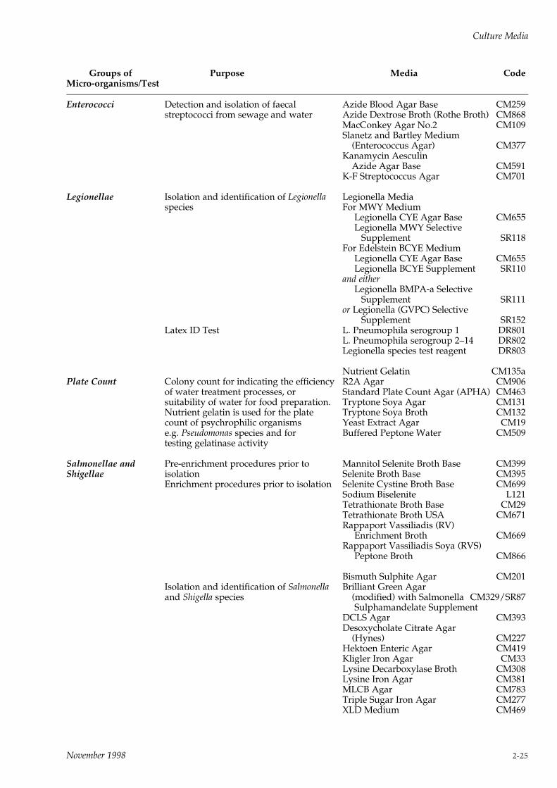
Groups of Purpose Media CodeMicro-organisms/Test
Enterococci Detection and isolation of faecal Azide Blood Agar Base CM259streptococci from sewage and water Azide Dextrose Broth (Rothe Broth) CM868
MacConkey Agar No.2 CM109Slanetz and Bartley Medium
(Enterococcus Agar) CM377Kanamycin Aesculin
Azide Agar Base CM591K-F Streptococcus Agar CM701
Legionellae Isolation and identification of Legionella Legionella Mediaspecies For MWY Medium
Legionella CYE Agar Base CM655Legionella MWY Selective
Supplement SR118For Edelstein BCYE Medium
Legionella CYE Agar Base CM655Legionella BCYE Supplement SR110
and eitherLegionella BMPA-a Selective
Supplement SR111or Legionella (GVPC) Selective
Supplement SR152Latex ID Test L. Pneumophila serogroup 1 DR801
L. Pneumophila serogroup 2±14 DR802Legionella species test reagent DR803
Nutrient Gelatin CM135aPlate Count Colony count for indicating the efficiency R2A Agar CM906
of water treatment processes, or Standard Plate Count Agar (APHA) CM463suitability of water for food preparation. Tryptone Soya Agar CM131Nutrient gelatin is used for the plate Tryptone Soya Broth CM132count of psychrophilic organisms Yeast Extract Agar CM19e.g. Pseudomonas species and for Buffered Peptone Water CM509testing gelatinase activity
Salmonellae and Pre-enrichment procedures prior to Mannitol Selenite Broth Base CM399Shigellae isolation Selenite Broth Base CM395
Enrichment procedures prior to isolation Selenite Cystine Broth Base CM699Sodium Biselenite L121Tetrathionate Broth Base CM29Tetrathionate Broth USA CM671Rappaport Vassiliadis (RV)
Enrichment Broth CM669Rappaport Vassiliadis Soya (RVS)
Peptone Broth CM866
Bismuth Sulphite Agar CM201Isolation and identification of Salmonella Brilliant Green Agarand Shigella species (modified) with Salmonella CM329/SR87
Sulphamandelate SupplementDCLS Agar CM393Desoxycholate Citrate Agar
(Hynes) CM227Hektoen Enteric Agar CM419Kligler Iron Agar CM33Lysine Decarboxylase Broth CM308Lysine Iron Agar CM381MLCB Agar CM783Triple Sugar Iron Agar CM277XLD Medium CM469
Culture Media
November 1998 2-25

Groups of Purpose Media CodeMicro-organisms/Test
Vibrios Isolation and identification of the Cholera Medium (TCBS) CM333Vibrionaceae Aeromonas Medium Base (Ryan) CM833
Supplement ±Ampicillin Supplement SR136
For presumptive identification ofvibrio species
0129 Discs ± 10mg per disc DD140129 Discs ± 150mg per disc DD15
Diluents used in For preparation of 1/4 strength Ringer Ringer Solution (1/4 strength Ringerwater and sewage Solution, used as a diluent or rinse Solution tablets) BR52bacteriology
Preparation of a sodium 'Calgon' Ringer Tablets BR49hexametaphosphate Ringer Solution,used as a solvent diluent solution inconjunction with calcium alginateswabs
Preparation of chlorine-neutralising Thiosulphate Ringer Tablets BR48Ringer Solution, used as a diluent orrinse after hypochlorites or otherchlorine sources
Culture Media
2-26 November 1998

OXOID PRODUCTS FOR SPECIFIC GROUPS OF MICRO-ORGANISMS
Blood Agar Base MediaBlood Agar Base CM55Blood Agar Base No.2 CM271Blood Agar Base (sheep) CM854Columbia Agar Base CM331Tryptone Soya Agar CM131Tryptose Blood Agar Base CM233
OrganismActinomycetes:
Nocardia speciesStreptomyces species
Isolation/CultivationBrain Heart Infusion Agar/Broth CM375/CM225Czapek-Dox Agar CM97Sabouraud Dextrose/Maltose Agar CM41/CM41aTryptone Soya Agar/Broth CM131/CM129Thioglycollate Broth CM173
DifferentiationNutrient Gelatin CM135a
OrganismAnaerobes:
Actinomyces speciesBacteroides speciesClostridium speciesFusiformis speciesStreptococcus species
Isolation/CultivationAmies Transport Medium CM425Blood Agar Base Media: see aboveBrain Heart Infusion Agar/Broth CM375/CM225Brucella Agar Base (USA) CM691Clausen Medium CM353Clostridium difficile Agar Base CM601+ supplement SR96Cooked Meat Medium CM81Iron Sulphite Agar CM79Liver Broth CM77Nutrient Broth CM67Perfringens Agar Base OPSP CM543+ supplements SR76/SR77Perfringens Agar Base TSC/SFP CM587+ supplements SR88/SR93Reinforced Clostridial Agar/Broth CM151/CM149Schaedler Anaerobe Agar/Broth CM437/CM497Thioglycollate Medium (Brewer) CM23Thioglycollate Medium (USP) CM173Thioglycollate Broth (USP Alternative) CM391Tryptone Soya Agar/Broth CM131/CM129Wilkens-Chalgren Anaerobe Agar/Broth CM619/CM643 + supplements SR107/SR108Anaerobic Basal Agar CM972Anaerobic Basal Broth CM957
DifferentiationAn-Ident Discs DD6Egg Yolk Emulsion SR47Cooked Meat Medium CM81Fildes Extract SR46
Iron Sulphite Agar CM79Crossley Milk Medium CM213Liver Broth CM77SPS Discs DD16Tryptone Water CM87Wilkens-Chalgren Anaerobe Agar CM619/CM643+ supplements SR107/SR108Enterotoxin Detection PET-RPLA TD900
OrganismBacillus species
Isolation/CultivationBacillus cereus Selective Agar Base CM617+ supplements SR99/SR47Dextrose Tryptone Agar/Broth CM75/CM73Nutrient Agar/Broth CM17/CM15Tryptone Soya Agar/Broth CM131/CM129
DifferentiationBacillus cereus Selective Agar Base CM617+ supplements SR99/SR47Crossley Milk Medium CM2l3Egg Yolk Emulsion SR47Enterotoxin Detection BCET-RPLA TD950
OrganismBordetella species
Isolation/CultivationCharcoal Agar CM119+ supplement SR82`Chocolate' Agar CM271
DifferentiationX and V factor discs DD3-DD5 on Blood Agar BaseCM55
OrganismBrucella species
Isolation/CultivationBlood Agar Base No.2 CM271Columbia Blood Agar Base CM331Brucella Medium Base CM169+ supplements SR83/SR35Liver Broth CM77Tryptone Soya Agar/Broth CM131/CM129
DifferentiationBrucella Medium Base CM169 with fuchsin andthionin dyes
OrganismCampylobacter species
Isolation/DifferentiationBases:Blood Agar Base No.2 CM271Blood Free Campylobacter Selective Agar BaseCM739Campylobacter Agar Base CM689Columbia Agar CM331
Supplements:Blaser-Wang supplement SR98Butzler supplement SR85
Culture Media
November 1998 2-27
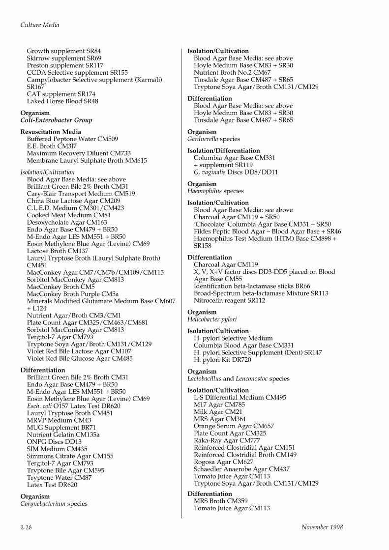
Growth supplement SR84Skirrow supplement SR69Preston supplement SR117CCDA Selective supplement SR155Campylobacter Selective supplement (Karmali)SR167CAT supplement SR174Laked Horse Blood SR48
OrganismColi-Enterobacter Group
Resuscitation MediaBuffered Peptone Water CM509E.E. Broth CM3l7Maximum Recovery Diluent CM733Membrane Lauryl Sulphate Broth MM615
Isolation/CultivationBlood Agar Base Media: see aboveBrilliant Green Bile 2% Broth CM31Cary-Blair Transport Medium CM519China Blue Lactose Agar CM209C.L.E.D. Medium CM301/CM423Cooked Meat Medium CM81Desoxycholate Agar CM163Endo Agar Base CM479 + BR50M-Endo Agar LES MM551 + BR50Eosin Methylene Blue Agar (Levine) CM69Lactose Broth CM137Lauryl Tryptose Broth (Lauryl Sulphate Broth)CM451MacConkey Agar CM7/CM7b/CM109/CM115Sorbitol MacConkey Agar CM813MacConkey Broth CM5MacConkey Broth Purple CM5aMinerals Modified Glutamate Medium Base CM607+ L124Nutrient Agar/Broth CM3/CM1Plate Count Agar CM325/CM463/CM681Sorbitol MacConkey Agar CM813Tergitol-7 Agar CM793Tryptone Soya Agar/Broth CM131/CM129Violet Red Bile Lactose Agar CM107Violet Red Bile Glucose Agar CM485
DifferentiationBrilliant Green Bile 2% Broth CM31Endo Agar Base CM479 + BR50M-Endo Agar LES MM551 + BR50Eosin Methylene Blue Agar (Levine) CM69Esch. coli O157 Latex Test DR620Lauryl Tryptose Broth CM451MRVP Medium CM43MUG Supplement BR71Nutrient Gelatin CM135aONPG Discs DD13SIM Medium CM435Simmons Citrate Agar CM155Tergitol-7 Agar CM793Tryptone Bile Agar CM595Tryptone Water CM87Latex Test DR620
OrganismCorynebacterium species
Isolation/CultivationBlood Agar Base Media: see aboveHoyle Medium Base CM83 + SR30Nutrient Broth No.2 CM67Tinsdale Agar Base CM487 + SR65Tryptone Soya Agar/Broth CM131/CM129
DifferentiationBlood Agar Base Media: see aboveHoyle Medium Base CM83 + SR30Tinsdale Agar Base CM487 + SR65
OrganismGardnerella species
Isolation/DifferentiationColumbia Agar Base CM331+ supplement SR119G. vaginalis Discs DD8/DD11
OrganismHaemophilus species
Isolation/CultivationBlood Agar Base Media: see aboveCharcoal Agar CM119 + SR50`Chocolate' Columbia Agar Base CM331 + SR50Fildes Peptic Blood Agar ± Blood Agar Base + SR46Haemophilus Test Medium (HTM) Base CM898 +SR158
DifferentiationCharcoal Agar CM119X, V, X+V factor discs DD3-DD5 placed on BloodAgar Base CM55Identification beta-lactamase sticks BR66Broad-Spectrum beta-lactamase Mixture SR113Nitrocefin reagent SR112
OrganismHelicobacter pylori
Isolation/CultivationH. pylori Selective MediumColumbia Blood Agar Base CM331H. pylori Selective Supplement (Dent) SR147H. pylori Kit DR720
OrganismLactobacillus and Leuconostoc species
Isolation/CultivationL-S Differential Medium CM495M17 Agar CM785Milk Agar CM21MRS Agar CM361Orange Serum Agar CM657Plate Count Agar CM325Raka-Ray Agar CM777Reinforced Clostridial Agar CM151Reinforced Clostridial Broth CM149Rogosa Agar CM627Schaedler Anaerobe Agar CM437Tomato Juice Agar CM113Tryptone Soya Agar/Broth CM131/CM129
DifferentiationMRS Broth CM359Tomato Juice Agar CM113
Culture Media
2-28 November 1998
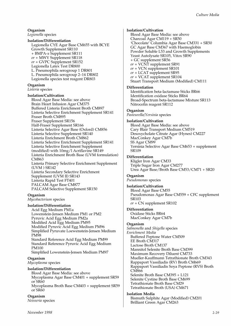
OrganismLegionella species
Isolation/DifferentiationLegionella CYE Agar Base CM655 with BCYEGrowth Supplement SR110+ BMPA-a Supplement SR111or + MWY Supplement SR118or + GVPC Supplement SR152Legionella Latex Test DR800L. Pneumophila serogroup 1 DR801L. Pneumophila serogroup 2±14 DR802Legionella species test reagent DR803
OrganismListeria species
Isolation/CultivationBlood Agar Base Media: see aboveBrain Heart Infusion Agar CM375Buffered Listeria Enrichment Broth CM897Listeria Selective Enrichment Supplement SR141Fraser Broth CM895Fraser Supplement SR156Half-Fraser Supplement SR166Listeria Selective Agar Base (Oxford) CM856Listeria Selective Supplement SR140Listeria Enrichment Broth CM862Listeria Selective Enrichment Supplement SR141Listeria Selective Enrichment Supplement(modified) with 10mg/l Acriflavine SR149Listeria Enrichment Broth Base (UVM formulation)CM863Listeria Primary Selective Enrichment Supplement(UVM ) SR142Listeria Secondary Selective EnrichmentSupplement (UVM II) SR143Listeria Rapid Test FT401PALCAM Agar Base CM877PALCAM Selective Supplement SR150
OrganismMycobacterium species
Isolation/DifferentiationAcid Egg Medium PM1aLowenstein-Jensen Medium PM1 or PM2Pyruvic Acid Egg Medium PM2aModified Acid Egg Medium PM95Modified Pyruvic Acid Egg Medium PM96Simplified Pyruvate Loewenstein-Jensen MediumPM98Standard Reference Acid Egg Medium PM99Standard Reference Pyruvic Acid Egg MediumPM100Simplified Lowenstein-Jensen Medium PM97
OrganismMycoplasma species
Isolation/DifferentiationBlood Agar Base Media: see aboveMycoplasma Agar Base CM401 + supplement SR59or SR60Mycoplasma Broth Base CM403 + supplement SR59or SR60
OrganismNeisseria species
Isolation/CultivationBlood Agar Base Media: see aboveCharcoal Agar CM119 + SR50`Chocolate' Columbia Agar Base CM331 + SR50GC Agar Base CM367 with HaemoglobinPowder Soluble L53 and Growth SupplementsYeast Autolysate SR105, Vitox SR90+ GC supplement SR56or + VCNT supplement SR91or + VCN supplement SR101or + LCAT supplement SR95or + VCAT supplement SR104Stuart Transport Medium (Modified) CM111
DifferentiationIdentification beta-lactamase Sticks BR66Identification oxidase Sticks BR64Broad-Spectrum beta-lactamase Mixture SR113Nitrocefin reagent SR112
OrganismPasteurella/Yersinia species
Isolation/CultivationBlood Agar Base Media: see aboveCary Blair Transport Medium CM519Desoxycholate Citrate Agar (Hynes) CM227MacConkey Agar CM7bSS Agar CM99Yersinia Selective Agar Base CM653 + supplementSR109
DifferentiationKligler Iron Agar CM33Triple Sugar Iron Agar CM277Urea Agar Base/Broth Base CM53/CM71 + SR20
OrganismPseudomonas species
Isolation/CultivationBlood Agar Base CM55Pseudomonas Agar Base CM559 + CFC supplementSR103or + CN supplement SR102
DifferentiationOxidase Sticks BR64MacConkey Agar CM7b
OrganismSalmonella and Shigella speciesEnrichment Media
Buffered Peptone Water CM509EE Broth CM317Lactose Broth CM137Mannitol Selenite Broth Base CM399Maximum Recovery Diluent CM733Mueller-Kauffmann Tetrathionate Broth CM343Rappaport Vassiliadis (RV) Broth CM669Rappaport Vassiliadis Soya Peptone (RVS) BrothCM866Selenite Broth Base CM395 + L121Selenite Cystine Broth Base CM699Tetrathionate Broth Base CM29Tetrathionate Broth (USA) CM671
Isolation MediaBismuth Sulphite Agar (Modified) CM201Brilliant Green Agar CM263
Culture Media
November 1998 2-29
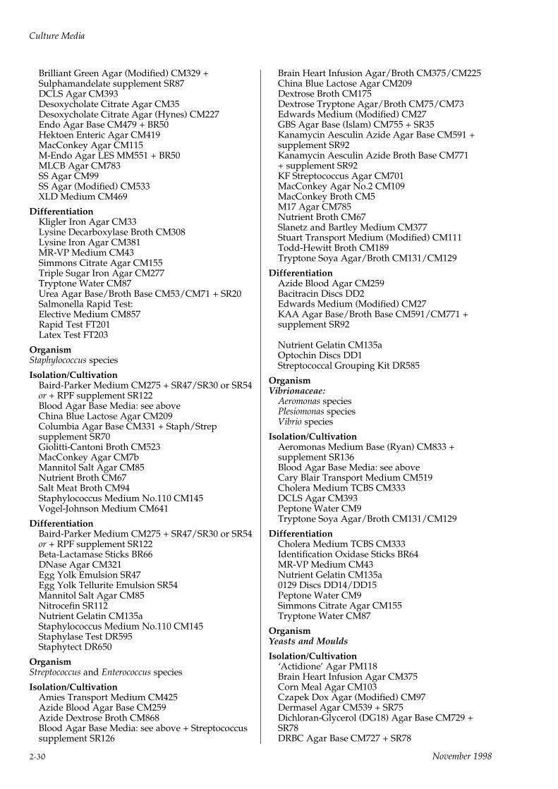
Brilliant Green Agar (Modified) CM329 +Sulphamandelate supplement SR87DCLS Agar CM393Desoxycholate Citrate Agar CM35Desoxycholate Citrate Agar (Hynes) CM227Endo Agar Base CM479 + BR50Hektoen Enteric Agar CM419MacConkey Agar CM115M-Endo Agar LES MM551 + BR50MLCB Agar CM783SS Agar CM99SS Agar (Modified) CM533XLD Medium CM469
DifferentiationKligler Iron Agar CM33Lysine Decarboxylase Broth CM308Lysine Iron Agar CM381MR-VP Medium CM43Simmons Citrate Agar CM155Triple Sugar Iron Agar CM277Tryptone Water CM87Urea Agar Base/Broth Base CM53/CM71 + SR20Salmonella Rapid Test:Elective Medium CM857Rapid Test FT201Latex Test FT203
OrganismStaphylococcus species
Isolation/CultivationBaird-Parker Medium CM275 + SR47/SR30 or SR54or + RPF supplement SR122Blood Agar Base Media: see aboveChina Blue Lactose Agar CM209Columbia Agar Base CM331 + Staph/Strepsupplement SR70Giolitti-Cantoni Broth CM523MacConkey Agar CM7bMannitol Salt Agar CM85Nutrient Broth CM67Salt Meat Broth CM94Staphylococcus Medium No.110 CM145Vogel-Johnson Medium CM641
DifferentiationBaird-Parker Medium CM275 + SR47/SR30 or SR54or + RPF supplement SR122Beta-Lactamase Sticks BR66DNase Agar CM321Egg Yolk Emulsion SR47Egg Yolk Tellurite Emulsion SR54Mannitol Salt Agar CM85Nitrocefin SR112Nutrient Gelatin CM135aStaphylococcus Medium No.110 CM145Staphylase Test DR595Staphytect DR650
OrganismStreptococcus and Enterococcus species
Isolation/CultivationAmies Transport Medium CM425Azide Blood Agar Base CM259Azide Dextrose Broth CM868Blood Agar Base Media: see above + Streptococcussupplement SR126
Brain Heart Infusion Agar/Broth CM375/CM225China Blue Lactose Agar CM209Dextrose Broth CM175Dextrose Tryptone Agar/Broth CM75/CM73Edwards Medium (Modified) CM27GBS Agar Base (Islam) CM755 + SR35Kanamycin Aesculin Azide Agar Base CM591 +supplement SR92Kanamycin Aesculin Azide Broth Base CM771+ supplement SR92KF Streptococcus Agar CM701MacConkey Agar No.2 CM109MacConkey Broth CM5M17 Agar CM785Nutrient Broth CM67Slanetz and Bartley Medium CM377Stuart Transport Medium (Modified) CM111Todd-Hewitt Broth CM189Tryptone Soya Agar/Broth CM131/CM129
DifferentiationAzide Blood Agar CM259Bacitracin Discs DD2Edwards Medium (Modified) CM27KAA Agar Base/Broth Base CM591/CM771 +supplement SR92
Nutrient Gelatin CM135aOptochin Discs DD1Streptococcal Grouping Kit DR585
OrganismVibrionaceae:
Aeromonas speciesPlesiomonas speciesVibrio species
Isolation/CultivationAeromonas Medium Base (Ryan) CM833 +supplement SR136Blood Agar Base Media: see aboveCary Blair Transport Medium CM519Cholera Medium TCBS CM333DCLS Agar CM393Peptone Water CM9Tryptone Soya Agar/Broth CM131/CM129
DifferentiationCholera Medium TCBS CM333Identification Oxidase Sticks BR64MR-VP Medium CM43Nutrient Gelatin CM135a0129 Discs DD14/DD15Peptone Water CM9Simmons Citrate Agar CM155Tryptone Water CM87
OrganismYeasts and Moulds
Isolation/Cultivation`Actidione' Agar PM118Brain Heart Infusion Agar CM375Corn Meal Agar CM103Czapek Dox Agar (Modified) CM97Dermasel Agar CM539 + SR75Dichloran-Glycerol (DG18) Agar Base CM729 +SR78DRBC Agar Base CM727 + SR78
Culture Media
2-30 November 1998

Eosin Methylene Blue Agar (Levine) CM69Lysine Medium CM191Malt Extract Agar/Broth CM59/CM57OGYE Agar Base CM545 + SR73Potato Dextrose Agar CM139Rose-Bengal Chloramphenicol Agar CM549 + SR78Sabouraud Media CM41/CM41a/CM147Tryptone Soya Agar/Broth CM131/CM129WL Nutrient Agar/Broth CM309/CM501Wort Agar CM247Yeast and Mould Agar CM920
DifferentiationCorn Meal Agar CM103Czapek Dox Agar (Modified) CM97Dermasel Agar CM539 + SR75Eosin Methylene Blue Agar (Levine) CM69Lysine Medium CM191Sabouraud Media CM41/CM41aWL Nutrient Agar CM309
Culture Media
November 1998 2-31
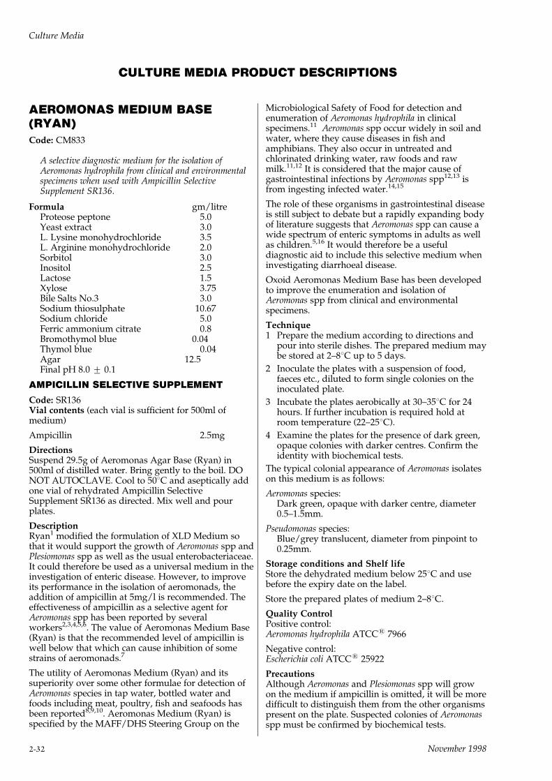
CULTURE MEDIA PRODUCT DESCRIPTIONS
AEROMONAS MEDIUM BASE(RYAN)Code: CM833
A selective diagnostic medium for the isolation ofAeromonas hydrophila from clinical and environmentalspecimens when used with Ampicillin SelectiveSupplement SR136.
Formula gm/litreProteose peptone 5.0Yeast extract 3.0L. Lysine monohydrochloride 3.5L. Arginine monohydrochloride 2.0Sorbitol 3.0Inositol 2.5Lactose 1.5Xylose 3.75Bile Salts No.3 3.0Sodium thiosulphate 10.67Sodium chloride 5.0Ferric ammonium citrate 0.8Bromothymol blue 0.04Thymol blue 0.04Agar 12.5Final pH 8.0 + 0.1
AMPICILLIN SELECTIVE SUPPLEMENT
Code: SR136Vial contents (each vial is sufficient for 500ml ofmedium)
Ampicillin 2.5mg
DirectionsSuspend 29.5g of Aeromonas Agar Base (Ryan) in500ml of distilled water. Bring gently to the boil. DONOT AUTOCLAVE. Cool to 508C and aseptically addone vial of rehydrated Ampicillin SelectiveSupplement SR136 as directed. Mix well and pourplates.
DescriptionRyan1 modified the formulation of XLD Medium sothat it would support the growth of Aeromonas spp andPlesiomonas spp as well as the usual enterobacteriaceae.It could therefore be used as a universal medium in theinvestigation of enteric disease. However, to improveits performance in the isolation of aeromonads, theaddition of ampicillin at 5mg/l is recommended. Theeffectiveness of ampicillin as a selective agent forAeromonas spp has been reported by severalworkers2,3,4,5,6. The value of Aeromonas Medium Base(Ryan) is that the recommended level of ampicillin iswell below that which can cause inhibition of somestrains of aeromonads.7
The utility of Aeromonas Medium (Ryan) and itssuperiority over some other formulae for detection ofAeromonas species in tap water, bottled water andfoods including meat, poultry, fish and seafoods hasbeen reported8,9,10. Aeromonas Medium (Ryan) isspecified by the MAFF/DHS Steering Group on the
Microbiological Safety of Food for detection andenumeration of Aeromonas hydrophila in clinicalspecimens.11 Aeromonas spp occur widely in soil andwater, where they cause diseases in fish andamphibians. They also occur in untreated andchlorinated drinking water, raw foods and rawmilk.11,12 It is considered that the major cause ofgastrointestinal infections by Aeromonas spp12,13 isfrom ingesting infected water.14,15
The role of these organisms in gastrointestinal diseaseis still subject to debate but a rapidly expanding bodyof literature suggests that Aeromonas spp can cause awide spectrum of enteric symptoms in adults as wellas children.5,16 It would therefore be a usefuldiagnostic aid to include this selective medium wheninvestigating diarrhoeal disease.
Oxoid Aeromonas Medium Base has been developedto improve the enumeration and isolation ofAeromonas spp from clinical and environmentalspecimens.
Technique1 Prepare the medium according to directions and
pour into sterile dishes. The prepared medium maybe stored at 2±88C up to 5 days.
2 Inoculate the plates with a suspension of food,faeces etc., diluted to form single colonies on theinoculated plate.
3 Incubate the plates aerobically at 30±358C for 24hours. If further incubation is required hold atroom temperature (22±258C).
4 Examine the plates for the presence of dark green,opaque colonies with darker centres. Confirm theidentity with biochemical tests.
The typical colonial appearance of Aeromonas isolateson this medium is as follows:
Aeromonas species:Dark green, opaque with darker centre, diameter0.5±1.5mm.
Pseudomonas species:Blue/grey translucent, diameter from pinpoint to0.25mm.
Storage conditions and Shelf lifeStore the dehydrated medium below 258C and usebefore the expiry date on the label.
Store the prepared plates of medium 2±88C.
Quality ControlPositive control:Aeromonas hydrophila ATCC1 7966
Negative control:Escherichia coli ATCC1 25922
PrecautionsAlthough Aeromonas and Plesiomonas spp will growon the medium if ampicillin is omitted, it will be moredifficult to distinguish them from the other organismspresent on the plate. Suspected colonies of Aeromonasspp must be confirmed by biochemical tests.
Culture Media
2-32 November 1998
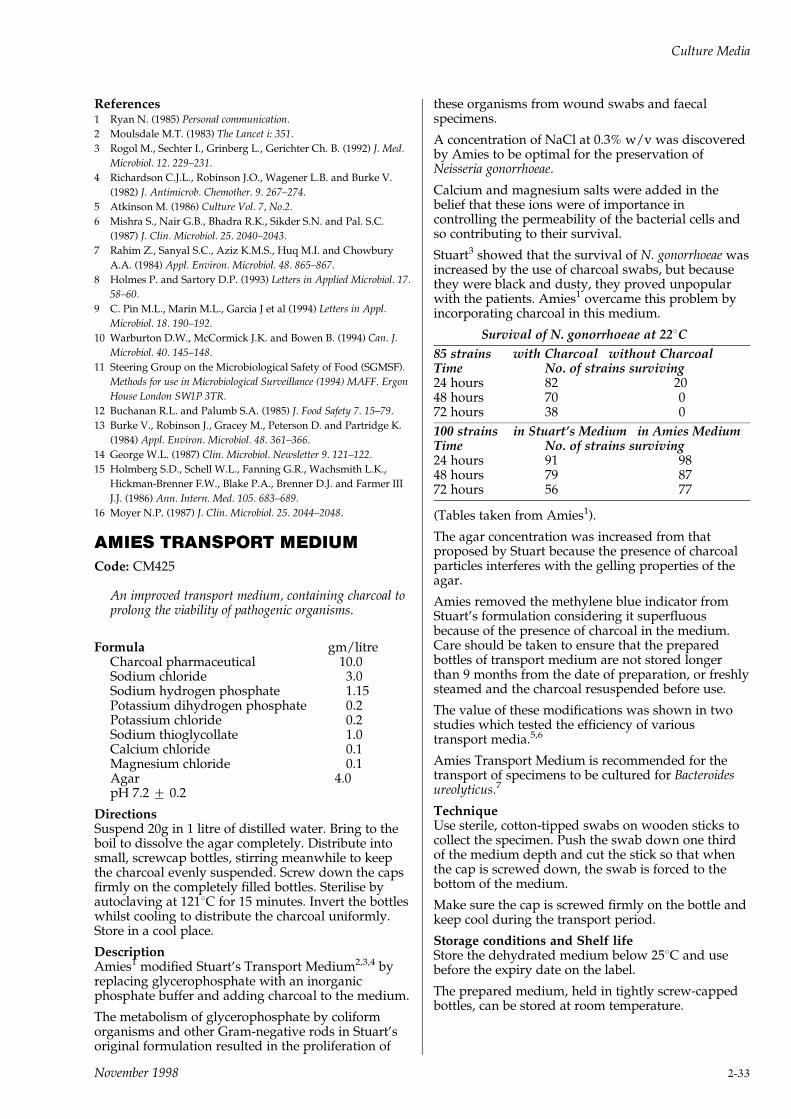
References1 Ryan N. (1985) Personal communication.
2 Moulsdale M.T. (1983) The Lancet i: 351.
3 Rogol M., Sechter I., Grinberg L., Gerichter Ch. B. (1992) J. Med.
Microbiol. 12. 229±231.
4 Richardson C.J.L., Robinson J.O., Wagener L.B. and Burke V.
(1982) J. Antimicrob. Chemother. 9. 267±274.
5 Atkinson M. (1986) Culture Vol. 7, No.2.
6 Mishra S., Nair G.B., Bhadra R.K., Sikder S.N. and Pal. S.C.
(1987) J. Clin. Microbiol. 25. 2040±2043.
7 Rahim Z., Sanyal S.C., Aziz K.M.S., Huq M.I. and Chowbury
A.A. (1984) Appl. Environ. Microbiol. 48. 865±867.
8 Holmes P. and Sartory D.P. (1993) Letters in Applied Microbiol. 17.
58±60.
9 C. Pin M.L., Marin M.L., Garcia J et al (1994) Letters in Appl.
Microbiol. 18. 190±192.
10 Warburton D.W., McCormick J.K. and Bowen B. (1994) Can. J.
Microbiol. 40. 145±148.
11 Steering Group on the Microbiological Safety of Food (SGMSF).
Methods for use in Microbiological Surveillance (1994) MAFF. Ergon
House London SW1P 3TR.
12 Buchanan R.L. and Palumb S.A. (1985) J. Food Safety 7. 15±79.
13 Burke V., Robinson J., Gracey M., Peterson D. and Partridge K.
(1984) Appl. Environ. Microbiol. 48. 361±366.
14 George W.L. (1987) Clin. Microbiol. Newsletter 9. 121±122.
15 Holmberg S.D., Schell W.L., Fanning G.R., Wachsmith L.K.,
Hickman-Brenner F.W., Blake P.A., Brenner D.J. and Farmer III
J.J. (1986) Ann. Intern. Med. 105. 683±689.
16 Moyer N.P. (1987) J. Clin. Microbiol. 25. 2044±2048.
AMIES TRANSPORT MEDIUMCode: CM425
An improved transport medium, containing charcoal toprolong the viability of pathogenic organisms.
Formula gm/litreCharcoal pharmaceutical 10.0Sodium chloride 3.0Sodium hydrogen phosphate 1.15Potassium dihydrogen phosphate 0.2Potassium chloride 0.2Sodium thioglycollate 1.0Calcium chloride 0.1Magnesium chloride 0.1Agar 4.0pH 7.2 + 0.2
DirectionsSuspend 20g in 1 litre of distilled water. Bring to theboil to dissolve the agar completely. Distribute intosmall, screwcap bottles, stirring meanwhile to keepthe charcoal evenly suspended. Screw down the capsfirmly on the completely filled bottles. Sterilise byautoclaving at 1218C for 15 minutes. Invert the bottleswhilst cooling to distribute the charcoal uniformly.Store in a cool place.
DescriptionAmies1 modified Stuart's Transport Medium2,3,4 byreplacing glycerophosphate with an inorganicphosphate buffer and adding charcoal to the medium.
The metabolism of glycerophosphate by coliformorganisms and other Gram-negative rods in Stuart'soriginal formulation resulted in the proliferation of
these organisms from wound swabs and faecalspecimens.
A concentration of NaCl at 0.3% w/v was discoveredby Amies to be optimal for the preservation ofNeisseria gonorrhoeae.
Calcium and magnesium salts were added in thebelief that these ions were of importance incontrolling the permeability of the bacterial cells andso contributing to their survival.
Stuart3 showed that the survival of N. gonorrhoeae wasincreased by the use of charcoal swabs, but becausethey were black and dusty, they proved unpopularwith the patients. Amies1 overcame this problem byincorporating charcoal in this medium.
Survival of N. gonorrhoeae at 228C
85 strains with Charcoal without CharcoalTime No. of strains surviving24 hours 82 2048 hours 70 072 hours 38 0
100 strains in Stuart's Medium in Amies MediumTime No. of strains surviving24 hours 91 9848 hours 79 8772 hours 56 77
(Tables taken from Amies1).
The agar concentration was increased from thatproposed by Stuart because the presence of charcoalparticles interferes with the gelling properties of theagar.
Amies removed the methylene blue indicator fromStuart's formulation considering it superfluousbecause of the presence of charcoal in the medium.Care should be taken to ensure that the preparedbottles of transport medium are not stored longerthan 9 months from the date of preparation, or freshlysteamed and the charcoal resuspended before use.
The value of these modifications was shown in twostudies which tested the efficiency of varioustransport media.5,6
Amies Transport Medium is recommended for thetransport of specimens to be cultured for Bacteroidesureolyticus.7
TechniqueUse sterile, cotton-tipped swabs on wooden sticks tocollect the specimen. Push the swab down one thirdof the medium depth and cut the stick so that whenthe cap is screwed down, the swab is forced to thebottom of the medium.
Make sure the cap is screwed firmly on the bottle andkeep cool during the transport period.
Storage conditions and Shelf lifeStore the dehydrated medium below 258C and usebefore the expiry date on the label.
The prepared medium, held in tightly screw-cappedbottles, can be stored at room temperature.
Culture Media
November 1998 2-33
Quality ControlPositive control:
Staphylococcus aureus ATCC1 25923.Escherichia coli ATCC1 25922
Negative control:Uninoculated medium.
PrecautionsIt is important that the charcoal is properlysuspended in the medium, invert the bottles when thebottles are cool but the agar still liquid.
During preparation of the medium, avoid prolongedheating in open flasks because thioglycollate isvolatile.
Old medium should be freshly steamed and thecharcoal resuspended before use.
Keep medium cool during transport but do not freeze.
References1 Amies C.R. (1967) Can. J. Pub. Hlth. 58. 296±300.
2 Stuart R.D. (1946) J. Path. Bact. 58. 343±345.
3 Stuart R.D. (1959) Pub. Hlth. Rep. 74. 431±435.
4 Stuart R.D., Toshach Sheila R. and Patsula Teresa M. (1954) Acta.
Pathol. Microbiol. Scand. 74. 371±374.
5 Gastrin L., Kallings O. and Marcetic A. (1968) Acta. Pathol.
Microbiol. Scand. 74. 371±374.
6 Barry A.L., Fay G.D. and Sauer R.L. (1972) Appl. Microbiol. 24.
31±33.
7 Bennett K.W., Eley A. and Woolley P.D. (1990) Eur. J. Clin.
Microbiol. Inf. Dis. 9. 237±238.
ANAEROBE BASAL AGARCode: CM972
A nutrient agar for the growth of anaerobic micro-organisms, particularly Bacteroides spp. and otherfastidious anaerobes.
Formula gm/litrePeptone 16.0Yeast extract 7.0Sodium chloride 5.0Starch 1.0Dextrose 1.0Sodium pyruvate 1.0Arginine 1.0Sodium succinate 0.5Sodium bicarbonate 0.4L-cysteine HCl 0.25Ferric pyrophosphate 0.5Haemin 0.005Vitamin K 0.0005Dithiothreitol 0.25Agar 12.0pH 7.0 + 0.2
DirectionsSuspend 46g in 1 litre of distilled water. Sterilise byautoclaving at 1218C for 15 minutes. Cool to 50±558Cand aseptically add 5±10% sterile Defibrinated HorseBlood SR50.
Mix well and pour in sterile petri dishes.
500g of this medium makes 10.8 litres.
DescriptionAnaerobe Basal Agar CM972 contains peptones,carefully selected to support good growth ofanaerobic bacteria and yeast extract as a vitaminsource. Starch is present to absorb any toxicmetabolites1. Sufficient arginine is added to ensurethe growth of Eubacterium lentum2, whilst haemin andvitamin K are growth factors required by manyBacteroides species3. Haemin is also required byPorphyromonas species. Sodium succinate improvesthe growth of Prevotella melaninogenica and Bacteroidesspecies4. Sodium pyruvate is added as an energysource for asaccharolytic cocci such as Veillonella. Italso acts similarly to catalase and degrades traces ofhydrogen peroxide, which may be produced by theaction of molecular oxygen on media components5. L-cysteine hydrochloride and dithiothreitol are reducingagents, and cysteine has also been shown to stimulatethe growth of some anaerobes6.
TechniqueInoculate the medium by surface plating to obtainsingle colonies. Incubate anaerobically for up to 5days at 378C. Anaerobic conditions can be achievedusing the Oxoid AnaeroGen Atmosphere GenerationSystem AN025A with the Oxoid AnaeroJar AG025A.
The prepared medium may be stored for up to 3weeks at 2±88C in the dark.
The medium may be rendered selective for Gram-negative anaerobes by the addition of G-Nsupplement SR108 and for non-sporing anaerobes bythe addition of N-S supplement SR107 with Tween 80.Neomycin Selective Supplement SR163 can be addedto select for Clostridia.
Storage conditions and Shelf lifeAnaerobe Basal Agar CM972 should be stored tightlycapped in the original container at 108C±258C. Whenstored as directed, the medium will remain stableuntil the expiry date printed on the bottle.
Quality ControlPositive control:Peptostreptococcus anaerobius ATCC1 27337Prevotella melaninogenicus ATCC1 15930Clostridium perfringens ATCC1 13124
Negative control:Uninoculated medium.
PrecautionsAnaerobe Basal Agar CM972 should be used for invitro purposes only. Do not use beyond the statedexpiry date, or if the product is caked, discoloured orshows any sign of deterioration.
References1 Ajello GW. Geely JC. Hayes PS. et al. Trans-isolate medium: a
new medium for primary culturing and transport of Neisseria
meningitidis, Streptococcus pneumoniae and Haemophilus influenzae.
J. Clin. Micro. 1984:20:55±8.
2 Sperry JF. Wilkins TD. Arginine, a growth-limiting factor for
Eubacterium lentum. J. Bacteriol 1976:127:780±4.
3 Gibbons RJ. and MacDonnald JB. Haemin and vitamin K
compounds as required factors for the cultivation of certain
strains of Bacteroides melaninogenicus. J. Bact. 1960:80:164±170.
Culture Media
2-34 November 1998
4 Lev M. Keudell KC. and Milford AF. Succinate as a growth
factor for Bacteroides melaninogenicus. J. Bact. 1971:108:175±8.
5 Neilson PA. Role of reduced sulphur compounds in nutrition of
Proprionobacterium acnes. J. Clin. Micr. 1983:17:276±9.
6 Shanson DC. and Singh J. Effect of adding cysteine to brain-
heart infusion broth on the isolation of Bacteroides fragilis from
experimental blood cultures. J. Clin. Path. 1981:34:221±3.
Anaerobe Basal BrothCode: CM957
A nutrient broth for the growth of anaerobic micro-organisms, particularly Bacteroides spp. and otherfastidious anaerobes.
Formula gm/litrePeptone 16.0Yeast extract 7.0Sodium chloride 5.0Starch 1.0Dextrose 1.0Sodium pyruvate 1.0Arginine 1.0Sodium succinate 0.5L-cysteine HCl 0.5Sodium bicarbonate 0.4Ferric pyrophosphate 0.5Haemin 0.005Vitamin K 0.0005Sodium thioglycollate 0.5Dithiothreitol 1.0
DirectionsSuspend 35.4g of Anaerobe Basal Broth in 1 litre ofdistilled water. Bring to the boil to dissolvecompletely. Distribute into final containers. Steriliseby autoclaving at 1218C for 15 minutes.
DescriptionOxoid Anaerobe Basal Broth CM957 is formulatedfrom a range of nutrients which have been selected tooptimise the recovery and growth of the majority ofanaerobic organisms of clinical importance.
The formulation includes yeast extract as a source ofvitamins and starch is included to absorb toxicproducts1. Sufficient arginine is added to ensuregrowth of Eubacterium lentum2, whilst haemin andvitamin K are present as they are essential for thegrowth of Bacteroides spp3. Pyruvate is present as anenergy source for asaccharolytic cocci such asVeillonella spp. It also eliminates traces of hydrogenperoxide which may be produced by the action ofmolecular oxygen on medium constituents4. Sodiumsuccinate improves the growth of Prevotellamelaninogenica and Bacteroides spp5. It is includedtogether with the reducing agent L-cysteinehydrochloride, which has been shown to stimulatedirectly the growth of some anaerobes6.
TechniqueIt is preferable to use freshly reconstituted and sterilemedium which is inoculated as soon as it has cooledto room temperature. Tubes which are not used onthe day of preparation should be placed in a boilingwater bath or steamer for approximately 15 minutes
to remove dissolved oxygen. They should be allowedto cool without agitation and then inoculated.
Storage conditions and Shelf lifeAnaerobe Basal Broth CM957 should be stored tightlycapped in the original container at 108C±258C. Whenstored as directed, the medium will remain stableuntil the expiry date printed on the bottle.
Quality ControlPositive control:Peptostreptococcus anaerobius ATCC1 27337Prevotella melaninogenica NCTC 11321Clostridium perfringens ATCC1 13124
Negative control:Uninoculated medium
PrecautionsAnaerobe Basal Broth CM957 should only be used forin vitro diagnostic purposes.
Do not use beyond the stated expiry date, or if theproduct is caked, discoloured or shows any sign ofdeterioration.
References1 Ajello GW. Geely JC. Hayes PS. et al. Trans-isolate medium: a
new medium for primary culturing and transport of Neisseria
meningitidis, Streptococcus pneumoniae and Haemophilus influenzae.
J. Clin. Micro. 1984:20:55±8.
2 Sperry JF. Wilkins TD. Arginine, a growth-limiting factor for
Eubacterium lentum. J. Bacteriol 1976:127:780±4.
3 Gibbons RJ. and MacDonnald JB. Haemin and vitamin K
compounds as required factors for the cultivation of certain
strains of Bacteroides melaninogenicus. J. Bact. 1960:80:164±170.
4 Neilson PA. Role of reduced sulphur compounds in nutrition of
Proprionobacterium acnes. J. Clin. Micr. 1983:17:276±9.
5 Lev M. Keudell KC. and Milford AF. Succinate as a growth
factor for Bacteroides melaninogenicus. J. Bact. 1971:108:175±8.
6 Shanson DC. and Singh J. Effect of adding cysteine to brain-
heart infusion broth on the isolation of Bacteroides fragilis from
experimental blood cultures. J. Clin. Path. 1981:34:221±3.
Antibiotic Assay MediaCodes: CM327/CM335/CM287
The methods and media required for the bio-assay ofantibiotics in pharmaceutical products, foods andother materials are specified in the US PharmacopoeiaXX1 and by the FDA2 as well as other works ofreference3. Grove and Randall4 defined the mediaformulations which are best for the different assayorganisms used when testing for different antibioticsand these are numbered 1 to 13, 19, 20 and 21. Toavoid confusion the original numbers given by Groveand Randall are retained but some of the formulationsare less widely used than others.
Antibiotic media Nos. 1, 2 and 3 are the most widelyused and there is the advantage that antibiotic mediaNos. 5, 8 and 11 can be prepared from them by simplepH adjustment.
pH ranges of Antibiotic Assay Media5.6±5.7 6.6 7.0 7.9±8.0No. 8 Nos. 1 & 2 No. 3 Nos. 5±11
Culture Media
November 1998 2-35

The assay of antibiotics is a highly skilled processwhich requires very close attention to the detailsspecified in the official publications and these must beconsulted. The general principles are, however,straightforward and can be divided into (1) diffusionplate assay and (2) turbidometric assay.
Diffusion plate assays depend upon the diffusion ofantibiotics from reservoirs on or in the agar layer.Measurement of the amount of diffusion is made bycomparing the sizes of zones of growth inhibition,using standard strains of susceptible organisms andknown amounts of reference antibiotics.
The results depend on critical rates of diffusion of theantibiotic, critical growth rates of the standardorganisms and critical minimal inhibitory coefficientlevels of each organism.
The reservoirs may be holes cut into the agar andfilled with known concentrations of antibiotic, ormetal cylinders placed on the surface of the agar andsimilarly filled, or paper discs charged with antibiotic.
Large square plates (250 x 250mm) or flat sheets ofglass are commonly used and a 4mm depth of baseagar is poured and allowed to set. Then a thin (1mm)layer of seeded agar is poured over the base layer andallowed to set. The holes are punched into theselayers or the various reservoirs placed on the seedlayer. It is essential that the layers of agar are even indepth and care must be taken to pour them onproperly levelled benches. Up to 36 separatereservoirs can be placed in dishes of this size.
ANTIBIOTIC MEDIA USED FOR MICROBIOLOGICAL ASSAYS
Antibiotic Organism Inoc. Diffusion Turbidomet.Base Seed
Amikacin Staph. aureus 1 3ATCC1 6538P
Amoxicillin Micro. luteus 1 11 11ATCC1 9341
Ampicillin Micro. luteus 1 11 11ATCC1 9341
Cephalosporins Staph. aureus 1 2 1ATCC1 6538P
Chloramphenicol Esch. coli 1 3ATCC1 10536
Cloxacillin Staph. aureus 1 2 1ATCC1 6538P
Erythromycin Micro. luteus 1 11 11ATCC1 9341
Gentamycin Staph. epiderm. 1 11 11ATCC1 12228
Kanamycin Staph. aureus 1 3ATCC1 6538P
Methicillin Staph. aureus 1 1 2ATCC1 6538P
Neomycin Staph. aureus 1 11 11ATCC1 6538P
Penicillin Staph. aureus 1 2 1ATCC1 6538P
Streptomycin Bac. subtilis 1 5 5ATCC1 6633
Kleb. pneumo. 1 3ATCC1 10031
Tetracycline Staph. aureus 1 3ATCC1 6538P
Tobramycin Staph. aureus 1 3ATCC1 6538P
Vancomycin Bac. subtilis 1 8 8ATCC1 6633
ATCC1 is a registered trade mark of the American Type Culture Collection.
Culture Media
2-36 November 1998
Turbidimetric assays are an alternative method withadvantages of short incubation times (3±4 hours) butthey lack the accuracy of the diffusion assay5.Samples must be clear and not absorb the wavelengthof light used in the spectrophotometer.
Dilutions of sample and dilutions of the antibiotic aremade in parallel in 1ml volumes and 9ml volumes ofseeded broth are added to each tube. The tubes arethen incubated in a water bath for 3±4 hours at theappropriate temperature and at the end of theincubation period the growth is stopped by theaddition of formaldehyde to each tube. The amount ofgrowth that has taken place within the incubationperiod is measured by light transmission in aspectrophotometer. Comparison of the readings of theunknown sample and those of the known dilutions ofthe antibiotic will establish the level of antibiotic inthe sample.
Standard organism cultures used in assays arenormally maintained on agar slopes and transferredat 1±2 week intervals. The inoculum used for an assayis usually washed from an overnight culture on anagar slope, using saline or buffered peptone waterand further diluted to a standard density. Sporesuspensions of Bacillus subtilis and B. cereus are usedand it is important to cultivate these organisms onmedia which enhance spore production e.g. AntibioticMedium No.l with 300mg of manganese sulphate perlitre added to the medium. Surface seed agar platesand examine the growth microscopically during thedays of incubation until the majority of the organismsare seen to be bearing spores. Pasteurise the sporesuspension to destroy the vegetative organisms andthus ensure clean edges to the zones of inhibition.
References1 The United States Pharmacopeia, Biological Tests and Assays.
XX. Revision, Rockville Md.: United States Pharmacopeial
Convention, 1980, pp. 882±888.
2 Tests and Methods of Assay of Antibiotics and Antibiotic-
Containing drugs, FDA, CFR, Title 21, Part 436, Subpart D,
Washington DC: US Government Printing Office, paras. 436.100±
436.106, p. 242±259 (April 1) 1983.
3 Hausler W. J. (Ed) Standard Methods for the Examination of Dairy
Products, 13th edn. Washington D.C.: American Public Health
Association, 1972.
4 Grove D.C. and Randall W.A. Assay Methods of Antibiotics,
New York: Medical Encyclopedia 1955.
5 Reeves D.S. and Bywater M.J. (1975) J. Antimicrob. Chemoth. 1.
103±107.
ANTIBIOTIC MEDIUM NO. 1
SEED AGAR
Code: CM327
A medium recommended for the seed layer in thepreparation of plates for the microbiological assay ofantibiotics.
Formula gm/litrePeptone 6.0Tryptone 4.0Yeast extract 3.0`Lab-Lemco' powder 1.5Glucose 1.0Agar No.1 11.5pH 6.5 + 0.2
DirectionsSuspend 27g in 1 litre of distilled water and bring tothe boil to dissolve completely. Sterilise byautoclaving at 1218C for 15 minutes.
DescriptionThis is perhaps the most important medium inantibiotic assay work and it is in a specially modifiedform to take advantage of the properties of OxoidAgar No.1.
Hanus, Sands and Bennett1 drew attention to theinhibitory properties which certain agars havetowards some antibiotics, particularly streptomycin,kanamycin, polymyxin B and neomycin. BecauseOxoid Agar No.1 does not share these inhibitoryproperties it is especially suited to antibiotic assaywork. In addition, this agar, with its superiordiffusion properties, produces more clearly definedinhibition zones.
NB In the Oxoid formulation, only 11.5 grams of Agar No.1 are used to give the equivalent gel strength of 15 gramsordinary agar.
This medium is used as a seed agar with Micrococcusflavus for the plate assay of bacitracin; with Sarcinalutea for the plate assay of chloramphenicol and withStaphylococcus aureus for the assay of kanamycinsulphate, penicillin G, sodium methicillin and sodiumoxacillin.
It is also employed as a base agar in the assay of thefollowing drugs: chloramphenicol, kanamycinsulphate, colistin sulphate, sodium methicillin,sodium oxacillin and vancomycin hydrochloride.
Reference1 Hanus F. J., Sands J. G. and Bennett E. O. (1967) Applied
Microbiology 15(1) 31±34.
Culture Media
November 1998 2-37
ANTIBIOTIC MEDIUM NO. 2
BASE AGAR
Code: CM335
A standard medium for use as a base layer in thepreparation of plates for the microbiological assay ofantibiotics.
Formula gm/litrePeptone 6.0Yeast extract 3.0`Lab-Lemco' powder 1.5Agar No.1 10.0pH 6.5 + 0.2
DirectionsSuspend 20.5g in 1 litre of distilled water. Bring to theboil to dissolve completely. Sterilise by autoclaving at1218C for 15 minutes.
DescriptionThis new formulation replaces the older productlabelled `Base Agar' which has been withdrawn.
Medium No.2 is used as the base agar in the plateassay of bacitracin and in the assay of penicillin G.
ANTIBIOTIC MEDIUM NO. 3
ASSAY BROTH
Code: CM287
Used in the serial dilution assay of penicillin and otherantibiotics.
Formula gm/litrePeptone 5.0Yeast extract 1.5`Lab-Lemco' powder 1.5Glucose 1.0Sodium chloride 3.5Dipotassium hydrogen phosphate 3.68Potassium dihydrogen phosphate 1.32pH 7.0 + 0.2
DirectionsAdd 17.5 grams to 1 litre of warm (608C) distilledwater and mix well to dissolve. Distribute andsterilise by autoclaving at 1218C for 15 minutes.
DescriptionMedium No.3 is used in the turbidimetric assay ofpenicillin and tetracycline with Staphylococcus aureus.
Taking advantage of modern technology, thismedium is based on the original `Penassay Broth'which has been withdrawn.
ANTIBIOTIC MEDIUM NO. 5
STREPTOMYCIN ASSAY AGAR
This is very easily made by changing the pH ofAntibiotic Medium No.2. To one litre of Base Agaradd 5ml of normal caustic soda solution. This may becarried out either prior to autoclaving or asepticallyafter sterilisation. This will bring the pH to 7.9, but nofiltration will be necessary. This medium is suitablefor the streptomycin assay, using the cylinder platetechnique and a test organism of Bacillus subtilis andgives clearly defined inhibition zones.
After this base layer has gelled, it is overlaid with thesame medium already inoculated with spores of thetest organism.
ANTIBIOTIC MEDIUM NO. 8
FOR THE PLATE ASSAY OF TETRACYCLINEAND VANCOMYCIN
This can readily be made by a small adjustment of thepH of Medium No.2. To convert Medium No.2 intoMedium No.8 aseptically add 1.25ml of N/1 HCl,after sterilisation, to the still molten Medium No.2 at50±558C. This will bring the pH down to 5.6±5.7. Nofiltration will be necessary. Medium No.8 is used forthe base agar and seed agar for the plate assay oftetracycline. It is also used as the seed agar forvancomycin hydrochloride.
ANTIBIOTIC MEDIUM NO.11
FOR THE PLATE ASSAY OF NEOMYCINSULPHATE
This can readily be made by a small change of pH ofMedium No.l. To one litre of Base Agar add 5ml ofnormal caustic soda solution. This may be carried outeither prior to autoclaving or aseptically aftersterilisation. The pH will then be 7.9±8.0. No filtrationwill be necessary. Medium No. 11 is used as the baseagar and seed agar for the plate assay of neomycinsulphate.
Culture Media
2-38 November 1998

ARCOBACTER BROTHCode: CM965
An enrichment broth for Arcobacter species.
Formula gm/litrePeptone 18.0Yeast extract 1.0Sodium chloride 5.0
Directions for use with CAT Supplement SR174Dissolve 12g of Arcobacter Broth CM965 in 500ml ofdistilled water. Sterilise at 1218C for 15 minutes.
Allow to cool to 508C and add one vial of CATSelective Supplement SR174E reconstituted asdirected.
Dispense into sterile containers.
pH 7.2 � 0.2
Directions for use with CCDA Supplement (SR155)Dissolve 12g of Arcobacter Broth CM965 in 500ml ofdistilled water. Sterilise at 1218C for 15 minutes.Allow to cool to 508C and add one vial of CCDASelective Supplement SR155 reconstituted as directed.
Dispense into sterile containers.
Incubate at 308C aerobically for 24 hours.
DescriptionOxoid Arcobacter Broth CM965 is intended for usewith Cefoperazone, Amphotericin B, Teicoplanin(CAT) Selective Supplement SR174 as a selectiveenrichment broth for the growth of Arcobacter speciesand with the more selective CCDA SR155 for theselective enrichment of Arcobacter butzleri.
Peptones in the base medium are specificallydesigned to provide the ideal growth conditions forArcobacter species. The incubation conditions and theabsence of blood or charcoal supplements suppressthe growth of Campylobacter species. Cefoperazone,Amphotericin B and Teicoplanin are added tosuppress the growth of competing flora, but allow thegrowth of Arcobacter species. CCDA SelectiveSupplement SR155 is substituted for CAT toselectively isolate Arcobacter butzleri1,2.
Arcobacters are micro aerophilic, Gram-negative rods,which were formerly classified as Campylobacter3.
Four Arcobacter species have been identified: A.butzleri, A. cryaerophilus, A. skirrowii and A. nitrofigilis,all of which have a greater propensity to grow in airthan Campylobacter spp.
A. butzleri, A. cryaerophilus, and A. skirrowii have beenassociated with disease in humans4,5, and typicallyare isolated from faecal samples.
A. butzleri has been isolated from patients withbacteraemia, peritonitis, endocarditis and diarrhoea.Patients with A. butzleri-associated diarrhoea typicallysuffer from abdominal pain and nausea, fever, chills,vomitting and malaise, but the organism has alsobeen implicated in an outbreak of recurrentabdominal cramps without diarrhoea6. The source ofinfection is usually contaminated water or sewage6.
A. cryaerophilus group 1B has been isolated frompatients with bacteraemia and diarrhoea4,5, althoughit is a much less common human isolate than A.butzleri6.
A. nitrofigilis has only been isolated from marsh grassto date, never from humans or animals. It is notthought to be clinically significant7.
Storage conditions and Shelf lifeArcobacter Broth CM965 should be stored at roomtemperature. When stored as directed, the reagentsremain stable until the expiry date shown on thelabel.
Quality ControlPositive control:Arcobacter butzleri ATCC1 12481 ± white/grey
colonies
Negative control:Escherichia coli ATCC1 25922 ± inhibited
PrecautionsArcobacter Broth CM965 should only be used for invitro diagnostic purposes.
References1 Data on file.
2 Lammerding, A.M., Harris, J.E., Lior, D.L. et al. Presented at the
81st annual meeting of IAMFES (1994).
3 Vandamme, P., Falsen, E., Rossau, R., Hoste, B., Segers, P.,
Tytgat, R., De Ley, J. (1991). Int. J. Syst. Bacteriol. 41:88±103.
4 Kiehlbauch, J.A., Brenner, D.J., Nicholson, M.A., Baker, C.N.,
Patton, C.M., Steigerwalt, A.G., Wachsmuth, I.K. (1991). J. Clin.
Microbiol. 29:376±385.
5 Vandamme, P., Vancanneyt, M., Pot, B., Mels, L., Hoste, B.,
Dewettinck, D., Vlaes, L., Van den Borre, C., Higgins, R.,
Hommer, J. (1992). Int. J. Syst. Bacteriol. 42:344±356.
6 Vandamme, P., Pugina, P., Benzi, G., Van Etterick, R., Vlaes, L.,
Kersters, K., Butzler, J., Lior, H., Lauwers, S. (1992). J. Clin.
Microbiol. 30:2335±2337.
7 Atabay, H.I and Corry, J.E.L. (1998) Int. J. Food Microbiol, 41, 53±
58.
AZIDE BLOOD AGAR BASECode: CM259
A selective medium for the detection and isolation ofstreptococci and staphylococci from faeces, sewage andother specimens. With added blood, the medium may beemployed for the simultaneous determination ofhaemolytic reactions.
Formula gm/litreTryptose 10.0`Lab-Lemco' powder 3.0Sodium chloride 5.0Sodium azide 0.2Agar 12.0pH 7.2 + 0.2
DirectionsSuspend 30g in 1 litre of distilled water and bring tothe boil to dissolve completely. Sterilise byautoclaving at 1218C for 15 minutes. For azide bloodagar, cool to 45±508C and add 5% of sterile blood.
Culture Media
November 1998 2-39
DescriptionA selective medium for the detection and isolation ofstreptococci from faeces, sewage, and other specimenscontaining a mixed flora. Azide Blood Agar Base issimilar to the medium used by Edwards1 for theisolation of mastitis streptococci. The sodium azidehas a bacteriostatic effect on most Gram-negativeorganisms but permits growth of Gram-positiveorganisms such as streptococci and some strains ofstaphylococci. Proteus species are slightly moreresistant than other Enterobacteriaceae but swarmingis prevented (Snyder and Lichstein2, Lichstein andSnyder3).
At the above concentration and pH, sodium azideexerts no appreciable effect on haemolysis so that themedium, with added blood, may be used for thesimultaneous determination of haemolytic reactions.
Azide blood agar is recommended by the AmericanPublic Health Association4 for the isolation ofstreptococci from cheese. The plates, inoculated withdilutions of emulsified cheese, are incubated at 358Cand representative colonies subcultured forsubsequent identification.
There are variations in formula of Azide Blood AgarBase which have been recommended for differentpurposes:
1 Packer5 increased the sodium azide concentration to0.9g per litre and added 0.002g per litre crystal violet.The pH was also adjusted to 6.8+ 0.1. This is a moreselective medium for faecal streptococci in foods6.
2 Packer5 and Wood7 used the above formulationwith 5% blood and the crystal violet increased to0.01g per litre, for the isolation of Erysipelothrixrhusiopathiae and Streptococcus pneumoniae.
3 Dale8 and Bohm9 recommended the addition ofphenol (1.0 to 2.5g per litre) to Packer's formulationto isolate E. rhusiopathiae.
Storage conditions and Shelf lifeStore the dehydrated medium below 258C and usebefore the expiry date on the label.
Store prepared blood agar plates of medium at 2±88C.
Quality ControlPositive control:
Enterococcus faecalis ATCC1 29212Staphylococcus aureus ATCC1 25923
Negative control:Proteus vulgaris ATCC1 13315Escherichia coli ATCC1 25922
PrecautionsProteus and Escherichia species may not always beinhibited on the Edward's formulation.
Always use a light inoculum for best selective results.
Anaerobic incubation will enhance haemolytic reactions.
Haemolytic reactions will not be typical on Packer'smodification of Azide Blood Agar Base. Streptococcuslactis will not grow on Packer's modification with 5%sheep blood.
Read the section on Hazard Precautions 2.7 for azide-containing media.
References1 Edwards S. J. (1933) J. Comp. Path. Therap. 46(4) 211±217.
2 Snydar M. L. and Lichstein H. C. (1940) J. Infect. Dis. 67(2) 113±
115.
3 Lichstein H. C. and Snyder M. L. (1941) J. Bact. 42(5) 653±664.
4 American Public Health Association (1978) Standard Methods for
the Examination of Dairy Products. 14th Edn. APHA Inc. New York.
5 Packer R. A. (1943) J. Bact. 46. 343±349.
6 Mossel D. A. A., Diepen H. M. J. van and De Bruin A. S. (1957) J.
Appl. Bact. 20(2) 265±272.
7 Wood R. L. (1965) Amer. J. Vet. Res. 26. 1303±1308.
8 Dale C. N. (1940) J. Bact. 40. 228±231.
9 Bohm K. H. (1971) Zbl. Bakt. I. Orig. 218. 330±334.
AZIDE DEXTROSE BROTH(ROTHE)Code: CM868
For the detection of enterococci in water.
Formula gm/litrePeptone 20.0Glucose 5.0Sodium chloride 5.0Di-potassium hydrogen phosphate 2.7Potassium dihydrogen phosphate 2.7Sodium azide 0.2Final pH 6.8 + 0.2
DirectionsAdd 35.6g to one litre of distilled water for singlestrength broth or 71.2g for double strength broth.Heat gently to dissolve. Dispense into final containersand sterilise by autoclaving at 1218C for 15 minutes.
DescriptionAzide Dextrose Broth (Rothe) is used for the detectionof enterococci in water and sewage1.
The presence of enterococci serves as an indicator offaecal contamination. Enterococci are better indicatorsthan E. coli of sewage pollution in chlorinated watersbecause they have a greater resistance to chlorine.
Mallmann and Seligmann2 recommended AzideDextrose Broth for the quantitative determination ofenterococci in water, sewage, foods and othermaterials suspected of contamination with sewage.
A blend of peptone and glucose render AzideDextrose Broth highly nutritious, and sodiumchloride maintains osmotic equilibrium. The use ofsodium azide as an inhibitor of Gram-negativeorganisms has been reported by several workers2,3,4,and the concentration selected provides optimumprotection for the enterococci while largelysuppressing the Gram-negative flora. The phosphatebuffer system controls pH.
TechniqueInoculate 10ml of medium with 1ml of the testsample. Inoculate a further three tubes with 0.1ml,0.01ml and 0.001ml sample respectively. For samplesof 10ml or more, use double strength broth. Incubateall tubes at 358C and examine for turbidity after 24and 48 hours. For a more detailed description pleaseconsult `Standard Methods for the Examination ofWater and Wastewater'.
Culture Media
2-40 November 1998

The presence of enterococci in the sample is indicatedby turbidity in the broth. Positive cultures should beinoculated into Ethyl Violet Azide Broth (Litsky)CM869 to confirm the presence of enterococci.
Storage conditions and Shelf lifeStore the dehydrated medium below 258C and usebefore the expiry date on the label.
Store the prepared medium at 2±88C.
Quality ControlPositive control
Enterococcus faecalis ATCC1 29212
Negative controlEscherichia coli ATCC1 25922
PrecautionsThis product contains less than 1% azide and has lowtoxicity. However, when handling the powder, weargloves, mask and eye protection. When washingazide products down sinks, use sufficient water toprevent accumulation of azide in the plumbing.
References1 Greenberg A. E. et al (ed). (1985) Standard Methods for the
Examination of Water and Wastewater, 16th ed. APHA,
Washington, D.C.
2 Mallmann W. L. and Seligmann E. B. (1950) Am. J. Public Health
40. 286.
3 Edwards S. J. (1933) J. Comp. Path. Therap. 46. 211.
4 Hartman G. (1937) Milchw. Forsch. 18. 166.
BACILLUS CEREUS SELECTIVEAGAR BASECode: CM617
A selective and diagnostic medium for the isolation andenumeration of Bacillus cereus.
Formula gm/litrePeptone 1.0Mannitol 10.0Sodium chloride 2.0Magnesium sulphate 0.1Disodium hydrogen phosphate 2.5Potassium dihydrogen phosphate 0.25Bromothymol blue 0.12Sodium pyruvate 10.0Agar 14.0pH 7.2 + 0.2
BACILLUS CEREUS SELECTIVESUPPLEMENT
Code: SR99
Vial contents (each vial is sufficient for 500ml ofmedium)
Polymyxin B 50,000 IU
DirectionsSuspend 20.5g in 475ml of distilled water and bringgently to the boil to dissolve completely. Sterilise byautoclaving at 1218C for 15 minutes. Cool to 508C andaseptically add the contents of one vial of OxoidBacillus cereus Selective Supplement SR99reconstituted with 2ml of sterile distilled water, then
add 25ml of sterile Egg Yolk Emulsion SR47. Mix welland pour into sterile petri dishes.
DescriptionBacillus Cereus Selective Agar CM617, is based on thehighly specific diagnostic and selective PEMBAmedium, developed by Holbrook and Anderson1 forthe isolation and enumeration of Bacillus cereus infoods. It meets the requirements for a medium that issufficiently selective to be able to detect small numbersof B. cereus cells and spores in the presence of largenumbers of other food contaminants. The medium isalso sufficiently diagnostic that colonies of B. cereus arereadily identified and confirmed by microscopicexamination.
The role of B. cereus in food poisoning, particularlyfrom the consumption of contaminated rice, is nowwell documented.2,3,4 The organism has also beenimplicated in eye infections5,6 and a wide range of otherconditions including abscess formation, meningitis,septicaemia and wound infection. B. cereus isrecognised as a significant pathogen in post-operativeand post-traumatic wounds of orthopaedic patients.7
Amongst veterinarians, B. cereus is a known cause ofdisease, especially mastitis, in ewes and heifers.8
In the formulation of Bacillus cereus Selective Agar apeptone level of 0.1% and the addition of sodiumpyruvate improve egg yolk precipitation and enhancesporulation. Bromothymol blue is added as a pHindicator to detect mannitol utilisation. The medium ismade selective by addition of Bacillus cereus SelectiveSupplement SR99, which gives a final concentration of100 IU of polymyxin B per ml of medium. Polymyxin Bas a selective agent for the isolation of B. cereus hasbeen previously suggested by Donovan9 and found tobe satisfactory by Mossel.10 It is recommended that,where large numbers of moulds are expected in theinoculum, filter-sterilised cycloheximide is added tothe medium at a final concentration of 40mg/ml.
The primary diagnostic features of the medium arethe colonial appearance, precipitation of hydrolysedlecithin and the failure of B. cereus to utilise mannitol.
The typical colonies of B. cereus are crenated, about5mm in diameter and have a distinctive turquoise topeacock blue colour surrounded by a good egg yolkprecipitate of the same colour.
These features distinguish B. cereus from other Bacillusspecies except B. thuringiensis. Other egg yolk-reactingorganisms which can grow on the medium, includingStaphylococcus aureus, Serratia marcescens and Proteusvulgaris are distinguished from B. cereus by colonyform and colour. These organisms also produce anegg yolk-clearing reaction in contrast to egg yolkprecipitate produced by B. cereus.
Microscope examination for presence of lipid globulesin the vegetative cells is recommended as a rapid andconfirmatory test for B. cereus and replaces the need forbiochemical testing. Holbrook and Anderson1 haveconfirmed that only B. cereus of the Bacillus species arecapable of possessing lipid globules in their vegetativecells when grown on the selective medium. One furtheradvantage of this test is that strains of B. cereus thatreact only weakly or not at all with egg yolk can bedetected and confirmed.
Culture Media
November 1998 2-41

Technique1 Homogenise 10g of the food sample for 30 seconds
in 90ml of 0.1% Peptone Water CM9 using aStomacher 400.11 Dried foods should first berehydrated by soaking 20g in 90ml of Tryptone saltsolution (Tryptone L42 0.3% and sodium chloride0.8%, pH 7.3) for 50 minutes at room temperature.Add a further 90ml of 0.1% peptone water to give afinal dilution of 10-1. Homogenise for 30 secondsusing the Stomacher 400.
2 Further dilutions of the homogenate should bemade in 0.1% peptone water.
3 Inoculate 0.1ml amounts of the 10-1 and higherdilutions on to the surface of the medium.
4 Incubate the plates at 358C for 24 hours.
5 Examine for typical colonies of B. cereus.
6 Leave the plates for a further 24 hours at roomtemperature in order to detect all the Bacillus cereuscolonies.
7 Confirm the presumptive identification of B. cereusby the Rapid Confirmatory Staining Procedure.
8 Report the results as the number of B. cereuscolonies per gram weight of the food sample.
The medium may also be used for detecting B. cereus inmilk. When necessary, decimal dilutions of the samplesshould be made in 0.1% peptone water. Undiluted anddiluted samples are inoculated directly on to plates ofagar and incubated. An incubation temperature of308C for 18 hours is recommended as optimal forpromoting the growth of B. cereus relative to that ofother organisms.9
For examining clinical specimens plates may beinoculated in the usual way.
Rapid Confirmatory Staining ProcedureThis staining method was developed by Holbrookand Anderson1 combining the spore stain of Ashby12
and the intracellular lipid stain of Burdon.13 Forreasons of safety, Citroclear* replaces xylene in theoriginal technique.
Procedure1 Prepare films from the centre of a 1 day old
colony or from the edge of a 2 day colony.
2 Air-dry the film and fix with minimal heating.
3 Flood the slide with aqueous 5% w/v malachitegreen and heat with a flaming alcohol swab untilsteam rises. Do not boil.
4 Leave for 2 minutes without re-heating.
5 Wash the slide with running water and blot dry.
6 Flood the slide with 0.3% w/v Sudan black in70% ethyl alcohol. Leave for 15 minutes.
7 Wash the slide with running Citroclear* from awash bottle for 5 seconds.
8 Blot dry using filter paper.
9 Flood the slide with aqueous 0.5% w/v safraninfor 20 seconds.
10 Wash under running water.
11 Blot dry and examine under the microscope usingthe oil immersion lens. A blue filter may be used toaccentuate the appearance of the lipid granules butthis will give a blue colour cast to the red of thecytoplasm.
* Citroclear is available from:
H.D. Supplies44 Rabans CloseRabans Lane Industrial EstateAylesburyBuckinghamshireHP19 3RS
Characteristic appearance of B. cereus vegetativecells.(i) Cells are 4±5 micron long and 1.0±1.5 micron
wide with square ends and rounded corners.
(ii) The spores stain pale green to mid green, arecentral or paracentral in position and do notswell the sporangium.
(iii) Lipid globules are black and the vegetativecytoplasm red.
The appearance, together with the typical colonyform, confirms the identification of B. cereus.
Storage conditions and Shelf lifeStore the dehydrated medium below 258C and usebefore the expiry date on the label.
The prepared medium may be stored at 2±88C.
Quality ControlPositive Control:
Bacillus cereus ATCC1 10876Bacillus subtilis ATCC1 6633 (should be readily
differentiated by colony form and colour)
Negative control:Bacillus coagulans ATCC1 7050
PrecautionsOn this medium B. cereus is indistinguishable fromB. thuringiensis.
Identify B. cereus by colony form, colour, egg yolkhydrolysis and confirm with cell and sporemorphology.14
Occasional strains of B. cereus show weak or negativeegg yolk reactions.
References1 Holbrook R. and Anderson J.M. (1980) Can. J. Microbiol., 26 (7)
753±759.
2 Brit. Med. J., 15 January, 1972, 189.
3 Brit. Med. J., 22 September. 1973. 647.
4 Mortimer P.R. and McCann G., 25 May, 1974, Lancet, 1043±1045.
5 Davenport R. and Smith C. (1952) Brit. J. Ophthal. 36. 39.
6 Bouza E., Grant S., Jordan C., Yook R. and Sulit H. (1979) Arch.
Ophthalmol. 97. 498±499.
7 Akesson A., HedstroÈm S.A. and Ripa T. (1991) Scand. J. Inf. Dis.
23. 71±77.
8 Wohlgemuth K., Kirkbride C.A., Bicknell E.J. and Ellis R.P.
(1972) J. Amer. Vet. Med. Ass., 161. 1691±1695.
9 Donovan K.O. (1958) J. Appl. Bacteriol., 21 (1) 100±103.
10 Mossel D.A.A., Koopman M.J. and Jongerius E. (1967) J. Appl.
Microbiol., 15 (3) 650±653.
11 Supplied by A.J. Seward, Pharm. Mfrs. and Distrib., UAC
House, 8±16 Blackfriars Road, London SE1.
12 Ashby G.K. (1938) Science, 87, 433±435.
13 Burdon K.L. (1946) J. Bacteriol., 52. 665±678.
14 DeaÂk T. and TimaÂr E. (1988) Int. J. Food Microbiology. 6. 115±125.
Culture Media
2-42 November 1998
BAIRD-PARKER AGAR BASECode: CM275
A selective and diagnostic medium for the isolation andenumeration of Staphylococcus aureus in foods.
Formula gm/litreTryptone 10.0`Lab-Lemco' powder 5.0Yeast extract 1.0Sodium pyruvate 10.0Glycine 12.0Lithium chloride 5.0Agar 20.0pH 6.8 + 0.2
DirectionsSuspend 63g in one litre of distilled water and boil todissolve the medium completely. Dispense into tubesor flasks and sterilise by autoclaving at 1218C for 15minutes. Cool to 508C and aseptically add 50ml ofEgg Yolk-Tellurite Emulsion SR54. Mix well beforepouring.
Prepared plates may be stored at 48C.
Description (with E-Y-T Emulsion SR54)Baird-Parker1 developed this medium from thetellurite-glycine formulation of Zebovitz et al.2 andimproved its reliability in isolating Staph. aureus fromfoods.
Baird-Parker added sodium pyruvate, to protectdamaged cells and aid their recovery2 and egg yolkemulsion as a diagnostic agent. It is now widelyrecommended by national and international bodiesfor the isolation of Staph. aureus4.
The selective agents glycine, lithium and telluritehave been carefully balanced to suppress the growthof most bacteria present in foods, without inhibitingStaph. aureus.
Egg yolk emulsion makes the medium yellow andopaque. Staph. aureus reduces tellurite to form grey-black shiny colonies and then produces clear zonesaround the colonies by proteolytic action. This clearzone with typical grey-black colony is diagnostic forStaph. aureus. On further incubation, most strains ofStaph. aureus form opaque haloes around the colonies.and this is probably the action of a lipase. Not allstrains of Staph. aureus produce both reactions. Somestrains of Staph. saprophyticus produce both clearzones and opaque haloes but experienced workerscan distinguish these from Staph. aureus by the longerincubation time required5.
Colonies typical of Staph. aureus but without an eggyolk reaction should also be tested for coagulaseproduction6.
Egg yolk reaction negative strains of Staph. aureusmay occur in some foods, especially cheese.
Smith and Baird-Parker7 found that the addition of 50mgof sulphametazine per ml of medium suppressed thegrowth and swarming of Proteus species. Small numbersof Staphylococcus aureus could then be recovered fromspecimens containing mixed Proteus strains.
Baird-Parker and Davenport8 showed that therecovery of damaged staphylococci was greater onBaird-Parker medium than on other recovery mediatested.
Broeke9 and de Waart et al.10 found Baird-Parkermedium valuable in ecological studies on foodsincriminated in staphyloenterotoxicosis. 97.5% of the522 strains of Staph. aureus tested, isolated fromhuman and food origins developed characteristicallyand quantitatively on Baird-Parker medium.
Colony characteristics of typical organisms onBaird-Parker Egg Yolk-Tellurite Medium
OrganismStaph. aureus
GrowthGood
ColonyGrey-black shiny convex 1±1.5mm diameter (18hours) up to 3mm (48 hours) narrow white entiremargin surrounded by zone of clearing 2±5mm.
OrganismStaph. epidermidis
GrowthVariable
ColonyNot shiny black and seldom produces clearing.
OrganismStaph. saprophyticus
GrowthVariable
ColonyIrregular and may produce clearing. Wide opaquezones may be produced in 24hrs.
OrganismMicrococcus species
GrowthVariable
ColonyVery small in shades of brown and black. Noclearing.
OrganismBacillus species
GrowthVariable
ColonyDark brown matt with occasional clearing after48hrs.
OrganismEscherichia coli.
GrowthVariable
Culture Media
November 1998 2-43
ColonyLarge brown-black.
OrganismProteus species
GrowthVariable
ColonyBrown-black with no clearing.
OrganismYeasts.
GrowthVariable
ColonyWhite, no clearing.
Technique1 Dry the surface of agar plates for a minimal period
of time prior to use.
2 With a glass spatula, spread 0.1ml aliquots of fooddilutions made up in buffered peptone water onthe agar surface until it is dry. Up to 0.5ml may beused on larger dishes (24 cm).
3 Incubate the inverted dishes at 358C. Examine after24 hours and look for typical colonies of Staph.aureus. Re-incubate negative cultures for a further24 hours.
Quantitative resultsIncubate the dishes for 48 hours and select those with20±200 colonies.
Count the Staph. aureus-like colonies and test them forcoagulase reaction.
Report Staph. aureus results per gram of food.
RPF SUPPLEMENT
Code: SR122
This supplement is used as an alternative to Egg Yolk-Tellurite Emulsion SR54 in Baird-Parker medium toidentify coagulase-positive staphylococci.
Formula (per vial sufficient for 100ml of medium)
Bovine fibrinogen 0.375gRabbit plasma 2.5mlTrypsin inhibitor 2.5mgPotassium tellurite 2.5mg
DirectionsTo one vial aseptically add 10ml of sterile distilledwater. Turn the vial end-over-end to dissolve. Avoidfrothing the solution. Dissolution is not obtainedimmediately. Leave for one to two hours to dissolvecompletely. Aseptically add the vial contents to 90mlof sterile Baird-Parker Agar Base (Oxoid CM275)cooled to 488C. Mix well and use immediately.
Description (with RPF Supplement SR122)
A recognised disadvantage of Baird-Parker mediumwith egg yolk is that the identity of the staphylococciisolated must be confirmed as Staph. aureus with acoagulase reaction. Also there are some strains ofStaph. aureus which give negative egg yolk reactionsand therefore all suspicious colonies should betested.11,12
Attempts were made to substitute pig plasma for eggyolk in Baird-Parker medium, to obtain an in-situcoagulase test13. Hauschild14 improved the reliabiltyof the coagulase reaction by adding bovine fibrinogenand trypsin inhibitor to the pig plasma. Beckers etal.15 showed that rabbit plasma used in place of pigplasma overcame the problems of false positive andfalse negative reactions in the medium. A rabbitplasma- fibrinogen-trypsin inhibitor-telluritesupplement for Baird-Parker medium was created byBeckers et al.15 which gave reliable coagulasereactions for Staph. aureus.
Sawhney16 observed variation in yields of Staph.aureus cultures when comparing RPF supplementwith egg yolk supplement. He showed that it wasnecessary to reduce the tellurite level in the RPFsupplement because it lacked the protective factor(s)in egg yolk.
Oxoid RPF supplement SR122 contains therecommended reduced level of tellurite.
The addition of RPF supplement to Baird-Parkermedium gives a translucent agar plate in which theopaque zones of the coagulase reaction can clearly beseen.
THIS IS THE DIAGNOSTIC REACTION. DO NOTADD EGG YOLK TELLURITE EMULSION.
Colony characteristics on Baird-Parker RPF MediumIt is important to note that the reduction in telluritelevel means that black colonies may not be formed.Staph. aureus colonies may be white, grey or black,surrounded by an opaque halo of fibrin precipitationi.e. the coagulase reaction. Staph. epidermidis will notshow the coagulase reaction at 24 hours incubationbut may produce zones at 40 hours.
Most Staph. aureus cultures will be detected at 24hours incubation.
TechniqueSurface Inoculation Method1 Prepare the RPF Agar plates as directed.
2 Process the food sample in a stomacher or Waringblender using the recommended sample size anddiluent.
3 Separate plates are inoculated with 0.1ml of theprepared samples and the subsequent decimaldilutions of them.
4 Incubate at 358C and examine after 24 and 48hours incubation.
5 Count all the colonies that have an opaque halo ofprecipitation around them. Do not limit the countto black colonies.
6 Report as number of coagulase positivestaphylococci isolated per gram of food.
Culture Media
2-44 November 1998

Pour Plate Method1 Prepare the RPF Agar as directed and hold at 488C.
2 Process the food sample in a stomacher or Waringblender using the recommended sample size anddiluent.
3 Add 1ml of the prepared sample (initial suspensionand subsequent decimal dilution) into each sterilepetri dish.
4 Add aseptically 20ml of sterile RPF Agar andprepare pour plates.
5 Incubate at 358C and examine after 24 to 48 hours.
6 Count all the colonies that have an opaque halo ofprecipitation around them.
7 Report as the number of coagulase positivestaphylococci per gram of food.
Storage conditions and Shelf lifeStore the dehydrated medium below 258C and usebefore the expiry date on the label.
Prepared plates of medium are best used freshlyprepared17.
Quality ControlPositive control:
Staphylococcus aureus ATCC1 25923
Negative control:Bacillus subtilis ATCC1 6633Staphylococcus epidermidis ATCC1 155
PrecautionsRegard all suspicious colonies as Staph. aureusregardless of negative reactions in the medium andcarry out further tests.
Colonies of some contaminating organisms growingin close proximity to the coagulase positive coloniesmay partially digest the coagulase halo reaction.
References1 Baird-Parker A. C. (1962) J. Appl. Bact. 25. 12±19.
2 Zebovitz E., Evans J. B. and Niven C. F. (1955) J. Bact. 70.
686±689.
3 Baird-Parker A. C. (1963) J. Gen. Microbiol. 30. 409±413.
4 Chopin A., Malcolm S., Jarvis G., Asperger H., Beckers H. J.,
Bertona A. M., Cominazzini C., Carini S., Lodi R., Hahn G.,
Heeschen W., Jans J. A., Jervis D., I., Lanier J. M., O'Connor F.,
Rea M., Rossi J., Seligmann R., Tesone S., Waes G., Mocquot G.
and Pivnick H. (1985) ICMSF Methods studies XV. J. Food Protect.
48. 21±27.
5 Shaw S., Scott M. and Cowan T. (1957) J. Gen. Microbiol. 5. 1010±
1023.
6 Devries L. A. and Hajek V. (1960) J. Appl. Bact. 49. 1±11.
7 Smith B. A. and Baird-Parker A. C. (1964) J. Appl.Bact. 27. 78±82.
8 Baird-Parker A. C. and Davenport E. (1965) J. Appl.Bact. 28. 390±
402.
9 Broeke R. Ten (1967) Antonie van Leeuwenhoek 33. 220±236.
10 Waart J., de Mossel D. A. A., Broeke R. Ten and Moosdijk A.
van de (1968) J. Appl. Bact. 31. 276±285.
11 Owens J. J. and John P. C. L. (1975) J. Appl. Bact. 39. 23±30.
12 Stadhauders J., Hassing F. and van Aalst-van Maren (1976)
Netherlands Milk and Dairy Journal 30. 222±229.
13 Devoyed J. J., Millet L. and Ocquot G. (1976) Canad. J. Microbiol.
22. 1603±1611.
14 Hauschild A. H. U., Park C. E. and Hilscheimer R. (1979) Canad.
J. Microbiol. 25. 1052±1057.
15 Beckers N. J., Leusden F. M., Bindscheidler O. and Guerraz D.
(1984) Canad. J. Microbiol. 30. 470±474.
16 Sawhney D. (1986) J. Appl. Bact. 61. 149±155.
17 Holbrook R., Anderson J. M. and Baird-Parker A. C. (1965) J.
Appl. Bact. 32. 187±191.
BIGGY AGARCode: CM589
For the isolation and presumptive identification ofCandida species.
Formula gm/litreYeast extract 1.0Glycine 10.0Glucose 10.0Sodium sulphite 3.0Bismuth ammonium citrate 5.0Agar 13.0pH 6.8 + 0.2
DirectionsSuspend 42g in 1 litre of distilled water and bringgently to the boil to dissolve the agar. Allow to cool to50±558C. Mix gently to disperse the flocculantprecipitate and pour into sterile petri dishes.
DO NOT AUTOCLAVE THE MEDIUM.
DescriptionBIGGY, Bismuth Sulphite Glucose Glycine YeastAgar, is based on the formulation developed byNickerson1 and may be used for the isolation andpresumptive identification of Candida species.
In a study of sulphite reduction by yeasts, the abilityof many yeasts to reduce a bismuthyl hydroxypolysulphite was noted. This was demonstrated to bemost evident in Candida species. but strong reducingability was confined to Candida albicans, Candida kruseiand Candida tropicalis. Growth on an acidic or neutralmedium containing bismuth sulphite produced blackcolonies because of the extra-cellular reduction of thebismuth sulphite, to bismuth sulphide.
The bismuth sulphite complex confers a high degreeof selectivity to the medium, and most strains ofbacteria are inhibited on BIGGY Agar.
Barr and Collins2 described the addition of neomycinsulphate to the medium at 2mg per litre to improveinhibition of accompanying bacterial flora.
The medium may be used for the isolation andpresumptive identification of C. albicans andC. tropicalis from sputum2,3 and vaginal smears4. It isa recommended medium for the quality assessment ofpharmaceutical and cosmetic products5.
TechniqueReconstitute the medium as directed and pour intosterile petri dishes to contain approximately 20ml ofmedium.
Freshly prepared plates should be used. Reactions onslant cultures are unsatisfactory1.
Incubate the plates at 28±308C and examine daily forevidence of sulphite reduction.
Culture Media
November 1998 2-45
Colony appearance on BIGGY Agar (48 hours)
C. albicansColony morphologySmooth, circular brown-black, slight mycelialfringe; no colour diffusion into surroundingmedium; no sheen.
C. tropicalisColony morphologySmooth, dark brown with black centres; slightmycelial fringe; diffuse blackening of medium after72 hours; sheen.
C. kruseiColony morphologyLarge, flat, wrinkled silvery brown-black withbrown peripheries; yellow halo diffused intomedium.
C. pseudotropicalisColony morphologyMedium size, flat, dark reddish-brown glistening;slight mycelial fringe; no diffusion.
C. parakruseiColony morphologyMedium size, flat, wrinkled, glistening darkreddish-brown with lighter periphery; extensiveyellow mycelial fringe.
C. stellatoideaColony morphologyMedium size, flat, dark brown; very light mycelialfringe.
Storage conditions and Shelf lifeStore the dehydrated medium below 258C and usebefore the expiry date on the label.
Medium should be freshly prepared just prior to use.
Quality ControlPositive control:
Candida albicans ATCC1 10231Candida tropicalis ATCC1 750
Negative control:Escherichia coli ATCC1 25922Staphylococcus aureus ATCC1 25923
PrecautionsCarry out further tests to confirm identity of isolatedyeasts.
Do not use slants of medium because the reactions areunsatisfactory.
The flocculent precipitate present in the moltenmedium must be evenly suspended whilst dispensingthe agar.
References1 Nickerson W. J. (1953) J. Inf. Dis. 93. 43±56.
2 Barr F. S. and Collins G. F. (1966) South. Med. J. 59. 694±697.
3 Haley L. D. (1959) Trans. N. Y. Academy Sci. Series 11.
4 Mendel E. B., Naberman S. and Hall D. K. (1960) Obstet. &
Gynec. 16. 180±184.
5 Code of Good Practice for the Toiletry and Cosmetic Industry
(1975). Recommended Microbiological Limits and Guidelines to
Microbiological Quality Control.
BILE AESCULIN AGARCode: CM888
A differential medium for the isolation and presumptiveidentification of enterococci/Group D streptococci.
Formula gm/litrePeptone 8.0Bile salts 20.0Ferric citrate 0.5Aesculin 1.0Agar 15.0pH 7.1 + 0.2
DirectionsSuspend 44.5g in 1 litre of distilled water and bringgently to the boil to dissolve completely. Sterilise byautoclaving at 1218C for 15 minutes.
DescriptionThe major use of Bile Aesculin Agar is to differentiatebetween enterococci/Group D streptococci and nonGroup D streptococci. It may also be used for thepresumptive identification of other groups oforganisms.
Enterococci/Group D streptococci hydrolyse aesculinto form aesculetin and dextrose. Aesculetin combineswith ferric citrate in the medium to form a darkbrown or black complex which is indicative of apositive result. Bile salts will inhibit Gram-positivebacteria other than enterococci/Group D streptococci.
The value of bile tolerance together with hydrolysis ofaesculin as a means of presumptively identifyingenterococci/Group D streptococci is widelyrecognised1±5.
The use of these parameters forms the basis of BileAesculin Agar and was described by Swan6 whoconcluded that the use of this medium is a validalternative to Lancefield grouping for the recognitionof enterococci/Group D streptococci.
Facklam7 further confirmed its usefulness indifferentiating enterococci/Group D streptococci fromnon Group D streptococci while other workers haveused the medium for presumptive identification ofthe Klebsiella-Enterobacter-Serratia group amongstthe Enterobacteriaceae8±10.
TechniqueUsing a sterile loop inoculate the medium with 4±5colonies and incubate at 378C for 18±24 hours.
The result is positive for bile salt tolerance andaesculin hydrolysis if blackening of the mediumoccurs.
Storage conditions and Shelf lifeStore the dehydrated medium below 258C and usebefore the expiry date on the label.
Quality ControlPositive Control:
Enterococcus faecalis ATCC1 19433Enterobacter aerogenes ATCC1 13048
Negative Control:Streptococcus pyogenes ATCC1 19615
Culture Media
2-46 November 1998

References1 Facklam R. R. and Moody M. D. (1970). Appl. Microbiol. 20, 245±
250.
2 Isenberg H. D., Goldberg D. and Sampson J. (1970). Appl.
Microbiol. 20, 433±436.
3 Sabbaj J., Sutter V. L. and Finegold S. M. (1971). Appl. Microbiol.
22, 1008±1011.
4 Facklam R. (1972). Appl. Microbiol. 23, 1131±1139.
5 Facklam R. et al (1974). Appl. Microbiol. 27, 107±113.
6 Swan A. (1954). J. Clin. Path. 7, 160±163.
7 Facklam R. (1973). Appl. Microbiol. 26, 138±145.
8 Wasilauskas B. L. (1971). Appl. Microbiol. 21, 162±163.
9 Lindell S. S. and Quinn P. (1975). J. Clin. Microbiol. 1, 440±443.
10 Chan P. C. K. and Porschen R. K. (1977). J. Clin. Microbiol. 6,
528±529.
BISMUTH SULPHITE AGARCode: CM201
A modification of the original Wilson and Blair Mediumfor the isolation of Salmonella typhi and othersalmonellae. It is particularly useful for the isolation oflactose-fermenting salmonellae.
Formula gm/litrePeptone 5.0`Lab-Lemco' powder 5.0Glucose 5.0Disodium phosphate 4.0Ferrous sulphate 0.3Bismuth sulphite indicator 8.0Brilliant green 0.016Agar 12.7pH 7.6 + 0.2
Directions (half litre volume)Suspend 20g in 500ml of distilled water in a 1 litreflask. Heat gently with frequent agitation until themedium just begins to boil and simmer for 30 secondsto dissolve the agar. Cool to 508±558C, mix well todisperse suspension and pour thick plates (25mlmedium per plate). Allow the medium to solidifywith the dish uncovered. Larger volumes may beprepared if great care is taken and adequate headspace provided.
Dry the plates before use but take care to avoidoverdrying. Correctly prepared plates should have asmooth, cream-like opacity with a pale straw colour.There should be no sedimentation of the indicator.
DO NOT OVERHEAT ± DO NOT AUTOCLAVE
DescriptionBismuth Sulphite Agar is a modification of theoriginal Wilson and Blair1 selective medium for theisolation and preliminary identification of Salmonellatyphi and other salmonellae from pathologicalmaterial, sewage, water supplies, food and otherproducts suspected of containing these pathogens.
In this medium freshly precipitated bismuth sulphiteacts together with brilliant green as a selective agentby suppressing the growth of coliforms, whilstpermitting the growth of salmonellae. Sulphurcompounds provide a substrate for hydrogen
sulphide production, whilst the metallic salts in themedium stain the colony and surrounding mediumblack or brown in the presence of hydrogen sulphide.
Atypical colonies may appear if the medium isheavily inoculated with organic matter. Such asituation may be prevented by suspending the samplein sterile saline and using the supernatant forinoculation.
The freshly prepared medium has a strong inhibitoryaction2 and is suitable for heavily contaminatedsamples. Storing the poured plates at 48C for 3 dayscauses the medium to change colour to green, makingit less selective with small numbers of salmonellaebeing recovered3. However, for Salmonella typhirecovery the latter technique is not recommended4.
Where the number of salmonellae is expected to besmall, enrichment methods may be employed.
The use of this medium is advocated by severalauthorities5,6,7.
TechniqueBismuth Sulphite Agar may be used in conjunctionwith other selective enteric agars for the isolation ofsalmonellae by direct plating or from enrichmentmedia8. Thus the following scheme may be adopted.
Inoculate directly on Bismuth Sulphite Agar and oneor more of the following:
Desoxycholate Citrate Agar CM227 or DCLS AgarCM393
XLD Agar CM469
Brilliant Green Agar CM329
MacConkey Agar No.3 CM115
At the same time inoculate an enrichment broth, suchas Selenite Broth Base CM395 + Sodium BiseleniteL121 or Tetrathionate Broth CM343. Subculture on toBismuth Sulphite Agar and any other selectivemedium after 12±18 hours incubation. Examine theplates after 18 hours incubation and subculturesuspect colonies to identification media, e.g. KliglerIron Agar CM33.
All negative plates should be incubated for 48hours.
Salmonella typhiAppearanceBlack `rabbit-eye' colonies with a black zone andmetallic sheen surrounding the colony after 18hours. Uniformly black after 48 hours incubation.
Other Salmonella speciesAppearanceVariable colony appearance after 18 hours, theymay be black, green or clear and mucoid.Uniformly black colonies are seen after 48 hours,often with widespread staining of the medium anda pronounced metallic sheen.
Other organisms, e.g. coliform bacteria, Serratia,Proteus species
AppearanceUsually inhibited but occasional strains give dullgreen or brown colonies with no metallic sheen orstaining of the surrounding medium.
Culture Media
November 1998 2-47
Storage conditions and Shelf lifeStore the dehydrated medium below 258C and usebefore the expiry date on the label. Note the followingcomments:
Due to its contents of reactive and hygroscopicsubstances, dehydrated Bismuth Sulphite Agarquickly deteriorates when exposed to the atmosphere.This is usually indicated by aggregation into a solidnon-friable mass, and by the development of a browncoloration. Medium reconstituted from such materialis brown, does not become green on storage, and ischaracterised by loss of differential and selectiveproperties. For this reason the powder should bestored in a cool, dry place and after use the containershould be properly closed.
Prepared mediumIt is recommended that the medium should be usedon the day of preparation.
Quality ControlSalmonella typhi should be used only in a Class IIlaboratory, not for routine testing or in foodlaboratories.
Positive Control:Salmonella enteritidis ATCC1 13076S. typhi-murium ATCC1 14028
Negative control:Escherichia coli ATCC1 25922Citrobacter freundii ATCC1 8090
PrecautionsPrepared plates of medium should not be stored forlonger than two days at 2±88C; after which time thedye oxidises to give a green medium that can beinhibitory to some salmonellae.
Shigella species are usually completely inhibited.
Salmonella sendai, S. cholera-suis, S. berta, S. gallinarumand S. abortus-equi are markedly inhibited9.
It is important that the spreading technique yieldswell separated colonies. The typical colonialcharacteristics will not develop if the growth is tooheavy or confluent; S. typhi colonies will appear lightgreen in these circumstances. Therefore, when indoubt, almost any growth on the medium should besubject to further tests.
References1 Wilson W. J. and Blair E. M. McV (1927) J. Hyg. Camb. 26. 374.
2 Cook G. T. (1952) J. Path. Bact. 64. 559.
3 McCoy J. M. and Spain G. E. (1969) in Isolation Methods for
Microbiologists, p. 20. Ed. by Shapton D. A. and Gould G. W.
Academic Press London.
4 Hobbs B. C., King G. C. G. and Allison V. D. (1945) Monthly
Bulletin of the Ministry of Health and Emergency Public Health Lab.
Service 4. 40.
5 Anon (1981) Int. Standard ISO 6579±1981. Geneva. Internat.
Organization for Standardization.
6 ICMSF (1978) Micro-organisms in Food 1. 2nd Edn. University of
Toronto Press, Ontario.
7 Speck M. L. (1984) Compendium of methods for the micro-biological
examination of foods. 2nd Edn. American Public Health Association.
8 Harvey R. W. S. and Price T. M. (1974) Public Health Laboratory
Service Monograph Series No.8. Isolation of Salmonellas. HMSO
London.
9 Hajna A. A. (1951) Pub. Hlth. Rep. 9. 48±51.
BLOOD AGAR BASECode: CM55
A non-selective general purpose medium which may beenriched with blood or serum.
Formula gm/litre`Lab-Lemco' powder 10.0Peptone Neutralised 10.0Sodium chloride 5.0Agar 15.0pH 7.3 + 0.2
DirectionsSuspend 40g in 1 litre of distilled water. Bring to theboil to dissolve completely. Sterilise by autoclaving at1218C for 15 minutes.
For blood agar, cool the Base to 508C and add 7% ofDefibrinated Horse Blood SR50. Mix with gentlerotation and pour into petri dishes or othercontainers.
DescriptionOxoid Blood Agar Base is a non-selective generalpurpose medium widely employed for the growth ofpathogenic and non-pathogenic bacteria:
(i) Without additions, the medium may beemployed as a nutrient agar (a richer mediumthan Nutrient Agar CM3), or as a medium for theshort-term maintenance of stock cultures.
(ii) With added serum or other enrichments, themedium becomes suitable for the cultivation ofmany fastidious organisms. Serum and otherthermolabile enrichments should be added to thesterilised medium cooled to 45±508C.
(iii) With added blood, the medium is not onlyenriched, but becomes suitable for thedetermination of the typical haemolytic reactionswhich are important diagnostic criteria forstreptococci, staphylococci, and other organisms.For blood agar, 7% of sterile blood should beadded to the sterilised medium cooled to45±508C.
Blood Agar Base was used during investigations onirradiated Escherichia coli and other bacteria1,2. It wasthe most suitable medium for investigating thephages of Clostridium perfringens3 and as the basis of aselective medium for Cl. perfringens4. It was used withadded phenolphthalein phosphate for the detection ofphosphatase-producing staphylococci5 and withadded salt and agar for the assessment of surfacecontamination on equipment and pig carcasses6. Itwas used for determining the salinity range of growthof marine flavobacteria7.
Storage conditions and Shelf lifeStore the dehydrated medium below 258C and usebefore the expiry date on the label.
Store the prepared plates of medium at 2±88C.
Culture Media
2-48 November 1998

Quality ControlPositive control:
Staphylococcus aureus ATCC1 25923Streptococcus pyogenes ATCC1 19615Streptococcus pneumoniae ATCC1 6303
Negative control:Uninoculated plate.
PrecautionsThe haemolytic reactions of organisms inoculated onto this medium will be affected by the animal bloodused e.g. horse or sheep and the incubationconditions e.g. aerobic, capnoeic or anaerobic8.
When horse blood is added to the mediumHaemophilus haemolyticus colonies will produce beta-haemolysis and mimic Streptococcus pyogenes8.
References1 Alper T. and Gillies N. E. (1960) J. Gen. Microbiol. 22 113±128.
2 Hodgkins Brenda and Alper T. (1963) J. Gen. Microbiol. 30 307±
315.
3 Williams Smith H. (1959) J. Gen. Microbiol. 21 622±630.
4 Noble W. C. (1961) J. Path. Bact. 81 523±526.
5 Noble W. C. (1962) J. Clin. Path. 15 552±558.
6 Hansen N. H. (1962) J. Appl. Bact. 25 46±53.
7 Hayes P. R. (1963) J. Gen. Microbiol. 30 1±19.
8 Facklam R. R. (1980) in Manual of Clinical Microbiology. Eds.
Lennette E. H., Balows A., Hausler W. J. & Truant J. P. 3rd Edn.
Amer. Soc. for Microbiology. Washington D.C. pp. 88±110.
BLOOD AGAR BASE NO.2Code: CM271
An improved Blood Agar Base possessing enhancednutritional properties suitable for the cultivation offastidious pathogens and other micro-organisms.
Formula gm/litreProteose peptone 15.0Liver digest 2.5Yeast extract 5.0Sodium chloride 5.0Agar 12.0pH 7.4 + 0.2
DirectionsSuspend 40g in 1 litre of distilled water. Bring to theboil to dissolve completely. Sterilise by autoclaving at1218C for 15 minutes. Cool to 45±508C and add 7%sterile blood.
Mix with gentle rotation and pour into petri dishes orother containers.
DescriptionOxoid Blood Agar Base No.2 was developed to meetthe demand for an especially nutritious blood agarbase which would permit the maximum recovery ofdelicate organisms without interfering with theirhaemolytic reactions. In comparison with fresh digestagar, Blood Agar Base No.2 may be shown to haveequal or superior growth promoting properties andchromogenic bacteria grown on the Oxoid mediumshow enhanced pigment formation. Comparison withmany other blood agars has shown that with Oxoid
Blood Agar Base No.2 growth of many bacteria ±especially the fastidious streptococci andpneumococci ± is considerably improved, as shownby luxuriant and early colonial development.
Oxoid Blood Agar Base No.2 is specified by theAmerican Food and Drug Administration for thepreparation of sheep blood agar.1
Phillips2, described an improved medium forsporulation of Clostridium perfringens based on BloodAgar Base No.2 to which are added lysed horseblood, bile, sodium bicarbonate and quinoline.
The medium induced significant sporulation in all of100 strains of Clostridium perfringens isolated fromhuman faeces.
Brucella:To prepare a selective medium add Brucella SelectiveSupplement SR83 to 500ml of sterile, molten BloodAgar Base No.2 containing 5±10% v/v inactivatedhorse serum and 1% w/v dextrose.2,3
Campylobacter:To prepare a selective medium add CampylobacterSupplement (Skirrow) 5 SR69 or CampylobacterSupplement (Butzler) 6 SR85 or CampylobacterSupplement (Blaser-Wang)7 to 500ml of sterile,molten Blood Agar Base No.2 containingCampylobacter Growth Supplement SR848 and 5±7%v/v horse blood.
Haemophilus:For the primary isolation of Haemophilus species fromspecimens containing a mixed flora, use Blood AgarBase No.2 with added Defibrinated Horse BloodSR50. Even better results may be obtained using thehorse blood agar plates with half of each spread with2 drops of 10% saponin.9 Where haemolytic reactionsare not important, for example when dealing withpure cultures, the Base may be used to preparechocolate agar. Add 10% of Defibrinated Horse Bloodcode SR50 to the Base at 808C and maintain at thistemperature for 5 to 10 minutes, agitating frequently.Cool to 508C, mix well and pour plates.
Roberts, Higgs and Cole used Blood Agar Base No.2as the basis of a medium which is selective forHaemophilus spp. in primary culture of clinicalspecimens. The medium distinguishes Haemophilusinfluenzae and Haemophilus parainfluenzae bydifferences in colony colour.10
A selective chocolate blood agar for the culture ofHaemophilus influenzae from respiratory secretions ofcystic fibrosis patients has been described.11 Themedium is based on Blood Agar Base No. 2 to which7% v/v horse blood and 8mg/litre of cefsulodin isadded. Growth of Pseudomonas aeruginosa andStaphylococcus aureus on this medium is inhibited.
Storage conditions and Shelf lifeStore the dehydrated medium below 258C and usebefore the expiry date on the label.
Store the prepared plates of medium at 2±88C.
Culture Media
November 1998 2-49

Quality ControlBlood AgarPositive control:
Staphylococcus aureus ATCC1 25923Streptococcus pyogenes ATCC1 19615Haemophilus influenzae ATCC1 35056
Negative control:Uninoculated plate.
Brucella AgarPositive control:
Brucella abortus ATCC14315
Negative control:Escherichia coli ATCC1 25922
Campylobacter AgarPositive control:
Campylobacter jejuni ATCC1 29428
Negative control:Escherichia coli ATCC1 25922
PrecautionsBrucella cultures are highly infective and must behandled under properly protected conditions.Incubate in 5±10% carbon dioxide atmosphere for24±48 hours.
References1 F.D.A. Bacteriological Analytical Manual (1992) 7th Edition F.D.A.
Washington D.C.
2 Phillips K.D. (1986) Lett. Appl. Microbiol 3 77±79.
3 Farrell I.D. and Robinson L. (1972) J. Appl. Bact. 35. 625±630.
4 Hunter D. and Kearns M. (1977) Brit. Vet. J. 133. 486±489.
5 Skirrow M.B. (1977) BMJ (ii) 9±11.
6 Butzler J.P. and Skirrow M.B. (1979) Clins. Gastroenterol. 8 737±
65.
7 Blaser M.J., Hardesty H.L., Powers B. and Wang W.L.L. (1980) J.
Clin. Microbiol. 11 309±313.
8 George H.A., Hoffman P.S. and Krieg N.R. (1978) J. Clin.
Microbiol. 8. 36±41.
9 Waterworth Pamela M. (1955) Brit. J. Exp. Path. 36. 186±194.
10 Roberts D.E., Higgs E. and Cole P.J. (1987) J. Clin. Pathol. 40, 75±
76.
11 Smith A. and Baker M. (1997) J. Med. Microbiol. 46, 883±885.
BLOOD AGAR BASE (SHEEP)Code: CM854
A Blood Agar Base that has been specifically formulatedto give improved haemolytic reactions with sheep blood.
Formula gm/litreTryptone 14.0Peptone Neutralised 4.5Yeast extract 4.5Sodium chloride 5.0Agar 12.5Final pH 7.3 + 0.2
DirectionsSuspend 40g in 1 litre of distilled water and bring tothe boil to dissolve completely. Sterilise byautoclaving at 1218C for 15 minutes. Cool to 508C andaseptically add 7% sterile sheep blood.
DescriptionThe Sheep Blood Agar Base CM854 was developed tomeet the demand for an especially nutritious bloodagar base which would permit the maximumrecovery of organisms without interfering with theirhaemolytic reactions when used with sheep blood.Sheep Blood Agar Base is based on the formulation ofBlood Agar Base No. 2 CM271.
Blood Agar Base No. 2 when supplemented withsheep blood was occasionally found to result in amixed haemolytic reaction (alpha and betahaemolysis) for some Group A Streptococci(Streptococcus pyogenes). These mixed haemolyticreactions were due to trace amounts of fermentablecarbohydrates in yeast extract and the physiologicaldifferences of sheep blood when compared to horseblood1.
Having identified the causes of the mixed haemolyticreactions, the Sheep Blood Agar Base was formulatedto be compatible with sheep blood. Comparisons withother blood agar bases supplemented with sheepblood have shown that with Sheep Blood Agar Basethe growth of many bacteria ± especially thefastidious streptococci ± is considerably improved,and the expected beta haemolytic reaction is achievedwith Streptococcus pyogenes.
Storage conditions and Shelf lifeStore the dehydrated medium below 258C and usebefore the expiry date on the label.
Store the prepared medium at 2±88C.
Quality ControlPositive control:
Streptococcus pyogenes ATCC119615Streptococcus pneumoniae ATCC16303
Negative control:Uninoculated medium.
Reference1 Spector W S (1961) Handbook of Biological Data, P51 and 53. In
W S Spector (Ed.) Handbook of Biological Data. W B Saunder
Company, Philadelphia and London.
BORDETELLA PERTUSSISSELECTIVE MEDIA
Selective media prepared from Charcoal Agar with theaddition of cephalexin.
BASE MEDIA
CHARCOAL AGAR
Code: CM119
Formula gm/litre`Lab-Lemco' powder 10.0Peptone 10.0Starch 10.0Charcoal bacteriological 4.0Sodium chloride 5.0Nicotinic acid 0.001Agar 12.0pH 7.4 + 0.2
Culture Media
2-50 November 1998
DirectionsSuspend 25.5g in 500ml of distilled water. Bring to theboil to disssolve completely. Sterilise by autoclavingat 1218C for 15 minutes.
BORDETELLA SELECTIVESUPPLEMENTCode: SR82Vial contents (each vial is sufficient for 500ml ofmedium)
Cephalexin 20mg
DirectionsCharcoal AgarTo one vial add 2ml of sterile distilled water anddissolve the contents completely. Add this solution to500ml of sterile, molten Charcoal Agar CM119, cooledto 508C, together with 10% v/v defibrinated horseblood SR50. Mix well before pouring into sterile petridishes.
Transport Medium for B. pertussisThe vial contents may also be added to 500ml of halfstrength Charcoal Agar +10% v/v lysed defibrinatedhorse blood SR48 for use as a transport medium forBordetella pertussis.
DescriptionCharcoal Agar CM119 with added blood may be usedfor the isolation of B. pertussis.
Bradford and Slavin1 concluded that poor results withthe cultivation of post-nasal swabs for B. pertussiswere due to overgrowth by competing organismsrather than the absence of B. pertussis.
Lacey2 was able to confirm this. He noted that evenwith the addition of 0.25 IU/ml of penicillin3, thedetection of B. pertussis was often made tedious anddifficult because of overgrowth by penicillininsensitive flora.
The fact that this occurred much more often withpost-nasal rather than per-nasal swabs was felt toaccount for the relatively small use made of per-nasalswabs in the diagnosis of whooping cough.
Lacey2 then found that addition of M&B 938(4,4',diamidino-diphenylamine dihydrochloride), at alevel of 12mg/ml in a partially defined agar base,together with penicillin, increased the selectivity ofthe medium by partially inhibiting the growth ofpenicillin insensitive strains of Haemophilus,Staphylococcus and Neisseria and led to a higherisolation rate for B. pertussis.
Addition of 40mg/ml cephalexin to the medium4
instead of penicillin and M&B 938 makes it even moreselective and significantly increases the isolation rateof Bordetella species. Coliform organisms are inhibitedand many flora are totally suppressed; someexceptions are Pseudomonas aeruginosa and fungi, butfor suppression of fungi the addition of 50mg/ml ofamphotericin B is recommended.
The use of cephalexin instead of penicillin allows agreater recovery of stressed B. pertussis cells thusfurther increasing the isolation rate.
Cephalexin has also been recommended for use in atransport medium for B. pertussis. Half strengthCharcoal Agar together with 10% v/v horse bloodand 40mg/ml cephalexin acts both to suppress growthof contaminating bacteria and as an enrichmentmedium to allow an increase in the numbers of viableB. pertussis and B. parapertussis organisms.
Enhanced isolation rates for B. pertussis may beachieved using the enrichment transport mediumfollowed by isolation on cephalexin ± charcoal agar.
TechniqueThe supplemented Charcoal Agar plate may beinoculated with either post or per-nasal swabs andincubated for up to 3 days at 358C.5
B. pertussis grows in the `smooth phase' on thismedium and produces small, pale, drop-likeglistening colonies which may be identified bymicroscopy and agglutination.
A `cough-plate' technique may be used with platesfilled with supplemented Charcoal Agar.
1 Distribute 30±40ml of the complete medium in a9±10cm petri dish. The poured plate should bebright cherry red and should not be dried.
2 Inoculate the agar by holding the opened plateabout 10cms in front of the patient's mouth andasking him to cough on it. Alternatively, inoculatethe plate with material collected on a naso-pharyngeal or per-nasal swab.
3 Incubate the plates at 358C in a moist chamber toprevent desiccation of the medium.
Examine the plates for B. pertussis after 40 hours andtwice daily thereafter. Do not discard the plates asnegative until they have been incubated for six days.B. pertussis colonies are almost transparent with a`bisected pearl' appearance. They are surrounded by azone of haemolysis with an indefinite periphery.
References1 Bradford W. L. and Slavin B. (1940) Proc. Soc. Exp. Biol. 43. 590.
2 Lacey B. W. (1954) J. Hyg. 52. 273±303.
3 Fleming A. (1932) J. Path. Bact. 35. 831.
4 Regan J. and Lowe F. (1977) J. Clin. Microbiol. 6. 303±309.
5 Bradford W. L. and Brooks A. M. (1941) Am. J. Dis. Child. 62.
436±438.
BRAIN HEART INFUSIONCode: CM225
A highly nutritious infusion medium recommended forthe cultivation of streptococci, pneumococci,meningococci and other fastidious organisms. Suitablefor blood culture work.
Formula gm/litreCalf brain infusion solids 12.5Beef heart infusion solids 5.0Proteose peptone 10.0Glucose 2.0Sodium chloride 5.0Disodium phosphate 2.5pH 7.4 + 0.2
Culture Media
November 1998 2-51

DirectionsDissolve 37g in 1 litre of distilled water. Mix well anddistribute into final containers. Sterilise byautoclaving at 1218C for 15 minutes.
DescriptionA versatile liquid infusion medium which is suitablefor the cultivation of streptococci, pneumococci,meningococci, and other fastidious organisms. Thismedium is recommended for blood culture work and,with the additions described below, for the isolationand cultivation of pathogenic fungi.
Oxoid Brain Heart Infusion is essentially a bufferedinfusion broth giving similar results to the braindextrose broths originally employed for thecultivation of streptococci1, and for the cultivation ofdental pathogens2.
The addition of 0.1% of agar will serve to reduceconvection currents and so create conditions ofvarying oxygen tension which favour the growth andprimary isolation of aerobes and anaerobes3, whileeven easily cultivated organisms show improvedgrowth4.
Brain Heart Infusion was used in a test for thepathogenicity of streptococci5,6 and the same mediumwas enriched with ascitic fluid for the cultivation ofgonococci7.
Oxoid Brain Heart Infusion is especially useful as agrowth and suspension medium for staphylococciwhich are to be tested for coagulase production;Newman8 employed a similar medium for thispurpose in an investigation of food poisoning causedby dairy products.
A satisfactory medium for blood culture can beprepared by adding 1g of agar per litre of Brain HeartInfusion. Ensure that the agar is uniformly distributedin the sterile broth before dispensing into bottles.More conveniently, add 1 Agar Tablet CM49 to each100ml of Brain Heart Infusion and sterilise byautoclaving for 15 minutes at 1218C. Cool to 60±708Cand mix gently to ensure uniform distribution of theagar.
Tubes of Brain Heart Infusion which are not used thesame day as sterilised should be placed in a boilingwater bath for several minutes to remove absorbedoxygen, and cooled rapidly without shaking, justbefore use.
Further supplements to improve the recovery oforganisms from blood can be added beforesterilisation or aseptically post-sterilisation. Co-enzyme 1 (NAD), penicillinase and p-amino benzoicacid are examples.
Brain Heart Infusion supplemented with yeast extract,haemin and menadione was consistently better inproducing heavy growth of five species of Bacteroidesthan three standard anaerobic broths. Furthermore,microscopy of overnight cultures showed normalmorphology in Brain Heart Infusion but abnormalmorphology in the three anaerobic broths9.
Storage conditions and Shelf lifeStore the dehydrated medium below 258C and usebefore the expiry date on the label.
Store tubed or bottled medium in the dark and below208C.
Quality ControlPositive controls:
Streptococcus pneumoniae ATCC1 6303Candida albicans ATCC1 10231
Negative control:Uninoculated medium.
References1 Rosenow E. C. (1919) J. Dental Research l. 205±249.
2 Haden R. L. (1923) Arch. Internal Med. 32. 828±849.
3 Hitchens A. P. (1921) J. Infectious Diseases 29. 390±407.
4 Falk C. R. et al. (1939) J. Bact. 37. 121±131.
5 Chapman G. H. et al. (1944) Am. J. Clin. Path. 9: Tech. Suppl. 3.
20±26.
6 Chapman G. H. (1946) Am. J. Digestive Diseases 13. 105±107.
7 Reitzel R. J. and Kohl C. (1938) J. Am. Med. Assoc. 110. 1095±
1098.
8 Newman R. W. (1950) J. Milk and Food Tech. 13. 226±233.
9 Eley A., Greenwood D. and O'Grady F. (1985) J. Med. Microbiol.
19. 195±201.
BRAIN HEART INFUSION AGARCode: CM375
A solid medium which contains the highly nutritiousinfusions recommended for the cultivation of fastidiousorganisms.
Formula gm/litreCalf brain infusion solids 12.5Beef heart infusion solids 5.0Proteose peptone 10.0Sodium chloride 5.0Glucose 2.0Disodium phosphate 2.5Agar 10.0pH 7.4 + 0.2
DirectionsSuspend 47 grams in 1 litre of distilled water. Boil todissolve the medium completely. Distribute into tubesor flasks and sterilise by autoclaving at 1218C for 15minutes.
DescriptionBrain Heart Infusion Agar may be recommended forthe cultivation of streptococci, neisseria and otherfastidious organisms.
Seth1 described the use of Oxoid Brain Heart Infusionwith agar for the isolation of N. gonorrhoeae. OxoidBrain Heart Infusion Agar was designed to beequivalent in performance. The addition of 10% v/vhorse blood plus vancomycin 3.0mg/ml, colistinmethane sulphonate 7.5mg/ml, nystatin 12.5 IU/mland trimethoprim lactate 8.0mg/ml produced aspecific medium which prevented the growth ofProteus species without significantly affecting N.gonorrhoeae.
The addition of blood and antibiotics also makesBrain Heart Infusion Agar suitable for the isolation ofthe tissue phase of Histoplasma capsulatum and otherpathogenic fungi, including Coccidioides immitis2,3.
Culture Media
2-52 November 1998
For the selective isolation of fungi, without blood,cyclohexamide 0.5mg/ml and chloramphenicol0.05mg/ml of Brain Heart Infusion Agar may beadded4,5.
Storage conditions and Shelf lifeStore the dehydrated medium below 258C and usebefore the expiry date on the label.
Store the prepared plates of medium at 2±88C.
Quality Control
with bloodPositive control:
Neisseria meningitidis ATCC1 13090Streptococcus pneumoniae ATCC1 6303
Negative control:Uninoculated medium.
with antibioticsPositive control:
Aspergillus niger ATCC1 9642
Negative control:Escherichia coli ATCC1 25922
PrecautionsWhen using this medium to isolate H. capsulatum, C.immitis or other pathogenic fungi which can producefree infective spores, extreme care must be taken toavoid dissemination of infective particles in thelaboratory. The cultures should be examined only in aclosed, filtered air cabinet.
References1 Seth A. (1970) Brit. J. Vener. Dis. 46. 201±202.
2 Howell A. (1948) Public Health Reports 63. 173±178.
3 Creitz J. R. and Puckett T. F. (1954) Amer. J. Clin. Path. 24. 1318±
1323.
4 Ajello L., Georg L. K., Kaplan W. and Kaufman L. (1960) in
Laboratory Manual for Medical Mycology (CDC) Atlanta Ga.
US.DHEW. Center for Disease Control.
5 McDonough E. S., Georg L. K., Ajello L. and Brinkman A. (1960)
Mycopath. Mycol. Appl. 13. 113±116.
BRILLIANT GREEN AGARCode: CM263
A selective medium for the isolation of salmonellae, otherthan S. typhi.
Formula gm/litreProteose peptone 10.0Yeast extract 3.0Lactose 10.0Sucrose 10.0Sodium chloride 5.0Phenol red 0.08Brilliant green 0.0125Agar 12.0pH 6.9 + 0.2
DirectionsSuspend 50g in 1 litre of distilled water. Bring to theboil to dissolve completely. Sterilise by autoclaving at1218C for 15 minutes.
DescriptionBrilliant Green Agar was first described as a selectiveisolation medium for Salmonella species by Kristensenet al.1 Kauffmann2 modified their formula to give ahighly selective plating medium for the isolation andidentification of salmonellae from faeces and otherpathological material, and from food and dairyproducts. This medium was not designed for theisolation of Salmonella typhi or Shigella species andwhere these may be encountered, Brilliant GreenAgar should be used in parallel with other selectiveplating media such as Desoxycholate Citrate Agar(Hynes) CM227, Hektoen Enteric Agar CM419, XLDAgar CM469. Bismuth Sulphite Agar (Modified)CM201 is specifically recommended for Salmonellatyphi.
The use of enrichment/selective broths prior tosubculture on Brilliant Green Agar will improve theprobability of isolating salmonellae. TetrathionateBroth Base CM29, Tetrathionate Broth USA CM671,Selenite Broth Base CM395 and Muller-KauffmannTetrathionate Broth Base CM343 may be used inconjunction with Brilliant Green Agar.
Brilliant Green Agar corresponds to the mediumrecommended by the APHA3,4 and the AOAC5.
The addition of sulphonamides to Brilliant GreenAgar helps improve the isolation of salmonellae6. Toone litre of Brilliant Green Agar add 1.0g ofsulphapyridine or 0.8g sulphadiazine and sterilise inthe normal way.
Technique
Examination of faeces, or similar material, forsalmonellae:1 Heavily inoculate a Brilliant Green Agar plate. At
the same time, inoculate other plating media andtubes of Selenite Broth and Tetrathionate Broth.
2 Incubate the Brilliant Green Agar plate for 18±24hours at 358C.
3 Examine the plates and identify suspect coloniesusing differential tests for serological methods.
4 If no non-lactose fermenters are observed on theprimary plate cultures, inoculate Brilliant GreenAgar and other media with the enrichment cultures± then proceed as in paragraph 3.
Examination of Foods1 Pre-enrich four 25g aliquots of food in 75ml of
Buffered Peptone Water CM509 and incubate at358C for 4±6 hours.
2 Add to each sample 75ml of double-strengthSelenite Cystine Broth CM699 and incubate at 438Cfor 24 hours.
3 Subculture to plates of Brilliant Green Agar andBismuth Sulphite Agar (Modified) CM201.
4 Incubate the plates at 358C and examine theBrilliant Green Agar after 24 hours and theBismuth Sulphite Agar after 48 hours.
5 Look for colonies with salmonella characteristicsand confirm their identity with biochemical andserological tests.
Culture Media
November 1998 2-53
Examination of food for salmonellae (enumeration)4
This is carried out by adding equal volumes ofdecimal dilutions of the homogenised sample to tubesof double strength Selenite Broth. After incubation, aloopful from each tube is plated on Bismuth SulphiteAgar and Brilliant Green Agar. Colonies withsalmonellae characteristics are identified and the mostprobable number of salmonellae per gram of sampleis calculated from the three highest sample dilutionswhich yield salmonellae on subculture.
Examination of dairy products for salmonellae3
Milk and liquid milk products, dried milk, cheese,eggs and egg products ± Brilliant Green Agar isemployed, with and without an enrichment phase, inconjunction with other selective media for entericbacteria.
Colonial CharacteristicsNon-lactose/sucrose-fermenting organismsRed-pink-white opaque coloured colonies surroundedby brilliant red zones in the agar ± most probablysalmonella (but not S. typhi).
Proteus and Pseudomonas speciesThese may grow as small red colonies.
Lactose/sucrose-fermenting organisms (normallyinhibited)Yellow to greenish-yellow coloured coloniessurrounded by intense yellow-green zones in the agar± E. coli or Klebsiella/Enterobacter group.
Storage conditions and Shelf lifeStore the dehydrated medium below 258C and usebefore the expiry date on the label.
Store the prepared plates of medium at 2±88C.
Quality ControlPositive control:
Salmonella typhimurium ATCC1 14028
Negative control:Escherichia coli ATCC1 25922Proteus vulgaris ATCC1 13315
PrecautionsLactose-fermenting salmonella (S. arizona) may bepresent in foods7.
Salmonella typhi and Shigella species may not grow onthis medium, use the cited alternative media.
Proteus, Citrobacter and Pseudomonas species maymimic enteric pathogens by producing small redcolonies.
References1 Kristensen M., Lester V. and Jurgens A. (1925) Brit. J. Exp. Pathol.
6. 291±297.
2 Kauffman F. (1935) Seit. F. Hyg. 177. 26±34.
3 American Public Health Association (1976) Compendium of
Methods for the Microbiological Examination of Foods. APHA Inc.
Washington D.C.
4 American Public Health Association (1978) Standard Methods for
the Examination of Dairy Products. 14th Edn. APHA Inc.
Washington D.C.
5 Association of Official Analytical Chemists (1978) Bacteriological
Analytical Manual 5th Edn. AOAC. Washington D.C.
6 Osborn W. W. and Stokes J. L. (1955) Appl. Microbiol. 3. 295±301.
7 Harvey R. W. S., Price T. H. and Hall L. M. (1973) J. Hyg. Camb.
71. 481±486.
BRILLIANT GREEN AGAR(MODIFIED)Code: CM329
A selective and diagnostic agar for salmonellae (otherthan S. typhi) from food and feeds.
Formula gm/litre`Lab-Lemco' powder 5.0Peptone 10.0Yeast extract 3.0Disodium hydrogen phosphate 1.0Sodium dihydrogen phosphate 0.6Lactose 10.0Sucrose 10.0Phenol red 0.09Brilliant green 0.0047Agar 12.0pH 6.9 + 0.2
DirectionsSuspend 52 grams in 1 litre of distilled water. Heatgently with occasional agitation and bring just to theboil to dissolve the medium completely. DO NOTAUTOCLAVE. Cool to 508C, mix well and pourplates.
SULPHAMANDELATE SUPPLEMENT
Code: SR87
Vial contents (each vial is sufficient for 500ml ofmedium)
Sodium sulphacetamide 500mgSodium mandelate 125mg
DirectionsTo one vial add 5ml of sterile distilled water and mixgently to dissolve the contents completely. Avoidfrothing. Add the solution to 500ml of sterile OxoidBrilliant Green Agar (Modified) CM329 cooled to50±558C. Mix gently and pour into sterile petri dishes.
DescriptionBrilliant Green Agar (Modified) was developed froma formula supplied by the Rijks Instituut voor deVolksgezondheid (National Institute for PublicHealth), Utrecht1,2.
The medium has been widely assessed in Europe andis now used in the ISO standards3,4,5.
The advantages claimed for the medium are thegreater inhibition of Escherichia coli and Proteusspecies than other formulations: the restriction ofgrowth of Pseudomonas species, whose colonies mayresemble salmonellae on Brilliant Green Agar andcause confusion or much extra work to confirm theiridentity: the absence of inhibitory properties towardssmall numbers of salmonellae6.
Culture Media
2-54 November 1998

SELECTIVE BRILLIANT GREEN AGAR(MODIFIED)
Watson and Walker7 incorporated a combination ofsulphacetamide (at 1.0mg/ml) and mandelic acid (at0.25mg/ml) into Oxoid Brilliant Green Agar(Modified) to obtain maximum recovery ofsalmonellae from Muller-Kauffmann TetrathionateBroth whilst giving maximum suppression ofcontaminating organisms.
Oxoid Salmonella Sulpha-Mandelate Supplement,SR87 used for the isolation and enumeration ofsalmonellae from sewage and sewage sludge, is basedon the formulation of Watson and Walker1,7. Theseauthors showed that the use of Brilliant Green Agar(Modified) CM329 incorporating a combination ofsulphacetamide (1.0mg/ml) and mandelic acid (0.25mg/ml) incubated at 438C resulted in maximumrecovery of salmonellae from Muller-KauffmannTetrathionate Broth.
The method described7 has been shown to be a quickand reliable technique for the isolation of sub-lethallydamaged salmonellae from treated sewage andsewage sludge.
Use of antibiotic supplemented Brilliant Green Agar ismade necessary because the pre-enrichment of thesewage in phosphate buffered peptone (PBP) waterwill encourage not only the growth of stressedsalmonellae but many competing organisms.
The inhibitory properties of Muller-KauffmannTetrathionate Broth are not sufficient by themselves tosuppress the growth of the latter. The advantageclaimed for Selective Brilliant Green Agar is its greaterinhibition of contaminating organisms and a lowerincidence of false positives.
This advantage was confirmed by Fricker and his co-workers when using Brilliant Green Agar (Modified)CM329 containing sodium sulphacetamide andsodium mandelate for plating enrichment cultures inRappaport Broth, from sewage and sewage pollutedwater8,11, seagull faeces9 and chicken10,12.
Vassilliadis et al.13 added 2.5g of sodiumdesoxycholate L57 to one litre of Brilliant Green Agar(Modified) to prevent swarming by Proteus hauseri,during examination of sewage effluents. They founddesoxycholate to be superior to sulphonamides insuppressing swarming without affecting the growthof a wide range of salmonellae serotypes.
Colonial CharacteristicsSalmonellae ± red colonies surrounded by bright redmedium.
Lactose/Sucrose fermenters ± inhibited to a certainextent, but producing yellow green colonies whengrowth is evident.
Proteus ± almost completely inhibited, those coloniesthat grow produce red colonies without swarming.
Pseudomonas ± inhibited growth of small, crenatedred colonies.
TechniqueTechnique for food and feedsAn outline of the method used by Edel andKampelmacher2 in their trials is as follows:
1 One part of the food sample was added to 20 partsof Muller-Kauffmann Tetrathionate MediumCM343.
2 After agitation, the flask of broth was placed into a458C waterbath for 15 minutes only.
3 The flask was then transferred to a 438C incubator.
4 The broth was subcultured to Brilliant Green Agar(Modified) after 18 and 48 hours.
A single loopful of broth was used to streakinoculate either two 9cm diameter plates (withoutrecharging the loop between plates) or one 14cmdiameter plate.
5 The plates were incubated at 358C for 18±24 hours.
6 Red colonies, resembling salmonellae, were pickedoff the plates and subcultured to LysineDecarboxylase Broth CM308 and Triple Sugar IronAgar CM277. These media were incubated at 358Cfor 18±24 hours.
If the reactions on these media were positive forsalmonellae then slide agglutination tests were carriedout on the surface growth of the Triple Sugar IronAgar.
Technique for sewage7
1 Take a representative sample of sewage or sludgefor examination.
2 Homogenise a suitable volume in a macerator orstomacher.
3 Inoculate five 10ml samples into 35ml of BufferedPeptone Water CM509, five 1ml samples and five0.1ml samples into 10ml of Buffered PeptoneWater. Incubate at 358C overnight.
4 Transfer 10ml portions into 35ml of Muller-Kauffmann Tetrathionate Broth and incubate at438C.
5 Subculture the broths on to Brilliant Green Agar(Modified) containing Sulphamandelate SelectiveSupplement SR87 after 24 and 48 hours incubation.
6 Incubate the Brilliant Green Agar plates overnightat 438C.
7 Identify suspicious (red) colonies using furtherdiagnostic tests.
The Sulphamandelate Selective Supplement SR87inhibits competing organisms which multiply duringthe resuscitation and recovery stages in BufferedPeptone Water.
Storage conditions and Shelf lifeStore the dehydrated medium below 258C and usebefore the expiry date on the label.
Store the prepared plates of medium at 2±88C.
Quality ControlPositive control:
Salmonella typhimurium ATCC1 14028
Negative control:Escherichia coli ATCC1 25922Proteus vulgaris ATCC1 13315
Culture Media
November 1998 2-55
PrecautionsLactose-fermenting salmonellae may be present infoods.
Salmonella typhi and Shigella species may not grow onthis medium.
Proteus, Citrobacter and Pseudomonas species maymimic enteric pathogens by producing small redcolonies.
References1 Edel W. and Kampelmacher E. H. (1968) Bull. Wld Hlth. Org. 39.
487±491.
2 Edel W. and Kampelmacher E. H. (1969) Bull. Wld Hlth. Org. 41.
297±306.
3 Anon. (1975) International Organization for Standardization. Meat
and Meat products ± detection of Salmonella. Ref. method ISO 3565±
1975(E).
4 Anon. (1981) International Organization for Standardization.
Microbiology ± General guidance on methods for the detection of
Salmonella. Ref. method ISO 6579±1981(E).
5 Anon. (1985) International Organization for Standardization. Milk
and Milk products ± detection of Salmonella. Ref. method ISO 6785±
1985.
6 Read R. B. and Reyes A. L. (1968) Appl. Microbiol. 16. 746±748.
7 Watson U. C. and Walker A. P. (1978) J. Appl. Bact. 45. 195±204.
8 Fricker C.R. (1984) Zbl. Bakt. Hyg. Abt. I. Orig.B. 179. 170±178.
9 Fricker C.R. (1984) lnt. J. Food Microbiol. 1. 171±177.
10 Fricker C.R. and Girdwood R.W.A. (1985) J. Appl. Bact. 58. 343±
346.
11 Fricker C.R., Quail E., McGibbon L. and Girdwood R.W.A.
(1985) J. Hyg. 95. 337±344.
12 Vassilliadis P., Trichopoulos J., Papadakis V. K. and Ch. Serie.
(1979) Ann. Soc. belge. Med. trop. 59. 117±120.
13 Harvey R. W. S., Price T. H. and Hall L. M. (1973) J. Hyg. Camb.
71. 481±486.
BRILLIANT GREEN BILE (2%)BROTHCode: CM31
This medium is used to detect or confirm the presence ofmembers of the coli-aerogenes group; the brilliant greencontent suppresses anaerobic lactose fermenters, such asClostridium perfringens, and the medium isrecommended for the 448C confirmatory test forEscherichia coli.
Formula gm/litrePeptone 10.0Lactose 10.0Ox bile (purified) 20.0Brilliant green 0.0133pH 7.4 + 0.2
DirectionsDissolve 40g in 1 litre of distilled water. Mix well,distribute into containers fitted with Durham's tubesand sterilise by autoclaving at 1218C for 15 minutes.
An alternative procedure is to heat the dissolvedbroth at 1008C for 30 minutes, a recommendedprocedure when preparing double-strength broth1.
DescriptionThis medium was formulated by Durham andSchoenlein2 to select organisms of the coli-aerogenes
group. The bile and brilliant green components inhibitthe Gram-positive organisms, whilst the coli-aerogenes group are recognised by the rapidformation of gas during lactose fermentation3.
It is important that the inhibitory agents in themedium are balanced with the nutrient and mineralcomponents, so that Clostridia and Bacillus spores willnot give false positive reactions in the medium i.e. gasformation.
Brilliant Green Bile Broth is used in water, dairy andfood analysis4,5,6,7,8.
MUG Reagent BR71 ± The addition of4-methylumbelliferyl-b-D-glucuronide (MUG) BR71to this medium will enhance the detection ofEscherichia coli. See MUG Reagent BR71 underBiological Reagents for further details.
TechniqueTo indicate the presence of Escherichia coli, BrilliantGreen Bile Broth is incubated at 44+18C for 48 hours.Turbidity in the broth and gas production in theinverted tube are positive signs. An indole productiontest at 448C is also carried out in Tryptone WaterCM87 or Peptone Water CM9 to confirm the identityof E. coli.
In water plant control tests where <1ml to 10mlvolumes of water are used, it is important not tooverdilute the medium. Thus 1ml or less volumes ofwater can be added to 10ml of Brilliant Green BileBroth. For 10ml volumes of water, double-strengthBrilliant Green Bile Broth should be used in equalvolumes.
When incubated at 358C for 48 hours, gas formationpresumptively indicates coli-aerogenes organisms.
Food macerates are decimally diluted and added tothe broth in the proportion 1:10. Double strengthbroth can be used for large volume samples.
Incubation is carried out at 448C for 48 hours to detectE. coli. At 328C for 25±48 hours to detect coli-aerogenes organisms1,10 or at 48C for 10 days to detectpsychrotrophic coliform organisms.
The medium becomes turbid and yellowish-green incolour when bacteria are growing and whenaccompanied by copious gas formation, there ispresumptive evidence of coli-aerogenes organisms.Confirmatory tests should be carried out.
Storage conditions and Shelf lifeStore the dehydrated medium below 258C and usebefore the expiry date on the label.
Store the prepared tubes of broth at 2±88C.
Quality ControlPositive control:
Escherichia coli ATCC1 25922Enterobacter aerogenes ATCC1 13048
Negative control:Staphylococcus aureus ATCC1 25923Bacillus cereus ATCC1 10876
Culture Media
2-56 November 1998
PrecautionsDo not autoclave double-strength broth.
Gram-positive sporing organisms may produce gas ifthe bile/brilliant green inhibition is attenuated byfood material.
References1 Baird R. M., Corry J. E. L. and Curtis G. D. W. Editors. (1987)
`Pharmacopoeia of Culture media for Food Microbiology' Internat. J.
Food Microbiol. 5. 206±207.
2 Durham H. G. and Schoenlein H. W. (1926) Stain Techn. 1. 129±
134.
3 Mackenzie E. F. W., Windle Taylor E. and Gilbert W. E. (1948) J.
Gen. Microbiol. 2. 197±204.
4 American Public Health Association (1978) Standard Methods for
the Examination of Dairy Products. 14th Edn. APHA Inc.
Washington D.C.
5 American Public Health Association (1976) Compendium of
Methods for the Microbiological Examination of Foods. APHA Inc.
Washington D.C.
6 American Public Health Association (1986) Standard Methods for
the Examination of Water and Wastewater. 15th Edn. APHA Inc.
Washington D.C.
7 Association of Official Analytical Chemists (1978) Bacteriological
Analytical manual. 5th Edn. AOAC Washington D.C.
8 Labots H. and Galesloot Th. E. (1960) Rapp. Ned. Inst.
Zuivelonderz. 25±31.
9 Joint Circular 20/82, Departments of the Environment (1982)
incorporating EC Directive relating to the Quality of Water intended
for human consumption. (80/778/EEC) HMSO London England.
10 Lightbody L. G. (1963) Aust. J. Dairy Techn. 18. 202±203.
BRUCELLA SELECTIVE MEDIAFor the selective isolation of Brucella species from milk.
BASE MEDIA
BLOOD AGAR BASE NO. 2
Code: CM271
Formula gm/litreProteose peptone 15.0Liver digest 2.5Yeast extract 5.0Sodium chloride 5.0Agar 12.0pH 7.4 + 0.2
DirectionsSuspend 20g in 500ml of distilled water, bring to theboil to dissolve completely. Mix and sterilise byautoclaving at 1218C for 15 minutes.
Or
COLUMBIA AGAR BASE
Code: CM331
Formula gm/litreSpecial peptone 23.0Starch 1.0Sodium chloride 5.0Agar 10.0pH 7.3 + 0.2
DirectionsSuspend 19.5g in 500ml of distilled water. Boil todissolve the medium completely. Sterilise byautoclaving at 1218C for 15 minutes.
Or
BRUCELLA MEDIUM BASE
Code: CM169
Formula gm/litrePeptone 10.0`Lab-Lemco' powder 5.0Glucose 10.0Sodium chloride 5.0Agar 15.0pH 7.5 + 0.2
DirectionsSuspend 22.5g in 500ml of distilled water. Bring to theboil to dissolve completely. Sterilise by autoclaving at1218C for 15 minutes.
BRUCELLA SELECTIVE SUPPLEMENT
Code: SR83
Vial contents (each vial is sufficient for 500ml ofmedium)
Polymyxin B 2500 IUBacitracin 12500 IUCycloheximide 50mgNalidixic acid 2.5mgNystatin 50000 IUVancomycin 10mg
DirectionsTo one vial add 10ml of a 50:50 mixture of methanoland sterile distilled water to form a suspension.Incubate for 10±15 minutes at 358C. Mix thoroughlyand add to 500ml of sterile medium.
Use Blood Agar Base No. 2 CM271 (or ColumbiaAgar Base CM331) plus 5±10% v/v inactivated horseserum and 1% w/v sterile dextrose cooled to 50±558C.
DescriptionThe slow growth of Brucella species, combined withtheir requirement for highly nutritious media meansthat selective agents must be incorporated to preventovergrowth of contaminant organisms from milk orveterinary tissues.
Jones and Brinley Morgan1 showed that mediacontaining bacteriostatic dyes are inhibitory to strainsof Brucella abortus biotype 2 and other fastidiousstrains.
They also showed that antibiotics used in place ofdyes enabled growth of all biotypes of Brucella speciesto appear on selective media. However, Leech et al.2
showed that the serum-glucose-antibiotic formulationof Jones and Brinley Morgan was not sufficientlyselective and was less efficient than guinea-piginoculation.
Barrow and Peel3 modified a selective mediumdevised by Mair4. This contained both antibiotics andgentian violet. Failure of some strains of Brucellaabortus to grow confirmed their sensitivity to very lowconcentrations of the dye recognised by Mair.
Culture Media
November 1998 2-57

Farrell7 developed a highly selective antibioticcontaining nutrient medium which incorporatedbacitracin 25 IU/ml, vancomycin 20mg/ml, polymixin5 IU/ml, nalidixic acid 5mg/ml, cycloheximide100mg/ml and nystatin 100 IU/ml, in a serum-glucoseagar base.
In comparative trials5 the medium was shown to givea rate of isolation equivalent to that achieved byguinea-pig inoculation. It also supported the growthof Brucella abortus biotype 2 strains.
Oxoid Brucella Selective Supplement SR83 is based onthe superior formulation of Farrell. Its greaterefficiency at suppressing bacterial contamination thaneither serum glucose agar or Barrow and Peels'Medium was shown in a further trial6.
Technique1 For direct culture of Brucella species from milk
transfer the samples to sterile tubes and holdovernight at 408C.
2 Withdraw an aliquot of gravity cream with a spiralwire4 and spread over a plate of supplementedagar with a bent sterile glass rod.
3 Incubate the plates at 358C in an atmospherecontaining 10±20% (v/v) carbon dioxide andexamine every two days for ten days.
4 Brucella colonies appear as 1±2mm diameterconvex colonies with round entire edges, and maybe identified by slide agglutination.
References1 Jones L. M. and Brinley Morgan W. J. (1958) Bull. Wld Hlth Org.
19. 200. p.576.
2 Leech F. B., Vessey M. P., Macrae W. D., Lawson J. R.,
MacKinnon D. J. and Brinley Morgan W. J. (1964) Animal
Disease: Survey No. 4, HMSO, London, p. 17.
3 Barrow G. I. and Peel M. (1967) Mon. Bull. Minist. Hlth 26. 192±
196.
4 Mair N. S. (1955) Mon. Bull. Minist. Hlth 14. 184±191.
5 Farrell I. D. and Robinson L. (1972) J. Appl. Bact. 35. 625±630.
6 Hunter D. and Kearns M. (1977) Br. Vet. J. 133. 48&489.
7 Farrell I. D. (1969) PhD Thesis, University of Liverpool, cited in
Reference 4.
BRUCELLA MEDIUM BASECode: CM169
For the preparation of serum-dextrose-antibioticmedium for the cultivation and isolation of Brucellausing Brucella Selective Supplement SR83. Withoutantibiotics, it may be used in conjunction with theCruickshank dyestrip method for differentiation betweenstrains.
Formula gm/litrePeptone 10.0`Lab-Lemco' powder 5.0Glucose 10.0Sodium chloride 5.0Agar 15.0pH 7.5 + 0.2
DirectionsSuspend 45g in 1 litre of distilled water. Bring to theboil to dissolve completely. Sterilise by autoclaving at1218C for 15 minutes. Cool to 508C and add 5% ofinactivated Horse Serum (i.e. serum held at 568C for30 minutes). Mix well before pouring.
BRUCELLA SELECTIVE SUPPLEMENT
Code: SR83
Vial contents (each vial is sufficient for 500ml ofmedium)
Polymyxin B 2,5000 IUBacitracin 12,500 IUCycloheximide 50mgNalidixic acid 2.5mgNystatin 50,000 IUVancomycin 10mg
DirectionsAdd 10ml of a 50:50 mixture of methanol and steriledistilled water to form a suspension. Incubate for10±15 minutes at 358C. Mix thoroughly andimmediately add the vial contents to 500ml of sterileOxoid Brucella Medium Base CM169, cooled to 508Ctogether with 5±10% v/v sterile inactivated horseserum SR35 and 1.5% w/v of a filter-sterilised 10%solution of dextrose. Mix well and pour into sterilepetri dishes.
DescriptionOxoid Brucella Medium Base may be used to preparethe serum-dextrose-antibiotic medium described byJones and Brinley Morgan1 for the cultivation andisolation of Brucella, including fastidious types.Brucella medium with antibiotics has advantages overthe media described by Kuzdas and Morse2 andRenoux3 in that it will support growth of fastidioustypes and it is more effective as a selective medium.During investigations, Jones and Brinley Morganshowed that serum-glucose agar with antibiotics gaveexcellent growth of all Brucella strains and permittedbetter growth of Brucella abortus biotype 2 ± a strainwhich had been difficult or impossible to cultivate.
TechniqueThe addition of dyes (i.e. malachite green and gentianviolet) as selective agents, is not recommended, as itmay result in poor growth of many Brucella strains.Where a non-selective medium is required, the Oxoidbase may be employed with the addition of serumonly (i.e. without antibiotics), and for subsequentdifferentiation between strains of Brucella thismedium is recommended for use in conjunction withthe Cruickshank dyestrip method4:
1 Impregnate filter paper strips with 1:200 BasicFuchsin or 1:600 Thionin. Dry.
2 Place the strips parallel on the surface of theserum-dextrose agar and then cover with a thinlayer of the same medium. Then allow the mediumto solidify.
3 Make stroke inoculations of the Brucella strains tobe tested, at right angles to the strips.
Culture Media
2-58 November 1998
4 Incubate in 10% carbon dioxide for 2±3 days at358C.
5 Examine. Resistant strains grow right across thestrip, but sensitive strains show inhibition ofgrowth up to 10mm from the strip. Typical growthpatterns are then as follows:
Basic Fuchsin Thionin
1:200 1:600
Brucella abortus growth no growth
Brucella melitensis growth growth
Brucella suis no growth growth
However, there are exceptions to the above and it istherefore advisable to base identification on manycharacteristics5.
The slow growth of Brucella species, combined withtheir requirement for highly nutritious media meansthat selective agents must be incorporated to preventovergrowth of contaminant organisms from milk orveterinary tissues.
Media containing bacteriostatic dyes are inhibitory tostrains of Brucella abortus biotype 2 and otherfastidious strains. Antibiotics used in place of dyesenabled growth of all biotypes Brucella species toappear on selective media1. However, Leech et al.6
showed that the serum-glucose-antibioticformulation1 was not sufficiently selective and wasless efficient than guinea-pig inoculation.
Barrow and Peel7 modified a selective mediumdevised by Mair8. This contained both antibiotics andgentian violet. Failure of some strains of Brucellaabortus to grow confirmed their sensitivity to very lowconcentrations of the dye recognised by Mair.
Farrell9 developed a highly selective antibiotic-containing nutrient medium which incorporatedbacitracin 25iu/ml, vancomycin 20mg/ml,cycloheximide 100mg/ml and nystatin 100iu/ml, in aserum-glucose agar base.
In comparative trials10 the medium was shown togive a rate of isolation equivalent to that achieved byguinea-pig inoculation. It also supported the growthof Brucella abortus biotype 2 strains.
Oxoid Brucella Selective Supplement SR83 is based onthe superior formulation of Farrell. Its greaterefficiency at suppressing bacterial contamination thaneither serum glucose agar or Barrow and Peel'sMedium was shown in a further trial11.
1 For direct culture of Brucella species from milktransfer the samples to sterile tubes and holdovernight at 408C.
2 Withdraw an aliquot of gravity cream with a spiralwire and spread over a plate of supplemented agarwith a bent sterile glass rod.
3 Incubate the plates at 358C in an atmospherecontaining 10±20% (v/v) carbon dioxide andexamine every two days for ten days.
4 Brucella colonies appear as 1±2mm diameterconvex colonies with round entire edges, and maybe identified by slide agglutination.
References1 Jones Lois M. and Brinley Morgan W.J. (1958) Bull. Wld Hlth
Org. 19. 200±203.
2 Kuzdas C.D. and Morse E.V. (1953) J. Bact. 66(4). 502±504.
3 Renoux G. (1954) Ann. Inst. Pasteur 87(3). 325±333.
4 Cruickshank J.C. (1948) J. Path. Bact. 60. 328±329.
5 Stableforth A.W. and Jones Lois M. (1963) Internat. Bull. Bact.
Nomen. Taxon. 13. 145±158.
6 Leech F.B., Vessey M.P., Macrae W.D., Lawson J.R., MacKinnon
D.J. and Brinley Morgan W.J. (1984) Animal Disease: Survey No 4
HMSO. London. p17.
7 Barrow G.I. and Peel M. (1967) Mon. Bull. Minist. Hlth 26. 192±
196.
8 Mair N.S. (1955) Mon. Bull. Minist. Hlth 14. 184±191.
9 Farrell I.D. (1969) PhD Thesis, University of Liverpool.
10 Farrell I.D. and Robinson L. (1972) J. Appl. Bact. 35. 625±630.
11 Hunter D. and Kearns M. (1977) Br. Vet. J. 133. 486±489.
BUFFERED PEPTONE WATERCode: CM509
A pre-enrichment medium to be used prior to selectiveenrichment for the isolation of Salmonella species fromfoods.
Formula gm/litrePeptone 10.0Sodium chloride 5.0Disodium phosphate 3.5Potassium dihydrogen phosphate 1.5pH 7.2 + 0.2
DirectionsAdd 20g to 1 litre of distilled water. Mix well anddistribute into final containers. Sterilise byautoclaving at 1218C for 15 minutes. It is extremelyimportant that the distilled water used is of a highquality with a low mineral content/conductivity.
DescriptionOxoid Buffered Peptone Water CM509 may be usedas a pre-enrichment medium, prior to selectiveenrichment in the isolation of salmonellae from foods.It also provides conditions for resuscitation of cellsthat have been injured by processes of foodpreservation.
It was noted by Edel and Kampelmacher1 thatsublethal injury to salmonellae may occur in manyfood processes. In a survey involving isolation ofsalmonellae from meat that had been artificiallycontaminated with sublethally injured organisms,pre-enrichment in buffered peptone water at 378C for18 hours before selection in Brilliant Green-Tetrathionate-Bile Broth showed superior resultscompared with a direct selection method.
Pietzsch2 found that isolation of salmonellae wasmuch improved by pre-enrichment of egg samples inbuffered peptone water at 378C for 18 hours followedby incubation of 10ml of this sample in 100ml SeleniteCystine Broth CM699 or Muller-KauffmannTetrathionate Broth CM343 at 438C for 48 hours.
Sadovski3 reported that in experiments involvingisolation of salmonellae from frozen vegetables therapid drop in pH when using lactose broth4 as a pre-enrichment medium was detrimental to the recovery
Culture Media
November 1998 2-59
of salmonellae. This was due to the enhancedsensitivity to low pH of freeze-injured salmonellaewhich may contaminate frozen vegetables. Pre-enrichment with buffered peptone water maintaineda high pH over a period of 24 hours incubation.Vegetable tissue has a low buffering capacity and themedium overcame this problem.
A shortened enrichment time of 6 hours wasinvestigated6 but in circumstances where heavilycontaminated materials were examined, the additionof 0.1g of malachite green per litre of BufferedPeptone Water was advised.
The addition is important where small numbers ofsalmonellae may have their generation time increasedbecause of competitive growth and may not reach theminimum number for successful isolation.
For cocoa products the inclusion of casein in the pre-enrichment medium is necessary to inhibitbactericidal substances present in cocoa7. Acomparative collaborative study confirmed the valueof adding casein and malachite green to bufferedpeptone water when examining cocoa bean dust andchocolate for Salmonella8.
Technique for the isolation of Salmonella5
1 Add 10g of sample to 50ml of Buffered PeptoneWater and mix thoroughly.
2 Incubate at 358C for 18 hours.
3 Add 10ml of incubated BPW to 100ml of Muller-Kaufmann Tetrathionate Broth CM343.
4 Incubate at 438C.
5 Subculture to Brilliant Green Agar CM263 orBrilliant Green Agar (Modified) CM329, after 24and 48 hours incubation.
6 Incubate the Brilliant Green Agar plates at 358C for18 hours.
7 Examine the plates for colonies of Salmonella spp.
Storage conditions and Shelf lifeStore dehydrated medium below 258C and use beforethe expiry date of the label.
Store prepared medium at 2±88C.
Quality ControlPositive control:
Salmonella typhimurium ATCC1 14028
Negative control:Uninoculated medium.
PrecautionsObserve the safety precautions required whencultivating salmonellae.
Liquid cultures are more infective than plates andspecial care should be taken if the 438C incubationtakes place in a water bath.
Do not use malachite green if Salmonella typhi may bepresent in the test material.
References1 Edel W. and Kampelmacher E.H. (1973) Bull. Wld Hlth Org. 48.
167±174.
2 Pietzsch O., Kretschmer F.J. and Bulling E. (1975) Zbl. Bakt. Abt.
I. Orig. 232. 232±246.
3 Sadovski A.Y. (1977) J. Food Technol. 12. 85±91.
4 Angelotti R. (1963) `Microbiological Quality of Foods' Academic
Press, New York, p. 149.
5 American Public Health Association (1976) Compendium of
Methods for the Microbiological Examination of Foods. A.P.H.A. Inc.
Washington D.C.
6 van Schothorst M. and Renaud A.M. (1985) J. Appl. Bact. 59.
223±230.
7 Zapatka F.A., Varney G.W. and Sinskey A.J. (1977) J. Appl. Bact.
42. 21±25.
8 De Smedt J.M., Chartron S., Cordier J.L. et al (1991) Int. J. Food
Microbiol. 8. 301±308.
Culture Media
2-60 November 1998
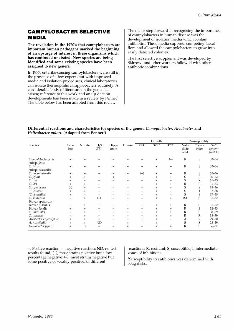
CAMPYLOBACTER SELECTIVEMEDIAThe revelation in the 1970's that campylobacters areimportant human pathogens marked the beginningof an upsurge of interest in these organisms whichhas continued unabated. New species are beingidentified and some existing species have beenassigned to new genera.
In 1977, enteritis-causing campylobacters were still inthe province of a few experts but with improvedmedia and isolation procedures, clinical laboratoriescan isolate thermophilic campylobacters routinely. Aconsiderable body of literature on the genus hasarisen; reference to this work and an up-date ondevelopments has been made in a review by Penner1.The table below has been adapted from this review.
The major step forward in recognising the importanceof campylobacters in human disease was thedevelopment of isolation media which containantibiotics. These media suppress competing faecalflora and allowed the campylobacters to grow intoeasily detected colonies.
The first selective supplement was developed bySkirrow1 and other workers followed with otherantibiotic combinations.
Differential reactions and characteristics for species of the genera Campylobacter, Arcobacter andHelicobacter pylori. (Adapted from Penner1)
Growth Susceptibility
Species Cata- Nitrate H2S Hipp- Urease 258C 378C 428C Nali- Cephol- G+Clase (TSI) urate dixic othin content
acid (mol%)
Campylobacter fetus + + ± ± ± + + (±) R S 33±34subsp. fetusC. fetus + + ± ± ± + + ± R S 33±34subsp. venerealisC. hyointestinalis + + + ± ± (+) + + R S 35±36C. jejuni + + ± + ± ± + + S R 30±32C. coli + + ± ± ± ± + + S R 31±33C. lari + + ± ± ± ± + + R R 31±33C. upsaliensis (±) + ± ± ± ± + + S S 35±36`C. cinaedi' + + ± ± ± ± + ± S I 37±38`C. fennelliae' + ± ± ± ± ± + ± S S 37±38C. sputorum ± + (+) ± ± ± + + (S) S 31±32Biovar sputorumBiovar bubulus ± + + ± ± ± + + R S 31±32Biovar fecalis + + + ± ± ± + + R S 32±33C. mucosalis ± + + ± ± ± + + R S 38±39C. concisus ± + + ± ± ± + + R R 38±39Arcobacter cryaerophila + + ± ± ± + + ± d R 29±30A. nitrofigilis + + ND ± + + + ± S S 28±29Helicobacter pylori + d ± ± + ± + + R S 36±37
+, Postive reaction; ±, negative reaction; ND, no testresults found; (+), most strains positive but a lowpercentage negative: (±), most strains negative butsome positive or weakly positive; d, different
reactions; R, resistant; S, susceptible; I, intermediatezones of inhibitions.aSusceptibility to antibiotics was determined with30mg disks.
Culture Media
November 1998 2-61
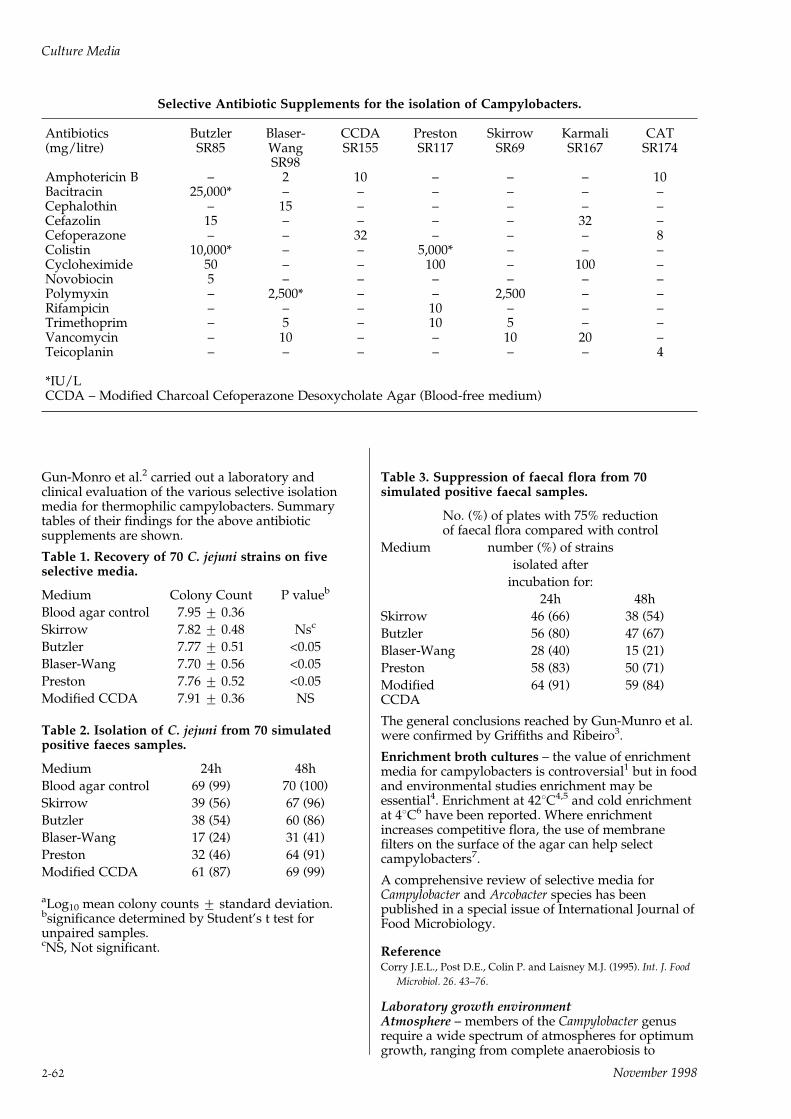
Selective Antibiotic Supplements for the isolation of Campylobacters.
Antibiotics Butzler Blaser- CCDA Preston Skirrow Karmali CAT(mg/litre) SR85 Wang SR155 SR117 SR69 SR167 SR174
SR98Amphotericin B ± 2 10 ± ± ± 10Bacitracin 25,000* ± ± ± ± ± ±Cephalothin ± 15 ± ± ± ± ±Cefazolin 15 ± ± ± ± 32 ±Cefoperazone ± ± 32 ± ± ± 8Colistin 10,000* ± ± 5,000* ± ± ±Cycloheximide 50 ± ± 100 ± 100 ±Novobiocin 5 ± ± ± ± ± ±Polymyxin ± 2,500* ± ± 2,500 ± ±Rifampicin ± ± ± 10 ± ± ±Trimethoprim ± 5 ± 10 5 ± ±Vancomycin ± 10 ± ± 10 20 ±Teicoplanin ± ± ± ± ± ± 4
*IU/LCCDA ± Modified Charcoal Cefoperazone Desoxycholate Agar (Blood-free medium)
Gun-Monro et al.2 carried out a laboratory andclinical evaluation of the various selective isolationmedia for thermophilic campylobacters. Summarytables of their findings for the above antibioticsupplements are shown.
Table 1. Recovery of 70 C. jejuni strains on fiveselective media.
Medium Colony Count P valueb
Blood agar control 7.95 + 0.36
Skirrow 7.82 + 0.48 Nsc
Butzler 7.77 + 0.51 <0.05
Blaser-Wang 7.70 + 0.56 <0.05
Preston 7.76 + 0.52 <0.05
Modified CCDA 7.91 + 0.36 NS
Table 2. Isolation of C. jejuni from 70 simulatedpositive faeces samples.
Medium 24h 48h
Blood agar control 69 (99) 70 (100)
Skirrow 39 (56) 67 (96)
Butzler 38 (54) 60 (86)
Blaser-Wang 17 (24) 31 (41)
Preston 32 (46) 64 (91)
Modified CCDA 61 (87) 69 (99)
aLog10 mean colony counts + standard deviation.bsignificance determined by Student's t test forunpaired samples.cNS, Not significant.
Table 3. Suppression of faecal flora from 70simulated positive faecal samples.
No. (%) of plates with 75% reductionof faecal flora compared with control
Medium number (%) of strains
isolated after
incubation for:
24h 48h
Skirrow 46 (66) 38 (54)
Butzler 56 (80) 47 (67)
Blaser-Wang 28 (40) 15 (21)
Preston 58 (83) 50 (71)
Modified 64 (91) 59 (84)CCDA
The general conclusions reached by Gun-Munro et al.were confirmed by Griffiths and Ribeiro3.
Enrichment broth cultures ± the value of enrichmentmedia for campylobacters is controversial1 but in foodand environmental studies enrichment may beessential4. Enrichment at 428C4,5 and cold enrichmentat 48C6 have been reported. Where enrichmentincreases competitive flora, the use of membranefilters on the surface of the agar can help selectcampylobacters7.
A comprehensive review of selective media forCampylobacter and Arcobacter species has beenpublished in a special issue of International Journal ofFood Microbiology.
ReferenceCorry J.E.L., Post D.E., Colin P. and Laisney M.J. (1995). Int. J. Food
Microbiol. 26. 43±76.
Laboratory growth environmentAtmosphere ± members of the Campylobacter genusrequire a wide spectrum of atmospheres for optimumgrowth, ranging from complete anaerobiosis to
Culture Media
2-62 November 1998

ambient air tolerance8. Most species, however, liebetween these extremes and are micro-aerophilic. TheOxoid Campylobacter Gas Generating Kits (BR56 andBR60) ensure that the correct oxygen and carbondioxide levels are produced for the optimum growthof these micro-aerophilic organisms. Alternatively useCampyGen CN025A or CN035A. CampyGen doesnot require the addition of water or a catalyst.
Temperature ± the temperature range for incubationof Campylobacter species and related organisms variesfrom 158C for Arcobacter cryaerophilea to 428C for thethermophilic species. However, most strains have aconsiderable tolerance of growth temperaturesaround those required for optimum growth.
Helicobacter pylori ± this organism is implicated as acause of gastritis and peptic ulceration9,10,11.
A specific selective culture medium, prepared fromHelicobacter pylori Selective Supplement (Dent)SR147 and Columbia Blood Agar Base CM331 isrequired to isolate this organism from gastric biopsyspecimens.
References1 Penner J. L. (1988) Clin. Microbiol. Reviews. 1. 157±172.
2 Gun-Munro J., Rennie R. P. , Thornley J. H., Richardson H.L.,
Hodge D. and Lynch J. (1987) J. Clin. Microbiol. 25. 2274±2277.
3 Griffiths A. and Ribeiro C. D. (1988) J. Clin. Path. 41. 704±705.
4 Marinescu M., Festy B., Derimay R. and Megraud F. (1987) Eur.
J. Clin. Microbiol. 6. 693±694.
5 Bolton F. J., Coates D., Hinchliffe P. M. and Robertson L. (1983)
J. Clin. Path. 36. 78±83.
6 Rubin S. J. and Woodward N. (1983) J. Clin. Microbiol. 18. 1008±
1010.
7 Steele T. W. and McDermott S. N. (1984) Pathology 16. 263±265.
8 Neill S. D., Campbell J. N., O'Brien J. J., Weatherup S. T. and
Ellis W. A. (1985) Int. J. Sys. Bacteriol. 35. 342±356.
9 Marshall B. K., Warren J. R., Blincow E. D., Phillips M.,
Goodwin C. S., Murray R., Blackbourne S. J. and Waters T. E.
(1988) Lancet ii. 626±627.
10 Dent J. C. and McNulty C. A. M. (1988) Eur. J. Clin. Microbiol.
Infect. Dis. 7. 555±568.
11 Buck G. E. (1988) Lab. Managemt. 26. 9.
CAMPYLOBACTER SELECTIVEMEDIA
For the isolation of campylobacters.
BASE MEDIA
BLOOD AGAR BASE NO. 2
Code: CM271
Formula gm/litreProteose peptone 15.0Liver digest 2.5Yeast extract 5.0Sodium chloride 5.0Agar 12.0pH 7.4 + 0.2
DirectionsSuspend 20g in 500ml of distilled water, bring to theboil to dissolve completely. Mix and sterilise byautoclaving at 1218C for 15 minutes.
Or
COLUMBIA AGAR BASE
Code: CM331
Formula gm/litreSpecial peptone 25.0Starch 1.0Sodium chloride 5.0Agar 10.0pH 7.3 + 0.2
DirectionsSuspend 19.5g in 500ml of distilled water. Boil todissolve the medium completely. Sterilise byautoclaving at 1218C for 15 minutes.
CAMPYLOBACTER AGAR BASECode: CM689
Formula gm/litre`Lab-Lemco' powder 10.0Peptone 10.0Sodium chloride 5.0Agar 12.0pH 7.5 + 0.2
DirectionsFor preparation of Preston Campylobacter SelectiveAgar Base suspend 18.5g in 475ml of distilled waterand bring to the boil to dissolve completely. Steriliseby autoclaving at 1218C for 15 minutes. Cool to 508Cor below. Aseptically add 25ml of Lysed Horse BloodSR48.
See Campylobacter Selective Agar (Preston) forfurther information.
CAMPYLOBACTER SELECTIVESUPPLEMENTS
Code: SR69 (Skirrow)Code: SR85 (Butzler)Code: SR98 (Blaser-Wang)Code: SR117 (Preston)
Freeze-dried antibiotic supplements for the isolationof campylobacters.
CAMPYLOBACTER SELECTIVESUPPLEMENT (SKIRROW)
Code: SR69 (Skirrow)
Vial contents (each vial is sufficient for 500ml ofmedium)
Vancomycin 5mgPolymyxin 1,250 IUTrimethoprim 2.5mg
DirectionsTo rehydrate the contents of the vial, add aseptically2ml of sterile distilled water and turn end-over-end todissolve. Avoid frothing the solution.
Culture Media
November 1998 2-63

Add the contents of one vial to 500ml of sterile bloodagar, cooled to 50±558C, prepared from OxoidColumbia Agar CM331 or Blood Agar Base No.2CM271, with 5±7% lysed defibrinated horse blood.Mix gently and pour into sterile petri dishes.
DescriptionCampylobacter Selective Supplement SR69 is basedon the formulation of Skirrow1.
The antibiotic supplement is designed to be used at428C for optimum selective effect.
Reference1 Skirrow M. B. (1977) BMJ 2. 9±11.
CAMPYLOBACTER SELECTIVESUPPLEMENT (BUTZLER)
Code: SR85 (Butzler)
Vial contents (each vial is sufficient for 500ml ofmedium)
Bacitracin 12,500 IUCycloheximide 25mgColistin sulphate 5,000 IUCephazolin sodium 7.5mgNovobiocin 2.5mg
DirectionsTo rehydrate the vial aseptically add 3ml of 50/50ethanol/water and turn end-over-end to dissolve.Avoid frothing of the solution.
Add the contents of one vial to 500ml of sterile bloodagar cooled to 50±558C prepared from OxoidColumbia Agar CM331, with 5±7% defibrinated horseor sheep blood. Mix gently and pour into sterile petridishes.
DescriptionCampylobacter Selective Supplement SR85 is basedon the formulation of Lauwers, De Boeck andButzler1.
Cephalothin (15mg/ml) has been replaced in theOxoid Supplement by cephazolin (15mg/ml) as thishas been found to be more inhibitory to Pseudomonasspecies1.
This supplement differs from Skirrow SupplementSR69 by its selective action at 378C. It thereforeovercomes the need to use incubators at 428C and itselectively isolates those strains of Campylobacterspecies that fail to grow at 428C2 (e.g. C. fetus sub.sp.fetus).
References1 Lauwers S., De Boeck M. and Butzler J.P. (1978) Lancet, 1. 604±
605.
2 Butzler J.P., Dekeyser P., Detrain M. and Dehaen F. (1973) J.
Pediat. 82. 493.
CAMPYLOBACTER SELECTIVESUPPLEMENT (BLASER-WANG)
Code: SR98 (Blaser-Wang)
Vial contents (each vial is sufficient for 500ml ofmedium)
Vancomycin 5mgPolymyxin B 1,250 IUTrimethoprim 2.5mgAmphotericin B 1mgCephalothin 7.5mg
DirectionsTo rehydrate the contents of the vial, add aseptically2ml of sterile distilled water and turn end-over-end todissolve. Avoid frothing the solution.
Add the contents of one vial to 500ml of a sterilenutrient medium cooled to 50±558C prepared fromColumbia Agar Base CM331 or Blood Agar Base No.2CM271, with 10% sheep blood or 5±7% laked horseblood SR48. Mix gently and pour into sterile petridishes.
DescriptionCampylobacter Selective Supplement (Blaser-Wang)SR98 is based on the formulation of Skirrow1, butwith the addition of amphotericin B and cephalothin2.The inclusion of amphotericin B inhibits the growth ofCandida albicans and cephalothin improves theselectivity of the supplement.
References1 Skirrow M.B. (1977) BMJ 2. 9±11.
2 Blaser M.J., Hardesty H.L., Powers B. and Wang W.L.L. (1980) J.
Clin. Micro. 11. 309±313.
CAMPYLOBACTER GROWTH SUPPLEMENT
Code: SR84
FBP Supplement for the enhanced growth andaerotolerance of campylobacter.
Vial contents (each vial is sufficient for 500ml ofmedium)
Sodium pyruvate 0.125gSodium metabisulphite 0.125gFerrous sulphate (hydrated salt) 0.125g
DirectionsTo rehydrate the contents of the vial, aseptically add2ml of sterile distilled water and invert to dissolve.Avoid frothing of solution.
Add the contents of one vial to 500ml of a sterilenutrient medium cooled to 50±558C prepared fromOxoid Columbia Agar CM331, Blood Agar Base No.2CM271, or Campylobacter Agar Base CM689, with5±7% lysed defibrinated horse or sheep blood, and therehydrated contents of one vial of CampylobacterAntibiotic Supplement SR69, SR85 or SR98. Mixgently and pour aseptically into sterile petri dishes.
DescriptionCampylobacter species, which require less oxygen (i.e.microaerophilic) are inhibited at the normalatmospheric oxygen level. The optimum level ofoxygen required for growth has been reported to be6%1. This exacting level, together with a carbon
Culture Media
2-64 November 1998
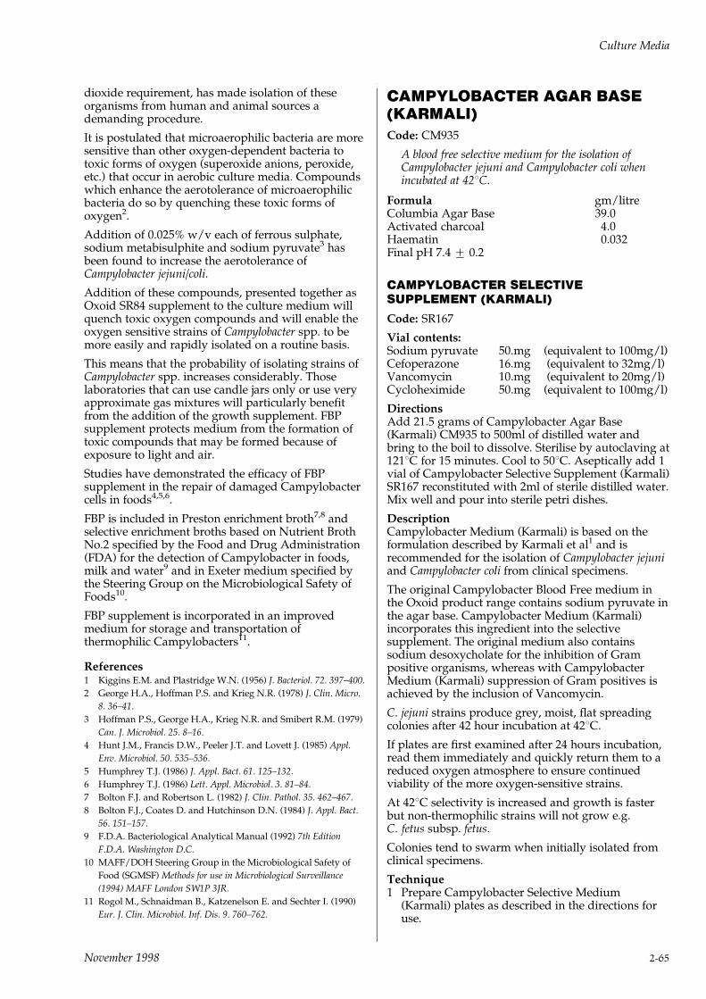
dioxide requirement, has made isolation of theseorganisms from human and animal sources ademanding procedure.
It is postulated that microaerophilic bacteria are moresensitive than other oxygen-dependent bacteria totoxic forms of oxygen (superoxide anions, peroxide,etc.) that occur in aerobic culture media. Compoundswhich enhance the aerotolerance of microaerophilicbacteria do so by quenching these toxic forms ofoxygen2.
Addition of 0.025% w/v each of ferrous sulphate,sodium metabisulphite and sodium pyruvate3 hasbeen found to increase the aerotolerance ofCampylobacter jejuni/coli.
Addition of these compounds, presented together asOxoid SR84 supplement to the culture medium willquench toxic oxygen compounds and will enable theoxygen sensitive strains of Campylobacter spp. to bemore easily and rapidly isolated on a routine basis.
This means that the probability of isolating strains ofCampylobacter spp. increases considerably. Thoselaboratories that can use candle jars only or use veryapproximate gas mixtures will particularly benefitfrom the addition of the growth supplement. FBPsupplement protects medium from the formation oftoxic compounds that may be formed because ofexposure to light and air.
Studies have demonstrated the efficacy of FBPsupplement in the repair of damaged Campylobactercells in foods4,5,6.
FBP is included in Preston enrichment broth7,8 andselective enrichment broths based on Nutrient BrothNo.2 specified by the Food and Drug Administration(FDA) for the detection of Campylobacter in foods,milk and water9 and in Exeter medium specified bythe Steering Group on the Microbiological Safety ofFoods10.
FBP supplement is incorporated in an improvedmedium for storage and transportation ofthermophilic Campylobacters11.
References1 Kiggins E.M. and Plastridge W.N. (1956) J. Bacteriol. 72. 397±400.
2 George H.A., Hoffman P.S. and Krieg N.R. (1978) J. Clin. Micro.
8. 36±41.
3 Hoffman P.S., George H.A., Krieg N.R. and Smibert R.M. (1979)
Can. J. Microbiol. 25. 8±16.
4 Hunt J.M., Francis D.W., Peeler J.T. and Lovett J. (1985) Appl.
Env. Microbiol. 50. 535±536.
5 Humphrey T.J. (1986) J. Appl. Bact. 61. 125±132.
6 Humphrey T.J. (1986) Lett. Appl. Microbiol. 3. 81±84.
7 Bolton F.J. and Robertson L. (1982) J. Clin. Pathol. 35. 462±467.
8 Bolton F.J., Coates D. and Hutchinson D.N. (1984) J. Appl. Bact.
56. 151±157.
9 F.D.A. Bacteriological Analytical Manual (1992) 7th Edition
F.D.A. Washington D.C.
10 MAFF/DOH Steering Group in the Microbiological Safety of
Food (SGMSF) Methods for use in Microbiological Surveillance
(1994) MAFF London SW1P 3JR.
11 Rogol M., Schnaidman B., Katzenelson E. and Sechter I. (1990)
Eur. J. Clin. Microbiol. Inf. Dis. 9. 760±762.
CAMPYLOBACTER AGAR BASE(KARMALI)Code: CM935
A blood free selective medium for the isolation ofCampylobacter jejuni and Campylobacter coli whenincubated at 428C.
Formula gm/litreColumbia Agar Base 39.0Activated charcoal 4.0Haematin 0.032Final pH 7.4 + 0.2
CAMPYLOBACTER SELECTIVESUPPLEMENT (KARMALI)
Code: SR167
Vial contents:Sodium pyruvate 50.mg (equivalent to 100mg/l)Cefoperazone 16.mg (equivalent to 32mg/l)Vancomycin 10.mg (equivalent to 20mg/l)Cycloheximide 50.mg (equivalent to 100mg/l)
DirectionsAdd 21.5 grams of Campylobacter Agar Base(Karmali) CM935 to 500ml of distilled water andbring to the boil to dissolve. Sterilise by autoclaving at1218C for 15 minutes. Cool to 508C. Aseptically add 1vial of Campylobacter Selective Supplement (Karmali)SR167 reconstituted with 2ml of sterile distilled water.Mix well and pour into sterile petri dishes.
DescriptionCampylobacter Medium (Karmali) is based on theformulation described by Karmali et al1 and isrecommended for the isolation of Campylobacter jejuniand Campylobacter coli from clinical specimens.
The original Campylobacter Blood Free medium inthe Oxoid product range contains sodium pyruvate inthe agar base. Campylobacter Medium (Karmali)incorporates this ingredient into the selectivesupplement. The original medium also containssodium desoxycholate for the inhibition of Grampositive organisms, whereas with CampylobacterMedium (Karmali) suppression of Gram positives isachieved by the inclusion of Vancomycin.
C. jejuni strains produce grey, moist, flat spreadingcolonies after 42 hour incubation at 428C.
If plates are first examined after 24 hours incubation,read them immediately and quickly return them to areduced oxygen atmosphere to ensure continuedviability of the more oxygen-sensitive strains.
At 428C selectivity is increased and growth is fasterbut non-thermophilic strains will not grow e.g.C. fetus subsp. fetus.
Colonies tend to swarm when initially isolated fromclinical specimens.
Technique1 Prepare Campylobacter Selective Medium
(Karmali) plates as described in the directions foruse.
Culture Media
November 1998 2-65
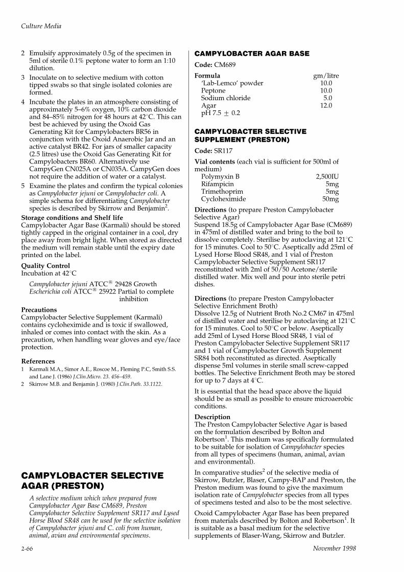
2 Emulsify approximately 0.5g of the specimen in5ml of sterile 0.1% peptone water to form an 1:10dilution.
3 Inoculate on to selective medium with cottontipped swabs so that single isolated colonies areformed.
4 Incubate the plates in an atmosphere consisting ofapproximately 5±6% oxygen, 10% carbon dioxideand 84±85% nitrogen for 48 hours at 428C. This canbest be achieved by using the Oxoid GasGenerating Kit for Campylobacters BR56 inconjunction with the Oxoid Anaerobic Jar and anactive catalyst BR42. For jars of smaller capacity(2.5 litres) use the Oxoid Gas Generating Kit forCampylobacters BR60. Alternatively useCampyGen CN025A or CN035A. CampyGen doesnot require the addition of water or a catalyst.
5 Examine the plates and confirm the typical coloniesas Campylobacter jejuni or Campylobacter coli. Asimple schema for differentiating Campylobacterspecies is described by Skirrow and Benjamin2.
Storage conditions and Shelf lifeCampylobacter Agar Base (Karmali) should be storedtightly capped in the original container in a cool, dryplace away from bright light. When stored as directedthe medium will remain stable until the expiry dateprinted on the label.
Quality ControlIncubation at 428C
Campylobacter jejuni ATCC1 29428 GrowthEscherichia coli ATCC1 25922 Partial to complete
inhibition
PrecautionsCampylobacter Selective Supplement (Karmali)contains cycloheximide and is toxic if swallowed,inhaled or comes into contact with the skin. As aprecaution, when handling wear gloves and eye/faceprotection.
References1 Karmali M.A., Simor A.E., Roscoe M., Fleming P.C, Smith S.S.
and Lane J. (1986) J.Clin.Micro. 23. 456±459.
2 Skirrow M.B. and Benjamin J. (1980) J.Clin.Path. 33.1122.
CAMPYLOBACTER SELECTIVEAGAR (PRESTON)
A selective medium which when prepared fromCampylobacter Agar Base CM689, PrestonCampylobacter Selective Supplement SR117 and LysedHorse Blood SR48 can be used for the selective isolationof Campylobacter jejuni and C. coli from human,animal, avian and environmental specimens.
CAMPYLOBACTER AGAR BASE
Code: CM689
Formula gm/litre`Lab-Lemco' powder 10.0Peptone 10.0Sodium chloride 5.0Agar 12.0pH 7.5 + 0.2
CAMPYLOBACTER SELECTIVESUPPLEMENT (PRESTON)
Code: SR117
Vial contents (each vial is sufficient for 500ml ofmedium)
Polymyxin B 2,500IURifampicin 5mgTrimethoprim 5mgCycloheximide 50mg
Directions (to prepare Preston CampylobacterSelective Agar)Suspend 18.5g of Campylobacter Agar Base (CM689)in 475ml of distilled water and bring to the boil todissolve completely. Sterilise by autoclaving at 1218Cfor 15 minutes. Cool to 508C. Aseptically add 25ml ofLysed Horse Blood SR48, and 1 vial of PrestonCampylobacter Selective Supplement SR117reconstituted with 2ml of 50/50 Acetone/steriledistilled water. Mix well and pour into sterile petridishes.
Directions (to prepare Preston CampylobacterSelective Enrichment Broth)Dissolve 12.5g of Nutrient Broth No.2 CM67 in 475mlof distilled water and sterilise by autoclaving at 1218Cfor 15 minutes. Cool to 508C or below. Asepticallyadd 25ml of Lysed Horse Blood SR48, 1 vial ofPreston Campylobacter Selective Supplement SR117and 1 vial of Campylobacter Growth SupplementSR84 both reconstituted as directed. Asepticallydispense 5ml volumes in sterile small screw-cappedbottles. The Selective Enrichment Broth may be storedfor up to 7 days at 48C.
It is essential that the head space above the liquidshould be as small as possible to ensure microaerobicconditions.
DescriptionThe Preston Campylobacter Selective Agar is basedon the formulation described by Bolton andRobertson1. This medium was specifically formulatedto be suitable for isolation of Campylobacter speciesfrom all types of specimens (human, animal, avianand environmental).
In comparative studies2 of the selective media ofSkirrow, Butzler, Blaser, Campy-BAP and Preston, thePreston medium was found to give the maximumisolation rate of Campylobacter species from all typesof specimens tested and also to be the most selective.
Oxoid Campylobacter Agar Base has been preparedfrom materials described by Bolton and Robertson1. Itis suitable as a basal medium for the selectivesupplements of Blaser-Wang, Skirrow and Butzler.
Culture Media
2-66 November 1998

Preston Campylobacter Selective Supplement SR117can also be used to prepare Preston CampylobacterSelective Enrichment Broth2.
The selective enrichment technique is recommendedfor specimens and food samples that are expected tobe heavily contaminated and/or carry small numbersof viable colony forming units. The PrestonCampylobacter Selective Enrichment Broth which issupplemented with the growth supplement SR84,made to the formulation of George et al.3 effectivelyquenches toxic compounds which may form onexposure of the medium to light and air4.
TechniqueDirect Selective Plating Method1 Prepare the Preston Campylobacter Selective Agar
as directed from CM689, SR117 and Lysed Blood.
2 Emulsify the specimen under test in 2ml of 0.1%peptone water.
3 Inoculate onto the selective medium with cottontipped swabs so that single isolated colonies areformed.
4 Incubate the plates in an atmosphere consisting ofapproximately 5±6% oxygen, 10% carbon dioxideand 84±85% nitrogen for 24±48 hours* at 428C. Thiscan best be achieved by using the Oxoid GasGenerating Kit for Campylobacters BR56 inconjunction with the Oxoid Anaerobic Jar and anactive catalyst. For jars of smaller capacity (2.5litres) use the Oxoid Gas Generating Kit forCampylobacters BR60. Alternatively useCampyGen CN025A or CN035A. CampyGen doesnot require the addition of water or a catalyst.
5 Examine the plates and confirm the typical coloniesas Campylobacter jejuni or C. coli by the standardmethods.
* When few Campylobacter colony forming units arepresent 48 hours incubation is necessary.
Selective Enrichment Broth Technique1 Prepare the Preston Selective Enrichment Broth as
directed from CM67, SR117, SR84 and Lysed Blood.
2 Emulsify the specimen under test in the selectiveenrichment broth.
3 Incubate the broth aerobically at 428C for 24 hours.
4 Subculture on to Preston Campylobacter SelectiveAgar or Campylobacter Blood-Free Selective Agar.
CAMPYLOBACTER TRANSPORT MEDIUM
Campylobacter Selective Supplement (Preston) isincorporated in an improved medium for storage andtransportation of thermophilic Campylobacters5.
References1 Bolton F.J. and Robertson L. (1982) J. Clin. Pathol. 35. 462±467.
2 Bolton F.J., Coates D., Hinchliffe P.M. and Robertson L. (1983) J.
Clin. Pathol. 36. 78±83.
3 George H.A., Hoffman P.S., Kreig N.R. and Smibert R.M. (1979)
Can. J. Microbiol. 25. 8±16.
4 Bolton F.J., Coates D. and Hutchinson D.N. (1984) J. Appl. Bact.
56. 151±157.
5 Rogol M., Schnaidman B., Katzenelso E. and Sechter I. (1990)
Eur. J. Clin. Microbiol. Inf. Dis. 9. 760±762.
CAMPYLOBACTER BLOOD-FREESELECTIVE MEDIUM (MODIFIEDCCDA-PRESTON)
A medium, which when prepared from CampylobacterBlood-Free Selective Agar Base CM739 and CCDASelective Supplement SR155, can be used for theisolation of Campylobacter jejuni, C. coli and C. laridis.
CAMPYLOBACTER BLOOD-FREE SELECTIVEAGAR BASE
Code: CM739
Formula gm/litreNutrient Broth No.2 25.0Bacteriological charcoal 4.0Casein hydrolysate 3.0Sodium desoxycholate 1.0Ferrous sulphate 0.25Sodium pyruvate 0.25Agar 12.0pH 7.4 + 0.2
CCDA SELECTIVE SUPPLEMENT
Code: SR155
An improved selective supplement for blood-freeCampylobacter Agar.
Vial contents (each vial is sufficient to supplement500ml of medium).Cefoperazone 16mg equivalent to 32mg/litreAmphotericin B 5mg equivalent to 10mg/litre
DirectionsSuspend 22.75g of Campylobacter Blood-FreeSelective Agar Base in 500ml of distilled water andbring to the boil to dissolve. Sterilise by autoclaving at1218C for 15 minutes. Cool to 508C. Aseptically add 1vial of CCDA Selective Supplement SR155reconstituted with 2ml of sterile distilled water. Mixwell and pour into sterile petri dishes.
DescriptionModified CCDA Medium is based on the originalformulation described by Bolton et al1 which wasdeveloped to replace blood with charcoal, ferroussulphate and sodium pyruvate. Improved selectivitywas achieved when cephazolin in the originalformulation was replaced by cefoperazone as theselective agent2. More recent work has shown anincreased isolation rate can be achieved if the platesare incubated at 378C rather than 428C3.
Amphotericin B has been added to the formula tosuppress the growth of yeast and fungalcontaminants that may occur at 378C.
Modified CCDA medium and Campy-BAP mediumwere equal in performance in a rapid colony-liftprocedure for detection of thermophiliccampylobacters in which membranes are blotted onagar cultures and then subjected to immunoassay5.
In a study of healthy puppies and kittens for carriageof Campylobacter species6, modified CCDA mediumwas found to be a suitable medium and more
Culture Media
November 1998 2-67
productive for C. upsaliensis in this application thanCAT medium. Modified CCDA medium has beenconfirmed as suitable for isolation of Campylobacterspp. from non-clinical samples following enrichmentin Exeter broth7.
The use of Campylobacter Blood-Free Medium isspecified by the U.K. Ministry of Agriculture,Fisheries and Food (MAFF) in a validated method forisolation of Campylobacter from foods4.
Technique1 Prepare Campylobacter Blood-Free Selective Agar
as described in the directions.
2 Emulsify approximately 0.5g of the specimen in5ml of sterile 0.1% peptone water to form anapproximate 1:10 dilution.
3 Inoculate onto the selective medium with cottontipped swabs so that single isolated colonies areformed.
4 Incubate the plates in an atmosphere consisting ofapproximately 5±6% oxygen, 10% carbon dioxideand 84±85% nitrogen for 48 hours at 378C. This canbest be achieved by using the Oxoid GasGenerating Kit for Campylobacters (BR56) inconjunction with the Oxoid Anaerobic Jar and anactive catalyst (BR42). For jars of smaller capacity(2.5 litres) use the Oxoid Gas Generating Kit forCampylobacters (BR60). Alternatively useCampyGen CN023A or CN035A which does notrequire the use of a catalyst or the addition ofwater.
The colonial morphology of campylobacters can beused as a guideline for identification to species level.C. jejuni strains produce grey, moist flat spreadingcolonies. Some strains may have a green hue or a dryappearance, with or without a metallic sheen. C. colistrains tend to be creamy-grey in colour, moist,slightly raised and often produce discrete colonies.
Colonies tend to swarm when initially isolated fromclinical specimens.
Storage conditions and Shelf lifeStore the dehydrated medium below 258C and usebefore the expiry date on the label.
Store the selective supplement in the dark at 2±88Cand use before the expiry date on the label.
The prepared medium may be stored for up to 2weeks at 2±88C.
Quality ControlIncubation at 378C for 48 hours.
Positive control:Campylobacter jejuni ATCC1 29428
Negative control:Escherichia coli ATCC1 25922
References1 Bolton, F.J., Hutchinson, D.N. and Coates, D. (1984) J. Clin.
Microbiol. 19, 169±171.
2 Hutchinson, D.N. and Bolton, F.J. (1984) J. Clin. Path. 34, 956±
957.
3 Bolton, F.J., Hutchinson, D.N. and Parker, G. (1988) Eur. J. Clin.
Microbiol. Infect. Dis. 7. 155±160.
4 MAFF Validated Methods for the Analysis of Foodstuffs:
Method for the detection of thermotolerant Campylobacter in
Foods (v30) J. Assoc. Publ. Analysts (1993) 29. 253±262.
5. Rice, B.E., Chinta Lamichhane, Joseph, S.W. and Rollins, D.M.
(1996) Clin. Diag. Lab. Immunol. 3, 669±677.
6. Hald, B. and Madsen, M. (1997) J. Clin. Microbiol. 35, 3351±3352.
7. Humphrey, T.J., Martin, K.W. and Mason, M.J. (1997) PHLS
Microbiology Digest 13 (2), 86±88.
CEFOPERAZONE, AMPHOTERICINB, TEICOPLANIN SUPPLEMENT(CAT)Code: SR174
A selective supplement for the isolation of thermophilicCampylobacter spp. and improved recovery ofCampylobacter upsaliensis from faeces.
Vial contents (each vial is sufficient for 500ml ofmedium)
CAT Supplement gm/litreCefoperazone 8.0Teicoplanin 4.0Amphotericin B 10.0
DirectionsAseptically add 4ml of sterile distilled water to thevial. Mix gently to resuspend the supplement.
Prepare 500ml of sterile Blood-Free CampylobacterAgar Base CM739 as directed. Cool to 508C andaseptically add one vial of SR174E reconstituted asdirected above.
Mix well and pour the resulting CAT medium intosterile petri dishes. Incubate cultures at 378C for48±72 hours in a microaerobic atmosphere.
DescriptionBecause of the sensitivity of C. upsaliensis to a widerange of antibiotics, isolation of the organism fromfaeces using selective media has hitherto beendifficult. The recommended isolation method uses amembrane filter culture technique on non-selectiveagar. This does not give good recovery from faecescontaining less than 105 CFU/g5 however, and is atechnically demanding method which is relativelyslow to perform.
CAT Supplement SR174 is based on the formulationdescribed by Aspinall et al7. When added to Blood-Free Campylobacter Agar Base CM739 whichcontains charcoal, it gives good isolation ofthermophilic Campylobacter spp., and makes theisolation of C. upsaliensis possible on a selectivemedium because CAT Supplement contains reducedlevels of cefoperazone compared to otherCampylobacter supplements. This inhibits mostEnterobacteriaceae, but not enterococci. Teicoplanin isincluded to inhibit enterococci. Amphotericin B isadded as an antifungal agent.
Further work confirmed the effectiveness of CATmedium as an alternative to membrane filtrationculture for selective isolation of thermophiliccampylobacters including C. upsaliensis8.
Atabay, Corry and On9 isolated a previously
Culture Media
2-68 November 1998
unknown catalase-negative, urease-positiveCampylobacter from cattle faeces using CATmedium. This organism could not be cultured onblood-free Campylobacter medium (CCDA).
A study in which the productivity of CAT medium, 2blood-free media and semi-solid medium werecompared, showed that CAT medium, used inparallel with membrane filtration on non-selectiveblood agar, is likely to be the most productive methodfor recovery of the greatest number of Campylobacterand Arcobacter species10.
Storage conditions and Shelf lifeCAT Supplement SR174 should be stored at 28C±88Cin the dark. When stored as directed, the reagentsremain stable until the stated expiry date shown onthe packaging.
Quality ControlPositive control:C. upsaliensis NCTC 11926 (pale grey colonies)C. jejuni ATCC1 29428 (grey colonies)
Negative control:Enterococcus faecalis ATCC1 33186 (inhibited)
References1 Atabay, I., Corry, J.E.L., Post, D. 8th International
Campylobacter Workshop 1995.
2 Sandstedt, K., Ursing, J., Walder, M. (1983). Curr. Microbiol. 8:
209±213.
3 Sandstedt, K. and Ursing, J. (1991). Sys. Appl. Microbiol. 14: 39±
45.
4 Patton, C.M., Shaffer, N., Edmonds, P. et al. (1989). J. Clin.
Microbiol. 27: 66±73.
5 Goosens, H., Vlaes, L., Butzler, J.P. et al. (1991). Lancet. 337:
1486±7.
6 Bolton, F.J., Hutchinson, D.N., Parker, G. (1987). J. Clin. Pathol.
40: 702±3.
7 Aspinall, S.T., Wareing, D.R.A., Hayward, P.G. and Hutchinson,
D.N. (1993). J. Clin. Pathol. 46: 829±831.
8 Aspinall, S.T., Wareing, D.R.A., Hayward, P.G. and Hutchinson,
D.N. (1996). J. Appl. Bact. 80: 667±672.
9 Atabay, H.I., Corry, J.E.L. and On, S.L.W. (1997). Lett. Appl.
Microbiol. 24: 59±64.
10 Atabay, H.I., Corry, J.E.L. and Post, D.E. (1996).
Campylobacters, Helicobacters and Related Organisms. Newell,
D.G., Ketley, J.M. and Feldman, R.A. (eds) Part 1±5. Plenum
Press, New York.
CARY-BLAIR MEDIUM
Code: CM519A transport medium for Gram negative and anaerobicorganisms.
Formula gm/litreDisodium hydrogen phosphate 1.1Sodium thioglycollate 1.5Sodium chloride 5.0Calcium chloride 0.09Agar 5.6pH 8.4 + 0.2
DirectionsSuspend 13.3g in 1 litre of distilled water and bringgently to the boil to dissolve the agar. Distribute into
small, screw-cap bottles and sterilise by immersing infree-steam for 15 minutes. Allow to cool and tightenthe screw caps to prevent water loss.
DescriptionOxoid Cary-Blair Medium is a transport medium forthe collection and shipment of clinical specimensbased on the formulation of Cary and Blair1.
The low nutrient content of the medium andutilisation of phosphate as a buffering agent insteadof sodium glycerophosphate, prevents bacterialovergrowth by Escherichia coli, Citrobacter freundii andKlebsiella aerogenes.
This sometimes happens when using Stuart TransportMedium because these organisms possess specificglycerophosphate dehydrogenases2. The lowoxidation-reduction potential of the medium ensuresbacterial survival over long periods3.
Cary and Bair1 reported recovery of cholera vibriosup to 22 days, salmonellae and shigellae after 49 daysand Yersinia pestis up to 75 days storage at 288C.
Cary-Blair Medium is particularly suitable in fieldepidemiological surveys for Vibrio parahaemolyticus,especially where rectal swabs are to be transported toa central diagnostic laboratory4,5. Survival of V.parahaemolyticus in Cary-Blair medium has beenreported after a 35-day period at a temperature of70±808F6.
The medium can be modified to improve thetransport and survival of Campylobacter species.
(i) by the addition of 1% w/v sodium pyruvate(10g/1itre) to the formula7.
(ii) reducing the agar content from 5g to 1.6g perlitre8.
For the transport of fastidious anaerobic bacteria themedium may be prepared as directed and filled intolong narrow screw-capped tubes9. It may also beprepared as a pre-reduced anaerobic sterilisedmedium (PRAS)10. Methods of producing PRASmedia are described by Holdeman and Moore11.
TechniqueUse sterile, cotton-tipped swabs on wooden sticks tocollect the specimen. Push the swabs down one thirdof the medium depth and cut the stick. Screw the capfirmly on the bottle.
Label the bottle and send it to the laboratory withoutdelay.
The recovery of Shigella species is higher when thetransport medium is held at 48C or frozen12.
Storage conditions and Shelf lifeStore the dehydrated medium below 258C and usebefore the expiry date on the label.
The prepared medium should be stored away fromlight at 2±88C or at room temperature (22±258C) up to19 months13.
Quality ControlPositive control:
Shigella sonnei ATCC1 25931Vibrio parahaemolyticus NCTC 11344
Culture Media
November 1998 2-69
Negative control:Uninoculated medium.
PrecautionsThe medium should not be incubated to checksterility, prior to use. This should be carried out onseparate quality control samples.
The medium can maintain the viability of fastidiousorganisms for transport purposes but it should not beused as a storage or enrichment medium.
The results obtained from the medium are dependenton the quality of the specimen material. Commensalanaerobic organisms may overgrow in the mediumand cause misleading results.
References1 Cary S. G. and Blair E. B. (1964) J. Bact. 88. 96±98.
2 Crookes E. M. and Stuart R. D. (1959) J. Pathol. Bacteriol. 78. 283±
288.
3 Stuart R. D. (1959) Public Health Reports 74. 431±438.
4 Cary S. G., Fusillo M. H. and Harkins C. (1965) Am. J. Clin. Path.
43. 294±295.
5 DeWitt W. E., Gangarosa E.J., Huq I. and Zarifi A. (1971) Amer.
J. Trop. Med. Hyg. 20. 685±688.
6 Neumann D. A., Benenson M. W., Hubster E. and Tuan N. T. N.
(1971) Am. J. Clin. Path. 57.
7 Patton C. M., Mitchell S. W., Potter M. E. and Kauffmann A. F.
(1981) J. Clin. Microbiol. 13. 326±328.
8 Luechtefeid M.W., Wang W. L. L., Blaser M. J. and Reller L. B.
(1981) J. Clin. Microbiol. 13. 438±439.
9 Wren M. W. D., Baldwin A. W. F., Eldon C. P. and Sanderson P.
J. (1977) J. Med. Microbiol. 10. 49±61.
10 Wren M. W. D. J. Med. Microbiol. 10. 195±201.
11 Holdeman L. V. and Moore W. E. C. (1975) Anaerobe Laboratory
Manual, Virginia Polytechnic Institute Anaerobe Laboratory, 3rd Ed.
12 Wells J. G. and Morris G. K. (1981) J. Clin. Microbiol. 13. 789±791.
13 Morris G. K and Heck J. (1978) J. Clin. Microbiol. 13. 438±440.
CHARCOAL AGARCode: CM119
A medium for the cultivation and isolation of Bordetellapertussis and Haemophilus influenzae.
Formula gm/litre`Lab-Lemco' powder 10.0Peptone 10.0Starch 10.0Charcoal bacteriological 4.0Sodium chloride 5.0Nicotinic acid 0.001Agar 12.0pH 7.4 + 0.2
DirectionsSuspend 51g in 1 litre of distilled water. Bring to theboil to dissolve completely. Sterilise by autoclaving at1218C for 15 minutes. Cool to 508C, add 10% ofdefibrinated blood and mix gently. The medium ismade selective for the isolation of Bordetella pertussisand B. parapertussis by the addition of BordetellaSelective Supplement SR82.
To one vial add 2ml of sterile distilled water anddissolve the contents completely. Add this solution to500ml of sterile, molten Charcoal Agar CM119, cooledto 508C, together with 10% v/v defibrinated horseblood SR50. Mix well before pouring into sterile petridishes.
For Haemophilus influenzae, omit the selective agentsand convert to `chocolate' agar.
Transport Medium for B. pertussisThe vial contents may be added to 500ml of half-strength Charcoal Agar + 10% v/v defibrinated horseblood SR50 for use as a transport medium for B.pertussis.
DescriptionCharcoal Agar CM119 was developed by Oxoid toprovide a non-blood containing medium for thecultivation of Bordetella pertussis and Haemophilusinfluenzae. Proom1 showed that nicotinic acid was anessential growth factor for the bordetellae. Ensmingeret al.2 used a charcoal medium for the growth of B.pertussis in vaccine production and found that themedium could replace Bordet-Genou. Mishulow etal.3 used charcoal agar for B. pertussis cultivation.
H. influenzae is cultivated on the medium containing10% `chocolated' blood but no antibiotics. Theinoculated plate is incubated for 2 to 3 days at 378C.The colonies are usually small, transparent anddroplet-like, but some transformation to the `rough'type colony may occur. Species differentiation isperformed by examination of the need for X and Vgrowth factors, on Blood Agar Base CM55.
The greatest problem in the isolation of Bordetellaspecies from naso-pharyngeal secretions is thesuppression of unwanted flora during the longincubation period on very nutritious media.
Fleming's first in vitro demonstration of penicillin wasto show that it could help isolate B. pertussis onmedia4. Lacey5 confirmed this but found that thepenicillin-resistant flora still caused problems. Hesupplemented penicillin with 2mg/ml 4,4' diamidino-diphenylamine dihydrochloride (M & B 938) therebyincreasing the selectivity of this medium.
Broome et al.6 found methicillin to be superior topenicillin in suppressing unwanted naso-pharyngealflora but the earlier publication of Sutcliffe andAbbott7 where cephalexin (40mg/ml) was shown to besuperior to penicillin, has proved to be the mostsignificant advance.
The benefits of cephalexin as a selective agent for B.pertussis have been confirmed8,9,10,11. The ability torecover stressed cells and the much longer shelf life(6±8 weeks) are added benefits to its superiority atsuppressing unwanted naso-pharyngeal growth.
Regan and Lowe8 showed that half-strength OxoidCharcoal Agar, supplemented with 40mg/mlcephalexin SR82 v/v lysed, defibrinated horse bloodwas an excellent enrichment and transport medium.
The efficacy of this transport medium has beenconfirmed by other workers12.
Culture Media
2-70 November 1998
TechniqueThe following technique for the laboratory diagnosisof Pertussis is recommended11.
1 Collect pernasal swabs in the early stage of theillness and place in tubes of half-strength CharcoalAgar supplemented with 10% v/v lysed,defibrinated horse blood and 40mg/ml cephalexin.
2 Generously inoculate the swabs on to thick layersof Charcoal Agar CM119 containing 10% v/vdefibrinated horse blood and 40mg/ml cephalexin(SR82).
A non-selective medium in which the cephalexin isomitted may be used in addition.
3 Perform direct fluorescent antibody (DFA) tests onthe secretions, using B. pertussis and B.parapertussis-conjugated antisera, to help make anearlier diagnosis.
4 Replace the swabs in the original transportmedium and hold at room temperature. If theculture plates become overgrown with commensalflora or fungi, use the swabs to inoculate freshplates of medium.
5 Incubate the plates at 358C in a moist atmosphere(60±70% humidity) for up to six days. Examine theplates after 40 hours incubation and twice-dailythereafter.
6 Look for small, shiny, greyish-white, round convexcolonies. Suspicious colonies should be Gramstained, using a two-minute safranin counterstain.Some pleomorphic cells may be seen, caused by thecephalexin in the selective medium.
7 Confirm the identification with DFA tests on thesuspicious colonies.
PrecautionsStuart's transport medium or similar formulationmedia should not be used for Bordetella-containingspecimens13.
Two pernasal swabs should be taken from eachpatient, one through each nostril14.
Make sure the charcoal remains in suspension whendispensing the medium by gently swirling the flask.
Lysed horse blood is used in the transport mediumbut whole blood is used in the isolation medium.
Most naso-pharyngeal flora are inhibited bycephalexin but Pseudomonas aeruginosa and somefungi may grow through. Amphotericin B can beadded (12mg/ml) as an antifungal agent to preventthe growth of filamentous fungi. However, this levelof amphotericin B can be inhibitory to B. pertussis andshould not be used routinely.
METRONIDAZOLE SUSCEPTIBILITY TESTFOR
Helicobacter pylori
Charcoal agar supplemented with a concentrate ofessential growth factors has been reported to be areliable testing medium for determiningmetronidazole resistance in Helicobacter pylori15.
Storage conditions and Shelf lifeStore the dehydrated medium below 258C and usebefore the expiry date on the label.
Store the prepared plates of medium at 2±88C.
Quality ControlWith Cephalexin:Positive control:
Bordetella pertussis ATCC1 8467Bordetella parapertussis ATCC1 10521
Negative control:Staphylococcus aureus ATCC1 25923Klebsiella pneumoniae ATCC1 13883
Without Antibiotics:Positive control:
Haemophilus influenzae ATCC1 35056
Negative control:Uninoculated medium
References1 Proom H. (1955) J. Gen. Microbiol. 12 (1). 63±75.
2 Ensminger P.W., Culbertson C.G. and Powell H.M. (1953) J.
Infect. Dis. 93 (3). 266±268.
3 Mishulow Lucy, Sharpe L.S. and Choen Lillian L. (1953) Amer. J.
Pub. Health 43 (11). 1466±1472.
4 Fleming A. (1932) J. Path. Bact. 35. 831±842.
5 Lacey B.W. (1954) J. Hyg. 59. 273±303.
6 Broome C.V., Fraser D.W. and English J.W. (1979) In Internat.
Symp. on Pertussis DHEW J. Washington DC pp 19±29.
7 Sutcliffe E.M. and Abbott J.D. (179) BMJ ii. 732±733.
8 Regan J. and Lowe F. (177) J. Clin. Microbiol. 6. 303±309.
9 Stauffer L.R., Brown D.R. and Sandstrom R.E. (1983) J. Clin.
Microbiol. 17. 60±62.
10 Giligan P.H. and Fisher M.C. (1984) J. Clin. Microbiol. 20. 891±
893.
11 Young S.A., Anderson G.L. and Mitchell P.D. (1987) Clin.
Microbiol. Newsletter 9. 176±179.
12 Hoppe J.E., Worz S. and Botzenhart K. (1986) Eur. J. Clin. Micro.
5. 671±673.
13 Gastrin L., Kallings O. and Marcetic A. (1968) Acta. Path.
Microbiol. Scand. 74. 371±375.
14 Regan J. (1980) Clin. Microbiol. Newsletter 2. 1±3.
15. Henriksen T.H., Brorson O, Schoyen R. et al. (1997) J. Clin.
Microbiol. 35. 1424±1426.
CHINA BLUE LACTOSE AGARCode: CM209
A standard, non-inhibitory solid medium forenumeration and differentiation of bacteria in dairyproducts.
Formula gm/litrePeptone 5.0`Lab-Lemco' powder 3.0Lactose 10.0Sodium chloride 5.0China blue q.s.Agar 12.0pH 7.0 + 0.2
Culture Media
November 1998 2-71
DirectionsSuspend 35g in 1 litre of distilled water. Boil todissolve completely. Sterilise by autoclaving at 1218Cfor 15 minutes.
DescriptionChina Blue Lactose Agar was formulated by Brandland Sobeck-Skal1. A standard, non-inhibitory solidmedium for the differentiation and enumeration ofbacteria in milk, proposed by theMethodenkommission fuÈ r Milchwirtschaft2. The chinablue serves as a pH indicator, to differentiate betweenlactose fermenters and non-lactose fermenters, butdoes not suppress the growth of cocci; therefore thismedium may be used for the detection of streptococciand staphylococci as well as the coli-aerogenes group.
Plates may be streak-inoculated or decimal dilutionsof milk may be added to the molten, cooled mediumin a pour-plate technique.
After 18 hours incubation at 358C colony appearancesare:
Colour Micro-organismsBlue Lactose fermenters e.g.
Escherichia coli and coliform bacteria:3±4mm diameter.Staphylococci: 1mm diameterStreptococci: 0.5mm diameter.
Colourless Non-lactose fermenters e.g.Salmonella, Serratia, Proteusspecies and others.
Storage conditions and Shelf lifeStore the dehydrated medium below 258C and usebefore the expiry date on the label.
Store the prepared medium at 2±88C.
Quality ControlPositive control:
Enterococcus faecalis ATCC1 29212Escherichia coli ATCC1 25922
Negative control:Uninoculated medium.
PrecautionsIt is important to remember that Gram positive andnegative cocci and bacilli can grow on this medium.Always confirm the organism morphology and Gramreaction.
References1 Brandl E. and Sobeck-Skal E. (1963) Milchwiss. Ber. 13. 1±9.
2 Methodenbuch Band VI. Verband Deutscher
Landwirtschaftlicher Untersuchungs und Forschungsanstalten.
1970.
CHOLERA MEDIUM TCBSCode: CM333
A selective isolation medium for pathogenic vibrios.
Formula gm/litreYeast extract 5.0Bacteriological peptone 10.0Sodium thiosulphate 10.0Sodium citrate 10.0Ox Bile 8.0Sucrose 20.0Sodium chloride 10.0Ferric citrate 1.0Bromothymol blue 0.04Thymol blue 0.04Agar 14.0pH 8.6 + 0.2
DirectionsSuspend 88 grams in 1 litre of distilled water. Boil todissolve the medium completely. DO NOTAUTOCLAVE.
Pour plates without further heating and dry beforeuse.
DescriptionKobayashi, Enomoto, Sakazaki and Kuwahara1
developed TCBS media from the selective isolationagar of Nakanishi2.
The Oxoid TCBS medium conforms to theformulation of Kobayashi et al., except that it containsspecially processed ox bile, free from the defectsnoted by Nakanishi and Kobayashi.
The complexity of the composition of this mediummeans that uniformity of growth is a difficultstandard to maintain. Several investigations haveshown variation between batches of TCBS Mediummade by different companies3,4,5,6.
Quality control by the manufacturers of this mediumis especially important because satisfactory inhibitionof normal gut flora and lack of inhibition of certainVibrio species is very critical. West et al.7 showed thatOxoid TCBS Medium came closest to their criteria fora satisfactory product.
WHO has established a minimum acceptableguideline for the recovery of Vibrio species on TCBSMedium8.
The Oxoid medium is suitable for the growth of Vibriocholerae, V. parahaemolyticus, and most other vibrios9.
Most of the Enterobacteriaceae encountered in faecesare totally suppressed for at least 24 hours. Slightgrowth of Proteus species and Strept. faecalis mayoccur but the colonies are easily distinguished fromvibrio colonies.
Oxoid TCBS Medium is complete and requires noadditives or aseptic additions of blood. It thereforeshows a considerable advantage over Lauryl SulphateTellurite Agar which requires further additions aftersterilisation. Apart from this convenience factor, italso possesses superior growth characteristics forVibrio species, compared with tellurite media. Whilstinhibiting non-vibrios, it promotes rapid growth of
Culture Media
2-72 November 1998
pathogenic vibrios after overnight incubation at 358C.For the isolation of other vibrios from environmentalsamples, incubation at lower temperatures, 20±308C isneeded.
Colonial appearance of organisms on TCBSMedium24 hours incubation at 358C.
Organisms Colonies
V. cholera Yellow, flat,and El Tor type 2±3mm diameter
V. parahaemolyticus Blue-green,3±5mm diameter
V. alginolyticus Yellow,3±5mm diameter
V. metschnikovii10 Yellow,3±4mm diameter
V. fluvialis11 Yellow,2±3mm diameter
V. vulnificus12 Blue-green,2±3mm diameter
V. mimicus13 Blue-green,2±3mm diameter
Enterococcus species Yellow,1mm diameter
Proteus species Yellow-green,1mm diameter
Pseudomonas species Blue-green,1mm diameter
Some strains of Aeromonas hydrophila grow producingyellow colonies but Plesimonas shigelloides does notusually grow well on TCBS.
TechniqueStreak the faeces or a subculture from enrichmentmedia, e.g. Alkaline Peptone Water across the surfaceof Oxoid TCBS Cholera Medium and incubate for18±24 hours at 358C for clinical specimens or lowertemperature for environmental samples.
Cultures grown on TCBS should be examined quicklyafter removal from an incubator as the yellowcolonies of cultures of vibrios e.g. V. cholerae mayrevert to a green colour when left at roomtemperature9.
Storage conditions and Shelf lifeStore the dehydrated medium below 258C and usebefore the expiry date on the label.
Store the prepared medium at 2±88C.
Quality ControlPositive control:
Vibrio furnissii NCTC 11218 (a non-pathogenicstrain6)
Negative control:Escherichia coli ATCC1 25922
PrecautionsThe identification of the various Vibrio species onTCBS Medium is presumptive and further tests arerequired for confirmation.
Yellow colonies on TCBS Medium will giveunsatisfactory oxidase reactions.
Colonies taken from TCBS Medium are `sticky' andreact poorly in slide agglutination tests. Subculture tonutrient agar is required before slide agglutinationtests can be carried out.
References1 Kobayashi T., Enomoto S., Sakazaki R. and Kuwahara S. (1963)
Jap. J. Bacteriol. 18. 10±11, 387±311.
2 Nakanishi Y. (1963) Modern Media 9. 246.
3 McCormack W. M., DeWitt W. E., Bailey P. E., Morris G. K.,
Socharjono P. and Gangarosa E. J. (1974) J. Inf. Dis. 129. 497±500.
4 Morris G. K., Merson M. H., Huq I., Kibrya A. K. M. G. and
Black R. (1979) J. Clin. Microbiol. 9. 79±83.
5 Nicholls K. M., Lee J. V. and Donovan T. J. J. Appl. Bact. 41.
265±269.
6 Taylor J. A. and Barrow G. I. (1981) J. Clin. Path. 34. 208±212.
7 West P. A., Russek E., Brayton P. R. and Colwell P. R. (1982) J.
Clin. Microbiol. 16. 1110±1116.
8 WHO Scientific Working Group (1980) Bull. WHO 58. 353±374.
9 Furniss A. L., Lee J. V. and Donovan T. J. (1978) The Vibrios.
PHLS Monograph No. 11.
10 Lee J. V., Donovan T. J. and Furniss A. L. (1978) Int. J. Sys. Bact.
28. 99±111.
11 Lee J. V., Shread P., Furniss A. L. and Bryant T. N. (1981) J. Appl.
Bact. 50. 73±94.
12 Farmer J. J. 111 (1979) The Lancet. 2. 903.
13 Davis B. R., Fanning G. R., Madden J. M., Steigerwall A. G.,
Bashford H. B., Smith H. L. and Brenner D. J. (1981) J. Clin.
Microbiol. 14. 631±639.
CHROMOGENIC E. COLI/COLIFORM MEDIUMCode: CM956
A chromogenic medium to aid differentiation betweenEscherichia coli and other coliforms in culturesproduced from food and environmental samples.
Formula gm/litreChromogenic mix 20.3Agar 15.0Yeast extract 3.0Peptone 5.0Lactose 2.5Sodium chloride 5.0Di-sodium hydrogen phosphate 3.5Potassium di-hydrogen phosphate 1.5Neutral red 0.03
DirectionsSuspend 55.8g of Chromogenic Escherichia coli/Coliform Medium in 1 litre of distilled water. Steriliseby autoclaving at 1218C for 15 minutes. Cool to 508C.Mix well and pour into sterile petri dishes.
Dry the surface of the medium in the prepared plates.Prepare the food sample by diluting 1 in 5 or 1 in 10(as appropriate) with 0.1% (w/v) sterile Peptone
Culture Media
November 1998 2-73
Water CM9, and homogenise in a stomacher orlaboratory blender. Pipette 0.5ml or 1.0ml (asappropriate) of the homogenate on to the plate andspread over the surface with a sterile glass spreader.Incubate plates for 18±24 hours at 378C.
Multiply the number of purple colonies by thedilution factor and express the result as the number ofE. coli per gram of food.
DescriptionChromogenic Escherichia coli/Coliform MediumCM956 is a differential agar which providespresumptive identification of E. coli and coliforms infood and environmental samples.
The agar base uses two enzyme substrates to improvedifferentiation between E. coli and other coliforms.
One chromogen allows specific detection of E. colithrough the formation of purple colonies. Thissubstrate is cleaved by the enzyme glucuronidasewhich is produced by approximately 97% of E. colistrains.
The other chromogen is cleaved by the enzymegalactosidase, which is produced by the majority ofcoliforms, resulting in rose/pink colonies.
Storage conditions and Shelf lifeChromogenic Escherichia coli/Coliform MediumCM956 must be stored tightly capped in the originalcontainer at 108C±258C. When stored as directed, themedium will remain stable until the expiry dateprinted on the bottle.
Quality ControlPositive control:
Escherichia coli ATCC1 25922 ± purple coloniesKlebsiellapneumoniae ATCC1 11228 ± rose/pink colonies
Negative control:Pseudomonasaeruginosa ATCC1 27853 ± straw colonies
PrecautionsChromogenic Escherichia coli/Coliform MediumCM956 must only be used for in vitro diagnosticpurposes.
Do not use beyond the stated expiry date, or if theproduct is caked, discoloured or shows any sign ofdeterioration.
Wear dust mask when handling the dehydratedproduct. Avoid contact with eyes.
References1. Kilian M. and Bulow P. (1976). Acta. Pathol. Microbiol. Scand.
Sect. B 84: 245±251.
2. Kilian M. and Bulow P. (1979). Acta. Pathol. Microbiol. Scand.
Sect. B 87: 271±276.
3. Frampton E.W., Restaino L. and Blaszko N. (1988). J. Food Prot.
51(5): 402±404.
CHROMOGENIC URINARY TRACTINFECTION (UTI) MEDIUMCode: CM949
A chromogenic medium for the presumptiveidentification and differentiation of all the main micro-organisms that cause urinary tract infections (UTIs).
Formula gm/litrePeptone 15.0Chromogenic mix 26.3Agar 15.0Final pH 6.8 + 0.2
DirectionsSuspend 56.3 grams of Chromogenic UTI Medium in1 litre of distilled water, mix well and sterilise byautoclaving at 1218C for 15 minutes. Cool to 508C andmix well before pouring plates.
DescriptionChromogenic UTI Medium CM949 contains twospecific chromogenic substrates which are cleaved byenzymes produced by Enterococcus spp., E. coli andcoliforms. In addition, it contains phenylalanine andtryptophan which provide an indication oftryptophan deaminase activity, indicating thepresence of Proteus spp., Morganella spp. andProvidencia spp. It is based on electrolyte deficientCLED Medium which provides a valuable non-inhibitory diagnostic agar for plate culture of otherurinary organisms, whilst preventing the swarming ofProteus spp.
One chromogen, X-Gluc, is targeted towards b-glucosidase, and allows the specific detection ofenterococci through the formation of blue colonies.
The other chromogen, Red-Gal, is cleaved by theenzyme b-D galactosidase which is produced by E.coli, resulting in pink colonies. Any uncertainty inidentification may be resolved by removing suspect E.coli colonies from the plate and performing an indoletest using DMACA reagent.
Cleavage of both chromogens occurs in the presenceof coliforms, resulting in purple colonies.
The medium also contains tryptophan which acts asan indicator of tryptophan deaminase activity,resulting in colonies of Proteus, Morganella andProvidencia spp. appearing brown.
Table of typical colour reactions
Organism b-D b-glucosidase Tryptophan Colony colourgalactosidase deaminase
TDA
Enterococci + Blue
E. coli + Pink
Coliforms + + Purple
Proteus/ + BrownMorganellaand Providencia
Pseudomonas Fluoresce
Staphylococcus Normalpigmentation
Culture Media
2-74 November 1998

It should be noted that organisms with atypicalenzyme patterns may give anomalous reactions. Forexample, in a trial6, over 45% of Enterobacter cloacaewere shown to lack b-glucosidase, resulting in pinkcolonies which were indistinguishable from E. coli. Insuch cases, an indole test can be performed usingDMACA indole (do not use Kovac's as the colour ofthe E. coli colonies may interfere with the red colourof a positive indole test). The reagent should not beapplied directly to the plate, but the test should beperformed on filter paper. This test will distinguishbetween E. coli and Enterobacter, and also betweenProteus mirabilis and other species.
Storage conditions and Shelf lifeChromogenic UTI Medium CM949 must be storedtightly capped in the original container at roomtemperature. When stored as directed, the mediumwill remain stable until the expiry date printed on thebottle.
Quality ControlColour reaction
Escherichia coli ATCC1 25922 Pink
Klebsiella pneumoniae ATCC1 29665 Purple
Enterococcus faecalis ATCC1 29212 Blue
Proteus mirabilis ATCC1 10975 Brown
Staphylococcus aureus ATCC1 25923 Typicalappearance
PrecautionsChromogenic UTI Medium CM949 must only be usedfor in vitro diagnostic purposes.
Do not use beyond the stated expiry date, or if theproduct is caked, discoloured or shows any sign ofdeterioration.
Wear dust mask when handling the dehydratedproduct. Avoid contact with eyes.
References1 Pezzlo, M. (1998). Clinical Microbiology Reviews 1: 268±280.
2 Wilkie, M.E., Almond, M.K., Marsh, F.P. (1992). British Medical
Journal 305: 1137±1141.
3 Friedman, M.P. et al. (1991). Journal of Clinical Microbiology 29:
2385±2389.
4 Murray, P., Traynor, P., Hopson, D. (1992). Journal of Clinical
Microbiology 30: 1600±1601.
5 Soriano, F., Ponte, C. (1992). Journal of Clinical Microbiology 30:
3033±3034.
6 Data on file.
CLAUSEN MEDIUM
DITHIONITE-THIOGLYCOLLATE (HS-T)BROTH
Code: CM353
The Nordic Pharmacopoeia Board have recommendedthis new medium, containing neutralising compoundsand supplementary minerals, for sterility testing.
Formula gm/litreTryptone 15.0Yeast extract 6.0Soya peptone 3.0Glucose 6.0Sodium chloride 2.5Dipotassium hydrogen phosphate 2.0Sodium citrate 1.0L-cystine 0.5L-asparagine 1.25Sodium dithionite 0.4Sodium thioglycollate 0.5Lecithin 0.3Magnesium sulphate .7H2O 0.4Calcium chloride .2H2O 0.004Cobalt sulphate .7H2O 0.001Cupric sulphate .5H2O 0.001Ferrous sulphate .7H2O 0.001Zinc sulphate .7H2O 0.001Manganese chloride .4H2O 0.002Resazurin 0.001Agar 0.75pH 7.1 + 0.2
DirectionsSuspend 40g in a solution composed of Tween 80(polyethylene sorbitan mono-oleate) 3g: glycerol 5gand distilled water 1 litre. Bring to the boil to dissolvecompletely.
Distribute into tubes or bottles and sterilise byautoclaving at 1218C for 15 minutes.
THE MEDIUM MUST NOT BE RE-STERILISED.
DescriptionDithionite-thioglycollate (HS-T) Broth was developedby Clausen in Oslo University and has beenrecommended for sterility testing by the NordicPharmacopoeia Board. The problems of sterilitytesting by selecting random samples is recognised bythe Board and they refer to the process as theMicrobial-Contamination Test. The standardmicrobial-contamination test is designed solely toestablish that the number of non-sterile units, if any,in a batch is below a certain level.
The following description of the Standard Microbial-Contamination Test has been abridged from thedetailed description published as an addendum in theNordiska FarmakopenaÈmnden.
The tests must be performed with all precautionstaken to prevent laboratory contamination occurringmore than once at the most in every 100 tests. The useof laminar air-flow cabinets is recommended. Testsare to be made by qualified and experienced staff andthe efficiency of the methods used must be checked atregular intervals.
Culture Media
November 1998 2-75
A random sample of sufficient quantity to berepresentative of the whole bulk, should be examined.
Two methods of detecting non-sterile units may beused in the microbial-contamination test.
Membrane Filter MethodThe test substance is dissolved or suspended in 200mlof 0.1% w/v sterile (pH 7.0±7.2) CM9 andimmediately filtered through one or more membranefilters (average pore diameter 0.45m or less).
Each filter is then washed three times, by passing100ml volumes of peptone solution through themembrane.
After filtration the membranes are transferred totubes of media, containing at least 15ml of ClausenMedium and tubes of Tryptone Soya Broth (soybean-casein digest medium) CM129. If only one filter isused, this is divided into two and the two halvesplaced in separate tubes.
Tubes of Clausen Medium are incubated for at least14 days at 30±328C.
Tubes of Tryptone Soya Broth (soybean-casein digestmedium) are incubated for at least 14 days at20±258C.
Dilution MethodFrom each sample 1.0ml of material or suspension istransferred to each of at least 10 tubes containing aminimum quantity of 15ml of Clausen Medium.
One half of the number of tubes is incubated at30±328C for at least 14 days and the other halfat 20±258C for the same time.
If the medium becomes turbid on incubation,subcultures must be taken as soon as possible.Subcultures must also be taken after normalincubation and observed for a further period of 14days.
Assessment Of The ResultsThe standard microbial contamination test is passed ifgrowth is not observed in any of the tubes. If growthis observed, the test may be repeated with twice thenumber of samples. The test is then passed if nogrowth is observed in any of these tubes. Growth isdiagnosed by the appearance of turbidity in fluid orsemi-fluid media, by the formation of colonies onsolid media, or by microscopy of culture samples.
ControlsBoth methods of testing must be controlled formicrobial inhibitors by adding a small inoculum oforganisms (approximately 10 colony-formingbacteria) either to the tubes prepared in Method II orto peptone diluent, prior to filtration, in Method I.
If no growth occurs in the tubes containing the testorganisms then the test must be repeated with thegrowth-inhibitory effect inactivated.
The test organisms recommended are:Staphylococcus epidermidisClostridium sporogenesRhodotorula rubra
They are maintained on agar slants or deep agar stabsand the test inoculum is prepared from 24 hour
cultures grown in Clausen Medium at 30±328C. TheRh. rubra inoculum is prepared from a 48 hour culturegrown in the same broth at 20±258C.
Dithionite-Thioglycollate Broth was formulated byClausen as a highly nutritious medium containingreducing agents and essential metals for the recoveryof anaerobic spore-bearing organisms. It also containslecithin and Tween 80 to overcome the effects ofcationic agents which may show powerfulbacteriostatic effects in vitro.
The broth should be prepared as directed andtransferred to tubes or bottles in sufficient volumes (atleast 15ml) for the standard microbial-contaminationtest and rapidly cooled to 208C after sterilisation.
Storage conditions and Shelf lifeStore the dehydrated medium below 258C and usebefore the expiry date on the label.
Prepared medium: The medium should be stored in acool place (not at 48C) away from the light, for amaximum period of one month.
Quality ControlPositive control:
see Control section
Negative control:Uninoculated medium.
PrecautionsThe medium is yellowish in colour and almost clear.It turns pale-pink under aerobic conditions. Theupper third only of the medium should be pink bythe time it is to be used.
BibliographyClausen O. G., Aasgaard N. B. and Solberg O. (1973) Ann.
Microbiol. (Inst. Pasteur) 124 B. 205.
Christensen E. A., Kristensen H. and Jensen K. M. (1969) Arch.
Pharm. Chem. 76. 625.
Clausen O. G. (1973) `A study of the growth-promoting properties of
fluid and solid microbial-contamination test media on small numbers
of micro-organisms.' `Pharmaceutica Acta Helvetiae''48. 541±548.
Clausen O. G. (1973) `An examination of the Bactericidal and
Fungicidal Effects of Cetylpyridinium Chloride, separately and in
combinations embodying EDTA and Benzyl Alcohol'. Die. Pharm.
Ind. 35. Nr. 12 869±874.
Mohamed A. and Abdou F. (1974) `Comparative Study of Seven
Media for Sterility Testing'. Jnl. of Pharma. Sci. Vol. 63. No.1 Jan.
Mohamed A. and Abdou F. (1974) `Sterilitatstest III
Vergleichsuntersuchungen von 3 Medien zum Nachweis von
Bakterien'. Pharm. Ind. 36. Nr. 5. 337±334.
Culture Media
2-76 November 1998
CLED MEDIUMCode: CM301
Recommended for diagnostic urinary bacteriology. Themedium supports the growth of all urinary potentialpathogens giving good colonial differentiation and cleardiagnostic characteristics.
Formula gm/litrePeptone 4.0`Lab-Lemco' powder 3.0Tryptone 4.0Lactose 10.0L-cystine 0.128Bromothymol blue 0.02Agar 15.0pH 7.3 + 0.2
DirectionsSuspend 36.2g in 1 litre of distilled water. Bring to theboil to dissolve completely. Sterilise by autoclaving at1218C for 15 minutes. Mix well before pouring.
DescriptionA dehydrated Cystine-Lactose-Electrolyte Deficient(CLED) medium made to the formula described byMackey and Sandys1 as a modification for urinarybacteriology of the Electrolyte Deficient Mediumdeveloped by Sandys2.
This medium is recommended for urinarybacteriology, supporting the growth of all urinarypathogens and giving good colonial differentiationsand clear diagnostic characteristics. The presence ofimportant contaminants such as diphtheroids,lactobacilli and micrococci is also clearly elicited,giving an indication of the degree of contamination.
In the laboratory CLED Medium provides a valuablenon-inhibitory diagnostic agar for plate culture ofurinary organisms. It is electrolyte deficient to preventthe swarming of Proteus species.
The medium has been used successfully in the Dip-inoculum Transport Medium technique (Mackey andSandys1,3).
A variant of this technique has been described byGuttman and Naylor4 who employed media-coatedslides.
These techniques overcome false bacteriologicalresults associated with delay in the transport of thespecimens of urine to the laboratory and permit aclinically accurate routine differential viable count.They are, therefore, suitable for both generalpractitioner and hospital work including thescreening of ante-natal specimens for symptomlessbacteriuria.
For full details, the original papers should beconsulted.
Growth Characteristics on CLED Agar (18 hoursIncubation)
E. coli ± yellow, opaque colonies with a slightlydeeper coloured centre about 1.25mm diam. (Non-lactose fermenting strains ± blue colonies.)
Klebsiella species ± extremely mucoid coloniesvarying in colour from yellow to whitish-blue.
Proteus species ± translucent blue colonies usuallysmaller than E. coli.
Salmonella species ± flat blue colonies.
Ps. pyocyanea ± green colonies with typical mattsurface and rough periphery.
E. faecalis -- Yellow colonies about 0.5mm diameter.
Staph. aureus ± deep yellow colonies about 0.75mmdiameter, uniform in colour.
Coagulase negative Staphylococci ± pale yellow orwhite, more opaque than E. faecalis, often with palerperiphery.
Corynebacteria ± very small grey colonies.
Lactobacilli ± similar to corynebacteria but with arougher surface.
Storage conditions and Shelf lifeStore the dehydrated medium below 258C and usebefore the expiry date on the label.
Store the prepared medium at 2±88C.
Quality ControlPositive control:
Proteus mirabilis ATCC 1 10975Staphylococcus aureus ATCC1 25923
Negative control:Uninoculated medium
PrecautionsShigella species may not grow on electrolyte-deficientmedium.
References1 Mackey J. P. and Sand G. H. (1966) B.M.J. 1. 1173.
2 Sandys G. H. (1960) J. Med. Lab. Techn. 17. 224.
3 Mackey J. P. and Sandys G. H. (1965) B.M.J. 2. 1286±1288.
4 Guttman D. and Naylor G. R. E. (1967) B.M.J. 2. 343±345.
CLED MEDIUM (WITH ANDRADEINDICATOR)Code: CM423
A modification of the CLED Medium of Mackey andSandys (B.M.J. 1966, 1, 1173) containing AndradeIndicator to enhance the differentiation of colonycharacteristics.
Formula gm/litrePeptone 4.0`Lab-Lemco' powder 3.0Tryptone 4.0Lactose 10.0L-cystine 0.128Bromothymol blue 0.02Andrade Indicator 0.1Agar 15.0pH 7.5 + 0.2
DirectionsSuspend 36.2g in 1 litre of distilled water. Bring to theboil to dissolve completely. Sterilise by autoclaving at1218C for 15 minutes. Mix well before pouring.
Culture Media
November 1998 2-77
DescriptionThe formula for this medium is similar to that forCLED Medium, CM301, but with the addition of acidfuchsin which enhances the colonial appearance andaids in identification of the organisms. The colour ofthe medium differs from that of the standard mediumat various pH levels.
Bevis1 listed the pH and colour as follows:
pH Colour of Medium7.4 Deep blue7.0 Blue grey6.8 Pale slate grey6.6 Pinkish grey6.4 Bright red with slight
smokey tint6.0 Bright red
The medium should not be incubated for longer than24 hours since, if lactose fermenters predominate, thewhole of the medium may turn pink, masking thepresence of non-lactose fermenters.
Colonial Characteristics
E. coli ± Bright pink semi-translucent colonies with asurrounding pink halo in the medium.
P. mirabilis ± Blue-green translucent colonies.
K. aerogenes ± Grey-green mucoid colonies.
A. anitratus ± Small, grey-green, translucent colonies.
Staph. aureus ± Smooth, entire, opaque; bright goldenyellow colonies. Lactose fermenting.
Staph. albus ± Smooth, entire, opaque porcelain whiteor very pale pink colonies.
E. faecalis ± Similar to Staph. aureus but smaller and amuch deeper orange yellow colour.
Strep. pyogenes ± Small opaque grey-green colonies.
Furniss et al.2 describe the use of CLED Medium withAndrade's Indicator for rapidly distinguishing vibriosinto halophilic and non-halophilic groups. Non-halophilic vibrios grow; halophilic vibrios do not.
Storage conditions and Shelf lifeStore the dehydrated medium below 258C and usebefore the expiry date on the label.
Store the prepared medium at 2±88C.
Quality ControlPositive control:
Proteus mirabilis ATCC1 10975Staphylococcus aureus ATCC1 25923
Negative control:Uninoculated medium.
PrecautionsNote the incubation time limit of 24 hours if lactose-fermenting colonies present.
Shigella species may not grow on electrolyte-deficientmedium.
References1 Bevis T. D. (1968) J. Med. Lab. Technol. 25. 38±41.
2 Furniss A. L., Lee J. V. and Donovan T. J. (1978) P.H.L.S.
Monograph series, London, H.M.S.O., 11.
CLOSTRIDIUM DIFFICILE AGARBASECode: CM601
For the isolation of Cl. difficile when used with SelectiveSupplements SR96 or SR173.
Formula gm/litreProteose peptone 40.0Disodium hydrogen phosphate 5.0Potassium dihydrogen phosphate 1.0Magnesium sulphate 0.1Sodium chloride 2.0Fructose 6.0Agar 15.0pH 7.4 + 0.2
CLOSTRIDIUM DIFFICILE SELECTIVESUPPLEMENT
Code: SR96
Vial contents (each vial is sufficient for 500ml ofmedium)
D-cycloserine 125mgCefoxitin 4mg
DirectionsSuspend 34.5g in 500ml of distilled water and bringgently to the boil to dissolve completely. Sterilise byautoclaving at 1218C for 15 minutes. Allow to cool to508C and add aseptically the contents of 1 vial ofOxoid Clostridium Difficile Supplement SR96rehydrated with 2ml sterile distilled water, togetherwith 7% (v/v) Defibrinated Horse Blood SR50. SheepBlood SR51 may be used in place of Horse BloodSR50 but some strains of the organism will show aslightly reduced growth recovery. Mix well and pourinto sterile petri dishes.
DescriptionClostridium difficile was first isolated in 1935 by Halland O'Toole1 who proposed the name `difficile'because it was very difficult to isolate. In 1940Snyder2 isolated Cl. difficile from infants aged 10weeks to 1 year. No further isolations were reporteduntil 1960, when the organism was cultured byMcBee3 from the intestinal contents of a seal, and in1962 Smith and King4 reported its presence in humaninfections.
Toxicogenic isolates of Cl. difficile have beendemonstrated to be a major cause of antibiotic-associated ileo-caecitis in laboratory animals5 andpseudomembranous colitis in man6,7. Keighley8 foundCl. difficile was associated with colitis and diarrhoeawithout pseudomembranous changes after antibiotictherapy following gastrointestinal operations.
Hafiz and Oakley9 devised a medium for the selectiveisolation of Cl. difficile based on the observation thatthe organism has a high tolerance to cresol, which itproduces during its growth, and used ReinforcedClostridial Medium CM151 plus 0.2% phenol or p-cresol.
George et al.10 in a study of selective media for theroutine isolation of Cl. difficile from faecal specimens
Culture Media
2-78 November 1998
found this medium was inhibitory compared withgrowth on blood agar. They recommended the use ofa fructose containing nutrient medium plus egg yolk,with D-cycloserine and cefoxitin as selective agentsfor the isolation of Cl. difficile.
The combination of Oxoid Clostridium difficile AgarBase CM601 plus the Selective Supplement SR96 isbased on the formulation proposed by George et al.10
The selective agents D-cycloserine (500mg/ml) andcefoxitin (16mg/ml) inhibit growth of the majority ofEnterobacteriaceae, as well as Strep. faecalis,staphylococci, Gram-negative non-sporing anaerobicbacilli and Clostridia species. (except Cl. difficile) whichmay be found in large numbers in faecal samples.
Levett11, noting reports12,13 that some strains ofClostridium difficile had low minimum inhibitoryconcentrations to both cycloserine and cefoxitin,reduced the antibiotic concentrations to 125mg per mlcycloserine and 4mg per ml cefoxitin and combinedthis with alcohol shock14 to compensate for thereduction in selectivity. Cl. difficile was isolated fromall of the 33 faecal specimens plated on to CCFAMedium containing cycloserine and cefoxitin at 250mgper ml and 8mg per ml respectively, but from only 25/33 specimens plated on to medium containing 500mgper ml cycloserine and 16mg per ml cefoxitin.
The specimen should be treated with alcohol beforeinoculation (see technique).
It can be expected that medium containing the lowerconcentration of antibiotics will yield a greatergrowth of contaminating organisms if antibiotics areused alone, but Levett reported that there was nodifference in the growth of contaminating organismson plates containing either concentration of antibioticsfollowing alcohol shock treatment of the specimen.
Phillips and Rogers15 have described a simplemodification to the medium in which the ability of Cl.difficile to produce p-cresol from p-hydroxyphenylacetic acid is used for the rapid presumptiveidentification by gas chromatographic detection of thep-cresol.
Addition of 7% horse blood to the agar base increasesthe recovery of Cl. difficile and produces largercolonies compared with Egg Yolk Emulsion used byGeorge et al.10
Technique1 Lightly inoculate the medium with the faecal
sample spreading part of the original inoculum inorder to obtain well separated colonies.
2 Incubate plates at 358C for 18±24 hours in aconventional anaerobic gas jar. The use of theOxoid Anaerobic Jar HP11 with an H2/CO2 GasGenerating Kit is strongly recommended.Alternatively use Anaerogen AN025A or AN035A.Anaerogen does not require the addition of wateror a catalyst.
3 Colonies of Cl. difficile after 48 hours incubation are4±6mm diameter irregular, raised opaque, grey-white.
Technique for Alcohol Shock Treatment1 Mix equal parts of industrial methylated spirit or
absolute alcohol and the faecal specimen.
2 Homogenise using a vortex mixer.
3 Leave at room temperature for 1 hour.
4 Inoculate on to Clostridium Difficile Selective Agarand incubate anaerobically.
Storage conditions and Shelf lifeStore the dehydrated medium below 258C and usebefore the expiry date on the label.
Store the prepared medium at 2±88C no longer than5±7 days.
Quality ControlPositive control:
Clostridium difficile NCTC 11204
Negative control:Staphylococcus aureus ATCC1 25923Escherichia coli ATCC1 25922
PrecautionsColonies of Cl. difficile from faecal cultures are smallerwhen egg yolk is used in place of horse blood.
The Oxoid formula does not contain the neutral redindicator proposed by George et al.10 because it isdesigned for use with horse blood. On this mediumthe typical colour of the colony of Cl. difficile will notappear however there will be a fluorescent reaction.
Typical Gram stain morphology of Cl. difficile may notbe evident in colonies picked from this mediumbecause of the antibiotics present. Subculture to bloodagar to obtain characteristic morphology10.
CLOSTRIDIUM DIFFICILEMOXALACTAM NORFLOXACIN(CDMN) SELECTIVE SUPPLEMENTCode: CR173
Vial contents (each vial is sufficient for 500ml ofmedium).
Mg Mg/litre
Cysteine hydrochloride 250.0 500.0
Norfloxacin 6.0 12.0
Moxalactam 16.0 32.0
DirectionsAseptically add 2ml of sterile distilled water to a vialand mix gently to dissolve the supplementcompletely. Avoid frothing. Add to 500ml ofClostridium difficile Agar Base, CM601, prepared asdirected and cooled to 508C. Add 7% v/v ofDefibrinated Horse Blood SR50. Mix well and pourinto petri dishes.
DescriptionCl. difficile CDMN medium is an alternative selectivemedium based on a formula described by Aspinall etal16 for the isolation of Cl. difficile from faeces. It hasbeen found to be significantly more productive thanCCFA medium. Inclusion of cysteine hydrochloridespeeds the growth rate of Cl. difficile. CDMN medium
Culture Media
November 1998 2-79

was reported to isolate 20% more Cl. difficile strainsthan CCFA and the use of norfloxacin andmoxalactam as selective agents reduces the number ofcontaminating micro-organisms by 30% whencompared to CCFA16.
Pre-treatment of specimens with alcohol is notnecessary with this medium but its use will furtherenhance selectivity. See Cl. difficile SelectiveSupplement SR96 for the technique.
Storage conditions and Shelf lifeCDMN supplement SR173 should be stored at 2±88Cin the dark.
Quality ControlPositive control:
Clostridium difficile NCTC 11204
Negative control:Escherichia coli ATCC1 25922Clostridium perfringens ATCC1 13124
References1 Hall I. and O'Toole E. (1935) Am. J. Dis. Child. 49. 390.
2 Snyder M. L. (1940) J. Infect. Dis. 66. 1.
3 McBee R. H. (1960) J. Bact. 79. 311.
4 Smith L. D. S. and King E. O. (1962) J. Bact. 84. 65.
5 Bartlett J. G., Onderdonk A. B., Cisneros R. L. and Kasper D. L.
(1977) J. Infect. Dis. 136. 701±705.
6 Bartlett J. G., Chang T. W., Gurwith M., Gorbach S. L. and
Onderdonk A. B. (1978) N. Engl. J. Med. 298. 531±534.
7 George W. L., Sutter V. L., Goldstein E. C. J., Ludwig S. L. and
Finegold S. M. (1978) Lancet. i. 802±803.
8 Keighley M. R. B., Burdon D. W., Alexander Williams J. et al
(1978) Lancet ii. 1165±1167.
9 Hafiz S. and Oakley C. L. (1976) J. Med. Microbiol. 9. 129±136.
10 George W. L., Sutter V. L., Citron D. and Finegold S. M. (1976) J.
Clin. Microbiol. 9. 214±219.
11 Levett (1985) J. Clin. Pathol. 38. 233±234.
12 George W. L., Kirby B. D., Sutter V. L. and Finegold S. M. in
Schlessinger D. Editor Microbiology 1979 Washington D.C.
American Society for Microbiology, 2670271.
13 Dzink J. and Bartlett J. G. (1980) Antimicrob. Ag. Chemother. 17.
695±698.
14 Borriello S. P. and Honour H. (1981) J. Clin. Pathol. 34. 1124±
1127.
15 Philips K. D. and Rogers P. A. (1981) J. Clin. Pathol. 34. 643±644.
16 Aspinall S.T. and Hutchinson D.N. (1992) J. Clin. Pathol. 45.
812±814.
COLUMBIA BLOOD AGAR BASECode: CM331
A multi-purpose medium suitable for the cultivation offastidious organisms.
Formula gm/litreSpecial peptone 23.0Starch 1.0Sodium chloride 5.0Agar 10.0pH 7.3 + 0.2
DirectionsAdd 39g to 1 litre of distilled water. Boil to dissolvethe medium completely. Sterilise by autoclaving at1218C for 15 minutes. Cool to 508C and add 5% steriledefibrinated blood.
DescriptionTraditionally, blood agar bases have been eithercasein hydrolysate or meat infusion media. Theadvantage of the first lies in the rapid production oflarge colonies, and of the second in clearly definedzones of haemolysis and good colonial differentiation.
Columbia Agar Base (Ellner et al.1) combines thevirtues of both these types of media to give animproved all-round performance.
This new base has shown versatility and superiorperformance in several applications.
BRUCELLA
To prepare a selective medium add Brucella SelectiveSupplement SR83 to 500ml of sterile, moltenColumbia Blood Agar Base, containing 5±10% v/vinactivated horse serum and 1% w/v dextrose2,3.
CAMPYLOBACTER AND HELICOBACTER
To prepare a selective medium add:
Campylobacter Supplement (Skirrow) SR69 4or Campylobacter Supplement (Butzler) SR855,6
or Campylobacter Supplement (Blaser-Wang) SR987,8,9
or Helicobacter pylori Supplement SR147 to 500ml ofsterile, molten Columbia Agar Base containingCampylobacter Growth Supplement SR8410,11 and5±7% v/v horse or sheep blood.
Egg Yolk Emulsion Agar made using OxoidColumbia Agar Base and Egg Yolk Emulsion SR47has been shown to be a satisfactory isolation mediumfor Helicobacter pylori12.
GRAM-POSITIVE COCCI
see Staph/Strep Selective Medium on page 2-194.see Streptococcus Selective Medium on page 2-195.
GARDNERELLA
see Gardnerella vaginalis Selective Medium on page2-99.
OTHER APPLICATIONS
Elek TestColumbia Agar Base with added sterile serumprovides an efficient Corynebacterium diphtheriaevirulence test medium. After following theestablished technique, lines of toxin-antitoxinprecipitation are clearly visible in 48 hours.
Nagler TestThe addition of 5ml Fildes Extract SR46 and 10ml ofEgg Yolk Emulsion SR47 to 100ml of sterile, moltenColumbia Blood Agar Base will provide a diagnosticmedium for Clostridium perfringens, when used withC. perfringens antitoxin (Nagler reaction) andneomycin (100±125m/ml)13.
Reverse Camp Test for Clostridium perfringensThe reverse CAMP test14 is a highly sensitive andspecific test for C. perfringens which may be used asan alternative to the Nagler test.
TechniqueInoculate the culture suspected to be C. perfringens ina straight line across a plate of Columbia sheep blood
Culture Media
2-80 November 1998

agar. Streak an overnight (or older) culture ofStreptococcus agalactiae known to produce the CAMPfactor at right angles to the first inoculation takingcare that the lines do not touch. Incubateanaerobically at 35±378C for 18±24 hours.
A positive reverse CAMP test is indicated by theformation of an ``arrowhead'' of haemolysis betweenthe lines of the C. perfringens and Strep. agalactiaegrowth.
Storage conditions and Shelf lifeStore the dehydrated medium below 258C and usebefore the expiry date on the label.
Store the prepared plates of medium at 2±88C.
Quality ControlColumbia AgarPositive control:
Staph. aureus ATCC1 25923Strept. pyogenes ATCC1 19615
Negative control:Uninoculated plate
Brucella MediumPositive control:
*Brucella abortus ATCC1 4315
Negative control:Esch. coli ATCC1 25922
Campylobacter MediaPositive control:
C. jejuni ATCC1 29428
Negative control:Esch. coli ATCC1 25922
Staph Medium StrepPositive control:
Staph. aureus ATCC1 25923Strept. pyogenes ATCC1 19615
Negative control:Esch. coli ATCC1 25922
Streptococcus Selective MediumPositive control:
Strept. pyogenes ATCC1 19615
Negative control:Staph. aureus ATCC1 25923
Gardnerella Selective MediumPositive control:
G. vaginalis ATCC1 14018
Negative control:Bact. fragilis ATCC1 25285
Precautions*Brucella cultures are highly infective and must behandled under properly protected conditions.Incubate in 5±10% carbon dioxide atmosphere for24±48 hours.
Campylobacter species are best grown at 428C (exceptC. fetus subsp. fetus) in a micro-aerophilic atmosphere(Oxoid Campylobacter Gas Generating Kit BR56 orBR60 or CampyGen CN025/CN035).
Staph./Strep. supplemented plates should beincubated aerobically at 358C for 18 hours. Incubationin carbon dioxide-enriched air will cause inhibition ofstaphylococcal growth15.
Strep. supplemented plates may be incubatedaerobically or anaerobically at 358C for 18 hours.
Prepared plates of both supplemented media shouldbe used within 18 hours of preparation for maximumselectivity. Gardnerella supplemented plates shouldbe incubated at 358C for 48 hours in an atmospherecontaining 7% carbon dioxide.
Carry out confirmatory tests on all colonies fromhorse blood medium and on beta-haemolytic coloniesfrom human or rabbit blood medium.
Incubate plates of Clostridium E-Y Agar anaerobicallyat 358C for 18 hours, look for lecithinase activity(pearly layer) and for proteolysis. Lecithinase activityis inhibited in the presence of specific anti-toxin.
References1 Ellner P.D., Stoessel C.J., Drakeford E. and Vasi F. (1966) Tech.
Bull. Reg. Med. Techn. 36. No. 3, reprinted in Amer. J. Clin. Path.
(1966) 45. 502±504.
2 Farrel I.D. and Robinson L. (1972) J. Appl. Bact. 35. 625±630.
3 Hunter D. and Kearns M. (1977) Brit. Vet. J. 133. 486±489.
4 Skirrow M. B. (1977) B.M.J. (ii) 9±11.
5 DeKeyser P., Goussuin-Detrain M., Butzler J.P. and Sternon J.
(1972) J. Infect. Dis. 125. 390±392.
6 Butzler J.P., De Keyser P., Detrain M. and Dehaen F. (1973) J.
Pediat. 32. 493.
7 Blaser M.J., Hardesty H.L, Powers B. and Wang W. L. L. (1980)
J. Clin. Microbiol. 11. 309±313.
8 Blaser M.J., Berkowitz I.D., La Force F.M., Dravens J., Reller L.B.
and Wang W.L.L. (1979) Ann. Int. Med. 91. 179±185.
9 Blaser M.J., Cravens J., Powers B.U., La Force F.M. and Wang
W.L.L. (1979) Amer. J. Med. 67. 715±718.
10 George H.A., Hoffman P.S., Krieg M.R. and Smibert R.M. (1979)
Canad. J. Microbiol. 25. 8±16.
11 Hoffman P.S., George H.A., Krieg H.R. and Smibert R.M. (1979)
Canad. J. Microbiol. 25. 8±16.
12 Westblom T.U., Madan E. and Midkiff B.R. (1991) J. Clin.
Microbiol. 29. 819±821.
13 Lowbury E.J.L. and Lilly H.A. (1955) J. Path. Bact. 70. 105±108.
14 Hansen M.V. and Elliott L.P. (1980) J. Clin. Microbiol. 12. 617±
619.
15 Morton C.E.G. and Holt H.A. (1989) Med. Lab. Sci. 46. 72±73.
COOKED MEAT MEDIUMCode: CM81
An excellent medium for the primary growth andmaintenance of aerobic and anaerobic organisms.
Formula gm/litreHeart muscle 454.0Peptone 10.0`Lab-Lemco' powder 10.0Sodium chloride 5.0Glucose 2.0pH 7.2 + 0.2
Culture Media
November 1998 2-81

DirectionsSuspend 10g in 100ml of distilled water (or 1gamounts in 10ml volumes of water in tubes). Allow tostand for 15 minutes until the meat particles arethoroughly wetted. Sterilise by autoclaving at 1218Cfor 15 minutes. Do not cool the bottles rapidlybecause ebullition will expel the meat particles fromthe containers.
DescriptionCooked Meat Medium prepared from heart tissue is awell established medium for the cultivation ofanaerobic and aerobic organisms1.
It has the ability to initiate growth of bacteria fromvery small inocula and to maintain the viability ofcultures over long periods of time. Mixed cultures ofbacteria survive in Cooked Meat Medium withoutdisplacing the slower growing organisms. Theproducts of growth do not rapidly destroy theinoculated organisms and therefore it is an excellentmedium for the storage of aerobic and anaerobicbacteria.
The addition of glucose in the formulation allowsrapid, heavy growth of anaerobic bacteria in a shorttime and leads to a more rapid identification ofimportant anaerobes. The improved growth alsoenhances GLC identification of anaerobic bacteria.
The improved clarity of the supernatant broth permitsearlier detection of growth especially when combinedwith the increased growth of most organisms. Slowergrowing isolates will yield detectable growth in 45hours incubation.
TechniqueAnaerobic Culture. It is preferable to use freshlyreconstituted and sterile medium which is inoculatedas soon as it has cooled to approximately 358C. Tubeswhich are not used on the day of preparation shouldbe placed in a boiling water bath or steamer for about15 minutes to remove dissolved oxygen. They shouldbe allowed to cool without agitation and theninoculated.
Inoculation should be made near the bottom of thetube in the meat particles.
Clostridia may be divided into two main groups bytheir action on the medium.
(i) Saccharolytic OrganismsThere is rapid production of acid and gas but nodigestion of the meat. Cultures may have a slightlysour smell, with reddened protein.
(ii) Proteolytic OrganismsProteolysis causes decomposition of the meat withthe formation of foul-smelling sulphur compoundsand blackening. However, some saccharolyticstrains also produce H2S which will causeblackening but to a lesser degree.
Aerobic CultureThe tube of medium is incubated with the cap looseand no seal is required. Aerobes grow at the topwhilst more anaerobic species grow deeper in themedium.
IncubationAerobic organisms: incubate up to 7 days at 358Cwith loosened caps, examine daily for turbidity, gasor changes in the meat particles.
Anaerobic organisms: use freshly reduced mediumand incubate up to 21 days at 358C, examine daily forchanges in the medium; make films and subculture atintervals.
Maintenance of stock cultures: hold at roomtemperature after the initial incubation at 358C.Subculture every 4±6 months.
Storage conditions and Shelf lifeStore the dehydrated medium below 258C and usebefore the expiry date on the label.
Store the prepared medium at room temperature, inthe dark with tightened caps, up to 6 months.
Quality ControlPositive proteolysis:
Clostridium histolyticum ATCC1 19401
Positive saccharolysis:Clostridium perfringens ATCC1 13124
Negative control:Uninoculated medium
PrecautionsThe excellent recovery properties of Cooked MeatMedium mean that mixed cultures commonly resultfrom sample inoculation.
Blackening of the medium will not take place if thepH is acid.
Carbohydrate fermentation may inhibit proteolysis.
Reference1 Robertson M. (1916) J. Path. Bact. 20. 327±349.
CORN MEAL AGARCode: CM103
A recommended medium for chlamydospore productionby Candida albicans and for the maintenance of fungalstock cultures.
Formula gm/litreCorn Meal Extract 2.0(from 50 grams whole maize)Agar 15.0pH 6.0 + 0.2
DirectionsSuspend 17g in 1 litre of distilled water. Bring to theboil to dissolve completely. Sterilise by autoclaving at1218C for 15 minutes.
DescriptionCorn Meal Agar is a well established mycologicalmedium which is a suitable substrate forchlamydospore production by Candida albicans andthe maintenance of fungal stock cultures.
When grown on this medium, microscopicexamination of Candida albicans shows thecharacteristic chlamydospore production which is an
Culture Media
2-82 November 1998
accepted criterion for the identification of this species.Prospero and Reyes1 investigated the use of corn mealagar, soil extract agar, and purified polysaccharidemedium for the morphological identification of C.albicans. Out of 290 yeast colonies isolated onSabouraud agar, corn meal agar stimulated theproduction of chlamydospores in 149 colonies (51%),soil extract agar in 103 (36%) and purifiedpolysaccharide medium in 94 (32%).
The addition of `Tween 80' (e.g. 1%) to Corn MealAgar greatly enhances the development ofchlamydospores on the medium2,3,4,5,6.
Mackenzie7 found that all 163 isolates of Candidaalbicans obtained from laboratories in the UnitedKingdom produced chlamydospores on Oxoid CornMeal Agar but Dawson8 using only 27 isolates ofCandida albicans, found that Oxoid Czapek Dox Agarand rice infusion agar were slightly superior forchlamydospore production.
Corn meal agar is a nutritionally impoverishedmedium and so may be employed for themaintenance of stock cultures of fungi, especially theblack-pigmented varieties.
The addition of glucose (0.2g% w/v) to Corn MealAgar will enhance the chromogenesis of some speciesof Trichophyton e.g. T. rubrum9.
TechniqueA single petri dish containing Corn Meal Agar maybe used to identify four or five different colonies ofCandida grown on Sabouraud Dextrose Agar CM41.Using a straight wire, pick a colony off the surface ofthe latter medium and make a deep cut in the CornMeal Agar (i.e. a horizontal furrow). Repeat for eachcolony. Place a flamed sterile coverslip over the line ofinoculum. After incubation for 24 to 48 hours at 228C,the streaks are examined microscopically, through thecover slip, using a low power objective. Along suchstreaks, C. albicans produces mycelium-bearing ball-like clusters of budding cells and the characteristicthick-walled round chlamydospores9.
The addition of 0.001g% w/v Trypan blue to CornMeal Agar provides a contrasting background for theobservation of characteristic morphological features ofyeast cultures10.
Storage conditions and Shelf lifeStore the dehydrated medium below 258C and usebefore the expiry date on the label.
Store the prepared plates of medium at 2±88C.
Quality ControlChlamydospore ProductionPositive control:
Candida albicans ATCC1 10231
Negative control:Candida krusei ATCC1 6258
PrecautionsGlucose supplemented Corn Meal Agar should not beused for chlamydospore production.
Corn Meal Agar with `Tween 80' (or other wettingagents) will allow C. stellatoides and C. tropicalis toproduce chlamydospores.
Some Candida strains lose their ability to producechlamydospores after repeated subculturing.
References1 Prospero Magdalene T. and Reyes A. C. (1955) Acta Mel.
Phillipina 12(2). 69±74.
2 Rosenthal S. A. and Furnari D. (1958) J. Invest. Derm. 31. 251±
253.
3 Kelly J. P. and Funigiello (1959) J. Lab. Clin. Med. 53. 807±809.
4 Walker L. and Huppert M. (1959) Am. J. Clin. Path. 31. 551±558.
5 Walker L., Huppert M. and Woods A. (1960) Am. J. Clin. Path.
33. 190±194.
6 Gordon M.A. and Little G. N. (1962±63) sabouraudia 2. 171±175.
7 Mackenzie D. W. R. (1962) J. Clin. Path. 15(6). 563±565.
8 Dawson Christine O. (1962) sabouraudia 1(4). 214±219.
9 Conant N. F., Smith D. T., Baker R. D., Callaway J. L. and
Martin D. S. (1971) Manual of Clinical Mycology. 3rd Edn. W. B.
Saunders, Philadelphia, USA.
10 Washington J. A. (1981) Laboratory Procedures in Clinical
Microbiology. Springer-Verlag. New York, USA.
CROSSLEY MILK MEDIUMCode: CM213
This medium is suitable for use where Litmus Milk waspreviously specified.
Formula gm/litreSkim milk powder 100.0Peptone 10.0Bromocresol purple 0.1pH 6.8 + 0.2
DirectionsCream 110g of the powder with a little distilled waterand gradually dilute to 1 litre with continuousmixing. Tube in 10ml quantities and autoclave at1218C for 5 minutes.
DescriptionA simple medium originally described by Crossley1
for the routine examination of canned food samplesfor anaerobic bacteria.
This medium was evolved as the result ofcomparative trials carried out by Crossley withseveral standard media. It is capable of giving rapidgrowth without the use of special anaerobicapparatus, yet the bacteria detected may beprovisionally identified by their reactions upon themedium.
Crossley milk medium is recommended, in thesecond edition of Tanner's `The Microbiology ofFoods'2, for the examination of meat, meat products,and canned foods for sporing anaerobes.
Culture Media
November 1998 2-83
TechniqueThe following method of examination is suggested:Inoculate 10ml of Oxoid Crossley Milk Medium with1.5 to 2.0g of the sample. Incubate for 3 to 4 days at378C and examine for the following striking andcharacteristic reactions.
Reaction Organism Indicated1 Neutral or alkaline pH (purple colour),
gas production, soft curd followed byrapid digestion of casein, often to a clearbrown liquid, formation of blacksediment accompanied by typical foulodour.
Cl. putrificumCl. sporogenesCl. flabelliferumCl. oedematiensCl. histolyticum
2 No initial change of pH, formation of softcurd within 2±3 days, slight gasformation. Complete digestion later withalkaline reaction, no odour. Cl. centrosporogenes
3 Slight acidity (pale yellow colour),formation of soft curd, and whey. Slightgas production. Cl. sphenoides
4 Acid (bright yellow colour), formation offirm clot and gas. Bleaching of theindicator may sometimes occur. Cl. butyricum
5 (a) Acid, formation of `stormy' clot.(b) Acid and `stormy' clot but with lessgas and cloudy whey.
(a) Cl. welchii(Cl. perfringens)
(b) Usually Cl. tertium6 Strong alkaline pH, with peptonisation
commencing at the surface and spreadingdownwards. Digestion not complete, noblackening, no odour, no gas production. B. subtilis, B. vulgatus
7 Acid and clot, or slightly acid only.Peptonisation in some cases.
B. cereus, B. coagulans,B. silvaticus and variouscocci (More detailedtests required)
Crossley1 modified his medium, by the addition of20% (w/v) of autoclaved meat or fish paste, so that itwas suitable for the examination of vegetable anddairy products.
The medium recommended by Jepsen3 in `MeatHygiene' published by the World HealthOrganization, is Crossley Milk Medium modified bythe addition of 20% (w/v) of cooked fish; it is suitablefor the examination of meat products for clostridia,and gives diagnostic reactions essentially similar tothose outlined above. Riemann4 modified CrossleyMilk Medium by the addition of 0.08% of cysteinehydrochloride, before autoclaving, or by the additionof 1ml of a sterile 10% sodium thioglycollate solutionjust before use.
Any of the above additions may be used tosupplement Oxoid Crossley Milk Medium.
References1 Crossley E.L. (1941) J. Soc. Chem. Ind. 60, 131±136.
2 Tanner F.W. (1944) `The Microbiology of Foods' 2nd ed., Garrard
Press, London, pp. 893, 1001±1002.
3 Jepsen A. and Albertsen V.E. et al. (1957) `Meat Hygiene', World
Health Organization, Geneva, pp. 424±426, 439.
4 Riemann H. (1959) Personal communication.
CZAPEK DOX AGAR(MODIFIED)Code: CM97
A solid defined medium for the cultivation of those fungiand bacteria which are able to utilise sodium nitrate asthe sole source of nitrogen. The acidity of the mediummay be increased for the cultivation of acidophilicorganisms such as yeasts.
Formula gm/litreSodium nitrate 2.0Potassium chloride 0.5Magnesium glycerophosphate 0.5Ferrous sulphate 0.01Potassium sulphate 0.35Sucrose 30.0Agar 12.0pH 6.8 + 0.2
DirectionsSuspend 45.4g in 1 litre of distilled water. Bring to theboil to dissolve completely. Sterilise by autoclaving at1218C for 15 minutes. Mix well before pouring. If it isrequired to adjust the reaction to pH 3.5 + add 10mlof Lactic Acid 10% SR21 per litre after sterilisation.
DescriptionCzapek Dox Agar (Modified) is a medium containingsodium nitrate as the sole source of nitrogen, it is oneof the most useful solid media for the generalcultivation of fungi.
In the Oxoid medium magnesium glycerophosphateand potassium sulphate replace the magnesiumsulphate and potassium phosphate of the original.This modification prevents the precipitation ofmagnesium phosphate. The medium is also a highlysatisfactory substrate for chlamydospore productionby Candida albicans1.
Dawson1 employed Oxoid Czapek Dox Agar(Modified) in her technique for the identification ofCandida albicans by chlamydospore formation inprimary culture, using swabs taken from the mouthand from the vagina. Identification was usuallypossible within 24 hours. The Oxoid medium showedgood chlamydospore production whereas the originalformulation did not. After 24 hours incubation 23 outof 27 C. albicans strains had formed chlamydosporeson Oxoid Czapek Dox Agar (Modified), 21 on riceinfusion agar, 10 on Oxoid Corn Meal Agar and 10 ona corn meal agar made in the laboratory. After 48hours 25 strains had formed chlamydospores on boththe Oxoid medium and the rice agar, 24 on OxoidCorn Meal Agar and 20 on the laboratory medium.Dawson concluded that the Oxoid Czapek Doxmedium and the rice infusion agar were the mostsatisfactory media. None of 14 strains of unidentifiedyeasts formed chlamydospores on any medium.
Smith2 cited the following recommendations for theuse of Czapek Dox Agar for taxonomic studies: byThom and Church3 for Aspergillus; by Thom4 and byRaper and Thom5 for Penicillium; and by Wakesman6
for actinomycetes.
Culture Media
2-84 November 1998
TechniqueGeneral CultivationTo avoid excessive condensation cool the moltenmedium to 508C before pouring approximately 12mlinto each 9cm diameter petri dish. Store the pouredplates in an inverted position and inoculate usingneedle or wire, with the plate still inverted in order toavoid scattering stray fungal spores over the surfaceof the medium. Time and temperature of incubationvary considerably according to the species beingcultivated, as a general guide, incubate for 1±2 weeksat 258C. Most Penicillium species have an optimumgrowth temperature between 208 and 258C, whilstmany Aspergillus species grow best at about 308C.However, different fungi grow over a wide range oftemperatures; Aspergillus fumigatus grows well at508C (Smith2) and Cladosporium herbarum will grow onmeat at ±68C7,8.
Identification of Candida albicans1.1 Using an inoculating needle (previously flamed,
cooled and rubbed against the swab) cut acrossand through the medium in a Czapek Dox Agarplate to the base of the petri dish. With the sameneedle, raise the medium along the whole of oneside of the cut ± so that the inoculum is spreadbetween the agar and the base of the dish.
2 Incubate the inoculated plates for 24 hours at 288C.
3 Using a low-power objective, microscopicallyexamine the unopened plates for chlamydosporesthrough the base of each dish. Alternatively,remove the tops of the dishes, and examinethrough the top of the medium.
4 If no chlamydospores are seen, incubate for afurther 24 hours and re-examine.
CZAPEK DOX LIQUID MEDIUM(MODIFIED)Code: CM95
A defined fluid medium for the cultivation of those fungiand bacteria which are able to utilise sodium nitrate asthe sole source of nitrogen.
Formula gm/litreSodium nitrate 2.0Potassium chloride 0.5Magnesium glycerophosphate 0.5Ferrous sulphate 0.01Potassium sulphate 0.35Sucrose 30.0pH 6.8 + 0.2
DirectionsAdd 33.4g to 1 litre of distilled water. Mix well anddistribute into final containers. Sterilise byautoclaving at 1218C for 15 minutes.
DescriptionA defined fluid medium for the cultivation of fungiand bacteria capable of utilising sodium nitrate as thesole source of nitrogen.
Storage conditions and Shelf lifeStore dehydrated medium below 258C and use beforethe expiry date on the label.
Store the prepared agar plates at 2±88C.Store tubes of broth at 15±258C.
Quality ControlPositive control:
Aspergillus niger ATCC1 9642Candida albicans ATCC1 10231
Negative control:Uninoculated medium
References1 Dawson Christine O. (1962) Saboutaudia 1. 214±219.
2 Smith G. (1960) `An Introduction to Industrial Mycology' 5th ed.,
Edward Arnold Ltd., London.
3 Thom C. and Church M. B. (1926) `The Aspergilli' Williams and
Wilkins Co., Baltimore.
4 Thom C. (1930) `The penicillia' Williams and Wilkins Co., Baltimore.
5 Raper K. B. and Thom C. (1949) `Manual of the Penicillia' Williams
and Wilkins Co., Baltimore.
6 Wakesman S. A. (1931) `Principles of soil Microbiology' Bailliere
Tindall and Cox, London.
7 Brooks F. T. and Kidd M. N. (1921) Specia. Report No.6, Food
Invest. Board, DSIR, London.
8 Brooks F. T. and Handsford C. G. (1922) Trans. Brit. Mycol. Soc.
8. 113±142.
DCLS AGARCode: CM393
A modified DCA containing sucrose to improve theaccuracy of recognition of pathogenic enterobacteriaceae.
Formula gm/litreSpecial peptone 10.0Sodium citrate 10.5Sodium thiosulphate 5.0Lactose 5.0Sucrose 5.0Sodium desoxycholate 2.5Neutral red 0.03Agar 12.0pH 7.2 + 0.2
DirectionsSuspend 50g in 1 litre of distilled water. Bring to theboil to dissolve the medium completely. Cool to 508Cand pour plates. DO NOT AUTOCLAVE.
DescriptionDCLS Agar is a modified form of DesoxycholateCitrate Agar1 which includes sucrose in itsformulation. The addition of this fermentablecarbohydrate increases the usefulness of the mediumbecause non-pathogenic sucrose-fermentingorganisms may be recognised by their red colonies,e.g. some Proteus, Enterobacter and Klebsiella species.
DCLS Agar reduces the number of false-positive sub-cultures when picking colonies and thereforeimproves the efficiency of isolation.
Culture Media
November 1998 2-85

The Special peptone, used in DCLS Agar, includes thenucleic acid factors, vitamins and carbon compoundsof meat extract, as well as a rich variety ofpolypeptides. It has improved the growth of shigellaeand salmonellae, but it should be noted that Sh. sonneimay exhibit a translucent, pink colony which shouldnot be confused with the red Esch. coli colony.
The selectivity of DCLS Agar is similar toDesoxycholate Citrate Agar and it will grow Vibriospecies, as well as salmonellae and shigellae, whilstinhibiting the growth of Esch. coli.
DCLS Agar may be inoculated directly from thespecimen, or inoculated after enrichment throughSelenite Broth CM395 and L121, Muller-KauffmannTetrathionate Broth CM343 or Tetrathionate BrothCM29. The plates should be incubated overnight(18±24 hours) at 358C and examined for the presenceof pale, translucent or colourless colonies. Sub-cultures can be made into confirmatory media such asKligler Iron Agar CM33 or Triple Sugar Iron AgarCM277 or picked for transfer to nutrient broth forsubsequent motility tests and serologicalagglutinations.
Storage conditions and Shelf lifeStore the dehydrated medium below 258C and usebefore the expiry date on the label.
Store the prepared agar plates at 2±88C.
Quality ControlPositive control:
Lactose/sucrose fermentersProteus vulgaris ATCC1 13315Non-lactose/sucrose fermentersSalmonella typhimurium ATCC1 14028
Negative control:Staphylococcus aureus ATCC1 25923
PrecautionsBoil the medium for the minimal period of time to getthe agar into solution. Overheating reduces the agargel strength and increases the degree of inhibition. Itis therefore important not to hold the molten mediumat 508C for more than the short time required todistribute it into dishes.
Reference1 Leifson E. (1935) J. Path. Bact. 40. 581±599.
DERMASEL AGAR BASECode: CM539
A selective medium for dermatophyte fungirecommended for the examination of hair, skinscrapings, nails, etc.
Formula gm/litreMycological peptone 10.0Glucose 20.0Agar 14.5
DERMASEL SELECTIVE SUPPLEMENT
Code: SR75
Vial contents ( each vial is sufficient for 500ml ofmedium)
Cycloheximide 200mgChloramphenicol 25mg
DirectionsSuspend 44.5g in 1 litre of distilled water and heatgently to dissolve completely. Add the contents of 1vial of Antibiotic Supplement SR75, reconstitutedwith 3mls of ethanol, to each 500ml of medium togive a level of cycloheximide 0.4g/l andchloramphenicol 0.05g/l. Mix gently and sterilise byautoclaving at 1218C for 10 minutes. Avoidoverheating at any time.
DescriptionOxoid Dermasel Agar CM539 is used for the primaryisolation and identification of dermatophyte fungifrom hair, nails or skin scrapings.
Emmons1 suggested that media for growth ofdermatophytes should have a pH of 6.8±7.0 ratherthan pH 5.6 as is often recommended. A near neutralpH is better for the growth of some fungi and the acidpH used to suppress bacterial contaminants can bereplaced by antibiotics.
The addition to the medium of Oxoid AntibioticSupplement SR75 to give a level of cycloheximide0.4g/l and chloramphenicol 0.05g/l renders themedium selective for dermatophytes, inhibiting thegrowth of saprophytic fungi, yeasts and bacterial skinflora2.
The chloramphenicol and cycloheximide supplementSR75 reduces the potential risk to health from theseantibiotics. To include them in the powder mix couldallow them to be scattered as dust whilst weighingthe medium. It also ensures a fixed, accurate dose ofantibiotic that has been protected from degradationon storage.
The addition of cycloheximide and an anti-bacterialagent has been reported to improve considerably theisolation of dermatophytes, especially when theinoculum, such as horse hair was heavilycontaminated3,4. The presence of staphylococci, whichmay grow in the absence of the antibiotic has beenshown to prevent the in-vitro growth of Trichophytonrubrum5.
The presence of cycloheximide in the medium inhibitsthe growth of Trichosporon cutaneum, Candidaparasilosis, Candida krusei, Aspergillus, Penicillium,Fusarium and Cephalosporium species which have beenassociated with diseased nails6,7.
The incorporation of griseofulvin at a level of 20mg/ml into one of paired tubes of selective media hasbeen recommended as an additional aid in thediagnosis of dermatophytosis8. The absence ofgrowth, on the medium containing griseofulvinprovides presumptive identification of adermatophyte fungus.
Dermatophyte fungi cultured on Oxoid DermaselAgar show characteristic colonial morphology with
Culture Media
2-86 November 1998
typical pigmentation. Macroconidia and microconidiaare typical for the species when studiedmicroscopically.
TechniqueOxoid Dermasel Agar CM539 may be prepared asslopes in test tubes with loose caps to ensureadequate aeration, or in vented petri dishes.
Small, pin head sized samples of the test material arestabbed into the surface of the agar. A number ofsamples may be inoculated on to the same surface.
The medium is incubated at 228C to 308C andexamined at regular intervals for two to four weeks.
Storage conditions and Shelf lifeStore the dehydrated medium below 258C and usebefore the expiry date on the label.
Store the prepared agar plates at 2±88C.
Quality ControlPositive control:
Trichophyton rubrum ATCC1 28191Candida albicans ATCC1 10231
Negative control:Aspergillus niger ATCC1 9642Escherichia coli ATCC1 25922
PrecautionsThis medium should not be used if agents causingsystemic mycoses are being sought9. If such agentsare suspected e.g. cryptococcus, histoplasma etc.,either Dermasel Agar Base CM539 without antibioticsupplement must be used in parallel or Brain HeartInfusion Agar CM375.
If the fungal agent sought is suspected to benutritionally fastidious, the use of Brain HeartInfusion Agar is particularly helpful.
Supplement SR75 contains a toxic concentration ofcycloheximide. Note the precautions to be takenunder HAZARDS page 2±7.
References1 Emmons C. W., Binford C. H. and Utz J. P. (1963) Medical
Mycology. Henry Kimpton.
2 Georg L. K., Ajello L. and Papageorge C. (1954) J. Lab. Clin. Med.
44. 422.
3 Quaife R. A. (1968) J. Med. Lab. Technol. 25. 227±232.
4 Merz W. G., Berger C. L. and Silva-Huntar M. (1970) Arch.
Derm. 102. 545±547.
5 Silva M., Kesten B. M. and Benham R. W. (1955) J. Invest. Derm.
25. 311±328.
6 Zaias N. (1966) Sabouraudia 5. 99±103.
7 Rosenthal S. A., Stritzler R. and Villafane J. (1968) Arch. Derm.
97. 685±687.
8 Blank H. and Rewbell G. (1965) Arch. Derm. 92. 319±322.
9 McDonough E. S., Georg L. K., Ajello L. and Brinkman S. (1960)
Mycopath et Mycol. Appl. 13. 113±115.
DESOXYCHOLATE AGARCode: CM163
A differential medium for the enumeration of coliformsin dairy products. It may be employed as a non-selectivemedium for the isolation of enteric pathogens.
Formula gm/litrePeptone 10.0Lactose 10.0Sodium desoxycholate 1.0Sodium chloride 5.0Dipotassium hydrogen phosphate 2.0Ferric citrate 1.0Sodium citrate 1.0Neutral red 0.03Agar 15.0pH 7.1 + 0.2
DirectionsSuspend 45g in 1 litre of distilled water. Bring to theboil over gauze and flame to dissolve the mediumcompletely. Agitate to prevent charring.
THIS MEDIUM IS HEAT SENSITIVE. AVOIDEXCESSIVE OR PROLONGED HEATING DURINGRECONSTITUTION. DO NOT AUTOCLAVE ORREMELT.
DescriptionDesoxycholate Agar is a differential medium for thedirect count of coliforms in dairy products (AmericanPublic Health Association1). It may also be employedfor the isolation of enteric pathogens from rectalswabs, faeces, or other specimens.
The medium may be used in a `pour-plate' techniqueor as a surface inoculated medium. A thin layer ofuninoculated desoxycholate agar poured over thesurface of a gelled `pour-plate' assists subsequentcounting.
Technique
Enumeration of Coliforms in Milk and Cream(APHA1)
1 Pipette 1±4ml of the sample (or decimal dilution ofthe sample) into a sterile petri dish.
2 Cool freshly prepared Desoxycholate Agar to42±448C and add 10±20ml to each dish.
3 Mix the contents of the dishes by gentle tilting androtation.
4 Allow the plates to solidify and pour on an overlayof 3±4ml of uninoculated Desoxycholate Agar.
5 When the overlay has set, invert the plates andincubate them for 18±24 hours at 358C.
6 Count all dark red colonies measuring at least0.5mm in diameter, and calculate the number ofcoliform colonies per millilitre or gram of originalsample.
Isolation of EnterobacteriaceaeIt is advisable to use Desoxycholate Agar in parallelwith other plating media for this purpose.
Lightly inoculate a Desoxycholate Agar plate withfaeces, rectal swab, or enrichment culture. Incubatefor 18±24 hours at 358C and examine. Non-lactose
Culture Media
November 1998 2-87

fermenters of enteric origin form colourless colonies.Non-lactose fermenters which are not of enteric originare generally inhibited by the sodium desoxycholatein the medium. Identify suspect colonies in the usualmanner.
Storage conditions and Shelf lifeStore the dehydrated medium below 258C and usebefore the expiry date on the label.
Store the prepared agar plates at 2±88C.
Quality ControlPositive control:Lactose Fermenters
Escherichia coli ATCC1 25922Klebsiella oxytoca NCTC 8167
Non-Lactose FermentersShigella sonnei ATCC1 25931
Negative control:Staphylococcus aureus ATCC1 25923
PrecautionsAs with all desoxycholate media, this medium is heatsensitive. Observe the precautions stated underDirections.
Reference1 American Public Health Association (1978) `Standard Methods for
the Examination of Dairy Products' 14th ed,. APHA Inc., New York,
pp. 58±59.
DESOXYCHOLATE CITRATE AGARCode: CM35
A modification of Leifson's medium for the isolation ofintestinal pathogens.
Formula gm/litre`Lab-Lemco' powder 5.0Peptone 5.0Lactose 10.0Sodium citrate 5.0Sodium thiosulphate 5.0Ferric citrate 1.0Sodium desoxycholate 2.5Neutral red 0.025Agar 15.0pH 7.0 + 0.2
DirectionsSuspend 48.5g in 1 litre of distilled water. Withfrequent agitation bring to the boil over a gauze andflame to dissolve completely. Mix well and pourplates immediately. Dry the agar surface before use.
THIS MEDIUM IS HEAT SENSITIVE. AVOIDEXCESSIVE OR PROLONGED HEATING DURINGRECONSTITUTION. DO NOT AUTOCLAVE, ORREMELT.
DescriptionAn Oxoid modification of Leifson medium1, for theisolation and maximum recovery of intestinalpathogens. It is less selective and inhibiting thanDesoxycholate Citrate Agar (Hynes) but colonialcharacteristics are identical on the two media.
See Desoxycholate Citrate Agar (Hynes) CM227 forthe description of colonies but note that DCA CM35provides an opaque background against which onemay more easily discern the clearing produced byalkali-producing pathogens.
The use of a less selective medium for direct samplingof faeces and a more selective medium for post-enrichment sampling, would be advantageous.Similarly, the less inhibitory medium is oftenpreferable when shigellae are being sought as well assalmonellae2.
Storage conditions and Shelf lifeStore the dehydrated medium below 258C and usebefore the expiry date on the label.
Store the prepared agar plates at 2±88C.
Quality ControlPositive controls:
Salmonella typhimurium ATCC1 14028Shigella sonnei ATCC1 25931
Negative control:Enterococcus faecalis ATCC1 29212
PrecautionsObserve the precautions about overheating shownunder Directions.
The medium is best used freshly prepared.Stock cultures of Shigella species may becomepredominantly in the R-phase when subculturedaway from DCA media. Such cultures are difficult touse for control purposes without first heavilystreaking the cultures on DCA plates and picking offthe few S-phase colonies i.e. the macro-colonies on theagar surface, for further subculture.
When making biochemical tests on colonies pickedfrom the surface of DCA plates, purity subculturesshould be carried out because the colony may becontaminated with Escherichia coli present as micro-colonies.
References1 Leifson E. (1935) J. Path. Bact. 40. 581±599.
2 Fricker C.R. (1987) J. Appl. Bact. 63. 99±116.
DESOXYCHOLATE CITRATE AGAR(HYNES)Code: CM227
A selective medium for the isolation of Salmonella andShigella organisms.
Formula gm/litre`Lab-Lemco' Powder 5.0Peptone 5.0Lactose 10.0Sodium citrate 8.5Sodium thiosulphate 5.4Ferric ammonium citrate 1.0Sodium desoxycholate 5.0Neutral red 0.02Agar 12.0pH 7.3 + 0.2
Culture Media
2-88 November 1998
DirectionsSuspend 52g in 1 litre of distilled water. Bring to theboil over gauze and flame, to dissolve completely.Agitate to prevent charring. Dry the agar surfacebefore use.
THIS MEDIUM IS HEAT SENSITIVE: AVOIDEXCESSIVE OR PROLONGED HEATING DURINGRECONSTITUTION. DO NOT AUTOCLAVE ORREMELT.
DescriptionAn improved medium, based on the Hynes1
modification of Leifson medium for the isolation ofsalmonellae and shigellae.
The improvement gives larger and more numerouscolonies of Shigella species which can easily be pickedoff and emulsified in saline for slide agglutinationtests.
Desoxycholate Citrate Agar (Hynes) is more selectivethan CM35. In particular, CM227 is more inhibitory tocoliforms and Proteus species.
TechniqueInoculate the medium heavily with faeces or rectalswabs, spreading part of the original inoculum inorder to obtain well separated colonies on someportion of the plate. Incubate for 18±24 hours at 358C.If organisms are late developers or if no non-lactosefermenters are observed, incubate for a further 24hours.
Colonies may be picked directly off the medium forserological and biochemical tests. It should be notedthat Escherichia coli survives on the medium eventhough it does not usually grow ± therefore colonialpurity should be established by subculture on to adifferential but less inhibitory medium, e.g.MacConkey Agar CM7.
Colonial Characteristics(Following incubation at 358C.)The medium is clear and pale pink. Lactosefermenting organisms produce pink colonies and maybe surrounded by a zone of precipitated desoxycholicacid, which is due to acid production. The colonies ofnon-lactose fermenters are colourless, and due to theiralkaline reaction they are surrounded by a clearorange-yellow zone of medium.
Escherichia coli -- Most strains are inhibited, but thefew strains which grow produce pink umbilicatedcolonies 1±2mm in diameter which may besurrounded by a zone of precipitation. Aerogenescolonies are domed and mucoid.
Shigella sonnei -- the colonies grow from 1mmdiameter after 18 hours incubation to 2mm after 38hours; they are smooth and initially colourless,becoming pale pink on further incubation due to latelactose fermentation.
Shigella flexneri -- colonies are colourless and similarin appearance to those of Shig. sonnei, but often with anarrow plane periphery round a central dome.
Salmonella paratyphi B ± from 1mm diameter after18 hours incubation to 2±4mm on the second day,when they are slightly opaque, dome-shaped, with acentral black dot.
Salmonella typhosa ± 0.25 to 1mm in diameter after18 hours and pale pink, a day later they are flat,conical, 2mm in diameter, colourless and slightlyopaque, often with a central grey dot.
Other Salmonella colonies -- similar to those of Salm.paratyphi B. Non-pathogenic non-lactose fermenters,such as Proteus and Pseudomonas species, grow on themedium and may produce colonies which closelysimulate those of the salmonellae or shigellae. Proteuscolonies are often glossy (more translucent than thoseof the pathogens), with a large central black dot and a`fishy' odour.
Storage conditions and Shelf lifeStore the dehydrated medium below 258C and usebefore the expiry date on the label.
Store the prepared agar plates at 2±88C.
Quality ControlPositive control:
Salmonella typhimurium ATCC1 14028Shigella sonnei ATCC1 25931
Negative control:Enterococcus faecalis ATCC1 29212
PrecautionsNote the precautions listed under DesoxycholateCitrate Agar CM35.
Reference1 Hynes M. (1942) J. Path. Bact. 54. 193±207.
DEXTROSE TRYPTONE AGARCode: CM75
For the detection and enumeration of `flat-sour'thermophiles and mesophiles in food products. Acidproducing organisms such as `flat-sour' thermophilesform yellow colonies surrounded by a yellow zone.
Formula gm/litreTryptone 10.0Dextrose 5.0Bromocresol purple 0.04Agar 12.0pH 6.9 + 0.2
DirectionsSuspend 27g in 1 litre of distilled water and bring tothe boil to dissolve completely. Sterilise byautoclaving at 1218C for 15 minutes.
DescriptionA bacteriologically controlled medium for thedetection and enumeration of thermophilic andmesophilic organisms in food products, etc.
Dextrose Tryptone Agar, evolved as the result ofseveral years research by Williams1 is most suitablefor the cultivation and enumeration of thethermophilic bacteria causing `flat-sour' spoilage ofcanned food. Its use for routine cultural purposes isrecommended by Cameron2 and the Association of
Culture Media
November 1998 2-89
Official Analytical Chemists3. Dextrose TryptoneAgar is also recommended by:
1 Tanner4 for the examination of canned food, sugar,and starch for thermophilic bacteria of the Bacillusstearothermophilus type (i.e. `flat-sour' spoilagebacteria).
2 The American Public Health Association5 for theenumeration of mesophilic and thermophilicaerobic bacteria in sweetening agents used infrozen dairy foods.
3 The National Canners Association6 fordetermination of the total plate and `flat-sour'count of thermophilic bacteria spores iningredients, such as sugar and starch.
4 The American Public Health Association7 for theenumeration of mesophilic organisms and `flat-sour' spores in sugars, starches and other complexcarbohydrates; and for the enumeration of `flat-sour' thermophiles in cereals and cereal products,dehydrated fruits and vegetables, and spices.
5 Baumgartner and Hersom8 for the examination oflow and medium-acid canned food (above pH 4.5)for `flat-sour' thermophiles, mesophilic aerobes,and facultative anaerobes.
Bashford9 reported that the addition of 0.5±1% ofmeat extract greatly improves the medium.
Townsend et al. (National Canners Association10)showed that some batches of bromo-cresol purple aremore inhibitory than others but this variability isovercome in the Oxoid medium by stringentbiological control.
TechniqueThe instructions given below are included only as anindication of the mode of use of Dextrose TryptoneAgar, and will vary according to the original sampleand the exact purpose of the investigation. For moreexact details of technique it is advisable to consult oneof the standard manuals mentioned in the references.
Enumeration of Mesophiles ± into each of 5 petridishes, pipette dilutions of the sample to be tested.Cover and mix the inoculum with sterile DextroseTryptone Agar and incubate for 72 hours at 328C.Count the total number of colonies, with separatetotals for acid producing (yellow halo) and non-acidproducing colonies.
Enumeration of `flat-sour' Thermophiles ± inoculateas above and incubate for 48 hours at 558C. `Flat-sour'colonies (e.g. Bacillus stearothermophilus) are typicallyround, 2±5mm in diameter, with an opaque centre,and surrounded by a yellow zone in contrast with thepurple medium.
Storage conditions and Shelf lifeStore the dehydrated medium below 258C and usebefore the expiry date on the label.
Store the prepared agar plates at 2±88C.
Quality ControlPositive control:
Bacillus stearothermophilus NCIB 8919/ATCC1
12976
Negative control:Uninoculated medium
PrecautionsIncubation at 558C must be carried out under humidconditions e.g. wrapped dishes or in a high humidityenvironment.
References1 Williams O. B. (1936) Food Res. 1(3) 217±221.
2 Cameron E. J. (1936) J. Assoc. Official Agr. Chem. 19. 433±438.
3 Association of Official Analytical Chemists (1978) Bacteriological
Analytical Manual 5th Edn. AOAC Washington DC.
4 Tanner F. W. (1944) `The Microbiology of Foods' 2nd ed., Garrard
Press, Champaers pp. 762±763 and 1127±1128.
5 American Public Health Association (1972) Standard Methods for
the Examination of Dairy Products. 13th Edn. APHA. Washington
DC.
6 National Canners Association (1968) Laboratory Manual for Food
Canners and Processors. Vol.1. p 13.
7 American Public Health Association (1976) Compendium of
Methods for the Microbiological Examination of Foods. APHA.
Washington DC.
8 Baumgartner J. G. and Hersom A. C. (1956) `Canned Foods' 4th
ed., Churchill Ltd., London, pp. 229±230 and 247.
9 Bashford T. E. (1948) Personal Communication.
10 National Canners Association (1954) `A Laboratory Manual for the
Canning Industry' 1st ed., National Canners Association,
Washington.
DEXTROSE TRYPTONE BROTHCode: CM73
A liquid medium for the bacteriological examination ofcanned foods etc. Acid producing organisms such as`flat-sour' thermophiles change the colour of the mediumfrom purple to yellow.
Formula gm/litreTryptone 10.0Dextrose 5.0Bromocresol purple 0.04pH 6.9 + 0.2
DirectionsAdd 15g to 1 litre of distilled water. Mix well anddistribute into final containers. Sterilise byautoclaving at 1218C for 15 minutes.
DescriptionDextrose Tryptone Broth is widely recommended forthe aerobic cultivation and detection of manydifferent organisms causing spoilage in canned foodsand other products.
The American Public Health Association1 andBaumgartner and Hersom2 recommended thisformulation for the bacteriological examination of lowand medium-acid canned foods (pH 4.5 and above).Both methods include inoculation of 10ml amounts ofthe broth with one or two grams of the food product.For food products in this pH range, the suggestedprocedure is aerobic cultivation in Dextrose TryptoneBroth in parallel with anaerobic cultivation in othermedia; Liver Broth CM77 is most suitable for thispurpose. Duplicate sets of tubes are incubated at 358C
Culture Media
2-90 November 1998

and at 558C. Organisms which produce acid fromdextrose, such as Bacillus stearothermophilus and other`flat-sour' organisms, are detected by the colourchange of the medium from purple to yellow.
Storage conditions and Shelf lifeStore the dehydrated medium below 258C and usebefore the expiry date on the label.
Store the prepared broth below 258C.
Quality ControlPositive control:
Bacillus stearothermophilus NCIB 8919
Negative control:Uninoculated medium
References1 American Public Health Association (1976) Compendium of
Methods for the Microbiological Examination of Foods. APHA
Washington DC.
2 Baumgartner J. G. and Hersom A. C. (1956) `Canned Foods' 4th
ed., Churchill Ltd. London, pp. 229±230 and 247.
DIAGNOSTIC SENSITIVITY TESTAGAR (DST AGAR)Code: CM261
A susceptibility test agar for antimicrobial testing.
Formula gm/litreProteose peptone 10.0Veal infusion solids 10.0Glucose 2.0Sodium chloride 3.0Disodium phosphate 2.0Sodium acetate 1.0Adenine sulphate 0.01Guanine hydrochloride 0.01Uracil 0.01Xanthine 0.01Aneurine 0.00002Agar 12.0pH 7.4 + 0.2
DirectionsAdd 40g to 1 litre of distilled water. Bring to the boilto dissolve completely. Sterilise by autoclaving at1218C for 15 minutes.
For blood agar, cool the base to 508C and add 7% ofDefibrinated Horse Blood SR50. Mix with gentlerotation and pour into petri dishes (12ml for a 9cmdish) or other containers. RECONSTITUTION ANDMIXING SHOULD BE PERFORMED IN A FLASKAT LEAST 2.5 TIMES THE VOLUME OF MEDIUMTO ENSURE ADEQUATE AERATION OF THEBLOOD.
DescriptionDiagnostic Sensitivity Test Agar was developed inOxoid as a dual purpose medium which wouldsatisfy both diagnostic and susceptibilityrequirements.
The diagnostic role was supported by the nutritionalamino-acid base with glucose to encourage early
growth. The inclusion of the buffers (disodiumphosphate and sodium acetate) helped preventexcessive movements of pH values which could resultfrom utilisation of glucose or amino-acids. Such pHmovements would interfere with haemolyticreactions1 and the MIC values of pH-susceptibleantimicrobials2.
Long before the mechanisms of folate antagonism hadbeen discovered, the addition of the bases adenine,guanine, uracil and xanthine were shown to improvethe performance of the medium as an antimicrobialtest medium.
Aneurine, added as a general purpose vitamin,improved the growth of several organisms especiallystaphylococci.
The agar used in the formulation has been speciallyprocessed to allow unimpeded diffusion ofantimicrobials from discs3.
DSTA CM261 is now primarily used for susceptibilitytests and its role in diagnostic microbiology i.e. theprimary isolation of organisms from clinical samples,has diminished.
An essential requirement for satisfactoryantimicrobial susceptibility media is that the reactivelevels of thymidine and thymine must be sufficientlyreduced to avoid antagonism of trimethoprim andsulphonamides4.
DSTA meets this requirement and in the presence oflysed horse blood (or defibrinated horse blood if theplates are stored long enough to allow some lysis ofthe erythrocytes) the level of thymidine will be furtherreduced. This is caused by the action of the enzymethymidine phosphorylase which is released from lysedhorse erythrocytes5. Thymidine is an essential growthfactor for thymidine-dependent organisms and theywill not grow in its absence or they will grow poorlyin media containing reduced levels6. It is importantthat users of DSTA are aware of this limitation ofthymidine which now exists in the medium and theeffect it will have on a small proportion of organisms.
Details of the function of the medium and themethodology used for antimicrobial susceptibilitytests are discussed in the Section `SusceptibilityTesting'.
Storage conditions and Shelf lifeStore the dehydrated medium below 258C and usebefore the expiry date on the label.
Store the prepared agar plates at 2±88C.
Quality ControlPositive control:
Streptococcus pneumoniae ATCC1 6303
with bloodNeisseria meningitidis ATCC1 13090Staphylococcus aureus ATCC1 25923
without bloodEnterococcus feacalis ATCC1 29212Pseudomonas aeruginosa ATCC1 27853
Negative control:Uninoculated medium
Culture Media
November 1998 2-91

PrecautionsDiagnostic Sensitivity Test Agar has reducedthymidine activity and this will affect its performanceas a primary isolation medium.
References1 Expert Committee on Antibiotics (1961) World Health
Organization Technical Report Series No.210. WHO, Geneva.
2 Bechtle R. M. and Scherr G. H. (1958) Antibiotics and
Chemotherapy 8(12). 599±606.
3 Marshall J. H. and Kelsey J. C. (1960) J. Hyg., Camb. 58. 367±372.
4 Ferone R., Bushby S. R. M., Burchall J.J., Moore W.D. and Smith
D. (1975) Antimicrob. Agents Chemotherap. 7. 91±98.
5 Ferguson R. W. and Weissfeld A. S. (1984) J. Clin. Microbiol. 19.
85±86.
6 Stokes E. J. and Ridgway G. L. (1980) `Clinical Bacteriology' 5th
Edn. Arnold. London. p.54.
DICHLORAN-GLYCEROL (DG18)AGAR BASECode: CM729
A selective low water activity (aw) medium forxerophilic moulds from dried and semi-dried foods.
Formula gm/litrePeptone 5.0Glucose 10.0Potassium dihydrogen phosphate 1.0Magnesium sulphate 0.5Dichloran 0.002Agar 15.0Final pH 5.6 + 0.2
CHLORAMPHENICOL SELECTIVESUPPLEMENT
Code: SR78
Vial contents (each vial is sufficient for 500ml ofmedium)Chloramphenicol 50mg
DirectionsSuspend 15.75g in 500ml of distilled water and heat todissolve completely. Add 110g of Glycerol (AnalyticalReagent grade). Rehydrate 1 vial of ChloramphenicolSupplement SR78 as directed and add to the DG18Agar Base. Sterilise by autoclaving at 1218C for 15minutes. Cool to 508C, mix well and pour into sterilepetri dishes.
DescriptionDichloran-Glycerol (DG18) Agar Base CM729 is basedon the formulation described by Hocking and Pitt1
and is recommended for the enumeration andisolation of xerophilic moulds from dried and semi-dried foods. Examples of these are dried fruits, spices,confectionery, cereals, nuts and dried meat and fishproducts.
The medium formulation contains glycerol at 16%(w/w) which lowers the water activity (aW) from0.999 to 0.95. Glycerol was chosen because ofadvantages it showed over sodium chloride andsugars which have traditionally been used toformulate media of reduced aW
1. The medium also
contains dichloran which inhibits spreading ofmucoraceous fungi and restricts the colony size ofother genera. This restrictive characteristic makes themedium especially suitable for enumeration becauseit allows unobscured growth of organisms thatordinarily form small colonies.
A modification to the formula has been described inwhich the addition of Triton-X to DG18 agar increasesthe inhibition of vigorously-spreading fungi2.
In a comparative study carried out between DG18and DRBC, (a medium of higher aW) greater recoveryof xerophilic moulds was achieved on the DG18medium1. In this study it was found that two of thefungi commonly isolated from dried foods in highnumbers, Aspergillus penicilloides and Wallemia sebi,grow very poorly or not at all on DRBC.
Technique1 Prepare the DG18 medium as directed using
CM729, SR78 and glycerol.
2 Process the food sample in a Seward `Stomacher'adding 40g to 200ml of 0.1% peptone water. Forpowdered products shake periodically for 30minutes with 0.1% peptone water.
3 Dilute the sample 1:10 in 0.1% peptone water.
4 Surface plate 0.1ml of the prepared sample perplate.
5 Incubate at 258C and examine after 4, 5 and 6 days.
6 Report as number of xerophilic colonies per gramof food.
Further experience with this medium has shown it tobe a good general purpose medium. In a collaborativeexercise in Holland the DG18 medium gave the bestresults for yeasts and moulds isolated fromfoodstuffs2.
Storage conditions and Shelf lifeStore the dehydrated medium below 258C and usebefore the expiry date on the label.
Store the prepared plates at 2±88C.
Quality ControlPositive control:
Mucor racemosus ATCC1 42647Saccharomyces cerevisiae ATCC1 9763
Negative control:Escherichia coli ATCC1 25922Bacillus subtilis ATCC1 6633
PrecautionsThe dichloran compound used in this medium isBotran1 2,6-Dichloro-4-Nitro-Analine (CAS: 99±30±9).
References1 Hocking A. D. and Pitt J. I. (1980) J. Appl. & Env. Microbiol. 39.
488±492.
2 Beuchat L. R. and Hwang C. A. (1996) Int. J. Food Microbiol. 29.
161±166.
3 Beckers H. J., Boer E., van Eikelenboom C., Hartog B. J., Kuik D.,
Mol N., Nooitgedagt A. J., Northolt M. O. and Samson R. A.
(1982) Intern. Stand. Org. Document ISO/TC34/SC9/N151.
Culture Media
2-92 November 1998
DNASE AGARCode: CM321
For the detection of microbial deoxyribonucleaseenzymes, particularly from staphylococci.
Formula gm/litreTryptose 20.0Deoxyribonucleic acid 2.0Sodium chloride 5.0Agar 12.0pH 7.3 + 0.2
DirectionsSuspend 39g in 1 litre of distilled water and bring tothe boil to dissolve completely. Sterilise byautoclaving at 1218C for 15 minutes.
DescriptionWeckman & Catlin1 suggested that DNase activitycould be used to identify pathogenic staphylococciafter they had established a close correlation withcoagulase production. Jeffries et al.2 incorporatedDNA in the agar medium to provide a simple methodof detecting DNase activity. Organisms are streakedon to the surface of the agar medium and incubated.The growth on the surface of the agar is then floodedwith 1N hydrochloric acid. Polymerised DNAprecipitates in the presence of 1N HCl and makes themedium opaque. If the organisms produce DNaseenzymes, in sufficient quantity to hydrolyse the DNA,then clear zones are seen around the colonies.
Good correlation was shown between DNaseproduction and coagulase activity when testingStaphylococcus aureus strains from clinical samples2,3,4.Both Staph. aureus and Staph. epidermis produceextracellular DNase5,6,7 but Staph. aureus producesgreater quantities1,7.
A modification of the medium is to add mannitol (1%w/v) and phenol red or bromothymol blue (0.0025%w/v) as an indicator of mannitol fermentation9. ThepH reaction around the colonies must be read beforethe plate is flooded with acid.
The DNase reaction helps in the differentiation andidentification of non-pigmented Serratia marcescens8
(positive DNase reaction) from Klebsiella-Enterobacter(negative DNase reaction).
Normal HCl is bactericidal and the organisms cannotbe recovered from the surface of the agar afterflooding. The incorporation of dyes into the mediumwhich can distinguish hydrolysis of DNA is a furthermodification which avoids the use of acid. Toluidineblue8 and methyl green10 form coloured complexeswith polymerised DNA; these colours change as theDNA is hydrolysed.
It should be noted that toluidine blue inhibits Grampositive organisms and it is used to detect DNaseproduction by the Enterobacteriaceae. It has beenused with ampicillin (30mg/litre) to demonstrateDNase production by Aeromonas hydrophila fromfaeces11.
TechniqueInoculate the plates by spotting the organism onto thesurface of the agar so that a thick plaque of growth isevident after 18 hours incubation.
Examine plates for colour changes in or around thecolonies if mannitol/indicator or dyes have beenadded to the medium. In the absence of dyes, floodthe plates with 1N HCl and allow them to stand onthe bench (lids uppermost) for a few minutes. Lookfor zones of clearing around the colonies.
Appearance of colonies with media modifications
1 Mannitol/pH indicator:Yellow, with yellow zones Mannitol +Same colour as medium Mannitol ±
2 Toluidine blue:Pink zones in blue medium DNase +No zones DNase ±
3 Methyl green:Almost colourless zones DNase +No zones DNase ±
4 Acid flood:Well defined clear zones DNase +No clear zones DNase ±
Storage conditions and Shelf lifeStore the dehydrated medium below 258C and usebefore the expiry date on the label.
Store the prepared plates of medium at 2±88C.
Quality ControlPositive control:
Staphylococcus aureus ATCC1 25923Serratia marcescens ATCC1 8100
Negative control:Staph. epidermidis ATCC1 12228Klebsiella pneumoniae ATCC1 13883
PrecautionsThe DNase reaction for staphylococci is an indicationof pathogenicity, it cannot be used as the solecriterion for identification.
Small zones of clearing may be caused by otherenzymes or organic acid production7.
Other organisms than staphylococci, serratia andaeromonads can produce DNases.
Once the hydrochloric acid has been applied to themedium the plate must be read within a few minutesand further testing cannot be carried out by re-incubation.
The methyl green must be purified by extraction withchloroform10.
Toluidine blue varies in performance according tosource.
Merck Toluidine blue 1273 is satisfactory. Note thatthis dye cannot be used for Gram positiveorganisms.
Culture Media
November 1998 2-93
References1 Weckman B. G. and Catlin B. W. (1957) Journal of Bacteriology 73.
747±753.
2 Jeffries C. D., Holtman D. F. and Guse D. G. (1957) Journal of
Bacteriology 73. 590±591.
3 DiSalvo J. W. (1958) Med. Techns. Suppl. to U.S. Armed Forces
Medical Journal 9. 191±196.
4 Blair E. B., Emerson J. S. and Tull A. H. (1967) American Journal
of Clin. Path. 47. 30±39.
5 Baird-Parker A. C. (1965) J. Gen. Microbiol. 38. 363±3670.
6 Raymond E. A. and Traub W. H. (1970) Appl. Microbiol. 19.
919±921.
7 Zierdt C. H. and Gold D. W. (1970) Appl. Microbiol. 20. 54±57.
8 Schreir J. B. (1969) Amer. J. Clin. Path. 51. 711±716.
9 Coobe E.R. (1968) Ulster Med. J. 37. 146±149.
10 Smith P. B., Hancock G. A. and Rhoden D. L. (1969) Appl.
Microbiol. 18. 991±994.
11 von Graevenitz A. and Zinterhofer L. (1970) Health Lab. Sci. T.
124±127.
DRBC AGAR BASECode: CM727
Dichloran Rose-Bengal Chloramphenicol Agar is aselective medium for yeasts and moulds associated withfood spoilage.
Formula gm/litrePeptone 5.0Glucose 10.0Potassium dihydrogen phosphate 1.0Magnesium sulphate 0.5Dichloran 0.002Rose-Bengal 0.025Agar 15.0pH 5.6 + 0.2
CHLORAMPHENICOL SELECTIVESUPPLEMENT
Code: SR78
Vial contents (each vial is sufficient for 500ml ofmedium)Chloramphenicol 50.mg
DirectionsSuspend 15.75g in 500ml of distilled water and heat todissolve completely. Rehydrate 1 vial ofChloramphenicol Supplement SR78 as directed andadd to the DRBC Agar Base. Sterilise by autoclavingat 1218C for 15 minutes. Cool to 508C, mix well andpour into sterile petri dishes.
DescriptionDichloran Rose-Bengal Chloramphenicol Medium(DRBC) CM727 is based on the formulation describedby King et al.1,2, and is recommended as a selectivemedium for the isolation and enumeration of yeastsand moulds that are of significance in food spoilage.
DRBC is a modification of Rose-BengalChloramphenicol Medium3 and differs as follows: pHis lowered to 5.6, the Rose-Bengal content is reducedby 50% and Dichloran is added.
The cumulative effect of these modifications is tofurther inhibit bacterial growth, inhibit spreadingmoulds such as Rhizopus and Mucor and make themedium capable of supporting the growth of thosespecies that cannot be isolated on Rose-BengalChloramphenicol Agar or acidified Potato DextroseAgar1. The inhibition of spreading moulds and thegeneral restriction of colony size results in improvedenumeration and detection of mycotoxigenic mouldsand other species of significance in food spoilage6.
In a collaborative exercise in the UK between ninelaboratories, in which mould and yeast counts weremade on different samples of food and feed, DBRCcame out best of the five different media tested7.
Rose-Bengal Chloramphenicol Agar should be used inaddition where it is necessary to gain an overallimpression of the fungal flora, including spreadingtypes, when the use of DRBC Agar alone wouldinhibit these.
The reduced pH of DRBC Agar increases theinhibition of yeasts by Rose-Bengal1 and the use ofRose-Bengal Chloramphenicol Agar (pH of 7.2) inparallel should be considered where it is necessary toenumerate yeasts in the presence of moulds.
Technique1 Prepare the DRBC Medium as directed using
CM727 and SR78.
2 Add 40ml of the food sample to 200ml of 0.1%peptone water and process in a Seward`Stomacher' for 30 seconds4 or alternatively weighinto 0.1% peptone water and leave for 30 minutesshaking periodically5.
3 Inoculate 0.1ml of the prepared sample on themedium surface.
4 Incubate the plates at 258C and examine after 3, 4and 5 days.
5 Report as number of colonies per gram of food.
Storage conditions and Shelf lifeStore the dehydrated medium below 258C and usebefore the expiry date on the label.
Store the prepared plates of medium at 2±88C.
Quality ControlPositive control:
Mucor racemosus ATCC1 42647Saccharomyces cerevisiae ATCC1 9763
Negative control:Escherichia coli ATCC1 25922Bacillus subtilis ATCC1 6633
PrecautionsROSE-BENGAL PHOTO-OXIDISES TO FORMTOXIC COMPOUNDS. STORE PLATES OF THEMEDIUM IN THE DARK AND AVOID EXPOSURETO LIGHT8.
Some strains of fungi may be inhibited on thismedium.
The dichloran compound used in this medium isBotran1 2,6-Dichloro-4-Nitro-Analine (CAS: 99±30±9).
Culture Media
2-94 November 1998
References1 King D. A. Jr., Hocking A. D. and Pitt J. I. (1979) J. Appl. &
Environ. Microbiol. 37. 959±964.
2 Pitt J. I. (1984) Personal Communication.
3 Jarvis B. (1973) J. Appl. Bact. 36. 723±727.
4 Sharp A. N. and Jackson A. K. (1972) J. Appl. Bact. 24. 175±178.
5 Sharf J. M. (ed) (1966) 2nd ed American Public Health Association,
New York.
6 Thomson G. F. (1984) Food Microbiol. 1. 223±227.
7 Seiler D. A. L. (1985) Int. J. Food Techn. 2. 123±131.
8 Kramer C. L and Pady S. M. (1961) Trans. Kan. Acad. Sci. 64.
110±116.
EDWARDS MEDIUM(MODIFIED)Code: CM27
A selective medium for the rapid isolation ofStreptococcus agalactiae and other streptococci involvedin bovine mastitis.
Formula gm/litre`Lab-Lemco' powder 10.0Peptone 10.0Aesculin 1.0Sodium chloride 5.0Crystal violet 0.0013Thallous sulphate 0.33Agar 15.0pH 7.4 + 0.2
POISON ± Contains Thallium Salt.
DirectionsSuspend 41g in 1 litre of distilled water. Bring to theboil to dissolve completely. Sterilise by autoclaving at1158C for 20 minutes. Cool to 508C, add 5 to 7% ofsterile bovine or sheep blood, mix well and pourplates.
DescriptionA selective medium for the rapid isolation ofStreptococcus agalactiae and other streptococci involvedin bovine mastitis.
Crystal violet or gentian violet and thallium saltshave long been used in selective media forstreptococci. Haxthausen1 employed a selectivecrystal violet medium for the isolation of skinstreptococci. Bryan2 using gentian violet blood agar,found that the growth of saprophytic milk bacteriawas prevented whilst that of streptococci wasunaffected. Edwards3 employed a crystal violetaesculin blood agar for the cultural diagnosis ofbovine mastitis, whilst McKenzie4 used a mediumcontaining thallium acetate for the same purpose.
Hauge et al.5 described a composite mediumcontaining all the components of modified EdwardsMedium.
Aesculin differentiates the negative Streptococcusagalactiae (blue colonies) from aesculin-positive GroupD streptococci (black colonies).
TechniqueInoculate the surface of the medium with centrifugeddeposits from milk samples and incubate at 358C.
Look for pale blue colonies which should then besubcultured for further identification tests.
Storage conditions and Shelf lifeStore the dehydrated medium below 258C. and usebefore the expiry date on the label.
Store the prepared plates at 2±88C.
Quality ControlPositive control:
Streptococcus agalactiae ATCC1 13813Enterococcus faecalis ATCC1 29212
Negative control:Escherichia coli ATCC1 25922Staphylococcus epidermidis ATCC1 12228
References1 Haxsthausen H. (1927) Ann. Derm. Syph. 8. 201.
2 Bryan C. S. (1932) Am. J. Pub. Hlth. 22. 749.
3 Edwards S. J. (1933) J. Comp. Path. Therap. 46 211±217.
4 McKenzie D. A. (1941) Vet. Rec. 53. 473±480.
5 Hauge S. T. and Kohler-Ellingsen J. (1953) Nord. Vet. Med. 5.
539±547.
EE BROTHCode: CM317
An enrichment medium for Enterobacteriaceae in thebacteriological examination of foods.
Formula gm/litrePeptone 10.0Glucose 5.0Disodium hydrogen phosphate anhyd. 6.45Potassium dihydrogen phosphate 2.0Ox Bile purified 20.0Brilliant green 0.0135pH 7.2 + 0.2
DirectionsAdd 43.5g to 1 litre of distilled water. Distribute100ml quantities in 250ml flasks and heat at 1008C for30 minutes only. Cool rapidly in cold running water.This medium is heat sensitive. DO NOTAUTOCLAVE.
DescriptionEE Broth (Buffered glucose ± Brilliant Green-bilebroth) is recommended as an enrichment medium forEnterobateriaceae in the bacteriological examination offoods1 and animal feed stuffs2. This medium is moreinhibitory to non-Enterobacteriaceae than othernon-selective media e.g. Mannitol broth3 or Lactosebroth4 by virtue of the presence of brilliant green andbile salts in the preparation.
The enumeration of Enterobacteriaceae is of greatimportance in monitoring the sanitary quality of foodand drugs but the reliability of the methods useddepends upon resuscitation of damaged cells. Suchweakened cells may arise from exposure todehydration, low pH and other unfavourableconditions5.
Incubation for 2 hours in well-aerated Tryptone SoyaBroth CM129 at 258C should precede enrichment in
Culture Media
November 1998 2-95
EE Broth. This procedure is recommended for driedfoods6, animal feeds7 and semi-preserved foods8.Occasionally, with a particular dry product, a longerincubation period is necessary but never over eighthours of resuscitation.
Oxoid EE Broth was formulated to overcome theunsatisfactory effects of inhibition on small numbersof Enterobacteriaceae cells due to bile salt variations.The inclusion of purified ox bile eliminated theseproblems and a preliminary assay can be used tocheck growth by inoculating approximately oneviable cell per medium unit9,10.
For the bacteriological evaluation of processed foodsthe entire Enterobacteriaceae group can be used asindicator organisms10. This will overcome thediscrepancies that can arise when lactose-negative,anaerogenic lactose-positive or late lactose fermentingEnterobacteria are present but are missed by thestandard `coli-aerogenes' tests. To overcome theseproblems lactose media have been replaced by thosecontaining glucose. Mossel et al.1 cited severalexamples in the literature which referred to variousfoods contaminated with salmonellae, althoughresults for coliforms were negative. A later examplequoted by Mossel9 involved an outbreak of diarrhoeacaused by French mould-fermented soft cheesecontaminated by Escherichia coli serotype 0124. Thisorganism is lactose-negative and therefore was notdetected in coliform tests but only recognised whenthe commodity was tested for Enterobacteriaceaesince it fermented glucose rapidly.
EE Broth should be used as an enrichment broth inconjunction with Violet Red Bile Glucose Agar CM485.When specific organisms, rather than Enterobacteriaceaein general, are required subcultures must be made ontolactose differential media e.g. Desoxycholate CitrateAgar CM35, Brilliant Green Agar CM329, or MacConkeyAgar CM7 for the detection of lactose-negative ordelayed organisms.
Sample size should not be less than 10 grams to yieldthe organisms being sought.
Technique1 Resuscitate debilitated cells by incubating 1:10
dilutions of the food samples under investigationin Tryptone Soya Broth CM129 at 258C for 2±8hours. The fluid layer should not be much deeperthan one centimetre. Shake the flask to disperse thecontents alternately in clockwise and anti-clockwise directions for 30 seconds on threesuccessive occasions.
2 After the period of time necessary for resuscitation,ten-fold volumes of EE Broth are added to theresuscitated suspensions.
3 Shake to disperse as above. For large samples it isdesirable to add the resuscitation mediumcontaining the product under examination, toequal volumes of double strength EE Broth.
4 Incubate at :448C for 18 hours for thermotrophic bacteria328C for 24/48 hours for mesophilic bacteria48C for 10 days for psychrotrophic bacteria
depending on the groups of Enterobacteriaceaesought.
5 Examine the tubes of broth and look for turbiditywith some change of colour towards yellowish-green for presumptive evidence ofEnterobacteriaceae.
6 Subcultures can be made on to Violet Red BileGlucose Agar CM485 or on to lactose-containingmedia for confirmation of LF or NLF status.Further tests must be made to confirm the identityof the isolate.
Storage conditions and Shelf lifeStore the dehydrated medium below 258C and usebefore the expiry date on the label.
Store the prepared broth at 2±88C.
Quality ControlPositive control:
Yersinia enterocolitica NCTC 10460Escherichia coli ATCC1 25922
Negative control:Staphylococcus aureus ATCC1 25923
PrecautionsAvoid overheating the medium, especially thedouble-strength broth.
References1 Mossel. D. A. A., Vissar M. and Cornellisen A. M. R. (1963) J.
Appl. Bact. 26(3). 444±452.
2 Van Schothurst M., Mossel D. A. A., Kampelmacher E. H. and
Drion E. F. (1966) Vet. Med. 13(3) 273±285.
3 Taylor W. I. (1961) Appl. Microbiol. 9. 487±490.
4 North W. R. (1961) Appl. Microbiol. 9. 188±195.
5 Mossel D. A. A. and Harrewijn G. A. (1972) Alimenta 11. 29±30.
6 Mossel D. A. A. and Ratto M. A. (1970) Appl. Microbiol. 20. 273±
275.
7 Mossel D. A. A., Shennan Jean L. and Vega Clare (1973) J. Sci.
Fd. Agric. 24. 499±508.
8 Mossel D. A. A. and Ratto M. A. (1973) J. Fd. Technol. 8. 97±103.
9 Mossel D. A. A., Harrewijn G. A. and Nesselrooy-van Zadelhoff
C. F. M. (1974) Health Lab. Sci. 11. 260±267.
10 Richard N. (1982) in Quality Assurance and quality control of
microbiological culture media. Ed. J.E.L. Corry. G.I.T. ± Verlag
Darmstadt. pp 51±57.
11 Mossel D. A. A. (1973) Food R. A. Technical Circular no 526,
February 1973.
2-96 November 1998
ENDO AGAR BASECode: CM479
A modified medium requiring the addition of basicfuchsin to form Endo Agar.
Formula gm/litrePeptone 10.0Lactose 10.0Di-potassium phosphate 3.5Sodium sulphite 2.5Agar 10.0pH 7.5 + 0.2
DirectionsSuspend 36g in 1 litre of distilled water. Add 4ml (oras directed by the supplier) of a 10% w/v alcoholicsolution of basic fuchsin BR50 (95% Ethyl Alcohol).Bring to the boil to dissolve completely. Sterilise byautoclaving at 1218C for 15 minutes. Mix well beforepouring.
BASIC FUCHSIN IS A POTENTIAL CARCINOGENAND CARE SHOULD BE TAKEN TO AVOIDINHALATION OF THE POWDERED DYE ANDCONTAMINATION OF THE SKIN.
Plates should be stored in the dark to preserve theirpale pink colour.
DescriptionEndo Agar is a long established medium which wasoriginally devised for the isolation of the typhoidbacillus. More reliable media for this purpose havesince been evolved, and the medium is now used forthe differentiation of lactose fermenting and non-lactose fermenting intestinal organisms, particularlyduring confirmation of the presumptive test forcoliforms. Production of both acid and aldehyde bylactose fermenting organisms, such as Escherichia coli,gives rise to the characteristic red coloration of thecolony and the surrounding medium.
TechniqueFor the confirmation of presumptive tests with liquidmedia, subculture tubes showing gas, or acid and gasformation, onto an Endo Agar plate. Incubate for 24hours at 358C.
Lactose fermenting coliforms (e.g. Escherichia coli) giverise to deep red colonies which colour thesurrounding medium and possess a golden metallicsheen.
Non-lactose fermenters form colourless translucentcolonies, against the pink to colourless medium.
Storage conditions and Shelf lifeStore the dehydrated medium below 258C and usebefore the expiry date on the label.
Store the prepared plates at 2±88C away from light.
Quality ControlPositive control:
Escherichia coli ATCC1 25922Enterobacter aerogenes ATCC1 13048Proteus vulgaris ATCC1 13315
Negative control:Staphylococcus aureus ATCC1 25923
PrecautionsWeigh out the basic fuchsin (BR50) in a fumecupboard and avoid inhalation of the powder orcontamination of the skin.
Keep the prepared medium away from light to avoidphoto-oxidation.
Endo Agar is quoted by the American Public HealthAssociation as a `Standard Methods' medium for usein water1 and dairy products2. Windle Taylor3
recommended the medium for the isolation anddifferentiation of coli-aerogenes bacteria from water.
References1 American Public Health Association (1980) Standard Methods for
the Examination of Water and Wastewater. 15th Edn. APHA Inc.
Washington DC.
2 American Public Health Association (1978) Standard Methods for
the Examination of Dairy Products. 14th Edn. APHA Inc.
Washington DC.
3 Windle Taylor E. (1958) `The Examination of Waters and Water
Supplies' 7th ed., Churchill Ltd., London, pp. 417. 440±441, 780±781.
EOSIN METHYLENE BLUE AGAR(MODIFIED) LEVINECode: CM69
An isolation medium for the differentiation of theEnterobacteriaceae.
Formula gm/litrePeptone 10.0Lactose 10.0Dipotassium hydrogen phosphate 2.0Eosin Y 0.4Methylene blue 0.065Agar 15.0pH 6.8 + 0.2
DirectionsSuspend 37.5g in 1 litre of distilled water. Bring to theboil to dissolve completely. Sterilise by autoclaving at1218C for 15 minutes. Cool to 608C and shake themedium in order to oxidise the methylene blue (i.e.restore its blue colour) and to suspend the precipitatewhich is an essential part of the medium.
DescriptionThis versatile medium, modified by Levine1,2, is usedfor the differentiation of Escherichia coli andEnterobacteria aerogenes, for the rapid identification ofCandida albicans, and for the identification ofcoagulase-positive staphylococci.
The medium is prepared to the formula specified bythe APHA3,4,5,6 for the detection and differentiation ofthe coliform group of organisms7,8.
Weld9,10 proposed the use of Levine eosin methyleneblue agar, with added chlortetracycline hydrochloridefor the rapid identification of Candida albicans inclinical materials. A positive identification of Candidaalbicans could be made after 24 to 48 hours incubationat 378C in 10% carbon dioxide from faeces, oral andvaginal secretions, and nail or skin scrapings. Vogeland Moses11 confirmed the reliability of Weld's
November 1998 2-97
method for the relatively rapid identification of C.albicans in sputum. They found that use of eosinmethylene blue agar was just as reliable as moreconventional methods for the identification of thisorganism in sputum. In addition, the mediumprovided a means for the identification of severalGram-negative genera. Doupagne12 also investigatedthe use of the Levine medium under tropicalconditions.
Haley and Stonerod13 found that Weld's method wasvariable so that Walker and Huppert14 advocated theuse of corn meal agar and a rapid fermentation test inaddition to the Levine medium. Using the combinedrapid technique they were able to obtain resultswithin 48 to 72 hours.
Subsequent to the findings of Vogel and Moses11,Menolasino et al.15 used Levine eosin methylene blueagar for the identification of coagulase-positivestaphylococci which grew as characteristic colourless,pin-point colonies. The Levine medium was moreefficient than tellurite glycine agar and showed goodcorrelation with the plasma coagulase test.
Colonial Characteristics
Escherichia coli ± isolated colonies, 2±3mm diameter,with little tendency to confluent growth, exhibiting agreenish metallic sheen by reflected light and darkpurple centres by transmitted light.
Enterobacter aerogenes ± 4±6mm diameter, raised andmucoid colonies, tending to become confluent,metallic sheen usually absent, grey-brown centres bytransmitted light.
Non-lactose fermenting intestinal pathogens ±translucent and colourless.
Candida albicans ± after 24 to 48 hours at 358C in10% carbon dioxide `spidery' or `feathery' colonies.Other Candida species produce smooth yeast-likecolonies. Since a typical appearance is variable it isadvisable to use a combined method such as that ofWalker and Huppert14.
Storage conditions and Shelf lifeStore the dehydrated medium below 258C and usebefore the expiry date on the label.
Store the prepared plates at 2±88C away from light.
Quality ControlPositive control:
Escherichia coli ATCC1 25922Enterobacter aerogenes ATCC1 13048Staphylococcus aureus ATCC1 25923
Negative control:Uninoculated medium
PrecautionsFurther tests are required to confirm the presumptiveidentity of organisms isolated on this medium.Some strains of Salmonella and Shigella species will notgrow in the presence of eosin and methylene blue.Store the medium away from light to prevent photo-oxidation.
References1 Levine M. (1918) J. Infect. Dis. 23. 43±47.
2 Levine M. (1921) `Bacteria Fermenting Lactose and the Significance
in Water Analysis' Bull. 62. Iowa State College Engr. Exp. Station.
3 American Public Health Association (1980) Standard Methods for
the Examination of Water and Wastewater. 15th Edn. APHA Inc.
Washington DC.
4 American Public Health Association (1978) Standard Methods for
the Examination of Dairy Products. 14th Edn. APHA Inc.
Washington DC.
5 American Public Health Association (1976) Compendium of
Methods for the Microbiological Examination of Foods. APHA Inc.
Washington DC.
6 American Public Health Association (1970) `Diagnostic
Procedures'. 5th Edn. APHA Inc. Washington DC.
7 American Society for Microbiology (1974) Manual of Clinical
Microbiology 2nd Edn. ASM Washington DC.
8 Windle Taylor E. (1958) `The Examination of Waters and Water
Supplies' 7th Ed., Churchill Ltd., London.
9 Weld Julia T. (1952) Arch. Dermat. Syph. 66. 691±694.
10 Weld Julia T. (1953) Arch. Dermat. Syph. 67(5). 473±478.
11 Vogel R. A. and Moses Mary R. (1957) Am. J. Clin. Path. 28. 103±
106.
12 Doupagne P. (1960) Ann. Soc. Belge de Med. Trop. 40(6). 893±897.
13 Haley L. D. and Stonerod M. H. (1955) Am. J. Med. Tech. 21. 304±
308.
14 Walker Leila and Huppert M. (1959) Am. J. Clin. Path. 31. 551±
558.
15 Menolasino N. J., Grieves Barbara, Payne Pearl (1960) J. Lab.
Clin. Med. 56. 908±910.
FRASER BROTHCode: CM895
A secondary selective diagnostic enrichment medium forthe isolation of Listeria spp. from food andenvironmental specimens.
Formula gm/litreProteose peptone 5.0Tryptone 5.0`Lab-Lemco' powder 5.0Yeast extract 5.0Sodium chloride 20.0Disodium hydrogen phosphate 12.0Potassium dihydrogen phosphate 1.35Aesculin 1.0Lithium chloride 3.0pH 7.2 + 0.2
FRASER SUPPLEMENTCode: SR156
Vial contents (each vial is sufficient to supplement500ml of medium)
Ferric ammonium citrate 0.25gNalidixic acid 10.0mgAcriflavine hydrochloride 12.5mg
DirectionsSuspend 28.7g in 500ml of distilled water. Sterilise byautoclaving at 1218C for 15 minutes. Cool to 508C andaseptically add the contents of one vial of FraserSelective Supplement SR156 reconstituted with 5ml ofethanol/sterile water (1:1). Mix well and distributeinto sterile containers.
Culture Media
2-98 November 1998
DescriptionFraser medium is a modification of the USDA-FSIS(United States Department of Agriculture-Food SafetyInspection Service) UVM secondary enrichment brothand is based on the formula described by Fraser andSperber1. It contains ferric ammonium citrate andlithium chloride. Blackening of the medium ispresumptive evidence of the presence of Listeria.Contrary to early indications, cultures which do notblacken cannot be assumed to be Listeria-free. AllFraser Broth enrichment cultures should be sub-cultured to plating medium.
The medium is intended for the isolation of Listeriaspp. from food and environmental samples whenused as the secondary enrichment medium in theUSDA-FSIS methodology for Listeria isolation.
It is generally accepted that the USDA-FSIS two stageenrichment method employing UVM primary andsecondary enrichment broths is the most suitable forthe examination of meat products. Fraser Broth hasproven to be remarkably accurate in detecting Listeriaspp. in food and environmental samples1,2.
All Listeria spp. hydrolyse aesculin to aesculetin.Aesculetin reacts with ferric ions which results inblackening. Another possible advantage to theaddition of ferric ammonium citrate is that it has beenshown that ferric ions enhance the growth of L.monocytogenes3.
Lithium chloride is included in the medium to inhibitthe growth of enterococci which can also hydrolyseaesculin.
Care must be taken when using Fraser Broth withDNA probe methodology because the high saltcontent of the medium may have an inhibitory effecton detection4.
Technique1 Inoculate 10ml of Fraser Broth with 0.1ml of the
primary enrichment broth (i.e. FDA or UVM Ienrichment broth) which has been incubated for 20to 24 hours.
2 Incubate at 358C for 26 + 2 hours in air.
3 Compare each inoculated tube to an inoculatedcontrol against a white background. Tubes thatdarken or turn black should be subcultured on toOxford Medium, Modified Oxford Medium (MOX)or PALCAM Medium. Tubes that retain theoriginal yellow colour should also be inoculated onplating media and confirmed as free from Listeriaspp. before discarding.
It should be emphasised that the incubation periodshould be controlled. Fraser Medium should beincubated for 26 + 2 hours to ensure at least 24 hoursincubation period to permit the development of theblack colour.
Storage conditions and Shelf lifeStore the dehydrated medium below 258C and usebefore the expiry date on the label.
Store the selective supplement in the dark at 28C to88C and use before the expiry date on the label.
The prepared medium may be stored for up to 2weeks at 28C to 88C.
Quality ControlPositive control:
Listeria monocytogenes ATCC1 19117
Negative control:Enterococcus faecalis ATCC1 29212
References1 Fraser J.A. and Sperber W.H. (1988) J. Food Protect. 51, No.10,
762±765.
2 McClain D. and Lee W.H. (1988) J. Assoc. Off. Anal. Chem. 71,
No.3, 660±664.
3 Cowart R.E. and Foster B.G. (1985) J. Infect. Dis. 151, 721±730.
4 Partis L., Newton K., Marby J. and Wells R.J. (1994) Appl. Env.
Microbiol. 60, 1693±1694
HALF FRASER SUPPLEMENT
Code: SR166E
Please see 4±6.
GARDNERELLA VAGINALISSELECTIVE MEDIUM
For the isolation of Gardnerella vaginalis.
BASE MEDIUM
COLUMBIA BLOOD AGAR BASE
Code: CM331
Formula gm/litreSpecial peptone 23.0Starch 1.0Sodium chloride 5.0Agar 10.0pH 7.3 + 0.2
DirectionsSuspend 19.5g in 450ml of distilled water. Boil todissolve completely. Sterilise by autoclaving at 1218Cfor 15 minutes. Cool to 508C and add 50ml of sterilehuman, rabbit or horse blood and the rehydratedcontents of 1 vial of Gardnerella Vaginalis SelectiveSupplement SR119.
GARDNERELLA VAGINALIS SELECTIVESUPPLEMENT
Code: SR119
Vial contents (each vial is sufficient for 500ml ofmedium)
Gentamicin sulphate 2mgNalidixic acid 15mgAmphotericin B 1mg
DirectionsTo rehydrate one vial of Gardnerella VaginalisSelective Supplement SR119 add 2ml of ethanol andsterile distilled water (1:1).
DescriptionGardnerella Vaginalis Selective Supplement SR119, isbased on the formulation of Ison et al.1 and isrecommended for the selective isolation of G. vaginalisfrom the vaginal discharge of patients with symptoms
Culture Media
November 1998 2-99
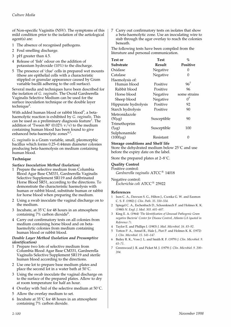
of Non-specific Vaginitis (NSV). The symptoms of thismild condition prior to the isolation of the aetiologicalagent(s) are:
1 The absence of recognised pathogens.
2 Foul smelling discharge.
3 pH greater than 4.5.
4 Release of `fish' odour on the addition ofpotassium hydroxide (10%) to the discharge.
5 The presence of `clue' cells in prepared wet mounts(these are epithelial cells with a characteristicstippled or granular appearance caused by Gramvariable bacilli adhering to the cell surface).
Several media and techniques have been described forthe isolation of G. vaginalis. The Oxoid GardnerellaVaginalis Selective Medium can be used for thesurface inoculation technique or the double layertechnique2.
With added human blood or rabbit blood3, a beta-haemolytic reaction is exhibited by G. vaginalis. Thiscan be used as a preliminary diagnosis feature1. Theaddition of `Tween 80' (0.02% v/v) to the mediumcontaining human blood has been found to giveenhanced beta-haemolytic zones4,5.
G. vaginalis is a Gram variable, small, pleomorphicbacillus which forms 0.25±0.44mm diameter coloniesproducing beta-haemolysis on medium containinghuman blood.
Technique
Surface Inoculation Method (Isolation)1 Prepare the selective medium from Columbia
Blood Agar Base CM331, Gardnerella VaginalisSelective Supplement SR119 and defibrinatedHorse Blood SR51, according to the directions. Todemonstrate the characteristic haemolysis withhuman or rabbit blood, substitute human or rabbitfor horse blood when preparing the medium.
2 Using a swab inoculate the vaginal discharge on tothe medium.
3 Incubate, at 358C for 48 hours in an atmospherecontaining 7% carbon dioxide6.
4 Carry out confirmatory tests on all colonies frommedium containing horse blood and on beta-haemolytic colonies from medium containinghuman blood or rabbit blood.
Double Layer Method (Isolation and Presumptiveidentification)1 Prepare two lots of selective medium from
Columbia Blood Agar Base CM331, GardnerellaVaginalis Selective Supplement SR119 and sterilehuman blood according to the directions.
2 Use one lot to prepare base medium plates andplace the second lot in a water bath at 508C.
3 Using the swab inoculate the vaginal discharge onto the surface of the prepared plates. Allow to dryat room temperature for half an hour.
4 Overlay with 5ml of the selective medium at 508C.
5 Allow the overlay medium to set.
6 Incubate at 358C for 48 hours in an atmospherecontaining 7% carbon dioxide.
7 Carry out confirmatory tests on isolates that showa beta-haemolytic zone. Use an inoculating wire tostab through the agar overlay to reach the coloniesbeneath.
The following tests have been compiled from theliterature and personal communication.
Test or Test %
Substrate Result Positive
Oxidase Negative 0
Catalase Negative 0
Haemolysis of:Human blood Positive 967
Rabbit blood Positive 96
Horse blood Negative some strains
Sheep blood Negative 07
Hippurate hydrolysis Positive 92
Starch hydrolysis Positive 90
Metronidazole(50mg) Susceptible 90
Trimethoprim(5mg) Susceptible 100
Sulphonamide(1000mg) Resistant 0
Storage conditions and Shelf lifeStore the dehydrated medium below 258C and usebefore the expiry date on the label.
Store the prepared plates at 2±88C.
Quality ControlPositive control:
Gardnerella vaginalis ATCC1 14018
Negative control:Escherichia coli ATCC1 25922
References1 Ison C. A., Dawson S. G., Hilton J., Csonka G. W. and Easmon
C. S. F. (1982) J. Clin. Path. 35. 550±554.
2 Spiegel C. A., Eschenbach D., Schoenknech F. and Holmes K. K.
(1980) N. Engl. J. Med. 303. 601±607.
3 King E. A. (1964) `The Identification of Unusual Pathogenic Gram
negative Bacteria' Center for Disease Control, Atlanta GA (quoted in
Reference 7).
4 Taylor E. and Phillips I. (1983) J. Med. Microbiol. 16. 83±92.
5 Totton P. A., Amsel R., Hale J., Piot P. and Holmes K. K. (1972)
J. Clin. Microbiol. 15. 141±147.
6 Bailey R. K., Voss J. L. and Smith R. F. (1979) J. Clin. Microbiol. 9.
65±71.
7 Greenwood J. R. and Picket M. J. (1979) J. Clin. Microbiol. 9. 200±
204.
Culture Media
2-100 November 1998
GBS AGAR BASE (ISLAM)Code: CM755
For the isolation and detection of Group B streptococci(GBS) in clinical specimens.
Formula gm/litreProteose peptone 23.0Soluble starch 5.0Sodium dihydrogen phosphate 1.5Di-sodium hydrogen phosphate 5.75Agar 10.0pH 7.5 + 0.1
DirectionsSuspend 45.2g in 1 litre of distilled water and bring tothe boil to disolve completely. Sterilise by autoclavingat 1218C for 15 minutes. Cool to 508C and asepticallyadd 50ml of sterile inactivated Horse Serum*. Mixwell and pour into petri dishes.*Sterile Inactivated Horse SerumHold sterile Horse Serum (Oxoid SR35) at 568C for 30minutes.
DescriptionGBS Agar Base CM755 is based on the formulationdescribed by Islam. The medium is designed toexploit the ability of most group B streptococci (GBS)to produce orange/red pigmented colonies whenincubated under anaerobic conditions.
Group B streptococci are a recognised cause of seriousneonatal infection acquired from the infected mother.A review2 of national data over an 8 year period bythe Public Health Laboratory Service showed thatgroup B streptococci accounted for 29.5% of allreports of neonatal bacterial meningitis withorganisms being isolated from CSF and blood.
Group B streptococci may also be isolated from adultsinfected in a variety of sites.
The pigment of group B streptococci has characteristicsof a carotenoid3 and was first noted by Lancefield in1934 in nine of twenty-four strains grownanaerobically. Modifications of media1,4,5 haveimproved the proportion of pigmented strains to about97%. Noble et al.6 reported that in their studies 99.5% ofbeta-haemolytic GBS strains produced pigment. GBSAgar also supports growth of other genital bacteria thatcause perinatal infections1, e.g. anaerobic streptococci,Bacteroides and Clostridium species.
Colonies of group B streptococci are 0.5 to 1mm indiameter, round, entire and pigmented orange/redafter 24 to 48 hours anaerobic incubation. Otherorganisms that can grow on this medium do notproduce the orange/red pigment.
de la Rosa et al.7 demonstrated the pigment-enhancing effect of trimethoprim/sulphonamidesadded to their medium. Work carried out in theOxoid laboratories has shown that this pigment-enhancing effect can also be demonstrated around asulphonamide disc placed on the inoculated plate.Standard discs of SF300 or SF500 can be used for thispurpose. No inhibition of growth occurs and thepigment effect is clearly seen over a radius of10±20mm.
Technique1 Swabs should be collected into Stuart's Transport
Medium CM111 and processed within 1/2±2 hoursof collection8.
2 Inoculate the swab on to the surface of GBS Agar.
3 If desired, apply a disc containing 300 or 500mg ofSulphafurazole on to an area of the plate wheregrowth can be expected to be moderately profuse.Such discs are available from Oxoid with productcode SF300 or SF500.
4 Incubate the plates anaerobically at 358C for 24 to48 hours. The Oxoid Anaerobic system with a GasGenerating Kit SR38 is recommended.
5 Report all orange/red pigmented colonies aspresumptive group B streptococcus.
6 Confirm identity with the Oxoid StreptococcalGrouping Kit DR585.
Storage conditions and Shelf lifeStore the dehydrated medium below 258C and usebefore the expiry date on the label.
Store the prepared plates at 2±88C away from light.
Quality ControlPositive control:
Streptococcus agalactiae ATCC1 13813
Negative control:Enterococcus faecalis ATCC1 29212
PrecautionsThe medium must be at its correct pH value to ensuregood pigmentation.
Some strains of Group B streptococci do not producepigmented colonies.
Do not hold the molten medium longer thannecessary to fill out the dishes.
References1 Islam A. K. M. S. (1977) Lancet i 256±7 (letter).
2 PHLS Communicable Disease Report (1954) CDR 84/38, 3±6.
3 Merrit K. and Jacobs N. J. (1978) J. Clin. Microbiol. 8. 105±7.
4 Fallon R. J. (1974) J. Clin. Pathol. 27. 902±5.
5 Merrit K. and Jacobs N. J. (1978) J. Clin. Microbiol. 4. 379±80.
6 Noble A. M., Bent J. M. and West A. B. (1983) J. Clin. Pathol. 36.
350±352.
7 de la Rosa M., Villareal R., Vega D., Miranda C. and
Martinezbrocal A. (1983) J. Clin. Microbiol. 18. 779±785.
8 Islam A. K. M. S. (1981) J. Clin. Pathol. 34. 78±81.
Culture Media
November 1998 2-101

GC SELECTIVE MEDIAIsolation techniques for Pathogenic Neisseria.
NEISSERIA ISOLATION MEDIA ANDTECHNIQUES
IntroductionThe probability of success in the isolation andidentification of pathogenic Neisseria from clinicalspecimens is related to five factors:
1 The amount of care taken in obtaining goodspecimens, their transport to the laboratory andcorrect inoculation on culture medium.
2 The provision of a culture medium capable ofgrowing small inocula of demanding strains.
3 The provision of optimal incubation temperatureand gaseous environment.
4 The inclusion of selective agents in the mediumwhich are capable of preventing overgrowth ofcommensal organisms but which will not inhibitthe growth of the species required.
5 Confirmatory tests to identify the species.
Importance of Accurate DiagnosisDiagnosis of gonorrhoea is achieved by microscopicexamination and cultivation of material from infectedsites. Presumptive gonococci have then to bedistinguished from other organisms of the Neisseriagroup which have a similar morphology and stainingcharacteristics.
Sampling SitesThe diagnosis is achieved by examining smears andcultures from a number of sampling sites. Theseinclude the urethra and rectum of males, the urethra,cervix and rectum of females and occasionally alsothe ducts of Bartholin's glands. The oropharynx ofboth sexes may also require sampling1,2. Repeatedexamination may be necessary before a diagnosis isachieved. The calcium alginate or cotton wool swabsthat are used must be of low toxicity forbacteriological purposes.
As with meningococcal infection, gonorrhoea maypresent as septicaemia with signs of rash, fever and/or arthritis and the responsible organism may beisolated from the blood, joints or (rarely) thecerebrospinal fluid. Ocular infections can occur inneonates and occasionally in adults.
InoculationThe plates are inoculated by rolling the culture swabacross a segment of a moist plate or preferably in alarge `Z' pattern so that an adequate area of the plateis inoculated. Streaking the plate with a sterileinoculating loop is carried out to ensure adequatedispersion of the organisms.
MediaMedia used for the cultivation of gonococci are agarsof high peptone and starch content enriched withfresh horse blood (lysed) or soluble haemoglobin andGC growth supplements (Yeast AutolysateSupplement SR105 and Vitox SR90) which have beenshown to stimulate growth from small inocula.
Several combinations of selective antibiotics (VCNT,
VCN, LCAT and VCAT) have been described that canbe added to culture media in order to suppress Gram-positive and Gram-negative contaminants.
The choice of the selective supplement is dependentupon the preference of the laboratory as well asregional and strain differences of the organism.
OXOID GC AGAR BASECode: CM367
Oxoid GC Agar Base has been formulated to includeSpecial Peptone L72 which is a mixture of meat andplant enzymatic digests. The presence of starch ensuresthat toxic metabolites produced by neisseria areabsorbed. Phosphate buffers are included to preventchanges in pH due to amine production that wouldaffect the survival of the organism.
Formula gm/litreSpecial peptone 15.0Corn starch 1.0Sodium chloride 5.0Dipotassium hydrogen phosphate 4.0Potassium dihydrogen phosphate 1.0Agar 10.0pH 7.2 + 0.2
SOLUBLE HAEMOGLOBIN POWDER
Code: L53
A specially prepared powder which will form a solutionof 2% w/v in water and remain stable after sterilisation.
YEAST AUTOLYSATE GROWTHSUPPLEMENT
Code: SR105
Yeast Autolysate Supplement is a sterile lyophilisedconcentrate of specially prepared yeast fractions withglucose and sodium bicarbonate.
Vial contents (each vial is sufficient for 500ml ofmedium)
Yeast autolysate 5.0gGlucose 0.5gSodium bicarbonate 0.075g
VITOX
Code: SR90/SR90B
Vitox is a sterile lyophilised concentrate of essentialgrowth factors. Many workers prefer such a chemicallydefined growth supplement to yeast extracts for thesupplementation of Thayer Martin Medium.
Culture Media
2-102 November 1998
VITOX (LYOPHILISED)
Code: SR90
Contents (per vial)Vitamin B12 0.1mgL-glutamine 100.0mgAdenine SO4 10.0mgGuanine HCl 0.3mgp-Aminobenzoic acid 0.13mgL-cystine 11.0mgNAD (Coenzyme 1) 2.5mgCocarboxylase 1.0mgFerric nitrate 0.2mgThiamine HCl 0.03mgCysteine HCl 259.0mgGlucose 1.0g
VITOX HYDRATION FLUID
Code: SR90B
Contents (per vial)Glucose 1.0gDistilled water 10.0ml
Vitox may be used satisfactorily at a finalconcentration of 1% v/v in culture media (1 vial to1,000ml of medium).
However, it was found at Oxoid Laboratories thatincreasing the concentation to 2% v/v (1 vial to 500mlof medium) in Thayer Martin Medium resulted infaster growth of Neisseria gonorrhoeae. Use at 2% v/vis therefore recommended.
STERILE SELECTIVE ANTIBIOTICSUPPLEMENTS
The following range of Selective Antibiotic Supplementsare available which can be added to prepare ThayerMartin Medium and derivatives of the New York CityMedium formulation.
VCN SELECTIVE SUPPLEMENT
Code: SR101
An antibiotic supplement for the isolation of N.gonorrhoeae and N. meningitidis.
Vial contents (each vial is sufficient for 500ml ofmedium)
Vancomycin 1.5mgColistin methane sulphonate 3.75mgNystatin 6,250 IU
Thayer and Martin3 described a medium to which theabove antibiotics are added. This selective mediumfor gonococci and meningococci has been widelyaccepted for the primary isolation of these organismsfrom conspicuously contaminated sites.
VCNT SELECTIVE SUPPLEMENT
Code: SR91
An antibiotic supplement for the isolation of N.gonorrhoeae and N. meningitidis.
Vial contents (each vial is sufficient for 500ml ofmedium)
Vancomycin 1.5mgColistin methane sulphonate 3.75mgNystatin 6,250 IUTrimethropin 2.5mg
Seth4 described a modification of Thayer MartinMedium in which Trimethoprim 5mg/ml was addedto the VCN antibiotics and was shown to be of valuein preventing Proteus species swarming. Several otherworkers5,6,7 confirmed the non-inhibitory effect uponN. gonorrhoeae and its value in preventing swarming.
GC SELECTIVE SUPPLEMENT
Code: SR56
GC Supplement is a sterile lyophilised concentrate ofspecially prepared yeast fractions, glucose and sodiumbicarbonate together with the selective agents VCNT.
Vial contents (each vial is sufficient for 500ml ofmedium)
Yeast autolysate 5.0gGlucose 0.75gSodium bicarbonate 0.075gVancomycin 1.5mgColistin sulphate 3.75mgNystatin 6,250 IUTrimethoprim 2.5mg
GC Supplement contains the appropriate growthfactors and antibiotics to prepare Thayer MartinMedium4,7. This supplement is recommended forthose laboratories that prefer to use yeast extract asthe source of essential growth factors required bypathogenic Neisseria.
LCAT SELECTIVE SUPPLEMENT
Code: SR95
An antibiotic supplement for the isolation of N.gonorrhoeae and N. meningitidis.
Vial contents (each vial is sufficient for 500ml ofmedium)
Lincomycin 0.5mgColistin 3.0mgAmphotericin B 0.5mgTrimethoprim 3.25mg
Young8 described a modification of the New YorkCity Medium to which the above antibiotics wereadded. Lincomycin was found to be less inhibitory togonococcus than vancomycin at 3mg/ml9,10. Nystatinwas replaced by amphotericin B which was found tobe a more active anti-fungal agent than nystatin,suppressing the yeast contaminants often found invaginal and urethral specimens11.
Culture Media
November 1998 2-103

MEDIUM CONSTITUENTS
SolubleHaemoglobin Growth Selectiveor Blood Supplement Supplement
Thayer Martin Oxoid GC Agar Soluble Vitox SR90 ±Medium Base CM367 Haemoglobin L53(non-selective)
Thayer Martin Oxoid GC Agar Soluble Vitox SR90 VCN AntibioticMedium Base CM367 Haemoglobin L53 Supplement
SR101
Thayer Martin Oxoid GC Agar Soluble Vitox SR90 VCNT AntibioticMedium (modified) Base CM367 Haemoglobin L53 Supplementwith Vitox SR91Thayer Martin Oxoid GC Agar Soluble GC Supplement GC SupplementMedium (modified) Base CM367 Haemoglobin L53 SR56 SR56with Yeast Fractions
`Transgrow' Medium Oxoid GC Agar Soluble with Vitox SR90 VCN SR101 orBase CM367 + Haemoglobin or VCNT SR91 or1% Agar Supplement SR56 GC Supplement GC Supplement
SR56 SR56
GC Medium Oxoid GC Agar Lysed Yeast Autolysate LCAT Antibioticderived from the Base CM367 Defibrinated Supplement SupplementNew York City horse blood or SR105 SR95 orformulation Laked horse VCAT Antibiotic
blood SR48 SupplementSR104
VCAT SELECTIVE SUPPLEMENT
Code: SR104
An alternative antibiotic supplement for the isolation ofN. gonorrhoeae and N. meningitidis.
Vial contents (each vial is sufficient for 500ml ofmedium)
Vancomycin 1.0mgColistin sulphate 3.75mgAmphotericin B 0.5mgTrimethoprim 1.5mg
Faur et al12 described a medium in which the aboveantibiotics were added.
Nystatin was replaced with amphotericin B whichwas found to be a more active anti-fungal agent,suppressing most yeast contaminants. Thevancomycin level was reduced from 3mg/ml to 2mg/ml to ensure that the 3% of gonococcus strains shownby Reyn9 to be sensitive to 3mg/ml of vancomycinwere not inhibited11.
Directions for preparation of the Variant MediaThayer Martin Medium with GC Supplement1 Suspend 18g of Oxoid GC Agar Base in 235ml of
distilled water and bring to the boil to dissolve theagar. Sterilise by autoclaving at 1218C for 15minutes.
2 Prepare a 2% solution of Soluble HaemoglobinPowder L53 by adding 250ml of warm distilledwater to 5g of haemoglobin powder. Continuallystir the solution during the addition of water.Sterilise by autoclaving at 1218C for 15 minutes.
3 Dissolve the contents of a vial of sterile GCSupplement SR56 in 15ml of sterile distilled water.
4 Aseptically add the GC Supplement Solution SR56to the 235ml of sterile GC Agar Base cooled to 508Cand mix gently. Add the 250ml of sterilehaemoglobin solution, cooled to 508C, to the GCAgar Base-Supplement solution. Mix gently toavoid trapping air bubbles in the agar and pourinto sterile petri dishes.
Thayer Martin Medium with Vitox and either VCN orVCNT Antibiotic Supplement.1 Suspend 18g of GC Agar Base in 240ml of distilled
water and bring gently to the boil to dissolve theagar. Sterilise by autoclaving at 1218C for 15minutes.
2 Prepare a 2% solution of Soluble HaemoglobinPowder L53 by adding 250ml of warm distilledwater to 5g of haemoglobin powder. Continuallystir the solution during the addition of water.Sterilise by autoclaving at 1218C for 15 minutes.
3 Dissolve the contents of one vial of Vitox SR90 asdirected on the vial label.
Culture Media
2-104 November 1998

4 Dissolve the contents of a vial of either VCNAntibiotic Supplement SR101 or VCNT AntibioticSupplement SR91 as directed on the vial label.
5 Aseptically add the Vitox solution to 240ml ofsterile GC Agar Base cooled to 508C.
6 Aseptically add the reconstituted antibioticsupplement VCN or VCNT to the GC Agar Base-Vitox solution.
7 Asceptically add the 250ml of sterile haemoglobinsolution, cooled to 508C to the GC Agar Base-Vitox-Antibiotic Supplement solution. Mix gentlyto avoid trapping air bubbles in the agar and pourinto sterile petri dishes.
`Transgrow' Medium`Transgrow' Medium is prepared in the same manneras described for Thayer Martin Medium variants,except that 5g of Agar is added to the 18g of GC AgarBase. The medium is dispensed as agar slants in glassbottles. The extra glucose required is contained in thegrowth supplements.
GC Medium derived from the New York CityFormulation with LCAT or VCAT antibioticsupplement1 Suspend 18g of Oxoid GC Agar Base in 425ml of
distilled water and bring gently to the boil todissolve the agar. Sterilise by autoclaving at 1218Cfor 15 minutes.
2 Lyse 50ml of Defibrinated Horse Blood SR50 with0.5% (by vol.) saponin.
3 Dissolve the contents of a vial of sterile YeastAutolysate Supplement SR105 in 15ml of steriledistilled water.
4 Dissolve the contents of either LCAT SR95 orVCAT SR104 in 10ml of sterile distilled water.
5 Aseptically add the lysed defibrinated horse blood,Yeast Autolysed Supplement and the AntibioticSupplement (LCAT or VCAT) to the sterile GCAgar Base cooled to 508C. Mix gently to avoidtrapping air bubbles in the agar and pour intosterile petri dishes.
This is a derivative of NYC Medium11,12,13 based onYoung's publication14 where the higher level ofglucose recommended by the originators was reducedto allow sugar fermentation test to be carried out15.
Technique1 Prepare the medium as directed and pour into
petri dishes.
2 Inoculate by rolling the culture swabs across asegment of the plate or preferably in a large `Z'pattern so that an adequate area of the plate isinoculated. Streaking of the plate is carried outwith a sterile loop to ensure adequate dispersion ofthe organisms.
3 Plates are incubated at 378C in a sealed jar with atleast 70% humidity and 5±10% carbon dioxideprovided by the introduction of the gas into theincubator or by the use of a candle jar. The aboveincubation conditions can be conveniently achievedby using the Oxoid Carbon Dioxide GasGenerating Kit BR39 in the Oxoid Gas Jar HP11.
4 Examine the plates after 24 hours incubation and ifnegative reincubate for a further 24 hours.
5 Presumptive gonococcus colonies are identified bythe Gram stain, oxidase and sugar fermentationreactions.
See also Mycoplasma page 2-120.
Typical reactions of N. gonorrhoeaeGram negative cocci, usually arranged in pairs withlong axes parallel. Oxidase positive.
Fermentation Reactions of the Neisseria Group
Glucose Maltose Lactose SucroseN. meningitidis + + ± ±N. gonorrhoeae + ± ± ±Bran. catarrhalis ± ± ± ±N. flava andrelated types + +/± ± +/±
+ = Acid production+/± = Variation in reaction among different types
List of products available from Oxoid for theGrowth and Identification of Neisseria Species
GC Agar Base CM367VCN Selective Supplement SR101
Agar No.1 L11VCNT Selective Supplement SR91
Soluble Haemoglobin Powder L53
LCAT Selective Supplement SR95
Sterile Yeast Autolysate Supplement SR105
VCAT Selective Supplement SR104
Vitox SR90
GC Supplement SR56(Yeast Fractions plus VCNT Antibiotics)
Defibrinated Horse Blood SR50
Oxidase Detection Papers BR54
Laked Horse Blood SR48
Storage conditions and Shelf lifeStore the dehydrated medium below 258C and usebefore the expiry date on the label.
Store the prepared plates at 2±88C.
Quality ControlPositive control:with antibiotics
Neisseria gonorrhoeae ATCC1 19424Neisseria meningitidis ATCC1 13090
without antibioticsHaemophilus influenzae ATCC1 35056
Negative control:with antibiotics
Proteus vulgaris ATCC1 13315Staphylococcus aureus ATCC1 25923
Please note that for the LCAT Selective SupplementSR95, Proteus vulgaris ATCC1 13315 as a negativecontrol is inoculum dependent.
without antibioticsUninoculated medium
Culture Media
November 1998 2-105

PrecautionsUse Dacron1 or calcium alginate swabs for specimencollection, avoid cotton wool.
Any suspected Neisseria-containing specimen shouldbe inoculated onto primary isolation mediaimmediately on collection. If this is not possible thenN. gonorrhoeae swabs are better held at 48C for notmore than 3 hours. N. meningitidis swabs should notbe chilled and they should be inoculated on to pre-warmed media.
It is a wise precaution to inoculate GC media withand without antibiotics in parallel because somestrains of neisseriae may fail to grow in the presenceof antibiotics, especially vancomycin16.
Humidity is essential for the successful isolation ofgonococci. If the prepared plates look dry moisten thesurface with a few drops of sterile broth and allow itto soak into the agar before inoculation. Do not floodthe plate with broth. Place damp gauze or papertowels in the CO2 container before incubation.
Hosty et al.17 showed that the usual transport mediaare not totally reliable for N. gonorrhoea. Inoculation ofthe sample on to the surface of medium slants inbottles is preferable. The incubator temperatureshould be set at 358C because many strains of N.gonorrhoea will not grow well at 378C on culturemedia18.
It has been shown that agars vary widely in theirtoxicity for N. gonorrhoea and may be a major factorpreventing the growth of gonococci on solid media19.
The Gram morphology, colony characteristics andoxidase tests form the presumptive identification forthe genus Neisseria. All presumptive neisseriae shouldbe confirmed by carbohydrate fermentation tests andfluorescent antibody or other serological tests20.
An ONPG test will detect lactose-utilising strains ofneisseriae e.g. N. lactamica21.
References1 Willcox R. R. (1979) Euro Reports and Studies, 12, World Health
Organization, Regional Office for Europe, Copenhagen.
2 'Neisseria gonorrhoeae and gonococcus infections' (1978) Report
of a WHO Group, Technical Report Series 616, World Health
Organization, Geneva.
3 Thayer J. D. and Martin J. E. (1966) Public Health Rep., 81. 6. 559±
562.
4 Seth A. (1970) Brit. J. Vener. Dis. 46. 201±202.
5 Riddel R. H. and Buck A. C. (1970) J. Clin. Path. 23. 481±483.
6 Odegaard K. (1971) Acta Path. Microbiol. Scand., Sect. B. 79. 545±
548.
7 Martin J. E. and Lester A. (1971) HSMHA Health Reports. 86. No.1
30±33.
8 Young H. (1978) Brit. J. Vener. Dis. 54. 36±40.
9 Reyn A. and Bentzon M. W. (1972) Brit. J. Vener. Dis. 48. 363±
368.
10 Mirrett S., Reller L. B. and Knapp J. S. (1981) J. Clin. Microbiol.
14. 94±99.
11 Faur Y. C., Wiesburd M. H., Wilson M. E. and May P. S. (1973)
Health Lab. Sci. 10. 44±54.
12 Faur Y. C., Weisburd M. H. and Wilson M. E. (1973) Health Lab.
Sci. 10. 55±60.
13 Faur Y. C., Weisburd M. H. and Wilson M. E. (1977) Health Lab.
Sci. 15. 22±27.
14 Young H. (1978) Brit. J. Ven. Diseases 54. 36±40.
15 Young H. (1978) J. Clin. Microbiol. 7. 247±250.
16 Faur Y. C., Weisburd M. H. and Wilson M. E. (1978) Health Lab.
Sci. 15. 22±26.
17 Hosty T., Freear M., Baker C. and Holston J. (1974) Amer. J. Clin.
Path. 62. 435±438.
18 Finegold S. M. and Martin W. J. (1982) Bailey and Scott's
Diagnostic Microbiology 6th Edn. C. V. Mosby & Co. St. Louis. p.
107.
19 Jones R. T. and Talley R. S. (1977) J. Clin. Microbiol. 5. 9±13.
20 CDC (1975) `General information to aid in handling Neisseria
gonorrhoea specimens in the laboratory' US DHEW Center for
Disease Control. Atlanta. Ga.
21 Hollis D. G., Wiggins G. T. and Weaver R. E. (1969) Appl.
Microbiol. 17. 71±77.
GIOLITTI-CANTONI BROTHCode: CM523
An anaerobic enrichment broth for Staphylococcusaureus.
Formula gm/litreTryptone 10.0`Lab-Lemco' powder 5.0Yeast extract 5.0Lithium chloride 5.0Mannitol 20.0Sodium chloride 5.0Glycine 1.2Sodium pyruvate 3.0pH 6.9 + 0.2
DirectionsSuspend 54.2 grams in one litre of distilled water andheat gently to dissolve. Dispense 19ml amounts into20mm x 200mm test tubes and sterilise by autoclavingat 1218C for 15 minutes. Cool rapidly then asepticallyadd to each tube 0.3ml of a sterile solution ofPotassium tellurite 3.5% SR30.
The medium requires the addition of a 3.5% solutionof Potassium tellurite when there is a direct additionof 1 gram of the sample to 19mls of broth. This levelof Potassium tellurite is necessary to suppress thehigh numbers of contaminating organisms that couldbe expected.
The use of a diluted solution of Potassium tellurite isapplicable when a 1 in 10 dilution of the food sampleis carried out1. In such cases the SR30 should first bediluted 1 in 10 with sterile distilled water.
The addition of 0.1% Tween 80 can be recommendedin order to improve recovery of heat injured Staph.aureus cells e.g. from milk powder. 1 gram of Tween80 should be added to 1 litre of CM523 prior toautoclaving2.
DescriptionOxoid Giolitti-Cantoni Broth, a tellurite-mannitol-glycine enrichment broth, based on the formulation ofGott and Cantoni3 is used for the selection andenrichment of Staphylococcus aureus from foodstuffs.Mannitol and sodium pyruvate are growth stimulantsfor staphylococci and aid detection of the organismwhen present in small numbers only4.
Culture Media
2-106 November 1998
The growth of Gram-negative lactose fermentingbacilli are inhibited by lithium chloride and Gram-positive bacilli are inhibited by Potassium tellurite incombination with glycine.
The creation of anaerobic conditions by overlayingwith 2cm of sterile paraffin wax inhibits the growth ofmicrococci.
Giolitti-Cantoni Broth is recommended for thedetection of Staphylococcus aureus in dried baby milkand other weaning foods where the organism shouldbe absent from 1 gram of test material6.
The medium is suitable for the examination of meatand meat products7. For this purpose theconcentration of the Potassium tellurite must bereduced to 0.35% and it is recommended that theweight of the test sample should be reduced to0.1±0.01 gram.
TechniqueThe medium should be inoculated as soon as it hasbeen cooled after autoclaving. If there is a delay inputting the medium to use it must be freed fromdissolved air by immersion in free flowing steam for20 minutes.
Inoculate 1 gram of sample material and 1ml aliquotsof a series of suitable decimal dilutions into tubescontaining 19ml of Giolitti-Cantoni Broth. Two tubesare used for the sample material and for each of thedilutions. This increases the likelihood of detectingStaphylococcus aureus when it is present in very smallnumbers.
The medium is overlaid with 2cm of molten sterileparaffin wax (melting temperature 42±448C) andincubated for 48 hours at 358C, examining daily. Theresult is considered negative for Staphylococcus aureusif no blackening of the medium is observed. Ifblackening does occur at the bottom of the tubes orgeneral blackening of the medium, the broth isstreaked on to a staphylococcal isolation medium,such as Baird-Parker Medium8 CM275 and incubatedat 358C for 24±48 hours. The result is consideredpositive if black colonies, with a narrow whitemargin, surrounded by a zone of clearing, are seen.
Storage conditions and Shelf lifeStore the dehydrated medium below 258C and usebefore the expiry date on the label.
Store the prepared medium at 2±88C.
Quality ControlPositive control:
Staphylococcus aureus ATCC1 25923
Negative control:Staphylococcus epidermidis ATCC1 12228
References1 IDF International Standard 60A:1978.
2 Chopin et al (1985) J. Food Prod. 48 No.1 21±27.
3 Giolitti C. and Cantoni C. (1966) J. Appl. Bact. 29. 395.
4 Baird-Parker A. C. (1962) J. Appl. Bact. 25. 12.
5 Lambin S. and German A. (1961) `PreÂcis de microbiologic' p.63,
Paris Masson.
6 Mossel D. A. A., Harrewijn G. A. and Elzebroek J. M. (1973)
UNICEF.
7 ISO/DIS 5551 (1177) Part 2.
8 De Waart J., Mossel D. A. A., Ten Broeke R. and Van de
Moosdijk A. (1968) J. Appl. Bact. 31. 276.
HAEMOPHILUS TEST MEDIUM(HTM)
HTM BASE
Code: CM898
HTM SUPPLEMENT
Code: SR158A medium specifically formulated for the susceptibilitytesting of Haemophilus influenzae.
HTM BASE (CM898)
Formula gm/litreMueller-Hinton Agar 38.0Yeast extract (specifically selectedfor low antagonist levels) 5.0pH 7.4 + 0.2
HTM SUPPLEMENT (SR158)
Vial contentsNAD 7.5mg (equivalent to 15mg/L)Haematin 7.5mg (equivalent to 15mg/L)
DirectionsSuspend 21.5g of Haemophilus Test Medium Base(CM898) in 500ml of distilled water. Bring to the boilto dissolve. Sterilise by autoclaving at 1218C for 15minutes. Cool to 508C and aseptically add thecontents of 1 vial of Haemophilus Test MediumSupplement (SR158), reconstituted with 2ml of steriledistilled water. Mix well and pour into sterile petridishes.
DescriptionHaemophilus Test Medium (HTM) has beenspecifically formulated for the susceptibility testing ofHaemophilus influenzae. The medium forms part of therecommended methods of the United States NationalCommittee for Clinical Laboratory Standards(NCCLS)1.
Haemophilus influenzae require complex media forgrowth. These complex media have aggravated theroutine susceptibility testing of H. influenzae becauseof antagonism between some essential nutrients andcertain antimicrobial agents. Difficulties ininterpreting inhibition zones may also arise.
Oxoid Haemophilus Test Medium (HTM) is based onthe formulation developed by Jorgensen et al2 whichis now recommended by the United States NCCLS.The results achieved using HTM have been found tobe highly reproducible3,4. Comparisons withMueller-Hinton Chocolate Agar have shown anoverall agreement of 99.6%5.
The transparency of the medium allows zones ofinhibition to be read easily through the bottom of thepetri dish. HTM contains low levels of antimicrobialantagonists, which allows testing of trimethoprim/sulphamethoxazole to be carried out with greaterconfidence.
Culture Media
November 1998 2-107
Technique1 Prepare the inoculum in Mueller-Hinton Broth
(Oxoid CM405) or 0.9% saline, to match theturbidity of 0.5 McFarland standard.
2 Using a swab saturated with the above inoculumsuspension, inoculate the surface of a HaemophilusTest Medium Agar plate to give confluent growth.
3 Apply the antimicrobial discs on to the surface ofthe Haemophilus Test Medium plate.
4 Incubate the plates at 358C in 5±7% carbon dioxidefor 16±18 hours and measure the zones ofinhibition.
NB: In the absence of a CO2 incubator, the CO2
enriched atmosphere can best be achieved by usingthe Oxoid Gas Generating Kit BR39 in conjunctionwith Oxoid Anaerobic Jar.
Storage conditions and Shelf lifeHTM Base should be stored tightly capped in theoriginal container in a cool, dry place away frombright light. When stored as directed the medium willremain stable until the expiry date printed on thelabel.
HTM Supplement as supplied should be stored in thedark below 08C. When stored as directed thesupplement is stable until the expiry date stated onthe label.
Quality ControlH. influenzae ATCC149766 Good growth
References1 NCCLS Documents M2-A4 Vol. 10. No. 7. and M7-A2 Vol. 10.
No 8.
2 Jorgensen J.H., Redding J.S., Maher L.A. and Howell A.W.
(1987) J. Clin. Micro. 25, 2105±2113.
3 Doern G.V., Jorgensen J.H., Thornsberry C. and Snapper H.
(1990). Eur. J. Clin. Microbiol. Infect. Dis. 9, 329±336.
4 Barry A.L., Jorgensen J.H. and Hardy D.J. (1991) J. Antimic.
Chem. 27, 295±301.
5 Evans G., Marsik F., Thompson L. and Fowler J. (1990) Abstracts
of ASM Meeting 1990 C-252.
HEKTOEN ENTERIC AGARCode: CM419
A differential, selective medium for the isolation ofShigella and Salmonella species from entericpathological specimens.
Formula gm/litreProteose peptone 12.0Yeast extract 3.0Lactose 12.0Sucrose 12.0Salicin 2.0Bile salts No.3 9.0Sodium chloride 5.0Sodium thiosulphate 5.0Ammonium ferric citrate 1.5Acid fuchsin 0.1Bromothymol blue 0.065Agar 14.0pH 7.5 + 0.2
DirectionsSuspend 76g of the medium in 1 litre of distilledwater and soak for 10 minutes. Heat gently and allowto boil for a few seconds to dissolve the agar. DONOT AUTOCLAVE. Cool to 508C and pour plates.
DescriptionHektoen Enteric Agar was developed by King &Metzger1. The high peptone content offsets theinhibitory effect of bile salts on Shigella species inparticular. The additional carbohydrates (sucrose andsalicin) give better differentiation than lactose aloneand the lower toxicity of the double indicatorimproves recovery. The increased lactose contenthelps early recognition of slow lactose-fermentingorganisms. The thiosulphate and ferric citrate arepresent to detect H2S-producing organisms.
Taylor & Schelhaut2 found the medium to be of valuein the differentiation of pathogenic organisms and forbetter growth of shigellae.
Hoben et al.3 added novobiocin 15mg/litre toimprove the selectivity of the medium by inhibitingCitrobacter and Proteus species.
Hektoen Enteric Agar meets the requirements of theAPHA4.
TechniqueInoculate the medium with fresh faeces suspended inRingers solution or inoculate directly with rectalswabs. Spread the inoculum to obtain well separatedcolonies. Incubate for 18±24 hours at 378C. Furtherincubation will improve differentiation betweenshigellae and salmonellae.
Organism characteristics:Shigella Green, moist raised colonies.Salmonella Blue-green colonies with
or without black centres.Coliforms (rapid Salmon-pink to orangelactose/sucrose/ colonies surrounded by a zonesalicin fermenters) of bile precipitation.
Storage conditions and Shelf lifeStore the dehydrated medium below 258C and usebefore the expiry date on the label.
Store the prepared plates at 2±88C.
Quality ControlPositive control:
Salmonella typhimurium ATCC1 14028Shigella flexneri ATCC1 12022
Negative control:Escherichia coli ATCC1 25922Enterococcus faecalis ATCC1 29212
PrecautionsDo not overheat the medium or hold it molten forlong periods.
Proteus species may resemble salmonellae or shigellae.Further testing must be carried out to confirm thepresumptive identification of organisms isolated onthis medium.
References1 King S. and Metzger W. I. (1968) Appl. Microbiol. 16. 577±561.
2 Taylor W. I. and Schelhaut D. (1971) Appl. Microbiol. 21. 32±37.
Culture Media
2-108 November 1998
3 Hoben D. A., Ashton D. H. A. and Peterson A. C. (1973) Appl.
Microbiol. 21. 126±129.
4 American Public Health Association (1976) Compendium of
Methods for the Microbiological Examination of Foods. APHA Inc.
Washington DC.
HELICOBACTER PYLORIISOLATION
HELICOBACTER PYLORI SELECTIVESUPPLEMENT (DENT)
Code: SR147
A selective supplement for the isolation of H. pylorifrom clinical specimens.
Vial contents (each vial is sufficient for 500ml ofmedium)
Vancomycin 5.0mgTrimethoprim lactate 2.5mgCefsulodin 2.5mgAmphotericin B 2.5mg
DirectionsTo one vial add 2ml of sterile distilled water anddissolve the powder completely. Avoid frothing. Addthe contents aseptically to 500ml of sterile ColumbiaBlood Agar Base CM331 at 508C. Add 35ml of LakedHorse Blood SR48 and mix well before pouring intosterile petri dishes.
DescriptionHelicobacter pylori is associated with a number ofgastric conditions, chiefly gastritis and pepticulcers1,2,3.
Helicobacter pylori Selective Supplement (Dent)SR147 was developed from Dent's selective mediumdescribed for the isolation of H. pylori from gastricbiopsies2. This is a modification of Skirrow's medium4
in which polymixin B is replaced by cefsulodin andamphotericin B is added to inhibit Candida species.
When used routinely in the laboratory for 100 gastricbiopsies, Dent's medium achieved a higher isolationrate for H. pylori and lower contamination by otherorganisms when compared with Skirrow's mediumand chocolate blood agar2. The provision of a goodselective medium for H. pylori will help establish therole of this organism in the aetiology of gastricdisease.
Technique1 Prepare the medium as directed. The plates can be
stored at 48C for three weeks but it is essential thatthey are kept moist. This can be achieved simplyby keeping the plates in a plastic bag.
2 Smear the specimen on to the medium.
Note ± the recovery of H. pylori from gastric biopsiesis improved by direct cultivation as soon as possibleafter collection. If transportation is necessary, thenplace the biopsy against the neck of a small, sterileglass bottle containing 0.1ml of sterile saline2. Thebiopsy should adhere to the glass but be protectedfrom dehydration by water vapour.
3 Incubate at 358C for three to five days under micro-aerophilic conditions. Use Campylobacter GasGenerating Kit BR56 or BR60 with an activecatalyst BR42. Alternatively use CampyGenCN025A or CN035A. CampyGen does not requirethe addition of water or a catalyst.
4 Examine for the presence of discrete, translucentand non-coalescent colonies. Note that colonies ofH. pylori do not resemble those of Campylobacterspecies.
5 Confirm the identity of the isolates with thefollowing tests:
Gram negative, curved or spiral bacillus.
Growth at 358C, no growth at 258C, variable growthat 428C. Urease positive, Catalase positive, Oxidasepositive, Hippurate negative.
Quality ControlPositive control:
Helicobacter pylori NCTC 11916/ATCC1 43526
Negative control:Candida albicans ATCC1 10231
References1 Marshall B. K., Warren J. R., Blincow E. D., Phillips M.,
Goodwin C. S., Murray R., Blackbourne S. J. and Waters T. E.
(1988) The Lancet, December 24/31, No 8626/8627.
2 Dent J. C. and McNulty C. A. M. (1988) Eur. J. Clin. Microbiol.
Infec. Dis. 7. 555±568.
3 Buck G. E. (1988) Laboratory Management, 26, No.9.
4 Skirrow M. B. (1977) BMJ, 1. 9±11.
HOYLE MEDIUM BASECode: CM83
A modification of Neill's medium for the isolation anddifferentiation of Corynebacterium diphtheriae types.
Formula gm/litre`Lab-Lemco' powder 10.0Peptone 10.0Sodium chloride 5.0Agar 15.0pH 7.8 + 0.2
DirectionsSuspend 40g in 1 litre of distilled water and bring tothe boil to dissolve completely. Sterilise byautoclaving at 1218C for 15 minutes. Cool to 558C andadd 50ml of Laked Horse Blood SR48 and 10ml of3.5% Potassium Tellurite solution SR30, mix, andpour plates.
DescriptionHoyle medium is the well known modification1 ofNeill's medium for the cultural isolation anddifferentiation of Corynebacterium diphtheriae types.Hoyle medium does not exert the inhibitory effectmanifested by Neill's on some mitis types, but givesvery rapid growth with all types of Corynebacteriumdiphtheriae, so that diagnosis is possible after 18 hours'incubation.
Culture Media
November 1998 2-109

TechniqueThis is a highly selective medium which is used inparallel with non-selective media such as blood agar(e.g. Blood Agar Base CM55 with 10% of horseblood). Unlike the non-selective media, Hoylemedium should be inoculated by rubbing the throatswab (or other material) over the entire surface ±spreading with a platinum loop is not necessary.
Normally incubation for 18 hours at 358C is sufficientbut, when a negative result is obtained, incubation forup to 72 hours may be advisable. Gentian violetstained films made from colonies picked straight offthe medium, are satisfactory for C. diphtheriaemorphology. For the demonstration of thecharacteristic morphology and staining reactions ofC. diphtheriae by Neisser's or Albert's method, it ispreferable to utilise colonies from Loeffler medium.The toxigenicity of C. diphtheriae strains may bedetermined by the Elek2 method.
Colonial CharacteristicsIt is best to examine with a low-power microscope,the colonies being illuminated from above bydaylight.
Type differentiation is good, and typical colonialappearances after 18 hours' incubation are as follows:
C. diphtheriae type gravis ± grey colonies, 1.5±2.5mmdiameter dull, matt surface. May be slightlyumbonate and show indented margins. Colonies canbe pushed along the surface of the medium.
C. diphtheriae type mitis -- grey colonies, 1.5±2.0mmdiameter with regular margins and shining surface.Variation in size common.
C. diphtheriae type intermedius ± grey colonies,0.5±0.75mm diameter with shining surface. Coloniesare very uniform in size with darker centres.
C. hofmannii ± usually rounded, white or greyish,0.5±0.75mm diameter. Colonies may be up to 1mmdiameter, when they are black in more heavilyinoculated areas and white when well isolated.
C. xerosis ± black shining colonies of variable size.
Streptococci ± minute black or brownish-blackcolonies.
Other organisms may occasionally grow whichresemble C. diphtheriae type intermedius but are larger,while sporing anaerobes may produce brownishmucoid colonies.
Storage conditions and Shelf lifeStore the dehydrated medium below 258C and usebefore the expiry date on the label.
Store the prepared plates at 2±88C.
Quality ControlPositive control:
Corynebacterium diphtheriae biotype gravis ATCC1
19409Corynebacterium diphtheriae biotype intermediusATCC1 14779
Negative control:Uninoculated medium.
PrecautionsIt should be noted that not all corynebacteria producethe typical colonies described above ± so in all cases itis advisable to use Hoyle medium in conjunction withthe additional media and tests mentioned above.
References1 Hoyle L. (1941) Lancet. i. 175±176.
2 Elek S. D. (1948) Brit. Med. J. 1. 493±496.
HIGH RESOLUTION (H.R.) MEDIUMCode: CM845
A chemically defined medium for fluconazolesusceptibility testing.
Formula gm/litreDextrose 19.98Potassium dihydrogen phosphate 1.99Ammonium sulphate 4.99L-Glutamine 0.58Magnesium sulphate (anhydrous) 0.99Sodium chloride 0.20Calcium chloride 0.20L-Lysine monohydrochloride 0.073Valine 0.047L-Arginine monohydrochloride 0.042L-Histidine 0.023Methionine (DL) 0.0189Tryptophane 0.020Inositol 0.00397Boric acid 0.00099Calcium D-pantothenic acid 0.00079Nicotinic acid 0.00079Pyridoxine hydrochloride 0.00079Aneurine hydrochloride 0.00079Manganous sulphate 0.00079Zinc sulphate 0.00144-Aminobenzoic acid 0.000395Riboflavin BP/USP 0.000395Ferric chloride 0.000395Cupric sulphate 0.00012Biotin crystalline 0.000004Folic acid 0.000395L-Isoleucine 0.052Sodium molybdate 0.00047Potassium iodide 0.00020L-Leucine 0.052Threonine 0.0476
DirectionsPART APrepare a 2% w/v solution of Agar Technical (OxoidL13) and buffer to pH 7.5 with 0.1ml phosphatebuffer. Sterilise at 1158C for 10 minutes.
PART BDissolve 29.34g of High Resolution Medium in 900mlof distilled water. Stir continuously and add 2g ofsodium bicarbonate (analar) and make up the volumeto 1 litre with distilled water. Sterilise by filtration.The medium can be kept at 48C for 2 weeks.
Aseptically mix equal volumes of molten HighResolution Medium Part A (cooled to 608C) and High
Culture Media
2-110 November 1998
Resolution Medium Part B. Mix thoroughly and pourinto sterile petri dishes.
DescriptionHigh Resolution Medium is a chemically definedmedium, specifically developed for the in vitro testingof fluconazole. The MIC values generated using themedium give sensible correlations with efficacy invitro and with clinical outcome.
In a comparison of a disc diffusion method against amicrodilution method correlation using HR mediumwas good for a number of antibiotics includingnystatin and amphotericin B but generally better fornew triazoles such as fluconazole than for the otherantifungals.6
TechniquePreparation of the inoculaThe preparation and standardisation of the inoculumvaries with different fungi:
CANDIDA spp1 Grow Candida spp overnight at 378C in Sabouraud
dextrose broth.
2 Vortex mix and make a 1 in 100 dilution of eachculture in normal saline and estimate the cellnumbers using a haemocytometer.
3 Appropriately dilute each culture in normal salineto give the following cell densities:
Candida albicans 105/mlCandida krusei 105/mlCandida tropicalis 105/mlCandida guillermondii 106/mlCandida parapsilosis 106/mlCandida pseudotropicalis 106/ml
Inoculation of the plates1 Surface inoculate each series of plates using a
multipoint inoculator which delivers 1ml of eachculture inoculum. It is possible to inoculate 36isolates per 9cm petri dish. Inoculate control platesat both the beginning and end of the inoculationrun.
2 Incubate the plates at 288C for 48 hours.
Endpoint determination1 Check the control plates to ensure that all the
organisms have grown adequately.
2 Read all plates against a standard background andrecord, for each isolate, the lowest concentration offluconazole that completely suppresses visible (tothe naked eye) growth. This is the MIC value.
DERMATOPHYTE spp1 Grow the dermatophytes on Sabouraud Dextrose
Agar (CM41) at 288C for 5±10 days.
2 Scrape off the mycelium from the agar surfaceusing a scalpel and place in a bijou bottlecontaining 4gm of glass beads (approximately2mm diameter) plus 2ml of 0.85% saline.
3 Vortex mix to evenly disperse.
4 Adjust the density of the suspension with 0.85%saline to give a 65% light transmission on anabsorptiometer.
5 Inoculate the plates using a multipoint inoculator.It is possible to inoculate 20 isolates, evenly spaced,per 9cm petri dish.
6 Incubate at 288C for 6 days.
7 Check the control plates to ensure that all isolateshave grown adequately.
8 Determine the endpoint as for Candida.
Candida albicans (ATCC1 76615) has an MIC value of1.56mg/ml against the antifungal agent fluconazole.
Storage conditions and Shelf lifeHigh Resolution Medium should be stored tightlycapped in the original container at 10 to 258C awayfrom bright light. When stored as directed themedium will remain stable until the expiry dateprinted on the label.
Please note shelf life of 2 years.
References1 Hoeprich P.D. and Finn P.D. (1972) J. Infect. Dis. 126, 353±361.
2 Cook R.A., McIntyre K.A. and Galgiani J.N. (1990). Antimicrob.
Agents and Chemother. 34, 1542±1545.
3 Pfaller M.A. et al. Antimicrob. Agents Chemother. 34, 1648±1654.
4 Pfaller M.A. et al. (1992) Antimicrob. Agents and Chemother. 36,
1805±1809.
5 Pfizer Ltd, private communication (1990).
6. Carillo-Munoz A.J., Tur C., Etervill D. et al (1995) Revista
EspanÄola de Quimioterapia 8, 221±228.
IRON SULPHITE AGARCode: CM79
A medium for the detection of thermophilic anaerobicorganisms.
Formula gm/litreTryptone 10.0Sodium sulphite 0.5Iron (III) citrate 0.5Agar 12.0pH 7.1 + 0.2
DirectionsSuspend 23.0g in 1 litre of distilled water and boil todissolve completely. Sterilise by autoclaving for 15minutes at 1218C. Mix well before pouring.
DescriptionThis medium is a modification of Cameron SulphiteAgar developed by the National Canners Associationof America1.
It had been shown that the medium was improved byreducing the concentration of sodium sulphite.Beerens2 showed that some strains of Clostridiumsporogenes would not tolerate 0.1% sulphite. This wasconfirmed by Mossel et al.3 who consequently usediron sulphite agar containing only 0.05% sulphite andobtained no apparent inhibition.
TechniqueIron Sulphite Agar is particularly suitable for thedetection of thermophilic anaerobic organismscausing sulphide spoilage in food. The medium
Culture Media
November 1998 2-111
should be dispensed in 10ml amounts in tubes fordeepshake cultures, and inoculated whilst fluid atabout 508C. Allow to set and incubate at 558C forthermophilic species. Desulfotomaculum nigrificans, thetype species, produces distinct black sphericalgrowths in the depth of the medium.
In the Attenborough and Scarr method4, dilutedsamples of the sugar were filtered through membranefilters which were then rolled up and placed in tubescontaining enough melted Iron Sulphite Agar (atapproximately 508C) to cover them. The medium wasallowed to solidify and the tubes were incubated at568C. After 48 hours the number of black colonies onthe membrane was counted. This membrane filtertechnique is quicker than the standard method but ofcomparable accuracy, and permits the examination ofa much larger sample5.
Storage conditions and Shelf lifeStore the dehydrated medium below 258C and usebefore the expiry date on the label.
Store the prepared medium below 258C.
Quality ControlPositive control:(blackening)Clostridium sporogenes ATCC1 19404Desulfotomaculum nigrificans ATCC1 19858
Negative control:(no blackening)Escherichia coli ATCC1 25922
PrecautionsThe blackening reaction is only presumptive evidenceof clostridial growth. Confirmation tests must becarried out to identify the organism growing in themedium.
References1 Tanner F. W. (1944) `The Microbiology of Foods' 2nd ed., Garrard
Press, Illinois p. 1127.
2 Beerens H. (1958) DSIR, Proc. 2nd Internat. Symp. Food Microbiol.
1957, HMSO, London, pp. 235±245.
3 Mossel D. A. A., Golstein Brouwers G. W. M. V. and de Bruin
A. S. (1959) J. Path. Bact. 78. 290±291.
4 Attenborough Sheila J. and Scarr M. Pamela (1957) J. Appl. Bact.
20. 460±466.
5 Bufton A. W. J. (1959) J. Appl. Bact. 22. 278±280.
`ISO-SENSITEST AGAR'Code: CM471
A closely defined medium with stabilised mineralcontent for antimicrobial susceptibility testing.
Formula gm/litreHydrolysed casein 11.0Peptones 3.0Glucose 2.0Sodium chloride 3.0Soluble starch 1.0Disodium hydrogen phosphate 2.0Sodium acetate 1.0Magnesium glycerophosphate 0.2Calcium gluconate 0.1Cobaltous sulphate 0.001Cupric sulphate 0.001Zinc sulphate 0.001Ferrous sulphate 0.001Manganous chloride 0.002Menadione 0.001Cyanocobalamin 0.001L-Cysteine hydrochloride 0.02L-Tryptophan 0.02Pyridoxine 0.003Pantothenate 0.003Nicotinamide 0.003Biotin 0.0003Thiamine 0.00004Adenine 0.01Guanine 0.01Xanthine 0.01Uracil 0.01Agar 8.0pH 7.4 + 0.2
DirectionsSuspend 31.4g in 1 litre of distilled water and bring tothe boil to dissolve the agar. Sterilise by autoclavingat 1218C for 15 minutes.
DescriptionOxoid `Iso-Sensitest Agar' was developed specificallyfor antimicrobial susceptibility tests. Its formulationwas carefully constructed to give a reproducible,semi-defined medium in which the undefinedcomponents were kept to a minimal level. However,it allows the growth of the great majority of micro-organisms without further supplementation.
It has been designed to overcome the objections madeabout Mueller-Hinton media by variousworkers1,2,3,4,5,6,7.
These objections were:
1 Different MIC values in the broth and agarversions of the medium.
2 Agar versions showing antagonistic effects towardstetracycline.
3 High levels of sulphonamide and trimethoprimantagonists.
4 Poor reproducibility with different manufacturers'peptones.
Culture Media
2-112 November 1998
5 Poor growth-supporting ability for streptococciand variable growth rates with gram-positiveorganisms generally.
Some mutant strains which are totally dependent onthymine and thymidine for their growth will,however, not grow in Oxoid `Iso-Sensitest Agar',which has these two compounds at very low levels, asthey are the naturally occurring antagonists oftrimethoprim. Care must be taken to recognise thesestrains8,9,10.
Oxoid `Iso-Sensitest Agar', CM471 was developedfrom Oxoid `Sensitest' Agar CM409 which has beenused successfully in several centres throughout theworld11,12,13.
The role of metal ions as antagonists to certainantibiotics has been described by manyworkers14,15,16,17,18,19. A stabilised mineral content insensitivity test media is, therefore, important.
Reller, Schoenknecht, Kenny & Sherris3 demonstratedthe contribution of cations provided in media byordinary agars. A considerable difference in mineralcontent between the agar and broth media wasshown. Only by using an agar, which is speciallyprocessed to remove the free anions and cations, can astabilised mineral content be obtained.
Oxoid `Iso-Sensitest Agar' has stability in mineralcontent and the ability to produce optimumantimicrobial zones of inhibition.
The amino-nitrogen base of acid-hydrolysed caseinand special peptones has been supplemented withdefined growth factors. Careful preparation of thenutrients ensures that trimethoprim andsulphonamide antagonists are at very low levels.
THE ADDITION OF LYSED HORSEERYTHROCYTES TO THE MEDIUM IS NOTREQUIRED WHEN CARRYING OUTANTIMICROBIAL SUSCEPTIBILITY TESTS WITHTHESE COMPOUNDS.
SUPPLEMENTAL NUTRIENTS FORNUTRITIONALLY DEPENDENT ORGANISMS
Some pathogenic organisms may be nutritionallydependent because of intrinsic demands for specialgrowth factors, e.g. streptococci, neisseria,haemophilus, campylobacter etc. Other organismsmay exhibit mutant characteristics, e.g. dwarf Staph.aureus, thymidine-dependent Esch. coli.
Supplemental nutrients can be added to `Iso-SensitestAgar' to obtain or improve growth of theseorganisms20.
Nutrient Micro-organismLaked Blood (5% v/v) Neisseria, Streptococcus
Fildes Peptic Digest Haemophilusof Blood (5% v/v)
Menadione (0.5mg/ml) Dwarf colonies ofThiamine (2mg/ml) Staph. aureus and coliform
organisms
Pyridoxine Symbiotic streptococcihydrochloride (1mg/ml)
Certain supplements interfere with antimicrobialactivity and tests must be made to measure theireffect.
Nutrient AntimicrobialThymidine TrimethoprimBlood Sulphonamides,
trimethoprim,aminoglycosides
CO2 Aminoglycosides,erythromycin,lincomycin, tetracyline,novobiocin
Cysteine and other Aminoglycosides-SH compoundsVitox/Isovitalex Aminoglycosides,
sulphonamides,trimethoprim
`Iso-Sensitest Agar' with the addition of 10% of horseblood has been recommended as a suitable mediumwhen testing susceptibility of Helicobacter pylori21.Although Mueller-Hinton agar performed nearly aswell, `Iso-Sensitest Agar' was preferred because itscontents are better defined.
References1 Ericsson H. M. and Sherris J. C. (1971) Acta. Pathol. Microbiol.
Scand. Suppl. 217. 1±90.
2 Garrod L. P. and Waterworth P. M. (1971) J. Clin. Path. 24. 779±
789.
3 Reller L. B., Schoenknecht F. D., Kenny M. A. and Sherris J. C.
(1974) J. Infect. Dis. 130. 454±463.
4 Duncan I. B. R. (1974) Antimicrob. Agents & Chemotherapy 5. 9±15.
5 Yourassowsky E., Vanderlinden M. P. and Schoutens E. (1974) J.
Clin. Path. 27. 897±901.
6 Neussil H. (1976) Chemotherapy Vol.2. 33±40.
7 Bridson E. Y. (1976) Arztl. Lab. 22. 373±376.
8 Tanner E. I. and Bullin C. H. (1974) J. Clin. Path. 27. 565±568.
9 Thomas M. and Bond L. (1973) Med. Lab. Technol. 30. 277±279.
10 Barker J., Healing D. and Hutchinson J. G. P. (1972) J. Clin. Path.
25. 1086±1088.
11 Reynolds A. V., Hamilton-Miller J. M. T. and Brumfitt W. (1974)
B.M.J. (ii) 778.
12 Stewart Sheila M., Anderson Isobel M. E. and Malcolm Margaret
G. G. (1975) J. Clin. Path. 28. 195±197.
13 Bell S. M. (1975) Pathology 7. Suppl. 1±48.
14 Garrod L. P. and Waterworth P. M. (1969) J. Clin. Path. 22. 534±
538.
15 Gilbert D. N., Kutscher E., Ireland P., Barnett J. A. and Sandford
J. P. (1971) J. Infect. Dis. 124. (suppl.), S37-S45.
16 Zimelis V. M. and Jackson G. G. (1973) J. Infect. Dis. 127. 663±
669.
17 Davis S. D., Ianetta A., Wedgewood R. J. (1971) J. Infect. Dis. 124.
610±612.
18 Brenner V. C. and Sherris J. C. (1972) Antimicrob. Agents
Chemother. 1. 116±122.
19 Traub W. H. (1970) Appl. Microbiol. 20. 98±102.
20 Acar J. F. (1980) Antibiotics in Laboratory Medicine, Lorian V. (Ed.)
Williams and Wilkens, Baltimore, USA, 48±51.
21 Hartzen S.H., Andersen L.P., Bremmelgaard A. et al (1997)
Antimicrob. Ag. and Chemother. 41. 2634±2639.
Culture Media
November 1998 2-113
`ISO-SENSITEST BROTH'Code: CM473
Formula gm/litreHydrolysed casein 11.0Peptones 3.0Glucose 2.0Sodium chloride 3.0Soluble starch 1.0Disodium hydrogen phosphate 2.0Sodium acetate 1.0Magnesium glycerophosphate 0.2Calcium gluconate 0.1Cobaltous sulphate 0.001Cupric sulphate 0.001Zinc sulphate 0.001Ferrous sulphate 0.001Manganous chloride 0.002Menadione 0.001Cyanocobalamin 0.001L-Cysteine hydrochloride 0.02L-Tryptophan 0.02Pyridoxine 0.003Pantothenate 0.003Nicotinamide 0.003Biotin 0.0003Thiamine 0.00004Adenine 0.01Guanine 0.01Xanthine 0.01Uracil 0.01pH 7.4 + 0.2
DirectionsAdd 23.4g to 1 litre of distilled water. Mix well anddistribute into tubes or bottles. Sterilise byautoclaving at 1218C for 15 minutes.
DescriptionOxoid `Iso-Sensitest Broth' has been produced inparallel with `Iso-Sensitest Agar'. The broth has anidentical nutrient formulation without the speciallypurified agar. Where studies on antimicrobialsusceptibilities are to be made in both broth and agar,this will be found to be of particular value.
Details of the function of the medium and themethodology used for antimicrobial susceptibilitytests are discussed in the Section `SusceptibilityTesting'.
Quality ControlPositive control:
Streptococcus pneumoniae ATCC1 6303
(with blood)Neisseria meningitidis ATCC1 13090
(without blood)Staphylococcus aureus ATCC1 25923Enterococcus faecalis ATCC1 29212Pseudomonas aeruginosa ATCC1 27853
Negative control:Uninoculated medium
PrecautionsNote the comments made in Description about theinhibition of thymine/thymidine dependentorganisms.
KANAMYCIN AESCULIN AZIDEAGAR BASECode: CM591
A selective medium when used with KanamycinSupplement SR92 for the isolation of enterococci infoodstuffs.
Formula gm/litreTryptone 20.0Yeast extract 5.0Sodium chloride 5.0Sodium citrate 1.0Aesculin 1.0Ferric ammonium citrate 0.5Sodium azide 0.15Agar 10.0Final pH 7.0 + 0.2
DirectionsSuspend 21.3g in 500ml of distilled water. Add onevial of Kanamycin Supplement SR92 reconstituted asdirected. Bring to the boil. Dispense into finalcontainers and sterilise by autoclaving at 1218C for 15minutes.
KANAMYCIN SULPHATE SELECTIVESUPPLEMENT
Code: SR92
Vial contents (each vial is sufficient for 500ml ofmedium)Kanamycin sulphate 10mg
DirectionsAdd 2ml of sterile distilled water to one vial and mixgently to dissolve completely. Add the contents to500ml of Kanamycin Aesculin Azide Agar BaseCM591.
DescriptionKanamycin Aesculin Azide Agar Base CM591replaces KAA Agar CM481. This change has beenmade to follow the Oxoid Health & Safety rules thatantibiotics should not be present in powdered culturemedia where they can be inhaled or contaminatesurfaces.
Kanamycin sulphate is added separately to 500ml ofreconstituted agar from freeze-dried vials (KanamycinSupplement SR92) containing the precise amount ofantibiotic required.
The medium contains the selective inhibitorycomponents kanamycin sulphate and sodium azide. Italso contains an indicator system to detect the growthof aesculin-hydrolysing streptococci. These organismsproduce black zones around the colonies from theformation of black iron phenolic compounds derivedfrom aesculin-hydrolysis products and ferrous iron.
Kanamycin Aesculin Azide Agar was designed byMossel et al.1,2 to detect enterococci in foodstuffs.
Culture Media
2-114 November 1998
Round, white or grey colonies about 2mm indiameter, surrounded by black zones of at least 1cmdiameter are considered to be enterococci(presumptive).
Incubation is carried out aerobically at 358C or 428C0.38C for 18±24 hours. The higher incubationtemperature increases the selectivity of the medium.
This medium was used by Mossel et al.3 in the DipSlide technique for bacteriological monitoring offoods.
Kanamycin-Aesculin-Azide Agar has been usedsuccessfully for the isolation of glycopeptide-resistantenterococci from clinical specimens and foods4,5.
TechniqueInoculation method for samples: spread 0.1ml ofsample dilutions over the whole of a pre-dried 9cmdiameter plate.
The following procedure for testing foodstuffs isadapted from Mossel, Harrewijn and Elzebroek4.
1 Prepare tubes of sterilised Tryptone Water CM87 in9ml volumes. Chill to 0±58C by storing in arefrigerator for 18 hours prior to use.
2 Add 1g or 1ml of the thoroughly mixed foodsample to a tube containing 9ml of pre-chilleddiluent (10-1 dilution). Shake well for 30 seconds.Sample 1ml of the contents, within 30 seconds aftermixing, into a fresh tube of diluent. Continue theprocess using fresh sterile pipettes until a dilutionis reached which will produce 100 colonies per1ml. Store the decimal dilutions in the refrigeratorand examine within 3 hours of their preparation.
3 Streak onto plates of Kanamycin Aesculin AzideAgar CM591 and incubate for 16±24 hours at 358C+ 18C. Consider the result positive for enterococciwhen colonies surrounded by black haloes aregrown.
4 Confirmatory tests may be carried out, e.g. catalasetest, utilisation of glucose, growth at 458C + 18C,chain-forming Gram-positive cocci.
Storage conditions and Shelf lifeStore the dehydrated medium below 258C and usebefore the expiry date on the label.
Store the prepared media at 2±88C.
Quality ControlPositive control:
Enterococcus faecium ATCC1 19434Enterococcus bovis ATCC1 27960
Negative control:Bacillus subtilis ATCC1 6633Escherichia coli ATCC1 25922
PrecautionsObserve the hazard precautions stated on page 2-7about sodium azide when disposing the medium.There is no universal medium which will isolate allstrains of enterococci7.
Unless a presumptive count is acceptable all isolatesshould have their identity confirmed with furthertests.
References1 Mossel D. A. A., Bijker P. G. H. and Eelderink I. (1978) Arch.
Lebensmittel-hyg. 29. 121±127.
2 Mossel D. A. A., Bijker P. G. H., Eelderink I. and van Spreekens
K. A. (1978) In: Streptococci. Eds. Skinner F.A. & Quesnel L.B. SAB
Symposium. Series No.7. Academic Press. London.
3 Mossel D. A. A., Eelderink I., de Vor H. and Keizer E.D. (1976)
Lab. Practice 25. 393±395.
4 Chadwick P.R., Brown D.F.J., Wilcox M.H. et al (1997) Clin.
Microbiol. Inf. 3. 559±563.
5 Van den Braak N., Van Belkum A., Van Keulen M. et al. (1998) J.
Clin. Microbiol. 36. 1927±1932.
6 Mossel D.A.A., Harrewijn G.A. and Elzebroek B.J.M. (1973)
UNICEF Geneva.
7 Reuter G. (1985) Inter. J. Food Microbiol. 2. 103±114.
KF STREPTOCOCCUS AGARCode: CM701
A selective medium for the isolation and enumeration ofenterococci.
Formula gm/litreProteose peptone 10.0Yeast extract 10.0Sodium chloride 5.0Sodium glycerophosphate 10.0Maltose 20.0Lactose 1.0Sodium azide 0.4Bromo-cresol purple 0.015Agar 20.0Final pH 7.2 + 0.2
DirectionsSuspend 76.4g in 1 litre of distilled water. Bring to theboil with frequent agitation. Boil for 5 minutes.* Coolto 508C and add aseptically 1ml of sterile aqueous 1%solution of 2,3,5-Triphenyltetrazolium chloride toeach 100ml of medium. Pour into sterile petri disheswhen using the membrane filtration method or holdat 458C when using the pour-plate method.
*Note: The medium can be autoclaved at 1218C for 10minutes if total selectivity is required.
DescriptionKF Streptococcus Agar is based on the formulationdescribed by Kenner et al.1 and is recommended2 forthe detection and enumeration of enterococci infaeces, milk, water and other materials by the pour-plate or membrane filtration methods. The presenceof enterococci in the material under test is indicativeof faecal pollution by man or animals.
KF Streptococcus Agar Medium is selective for thefollowing Group D and Group Q species.
Enterococcus Group
E. faecalis Group DE. faecalis subsp. liquefaciens Group DE. faecalis subsp. zymogenes Group DE. faecium Group DE. bovis Group DE. equinus Group DS. avium Group Q
Culture Media
November 1998 2-115
Streptococcus avium (Group Q) has been included inthe `enterococci' group as it has very similarbiochemical and antigenic characteristics to the GroupD species and also occurs in warm-blooded animals.
The detection of enterococci may provide morespecific information about the source of pollutionbecause certain enterococci are host specific. Forexample, a predominance of E. bovis and E. equinuswould indicate pollution due to animal excrement.
The detection of E. bovis and E. equinus species hasbeen found to be associated with pollution involvingmeat processing plants, dairy wastes and feedlot andfarmland run-off.
The detection of these enterococcal species isindicative of recent contamination as the organismssurvive for only a short period outside their naturalhabitat. The coliform/enterococci ratios may alsoprovide information on possible sources of pollution2.
Caution must be observed when assessing the qualityof marine recreational waters, particularly in tropicalclimates, because a high incidence of false-positivepresumptive counts for enterococci may occur.Anaerobic incubation of tropical marine watersamples is recommended3.
Colonies of enterococci on the membrane filter or agarplate are red or pink with a variation in diameterfrom 0.3 to 2mm. It is recommended that countingshould be done with the aid of a low power (10 to 15magnification) binocular wide field dissectingmicroscope or equivalent optical device.
TechniqueMembrane Filtration Technique1 Prepare the KF Streptococcus Agar Medium as
directed.
2 Filter samples through a sterile membrane to give20 to 100 colonies on the membrane surface. Usevolumes of 100, 10, 1, 0.1 or 0.01ml, depending onthe degree of pollution present.
3 Transfer the membrane filter directly to agarmedium in the petri dishes, avoiding the formationof air bubbles.
4 Invert the plates and incubate at 358C for 48 hours.
5 Count all red or pink colonies with the aid of a lowpower (10 to 15 magnification) binocular wide fielddissecting microscope.
6 Calculate the number of enterococci and report asfaecal streptococci per 100ml.
7 Confirm the colonies as enterococci.
Plate Count Method1 Prepare the KF Streptococcus Agar Medium as
directed.
2 Prepare dilutions to give a count of 30 to 300colonies. For most potable water samples platessuitable for counting will be obtained byinoculating 1ml and 0.1ml of undiluted sample and1ml of sample diluted 1:100m.
3 Measure the selected volume of sample into a petridish.
4 Pour 15ml of liquified medium into each plate.
5 Thoroughly mix the medium and sample to give auniform dispersion of the organisms.
6 Solidify the agar as rapidly as possible afterpouring.
7 Incubate the plates in an inverted position at 358Cfor 48 hours.
8 Count all red to pink surface and subsurfacecolonies.
9 Calculate the numbers of enterococci and report asfaecal streptococci per 100ml.
10 Confirm the colonies as enterococci2. Use aninoculating wire to stab through the agar to reachthe colonies.
Storage conditions and Shelf lifeStore the dehydrated medium below 258C and usebefore the expiry date on the label.
Store the prepared plates at 2±88C.
Quality ControlPositive control:
Enterococcus faecalis ATCC1 29212
Negative control:Escherichia coli ATCC1 25922
PrecautionsObserve the HAZARD precautions stated on page 2-7about sodium azide when disposing of the medium.The pH of the medium should not fall below 7.0 as itmay become inhibitory towards enterococci1. KFStreptococcus Agar is not specific for the presumptiveidentification of Group D streptococci. Further testsmust be made to confirm the identity of theorganisms isolated.
References1 Kennor G.A., Clark H.F. and Kabler P.W. (1961) J. Appl.
Microbiol. 9. 15±20.
2 American Public Health Association (1981) Standard Methods for
the Examination of Water and Wastewater, 15th Edn. APHA Inc.
Washington DC.
3 Fujioka R.S., Ueno A.A. and Narikawa O.T. (1984). Technical
Report number 168, Water Resources Research Center, University of
Hawaii at Manoa, Honolulu.
KLIGLER IRON AGARCode: CM33
A medium for the identification of Enterobacteriaceae,based on double sugar fermentation and hydrogensulphide production.
Formula gm/litre`Lab-Lemco' powder 3.0Yeast extract 3.0Peptone 20.0Sodium chloride 5.0Lactose 10.0Glucose 1.0Ferric citrate 0.3Sodium thiosulphate 0.3Phenol red 0.05Agar 12.0pH 7.4 + 0.2
Culture Media
2-116 November 1998
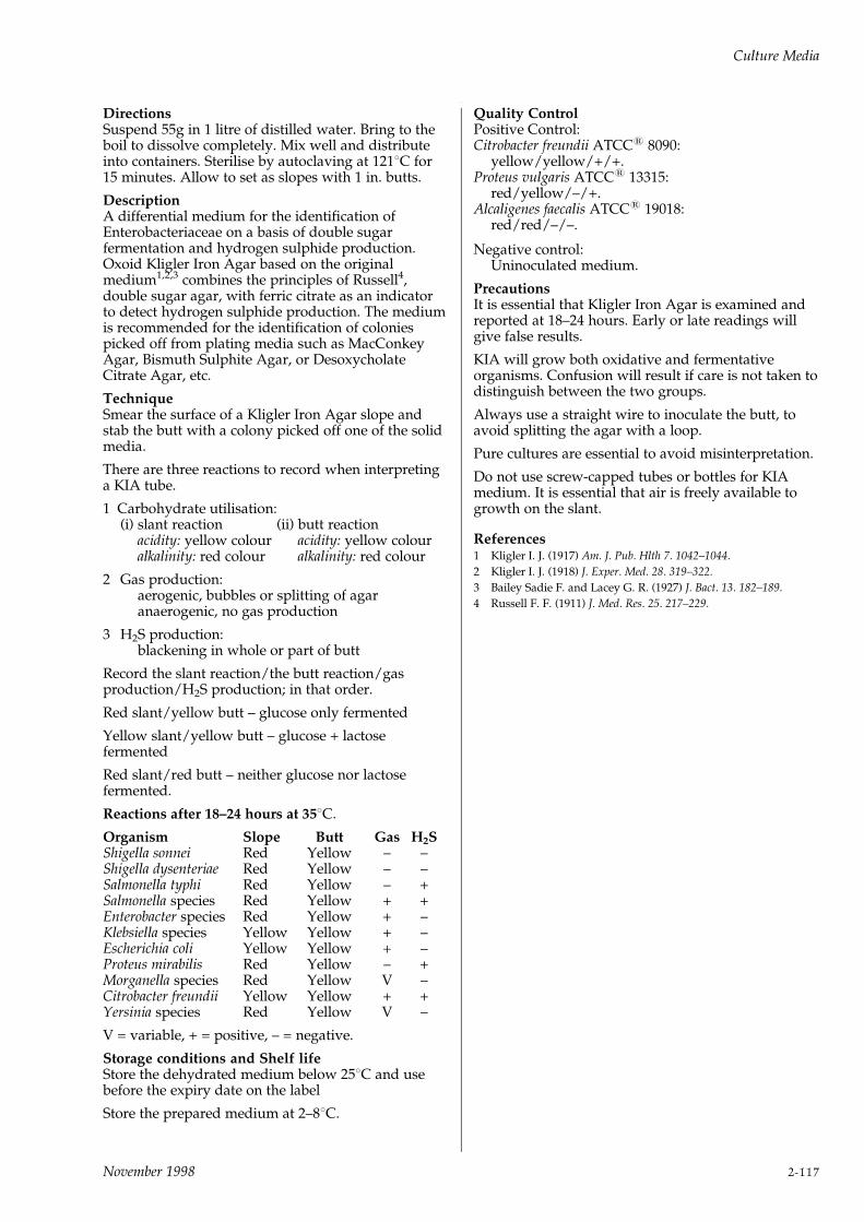
DirectionsSuspend 55g in 1 litre of distilled water. Bring to theboil to dissolve completely. Mix well and distributeinto containers. Sterilise by autoclaving at 1218C for15 minutes. Allow to set as slopes with 1 in. butts.
DescriptionA differential medium for the identification ofEnterobacteriaceae on a basis of double sugarfermentation and hydrogen sulphide production.Oxoid Kligler Iron Agar based on the originalmedium1,2,3 combines the principles of Russell4,double sugar agar, with ferric citrate as an indicatorto detect hydrogen sulphide production. The mediumis recommended for the identification of coloniespicked off from plating media such as MacConkeyAgar, Bismuth Sulphite Agar, or DesoxycholateCitrate Agar, etc.
TechniqueSmear the surface of a Kligler Iron Agar slope andstab the butt with a colony picked off one of the solidmedia.
There are three reactions to record when interpretinga KIA tube.
1 Carbohydrate utilisation:(i) slant reaction (ii) butt reaction
acidity: yellow colour acidity: yellow colouralkalinity: red colour alkalinity: red colour
2 Gas production:aerogenic, bubbles or splitting of agaranaerogenic, no gas production
3 H2S production:blackening in whole or part of butt
Record the slant reaction/the butt reaction/gasproduction/H2S production; in that order.
Red slant/yellow butt ± glucose only fermented
Yellow slant/yellow butt ± glucose + lactosefermented
Red slant/red butt ± neither glucose nor lactosefermented.
Reactions after 18±24 hours at 358C.
Organism Slope Butt Gas H2SShigella sonnei Red Yellow ± ±Shigella dysenteriae Red Yellow ± ±Salmonella typhi Red Yellow ± +Salmonella species Red Yellow + +Enterobacter species Red Yellow + ±Klebsiella species Yellow Yellow + ±Escherichia coli Yellow Yellow + ±Proteus mirabilis Red Yellow ± +Morganella species Red Yellow V ±Citrobacter freundii Yellow Yellow + +Yersinia species Red Yellow V ±
V = variable, + = positive, ± = negative.
Storage conditions and Shelf lifeStore the dehydrated medium below 258C and usebefore the expiry date on the label
Store the prepared medium at 2±88C.
Quality ControlPositive Control:Citrobacter freundii ATCC1 8090:
yellow/yellow/+/+.Proteus vulgaris ATCC1 13315:
red/yellow/±/+.Alcaligenes faecalis ATCC1 19018:
red/red/±/±.
Negative control:Uninoculated medium.
PrecautionsIt is essential that Kligler Iron Agar is examined andreported at 18±24 hours. Early or late readings willgive false results.
KIA will grow both oxidative and fermentativeorganisms. Confusion will result if care is not taken todistinguish between the two groups.
Always use a straight wire to inoculate the butt, toavoid splitting the agar with a loop.
Pure cultures are essential to avoid misinterpretation.
Do not use screw-capped tubes or bottles for KIAmedium. It is essential that air is freely available togrowth on the slant.
References1 Kligler I. J. (1917) Am. J. Pub. Hlth 7. 1042±1044.
2 Kligler I. J. (1918) J. Exper. Med. 28. 319±322.
3 Bailey Sadie F. and Lacey G. R. (1927) J. Bact. 13. 182±189.
4 Russell F. F. (1911) J. Med. Res. 25. 217±229.
Culture Media
November 1998 2-117
`LAB-LEMCO' AGARCode: CM17
A nutrient agar for general bacteriological use, it maybe used as the basis for selective, differential, or enrichedmedia.
Formula gm/litre`Lab-Lemco' powder 3.0Peptone 5.0Agar 15.0pH 7.4 + 0.2
DirectionsSuspend 23g in 1 litre of distilled water. Bring to theboil to dissolve completely. Sterilise by autoclaving at1218C for 15 minutes.
Description`Lab-Lemco' Agar is a general purpose medium usedfor the examination of water and dairy products1,2.
It is used as a nutrient meat extract agar for generalbacteriology and for the preservation of stockcultures. The absence of sodium chloride in theformulation prevents Proteus mirabilis forming`swarming' colonies.
When used as a basal medium, other substances canbe added to produce selective, differential or enrichedmedia.
Storage conditions and Shelf lifeStore the dehydrated medium below 258C and usebefore the expiry date on the label.
Store the prepared plates at 2±88C.
Quality ControlPositive control:
Staphylococcus aureus ATCC1 25923Escherichia coli ATCC1 25922
Negative control:Uninoculated medium
Precautions`Lab-Lemco' Agar is slightly more alkaline than themedium used in the APHA publications (pH 6.8).
If blood is to be added to the agar then it is necessaryto add sodium chloride (0.5% w/v) to preventhaemolysis.
References1 American Public Health Association (1980) Standard Methods for
the Examination of Water and Wastewater. 15th Edn. APHA Inc.
Washington DC.
2 American Public Health Association (1978) Standard Methods for
the Examination of Dairy Products. 14th Edn. APHA Inc.
Washington DC.
`LAB-LEMCO' BROTHCode: CM15
A nutrient broth for general bacteriological use, and forthe examination of water, sewage, and dairy products byAmerican standard methods.
Formula gm/litre`Lab-Lemco' powder 3.0Peptone 5.0pH 7.4 + 0.2
DirectionsDissolve 8g in 1 litre of distilled water and distributeinto final containers. Sterilise by autoclaving at 1218Cfor 15 minutes.
Description`Lab-Lemco' Broth is a general purpose liquidmedium used for the examination of water and dairyproducts1,2.
It is used as a nutrient meat extract broth for generalbacteriology.
Storage conditions and Shelf lifeStore the dehydrated medium below 258C and usebefore the expiry date on the label.
Store the prepared medium at room temperature(18±228C).
Quality ControlPositive control:
Staphylococcus aureus ATCC1 25923Escherichia coli ATCC1 25922
Negative control:Uninoculated medium.
Precautions`Lab-Lemco' Broth is slightly more alkaline than themedium used in the APHA publications (pH 6.8).
References1 American Public Health Association (1980) Standard Methods for
the Examination of Water and Wastewater. 15th Edn. APHA Inc.
Washington DC.
2 American Public Health Association (1978) Standard Methods for
the Examination of Dairy Products. 14th Edn. APHA Inc.
Washington DC.
LACTOSE BROTHCode: CM137
A liquid medium for use in the performance orconfirmation of the Presumptive Test for coliforms inwater, milk, etc. as specified by the American PublicHealth Association.
Formula gm/litre`Lab-Lemco' powder 3.0Peptone 5.0Lactose 5.0pH 6.9 + 0.2
DirectionsDissolve 13g in 1 litre of distilled water and distributeinto containers with fermentation tubes (Durham).Sterilise by autoclaving at 1218C for 15 minutes.
Culture Media
2-118 November 1998
DescriptionLactose broth is recommended for use in thepresumptive identification of coliform organisms inmilk, water and foods as specified by the AmericanPublic Health Association1,2,3.
Tubes of Lactose Broth are inoculated with dilutionsof the samples and incubated at 358C. Examinationfor gas formation is carried out after 24 and 48 hoursincubation. This presumptive evidence of coliformorganisms must be confirmed by further tests.
Storage conditions and Shelf lifeStore the dehydrated medium below 258C and usebefore the expiry date on the label.
Store the prepared medium at room temperature(18±228C).
Quality ControlPositive control:
Escherichia coli ATCC1 25922Enterobacter aerogenes ATCC1 13048
Negative control:Uninoculated medium
PrecautionsEnsure that the fermentation tubes are free from airbubbles before inoculation.
Large water samples may require double-strengthlactose broth to reduce the final volumes. Do notoverheat double-strength broth or inhibitory productswill be produced.
References1 American Public Health Association (1978) Standard Methods for
the Examination of Dairy Products. 14th Edn. APHA Washington
DC.
2 American Public Health Association (1980) Standard Methods for
the Examination of Water and Wastewater. 15th Edn. APHA Inc.
Washington DC.
3 American Public Health Association (1976) Compendium of
Methods for the Microbiological Examination of Foods. APHA Inc.
Washington DC.
LAURYL TRYPTOSE BROTH
(LAURYL SULPHATE BROTH)
Code: CM451
A medium for the detection of coliform organisms inwater and waste water, according to the formula of theAmerican Public Health Association.
Formula gm/litreTryptose 20.0Lactose 5.0Sodium chloride 5.0Dipotassium hydrogen phosphate 2.75Potassium dihydrogen phosphate 2.75Sodium lauryl sulphate 0.1pH 6.8 + 0.2
DirectionsDissolve 35.6g in 1 litre of distilled water anddistribute into containers with fermentation tubes(Durham). Sterilise by autoclaving at 1218C for 15minutes.
DescriptionLauryl Tryptose Broth provides a selective mediumwhich is used for the detection of coliform organismsin water, dairy products and other foods. The APHA1
recommend that Lauryl Tryptose Broth should beused for the Mean Probable Number PresumptiveTest of coliforms in waters, effluent or sewage as aconfirmatory test of lactose fermentation with gasproduction for milk samples (APHA2) and for thedetection of coliforms in foods (APHA3).
Surface active agents have long been used as theinhibitory ingredients in selective media. MacConkey4
introduced bile salts for this purpose and later Albusand Holm3 working with lactobacilli found thatsodium ricinoleate exerted a selective action. Thedevelopment of synthetic wetting agents opened upnew fields of investigation. Mallmann and Darby6
after comparative tests with a large number of thesecompounds, showed that sodium lauryl sulphategave the best results in selective media for thecoliform group.
Lauryl Tryptose Broth was designed to promote arich growth and copious gas production from smallinocula of coliform organisms. Aerobic sporingbacteria are completely inhibited. The advantage inusing this product is that in addition to giving thefermentation reaction typical of MacConkey Broth itcan also be directly tested for the presence of indole.Unlike MacConkey Broth, the medium contains noindicator, but this can be added (if required) afterincubation.
TechniqueFor details of the APHA standard methods pleaseconsult the references below.
Lauryl Tryptose Broth is recommended for thedetection and enumeration of coliform organisms inwater and milk products, especially in the control ofice-cream manufacture and in dairy hygiene. Asuggested procedure (Dyett7) is as follows:
Inoculate samples of ice-cream into tubes of LaurylTryptose Broth in the manner normally employed inthe MacConkey test. Examine the tubes afterovernight incubation at 358C and, if no gas is visible,examine again at the end of 48 hours' incubation. Forevery tube showing fermentation ('primaryfermentation') two further tubes of Lauryl TryptoseBroth are inoculated from a tube of the primaryfermenting broth, and these are incubated at 358C and448C respectively. It is advisable that the tube to beincubated at 448C be warmed up in a water bath atthis temperature before inoculation.
If the 448C incubated broth ferments after sevenhours, test for indole production with either Ehrlichor Kovac's reagent. Due to the lauryl sulphatepresent, shaking the reagent culture mixture forms apersistent emulsion which interferes with the test.This may be avoided by shaking with ether, whichseparates rapidly, and then adding Kovac's reagent tothe layer without shaking. If fermentation has notoccurred after seven hours, leave the tube overnightat 448C and test the following day. A positive indolereaction in a broth that has produced gas at 448Cindicates the presence of Escherichia coli. The tube at
Culture Media
November 1998 2-119
358C is incubated for 24 hours. If no fermentationoccurs, the primary fermentation is assumed to bedue to organisms other than coliforms. False positivesare not uncommon in the primary fermentation tubes,due to fermentation of the sucrose in the added ice-cream by organisms other than coliforms.
After the two tubes of Lauryl Tryptose Broth havebeen inoculated for secondary fermentation, test theoriginal primary fermentation tube (which wasinoculated directly with ice-cream) for indoleproduction. A positive reaction suggests the presenceof E. coli and confirmation will be obtained later withthe secondary fermentation from the 448C bath. Anegative indole reaction in the primary fermentationtube indicates the absence of E. coli.
MUG Reagent BR71 ± The addition of4-methylumbelliferyl-b-D-glucuronide (MUG) BR71to this medium will enhance the detection ofEscherichia coli.
The use of MUG in a Most Probable Number (MPN)technique for enumeration of presumptive E. coli inmilk and milk products has been specified in astandard procedure8. For further information aboutMUG see MUG Reagent BR71 under BiologicalReagents.
Storage conditions and Shelf lifeStore the dehydrated medium below 258C and usebefore the expiry date on the label.
Store the prepared medium at room temperature(18±228C).
Quality ControlPositive control:
Escherichia coli ATCC1 25922(Gas 358C and indole 448C)Enterobacter aerogenes ATCC1 13048(Gas 358C and no indole at 448C)
Negative control:Staphylococcus aureus ATCC1 25923
PrecautionsIf stored at 2±88C the broth will become cloudy orform a precipitate. This should clear at roomtemperature but gas formation is the criterion ofgrowth not turbidity.
References1 American Public Health Association (1980) Standard Methods for
the Examination of Water and Wastewater. 15th Edn. APHA Inc.
Washington DC.
2 American Public Health Association (1978) Standard Methods for
the Examination of Dairy Products. 14th Edn. APHA Inc.
Washington DC.
3 American Public Health Association (1976) Compendium of
Methods for the Microbiological Examination of Foods. APHA Inc.
Washington DC.
4 MacConkey A. T. (1938) J. Hyg. 8. 322±334.
5 Albus W. R. and Holm G. E. (1926) J. Bact. 12. 13±18.
6 Mallmann W. L. and Darby C. W. (1941) Am. J. Pub. Hlth. 31.
127±134.
7 Dyett E. J. (1957) Lab. Prac. 6(6). 327±328.
8 ISO Standard 11866±2 Milk and Milk Products - Enumeration of
presumptive Escherichia coli - part 2: Most probable number technique
using 4-methylumbelliferyl-b-D-glucuronide.
LEGIONELLA SELECTIVEMEDIA
For the isolation of Legionellaceae from clinical andenvironmental samples.
LEGIONELLA CYE AGAR BASECode: CM655
Charcoal Yeast Extract Agar for the isolation ofLegionellaceae when used with Legionella BCYEGrowth Supplement SR110 (Edelstein BYCE Agar).
Formula gm/litreActivated charcoal 2.0Yeast extract 10.0Agar 13.0
LEGIONELLA BCYE GROWTHSUPPLEMENTS
Supplement containing L-cysteine.
Code: SR110
SR110A Vial contents (each vial is sufficient for100ml of medium) gm/litre
ACES Buffer/Potassium hydroxide 10Ferric pyrophosphate 0.25L-cysteine HCl 0.4a-Ketogluterate 1
SR110C Vial contents (each vial is sufficient for500ml of medium) Supplement omitting L-cysteine, forpresumptive identification of Legionella spp.
Code: SR175Vial contents (each vial is sufficient for 100ml ofmedium)
gm/litreACES Buffer/Potassium hydroxide 10Ferric pyrophosphate 0.25L-cysteine hydrochloride Nila-Ketoglutarate 1.0
LEGIONELLA BMPA SELECTIVESUPPLEMENT
Code: SR111
Vial contents (each vial is sufficient for 100ml ofmedium)Cefamandole 400mgPolymyxcin B 8,000 IUAnisomycin 8mg
Culture Media
2-120 November 1998

LEGIONELLA MWY SELECTIVESUPPLEMENT
Code: SR118
Vial contents (each vial is sufficient for 100ml ofmedium).
Glycine 0.3gPolymyxin 5,000 IUAnisomycin 8.mgVancomycin 100mgBromothymol blue 1mgBromocresol purple 1mg
Directions to prepare BCYE AgarSuspend 2.5g of Legionella CYE Agar Base in 90ml ofdistilled water and bring gently to the boil to dissolvecompletely. Sterilise by autoclaving at 1218C for 15minutes. Allow to cool to 508C and aseptically addthe contents of one vial of Legionella BCYE GrowthSupplement SR110 reconstituted in 10ml of warmsterile distilled water (508C). Mix gently and pour intosterile petri dishes. The final pH of the medium willbe 6.9 + 0.1.
Directions to prepare BMPAa, MWY and GVPCLegionella Selective MediaSuspend 2.5g of Legionella CYE Agar Base in 90ml ofdistilled water and bring gently to the boil to dissolvecompletely. Sterilise by autoclaving at 1218C for 15minutes. Allow to cool to 508C and aseptically addthe contents of one vial of Legionella BYCE GrowthSupplement SR110 and one vial of either LegionellaBMPAa Selective Supplement SR111, reconstituted in2ml of sterile distilled water. Mix gently and pourinto sterile petri dishes. The final pH of both mediawill be 6.9 + 0.1.
DescriptionThe discovery of the causative organism ofLegionnaires' disease has been reviewed by Fallon1.Since that review further progress has been made inculturing the organism from clinical specimens andalso in the enumeration of Legionella species fromenvironmental samples. Feeley et al.2 described amodification of F-G Agar3 in which acid hydrolysedcasein was replaced by yeast extract as the source ofprotein and starch was replaced by activated charcoal(Norit A) at a final concentration of 0.2% (w/v). Thismedium, which they named CYE Agar2 has beenfurther supplemented with ACES Buffer and a-ketogluterate and is described in the literature asBCYE-a Medium4. BCYE-a Medium has been shownto yield optimal recovery of Legionellaceae in ashorter incubation period from environmentalsamples and clinical specimens5.
Oxoid BCYE Medium is based on the formulation ofEdelstein4 and is prepared from Legionella CYE AgarBase CM655 and Legionella BCYE GrowthSupplement SR110. The sterile lyophilisedsupplement contains ACES Buffer/potassiumhydroxide, a-ketogluterate, ferric pyrophosphate andL-cysteine HCl. When added to CYE Agar Base itstabilises the pH of the medium at 6.9 + 0.1 andprovides essential growth factors.
Additionally, a medium omitting L-cysteine may beprepared from Legionella CYE Agar Base CM655 andBYCE Growth Supplement SR175.
Legionellaceae have an absolute nutritionalrequirement for L-cysteine. Presumptive Legionellaspp. colonies can be subcultured onto both BCYEMedium with L-cysteine CM655 and SR110, andBCYE Medium without L-cysteine CM655 and SR175.
All plates are incubated at 358C. Colonies which havegrown on BCYE Medium with L-cysteine, but not onBCYE Medium without L-cysteine, can be regarded aspresumptive Legionella spp.
Quality Control
Legionella pneumophila Escherichia coliATCC1 33153 ATCC1 25922
SR175 ± +
SR110 + +
Edelstein and Edelstein later compared agars used inthe manufacture of buffered charcoal yeast extractmedium and found significant differences in growthoccurred. Oxoid Agar L11 gave the best results in theseries tested6.
1 BMPAa Medium containing polymyxin 80 IU/ml,anisomycin 80mg/ml and cefamandole 4mg/ml.This semi-selective medium was recommended byEdelstein4 for the isolation of L. pneumophila fromcontaminated clinical and environmentalspecimens and can be prepared by supplementingBCYE Agar with Legionella BMPAa SelectiveSupplement SR111.
2 Wadowsky and Yee Medium7 modified byEdelstein (MWY Medium)8 containing polymyxin50 IU/ml, anisomycin 80mg/ml, vancomycin1mg/ml and glycine 0.3% w/v. Bromocresolpurple 10mg/ml and Bromothymol blue 10mg/mlcolour the colonies and aid in the identification ofthe organisms9,10. Edelstein8 considered themedium to be the best for isolating L. pneumophilafrom potable water samples. This medium can beprepared by supplementing BCYE Agar withLegionella MWY Selective Supplement SR118.
Environmental samples should be pre-treated withan acid buffer (pH 2.2)11 or by heat treatment12.They should be plated both before and aftertreatment to maximise recovery (see Technique).
MWY medium has been used successfully forexamination of clinical specimens5.
3 GVPC Medium based on the formula by Dennis etal. This medium contains glycine, vancomycin,polymyxin B and cycloheximide. A completedescription follows on page 2-123.
Technique: Clinical SamplesFor the isolation of Legionella spp. from patients withclinical evidence of Legionnaires' disease, greatestsuccess has been achieved by the examination of lungtissue and bronchial aspirate.
1 Homogenise the patient's specimen in steriledistilled water.
Culture Media
November 1998 2-121
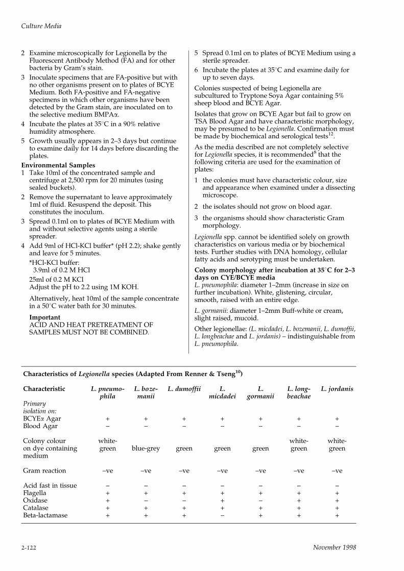
2 Examine microscopically for Legionella by theFluorescent Antibody Method (FA) and for otherbacteria by Gram's stain.
3 Inoculate specimens that are FA-positive but withno other organisms present on to plates of BCYEMedium. Both FA-positive and FA-negativespecimens in which other organisms have beendetected by the Gram stain, are inoculated on tothe selective medium BMPAa.
4 Incubate the plates at 358C in a 90% relativehumidity atmosphere.
5 Growth usually appears in 2±3 days but continueto examine daily for 14 days before discarding theplates.
Environmental Samples1 Take 10ml of the concentrated sample and
centrifuge at 2,500 rpm for 20 minutes (usingsealed buckets).
2 Remove the supernatant to leave approximately1ml of fluid. Resuspend the deposit. Thisconstitutes the inoculum.
3 Spread 0.1ml on to plates of BCYE Medium withand without selective agents using a sterilespreader.
4 Add 9ml of HCl-KCl buffer* (pH 2.2); shake gentlyand leave for 5 minutes.
*HCl-KCl buffer:3.9ml of 0.2 M HCl
25ml of 0.2 M KClAdjust the pH to 2.2 using 1M KOH.
Alternatively, heat 10ml of the sample concentratein a 508C water bath for 30 minutes.
ImportantACID AND HEAT PRETREATMENT OFSAMPLES MUST NOT BE COMBINED.
5 Spread 0.1ml on to plates of BCYE Medium using asterile spreader.
6 Incubate the plates at 358C and examine daily forup to seven days.
Colonies suspected of being Legionella aresubcultured to Tryptone Soya Agar containing 5%sheep blood and BCYE Agar.
Isolates that grow on BCYE Agar but fail to grow onTSA Blood Agar and have characteristic morphology,may be presumed to be Legionella. Confirmation mustbe made by biochemical and serological tests12.
As the media described are not completely selectivefor Legionella species, it is recommended8 that thefollowing criteria are used for the examination ofplates:
1 the colonies must have characteristic colour, sizeand appearance when examined under a dissectingmicroscope.
2 the isolates should not grow on blood agar.
3 the organisms should show characteristic Grammorphology.
Legionella spp. cannot be identified solely on growthcharacteristics on various media or by biochemicaltests. Further studies with DNA homology, cellularfatty acids and serotyping must be undertaken.
Colony morphology after incubation at 358C for 2±3days on CYE/BCYE mediaL. pneumophila: diameter 1±2mm (increase in size onfurther incubation). White, glistening, circular,smooth, raised with an entire edge.
L. gormanii: diameter 1±2mm Buff-white or cream,slight raised, mucoid.
Other legionellae: (L. micdadei, L. bozemanii, L. dumoffii,L. longbeachae and L. jordanis) ± indistinguishable fromL. pneumophila.
Characteristics of Legionella species (Adapted From Renner & Tseng10)
Characteristic L. pneumo-phila
L. boze-manii
L. dumoffii L.micdadei
L.gormanii
L. long-beachae
L. jordanis
Primaryisolation on:BCYEa Agar + + + + + + +Blood Agar ± ± ± ± ± ± ±
Colony colour white- white- white-on dye containing green blue-grey green green green green greenmedium
Gram reaction ±ve ±ve ±ve ±ve ±ve ±ve ±ve
Acid fast in tissue ± ± ± ± ± ± ±Flagella + + + + + + +Oxidase + ± ± + ± + +Catalase + + + + + + +Beta-lactamase + + + ± + + +
Culture Media
2-122 November 1998

Characteristic Gram morphology on first isolationGram-negative, short, pleomorphic rods 1mm long.
Carbol-fuchsin should be used as a counterstain in theGram films of legionellae because they resist stainingwith safranin and basic fuchsin.
Storage conditions and Shelf lifeStore the dehydrated medium below 258C and usebefore the expiry date on the label.
Store the prepared plates at 2±88C away from light.
Quality ControlPositive control:
Legionella pneumophila ATCC1 33152Legionella pneumophila NCTC 12821
Negative control:Escherichia coli ATCC1 25922Staphylococcus epidermidis ATCC1 12228
Please note for Legionella MWY Selective SupplementSR118, Escherichia coli ATCC1 25922 as a negativecontrol is inoculum dependent.
PrecautionsLegionella spp. are highly pathogenic organisms ifinhaled. Avoid creating aerosols and handle liquidcultures or suspensions of organisms in a protectivecabinet. Decontaminate working surfaces with 5%hypochlorite. Autoclave all materials beforediscarding.
Incubate cultures at 358C in 65% humidity up to 10days.
No CO2 is required for BCYE cultures but 2.5% CO2 isrecommended for other media and water samples7,13.
Organisms that grow on blood agar as well asLegionella media are not legionellae. Somethermophilic spore-bearing organisms mimicLegionella colonies after 358C incubation. Theseorganisms can be detected by incubating parallelplates at 358C and 558C when the thermophilicorganisms will grow at the higher temperature.Legionella species will not grow above 458C.
References1 Fallon J. Oxoid Limited. Culture September 1979, P. 3±4.
2 Feeley J.C., Gibson R.J., Gorman G.W., Langford N.C., Rasheed
J.W., Mackel D.C. and Baine W.B. (1979) J. Clin. Micro. 10. 437±
441.
3 Feeley J.C. Gorman G.W., Weaver R.E., Mackel D.C. and Smith
H.W. (1978) J. Clin. Micro. 8. 320±325.
4 Edelstein P.H. (1981) J. Clin. Micro. 14. 298±303.
5 PHLS Communicable Diseases Report (1983) CDR 83/49.
6 Edelstein P.H. and Edelstein M.A.C. (1991) J. Clin. Microbiol. 29.
190±191.
7 Wadowsky R.M. and Yee R.B. (1981) Clin. Microbiol. Newsletter
4. 768±772.
8 Edelstein P.H. (1982) J. Clin. Micro. 16. 697±699.
9 Vickers R.M., Brown A. and Garrity G.M. (1981) J. Clin. Micro.
13. 380±283.
10 Renner E.D. and Tseng C.H. (1982) Clin. Microbiol. Newsletter 4.
139, 142.
11 Bopp C.A., Sumner J.W., Morris G.K. and Wells J.G. (1981) J.
Clin. Micro. 13. 714±719.
12 Vesey G., Dennis P.J., Lee J.V. and West A.A. (1988) J. Appl. Bact.
65. 339±345.
LEGIONELLA (GVPC) SELECTIVESUPPLEMENT
Code: SR152
A freeze-dried selective supplement for the isolation ofLegionella spp. from environmental water samples.
Vial contents (each vial is sufficient to supplement500ml of BCYE Medium).
Glycine (Ammonia free) 1.5gVancomycin hydrochloride 0.5mgPolymixin B sulphate 39600 IUCycloheximide 40.0mg
DirectionsTo one vial aseptically add 10ml of sterile distilledwater and mix gently to dissolve completely. Add thecontents to 500ml of sterile BCYEa Medium (preparedusing Legionella CYE Agar Base CM655 to which onevial of Legionella BCYEa Growth SupplementSR110C has been added), cooled to 50±558C. Mixgently and pour into sterile petri dishes.
DescriptionL. pneumophila was first isolated by infecting guinea-pigs and fertile hens' eggs in 19771. Examination ofwater samples for the presence of legionellae hasfrequently been undertaken using directimmunofluorescence methods2. However, isolation isstill the most widely accepted method ofdemonstrating the presence of Legionellae and is ofparticular value in epidemiological studies.
Currently, a buffered charcoal yeast extract medium(BCYE) is generally considered to be the medium ofchoice3, to which various antimicrobials have beenadded to make it selective.
GVPC Selective Supplement is based on theformulation described by Dennis et al4. This selectiveformulation has been reported to be the most efficientin vitro method for the isolation of L. pneumophilawhen used in conjunction with acid or heatpretreatments. Cycloheximide is included because ithas a greater activity against fungi than anisomycinwhich is almost exclusively active against yeasts.Fungi commonly occur more frequently than yeasts inenvironmental waters examined for Legionella. Thisformulation is to be specified by the BSI (BritishStandard) for the detection and enumeration oflegionellae in water and related materials5.
The antimicrobials Glycine, Vancomycin andPolymixin will collectively inhibit most Gram-positiveand Gram-negative bacterial growth. Cycloheximidewill suppress the growth of fungi.
TechniqueFor each sample, three plates should be inoculated3,5:one after pretreatment with heat, one afterpretreatment with acid and one that has receivedneither pretreatment.
Heat pretreatment1 Take 10ml of concentrated sample3 and place in a
water bath at 508C for 30 minutes.
Acid pretreatment1 Take 10ml of concentrated sample3 and centrifuge
in sealed buckets at 2,500rpm for 20 minutes.
Culture Media
November 1998 2-123
2 Decant the supernatant to leave approximately1ml of fluid.
3 Add 9ml of HCl-KCl buffer (see below) andresuspend by gentle shaking. Leave to stand for 5minutes and inoculate without further delay.
HCl-KCl Buffer3.9ml of 1.2M HCl25ml of 0.2M KClAdjust to pH 2.2 using 1M KOH
No pretreatment1 Take 10ml of concentrated sample3 and do not
pretreat.
Directions1 Spread 0.1ml of each portion, described above,
onto plates of GVPC Selective Medium using asterile spreader.
2 Incubate the plates at 36 + 18C and examine ondays 3, 5, 7 and 10.
3 Suspect colonies should be verified as presumptiveLegionella by the following procedure: Selectseveral colonies of each type and subculture on to apair of plates, one of Buffered Charcoal YeastExtract Agar CM655 and one of Buffered CharcoalYeast Extract Agar CM655 containing LegionellaBCYE Growth Supplement SR110. Regard aspresumptive Legionella all colonies which grow onBuffered Charcoal Yeast Extract Agar containingthe supplement but which fail to grow on mediumnot containing the supplement.
4 Further confirmation of identity can be obtained bysubculturing colonies taken from the primaryculture plates on to Blood Agar. Isolates that fail togrow on Blood Agar and are small, poorly stainingGram-negative rods are presumptively identifiedas Legionella species.
5 Each presumptive Legionella colony should beconfirmed by serology.
L. pneumophila colonies are white-grey-blue inappearance and up to 2mm in diameter with aground-glass appearance. Colonies of all Legionellaspp. exhibit the same general appearance but theymay differ in colour. Colours include brown, limegreen, deep red and blue-purple.
Storage conditions and Shelf lifeStore the selective supplement in the dark at 28C to88C and use before the expiry date on the label.
Quality ControlThe medium may be tested for performance usingstable typical control cultures of organisms other thanLegionellae. However, when testing Legionellae,`wild' strains must be used because Legionellae easilybecome adapted to growth under laboratoryconditions and will grow on media that would notsupport the primary isolation of `wild' strains5. `Wild'strains can be defined as strains which have beensubcultured no more than twice following primaryisolation. GVPC Medium should not be tested usingL. pneumophila strains that may have becomelaboratory acclimatised.
Incubation at 35±378C + 0.58C for 5 days in air or inair enriched with 2.5% CO2
5 in conditions of highhumidity.
Positive control:Legionella species
Negative control:Staphylococcus epidermidis ATCC1 12228Escherichia coli ATCC1 25922
References1 McDade J. E. et al (1977) N. Engl. J. Med. 297. 1197±1203.
2 Fliermans C. B. et al. (1981) Appl. Environ. Microbiol. 41. 9±16.
3 Dennis P. J. L. (1988) Isolation of legionellae from environmental
specimens. In Harrison T. G. and Taylor A. G. (eds). A Laboratory
Manual for Legionella, John Wiley & Sons Limited, Chichester.
4 Dennis P. J. L., Bartlett C. L. R. and Wright A. E. (1984)
Comparison of isolation methods for Legionella spp. In Thornsbury C.
et al (eds). Legionella: Proceedings of the 2nd International
Symposium. Washington D. C. Am. Soc. Microbiol. pp 294±296.
5 BSI Document. Determination of Legionellae in water and related
materials. Method for their detection and enumeration. July 1989
DRAFT DOCUMENT. 89/53406.
LISTERIA SPECIES ANDLISTERIOSIS
Listeriosis is a disease which has been recognised forover 60 years, it is now known to affect a wide variety ofanimals as well as humans.
There are eight species in the Listeria genus:L. monocytogenes
the major species causing disease in humans andanimals1.
L. ivanoviiassociated with animal and a small number ofhuman infections2.
L. innocua, L. seeligerivery rarely associated with infection.
L. murrayii, L. welshimeri, L. denitrificansnot associated with naturally occurring infections.
L. monocytogenes is commonly found in theenvironment and is therefore common in food andhuman faeces. It can grow slowly at refrigeratortemperatures and has been isolated from a widevariety of foods. Fresh and frozen poultry (60%),cooked-chill foods (24%), salami and continentalsausages (24%), soft cheese (10%), pre-packed salads(7%)3. Although relatively few cases of listeriosis havebeen epidemiologically associated with food stuffs inthe UK3,4 a number of outbreaks associated with foodhave been reported in other countries5,6,7,8. Cautionshould be taken in assessing food-borne infectionsbecause the extent and route of such infections areunknown.
The annual number of cases of listeriosis reported tothe Central Public Health Laboratory has risen from50 in 1967 to 259 in 1987. Reported deaths are 38 in1983 rising to 59 in 19873. In human listeriosis there isa high rate of mortality (23%) and the disease islargely confined to pregnant women, neonates andpatients with underlying immuno-suppression2.
The use of efficient, selective media will undoubtedlyimprove the rate of isolation and may reveal the chainof infection which is still very obscure.
Culture Media
2-124 November 1998

TABLE 1 Differentiation of the species of the genus Listeria
L. mono- L. ivan- L. inno- L.welsh- L. seeli- L. grayi L. murr- L. denitri-cytogenes ovii cua imeri geri ayi ficans
Beta haemolysis on + ++ ± ± (+) ± ± ±blood agar
Nitrates reduced ± ± ± ± ± ± + +to nitrites
CAMP test with + ± ± ± (+) ± ± ±Staphylococcus aureus
CAMP test with ± + ± ± ± ± ± ±Rhodococcus equi
Acid produced from:
D-mannitol ± ± ± ± ± + + ±
L-rhamnose + ± V V ± ± V ±
D-xylose ± + ± + + ± ± +
a-methyl D-mannoside + ± + + V NS NS NS
V = Variable reaction; NS = Not stated; ( ) = Weak reaction
FIGURE 1 Dichotomous key for the identification of the species of Listeria
References1 McLauchlin J. (1988) Listeria Workshop DMRQ. Central Public
Health Service. 4/5th May.
2 McLauchlin J. (1987) J. Appl. Bact. 63. 1±11.
3 Hall S. M. et al. (1988) Lancet. ii.
4 Kerr K. G., Dealler S. F. and Lacey R. W. (1988) Lancet. ii. 1133.
5 Bille J. and Glauser M. P. (1988) Bull. Bund. fuÈ r Gesund. 3. 28±29.
6 Fleming D. W. et al. (1985) N. Engl. J. Med. 312. 404±407.
7 Linnan M. J. et al. (1988) N. Engl. J. Med. 319. 823±828.
8 Schlech W. F. et al. (1983) N. Engl. J. Med. 308. 203±206.
IDENTIFICATION OF LISTERIA SPECIES
(adapted with permission from reference 1.)
Criteria of identification for ListeriaeGram positive rod, VP +, urease ±, catalase +, oxidase± aesculin hydrolised, acid no gas from glucose andsalicin, tumbling motility below 308C.
Colony form ± non pigmented on nutrient agar withcharacteristic caramel/sour butter smell, 1±2mmdiameter with a ground glass appearance andemulsifies in saline to a smooth suspension.
Culture Media
November 1998 2-125
Motility test ± heavily inoculate brain-heart infusionbroth or nutrient broth and hold at room temperaturefor 4±6 hours. Examine by hanging drop technique. Ifnegative re-test after 18 hours at room temperature.
Catalase test ± most strains are strongly positive butsome may be weak or negative.
Haemolysis ± L. monocytogenes produces a narrowzone of b-haemolysis (1±2mm) on horse blood agar.
CAMP test ± use 5% v/v sheep blood in nutrient agarand pour a thin layer over nutrient agar base. StreakStaphylococcus aureus NCTC 1803 and Rhodococcus equiNCTC 1621 across the plates. The test strains oflisteriae are streaked at right angles to the S. aureus orR. equi inoculum. Incubate at 358C overnight. Positiveresults are when an enhanced zone of haemolysisoccurs between two cultures.
VP test ± some strains may require yeast extractadded to the VP broth to give sufficient growth forthis test.
Carbohydrate fermentation ± use API 50 CH system.Incubate up to 7 days at 358C.
Nitrate reduction ± Use the rapid method of BlazericD. N. & Ederer G. M. `Biochemical Tests in DiagnosticBacteriology' J. Wiley & Son Inc. New York. 1975.
Acknowledgement is gratefully made to the Central Public
Health Laboratory for permission to use this information and for
Table 1 with Figure 1.
BUFFERED LISTERIAENRICHMENT BROTHCode: CM897
A selective enrichment medium for the detection ofListeria monocytogenes when prepared from BufferedListeria Enrichment Broth base CM897 and ListeriaSelective Supplement SR141.
BUFFERED LISTERIA ENRICHMENT BROTHCM897
Formula gm/litreTryptone soya broth 30.0Yeast extract 6.0Potassium di-hydrogen
orthophosphate 1.35Disodium hydrogen
orthophosphate 9.60Final pH 7.3 + 0.2
LISTERIA SELECTIVE ENRICHMENTSUPPLEMENT (SR141)
Vial contents:
Nalidixic acid 20.0mg (equivalent to 40mg/l)
Cycloheximide 25.0mg (equivalent to 50mg/l)
Acriflavinehydrochloride 7.5mg (equivalent to 15mg/l)
Each vial is sufficient to supplement 500mls ofBuffered Listeria Enrichment Broth CM897.
DirectionsAdd 23.5g to 500ml of distilled water and mix well todissolve. Sterilise by autoclaving at 1218C for 15minutes. Cool to 508C and aseptically add thecontents of 1 vial of Listeria Selective EnrichmentSupplement SR141 reconstituted with 2ml of steriledistilled water. Mix well and aseptically distributeinto sterile containers in volumes as required.
DescriptionListeria Selective Enrichment Broth CM862 is basedon the formulation described by Lovett et al1 and isrecommended for the enrichment of Listeria species infood. Subsequent work has concluded that theenrichment properties can be improved by increasingthe buffering capacity of the medium by the additionof potassium di-hydrogen orthophosphate anddisodium hydrogen orthophosphate. Buffered ListeriaEnrichment Broth CM897 is therefore a modificationof the original medium.
Techniques1 Add 25g or 25ml samples to 225ml of Buffered
Listeria Enrichment Broth. Homogenise if required.
2 Incubate at 308C for 48 hours.
3 Subculture from the Buffered Listeria EmrichmentBroth onto Listeria Selective Agar plates (See Note)after 24 and 48 hours by:
(i) Direct plating onto Listeria Selective Agarplates.
(ii) Adding 1ml of the Buffered ListeriaEnrichment Broth to 9ml of 0.5% KOH, vortexmixing, and plating onto Listeria SelectiveAgar plates.
NoteSuitable Listeria Selective Media are:1 Listeria Selective Medium (Oxford formulation)
(Oxoid CM856 and Oxoid SR140).2 Listeria Selective Medium (MOX) (Oxoid CM856
and Oxoid SR157)3 PALCAM Medium (Oxoid CM877 and Oxoid
SR150)
Quality ControlPositive Control:
Listeria monocytogenes ATCC119117
Negative Control:Staphylococcus aureus ATCC125923
Storage conditions and Shelf lifeBuffered Listeria Enrichment Broth CM897 should bestored tightly capped in the original container in acool, dry place away from bright light. When storedas directed the medium will remain stable until theexpiry date printed on the label.
Listeria Selective Enrichment Supplement SR141 assupplied should be stored at 2±88C. When stored asdirected the reagents are stable until the expiry dateprinted on the label.
PrecautionsListeria Selective Enrichment Supplement containscycloheximide and is toxic if swallowed, inhaled orby skin contact. As a precaution when handling weargloves and eye/face protection.
Culture Media
2-126 November 1998
Acriflavine is activated by light and may formcompounds inhibitory for Listeria.
References1 Lovett J., Francis D.W. and Hunt J.M. (1987) J. Food Prot. 50.
188±192.
2 Curtis G.D.W., Nichols W.W. and Falla T.J. (1989) Lett. Appl.
Micro. 8. 169±172.
LISTERIA SELECTIVE AGAR
(OXFORD FORMULATION)
Code: CM856
A selective and diagnostic medium for the detection ofListeria monocytogenes, when prepared from ListeriaSelective Agar Base CM856 and Listeria SelectiveSupplement SR140.
Formula gm/litreColumbia Blood Agar Base 39.0Aesculin 1.0Ferric ammonium citrate 0.5Lithium chloride 15.0pH 7.0 + 0.2
LISTERIA SELECTIVE SUPPLEMENT(OXFORD FORMULATION)
Code: SR140
Vial contents (each vial is sufficient for 500ml ofmedium)
Cycloheximide 200mgColistin sulphate 10mgAcriflavine 2.5mgCefotetan 1.0mgFosfomycin 5.0mg
DirectionsSuspend 27.75 of the Listeria Selective Agar Base(Oxford Formulation) CM856 in 500ml of distilledwater. Bring gently to the boil to dissolve. Sterilise byautoclaving at 1218C for 15 minutes. Cool to 508C andaseptically add the contents of one vial of ListeriaSelective Supplement (Oxford Formulation) SR140reconstituted with 5ml of ethanol/sterile distilledwater (1:1). Mix well and pour into sterile petridishes.
Prepared plates may be stored for up to 10 days at48C in the dark room.
DescriptionFoodborne infection by Listeria monocytogenes hasprompted increased concern for detecting thisorganism in foods, in the environment and inpathological specimens from both human and animalsubjects.
Most infections in adult humans are symptomless andresult in intestinal, vaginal and cervical carriage.Infection during pregnancy may cause abortion,premature delivery and neonatal infection. Thepossibility of listeriosis should be considered in anywoman with unexplained recurrent miscarriage,premature labour or foetal death. The organismshould be sought in blood cultures and genital-tractswabs1.
The most common clinical manifestation in bothadults and neonates is meningitis. Widelydisseminated infection, abscesses, sub-acute bacterialendocarditis and opportunistic infections inimmunosuppressed patients occur lessfrequently.
Birds, fish and other animals are all susceptible toinfection with Listeria. It is of particular importance indomestic farm animals. In the Federal Republic ofGermany reporting of listeriosis in animals iscompulsory and meat inspection law in the samecountry requires examination for Listeria because ofits significance in meat hygiene.
Listeria monocytogenes is very widespread in theenvironment. Isolation has been reported frommilk2,3, cheese4, sewage and riverwater5, and silage6.Because Listeria is so widespread sources of infectionsare numerous. Uncooked vegetable foods have beenimplicated; an episode associated with consumptionof coleslaw7 was linked with cabbage from a farmusing sewage fertiliser. In outbreaks caused by dairyproducts, cattle with mastitis may be the source of theorganism. Of great importance to veterinarians is theconsiderable increase amongst sheep of infectionmanifesting as abortion or encephalitis due largely tochanging practices in silage manufacture8.
The ability to isolate the organism has been impededin the past by lack of an effective selective medium, asL. monocytogenes can be easily and completelyovergrown by competing flora.
Listeria Selective Medium (Oxford Formulation) isbased on the formulation described by Curtis et al9
and is recommended for the detection of Listeriamonocytogenes from clinical and food specimens.
The medium utilises:
(i) the selective inhibitory components lithiumchloride, acriflavine, colistin sulphate, cefotetan,cycloheximide and fosfomycin, and
(ii) the indicator system aesculin and ferrous iron forthe isolation or differentiation ofL. monocytogenes.
L. monocytogenes hydrolyses aesculin, producing blackzones around the colonies due to the formation ofblack iron phenolic compounds derived from theaglucon. Gram-negative bacteria are completelyinhibited. Most unwanted gram-positive species aresuppressed, but some strains of enterococci growpoorly and exhibit a weak aesculin reaction, usuallyafter 40 hours incubation. Some staphylococci maygrow as aesculin-negative colonies.
Typical L. monocytogenes colonies are almost alwaysvisible after 24 hours, but incubation should becontinued for a further 24 hours to detect slow-growing strains.
Techniques for isolation vary with the author and thematerial under examination10,11. For all specimensselective enrichment and cold enrichment have beenshown to increase isolation rates significantly12,13,14.The efficacy of Listeria Selective Medium (OxfordFormulation) has been confirmed for variousfoods15,16 following the methodology and using
Culture Media
November 1998 2-127
selective enrichment media described in theliterature16,17,18,19.
Oxford agar is a specified plating medium in theFDA/BAM isolation procedure20 and in thestandardised testing methods of other national andinternational bodies21.
Oxford agar base was used by Al-Zoreki and Sandineas the basal medium for their ASLM agar whichincorporates ceftazidime, moxalactam andcycloheximide as selective agents22.
TechniqueFaecal and Biological SpecimensThe sample is homogenised in 0.1% Peptone WaterCM9 (1 part to 9 parts peptone water).
Direct Surface Plate Method1 Inoculate 0.1ml of the homogenised specimen onto
the Listeria Selective Medium plates.
2 Incubate at 358C for up to 48 hours.
3 Examine for typical colonies of Listeria after 24 and48 hours incubation.
Selective Enrichment Method1 Add the homogenised specimen to the selective
enrichment broth and incubate at 308C for up to 7days.
2 Inoculate 0.1ml of the selective enrichment broth,after 24 hours, 48 hours and 7 days, onto theListeria Selective Medium plates.
3 Incubate the plates at 358C for up to 48 hours.
4 Examine for typical colonies of Listeria after 24 and48 hours incubation.
Food and Environmental SamplesTechniques for isolation vary with the author,material and authorities. For detection ofL. monocytogenes when present in small numbers, thetest samples must be inoculated into an enrichmentbroth to allow multiplication before isolation andidentification. Depending on the type of sampleunder test, an appropriate method and selectiveenrichment broth should be chosen prior toinoculation onto the Listeria Selective Medium plates.
1 Inoculate 0.1ml of the selective enrichment brothonto the Listeria Selective Medium plates.
2 Incubate at 358C for up to 48 hours.
3 Examine for typical colonies after 24 and 48 hoursincubation.
Colonies presumptively identified asL. monocytogenes must be confirmed by biochemicaland serological testing23.
NoteDifferences in susceptibility of L. monocytogenes,L. seeligeri and L. ivanovii to b-lactam antibiotics andfosfomycin have been observed dependent onwhether incubation is at 308C or 35±378C24.
Storage conditions and Shelf lifeStore the dehydrated medium below 258C and usebefore the expiry date on the label.
Store the prepared plates at 2±88C.
Quality ControlPositive control:
Listeria monocytogenes ATCC1 19117
Negative control:Staphylococcus aureus ATCC1 25923
PrecautionsL. monocytogenes is in ACDP Group 2 i.e. `might be ahazard to laboratory workers' and should be handledin a suitable environment only. It is alsorecommended that pregnant staff should be excludedfrom working with known cultures of listeriae.
Listeria media containing acriflavine should beprotected from light because photo-oxidation makes itinhibitory to listeria.
Supplement SR140 used in this medium contains atoxic concentration of cycloheximide. Note theprecautions to be taken under HAZARDS page 2-7.
References1 Lancet (1985 [2]) August 17. 364±365.
2 Hayes et al (1986) Appl. Env. Microbiol. 50. 438±440.
3 Fernandez Garayzabal J.F. et al (1986) Can. J. Microbiol. 32. 149±
150.
4 James S.M., Ferrin S.L. and Agee B.A. (1985) MMWR 34. 357±
359.
5 Watkins J. and Sleath K.P. (1981).
6 Gitter M. (1983) Vet. Rec. 112, 314.
7 Schlech W.F., Lavigne P.M. and Bortolussi R.A. (1983) N. Eng. J.
Med. 308. 203±206.
8 Appleyard W. (1986) Communicable Diseases, Scotland. April 1986.
CDS 86/13.
9 Curtis G.D.W., Mitchell R.G., King A. F. and Griffin E.J. (1989)
Letters in Appl. Microbiol. 8. 95±98.
10 van Netten P., van de Ven A., Perales I. and Mossel D.A.A.
(1988) Int. J. Food Microbiol. 6. 187±198.
11 Prentice G.A. and Neaves P. (1988) Bulletin of the International
Dairy Federation No. 223.
12 Hayes P.S., Feeley J.C. Graves L.M., Ajello G.W. and Fleming
D.W (1986) Appl. & Environ. Microbiol. 51. 438±440.
13 Garayzabal J.F.F. Rodriquez L.D., Boland J.A.V. Cancelo J.L.B.
and Fernendez G.S. (1986) Can. J. Microbiol. 32. 149±150.
14 Doyle M.P., Meske L.M. and Marth E.H. (1985) J. of Food
Protection, 48. 740±742.
15 Crowther J.S. (1988) Personal Communication, Unilever Research
Laboratory, Colworth House, Sharnbrook, Bedford, U.K.
16 Neaves P. and Prentice G.A. (1988) Personal Communication,
Technical Division, Milk Marketing Board, Thames Ditton, Surrey.
17 Lovett J., Francis D.W. and Hunt J.M. (1987) J. Food Prot. 50.
188±192.
18 Donelly C.W. and Baigent G.J. (1986) Appl. and Environ.
Microbiol. 52. 689±695.
19 Hammer P., Hahn G. and Heeschen W. (1988) Deutsch Mock-
Zeit. 50. 1700±1706.
20 Food and Drug Administration (FDA) Bacteriological Analytical
Manual 7th Edition 1992, AOAC Int. Publishers Arlington V.A.
21 Foodborne Pathogens. Monograph Number 2 ± Listeria, page 7.
Oxoid Ltd, Wade Road, Basingstoke, Hampshire, U.K.
22 Al-Zoreki N. and Sandine W.E. (1990) Appl. Env. Microbiol. 56.
3154±3157.
23 Bille J. and Doyle M.P. (1991) ``Listeria and Erysipelothrix'', 287±
295 in Balows A., Hauster W.J. Jnr., Herrman K.L., Isenberg H.D.
and Shadomy H.J. (Editors), Manual of Clinical Microbiology, 5th
Edition, American Society for Microbiology, Washington, D.C.
Culture Media
2-128 November 1998
24 Curtis G.D.W., Nichols W.W. and Falla T.J. (1989) Letters in
Appl. Microbiol. 8. 169±172.
LISTERIA ENRICHMENTBROTH BASECode: CM862
Formula gm/litreTryptone soya broth 30.0Yeast extract 6.0pH 7.3 + 0.2
LISTERIA SELECTIVE ENRICHMENTSUPPLEMENT
Code: SR141
Vial contents (each vial is sufficient for 500ml ofmedium)
Nalidixic acid 20.0mgCycloheximide 25.0mgAcriflavine hydrochloride 7.5mg
DirectionsSuspend 18g in 500ml of distilled water. Sterilise byautoclaving at 1218C for 15 minutes. Cool to 508C andaseptically add the contents of one vial of ListeriaSelective Enrichment Supplement SR141,reconstituted with 2ml of sterile distilled water. Mixwell and distribute into sterile containers in volumesas required.
Listeria Selective Enrichment Supplement (modifiedwith 10 mg/litre of Acriflavine) Code: SR149.
Vial contents (each vial is sufficient to supplement2.25 litres of CM862).
Nalidixic acid 90.0mgCycloheximide 112.5mgAcriflavine hydrochloride 22.5mg
DirectionsAseptically add 10ml of sterile distilled water to onevial and invert gently to dissolve. Aseptically add thevial contents to 2.25 litres of Listeria Enrichment BrothBase (CM862), cooled to 508C.
DescriptionListeria Selective Enrichment Medium is based on theformulation described by Lovett et al.1 and isrecommended for the selective enrichment of Listeriaspecies from food. The enrichment procedure hasbeen shown to recover an inoculum of less than10cfu/ml from raw milk.
In order to achieve a higher isolation rate it isrecommended that the enrichment broth issubcultured onto Listeria Selective Agar plates after 1,2 and 7 days. Agello et al.2, have shown thatextending the incubation period to 7 days allowsbetter recovery of environmentally stressed listeriafrom milk and milk products.
Technique1 Add 25g or 25ml samples to 225ml of Listeria
Selective Enrichment Broth. Homogenise ifrequired.
2 Incubate at 308C for 7 days.
3 Subculture from the Listeria Selective EnrichmentBroth onto Listeria Selective Agar plates (see Note)after 1, 2 and 7 days by:
(i) Direct plating onto Listeria Selective Agarplates.
(ii) Adding 1ml of the Listeria Selective EnrichmentBroth to 9ml of 0.5% KOH, vortex mixing, andplating onto Listeria Selective Agar plates.
Note:Suitable Listeria Selective Media are:
1 Listeria Selective Medium (Oxford formulation)(Oxoid CM856 and Oxoid SR140).
2 Listeria Selective Medium (MOX) (Oxoid CM856and Oxoid SR140).
3 PALCAM Medium (Oxoid CM877 and OxoidSR150).
Storage conditions and Shelf lifeStore the dehydrated medium below 258C and usebefore the expiry date on the label.
Store the prepared medium at 2±88C.
Quality ControlPositive control:
Listeria monocytogenes ATCC1 19117
Negative control:Staphylococcus aureus ATCC1 25923
PrecautionsNote the precautions stated under Listeria SelectiveMedium (Oxford) CM856 and SR140. Broth culturesare more dangerous than colonies on agar plates.
Store prepared medium away from light. Acriflavinecan photo-oxidise to form inhibitory compoundsagainst listeria.
Supplement SR141 used in this medium contains atoxic concentration of cycloheximide. Note theprecautions to be taken under HAZARDS page 2±7.
References1 Lovett J., Francis D. W. and Hunt J. M. (1987) Journal of Food
Protection 50. 188±192.
2 Agello G., Hayes P. and Feeley J. (1986) Abstracts of the Annual
Meeting, ASM, Washington DC p5.
LISTERIA ENRICHMENT BROTHBASE
(UVM FORMULATION)
Code: CM863
Formula gm/litreProteose peptone 5.0Tryptone 5.0`Lab-Lemco' powder 5.0Yeast extract 5.0Sodium chloride 20.0Disodium hydrogen phosphate 12.0Potassium dihydrogen phosphate 1.35Aesculin 1.0pH 7.4 + 0.2
Culture Media
November 1998 2-129

LISTERIA PRIMARY SELECTIVEENRICHMENT SUPPLEMENT (UVM I)
Code: SR142
Vial contents (each vial is sufficient for 500ml ofmedium)Nalidixic acid 10.0mgAcriflavine hydrochloride 12.5mg
DirectionsSuspend 27.2g in 500ml of distilled water. Sterilise byautoclaving at 1218C for 15 minutes. Cool to 508C.
To Prepare Listeria Primary Selective EnrichmentMedium (UVM I)Aseptically add 2ml of sterile distilled water to a vialof Listeria Primary Selective Enrichment Supplement(UVM I) Code SR142. Invert gently to dissolve.Aseptically add the vial contents to 500ml of sterileListeria Enrichment Broth Base (UVM formulation)Code CM863, cooled to 508C. Mix well and distributeinto sterile containers.
To Prepare Listeria Secondary Selective EnrichmentMedium (UVM II)Aseptically add 2ml of sterile distilled water to a vialof Listeria Secondary Selective EnrichmentSupplement (UVM II) Code SR143. Invert gently todissolve. Aseptically add the vial contents to 500ml ofsterile Listeria Enrichment Broth Base (UVMformulation) Code CM863, cooled to 508C. Mix welland distribute into sterile containers.
DescriptionThe Listeria Selective Enrichment Media (UVMformulations) are based on the original formulationdescribed by Donnelly and Baigent1, and itssubsequent modification2 which reduced the nalidixicacid concentration in both the primary and secondaryselective enrichment media and increased theconcentration of acriflavine hydrochloride in thesecondary selective enrichment medium.
This modification, and the two step selectiveenrichment method developed (USDA-FSIS method)2,results in a higher detection rate of Listeriamonocytogenes from meat products and has the addedadvantage of only taking 3±4 days.
UVM Broth has been recommended as a primaryenrichment broth for recovery of heat-injuredL. monocytogenes3.
Care must be taken when using UVM broth withDNA probe methodology because the high saltcontent of the medium may have an inhibitory effecton detection4.
TechniquePrimary Enrichment1 Add 25g or 25ml samples to 225ml of Listeria
Primary Selective Enrichment Medium (UVM I).Homogenise in a Stomacher for 2 minutes.
2 Incubate the prepared sample in the Stomacher bagat 308C.
3 From this bag, carry out the following procedures:
After 4 hours incubation, spread 0.2ml on ListeriaSelective Agar plates (see Note).
After 24 hours incubation,(i) transfer 0.1ml to 10ml of Listeria Secondary
Enrichment Medium (UVM II), and(ii) transfer 1ml to 4.5ml KOH solution. Vortex
mix and within one minute subculture ontoListeria Selective Agar plates. For details ofKOH preparation see below.
Secondary Enrichment4 Incubate the inoculated Listeria Secondary
Selective Enrichment Medium (UVM II) at 308C.See 3(i).
5 After 24 hours incubation,
(i) spread 0.2ml onto Listeria Selective Agarplates.
(ii) transfer 1ml to 4.5ml KOH solution. Vortexmix and within one minute subculture thismixture onto Listeria Selective Agar plates.
Preparation Of KOH SolutionDissolve 2.5g of KOH and 20g of NaCl in 1000ml ofdistilled water. Sterilise by autoclaving at 1218C andensure that the pH is above 12.0 before use.
NoteThe Listeria Selective Agar recommended for use inthe USDA method2 is LPM plating medium3.However, Oxoid laboratory studies5 have shown thatcomparable results can be achieved with ListeriaSelective Medium (Oxford formulation) CM856 andSR140.
Updated USDA methodology6 has replaced LPMmedium with Modified Oxford Medium (MOX).There is no longer a requirement to treat enrichmentculture with potassium hydroxide before plating.
Storage conditions and Shelf lifeStore the dehydrated medium below 258C and usebefore the expiry date on the label.
Store the prepared medium at 2±88C.
Quality ControlPositive control:
Listeria monocytogenes ATCC1 19117
Negative control:Staphylococcus aureus ATCC1 25923
PrecautionsNote the precautions stated under Listeria SelectiveMedium (Oxford) CM856 and SR140.
Broth cultures are more dangerous than colonies onagar plates.
Store prepared medium away from light. Acriflavinecan photo-oxidise to form inhibitory compoundsagainst listeriae.
References1 Donnelly C.W. and Baigent G.J. (1986) Appl. Environ. Microbiol.
52. 689±695.
2 McClain D. and Lee W.H. (1988) Assoc. Off. Anal. Chem. 71. 660±
664.
3 Bailey J.S., Fletcher D.L. and Cox N.A. (1990) J. Food Prot. 53.
473±477.
4 Partis L., Newton L., Marby J. and Wells R.J. (1994) Appl.
Environ. Microbiol. 60. 1693±1694.
5 Sawhney D.R. and Dodds L. (1988) Internal project report. Oxoid
R&D Laboratory.
Culture Media
2-130 November 1998

6 McLain D. and Lee W.H. (1989) FSIS Method for the isolation and
identification of Listeria monocytogenes from processed meat and
poultry products. Laboratory Communications number 57.
FRASER BROTHCode: CM895
A secondary selective diagnostic enrichment medium forthe isolation of Listeria spp. from food andenvironmental specimens.
Formula gm/litreProteose peptone 5.0Tryptone 5.0`Lab-Lemco' powder 5.0Yeast extract 5.0Sodium chloride 20.0Di-sodium hydrogen phosphate 12.0Potassium dihydrogen phosphate 1.35Aesculin 1.0Lithium chloride 3.0pH 7.2 + 0.2
FRASER SUPPLEMENT
Code: SR156
Vial contents (each vial is sufficient to supplement500ml of medium)
Ferric ammonium citrate 0.25gNalidixic acid 10.0mgAcriflavine hydrochloride 12.5mg
DirectionsSuspend 28.7g in 500ml of distilled water. Sterilise byautoclaving at 1218C for 15 minutes. Cool to 508C andaseptically add the contents of one vial of FraserSelective Supplement SR156 reconstituted with 5ml ofethanol/sterile water (1:1). Mix well and distributeinto sterile containers.
DescriptionFraser Medium is a modification of the USDA-FSIS(United States Department of Agriculture-Food SafetyInspection Service) UVM secondary enrichment brothand is based on the formula described by Fraser andSperber1. It contains ferric ammonium citrate andlithium chloride. Blackening of the medium ispresumptive evidence of the presence of Listeria.Contrary to early indications, cultures which do notblacken cannot be assumed to be Listeria-free. AllFraser Broth enrichment cultures should besubcultured to plating medium.
The medium is intended for the isolation of Listeriaspp. from food and environmental samples whenused as the secondary enrichment medium in theUSDA-FSIS methodology for Listeria isolation.
It is generally accepted that the USDA-FSIS two stageenrichment method employing UVM primary andsecondary enrichment broths is the most suitable forthe examination of meat products. Fraser Broth hasproven to be remarkably accurate in detecting Listeriaspp. in food and environmental samples1,2.
All Listeria spp. hydrolyse aesculin to aesculetin.Aesculetin reacts with ferric ions which results in
blackening. Another possible advantage to theaddition of ferric ammonium citrate is that it has beenshown that ferric ions enhance the growth of L.monocytogenes3.
Lithium chloride is included in the medium to inhibitthe growth of enterococci which can also hydrolyseaesculin.
Care must be taken when using Fraser Broth withDNA probe methodology because the high saltcontent of the medium may have an inhibitory effecton detection4.
Technique1 Inoculate 10ml of Fraser Broth with 0.1ml of the
primary enrichment broth (i.e. FDA or UVM Ienrichment broth) which has been incubated for 20to 24 hours.
2 Incubate at 358C for 26 + 2 hours in air.
3 Compare each inoculated tube to an inoculatedcontrol against a white background. Tubes thatdarken or turn black should be subcultured on toOxford Medium, Modified Oxford Medium (MOX)or PALCAM Medium. Tubes that retain theoriginal yellow colour should also be inoculated onplating media and confirmed as free from Listeriaspp. before discarding.
It should be emphasised that the incubation periodshould be controlled. Fraser Medium should beincubated for 26 + 2 hours to ensure at least 24 hoursincubation period to permit the development of theblack colour.
Storage conditions and Shelf lifeStore the dehydrated medium below 258C and usebefore the expiry date on the label.
Store the selective supplement in the dark at 28C to88C and use before the expiry date on the label.
The prepared medium may be stored for up to 2weeks at 28C to 88C.
Quality ControlPositive control:
Listeria monocytogenes ATCC1 19117
Negative control:Enterococcus faecalis ATCC1 29212
References1 Fraser J.A. and Sperber W.H. (1988) J. Food Protect. 51, No.10,
762±765.
2 McClain D. and Lee W.H. (1988) J. Assoc. Off. Anal. Chem. 71,
NO.3, 660±664.
3 Cowart R.E. and Foster B.G. (1985) J. Infect. Dis. 151, 721±730.
4 Partis L., Newton K., Marby J. and Wells R.J. (1994) Appl. Env.
Microbiol. 60, 1693±1694.
Culture Media
November 1998 2-131
PALCAM AGAR BASECode: CM877
A selective and differential diagnostic medium for thedetection of Listeria monocytogenes.
Formula gm/litreColumbia blood agar base 39.0Yeast extract 3.0Glucose 0.5Aesculin 0.8Ferric ammonium citrate 0.5Mannitol 10.0Phenol red 0.08Lithium chloride 15.0pH 7.2 + 0.2
PALCAM SELECTIVE SUPPLEMENT
Code: SR150
Vial contents
Code: SR150E Code: SR150B
To supplement To supplement
500ml 2.5 litres
Polymixin B 5mg 25mg
Acriflavine 2.5mg 12.5mghydrochloride
Ceftazidime 10mg 50mg
DirectionsSuspend 34.5g in 500ml of distilled water. Bringgently to the boil to dissolve completely. Sterilise byautoclaving at 1218C for 15 minutes. Cool to 508C andaseptically add the contents of one vial of PALCAMSelective Supplement SR150, reconstituted with 2mlof sterile distilled water. Mix well and pour intosterile petri dishes.
To prepare 2.5 litres of medium, suspend 172.5g in 2.5litres of distilled water. Sterilise and cool as aboveand add the contents of one vial of SR150B,reconstituted with 10ml of sterile distilled water.
The addition of 2.5% (v/v) Egg Yolk Emulsion (Oxoidcode SR47) to the medium may aid the recovery ofdamaged Listeria.
DescriptionPALCAM Medium is based on the formulationdescribed by Van Netten et al1 and is recommendedfor the isolation of Listeria monocytogenes from foods.
The heightened awareness and concern surroundingthe presence of Listeria monocytogenes in food hasresulted in the development of many media for itsisolation2±9. However, Cassiday and Brackett10
conclude that no single method currently available issuitable for use with all types of food.
PALCAM Medium is highly selective due to thepresence of Lithium chloride, Ceftazidime, PolymixinB and Acriflavine hydrochloride. It allows the easierdifferential diagnosis of Listeria monocytogenes byutilising the double indicator system:
1 Aesculin and ferrous iron
2 Mannitol and phenol red
Listeria monocytogenes hydrolyses aesculin resulting inthe formation of a black halo around colonies. Listeriamonocytogenes does not ferment mannitol so easydifferentiation from contaminants such as enterococciand staphylococci can be made as these will fermentmannitol and produce a change from red to yellow inthe pH indicator phenol red.
Incubation under micro-aerophilic conditions servesto inhibit strict aerobes such as Bacillus spp. andPseudomonas spp. that might otherwise appear on themedium.
A modification to PALCAM medium in whichincubated plates are overlaid with mediumcontaining blood enables haemolytic Listeria species tobe differentiated and enumerated7.
The addition of egg yolk to PALCAM medium hasbeen reported to aid repair of damaged cells3.
Incubation under microaerophilic conditions serves toinhibit strict aerobes such as Bacillus species andPseudomonas spp. that might otherwise appear on themedium.
TechniqueTechniques for the isolation of Listeria monocytogeneswill depend on the material under test. It is usual forthe test sample to be first inoculated into anenrichment broth to allow multiplication beforeisolation and identification. Depending on the type ofsample used, the appropriate method and selectiveenrichment broth should be used prior to inoculationonto PALCAM Medium plates. As a general rule useListeria Selective Enrichment Medium (Oxoid codesCM862 and SR149) for dairy products and ListeriaSelective Enrichment Media UVM and Fraser Broth(Oxoid codes CM863, SR142 and SR143; CM895 andSR156) for meats and poultry.
1 Inoculate one loopful of the selective enrichmentbroth onto the PALCAM Medium plates.
2 Incubate at 378C for 48 hours under micro-aerophilic conditions. The micro-aerophiliccondition can be best achieved by using OxoidCampylobacter Gas Generating Kit (BR56) inconjunction with the Oxoid Anaerobic Jar and anactive catalyst (BR42). For jars of smaller capacity(2.5 litres) use the Oxoid Campylobacter GasGenerating Kit (BR60). Alternatively useCampyGen CN025A or CN035A. CampyGen doesnot require the addition of water or a catalyst.
3 Examine for typical colonies of Listeria after 48hours incubation.
4 Colonies identified as presumptive Listeria speciesmust be confirmed by biochemical and serologicaltesting8.
After 48 hours incubation, typical Listeria spp. formcolonies that are approximately 2mm in diameter,grey-green in colour with a black sunken centre and ablack halo against a cherry-red medium background.
Occasional Enterococcus or Staphylococcus strainsdevelop on PALCAM Medium to form grey colonies
Culture Media
2-132 November 1998
with a brown-green halo or yellow colonies with ayellow halo.
Storage conditions and Shelf lifeStore the dehydrated medium below 258C and usebefore the expiry date on the label.
Store the selective supplement in the dark at 28C to88C and use before the expiry date on the label.
The prepared medium may be stored for up to 4weeks at 2±88C in the dark.
Quality ControlPositive control:
Listeria monocytogenes ATCC1 19112
Negative control:Escherichia coli ATCC1 25922Staphylococcus aureus ATCC1 25923Streptococcus faecalis ATCC1 29212
PrecautionsAcriflavine hydrochloride is activated by light whichmay cause it to become inhibitory to Listeria growth.
References1 van Netten P. et al (1989) Int. J. Food Microbiol. 8. (4) 299±316.
2 Farber J.M. and Peterkin P. (1991) Microbiol. Rev. 55. 476±511.
3 in't Veld P.H. and de Boer E. (1991) Int. J. Food Microbiol. 13.
295±300.
4 Gunasinghe C.P.G.L. Henderson C and Rutter M.A. (1994) Lett.
Appl. Microbiol. 18. 156±158.
5 Lund A.M., Zottola E.A. and Pusch D.J. (1991) J. Food Prot. 54.
602±606.
6 Cassiday P.K. and Brackett R.E. (1989) J. Food Prot. 52. 207±214.
7 van Netten P., van Gaal B. and Mossel D.A.A. (1991) Lett. Appl.
Microbiol. 12. 20±22.
8 Bille J. and Doyle M.P. (1991) ``Listeria and Erysipelothrix'' 287±
295 in Balows A., Hausler W.J. Jnr., Herrman K.L. Isenberg H.D. and
Shadomy H.J. (Eds) Manual of Clinical Microbiology, 5th Edition,
American Society for Microbiology, Washington D.C.
LIVER BROTHCode: CM77
A liquid medium, containing liver particles, for theexamination of foods for saccharolytic or putrefactivemesophilic and thermophilic anaerobes.
Formula gm/litreInfusion from fresh liver 23.0Peptone 10.0Potassium phosphate 1.0Extracted liver tissue 30.0pH 6.8 + 0.2
DirectionsSuspend 64 grams in 1 litre of distilled water andsoak for 15 minutes, with occasional stirring.Distribute into 18mm diameter tubes to a depth of50mm so that the bottom of the tube is filled withliver particles. Agitate frequently during distributionto keep the liver tissue in suspension. Sterilise byautoclaving for 20 minutes at 1158C. Inoculate whencool and then aseptically seal with a layer of sterile2% Oxoid Agar No.3 solution.
DescriptionA liver infusion medium, containing liver particles,for the examination of foods for saccharolytic orputrefactive mesophilic and thermophilic anaerobes.Also recommended for the maintenance of aerobesand anaerobes in pure culture.
TechniqueGillespie in communication with Scarr1 recommendedOxoid Liver Broth for the examination of canners'sugar for hydrogen swells caused by thermophilicanaerobes (Clostridium thermosaccharolyticum). A 20%w/v solution of the sugar is steamed for 30 minutesto destroy vegetative forms and inoculated into OxoidLiver Broth sealed with agar. The standard proposedwas a maximum of 1 positive tube in six ± with 20mlinocula incubated for 72 hours at 568C.
This medium should be made up only when requiredfor use. Storage of the reconstituted medium is notrecommended because air may be absorbed and there-steaming necessary for the restoration of anaerobicconditions darkens the medium. Liver broth is not anhomogeneous medium; consisting of a layer of liverparticles and a cloudy supernatant. Growth producesan obvious increase in turbidity and some organisms(e.g. Cl. thermosaccharolyticum) also produce gas whichoften pushes the agar plug towards the top of thetube. Some organisms also digest the solid livertissue.
Storage conditions and Shelf lifeStore the dehydrated medium below 258C and usebefore the expiry date on the label.
Store the prepared medium at 2±88C.
Quality ControlPositive control:
Clostridium thermosaccharolyticum ATCC1 7956
Negative control:Uninoculated medium
PrecautionsNote the comments on storage and reheating.
Reference1 Scarr M. Pamela (1958) DSIR, Proc. 2nd Internat. Symp. Food
Microbiol. 1957, HMSO London, pp. 29±33.
LYSINE DECARBOXYLASE BROTH
(TAYLOR MODIFICATION)
Code: CM308 (Tablets)
To detect lysine decarboxylase production bysalmonellae and some other Enterobacteriaceae.
Formula gm/litreYeast extract 3.0Glucose 1.0L-lysine 5.0Bromocresol purple 0.016pH 6.1 + 0.2
Culture Media
November 1998 2-133
DirectionsAdd 1 tablet to 5ml of distilled water in a 1/4 ozscrew-capped bottle. Sterilise by autoclaving at 1218Cfor 15 minutes.
Note Uninoculated the medium should be blue/greyin colour.
DescriptionLysine Decarboxylase Broth is a diagnostic mediumwhich distinguishes salmonellae (and some otherEnterobacteriaceae) by a distinct biochemical reaction.
Taylor's modification of the medium1 shows animproved performance over the formulationdescribed by Falkow2. This is achieved by omittingpeptone from the medium, thus eliminating falsepositives caused by Citrobacter freundii and itsparacolon types. These organisms utilise peptone as anitrogen source, produce an alkaline reaction andmask the absence of lysine decarboxylase.
Taylor's modification shares the advantages ofFalkow's formulation over that of Moller3 in that itdoes not require the special conditions of anaerobicculture and low pH and it is relatively easy to control.
During the initial stages of incubation, fermentationof glucose by the organism, with production of acid,results in a colour change in the indicator to yellow.On further incubation, if lysine is decarboxylated tocadaverine, there will be an alkaline reaction. Theindicator colour will then change to purple (positive).If the colour remains yellow, the reaction is negative.
Decarboxylase reactions of various members of theEnterobacteriaceae on lysine
LysineOrganism DecarboxylationSalmonella species +S. paratyphi A ±Shigella species ±Escherichia coli(including late-lactose variantsAlcalescens-Dispar) VCitrobacter species(including the Bethesda-Ballerup group) ±Providencia species ±Proteus species ±Serratia species VKlebsiella species* VEnterobacter species* V
TechniqueThe medium is inoculated with the organism andincubated for 24 hours at 358C.
Results after 24 hours:Purple colour ± positive reactionYellow colour ± negative reaction
Storage conditions and Shelf lifeStore the dehydrated medium below 258C and usebefore the expiry date on the label.
Store the prepared medium at 2±88C.
Quality ControlPositive control:
Edwardsiella tarda ATCC1 15947
Negative control:Citrobacter freundii ATCC1 8090
PrecautionsUse light inocula for the tubes of lysine medium.Note the negative reaction of Salmonella paratyphi A.Do not read the test under 24 hours incubation. Someorganisms may require prolonged incubation, up to 4days.
References1 Taylor W. I. (1961) Appl. Microbiol. 9. 487±490.
2 Falkow S. (1958) Amer. J. Clin. Path. 29. 598±600.
3 Moller V. (1955) Acta Path. Microbiol. Scand. 36. 158±172.
LYSINE IRON AGARCode: CM381
A diagnostic medium for salmonellae includingS. arizona.
Formula gm/litreBacteriological peptone 5.0Yeast extract 3.0Glucose 1.0L-lysine 10.0Ferric ammonium citrate 0.5Sodium thiosulphate 0.04Bromocresol purple 0.02Agar 14.5pH 6.7 + 0.2
DirectionsSuspend 34 grams in 1 litre of distilled water. Bring tothe boil to dissolve completely. Dispense into tubesand sterilise by autoclaving at 1218C for 15 minutes.Cool the tubes in an inclined position to form slantswith deep butts.
DescriptionLysine Iron Agar is a differential medium whichdetects salmonellae (including lactose fermentingS. arizona) by lysine decarboxylase activity and H2Sproduction. Edwards & Fife1 developed the mediumto detect lactose-fermenting salmonellae which willproduce pink colonies on lactose-containing mediae.g. DCA and BGA. In the usual examination forenteric pathogens these organisms would beoverlooked. Further, many of these cultures, whentransferred to Triple Sugar Iron (TSI) Agar slants,produced acid conditions in the medium so quicklythat the expected positive reaction for hydrogensulphide was suppressed. Since S. arizona strainswhich ferment lactose rapidly are found occasionallyin outbreaks of food infection, it is important todetermine their occurrence.
The only recognised groups of Enterobacteriaceaewhich regularly decarboxylate lysine rapidly andwhich produce large amounts of hydrogen sulphide,are the salmonellae2,3.
Lysine Iron Agar is therefore a sensitive medium forthe detection of lactose-fermenting and non lactose-fermenting salmonellae.
Culture Media
2-134 November 1998

TechniqueThe medium is tubed, sterilised and slanted so that ashort slant and deep butt are formed. It is inoculatedwith a straight needle by stabbing to the base of thebutt and streaking the slant. The caps of the tubesmust be replaced loosely so that aerobic conditionsprevail on the slant. Incubate at 358C overnight.
Cultures which rapidly produce lysine decarboxylasecause an alkaline reaction (purple colour) throughoutthe medium. Those organisms that do notdecarboxylate lysine produce an alkaline slant and anacid butt (yellow colour).
Cultures which produce hydrogen sulphide cause anintense blackening in the medium.
Due to deamination of the lysine, Proteus and Providencecultures produce a red slant over an acid butt.
Reactions
Cultures Slant Butt H2S
Salmonella Alkaline Alkaline +
Proteus Red Acid -
Providence Red Acid -
Citrobacter Alkaline Acid +
Escherichia Alkaline Acid or neutral -
Shigella Alkaline Acid -
Klebsiella Alkaline Alkaline -
Thatcher & Clark4 described a procedure for theisolation of salmonellae from foods in which suspectcolonies from selective agar plates were purified andthen inoculated into Lysine Iron Agar and TripleSugar Iron Agar. Using this combination of media agreater discrimination can be made between thecoliform organisms, e.g. Escherichia and Shigella.
Timms5 described the techniques of isolation andidentification of salmonellae infection in turkeys,using Lysine Iron Agar.
Storage conditions and Shelf lifeStore the dehydrated medium below 258C and usebefore the expiry date on the label.
Store the prepared medium at 2±88C.
Quality ControlEnterobacter aerogenes ATCC1 13048lysine decarboxylation
Proteus mirabilis NCTC 10975deamination
Negative control:Enterobacter cloacae ATCC1 23355
PrecautionsSalmonella paratyphi A does not produce lysinedecarboxylase and therefore will give an alkalineslant and an acid butt.
H2S-producing Proteus species do not blacken thismedium6.
References1 Edwards P. R. and Fife Mary A. (1961) Appl. Microbiol. 9. 478±480.
2 Moeller V. (1954) Acta. Pathol. Microbiol. Scand. 355. 259±277.
3 Ewing W. H., Davis B. R. and Edwards P. R. (1960) Pub. Hlth
Labs. 18. 77±83.
4 Thatcher F. S. and Clark D. S. (1968) University of Toronto Press,
p. 100.
5 Timms L. (1971) Med. Lab. Tachn. 28. 150±156.
6 Finegold S. M. & Martin W. J. (1982) Bailey & Scott's Diagnostic
Microbiology. 6th Edn. C. V. Mosby. St. Louis. p.63l.
LYSINE MEDIUMCode: CM191
A synthetic medium for the isolation and enumerationof wild yeasts encountered in brewing. On this medium,pitching yeasts are suppressed.
Formula gm/litreGlucose 44.5Potassium dihydrogen phosphate 1.78Magnesium sulphate 0.89Calcium chloride fused 0.178Sodium chloride 0.089Adenine 0.00178DL-methionine 0.000891L-histidine 0.000891DL-tryptophane 0.000891Boric acid 0.0000089Zinc sulphate 0.0000356Ammonium molybdate 0.0000178Manganese sulphate 0.0000356Ferrous sulphate 0.0002225Lysine 1.0Inositol 0.02Calcium pantothenate 0.002Aneurine 0.0004Pyridoxine 0.0004p-aminobenzoic acid 0.0002Nicotinic acid 0.0004Riboflavin 0.0002Biotin 0.000002Folic acid 0.000001Agar 17.8pH (see directions)
DirectionsSuspend 6.6g in 100ml distilled water containing1.0ml Potassium lactate 50% SR37. Bring to the boil todissolve completely. Agitate frequently to preventsuperheating. Cool to 508C and add 0.1ml of lacticacid 10% SR21 to adjust to pH 4.8 + 0.2. Dispenseinto petri dishes and remove surface moisture bydrying at 378C.
DescriptionA complex medium, originally described by Morris &Eddy1 for the isolation and enumeration of wildyeasts in pitching yeast. Walters and Thiselton2
examined 180 species of yeasts in a liquid syntheticmedium containing lysine as the sole nitrogen source.They found that no normal cerevisiae or carlsbergensisstrains utilised lysine whereas many other yeasts,including wild yeasts, did so. They kept their stockcultures on malt extract agar slopes or on malt extractchalk agar in the case of Brettanomyces species. Later,Morris & Eddy1 described a solid lysine medium forthe isolation and enumeration of wild yeasts inpitching yeast. Oxoid Lysine Agar is made to theirpublished formula.
Culture Media
November 1998 2-135
TechniqueWash and centrifuge the sample of pitching yeastthree times with distilled water. Remove 0.2ml of asuspension containing approximately 107 cells per mland spread with a bent platinum wire, over thesurface of a Lysine Medium plate. Incubate at 258Cand examine daily for evidence of growth. Count thenumber of colonies which develop, and express thedegree of contamination as the number of wild yeastcells per million cells of the original inoculum.
The number of cells in the inoculum is important as ithas been shown by Morris & Eddy that smallnumbers of cells (approximately 100 to 1,000) stillgrow to a limited extent on the medium. Where thenumber of brewing yeast cells exceeds approximately10,000, a count of the colonies developing provides adirect measure of the contamination by wild yeasts3.
Storage conditions and Shelf lifeStore the dehydrated medium below 258C and usebefore the expiry date on the label.
Store the prepared medium at 2±88C.
Quality ControlPositive control:
Pichia fermentans ATCC1 10651
Negative control:Saccharomyces (carlsbergensis) uvarum ATCC1 2700
PrecautionsThe pitching yeast may grow as a slight backgroundfilm with the `wild' yeast appearing as colonies on thefilm.
References1 Morris E. O. and Eddy A. A. (1957) J. Inst. Brew. 63(1) 34±35.
2 Walters L. S. and Thiselton M. R. (1953) J. Inst. Brew. 59. 401.
3 Fowell R. R. (1965) J. Appl. Bact. 28. 373±383.
M17 AGARCode: CM785
For improved growth of lactic streptococci and theirbacteriophages and selective enumeration ofStreptococcus thermophilus from yogurt.
Formula gm/litreTryptone 5.0Soya peptone 5.0`Lab-Lemco' powder 5.0Yeast extract 2.5Ascorbic acid 0.5Magnesium sulphate 0.25Di-sodium-glycerophosphate 19.0Agar 11.0pH 6.9 + 0.2
DirectionsSuspend 48.25g in 950ml of distilled water and bringgently to the boil. Sterilise by autoclaving at 1218C for15 minutes. Cool to 508C and add 50ml of sterilelactose solution (10% w/v).
Lactose solution 10% (w/v)Dissolve 10g of Lactose Code L70 in 100ml of distilledwater. Sterilise by autoclaving at 1218C for 15 minutesor by membrane filtration through a 0.2mmmembrane.
DescriptionM17 Agar CM785 is based on the formulationdescribed by Terzaghi and Sandine1 and isrecommended as an improved medium for thegrowth and enumeration of lactic streptococci andtheir bacteriophages.
Because it supports better host growth it allows thedemonstration of phenomena commonly associatedwith other bacterial virus systems but not previouslyreported for lactic streptococcal phages and makespossible detailed studies of plaque morphology andlysogeny.
Lactic streptococci are nutritionally fastidious andrequire complex media for optimal growth2,3. Theirhomofermentative acid-producing nature requiresthat the medium is well buffered so that the culturepH is maintained above 5.7 during active growth.This maintenance of the pH is important as lower pHcan result in injury and reduced recovery of Lacticstreptococci.
M17 Agar contains di-sodium-glycerophosphatewhich has sufficient buffering capacity to maintainthe pH above 5.7 of actively growing cultures evenafter 24 hours at 308C. This buffering agent alsoallows the addition of calcium without a precipitationcomplex being formed. The calcium-containingmedium is used for the assay of bacteriophages ofLactic streptococci1.
Shankar and Davies4 reported that M17 Agar wassuitable for the isolation and enumeration ofStreptococcus thermophilus from yogurt as the highconcentration of di-sodium-glycerophosphate resultedin suppression of Lactobacillus bulgaricus. M17 Agarhas been recommended5,6 by the International DairyFederation for the selective enumeration ofStreptococcus thermophilus from yogurt.
M17 Agar is also suitable for growing andmaintaining starter cultures for cheese and yogurtmanufacture as it has little deleterious effect on theirsubsequent acid-producing ability in milk at either308C or 228C1.
One further useful property of this agar is its ability todetect streptococcal mutants which are unable toferment lactose1. These mutant Lac- strains formmuch smaller colonies than the parent lactosefermenting strain.
TechniqueBacteriophage assay.Microbiologists wishing to assay phage activityshould consult the paper of Terzaghi and Sandine1 fora comprehensive description of the method.
For the enumeration of Streptococcus thermophilusin yogurt.1 Mix or blend the yogurt sample to obtain a
uniform homogenicity.
Culture Media
2-136 November 1998

2 Weigh 10 + 0.1 grams of the test sample into a200ml round bottom centrifuge tube made ofstrengthened glass, or the container of themechanical mixer.
3 Add sterile 0.1% (w/v) peptone solution* to thetest sample until the mass of the test sample anddiluent is 50 grams.
4 Prepare a suitable series of decimal dilutions of theyogurt suspension in 9ml volumes of sterile 0.1%(w/v) peptone solution.
5 (i) Inoculate duplicate plates of M17 Agar with aloopful from each dilution and spread to obtainsingle colonies.
(ii) Add duplicate 1ml aliquots of the dilution intoa petri dish and prepare pour plates with 14ml ofsterile M17 Agar cooled to 438C + 18C.
6 Incubate at 358C for 48 hours.
7 Examine the plates after 24 and 48 hoursincubation.
Streptococcus thermophilus colonies are visible after18±24 hours and after 48 hours incubation formcolonies of 1±2mm in diameter.
Lactobacillus bulgaricus do not grow or producevery restricted colonies.
8 Carry out the counts on pour plates and expressthe results as the number of colony forming unitsper gram of sample.
0.1% peptone water6 can be prepared as follows:*
L42 Tryptone 0.5 gL49 Peptone P 0.5 g
Distilled water 1 litre
Sterilise by autoclaving at 1218C for 15 minutes.
ConfirmationColonies isolated from milk products that aresuspected to be Streptococcus thermophilus can beconfirmed by the Gram stain (Gram positive cocci)and catalase test (negative).
Storage conditions and Shelf lifeStore the dehydrated medium below 258C and usebefore the expiry date on the label.
Store the prepared medium at 2±88C.
Quality ControlPositive control:
Streptococcus thermophilus ATCC1 14485
Negative control:Lactobacillus bulgaricus ATCC1 11842
References1 Terzaghi B. E. and Sandine W. E. (1975) Applied Microbiology 29.
807±813.
2 Anderson A. W. and Elliker P. R. (1953) J. Dairy Science 36. 161±
167.
3 Reiter B. and Oram J. D. (1962) J. Dairy Res. 29. 63±77.
4 Shankar P. A. and Davies F. L. (1977) J. Soc. Dairy Technology 30.
28±30.
5 International Dairy Federation (1981) Joint IDF/ISO/AOAC Group
E44.
6 International Organization for Standardization (1985) ISO/DIS
7889.
M17 BROTHCode: CM817
For improved growth of lactic streptococci and theirbacteriophages.
Formula gm/litreTryptone 5.0Soya peptone 5.0`Lab-Lemco' powder 5.0Yeast extract 2.5Ascorbic acid 0.5Magnesium sulphate 0.25Di-sodium-glycerophosphate 19.0pH 6.9 + 0.2
DirectionsSuspend 37.25g in 950ml of distilled water and bringgently to the boil. Sterilize by autoclaving at 1218C for15 minutes. Cool to 508C and aseptically add 50ml ofsterile lactose solution (10% w/v).
Lactose solution 10% (w/v)Dissolve 10g of Lactose Code L70 in 100ml of distilledwater. Sterilise by autoclaving at 1218C for 15 minutesor by membrane filtration through a 0.2mmmembrane.
DescriptionM17 Broth CM817 has been produced in parallel withM17 Agar CM785. Its use in conjunction with M17Agar in bacteriophage assays has been described byTerzaghi and Sandine1. These workers also suggestthat M17 Broth would be a suitable medium for themaintenance of starter cultures because of itsconsiderable buffering capacity and the little effect ithas on the subsequent acid-producing ability of thesecultures.
Storage conditions and Shelf lifeStore the dehydrated medium below 258C and usebefore the expiry date on the label.
Store the prepared medium at 2±88C.
Quality ControlPositive control:
Streptococcus thermophilus ATCC1 14485
Negative control:Lactobacillus bulgaricus ATCC1 11842
References1 Terzaghi B. E. and Sandine W. E. (1975) Applied Microbiology 29.
807±813.
2 Anderson A. W. and Elliker P. R. (1953) J. Dairy Science 36. 161±
167.
3 Reiter B. and Oram J. D. (1962) J. Dairy Res. 29. 63±77.
4 Shankar P. A. and Davies F. L. (1977) J. Soc. Dairy Technology 30.
28±30.
5 International Dairy Federation (1981) Joint IDF/ISO/AOAC Group
E44.
6 International Organization for Standardization (1985) ISO/DIS
7889.
Culture Media
November 1998 2-137
MACCONKEY AGARCode: CM7
A differential medium for the isolation of coliforms andintestinal pathogens in water, dairy products andbiological specimens.
Formula gm/litrePeptone 20.0Lactose 10.0Bile salts 5.0Sodium chloride 5.0Neutral red 0.075Agar 12.0pH 7.4 + 0.2
DirectionsSuspend 52g in 1 litre of distilled water. Bring to theboil to dissolve completely. Sterilise by autoclaving at1218C for 15 minutes. Dry the surface of the gel beforeinoculation.
DescriptionA differential medium for the detection, isolation andenumeration of coliforms and intestinal pathogens inwater, dairy products and biological specimens.MacConkey Agar CM7 corresponds to the mediumrecommended by the World Health Organization1,the Dept. of Health2 and by Windle Taylor 3 for thebacteriological examination of water.
Although principally used for coliforms, this mediummay also be employed for the differentiation of otherenteric bacteria (including pathogens) and is suitablefor the differentiation of Pasteurella species4.
TechniquePathological specimensDue to its ability to support the growth of pathogenicGram-positive cocci (e.g. staphylococci andenterococci) as well as Enterobacteriaceae,MacConkey Agar CM7 is particularly recommendedfor the cultivation of pathogens which may be presentin a variety of specimens such as urine, faeces andwound swabs. Whilst it is selective it does notsuppress a mixed bacterial flora to the same extent asother inhibitory media (including other MacConkeyagars). It provides a number of other diagnosticindications in addition to bile tolerance, such ascolony morphology and chromogenesis. MacConkeyAgar should be used in parallel with other selectiveindicator media such as Desoxycholate Citrate Agar,Bismuth Sulphite Agar, Brilliant Green Agar andBrilliant Green Bile (2%) Broth, and a non-selectivemedium such as Blood Agar.
Water Examination2,3
The medium may be used for the direct count of coli-aerogenes bacteria, using pour-plates prepared fromknown volumes of the water sample, but a more exactrole for the medium is for the differentiation oforganisms producing acid and gas in MacConkeyBroth at 358C: all positive broth tubes are plated onMacConkey Agar, the plates are incubated for 24hours at 358C and examined for typical colonies (seebelow). Colonies composed of Gram-negative non-
sporing rods are subcultured for furtheridentification.
The presence of enterococci in azide or tellurite mediamay be confirmed by subculture on MacConkeyAgar. See below for colonial morphology.
Yersinia and Pasteurella differentiationMacConkey Agar can be used to differentiate Yersiniaspecies from Pasteurella species4. Yersinia pestis, Y.pseudotuberculosis and Y. enterocolitica will showgrowth on MacConkey Agar after 24 hoursincubation at 358C5. Pasteurella species (includingP. multocida) will not grow on MacConkey Agar.
Pectinolytic Organisms (Stewart6)Stewart used Oxoid MacConkey Agar as the basis ofa selective-diagnostic medium for pectinolyticorganisms, in order to isolate soft-rot Erwinia speciesfrom specimens containing other Enterobacteriaceae.MacConkey Agar-calcium chloride plates (5.2g CM7powder, 0.4g CaCl2, 75ml distilled water) overlaidwith a pectate-EDTA layer (0.1% EDTA containing2% sodium polypectate) are inoculated and incubatedfor 48 hours at 258C. Lactose fermenting Erwiniaproduce red colonies in shallow pits formed bypectate liquefaction.
Colonial CharacteristicsAfter 24 hours at 358C typical colonies are as follows:
Organism Colour Remarks
Escherichia coli red non-mucoid
Aerobacter aerogenes pink mucoid
Enterococcus species red minute, round
Staphylococci pale pink opaque
Pseudomonas green- fluorescentaeruginosa brown growth
Storage conditions and Shelf lifeStore the dehydrated medium below 258C and usebefore the expiry date on the label.
Store the prepared plates of medium at 2±88C.
Quality ControlPositive control:
Enterococcus faecalis ATCC1 29212Staphylococcus aureus ATCC1 25923
Negative control:Uninoculated medium
PrecautionsThe colonial characteristics described givepresumptive identification only of the isolatedorganisms. It is necessary to subculture and carry outconfirmation tests for final identification.
To enhance the pigment of suspected Staphylococcusaureus, hold the plates on the bench at ambienttemperature for 12±l8 hours.
References1 World Health Organization (1963) International Standards for
Drinking Water 2nd ed. WHO, Geneva.
2 Departments of the Environment, Health, Social Security and
Public Health Laboratory Service (1982) The Bacteriological
Examination of Drinking Water Supplies. Report No.71. HMSO.
London.
Culture Media
2-138 November 1998
3 Windle Taylor E. (1958) `The examination of Waters and Water
Supplies' 7th ed., Churchill Ltd., London.
4 Hoogendijk J. L. (1962) Antonie van Leeuwenhoek J. Microbiol.
Serol. 28(3) 315±320.
5 Wilson G. S. and Miles A. A. (1964) `Topley and Wilson's
Principles of Bacteriology and Immunity' 5th Ed., Edward Arnold
Ltd., London. vol.2.
6 Stewart D. J. (1962) Nature 195(4845), 1023.
MACCONKEY AGAR
(WITHOUT SALT)
Code: CM7b
A differential medium on which swarming of Proteusspecies is suppressed. Recommended for urineexamination.
Formula gm/litrePeptone 20.0Lactose 10.0Bile salts 5.0Neutral red 0.075Agar 12.0pH 7.4 + 0.2
DirectionsSuspend 47g in 1 litre of distilled water. Bring to theboil to dissolve completely. Sterilise by autoclaving at1218C for 15 minutes. Mix well before pouring. Drythe surface of the gel before inoculation.
DescriptionThis medium has the same formulation asMacConkey Agar CM7 except that it does not containadded salt and therefore provides a `low electrolytemedium' on which most Proteus species do notspread. For this reason the medium has foundparticular favour for use in the examination of urineso that overgrowth of other organisms is prevented.
MACCONKEY AGAR NO.2Code: CM109
A modification of MacConkey Agar No.3 containingOxoid Bile Salts No.2 for the recognition of enterococci.
Formula gm/litrePeptone 20.0Lactose 10.0Bile salts No.2 1.5Sodium chloride 5.0Neutral red 0.05Crystal violet 0.001Agar 15.0pH 7.2 + 0.2
DirectionsSuspend 51.5g in 1 litre of distilled water. Bring to theboil to dissolve completely. Sterilise by autoclaving at1218C for 15 minutes.
DescriptionMacConkey Agar No.2 is a modification of theoriginal MacConkey solid medium and is especially
useful for the recognition of enterococci, in thepresence of coliforms and non-lactose fermenters fromwater, sewage, food products, etc.
On this medium enterococci appear as small intenselyred colonies with a pale periphery about 1mm indiameter. These organisms are frequently sought asan index of faecal pollution. Non-lactose fermentersare colourless. Bile tolerant micrococci, such asstaphylococci and non-faecal streptococci, arecompletely inhibited.
McGeachie & Kennedy1 employed Oxoid MacConkeyAgar No.2 in a simplified method for counting thebacteria in urine. Using a bacteriological loop(delivering a known volume) they streaked wellmixed uncentrifuged urine directly on to a blood agarand a MacConkey Agar plate ± and spread the urinein a 1cm wide strip across one edge of the plate using20 strokes. With a second sterile loop they spread a1cm wide portion to form a second strip at rightangles to the first. This was repeated to give a squarepattern of four 1cm wide strips around the edge ofthe plate. After incubation, growth was noted as +, ++, + + +, or + + + + depending on whether 1, 2, 3 or 4sides of the square showed colonies. The approximateestimate obtained agreed well with a morecomplicated pour-plate method and the simplifiedmethod was recommended for routine use.
Storage conditions and Shelf lifeStore the dehydrated medium below 258C and usebefore the expiry date on the label.
Store the prepared plates at 2±88C.
Quality ControlPositive control:
Enterococcus faecalis ATCC1 29212
Negative control:Uninoculated medium
Reference1 McGeachie J. and Kennedy A. C. (1963) J. Clin. Path. 16. 32±38.
MACCONKEY AGAR NO.3Code: CM115
A selective medium giving excellent differentiationbetween coliforms and non-lactose fermenters withinhibition of Gram-positive micrococci.
Formula gm/litrePeptone 20.0Lactose 10.0Bile salts No.3 1.5Sodium chloride 5.0Neutral red 0.03Crystal violet 0.001Agar 15.0pH 7.1 + 0.2
DirectionsSuspend 51.5g in 1 litre of distilled water. Bring to theboil to dissolve completely. Sterilise by autoclaving at1218C for 15 minutes.
Culture Media
November 1998 2-139

DescriptionA more selective modification of MacConkey mediumwhich is suitable for the detection and enumeration ofcoliform organisms and also for the detection andisolation of Salmonella and Shigella species occurringin pathological and food specimens. Due to theinclusion of a specially prepared fraction of bile saltsin addition to crystal violet, the medium givesimproved differentiation between coliforms and non-lactose fermenting organisms whilst Gram-positivecocci are completely inhibited.
This formulation corresponds with that recommendedby the American Public Health Association1 for thedirect plating of water samples for coliform bacilli, forthe examination of food samples for food poisoningorganisms2 and for the isolation of Salmonella andShigella species in cheese3.
Amongst other examples of the use of OxoidMacConkey Agar No.3 are: the count of coli-aerogenes bacteria in poultry faecal specimens4; thecount of coli-aerogenes bacteria in cattle and sheepfaeces5; the count of coli-aerogenes and non-lactosefermenting organisms in poultry carcases6; bacterialcounts on irradiated canned minced chicken7; therecognition of coli-aerogenes bacteria duringinvestigations on the genus Aeromonas8.
Anderson et al.9 added 10mg/ml of kanamycin toMacConkey Agar to isolate epidemic strains ofCitrobacter diversus which were causing neonatalmeningitis.
The addition of 100mg of 4-methylumbelliferyl-b-D-glucuronide to one litre of MacConkey Agar detectsthe enzyme b-glucuronidase10. The cleaved4-methylumbelliferyl moiety is fluorescent at 366nm.Thus colonies of Esch. coli can be detected rapidly inmixed cultures by examining the plate under a UVlamp after overnight incubation at 358C. However, itshould be remembered that other organisms may alsobe b-glucuronidase positive.
TechniqueAfter inoculation the plates are usually incubated for18 to 24 hours at 358C and for a further 24 hours ifnon-lactose fermenting organisms are sought andhave not appeared. Lower incubation temperaturesmay sometimes be used for more psychrophilicspecies. After 18 hours at 358C, coliforms produceintense violet-red colonies whilst non-lactosefermenters are colourless.
Storage conditions and Shelf lifeThe dehydrated medium should be stored below258C and used before the expiry date on the label.
Store the prepared medium at 2±88C.
Quality ControlPositive control:
Escherichia coli ATCC1 25922Shigella sonnei ATCC1 25931
Negative control:Enterococcus faecalis ATCC1 29212
PrecautionsProlonged incubation may lead to confusing results.Do not incubate beyond 48 hours.
Test the medium with a laboratory stock strain ofShigella species which is in the R-phase. R-phaseshigellae should grow satisfactorily on MacConkeyAgar.
References1 American Public Health Association (1980) Standard Methods for
the Examination of Water and Wastewater. 15th Edn. APHA Inc.
Washington DC.
2 American Public Health Association (1976) Compendium of
methods for the Microbiological Examination of Foods. APHA Inc.
Washington DC.
3 American Public Health Association (1978) Standard Methods for
the Examination of Dairy Products. 14th Edn. APHA Inc.
Washington DC.
4 Barnes Ella M. and Goldberg H. S. (1962) J. Appl. Bact. 25(1).
94±106.
5 Medrek T. F. and Barnes Ella M. (1962) J. Appl Bact. 25(2).
159±168.
6 Barnes Ella M. and Shrimpton D. H. (1957) J. Appl. Bact. 20(2).
273±285.
7 Thornley Margaret J. (1957) J. Appl. Bact. 20(2). 273±285.
8 Eddy B. P. (1960) J. Appl. Bact. 23(2). 216±249.
9 Anderson R. L., Graham D. R. and Dixon R. E. (1981) J. Clin.
Microbiol. 14. 161±164.
10 Trepeta A. W. and Edburg S. C. (1984) J. Clin. Microbiol. 19.
172±174.
11 Maddocks J. L. and Greenan M. J. (1975) J. Clin. Pathol. 28.
686±687.
SORBITOL MACCONKEY AGARCode: CM813
A selective and differential medium for the detection ofEscherichia coli O157.
Formula gm/litrePeptone 20.0Sorbitol 10.0Bile salts No.3 1.5Sodium chloride 5.0Neutral red 0.03Crystal violet 0.001Agar 15.0pH 7.1 + 0.2
DirectionsSuspend 51.5g in 1 litre of distilled water. Bring to theboil to dissolve completely. Sterilise by autoclaving at1218C for 15 minutes.
DescriptionSorbitol MacConkey Agar is based on the formulationdescribed by Rappaport and Henig1, and isrecommended for the isolation of pathogenic Esch. coliO157. The formulation is identical to MacConkeyAgar No.3 except that lactose has been replaced withsorbitol. Esch. coli O157 does not ferment sorbitol andtherefore produces colourless colonies. In contrast,most Esch. coli strains ferment sorbitol and form pinkcolonies. The efficiency of Sorbitol MacConkey Agarhas been confirmed by March and Ratnam2. Theseworkers reported that the detection of Esch. coli O157on this medium had a sensitivity of 100% and aspecificity of 85%. They recommended the medium as
Culture Media
2-140 November 1998
a simple, inexpensive, rapid and reliable means ofscreening Esch. coli O157.
Esch. coli O157 has recently been recognised as a causeof haemorrhagic colitis, an illness characterised bybloody diarrhoea and severe abdominal pain. There ismounting evidence linking Esch. coli O157 andhaemorrhagic colitis with haemolytic uraemicsyndrome (HUS)3,4,5,6,7.
Technique1 Make up the agar according to the directions and
pour into petri dishes. If necessary dry the surfaceof the agar.
2 Inoculate the plates with a suspension of the food,faeces, etc. to produce separated colonies.
3 Incubate at 358C for 24 hours. Doyle and Schoeni8
have reported that 35±378C is the optimaltemperature for growth of Esch. coli O157. At 44 to45.58C this Esch. coli serotype does not grow welleven after 48 hours incubation.
Delay in reading plates beyond 24 hours should beavoided because the colour intensity of sorbitol-fermenting colonies fades, reducing the contrast withnon-fermenting colonies.
Other Gram negative organisms includingPseudomonas, Proteus and Klebsiella species are able togrow on Sorbitol MacConkey Agar but may generallybe differentiated by the appearance of their colonies.
A diagnostic reagent Escherichia coli O157 latex testDR620 is available so that instant confirmatory testscan be made from suspicious colonies.
Colonial MorphologyEsch. coli O157 will form colourless but otherwisetypical Esch. coli colonies.
Storage conditions and Shelf lifeStore the dehydrated medium below 258C and usebefore the expiry date on the label.
Store the prepared plates at 2±88C.
Quality ControlPositive control:
Escherichia coli O157
Negative control:Escherichia coli ATCC1 25922
PrecautionsAlthough the great majority of Esch. coli O157 strainshave a typical appearance on Sorbitol MacConkeyAgar, some strains are atypical9.
Sorbitol MacConkey Agar cannot be used solely todetect VTEC strains of Esch. coli as some non-toxicstrains will not ferment sorbitol10.
References1 Rappaport F. and Henig E. (1952) J. Clin. Path. 5. 361.
2 March S. B. and Ratnam S. (1986) J. Clin. Microbiol. 23. 869±872.
3 Centers for Disease Control 1985 ± United States, 1984, Morbid
Mortal Weekly Rep., 34. 20±21.
4 Karmali M.A., Petric M., Lim C., Fleming P. C., Arbus G. S. and
Lior H. (1985) J. Infect. Dis. 151. 775±782.
5 Karmali M.A., Steele B.T., Petric M. and Lim C. (1983) Lancet i:
619±620.
6 Pai C. H., Gordon R., Sims H. V. and Bryant L. E. (1984) Ann.
Intern. Med. 101. 738±742.
7 Waters J. R. (1985) Can. Dis. Weekly Rep. 11. 123±124.
8 Doyle M. P. and Schoeni S. L. (1984) Appl. and Envir. Microbiol.
48. 855±856.
9 Karmali M. A. (1988) Culture 9. 2.
10 Lior H. and Borcryk A. (1987) Lancet. i. 333.
CEFIXIME-TELLURITESUPPLEMENTCode: SR172
A freeze-dried supplement for use with SorbitolMacConkey Agar, CM813, for the selective isolation ofE. coli O157:H7.
Vial contents Milligrams Mg/litre
Potassium tellurite 1.25 2.5
Cefixime 0.025 0.05
DirectionsAseptically add 2ml of sterile distilled water to 1 vialof Cefixime-Tellurite Supplement SR172E. Mix gentlyto dissolve the contents completely.
Add the vial contents to 500ml of SorbitolMacConkey Agar prepared as directed and cooled to508C. Mix well and pour the medium into petridishes.
DescriptionChapman and co-workers1, added cefixime andpotassium tellurite to Sorbitol MacConkey Agar toimprove the selectivity of the medium. The level ofpotassium tellurite selects serogroup O157 from otherE. coli serogroups and inhibits Providencia spp. andAeromonas spp. Cefixime is inhibitory to Proteus spp.
The use of cefixime and tellurite in SorbitolMacConkey Agar for isolation of E. coli O157:H7 isdescribed in the FDA Bacteriological AnalyticalManual2.
Storage conditions and Shelf lifeCefixime-Tellurite Supplement SR172 should bestored in the dark at temperatures below 08C.
Prepared medium may be stored for up to 2 weeks inplastic bags.
Quality ControlPositive control:
E. coli O157 NCTC 12079Negative control:
E. coli ATCC1 25922
References1 Zadik P.M., Chapman P.A. and Siddons C.A. (1993) J. Med.
Microbiol. 39. 155±158.
2 Food and Drug Administration (1995) Bacteriological Analytical
Manual. 8th Edition. AOAC International. Gaitherburg. MD.
Chapter 4, 20±23.
Culture Media
November 1998 2-141
MACCONKEY BROTHCode: CM5
A differential medium containing neutral red for thedetection of coliform organisms in water and milkexamination.
Formula gm/litrePeptone 20.0Lactose 10.0Bile salts 5.0Sodium chloride 5.0Neutral red 0.075pH 7.4 + 0.2
DirectionsTo prepare single strength broth, add 40g to 1 litre ofdistilled water. Mix well and distribute intocontainers fitted with fermentation (Durham) tubes.Sterilise by autoclaving at 1218C for 15 minutes.
DescriptionFor the past fifty years, MacConkey Broth has been thestandard medium for the primary isolation of coliformbacteria, and is still officially recommended for thispurpose by the Public Health Laboratory Service WaterCommittee7 and the World Health Organization1. TheOxoid product conforms to their specification for watertesting and also to the formulation specified by the Dept.of Health2 for milk grading.
The advantages of MacConkey Broth in thepresumptive coliform test are the low proportion offalse positive reactions (PHLS Water Subcommittee3)and the fact that most strains of Escherichia coliproduce a positive reaction within 24 hours4.Disadvantages, due to variability of the peptone andbile salts contained in the original medium, have beenovercome by large scale production, pooling ofbatches and careful quality control ± includingtitrimetric standardisation of the bile salts by amethod described by Burman5.
The neutral red is pre-tested for the absence of toxicsubstances before inclusion in the Oxoid medium.Childs & Allen6 have shown that some samples ofneutral red were inhibitory. For those who prefer, thismedium is also available with bromocresol purple asthe indicator ± for details of this alternative mediumand the presumptive coliform test see MacConkeyBroth (Purple) CM5a.
Storage conditions and Shelf lifeStore the dehydrated medium below 258C and usebefore the expiry date on the label.
Store the prepared medium at 2±88C.
Quality ControlPositive control:
Escherichia coli (Turbidity + Gas) ATCC1 25922
Negative control:Staphylococcus aureus ATCC1 25923
PrecautionsThe neutral red indicator is carefully selected for thisformulation and therefore shows no inhibitory effect.However, the more sensitive reaction of bromocresolpurple in MacConkey Broth (Purple) CM5a is oftenpreferred.
References1 World Health Organization (1963) International Standards for
Drinking Water 2nd ed., WHO, Geneva.
2 Dept. of Health (1937) Memo 139/Foods, HMSO, London.
3 Public Health Laboratory Service Water Subcommitee (1953) J.
Hyg. Camb. 51. 268±277.
4 Windle Taylor E. (1958) `The Examination of Waters and Water
Supplies' 7th ed., Churchill Ltd., London.
5 Burman N. P. (1955) Proc. Soc. Water Treat. Exam. 4. 10±20 and
discussion 20±26.
6 Childs Eileen and Allen L. A. (1953) J. Hyg. Camb. 51. 468±477.
7 Dept. of Health and Social Security (1969) 4th impression, HMSO
London.
MACCONKEY BROTH(PURPLE)Code: CM5a (Powder)Code: CM6a (Tablets)
A differential medium containing BCP for the detectionof coliform organisms in water and milk examination.
Formula gm/litrePeptone 20.0Lactose 10.0Bile salts 5.0Sodium chloride 5.0Bromocresol purple 0.01pH 7.4 + 0.2
DirectionsPowder To prepare single strength broth add 40g to 1litre of distilled water. Distribute into containers fittedwith fermentation (Durham) tubes. Sterilise byautoclaving at 1218C for 15 minutes.
Tablets Add 1 tablet to 10ml of distilled water. Insert afermentation (Durham) tube and sterilise byautoclaving at 1218C for 15 minutes.
DescriptionMacConkey Broth has long been used as apresumptive medium for the detection of the coli-aerogenes organisms. In the original medium, litmuswas employed as the indicator of acid production but,in later publications, MacConkey suggested neutralred as a more satisfactory alternative. Childs & Allen1
showed that some samples of neutral red exerted aninhibitory effect on the growth of Escherichia coli inthis medium.
Bromocresol purple is less inhibitory, and the colourchange from purple to yellow provides a moresensitive and definite indication of acid formation;therefore this indicator is used in Oxoid MacConkeyBroth (Purple), which corresponds to the alternativeformulations recommended in `The BacteriologicalExamination of Water Supplies'2 and `InternationalStandards for Drinking Water'3.
TechniqueThe presumptive coliform examination consists of theinoculation of measured volumes of water into tubesof MacConkey Broth (Purple) which are incubated at
Culture Media
2-142 November 1998

358C for 48 hours. Choice of volumes for inoculationwill depend on the bacteriological grade of the waterbeing tested; for `medium' waters the Public HealthLaboratory Service Water Committee (1961)recommend one 50ml, five 10ml and five 1mlquantities of water ± 50ml and 10ml amounts beingadded to their own volume of double-strengthMacConkey Broth while the 1ml amounts are eachadded to 5ml of single-strength MacConkey Broth.Acid formation is indicated by a yellow colouration ofthe broth, and gas formation is indicated by anamount of gas at least sufficient to fill the concavity atthe top of the Durham tube. From the number oftubes showing the presence of acid and gas, the mostprobable number of (presumed) coliform bacteriapresent in 100ml of the original water may beestimated by reference to probability tables; thesetables based on McCrady's computations, areincluded in Report No.71: `The BacteriologicalExamination of Water Supplies'2 and in many otherpublications dealing with this subject. For thedifferential coliform test, each MacConkey tubeshowing acid and gas is then subcultured into a freshtube of MacConkey Broth and incubated at 448C.Formation of gas within 48 hours is practicallyspecific for Escherichia coli and indicative of faecalpollution of the original water sample.
MacConkey Broth (Purple) is also suitable for thebacteriological examination of milk, as described byDavis4. This method, which is basically similar to thatused for the examination of water, consisting in theinoculation of suitable dilutions of the milk into tubesof this medium followed by incubation andinspection, was originally recommended by the Dept.of Health, London5.
MUG Reagent BR71 ± The addition of4-methylumbelliferyl-b-D-glucuronide (MUG) BR71to this medium will enhance the detection ofEscherichia coli. See MUG Reagent BR71 underBiochemical Reagents for further details.
Storage conditions and Shelf lifeStore the dehydrated medium below 258C and usebefore the expiry date on the label.
Quality ControlPositive control:
Escherichia coli (Acid + Gas) ATCC1 25922
Negative control:Staphylococcus aureus ATCC1 25923
References1 Childs Eileen and Allen L. A. (1953) J. Hyg. Camb. 51(4). 468±477.
2 Departments of the Environment, Health, Social Security and
Public Health Laboratory Service (1982) The Bacteriological
Examination of Drinking Water Supplies. Report No. 71. HMSO
London.
3 World Health Organization (1963) `International Standards for
Drinking Water' 2nd ed., WHO, Geneva.
4 Davis J. G. (1959) `Milk Testing' 2nd ed., Dairy Industries Ltd.,
London.
5 Dept. of Health (1937) Memo. 139/Foods, HMSO, London.
MALT EXTRACT AGARCode: CM59
A medium for the detection, isolation and enumerationof yeasts and moulds. Bacteria may be suppressed by theaddition of lactic acid.
Formula gm/litreMalt extract 30.0Mycological peptone 5.0Agar 15.0pH 5.4 + 0.2
DirectionsSuspend 50 grams in 1 litre of distilled water and boilto dissolve. Sterilise by autoclaving at 1158C for 10minutes.
If it is desired to adjust the reaction to pH 3.5, cool to558C and add 10% Lactic Acid SR21 to the MaltExtract Agar. Once acidified with lactic acid, themedium should not be re-heated.
DescriptionThis medium, similar to the one described byGalloway & Burgess1 is recommended for thedetection, isolation and enumeration of yeasts andmoulds. For mycological counts it may be desirable toprepare the more acid medium in order to suppressbacterial growth.
Also see Wort Agar.
MALT EXTRACT BROTHCode: CM57
A liquid medium recommended for the cultivation ofmoulds and yeasts, especially during tests for sterility.
Formula gm/litreMalt extract 17.0Mycological peptone 3.0pH 5.4 + 0.2
DirectionsAdd 20g to 1 litre of distilled water. Mix well,distribute into final containers and sterilise byautoclaving at 1158C for 10 minutes. This liquidmedium is recommended for the cultivation ofmoulds and yeasts, during tests for sterility, etc.
Storage conditions and Shelf lifeStore the dehydrated medium below 258C and usebefore the expiry date on the label.
Store the prepared medium at 2±88C.
Quality ControlPositive control:
Aspergillus niger ATCC1 9642Candida albicans ATCC110231
Negative control:Bacillus cereus (at pH 3.5) ATCC1 10876
PrecautionsAvoid overheating as the acid pH will soften the agarin the presence of heat.
Culture Media
November 1998 2-143
Reference1 Galloway L. D. and Burgess R. (1952) `Applied Mycology and
Bacteriology' 3rd ed., Leonard Hill, London, pp. 54 and 57.
MANNITOL SALT AGARCode: CM85
A selective medium for the isolation of presumptivepathogenic staphylococci. Most other bacteria areinhibited, with the exception of a few halophilic species.
Formula gm/litre`Lab-Lemco' powder 1.0Peptone 10.0Mannitol 10.0Sodium chloride 75.0Phenol red 0.025Agar 15.0pH 7.5 + 0.2
DirectionsSuspend 111g in 1 litre of distilled water and bring tothe boil to dissolve completely. Sterilise byautoclaving at 1218C for 15 minutes.
DescriptionA selective medium prepared according to therecommendations of Chapman1 for the isolation ofpresumptive pathogenic staphylococci. Most otherbacteria are inhibited by the high salt concentrationwith the exception of some halophilic marineorganisms. Presumptive coagulase-positivestaphylococci produce colonies surrounded by brightyellow zones whilst non-pathogenic staphylococciproduce colonies with reddish purple zones.
Mannitol Salt Agar is recommended for the detectionand enumeration of coagulase-positive staphylococciin milk2, in food3 and other specimens4.
Oxoid Mannitol Salt Agar has been used for theexamination of meat or fish5,6,7,8,9.
The addition of 5% v/v Egg Yolk Emulsion SR47 toMannitol Salt Agar enables the lipase activity ofstaphylococci to be detected as well as mannitolfermentation10. The high concentration of salt in themedium clears the egg yolk emulsion and lipaseproduction is detected as a yellow opaque zonearound colonies of staphylococci which produce thisenzyme.
TechniqueHeavily inoculate the Mannitol Salt Agar plate andincubate for 36 hours at 358C or for 3 days at 328C ±the latter is recommended by the APHA3.
Presumptive coagulase-positive staphylococciproduce colonies with bright yellow zones whilstcoagulase-negative staphylococci are surrounded by ared or purple zone. Pick off suspect colonies andsubculture in a medium not containing an excess ofsalt (e.g. Nutrient Broth No.2 CM67) to avoidinterference with coagulase or other diagnostic tests.
Storage conditions and Shelf lifeStore the dehydrated medium below 258C and usebefore the expiry date on the label.
Store the prepared plates at 2±88C.
Quality ControlPositive control:
Staphylococcus aureus ATCC1 25923Staphylococcus epidermidis ATCC1 12228
Negative control:Escherichia coli ATCC1 25922
PrecautionsA few strains of Staph. aureus may exhibit a delayedfermentation of mannitol. Negative plates should bere-incubated overnight before discarding.Presumptive Staph. aureus must be confirmed with acoagulase test (Staphylase Test DR595).
References1 Chapman G. H. (1945) J. Bact. 50. 201±203.
2 Davis J. G. (1959) `Milk Testing' 2 ed., Dairy Industries Ltd.,
London.
3 American Public Health Association (1966) `Recommended
Methods for the Microbiological Examination of Foods' 2nd Ed.,
APHA Inc., New York.
4 Silverton R. E. and Anderson M. J. (1961) `Handbook of Medical
Laboratory Formulae' Butterworths, London.
5 Barnes Ella M. and Shrimpton D. H. (1957) J. Appl. Bact. 20.
273±285.
6 Thornley Margeret J. (1957) J. Appl. Bact. 20. 286±298.
7 Bain Nora, Hodgkiss W. and Shewan J. M. (1958) DSIR, Proc.
2nd Internat. Symp. Food Microbiol., 1957, HMSO, London, pp.
103±116.
8 Spencer R. (1961) J. Appl. Bact. 24. 4±11.
9 Eddy B. P. and Ingram M. (1962) J. Appl. Bact. 25. 237±247.
10 Gunn B. A., Dunkelberg W. E. and Creitz J. R. (1972) Am. J. Clin.
Path. 57. 236±238.
MAXIMUM RECOVERYDILUENT
(PEPTONE SALINE DILUENT)
Code: CM733
A protective and isotonic diluent for maximal recoveryof micro-organisms (ISO/DIS 6649).
Formula gm/litrePeptone 1.0Sodium chloride 8.5pH 7.0 + 0.2
DirectionsDissolve 9.5g in 1 litre of distilled water. Dispenseinto the final containers and sterilise by autoclaving at1218C for 15 minutes.
DescriptionMaximum Recovery Diluent CM733 combines theprotective effect of peptone in the diluting solution1
with the osmotic support of physiological saline.
The low concentration of peptone does not causemultiplication of the organisms within 1±2 hours ofdilution of the sample.
The isotonic strength of the diluent ensures recoveryof organisms from various sources which may bevulnerable in distilled water or aqueous suspensions.
Culture Media
2-144 November 1998
Technique1 Prepare the medium according to the directions.
For the 150 method3 distribute the medium into90ml volumes or into 9ml volumes.
2 Put 10g of the test sample into a sterile blender jaror sterile plastic bag.
3 Add 90ml of sterile Maximum Recovery Diluent.
4 Operate the blender according to its speed forsufficient time to give a total number of 15,000 to20,000 revolutions. Alternatively operate aperistaltic type blender (Stomacher) for 2 minutes.
5 Within 15 minutes transfer 1ml of the macerate to9ml of sterile diluent and mix well (10-1 dilution).
6 Prepare additional decimal dilutions in the sameway.
7 Aseptically transfer 1ml of each dilution of theinitial suspension in duplicate to the centres ofpetri dishes.
8 Prepare pour plates with the medium of choice.
9 Allow the agar to solidify and incubate.
Storage conditions and Shelf lifeStore the dehydrated medium below 258C and usebefore the expiry date on the label.
Store the prepared medium at 2±88C.
Quality ControlUse a positive test sample divided between new andprevious lot/batch of diluent. Carry out duplicatetests as described in Technique and look forequivalent yield of organisms between the diluentbatches.
References1 Straker R. P. and Stokes J. L. (1957) Appl. Microbiol. 5. 21±25.
2 Patterson J. W. and Cassells J. A. (1963) J. Appl. Bact. 26. 493±497.
3 ISO/DIS 6649. Meat and Meat Products±Detection and
Enumeration of Clostridium perfringens.
MEMBRANE ENDO AGAR LESCode: MM551
A membrane filtration medium requiring Basic Fuchsinfor enumeration of coliform organisms in water, using a2-stage enrichment technique.
Formula gm/litreYeast extract 1.2Tryptone 3.7Peptone P 3.7Tryptose 7.5Lactose 9.4Dipotassium phosphate 3.3Monopotassium phosphate 1.0Sodium chloride 3.7Sodium desoxycholate 0.1Sodium lauryl sulphate 0.05Sodium sulphite 1.6Agar 10.0pH 7.2 + 0.2
Basic Fuchsin to be added at 0.8gm/litre.
BASIC FUCHSIN
Code: BR50
For use with Endo Agar Base CM479
For each litre of medium use 8ml of a 10% w/vsolution of this dye dissolved in 95% ethyl alcohol.
DirectionsSuspend 45 grams in 1 litre of distilled water. Add8ml of a 10% w/v alcoholic solution of Basic Fuchsin.Heat gently with frequent agitation until the mediumboils. DO NOT AUTOCLAVE. Cool to 458C anddispense into 50±60mm dishes in 4ml volumes. Forlarger dishes use sufficient medium to give anequivalent depth (approx. 1.5mm).
Plates should be protected from light and may bestored for up to two weeks in the refrigerator.
WARNINGBasic Fuchsin is a potential carcinogen and caremust be taken to avoid inhalation of the powdereddye and contamination of the skin.
DescriptionM-Endo Agar LES is prepared according to theLawrence Experimental Station formulation ofMcCarthy, Delaney and Grasso1 and used for theenumeration of coliform organisms in water2.
The value of the membrane filter technique for theenumeration of coliform organisms in water lies in itsgreater reliability and precision when compared withthe MPN multiple tube test3.
McCarthy, Delaney and Grasso1 have recommendeda two-stage process of enrichment to provide a non-toxic environment for maximal resuscitation of thecoliforms.
Calabrese and Bissonnette4 found thatsupplementation of M-Endo medium with catalaseand sodium pyruvate resulted in improved recoveryof coliform bacteria from chlorinated sewage effluent.
Experiments carried out by Noble5 indicated thatsodium sulphite and Basic Fuchsin can be extremelydetrimental to the stressed coliforms, reducing thetotal count.
Enrichment for a period of 2 hours + 0.5 hours insingle strength Lauryl Tryptose Broth CM451 willgive adequate resuscitation to the stressed coliformorganisms and provide the best assessment of thequality of the drinking water.
Enrichment is usually not necessary for theexamination of non-potable waters and sewageeffluents.
Selection of the sample volume is governed by theexpected bacterial density. An ideal quantity willresult in growth of more than 50 coliform coloniesand less than 200 colonies of all types.
All organisms which produce a colony with a golden-green metallic sheen within 24 hours incubation areconsidered members of the coliform group. The sheenmay cover the entire colony or be restricted to thecentral area or the periphery. The recommendeddepth of M-Endo Agar LES in plates restricts the
Culture Media
November 1998 2-145
colony size and hence facilitates carrying out thecolony count.
TechniqueThe water sample is filtered through a sterilemembrane filter6.
For the first stage of enrichment, place a sterileincubating pad in the upper half of a sterile petri dishand pipette onto this 2ml of Lauryl Tryptose BrothCM451. Aseptically place the filter membrane on tothe incubating pad and incubate, without invertingthe dish, for 1±1.5 hours at 358C in an atmospherehaving 100% humidity. Place petri dishes of M-EndoAgar LES in the incubator for the entire period so thatthey will be at the correct temperature when requiredfor the second stage of enrichment. The first stageenrichment culture is removed from the incubatorand the filter membrane is stripped aseptically fromthe incubating pad and transferred to the surface ofthe petri dish of M-Endo Agar LES. It is importantthat complete contact is made between the membraneand the agar surface. The plate is inverted andincubated for 22±24 hours at 358C.
Alternatively, the membrane filter incubating pad canbe placed inside the lid of the petri dish of M-EndoAgar LES and 2ml of Lauryl Tryptose Broth CM451pipetted onto the pad. The filter membrane is placedface upwards on the pad and incubated for 1±1.5hours at 358C.
To carry out the second stage of enrichment, the first-stage enrichment is removed from the incubator andthe filter membrane is stripped from the pad andplaced face upwards on the surface of the M-EndoAgar LES medium. The incubating pad is left in thelid and the plates are incubated in the invertedposition for 24 hours at 358C.
If preferred the second stage only may be used. Theprepared membrane filter is placed directly on theagar surface and incubated as described.
All the organisms which produce a colony with agolden-green metallic sheen within 24 hoursincubation may be considered as presumptivecoliforms.
Calculation of Coliform DensityReport the coliform density in terms of total coliform/100ml. Compute the count using those membranefilters with 20±80 coliform colonies and not more than200 of all types per membrane.
Total Coliform=
Coliform colonies x 100colonies/100ml ml of sample filtered
Storage conditions and Shelf lifeStore the dehydrated medium below 258C and usebefore the expiry date on the label.
Store the prepared medium in the dark and at 2±88C.
PrecautionsUse care when handling basic fuchsin to avoidinhaling the powder and staining the skin.
References1 McCarthy J.A., Delaney J.E., Grasso R.J. (1961) `Measuring
Coliforms in Water', Water and Sewage Works, 108. 238±243.
2 American Public Health Association (1980) Standard Methods for
the Examination of Water and Wastewater. 15th Edn, APHA Inc,
Washington DC.
3 McCarthy J.A., Thomas H.A.J., Delaney J.E. (1958) `Evaluation of
the Reliability of Coliform Density Tests'. AJPH, 48. 16±28.
4 Calabrese J.P. and Bissonnette G.M. (1990) Appl. Env. Microbiol.
56. 3558±3564.
5 Noble R.E. (1960) `Reliability of MPN Indexes for Coliform
organisms'. JAWWA, 52. 803.
6 Departments of the Environment, Health & Social Security and
PHLS (1982) The Bacteriological Examination of Drinking Water
Supplies. Report on Public Health and Medical Subjects No.71.
HMSO. London.
MEMBRANE LAURYLSULPHATE BROTHCode: MM615
A replacement medium for Membrane Enriched TeepolBroth for the enumeration of coliform organisms andEscherichia coli in water.
Formula gm/litrePeptone 39.0Yeast extract 6.0Lactose 30.0Phenol red 0.2Sodium lauryl sulphate 1.0pH 7.4 + 0.2
DirectionsDissolve 76.2 grams in 1 litre of distilled water.Distribute into final containers, e.g. 100ml screw capbottles. Sterilise by steaming for 30 minutes on threeconsecutive days or by autoclaving at 1218C for 15minutes.
DescriptionIn formulating Membrane Enriched Teepol Broth,Burman1 substituted Teepol in place of bile salts inthe membrane filtration test medium used to detectcoliform organisms in water. The use of Teepol inplace of bile salts had been previously recommendedby Jameson and Emberley2 and its value wasconfirmed by other workers (Jebb3 and Windle-Taylor4,5). It is essential to use one standard grade ofTeepol and Teepol 610 (BDH Ltd.) has beenrecommended.
Burman6 showed that resuscitation media are notrequired with Membrane Enriched Teepol Broth if apreliminary incubation is carried out at a lowertemperature. Thus non-chlorinated organisms benefitfrom 4 hours incubation at 308C, but chlorinatedorganisms require 6 hours incubation at 258C.
Membrane Lauryl Sulphate Broth is similar toMembrane Enriched Teepol Broth except that theselective agent Teepol 610 has been replaced bysodium lauryl sulphate.
In 1976 the production of Teepol 610 ceased andstudies were carried out to identify a suitablealternative selective agent that could be incorporatedinto the basal medium. As a result of this work it was
Culture Media
2-146 November 1998
recommended7 that Teepol 610 should be replaced bysodium lauryl sulphate (BDH No.44244) at aconcentration of 0.1% w/v.
Membrane Lauryl Sulphate Broth CM615 has beenrecommended7,8 as a standard medium for theenumeration of coliform organisms and Escherichiacoli from water and sewage by the membranefiltration technique.
The Medium and method are fully described in TheBacteriological Examination of Water Supplies Report719.
TechniqueThe coliform and Esch. coli count are made onseparate volumes of water. The volumes should bechosen so that the number of colonies to be countedon the membrane lies between 10 and 100. If thewater is suspected to contain less than 100 coliformorganisms per 100ml, then a 100ml sample should befiltered.
The water samples are filtered through a sterilemembrane filter (Report 719) and the membrane filteris placed face upwards on an absorbent padpreviously saturated with Membrane Lauryl SulphateBroth. The pad and membrane filter should beincubated in a vapour-tight container to preventevaporation.
Membranes to be incubated at 448C should be placedin watertight heavy containers and immersed in aclosely controlled water-bath.
Burman recommends the following incubationperiods and temperatures:
Unchlorinated watersColiform organisms 4 hours at 308C followed by
14 hours at 358C.Esch. coli 4 hours at 308C followed by
14 hours at 448C.
If rapid results are required, the membrane may beexamined after a total incubation time of 12 hours. Ifno colonies are present, a nil count can be assumed. Ifsmall colonies of indeterminate colour are present,then the membranes must be returned to theincubator for the full period.
Presumptive coliform organismsAfter incubation, yellow colonies from membranesincubated at 358C should be sub-cultured to LactosePeptone Water to confirm that they will produce gasat 358C after 43 hours incubation.
Presumptive Esch. ColiYellow colonies from membranes in 448C should besub-cultured to Lauryl Tryptose Mannitol BrothCM831, incubated at 448C to confirm gas productionand indole production at this temperature after 24hours incubation.
Storage conditions and Shelf lifeStore the dehydrated medium below 258C and usebefore the expiry date on the label.
Store the prepared medium at 2±88C.
Quality ControlPositive control:
Escherichia coli ATCC1 25922
Negative control:Bacillus subtilis ATCC1 6633
PrecautionsAvoid overheating.
References1 Burman N. P. (1967a) Proc. Soc. Wat. Treat Exam. 16. 40.
2 Jameson J. E. and Emberley N. W. (1956) J. Gen. Microbiol. 15.
198±204.
3 Jebb W. H. H. (1959) J. Hyg. Camb. 57. 184±192.
4 Windle Taylor E. (1959±60) `Glutamic acid media' 39th Ann. Rep.
Dir. Water Exam. Met. Water Board, London, pp. 27±30.
5 Windle Taylor E. (1961±62) `Glutamic acid medium' 40th Ann. Rep.
Dir. Water Exam. Met. Water Board, London, pp. 18±22.
6 Burman N. P. (1967b) `Rec. Adv. in Bacteriological Examination of
Water. Progress in Microbiological Techniques' edited by C. H.
Collins, London, Butterworth, p. 185.
7 Joint Committee of PHLS and The Standing Committee of
Analysts (1980) J. Hyg. Camb. 85. 181.
8 Stanfied G. and Irving T. E. (1981) Water Research 15. 469±474.
9 Departments of the Environment, Health & Social Security and
PHLS (1982) The Bacteriological Examination of Drinking Water
Supplies. Report on Public Health and Medical Subjects No.71,
HMSO, London.
MILK AGARCode: CM21
A nutrient medium enriched with milk solids for thedetermination of the viable micro-flora of dairy andwater samples.
Formula gm/litreYeast extract 3.0Peptone 5.0Milk solids(equivalent to 10ml fresh milk) 1.0Agar 15.0pH 7.2 + 0.2
DirectionsSuspend 24g in 1 litre of distilled water. Bring to theboil to dissolve completely. Sterilise by autoclaving at1218C for 15 minutes.
DescriptionOxoid Milk Agar is made to a formula correspondingto the Official medium described in Dept. of HealthMemo. 139/Foods1. It is recommended forperforming the plate count test on milks, rinse waters,milk products and ice cream, etc.
It also complies with the recommendations ofEUROGLACE (EEC Ice cream Industries) submittedto the EEC Commission for the examination of icecream2.
Statutory tests for milk must be carried out exactly asdescribed in the appropriate Statutory Instrument e.g.Statutory Instrument 1963, Food & Drugs, Milk andDairies: The Milk (Special Designation) (Amendment)Regulations. Ditto. (1965).
Culture Media
November 1998 2-147
TechniqueThe sample bottle is inverted 25 times, and, afterflaming the mouth, some of the milk sample isdiscarded ± while the remainder is re-shakenthoroughly and used for the preparation of decimaldilutions in 1
4 strength Ringer solution.
For milk, dilutions of 1/10, 1/100 and 1/1000 areprepared and 1ml of each pipetted aseptically intoseparate petri dishes. 10ml of molten Milk Agar,cooled to 458C, is then added to each dish and thecontents mixed by a combination of rapid to-and-froshaking and circular movements lasting 5±10 seconds.The recommended procedure is five to-and-fromovements followed by five circular movements in aclockwise direction, succeeded by five to-and-fromovements at right angles to the first set followed byfive anti-clockwise circular movements.
No more than fifteen minutes should elapse betweenpreparation of the dilutions and pouring the plates.
The plates are allowed to stand on the bench forabout an hour and then transferred to the incubator,where they are incubated in an inverted position for 2days at 358C or 3 days at 308C.
Appreciably higher counts may be obtained afterincubation at 228C and 308C than at 358C3,4,5.
After incubation the colonies are counted, within fourhours, and the result expressed as plate count per ml.
Storage conditions and Shelf lifeStore the dehydrated medium below 258C and usebefore the expiry date on the label.
Store the prepared medium at 2±88C.
Quality ControlCompare with previous lot/batch using pasteurisedand raw milk samples, incubated at 32±358C for 48hours.
PrecautionsMake sure that the procedures and media used inmilk product testing comply with the NationalRegulations required for each country.
References1 Dept. of Health (1987) Memo. 139/Foods.
2 Klose J. (1968) Susswaren. 14. 778±782.
3 Davis J. G. (1959) `Milk Testing' 2nd ed., Dairy Industries Ltd.,
London, pp. 175±187.
4 Thomas S. B. and Jenkins E. (1940) Proc. Soc. Appl. Agric. 38±40.
5 Wilson G. S. (1935) `Bacteriological Grading of Milk' HMSO,
London.
MINERALS MODIFIED MEDIUMBASE
(SODIUM GLUTAMATE ± L124)
Code: CM607
Formula gm/litre(double strength)Lactose 20.0Sodium formate 0.5L-cystine 0.04L(±)aspartic acid 0.048L(+)arginine 0.04Thiamine 0.002Nicotinic acid 0.002Pantothenic acid 0.002Magnesium sulphate 7H2O 0.200Ferric ammonium citrate 0.020Calcium chloride 2H2O 0.020Dipotassium hydrogen phosphate 1.80Bromocresol purple 0.020pH 6.7 + 0.1
DirectionsDouble Strength Dissolve 5 grams of Ammoniumchloride in 1 litre of distilled water. To this add 22.7grams of Minerals Modified Medium Base CM607,and 12.7 grams of Sodium glutamate L124. Mix todissolve completely. Sterilise by autoclaving for 10minutes at 1168C; alternatively heat to 1008C for 30minutes on three successive days.
Single strength Dissolve 2.5 grams of Ammoniumchloride in 1 litre of distilled water. To this add 11.4grams of Minerals Modified Medium Base CM607,and 6.4 grams of Sodium glutamate L124. Mix todissolve completely. Sterilise by autoclaving for 10minutes at 1168C; alternatively heat to 1008C for 30minutes on three successive days.
NoteTo improve the stability of the dehydrated mediumon storage the sodium glutamate L124 is suppliedseparately and must be added to the basal mediumCM607.
The pH of the final medium is critical for optimumperformance and the sterilised broth should bechecked to confirm that it is at pH 6.7 before use.
Differences in heating procedures cause differences infinal pH value. If necessary the heating procedureshould be adjusted so that the final pH, aftersterilisation is 6.71.
DescriptionA chemically defined medium based on glutamic acidwas first advocated by Folpmers2 for the enumerationof the coliform group of bacteria in water.
The Public Health Laboratory Service3 carried out atrial and concluded that glutamic acid mediacontaining glucose gave too many false positives in 48hours. Gray4 modified a glutamate mediumcontaining lactose and later published a formulationfor an improved Formate Lactose GlutamateMedium5.
Culture Media
2-148 November 1998

This latter medium was incorporated in another largetrial carried out by the PHLS6 in which threeglutamate media were compared with Teepol Broth(Jameson & Emberly7) and MacConkey Broth. Theresults showed that Gray's improved formate lactoseglutamate medium was superior to the otherglutamate media on trial.
The report carried criticism of the mineral content ofthe medium and it was considered that it could beimproved by modifying the amounts of minerals.
A co-operative investigation was carried out betweenthe Metropolitan Water Board Laboratories andOxoid Laboratories which resulted in a MineralsModified Glutamate Medium CM289.
The Oxoid Minerals Modified Glutamate Mediumwas used in further PHLS6 trials and the results withthe Oxoid medium confirmed the superiorperformance of glutamate media reported previously(PHLS6).
The superior performance of Minerals ModifiedGlutamate Medium over MacConkey Broth is duemainly to improved detection of Escherichia coli. Thetable (adapated from PHLS8) illustrates the resultsobtained in the trial.
The table shows that for chlorinated water, incubationfor >18 hours is required for glutamate media todemonstrate their superiority.
The medium and method are fully described in HerMajesty's Stationery Office Report 711.
More recently further trials showed MineralsModified Glutamate Medium to be the medium ofchoice for the detection of Esch. coli in chlorinatedwaters, especially where the numbers of organismsconcerned were small.
It was also found better than Lauryl Tryptose LactoseBroth for detection of small numbers of Esch. coli inother water, although the latter medium gave quickerresults (18±24 hours compared to the 48 hoursrequired by Minerals Modified Glutamate Medium).
Papadakis10 investigated the isolation of Esch. colifrom sea-water and found Minerals ModifiedGlutamate Medium to be better than MacConkeyBroth formulations. However, to avoid high saltconcentrations in the broth he recommended 1ml onlyof sea-water to be added to 10ml of single-strengthMMG medium. Higher volumes of sea-water must bediluted out 1/10 with MMG medium.
TechniqueThe technique known as the Multiple Tube Method,Dilution Method or the Most Probable Number(MPN) method is used with Minerals ModifiedGlutamate Medium. A trial comparing membranefiltration and multiple tube methods showedglutamate medium to is unsatisfactory for use withmembranes for enumerating coliform organisms inwater11.
With waters expected to be of good quality, themedium should be inoculated with one 50ml volumeand five 10ml volumes. With waters of more doubtfulquality, five 1ml volumes should be used in additionto the 50ml and 10ml volumes. Dilutions of the 1mlvolumes may be required for polluted water and the50ml volume may be omitted.
The larger volumes of water (10ml and 50ml) areadded to equal volumes of double-strength medium,whereas the 1ml volumes (or dilutions of them) areadded to 5ml of single-strength medium.
The tubes are incubated at 358C and examined after18±24 hours. All those tubes showing acid (yellowcolour in the medium) and gas in the inverted inner(Durham) tube should be regarded as `presumptivepositive' tubes, including those in which gas appearsafter tapping the tube. The tube may only have abubble of gas after tapping. The remaining tubesshould be re-incubated and examined after another 24hours. Any further tubes becoming `positive' shouldbe treated as `presumptive positives'.
Each `presumptive positive' tube should be sub-cultured to a tube of Brilliant Green Bile (2%) BrothCM31 and incubated for 24 hours at 448C.
Comparison of Minerals Modified Glutamate Medium and MacConkey Brothby Numbers of Positive Tubes
Number of tubes yielding
False positive Coliform Esch. colireactions organisms
18hr 24hr 48hr 18hr 24hr 48hr 18hr 24hr 48hr
Unchlorinated samplesMacConkey Broth 17 37 100 625 806 1060 467 528 582
Minerals ModifiedGlutamate Medium 2 20 97 557 858 1175 503 707 764
Chlorinated samplesMacConkey Broth 4 19 49 125 216 315 77 121 128
Minerals ModifiedGlutamate Medium 0 1 37 59 223 395 39 144 203
Culture Media
November 1998 2-149
At the same time a tube of 1% Tryptone Water CM87,should be inoculated for the production of indoleafter 24 hours at 448C.
The production of gas from lactose at 448C and theproduction of indole at 448C are accepted in theUnited Kingdom as evidence of Esch. coli.
Samples of chlorinated water giving `presumptivepositive' tubes must be tested to exclude false positiveresults due to aerobic or anaerobic spore-bearingorganisms that produce gas. Sub-cultures are madeinto Brilliant Green Bile (2%) Broth and incubated at358C for 48 hours. Production of gas within 48 hourscan be taken as sufficient confirmation that coliformorganisms are present. If the tubes are sub-cultured toMacConkey Agar CM7 at the same time, the colonialmorphology of the organisms can easily be obtainedfor further differential tests.
A further multi-laboratory trial has demonstrated theefficiency of Lauryl Tryptose Mannitol Broth as asingle tube confirmatory test of Esch. coli12.
The Most Probable Number of organisms can becalculated from the tables in Appendix C of HMSOReport 711.
Modified Direct Plate Method for counting Esch.coli in foodA direct plate method (DPM) for the rapidenumeration of Esch. coli. in foods has beendescribed13. This method was modified by aresuscitation procedure using Minerals ModifiedGlutamate Agar14. In the modified method 15g ofagar per litre is added to Oxoid Minerals ModifiedGlutamate Broth CM607 plus L124. Using thisresuscitation stage the authors have recovereddamaged cells from frozen, dried, heat-processed orlow pH foods.
Abbiss et al.15 made a comparative assessment of theperformance of Minerals Modified GlutamateMedium against three other enrichment broths in theenumeration of coliform organisms present in softcheese, cooked meat and paà teÂ. Minerals ModifiedGlutamate Medium was superior in sensitivity toLauryl Sulphate Tryptose Broth, MacConkey Brothand Brilliant Green Bile Broth.
Storage conditions and Shelf lifeStore the dehydrated medium below 258C and usebefore the expiry date on the label.
Store the prepared medium at 2±88C.
Quality ControlPositive control:
Escherichia coli (acid + gas) ATCC1 25922
Negative control:Enterobacter aerogenes (acid only) ATCC1 13048
PrecautionsPresumptive positive tubes must be sub-cultured toLauryl Tryptose Mannitol Broth CM831 andincubated at 448C to detect indole formation at thistemperature before the identification of Escherichia colican be made.
References1 Departments of the Environment, Health & Social Security and
PHLS (1982) The Bacteriological Examination of Drinking Water
Supplies. Report on Public Health and Medical Subjects No.71.
HMSO, London.
2 Folpmers T. (1948) Ant. v. Leeuwenhoek, J. Microbiol. Serol. 14. 58±
64.
3 PHLS Water Sub-Committee (1958) J. Hyg. Camb. 56. 377±388.
4 Gray R. D. (1959) J. Hyg. Camb. 57. 249±265.
5 Gray R. D. (1964) J. Hyg. Camb. 62. 495±508.
6 PHLS Standing Committee on Bacteriological Examination of
Water Supplies (1968) J. Hyg. Camb. 66. 67±82.
7 Jameson J. E. and Emberly N. W. (1956) J. Gen. Microbiol. 15.
198±204.
8 PHLS Standing Committee on the Bacteriological Examination
of Water Supplies (1969) J. Hyg. Camb. 67. 367±374.
9 Joint Committee of the PHLS and Standing Committee of
Analysts (1980) J. Hyg. Camb. 85. 35±48.
10 Papadakis J.A. (1982) 6th Workshop on Marine Pollution of the
Mediterranean, Cannes.
11 PHLS Standing Committee on the Bacteriological Examination
of Water Supplies (1972) J. Hyg. Camb. 70. 691±705.
12 Joint Committee of the PHLS and Standing Committee of
Analysts (1980) J. Hyg. Camb. 15. 51±57.
13 Anderson J. M. and Baird-Parker A. C. (1975) J. Appl. Bact. 39.
111±117.
14 Holbrook R., Anderson J. M. and Baird-Parker A. C. (1980) Food
Technology in Australia, 32. 78±83.
15 Abbiss J. S., Wilson J. M., Blood R. M. and Jarvis B. (1981) J.
Appl. Bact. 51. 121±127.
MLCB AGARCode: CM783
Mannitol Lysine Crystal Violet Brilliant Green Agarfor the isolation of salmonellae (not S. typhi or S.paratyphi A.).
Formula gm/litreYeast extract 5.0Peptone 10.0`Lab-Lemco' powder 2.0Sodium chloride 4.0Mannitol 3.0L-lysine hydrochloride 5.0Sodium thiosulphate 4.0Ferric ammonium citrate 1.0Brilliant green 0.0125Crystal violet 0.01Agar 15.0pH 6.8 + 0.2
DirectionsSuspend 49.0 grams in litre of distilled water. Mix andbring gently to the boil with frequent agitation todissolve the medium completely. Cool to 508C andpour approximately 20ml into sterile petri dishes.DO NOT AUTOCLAVE OR OVERHEAT.
DescriptionMannitol Lysine Crystal Violet Brilliant Green Agar(MLCB Agar) CM783 is based on the formula ofInoue et al.1 for the selective isolation of salmonellaefrom faeces and foods. Visual detection of very smallnumbers of hydrogen sulphide-producing strains iseasy because of the distinctive colonial appearance.
Culture Media
2-150 November 1998
The concentration of Mg++ appears to be critical formaximum growth of salmonellae on MLCB Agar. vanSchothorst et al.2 showed that Oxoid MLCB Agar didnot inhibit any of the Salmonellae speciesinvestigated.
Salmonellae serotypes that have a high incidence ofH2S-negative strains e.g. S. sendai, S. berta, S. pullorumand S. senftenberg may produce atypical pale colonies.MLCB Agar is not suitable for S. typhi and S. paratyphiA. because of the inhibitory concentration of brilliantgreen.
The medium may be inoculated directly with thespecimen or from an enrichment culture. Selectivity isrelatively weak and its performance may be adverselyeffected by heavily contaminated specimens. Becauseof these limitations MLCB Agar should not be usedalone.
MLCB Agar is specified as a plating mediumfollowing enrichment in Modified Semi-SolidRappaport Vassiliadis (MSRV) for isolation ofSalmonella spp. from human faeces3.
van Schothorst et al.2 reported MLCB Agar to beexcellent for the isolation of H2S-positive salmonellaeafter enrichment in Rappaport-Vassiliadis (RV)Enrichment Broth CM669. They found that theselectivity of MLCB Agar was substantially increasedafter RV broth enrichment. They suggested BrilliantGreen Agar and MLCB Agar should be used whenexamining heavily contaminated samples.
Salmonellae grow as large purple-black colonies dueto hydrogen sulphide production. Mannitol is utilisedby the organism and the resultant pH fall initiateslysine decarboxylation which controls furtherdownward pH movement and promotes blackening.
MLCB Agar does not depend on lactose fermentationand is therefore recommended when investigatinglactose-fermenting salmonellae (Salm. arizona).
Atypical Salmonella strains that produce little or nohydrogen sulphide grow as mauve-grey colonies andmay develop a central black `bulls eye'.
To assist the detection of these atypical strainsBrilliant Green Agar (modified) CM329 or BismuthSulphite Agar CM201 should also be used.
Gram-positive and most Gram-negative organismsare inhibited although some strains of Citrobacter spp.may grow sufficiently well to mimic the appearanceof Salmonella spp. and some Proteus spp. may swarm.
Most contaminating organisms that are able to growdevelop as small colourless colonies.
TechniqueDry the surface of the agar before use.
Inoculate the medium heavily with the specimen orenrichment culture and incubate for 18±24 hours at358C.
Examine for typical large purple-black colonies of H2Spositive salmonella. Search carefully for H2S negativestrains that atypically grow as large mauve-greycolonies with a cratered centre. A proportion mayshow a black `bulls eye'.
Pick all colonies presumed to be Salmonella spp. andconfirm by biochemical and serological testing.
Storage conditions and Shelf lifeStore the dehydrated medium below 258C and usebefore the expiry date on the label.
Store the prepared plates of medium at 2±88C.
Quality ControlPositive control:
Salmonella virchow NCTC 5742
Negative control:Escherichia coli ATCC1 25922
PrecautionsThe identity of colonies presumed from theirappearance to be Salmonella spp. must be confirmedby biochemical and serological testing. In commonwith other enteric media care must be taken to ensurethe purity of colonies taken for further testing asorganisms that are inhibited from developing intocolonies remain viable and may accidentally bepicked on sub-culture.
Reference1 Takao Inoue et al. (1968) Proceedings of the Japanese Society of
Veterinary Science. Number 169. Jap. J. Vet. Sci. 30.
2 van Schothorst M., Renaud A. and van Beek C. (1987) Food
Microbiol. 4. 11±18.
3 Aspinall S.T., Hindle M.A. and Hutchinson D.N. (1992) Eur. J.
Clin. Microbiol. Inf. Dis. 11. 936±939.
MODIFIED LAURYL SULPHATETRYPTOSE BROTH WITH MUGCode: CM967
A modified Lauryl Tryptose Broth, incorporating4-methylumbelliferyl-b-D-glucuronide (MUG) allowingthe enumeration of presumptive Escherichia coli as wellas other coliforms, using the Most Probable Number(MPN) method.
Formula gm/litreTryptose 20.0Lactose 5.0Dipotassium hydrogen phosphate 2.75Potassium dihydrogen phosphate 2.75Sodium chloride 5.0Sodium lauryl sulphate 0.14-methylumbelliferyl-b-D-glucuronide
(MUG) 0.1Tryptophan 1.0pH 6.8 + 0.2
DirectionsDissolve 36.7g of Modified Lauryl Sulphate TryptoseBroth with MUG CM967 in 1 litre of distilled water.
Dispense into final containers containing Durhamtubes.
Sterilise by autoclaving at 1218C for 15 minutes.
500g of this medium makes 13.3 litres of singlestrength medium.
Culture Media
November 1998 2-151
DescriptionOxoid Modified Lauryl Sulphate Tryptose Broth withMUG CM967 is formulated to allow use of the MPNtechnique for coliforms and also the enumeration ofpresumptive E. coli by means of a culture techniqueinvolving a liquid medium containing MUG.
The formulation contains 4-methylumbelliferyl-b-D-glucuronide (MUG), which is cleaved by the enzymeb-glucuronide to release 4-methylumbelliferone, ablue-green fluorophore exhibiting blue-greenfluorescence visible when viewed under long waveultra-violet (366nm). The inclusion of tryptophan actsas a substrate for indole production. Both reactionsare characteristic of E. coli and can therefore be usedto identify presumptive E. coli.
Coliform organisms will ferment lactose to producegas. This production of gas can be taken as positivefor the presence of coliforms.
TechniquePrepare a sufficient number of dilutions of originalsample to ensure tubes for the final dilution will yielda negative result. Inoculate each dilution into tubes ofModified Lauryl Sulphate Tryptose Broth with MUGcontaining inverted Durham tubes. For the MPNtechnique, inoculate each dilution in triplicate.
Incubate the tubes at 308C for 24±48 hours. Examinethe tubes for growth turbidity, gas production,fluorescence and formation of indole. Read as follows:
1 Tubes showing fluorescence gas and formation ofindole indicate presumptive E. coli.
2 Tubes showing gas formation indicate coliforms.
The MPN index can be determined from the numbersof positive tubes of selected dilutions by means of anMPB table, and a calculation of the MPN ofpresumptive E. coli or coliforms per gram or permillilitre of the original sample carried out.
Storage conditions and Shelf lifeModified Lauryl Sulphate Tryptose Broth with MUGCM967 should be stored tightly capped in the originalcontainer at 108C±258C. When stored as directed themedium will remain stable until the expiry dateprinted on the bottle.
Quality ControlPositive control:
Escherichia coli ATCC1 25922
Negative control:Staphylococcus aureus ATCC1 25923Enterobacter aerogenes ATCC1 13048*
* also MUG-ve.
PrecautionsModified Lauryl Sulphate Tryptose Broth with MUGCM967 should only be used for in vitro diagnosticpurposes.
Do not use beyond the stated expiry date, or if theproduct is caked, discoloured or shows any signs ofdeterioration.
References1 IDF-170L (1994) Milie & Milie products. Enumeration of
presumptive Escherichia coli.
2 ISO-11866±2: (1997) (E).
MODIFIED SEMI-SOLIDRAPPAPORT VASSILIADIS (MSRV)MEDIUM BASECode: CM910
A semi-solid medium for the detection of motileSalmonella spp. from food and environmental samples.
Formula gm/litreTryptose 4.59Casein hydrolysate 4.59Sodium chloride 7.34Potassium dihydrogen phosphate 1.47Magnesium chloride (anhydrous) 10.93Malachite green oxalate 0.037Agar 2.7pH 5.2 + 0.2
MSRV SELECTIVE SUPPLEMENT
Code: SR161
Vial contents (each vial is sufficient to supplement500ml of MSRV medium base)
Novobiocin 10mg
DirectionsSuspend 15.8g of MSRV Medium Base in 500ml ofdistilled water. Bring to the boil with frequentagitation. DO NOT AUTOCLAVE. Cool to 508C andaseptically add the contents of 1 vial of MSRVSelective Supplement reconstituted with 2ml of steriledistilled water. Mix well and pour into sterile petridishes. Air dry at room temperature for at least onehour. (Plates may be air-dried overnight prior tostorage at 28C to 88C.)
DescriptionModified Semi-solid Rappaport Vassiliadis (MSRV)Medium is based on the formulation described by DeSmedt et al which has been shown to detect moreSalmonella-positive samples than the traditionalenrichment procedures1,2. Further collaborativestudies have confirmed these findings3,4.
Motility enrichment on MSRV Medium has beendesigned as a simple, sensitive method for theisolation of salmonellae from food and environmentalsamples. The efficiency of the medium is based on theability of salmonellae to migrate through the selectivemedium ahead of competing motile organisms, thusproducing opaque halos of growth.
Further tests can be carried out directly from themigrated culture with the inoculum being taken fromthe edge of the growth. The Oxoid Salmonella LatexTest (FT203) is recommended for serologicalconfirmation of Salmonella species.
The medium is not suitable for the detection of non-motile strains of Salmonella (incidence <0.1%)5.(Figures obtained from records of the Department of
Culture Media
2-152 November 1998
Enteric Pathogens, Central Public Health Laboratory,Colindale, London. Dr. B. Rowe, PersonalCommunication, 1988.)
Technique1 Inoculate three drops (ca. 0.1ml) of the pre-
enrichment culture (after incubation for 16±20hours) in separate spots on the surface of theMSRV Medium plates.
2 Incubate the plates in an upright position at 428Cfor up to 24 hours. (Care should be taken not toexceed 24 hours.)
3 Examine the plates for motile bacteria which willbe shown by a halo of growth originating from theinoculation spot.
4 Sub-cultures can be taken from the outside edge ofthe halo to confirm purity and for furtherbiochemical and serological tests.
De Smedt6 reported that if MSRV medium iscontained in test tubes and incubation is carried outunder anaerobic conditions, visible migration zonesare produced in 6 hours enabling Salmonella in foodsto be detected in 24 hours.
Storage conditions and Shelf lifeStore the dehydrated medium below 258C and usebefore the expiry date on the label.
Store the selective supplement in the dark at 28C to88C and use before the expiry date on the label.
The prepared medium may be stored for up to 2weeks at 28C to 88C in the dark.
Quality ControlPositive control:
Salmonella typhimurium ATCC1 14028 ± Strawcolonies at site of inoculation surrounded by haloof growth.
Salmonella enteritidis ATCC1 13076 ± Strawcolonies at site of inoculation surrounded by haloof growth.
Negative control:Citrobacter freundii ATCC1 8090 ± Restricted or nogrowth.
PrecautionsThe basal medium is very hygroscopic. Whenhandling the powder a face mask and gloves must beworn.
References1 De Smedt J. M., Bolderdijk R., Rappold H. and Lautenschlaeger
D. (1986) J. Food. Prot. 49. 510±514.
2 De Smedt J. M., Bolderdijk R. (1987) J. Food. Prot. 50. 658±661.
3 De Zutter L. et al. (1991) Int. J. Food Micro. 13. 11±20.
4 De Smedt J. M. et al. (1991) Int. J. Food Micro. 13. 301±308.
5 Holbrook R., Anderson J. M., Baird-Parker A. C., Dodds L. M.,
Sawhney D., Struchbury S. H. and Swaine D. (1989) Lett. Appl.
Microbiol. 8. 139±142.
6 De Smedt, J. M. (1998) Abstract 1.5. Int. Symposium - Food borne
Pathogens: Detection and Typing. The Hague, The Netherlands
20th±21st April 1998.
MRS AGAR
(DE MAN, ROGOSA, SHARPE)
Code: CM361
A solidified version of MRS Broth for the culture of`lactic acid bacteria'.
Formula gm/litrePeptone 10.0`Lab-Lemco' powder 8.0Yeast extract 4.0Glucose 20.0Sorbitan mono-oleate 1mlDipotassium hydrogen phosphate 2.0Sodium acetate 3H2O 5.0Triammonium citrate 2.0Magnesium sulphate 7H2O 0.2Manganese sulphate 4H2O 0.05Agar 10.0pH 6.2 + 0.2
DirectionsSuspend 62 grams in 1 litre of distilled water. Boil todissolve the medium completely. Dispense into tubes,bottles or flasks and sterilise by autoclaving at 1218Cfor 15 minutes.
DescriptionThe MRS formulation was developed by de Man,Rogosa and Sharpe1 to replace a variable product(tomato juice) and at the same time to provide amedium which would support good growth oflactobacilli in general, even those strains whichshowed poor growth in existing media.
MRS medium is superior to the tomato juice mediumof Briggs2 and the meat extract tomato juice mediumof de Man. It gives more profuse growth of all strainsof lactobacilli, especially the difficult and slowgrowing strains of L. brevis and L. fermenti.
MRS Agar and Broth were designed to encourage thegrowth of the `lactic acid bacteria' which includesspecies of the following genera: Lactobacillus,Streptococcus, Pediococcus and Leuconostoc. All thesespecies can produce lactic acid in considerableamounts. They are Gram +ve, catalase and oxidase±ve and are fastidious in their nutritionalrequirements. Growth is enhanced considerably bymicro-aerophilic conditions.
Generally the `lactic acid bacteria' show delayedgrowth and smaller colony size than other micro-organisms. They may be overgrown in non-selectivemedia, especially if incubation is required for 2±4days.
Selection can be made by pH adjustment, thuslactobacilli will tolerate lower pH levels thanstreptococci (pH 5.0±6.5) with pediococci andleuconostocs growing best within this range.Inhibitors of the main groups of competitormicroflora include thallous acetate, sodium acetate,sorbic acid, acetic acid, sodium nitrite, cycloheximideand polymyxin. These substances can be used atvarying concentrations and combinations butinevitably a compromise has to be reached betweenselectivity and productivity of the organism sought3.
Culture Media
November 1998 2-153

MRS Agar with sorbic acid has been described3,4. Thisis MRS medium with its pH reduced to 5.7 and 0.14%w/v sorbic acid added (=0.2% w/v potassiumsorbate).
An evaluation of media for selective enumeration ofLactobacillus acidophilus and Bifidobacterium speciesshowed that minor adjustments to the basic formulaof MRS Agar can readily be made to optimise itsperformance for determining the content ofL. acidophilus and Bifidobacterium spp. in the presenceof other lactic acid bacteria which are present inyoghurt5.
The lactobacilli are micro-aerophilic and generallyrequire layer plates for aerobic cultivation on solidmedia. Submerged or surface colonies may becompact or feathery, and are small, opaque andwhite.
TechniqueProducts to be examined for lactobacilli content aremacerated or diluted in a diluent such as quarter-strength Ringer solution, and further dilutions aremade in MRS Broth.
1ml volumes of the diluted samples are added tosterile petri dishes, and molten MRS Agar (458C) ispoured into the dish and mixed thoroughly.
When the medium has set, another layer ofuninoculated MRS Agar is poured over the surface toproduce a layer-plate.
Plates are incubated as described below. It isimportant that adequate moisture vapour is presentin the atmosphere above the agar because drying ofthe plates during incubation will concentrate theselective factors on the surface and make the mediuminhibitory. The presence of carbon dioxide stimulatesgrowth and plates should be incubated in anatmosphere of 5% CO2. MRS medium is selective forlactobacilli but some growth of leuconostocs andpediococci may occur.
Incubation method428C thermophilic: 2 days358C mesophilic: 2 days308C + 228C mesophilic-psychrotrophic: 2+1 days258C psychrotrophic: 3 days
Incubation carried out under anaerobic or micro-aerophilic conditionsSelect isolated colonies on the agar medium and staina smear from each to identify the presumptiveLactobacillus colonies; pick these off into MRS Broth.An advantage of this broth is that any other micro-organisms, originally lying dormant in the selectiveagar, are not given the opportunity to multiply, asmay occur in a non-selective broth. Incubate thebroths at temperatures and times similar to thoseused for the MRS Agar; they can then be examinedmicroscopically and further sub-cultured to MRSAgar for subsequent confirmation and identificationof species.
References1 de Man J. C., Rogosa M. and Sharpe M. Elisabeth (1960) Appl.
Bact. 23. 130±135.
2 Briggs M. (1953) J. Dairy Res. 20. 36±40.
3 Reuter G. (1985) Intern. J. Food Microbiol. 2. 55±68.
4 ISO/TC 34/SC 6/WG 15, No.3 and 5 (1984) Enumeration of
Lactobacteriaceae in meat and meat products.
5 Lankaputhra W.E.V., Shah N.P. and Britz M.L. (1996) Food
Australia 48. 113±118.
MRS BROTH (DE MAN, ROGOSA,SHARPE)Code: CM359
A non-selective medium for profuse growth of `lacticacid bacteria'.
Formula gm/litrePeptone 10.0`Lab-Lemco' powder 8.0Yeast extract 4.0Glucose 20.0Sorbitan mono-oleate 1mlDipotassium hydrogen phosphate 2.0Sodium acetate 3H2O 5.0Triammonium citrate 2.0Magnesium sulphate 7H2O 0.2Manganese sulphate 4H2O 0.05pH 6.2 + 0.2
DirectionsAdd 52 grams to 1 litre of distilled water at 608C. Mixuntil completely dissolved. Dispense into finalcontainers and sterilise by autoclaving at 1218C for 15minutes.
DescriptionMRS Broth may be used for tests in the identificationof lactobacilli, such as temperature dependence,growth in 4% NaCl, growth in 0.4% Teepol, etc. asrecommended by Sharpe, Fryer and Smith1.
Storage conditions and Shelf lifeStore the dehydrated medium below 258C and usebefore the expiry date on the label.
Store the prepared medium at 2±88C.
Quality ControlPositive control:Lactobacillus gasseri ATCC1 19992
Negative control:Staphylococcus aureus ATCC1 25923
Reference1 Sharpe M. Elisabeth, Fryer T. F. and Smith D. G. (1966)
`Identification of the Lactic Acid Bacteria' in `Identification Method for
Microbiologists Part A' (Gibbs B. M. and Skinner F. A. eds.) London
and New York, Academic Press. Pages 65±79.
Culture Media
2-154 November 1998
MRVP MEDIUMCode: CM43
A medium recommended for the Methyl-red and Voges-Proskauer tests for the differentiation of the coli-aerogenes group.
Formula gm/litrePeptone 5.0Glucose 5.0Phosphate buffer 5.0pH 7.5 + 0.2
DirectionsAdd 15g to 1 litre of distilled water. Mix well,distribute into final containers and sterilise byautoclaving at 1218C for 15 minutes.
DescriptionThis glucose-phosphate medium is recommended forthe Methyl-red and Voges-Proskauer tests, for thedifferentiation of the coli-aerogenes group1.
Smith2 noted the low acid production of Enterobacteraerogenes cultures as compared with those ofEscherichia coli. Clark & Lubs3 employed methyl-redas a hydrogen-ion concentration indicator in order todifferentiate glucose phosphate peptone watercultures of members of the coli-typhoid group. Thistest, now known as the Methyl-red test, distinguishesthose organisms able to form large amounts of acidfrom glucose so that the pH falls below 4.4 and thoseorganisms which cannot produce a low pH level.
The difference in pH value is visualised by addingmethyl-red to the culture, (<pH 4.4 red: pH 5.0±5.8orange: >pH 6.0 yellow).
Methyl-red reactionColour OrganismOrange to red Escherichia coli(MR positive) Citrobacter species and others.
Orange to yellow Enterobacter species(MR negative) Klebsiella pneumoniae and
others.
Voges & Proskauer4 described a red fluorescentcoloration which appeared after the addition ofpotassium hydroxide to cultures of certain organismsin glucose medium. The coloration was shown to bedue to the oxidation of the acetylmethyl-carbinolproducing diacetyl which reacts with the peptone ofthe medium to give a red colour5,6. Durham7 notedthat Ent. aerogenes gave a positive reaction but thatEscherichia coli produce no coloration, and it laterbecame clear that there was a negative correlationbetween the Methyl-red and Voges-Proskauer tests8,9
for lactose-fermenting coliform organisms.
Voges-Proskauer reactionRed Enterobacter species and(Positive) others
No colour Escherichia coli and others.(Negative)
TechniqueInoculate a 10ml tube of MRVP Medium with twoloopfuls of a pure, 4±6 hours old, Peptone Water CM9culture of the organism under test.
Incubate not less than 48 hours at 358C for the MRtest but more usually 3±5 days at 308C. A heavyinoculum and 18±24 hours incubation at 358C maygive a rapid result10. A rapid VP test may be carriedout from a heavy inoculum and incubation in a waterbath at 358C for 4±5 hours. Some organisms (Hafniaalvei) require incubation at 258C to give a positive VPtest.
After incubation, test one portion of the broth with 5drops of 0.4% w/v methyl-red solution and read thecolour on the surface of the medium immediately.
The second portion of the broth is used for the VPreaction by one of the following methods:
1 Add 3ml of 5% w/v alcoholic a-naphthol solutionand 3 ml of 40% w/v KOH solution (Barritt'smethod12).
2 Add a trace amount of creatine (2 drops of a 0.3%w/v solution) and 5ml of 40% KOH solution(O'Meara's method 13).
A bright pink or eosin red colour will appear aftergentle shaking for 30 seconds. A pink colour ispositive; no colour is negative.
Barry & Feeney14 obtained rapid results by addingcreatine to Barritt's reagents.
Storage conditions and Shelf lifeStore the dehydrated medium below 258C and usebefore the expiry date on the label.
Store the prepared medium at 2±68C.
Quality ControlPositive MR:
Escherichia coli ATCC1 25922
Positive VP:Enterobacter cloacae ATCC1 23355
Negative MR:Klebsiella pneumoniae ATCC1 13883
Negative VP:Escherichia coli ATCC1 25922
PrecautionsThe MR-VP reactions are only part of the testsrequired to identify organisms.
Each laboratory should standardise on the inoculumdensity, volume of broth and the test container size.
MR tests require a minimum incubation of 48 hoursbefore the pH indicator is added.
When using Barritt's reagents add a-naphthol firstand KOH second; do not reverse this order.
Vaughn et al.15 warned of false positive VP reactionsif the completed tests are left standing for over anhour.
References1 DHSS Report 71 (1982) `The Bacteriological Examination of
Drinking Water Supplies' HMSO London.
2 Smith T. (1895) Amer. J. Med. Sci. 110. 283.
3 Clark W. M. and Lubs H. A. (1915) J. Inf. Dis. 17. 160±173.
4 Voges O. and Proskauer B. (1898) Z. f. Hyg. 28. 20±22.
5 Harden A. and Walpole G. S. (1906) Proc. Roy. Soc. B. 77. 399
6 Harden A. and Norris D. (1911) J. Physiol. 42. 332.
Culture Media
November 1998 2-155

7 Durham H. E. (1900±1901) J. Exper. Med. 5. 353±388.
8 Levine M. (1916a) J. Bact. 1. 87.
9 Levine M. (1916b) J. Bact. 1. 153±164.
10 Barry A. L., Bernsohn K. L., Adams A. B. and Thrupp L. D.
(1970) Appl. Microbiol. 20. 866±870.
11 Cowan S. T. & Steel K. J. (1966) Manual for the Identification of
Medical Bacteria. Cambridge University Press. pp. 32±33.
12 Barritt M. M. (1936) J. Path. Bact. 42. 441±454.
13 O'Meara R. A. Q. (1931) J. Path. Bact. 34. 401-
14 Barry A. L. and Feeney K. L. (1967) Appl. Microbiol. 15. 1138±
1141.
15 Vaughn R., Mitchell N. B. and Levine M. (1939) J. Amer. Water
Works Assoc. 31. 993-
MUELLER-HINTON AGARCode: CM337
An antimicrobial susceptibility testing medium whichmay be used in internationally recognised standardprocedures.
Formula* gm/litreBeef, dehydrated infusion from 300.0Casein hydrolysate 17.5Starch 1.5Agar 17.0pH 7.4 + 0.2
* modified to meet performance standards.
DirectionsAdd 38 grams to 1 litre of distilled water. Bring to theboil to dissolve the medium completely. Sterilise byautoclaving at 1218C for 15 minutes.
DescriptionMueller-Hinton Agar was designed to be areproducible culture medium for the isolation ofpathogenic Neisseria species (Mueller & Hinton1). Theinclusion of starch ensures that toxic factors foundduring growth will be absorbed and its presence isoften essential to establish growth from very smallinocula2.
However, GC selective media (GC Agar Base CM367plus supplements SR56/SR91/SR95/SR101/SR104/SR105) have replaced Meuller-Hinton Agar for thispurpose.
The major use of Mueller-Hinton Agar is forAntimicrobial Susceptibility Testing (AST). It hasbecome the standard medium for the Bauer-Kirbymethod3,4 and its performance is specified by theNCCLS5,6.
Oxoid Mueller-Hinton Agar meets the requirementsof WHO7,8. Criticisms have been made aboutvariation in performance of Mueller-Hinton Agarbetween and with manufacturers' batches/lots ofmedium9. The causes of such variation are:
1 The effects of differences in concentration ofdivalent cations Mg++ and Ca++. These effects areshown as MIC variations with aminoglycosidesagainst Pseudomonas aeruginosa and tetracyclineagainst staphylococci10,11,12.
2 Variation in thymine and thymidine content, whichaffect sulphonamide and trimethoprim MICvalues13,14.
3 Differences in the characteristics of the agar used inthe medium, especially diffusion properties15.
In the light of such criticisms the NCCLS calledinterested manufacturers together to discuss thestandardisation and stabilisation of Mueller-HintonAgar. Control methods were established wherebycritical antimicrobial/organism combinations had toyield consistent zones of inhibition within 2mm of thespecified diameters in the standards6.
The result of this cooperative effort is that Mueller-Hinton Agar is now a standard medium and declareson the label that it conforms to the NCCLS standardM6-A.
`This lot meets the NCCLS standard M6-A fordehydrated Mueller-Hinton agar.'
For further details of antimicrobial susceptibilitytesting see Section 6.
Mueller-Hinton Agar supplemented with yeast, NADand haematin is used specifically for the susceptibilitytesting of Haemophilus influenzae16. For further detailssee Haemophilus Test Medium (HTM), CM989.
Mueller-Hinton Agar and Broth are used as the basisof solid and liquid media containing cefoperazone,trimethoprim, piperacillin and cycloheximide forselective isolation of Arcobacter spp. from meats16.
Storage conditions and Shelf lifeStore the dehydrated medium below 258C and usebefore the expiry date on the label.
Store the prepared plates at 2±88C.
Quality ControlPositive controls:
Escherichia coli ATCC1 25922Pseudomonas aeruginosa ATCC1 27853Staphylococcus aureus ATCC1 25923Enterococcus faecalis ATCC1 29212
Negative control:Uninoculated medium.
PrecautionsIncubation in a CO2 enriched atmosphere is notrecommended because of its pH effect on themedium. If it is imperative to use CO2 then knowncontrol organisms should be included with the testplates to measure its effect.
Carbohydrates should not be added to Mueller-Hinton Agar because they may influence the rate ofgrowth of the organism and the resulting pH of themedium.
The addition of lysed horse blood to the medium mayfurther reduce the levels of thymidine and prevent thegrowth of thymidine-dependent organisms.
References1 Mueller J. H. and Hinton Jane (1941) Proc. Soc. Exp. Biol. and
Med. 48. 330±333.
2 Olsen A. M. and Scott W. J. (1946) Nature 157. 337.
3 Bauer A. W., Kirby W. M., Sherris J. C. and Turck M. (1966)
Amer. J. Clin. Path. 45. 493±496.
4 Ryan K. J., Schoenknecht F. D. and Kirby W. M. (1970) Hosp.
Pract. 5. 91±100.
Culture Media
2-156 November 1998
5 National Committee for Clinical Laboratory Standards (1990)
Performance Standards for Antimicrobial Disk Susceptibility Tests.
4th Edn. Approved Standard M2-A4. NCCLS. Villanova Pa.
6 Pollock H. M. et al. (1986) J. Clin. Microbiol. 24. 1±6.
7 WHO (1961) Standardization of Methods for Conducting Microbic
Sensitivity Tests. Geneva. Tech. Rep. Ser. No.210.
8 WHO (1977) Expert Committee on Biological Standardization.
Geneva. Tech. Rep. Ser. No.610.
9 Barry A. L. and Effinger L. J. (1974) Amer. J. Clin. Path. 62. 113±
117.
10 Reller L. B., Schoenknecht F. D., Kenny M. A. and Sherris J. C.
(1974) J. Infect. Dis. 130. 454±463.
11 D'Amato R. F., Thornsberry C., Baker N. and Kirven L. A. (1975)
Antimicrob. Agents Chemotherap. 7. 596±600.
12 D'Amato R. F. and Thornsberry C. (1979) Curr. Microbiol. 2.
135±138.
13 Ferone R., Bushby S. R. M., Burchall J. J., Moore W. D. and
Smith D. (1975) Antimicrob. Agents Chemotherap. 7. 91±98.
14 Ferguson R. W. and Weissfeld A. S. (1984) J. Clin. Microbiol. 19.
85±86.
15 Bridson E. Y. and Brecker A. (1970) in `Methods in Microbiology'
Eds. Norris and Ribbons. Vol.3A Academic Press London. pp. 257±
266.
16 Jorgensen J. H., Redding J. S., Maher L. A. and Howell A. W.
(1987) J. Clin. Microbiol. 25. 2105±2113.
17 De Boer E., Tilburg J. J. H. C., Woodward D. L., Lior H. and
Johnson W. M. (1996) Left. Appl. Microbiol. 23. 64±66.
MUELLER-HINTON BROTHCode: CM405
An antimicrobial susceptibility testing medium whichmay be used in internationally recognised standardprocedures.
Formula* gm/litreBeef, dehydrated infusion from 300.0Casein hydrolysate 17.5Starch 1.5pH 7.3 + 0.1
* modified to meet performance standards.
DirectionsDissolve 21g in 1 litre of distilled water. Sterilise byautoclaving at 1218C for 15 minutes.
DescriptionOxoid Mueller-Hinton Broth has been produced inparallel with Oxoid Mueller-Hinton Agar CM337.Where studies on antibiotic susceptibilities are beingmade both in broth and agar, it will be found to be ofparticular value to have media of identical nutrientformulation.
Mueller-Hinton Broth is recommended for brothdilution MIC studies1.
Oxoid Mueller-Hinton Broth will requiresupplementation with the divalent cations Mg++ andCa++ after sterilisation2.
Lysed horse blood or thymidine phosphorylase maybe added to the broth to improve the MIC endpointsof sulphonamides and trimethoprim3.
Mueller-Hinton Broth containing horse serum andagar added to create a semi-solid agar medium was
used in microtitre plates in an agar dilution methodfor determining the MIC for Helicobacter pylori of anumber of antibiotics. The method does not requireprolonged incubation in carbon dioxide-enriched airand results are available in 48 hours compared to 3±4days for agar diffusion testing on solid medium4.
For further details of antimicrobial susceptibilitytesting see Section 6.
Storage conditions and Shelf lifeStore the dehydrated medium below 258C and usebefore the expiry date on the label.
Store the prepared medium at 2±88C.
Quality ControlPositive control:
Escherichia coli ATCC1 25922Pseudomonas aeruginosa ATCC1 27853Staphylococcus aureus ATCC1 25923Enterococcus faecalis ATCC1 29212
Negative control:Uninoculated medium
PrecautionsMonitor the performance of the broth routinely usingthe standard QC organisms. If the broth does notyield the expected MIC values, modify the volumes ofMg++ and Ca++ solutions until the MIC valuesapproximate to those in Table 3 in reference1.
If the thymidine content is lowered, after the additionof lysed horse blood or thymidine phosphorylase, theMIC values may be lower.
References1 National Committee for Clinical Laboratory Standards (1985)
Methods for Dilution Antimicrobial Susceptibility Tests for bacteria
that grow Aerobically. Approved Standard M7-A. NCCLS. Villanova,
Pa.
2 Thornsberry C., Gavan T. L. and Gerlach E. H. (1977) Cumitech
6. American Society for Microbiology. Washington DC.
3 Swenson J. M. and Thornsberry C. (1978) Curr. Microbiol. l. 189±
193.
4 Kobayashi, Hasegawa M., Saika T. et al (1997) J. Antimicrob.
Chemother. 40. 713±716.
MULLER-KAUFFMANNTETRATHIONATE BROTH BASECode: CM343
An improved enrichment medium for the isolation ofsalmonellae and the suppression of Proteus species.
Formula gm/litreTryptone 7.0Soya peptone 2.3Sodium chloride 2.3Calcium carbonate 25.0Sodium thiosulphate 40.7Ox bile 4.75
DirectionsSuspend 82 grams in 1 litre of distilled water andbring to the boil. Cool below 458C and add, just priorto use, 19ml of iodine solution and 9.5ml of a 0.1%
Culture Media
November 1998 2-157
brilliant green solution. Mix well and fill out intosterile tubes or flasks.
Iodine SolutionIodine 20 gramsPotassium iodide 25 gramsDistilled water to 100ml
Dissolve the potassium iodide in approximately 5mlof distilled water, add the iodide and gently warm thesolution to completely dissolve it. Make up thevolume to 100ml with distilled water.
Brilliant Green SolutionBrilliant Green (BDH or Chroma) 0.1 gramsDistilled water 100ml
Add the brilliant green to the distilled water andshake to dissolve the dye. Heat the solution to 1008Cfor 30 minutes and shake from time to time whilstcooling, to ensure that the dye has completelydissolved. Store in a brown glass bottle or away fromlight.
DescriptionMuller1 developed this medium in 1923. It was latermodified by Kauffmann2,3 with the addition ofbrilliant green and ox bile to suppress commensalorganisms and thus improve the isolation ofsalmonellae.
The brilliant green dye used in the medium has beenshown to be critical and Chroma or BDH brandsshould be used. It is essential that the dye is added asdirected because heating the brilliant green orattempting to incorporate it in the basal mediumseriously impairs its selective action.
The addition of novobiocin at 40mg per litre of brothwas described by Jeffries4 to suppress the growth ofProteus species.
Muller-Kauffmann Tetrathionate Broth should not beused if Salmonella typhi is suspected.
Muller-Kauffmann Tetrathionate Broth was used in alarge-scale investigation between nine laboratories ineight different countries5.
Incubation of Muller-Kauffmann Broth at 438C wasshown to be essential in this trial and the techniqueused for enrichment of the salmonellae is as follows:
Add approximately 10 grams of sample to 100ml ofMuller-Kauffmann Broth. Shake vigorously andimmediately place the flasks of medium in a 458Cwater-bath for 15 minutes. Remove the flasks fromthe water-bath, without drying them, and place in anincubator or another water-bath at 438C.
Sub-culture the broth after 18±24 hours and againafter 48 hours. Take one loopful of broth from theedge of the surface of the fluid and inoculate eithertwo Oxoid Brilliant Green Agar (Modified) CM329plates (9cm diameter) without recharging the loopbetween plates, or one large plate (14cm diameter).
Incubate the plates at 358C for 18±24 hours.
Storage conditions and Shelf lifeStore the dehydrated medium below 258C and usebefore the expiry date on the label.
Store the prepared medium at 2±88C.
Quality ControlPositive control:
Salmonella typhimurium ATCC1 14028
Negative control:Escherichia coli ATCC1 25922
PrecautionsDo not autoclave the base broth.
Add the iodine solution and brilliant green just priorto use.
The medium is not suitable for the growth of S. typhi,S. sendai, S. pullorum and S. gallinarum.
References1 Muller L. (1923) C. R. Soc. Biol. (Paris) 89. 434±443.
2 Kauffmann F. (1930) Z. f. Hyg. 113. 148±157.
3 Kauffmann F. (1935) Z. f. Hyg. 117. 26±32.
4 Jeffries L. (1959) J. Clin. Path. 12. 568±570.
5 Edel W. and Kampelmacher E. H. (1969) Bull. Wld Hlth Org. 41.
297±306.
MYCOPLASMA AGAR BASECode: CM401
A basic medium which, after enrichment withsupplements, will support the growth of Mycoplasmaspecies.
Formula gm/litreBacteriological peptone 10.0`Lab-Lemco' powder 10.0Sodium chloride 5.0Mineral supplement 0.5Agar 10.0pH 7.8 + 0.2
DirectionsAdd 35.5g to 1 litre of distilled water. Boil to dissolvethe agar and distribute in 80ml volumes. Sterilise byautoclaving at 1218C for 15 minutes. Cool to 508C,and add the sterile supplement.
MYCOPLASMA SUPPLEMENT-G
Code: SR59
POISON ± CONTAINS THALLIUM SALT
Vial contents (each vial is sufficient for 80ml ofmedium)Horse serum 20mlYeast extract (25% w/v) 10mlThallous acetate 25mgPenicillin 20,000 IU
DirectionsThe sterile supplement is prepared by asepticallyadding 20ml of sterile distilled water to the vial andmixing gently. Add this to 80ml of sterilised OxoidMycoplasma Agar or Broth Base CM401/CM403,previously cooled to 508C.
Culture Media
2-158 November 1998
MYCOPLASMA SUPPLEMENT-P
Code: SR60
POISON ± CONTAINS THALLIUM SALT
Vial contents (each vial is sufficient for 80ml ofmedium)Horse serum 6mlYeast extract (25% w/v) 3mlThallous acetate 0.008gGlucose 0.3gPhenol red 0.0012gMethylene blue chloride 0.0003gPenicillin 12,000IUMycoplasma Broth Base CM403 0.145g
DirectionsPrepare the sterile supplement by aseptically adding20ml sterile distilled water to the vial and mix gently.Add 1ml of Oxoid Mycoplasma Base CM401 withoutsupplements to each of ten small bottles. Sterilise byautoclaving at 1218C for 15 minutes. Allow to set.Aseptically add 2ml of the reconstitutedSupplement-P to each bottle containing agar.
DescriptionOxoid Mycoplasma Agar Base CM401 wasformulated as a basal medium to be enriched withany satisfactory supplementary factors used for thegrowth of mycoplasmas (PPLO).
Edward1 stressed the importance of the absence oftoxic factors to mycoplasmas in the basal medium.Lynn & Morton2 paid special attention to theinhibitory factors which can be present in batches ofagar. Oxoid Mycoplasma Agar Base contains selectedconstituents shown to be free from such inhibitory ortoxic substances. It also contains a special mineralsupplement which improves the growth and colonycharacteristics of mycoplasmas without interferingwith the clarity of the medium.
The basal medium should be supplemented with 10%v/v of a 25% w/v extract of baker's yeast (Hayflick3)or Oxoid Yeast Extract L21 (Lemcke4). The majority ofmycoplasmas require media enriched with serum;horse serum (20% v/v) is commonly used. Swine orhuman sera may be substituted for horse serum butthe possible presence of antibodies or antibiotics inhuman serum make media control of greatimportance (Fallon6). The addition of DNA to themedium to encourage the growth of bovine generalgenital strains and other mycoplasmas was suggestedby Edward7. 20mg of sodium deoxyribonucleate perml of medium is quoted by Lemcke4.
Antibacterial agents are necessary to preventovergrowth of the slow-growing mycoplasmas bycontaminating organisms. Penicillin and thallousacetate are the most common agents used but T-strainmycoplasma* are sensitive to thallous acetate.Hutchinson5 and Fallon6 state that ampicillin at 1mgper ml of medium may be substituted for penicillinand thallous acetate.
Penicillin may be used at concentrations between 50and 500 units per ml and thallous acetate between1/2000 and 1/8000 (Lemcke)4. It is preferable to omit
thallous acetate when searching for T-strainmycoplasma* (Shepherd & Lanceford8).
The two pre-sterilised supplements, MycoplasmaSupplement-G and Mycoplasma Supplement-P havebeen developed for the improved growth ofmycoplasmas. Mycoplasma Supplement-G is ageneral supplement prepared to the formulation ofHayflick3 which, when added to Oxoid MycoplasmaBroth or Agar Base produces a complete selectivemedium for the propagation of sterol-requiringMycoplasma species of the classical type.
Mycoplasma Supplement-P is a liquid supplementbased on the formulation recommended by theMycoplasma Reference Laboratory, CPHLS,Colindale, which is used in conjunction withMycoplasma Agar Base to form a bi-phasic mediumfor the isolation and preliminary identification ofMycoplasma pneumoniae.
Many species of mycoplasmas are aerobes orfacultative anaerobes but some prefer micro-aerophilic conditions with the addition of carbondioxide, or strict anaerobiosis.
Pathogenic strains grow best at 358C whilesaprophytic strains often grow between 228C and308C, T-strains* have an optimal temperature of 368C.
Mycoplasma species grow best at pH 7.4±8.0 but T-strains* prefer pH 6.0±6.5.
* T-strain mycoplasma = Ureaplasma urealyticum
Material for cultivation is inoculated into broth oragar medium overlaid with broth. Agar plates areincubated in moist chambers aerobically,anaerobically and in 10% CO2±90% N2 atmosphere.Examine the agar surface after 7 days incubation witha dissecting microscope at 60x magnification, usingobliquely transmitted light. The colonies arecharacteristic with the centre of the colony embeddedbeneath the surface, giving a `fried-egg' appearance.
Purification of the organism by further cloning sub-cultures is essential before identification. This may becarried out by removing a plug of agar containing acolony from the plate and using it to inoculate furtherplates of medium.
Growth inhibition tests using specific antisera maythen be carried out (Clyde9).
Bi-phasic medium bottles should be inoculated with aswab or a fleck of sputum and incubated at 358C forup to three months. Any bottles showing grossturbidity due to growth of bacteria or fungi should bediscarded.
Growth of M. pneumoniae results in the reduction ofmethylene blue followed by production of acid due tothe fermentation of glucose, resulting in a colourchange of the phenol red indicator to yellow. Bottlesshowing such a colour change should be sub-culturedonto agar for further examination. OxoidMycoplasma Agar Base supplemented with OxoidMycoplasma Supplement-G SR59 is suitable for thispurpose.
Culture Media
November 1998 2-159
Storage conditions and Shelf lifeStore the dehydrated medium below 258C and usebefore the expiry date on the label.
Store the prepared medium at 2±88C.
Quality ControlPositive control:
Mycoplasma pneumoniae ATCC1 15531
Negative control:Ureaplasma urealyticum (when thallous acetate ispresent in the medium) ATCC1 27618
PrecautionsThallous acetate is toxic, observe the precautionsstated under HAZARDS page 2±7.
Do not use thallous acetate media to isolateU. urealyticum.
Mycoplasmas may be suspected if (1) typicalmorphology (2) no growth in media without serum(3) colonies are embedded below the agar surface.
References1 Edward D. G. ff. (1971) J. Gen. Microbiol. 69. 205±210.
2 Lynn R. J. and Morton H. E. (1965) Appl. Microbiol. 4. 339±341.
3 Hayflick L. (1965) Texas Rep. Biol. & Med. 23. suppl. 1. 285±303.
4 Lemcke Ruth M. (1965) `Media for the Mycoplasmataceae', Lab.
Pract. 14. 712.
5 Hutchinson D. (1969) J. Med. Lab. Technol. 26. 111±116.
6 Fallon R. J. (1969) S. A. B. Technical series 3. Academic Press. 41±
50.
7 Edward D. G. ff. (1954) J. Gen. Microbiol. 10. 27±64.
8 Shepard M. C. and Lanceford C. D. (1970) Appl. Microbiol. 2.
539±543.
9 Clyde W. A. (1964) J. Immun. 92. 958±962.
NEW YORK CITY MEDIUM
NYC Medium, primarily designed for the isolation ofpathogenic Neisseria, also readily supports the growthof Mycoplasma and Ureaplasma species.
The transparent, highly selective medium permitsdirect microscopic observation and presumptiverecognition of mycoplasmas. As a single medium itcan, therefore, be doubly useful in the diagnosis ofgonorrhoea and in the recognition of active orasymptomatic mycoplasma infections.
Urine specimens can be cultivated on NYC Mediumto detect the presence of Ureaplasma species (T-strainmycoplasma).
References1 Faur Y. C., Weisburd M. H., Wilson M. E. and May P. S. (1974)
Appl. Microbiol. 27. 1041±1045.
2 Hipp S. S., Rockwood L. D., Gaafar H. A. and Han Y. (1981) J.
Clin. Microbiol. 13. 135±138.
MYCOPLASMA BROTH BASECode: CM403
A basic medium which, after enrichment withsupplements can be used in the isolation and cultivationof mycoplasmas from clinical specimens.
Formula gm/litreBacteriological peptone 10.0`Lab-Lemco' powder 10.0Sodium chloride 5.0Mineral supplement 0.5pH 7.8 + 0.2
DirectionsDissolve 25.5 grams in 1 litre of distilled water. Mixwell and distribute in 80ml volumes. Sterilise byautoclaving at 1218C for 15 minutes. Cool to 508C andadd the sterile supplement.
DescriptionOxoid Mycoplasma Broth Base CM403 complementsOxoid Mycoplasma Agar Base CM401.
It requires supplementation with yeast extract, serumand antibiotics, as described for Oxoid MycoplasmaAgar Base.
Carbohydrate fermentation by mycoplasmas can betested by incorporating 1% w/v carbohydrate and0.005% w/v phenol red into the broth medium.
A Ureaplasma broth1 can be prepared by adding to95ml broth medium (pH adjusted to 6.0):
Horse Serum 4.mlUrea 0.05gPhenol red 0.001gPenicillin 100,000 units
A similar medium was described by Taylor-Robinsonet al.2 where reference is made to the use of HEPESbuffer to induce large colony-forming Ureaplasmastrains and for the isolation and titration of viablemycoplasma by the metabolism of arginine or glucoseand measuring the pH change in the medium.
A selective medium for Mycoplasma pneumoniae wasdescribed by Kraybill & Crawford3.
Mycoplasma Supplement-G SR59 and MycoplasmaSupplement-P SR60 may be used with this medium ±see Mycoplasma Agar Base CM401.
Most strains of mycoplasmas are encouraged bygrowth in a biphasic medium in which a layer ofBroth medium covers a basal layer of MycoplasmaAgar. (Reference Mycoplasma Agar Base CM401.)
Storage conditions and Shelf lifeStore the dehydrated medium below 258C and usebefore the expiry date on the label.
Store the prepared medium at 2±88C.
Quality ControlPositive control:
Mycoplasma pneumoniae ATCC1 15531Ureaplasma urealyticum (if thallous acetate is absent)
Negative control:Escherichia coli ATCC1 25922
Culture Media
2-160 November 1998
PrecautionsThallous acetate is toxic, observe the precautionsstated under Hazards on page 2±7.
Sub-culture the broth to agar as soon as the indicatorbegins to change colour before the pH changedestroys the organism.
References1 Shepard M. C. and Lanceford C. D. (1970) Appl. Microbiol. 20.
539±543.
2 Taylor-Robinson D., Martin-Bourgon C., Watanable T. and
Addey J. P. (1971) J. Gen. Microbiol. 68. 97±107.
3 Kraybill W. H. and Crawford Y. E. (1965) Proc. Soc. Exp. Biol.
Med. 118. 965±967.
NUTRIENT AGARCode: CM3
A general purpose medium which may be enriched with10% blood or other biological fluid.
Formula gm/litre`Lab-Lemco' powder 1.0Yeast extract 2.0Peptone 5.0Sodium chloride 5.0Agar 15.0pH 7.4 + 0.2
DirectionsSuspend 28g in 1 litre of distilled water. Bring to theboil to dissolve completely. Sterilise by autoclaving at1218C for 15 minutes.
DescriptionNutrient Agar is a basic culture medium used to sub-culture organisms for maintenance purposes or tocheck the purity of sub-cultures from isolation platesprior to biochemical or serological tests.
In semi-solid form, agar slopes or agar butts themedium is used to maintain control organisms1.
Nutrient Agar is suitable for teaching anddemonstration purposes. It contains a concentrationof 1.5% of agar to permit the addition of up to 10% ofblood or other biological fluid, as required. Themedium, without additions, may be used for thecultivation of organisms which are not exacting intheir food requirements.
For a medium which is richer in nutrients, see BloodAgar Base No.2 CM271.
Storage conditions and Shelf lifeStore the dehydrated medium below 258C and usebefore the expiry date on the label.
Store the prepared medium at 2±88C.
Quality ControlPositive control:
Staphylococcus aureus ATCC1 25923Escherichia coli ATCC1 25922
Negative control:Uninoculated medium
Reference1 Lapage S. P., Shelton J. E. and Mitchell T. G. (1970) in `Methods
in Microbiology' Eds. Norris J. R. and Ribbons D. W. Vol.3A.
Academic Press. London. p.116.
NUTRIENT BROTHCode: CM1
A general purpose fluid medium for the cultivation ofmicro-organisms not exacting in their nutritionalrequirements. Blood, serum, sugars, etc., may be addedas required for special purposes.
Formula gm/litre`Lab-Lemco' powder 1.0Yeast extract 2.0Peptone 5.0Sodium chloride 5.0pH 7.4 + 0.2
DirectionsAdd 13g to 1 litre of distilled water. Mix well anddistribute into final containers. Sterilise byautoclaving at 1218C for 15 minutes.
DescriptionLab-Lemco beef extract is combined with peptone andsodium chloride to form the basic bouillon describedby Loeffler and other early bacteriologists. Yeastextract is added to provide vitamins and minerals tohelp speed the growth of most organisms.
Nutrient Broth can be enriched with other ingredientssuch as carbohydrates, blood etc., for specialpurposes. See also Nutrient Broth No.2 CM67.
Storage conditions and Shelf lifeStore the dehydrated medium below 258C and usebefore the expiry date on the label.
Store the prepared medium below 258C.
Quality ControlPositive control:
Staphylococcus aureus ATCC1 25923Escherichia coli ATCC1 25922
Negative control:Uninoculated medium
NUTRIENT BROTH NO.2Code: CM67
A nutritious medium suitable for the cultivation offastidious pathogens and other micro-organisms.
Formula gm/litre`Lab-Lemco' powder 10.0Peptone 10.0Sodium chloride 5.0pH 7.5 + 0.2
DirectionsAdd 25g to 1 litre of distilled water. Mix well,distribute into final containers and sterilise byautoclaving at 1218C for 15 minutes.
Culture Media
November 1998 2-161
DescriptionThis medium is comparable to a meat infusion and isricher in nutrients than Nutrient Broth CM1. It givesgood growth from small inocula and is recommendedfor sterility testing for aerobic organisms. NutrientBroth No.2 complies with the recommendations in the`British Pharmacopoeia'1 for the composition of asterility testing medium for aerobes. The medium isideally suited for sub-culture, particularly as asecondary growth medium for staphylococci whichare to be tested for coagulase production. NutrientBroth No.2 made up at double strength correspondsto the medium recommended by the BritishStandards Institution2 for use in the determination ofthe Rideal-Walker Coefficient of Disinfectants.
Nutrient Broth No.2 is the basal medium for PrestonCampylobacter Enrichment Broth3,4.
Storage conditions and Shelf lifeStore the dehydrated medium below 258C and usebefore the expiry date on the label.
Store the prepared medium at 2±88C.
Quality ControlPositive control:
Staphylococcus aureus ATCC1 25923Pseudomonas aeruginosa ATCC1 27853
Negative control:Uninoculated medium
References1 British Pharmacopoeia (1980) London HMSO.
2 British Standard 541: (1934) `Determining the Rideal Walker
Coefficient of Disinfectants' BSI London, p.9.
3 Bolton F. J. and Robertson L. (1982) J. Clin. Pathol. 35. 462±467.
4 Bolton F. J., Coates D. and Hutchinson D. N. (1984) J. Appl. Bact.
56. 151±157.
NUTRIENT GELATIN (CM135a)Code: CM635
For determination of gelatin liquefaction, and for the208C plate count.
Formula gm/litre`Lab-Lemco' powder 3.0Peptone 5.0Gelatin 120.0pH 6.8 + 0.2
DirectionsSuspend 128g in 1 litre of distilled water. Bring to theboil to dissolve completely. Sterilise by autoclaving at1218C for 15 minutes. Mix well before pouring andcool below 208C or leave to set in a refrigerator.
DescriptionNutrient Gelatin CM635, a solid medium at 228C andbelow, is employed for the determination of gelatinliquefaction and for the 208C plate count1.
Gelatin is liquefied in a characteristic manner bycertain proteolytic organisms; whereas gelatin media
have been largely superseded by agar media for mostpurposes, nutrient gelatin is still employed fordifferentiation of micro-organisms by their proteolyticeffects.
TechniqueTest for gelatin liquefaction by incubating a NutrientGelatin stab or plate culture at 20±228C. Alternatively,incubate at a higher temperature (usually optimumfor the organism under investigation) and thentransfer the tube to a refrigerator or into cold waterbefore observation. The latter method not only allowsdeterminations to be carried out on organisms whichgrow slowly or not at all at 20±228C but also usuallyavoids false positive results produced by the releaseof enzymes after the death of the organisms2.
If the medium is incubated at a higher temperature itis necessary to employ uninoculated controls to allowfor the hydrolytic effect of heat and other factors.Rates of liquefaction vary considerably, so that someorganisms produce liquefaction within a few dayswhereas others require several weeks. For practicalpurposes, a maximum incubation period of 14 days issuggested3,4.
Considerably longer incubation may be necessary,some strains of Enterobacter cloacae liquefied gelatinonly after 3 months at 20±228C5. Especially whereprolonged incubation is necessary, it is important toensure adequate closure of the containers in order toprevent dehydration of the medium. Besides itspresence or absence, the shape and nature of theliquefied portion of the stab culture are often usefulidentifying characteristics.
Particularly with plate cultures, gelatin liquefactionmay be detected sooner by the `Stone reaction'(Stone6): add a drop of saturated aqueous ammoniumsulphate solution, or of fresh 20% aqueous sulpho-salicylic acid solution, to an individual colonygrowing on Nutrient Gelatin. A positive reaction (i.e.gelatin liquefaction) is indicated by the presence of aclear zone round the colony after 10 minutes contactwith either reagent. This method is slightly lesssensitive but several strains may be tested on oneplate. The `Stone reaction' is also employed withStaphylococcus Medium No.110, CM145 for thedifferentiation of staphylococci.
For the standard gelatin plate count on water(American Public Health Association1) dilute theoriginal sample with sterile tap water and place 0.5 or1ml of the dilutions in each dish of at least twoduplicate sets of sterile petri dishes. Cool the sterileprepared Nutrient Gelatin to approximately 428C andaseptically add 10ml to each petri dish. Mix thecontents by tilting and rotation, allow to solidify assoon as possible after pouring and immediately placein an incubator at 19±218C. Incubate for 48 + 3 hoursand count at least two plates made from the dilutiongiving between 30 and 300 colonies per plate.
Storage conditions and Shelf lifeStore the dehydrated medium below 258C and usebefore the expiry date on the label.
Store the prepared medium at 2±88C.
Culture Media
2-162 November 1998
Quality ControlPositive gelatinase:
Serratia liquefaciens ATCC1 27592
Negative gelatinase:Escherichia coli ATCC1 25922
PrecautionsDo not shake the gelatin tubes whilst they are warmbecause growth and liquefaction of gelatin frequentlyoccurs on the surface layer only7.
In routine diagnostic work report gelatin liquefactionor not. The type or shape of liquefaction is of lessimportance2.
References1 American Public Health Association (1946) Standard Methods for
the Examination of Water and Sewage. 9th Edn. APHA Inc.
Washington DC.
2 American Society for Microbiology (1981) Manual of Methods for
General Bacteriology. ASM. Washington DC.
3 Cowan S. T. and Steel K. J. (1966) Manual for the Identification of
Medical Bacteria. Cambridge University Press. Cambridge. pp. 19.
27±28, 116 and 156.
4 Wilson G. S. and Miles A. A (1964) Topley and Wilson's Principles
of Bacteriology and Immunity. 5th Edn. Vol.1. Edward Arnold.
London. pp. 493±494.
5 Windle Taylor. E. (1958) `The Examination of Waters and Water
Supplies' 7th ed., Churchill Ltd., London, pp. 414 and 422.
6 Stone R. V. (1935) Proc. Soc. Exper. Biol. Med. 185±187.
7 Frobisher M. (1957) Fundamentals of Microbiology. 6th Edn. W. B.
Saunders. Philadelphia. p. 39.
Culture Media
November 1998 2-163
ORANGE SERUM AGARCode: CM657
A medium for the isolation and enumeration of spoilageorganisms of citrus products.
Formula gm/litreTryptone 10.0Yeast extract 3.0Orange serum (equivalent solids) 3.5Glucose 4.0Di-potassium phosphate 2.5Agar 14.0pH 5.5 + 0.2
DirectionsSuspend 37 grams in 1 litre of distilled water andbring gently to the boil to dissolve completely.Dispense into final containers and sterilise byautoclaving at 1218C for 15 minutes.
DescriptionOrange Serum Agar was developed specifically forthe isolation and enumeration of micro-organismsthat are capable of surviving in citrus products1. Thelow pH of these products limits the growth of micro-organisms to those capable of tolerating the acidicenvironment.
Organisms known to grow in single strength andconcentrated citrus juices are lactic acid and aceticacid bacteria, yeasts and moulds2. Of this group oforganisms lactic acid bacteria have been mainlyimplicated as the cause of spoilage and these havebeen identified as Lactobacillus plantarum var. moblis,Lactobacillus brevis, Leuconostoc mesenteroides andLeuconostoc dextranicum3.
In comparative studies carried out by Murdock et al.4
Orange Serum Agar was found to be a superiormedium when compared to other media used for thetotal count and detection of spoilage organisms.
Technique1 Prepare the medium as directed.
2 Add 1ml of the test sample to a sterile petri dish.
3 Add approximately 20ml of cooled medium (508C)to each plate and mix according to the plate countmethod5.
4 Incubate at 308C and examine daily for up to sevendays.
5 Report as number of colony-forming units per mlof test material.
Storage conditions and Shelf lifeStore the dehydrated medium below 258C and usebefore the expiry date on the label.
Store the prepared medium at 2±88C.
Quality ControlPositive control:
Lactobacillus fermentans ATCC1 9338Saccharomyces cerevisiae ATCC19763
Negative control:Uninoculated medium
PrecautionsDo not overheat the medium.
References1 Hays G. L. (1951) Proc. Florida State Hort. Soc., 64th Ann. Meeting.
2 Murdock D. I. and Brokaw C. H. (1958) Food Tech. 12. 573±576.
3 Hays G. L. and Riester D. W. (1952) Food Tech. 6. 386±389.
4 Murdock D. I., Folinazzo J. F. and Troy V. S. (1952) Food Tech. 6.
181±185.
5 American Public Health Association (1976) Compendium of
Methods for the Microbiological Examination of Foods. APHA Inc.
Washington DC.
OXYTETRACYCLINE-GLUCOSE-YEAST EXTRACT AGAR(OGYE AGAR)Code: CM545
For the selective enumeration of moulds and yeasts.
Formula gm/litreYeast extract 5.0Glucose 20.0Biotin 0.0001Agar 12.0pH 7.0 + 0.2
OXYTETRACYCLINE GYE SELECTIVESUPPLEMENT
Code: SR73
Vial contents (each vial is sufficient for 500ml ofmedium)Oxytetracycline (in buffered base) 50mg
NB: When re-constituted the resultant solution isphoto-sensitive. It is recommended the solution isadded immediately to the prepared agar base. Failureto do so may result in the solution becoming cloudy.
DirectionsSuspend 18.5g in 500ml of distilled water and bringgently to the boil to dissolve completely. Sterilise byautoclaving at 1158C for 10 minutes. Allow themedium to cool to 508C and aseptically add thecontents of one vial of Oxytetracycline GYE SelectiveSupplement SR73 reconstituted as directed. Mixthoroughly and pour into sterile petri dishes.
DescriptionOxytetracycline-Glucose-Yeast Extract Agar isrecommended for the selection and enumeration ofyeasts and moulds from foodstuffs1,2. The mediumusing oxytetracycline as the selective agent is basedon the formulation developed by Mossel et al.3, whostated that the use of this antibiotic in a medium witha neutral pH gave increased counts of yeasts andmoulds from a variety of foodstuffs compared withmedia which relied on a low pH to suppress bacterialgrowth. Physically stressed yeast cells give highercounts on media which depend upon broad-spectrumantibiotics rather than a low pH for selectivity4. Inearlier work Mossel5 found that Glucose Yeast ExtractAgar was as favourable a basal medium as `Mycophil'Agar later recommended by Sharf6. Addition of theoxytetracycline was found to make the Glucose Yeast
Culture Media
2-164 November 1998
Extract Agar more selective than `Mycophil' Agar byinhibiting the growth of lactobacilli, most of whichgrow at the acid pH of the latter medium.
The choice of a suitable medium for enumeration ofyeasts and moulds is greatly dependent on the natureof the foodstuffs under investigation and theorganisms that occur on them7. Oxytetracycline-Glucose-Yeast Extract Agar remains bacteriostaticwhen inoculated with not greater than 1ml of a 10-1
dilution of foods and subsequently incubated for notgreater than 5 days at 258C as is the customarypractice in food mycology2.
For isolation of psychotrophic yeasts from chilledproteinaceous foods a combination of oxytetracyclineand gentamicin is effective8.
Very proteinaceous foods and the higher incubationtemperatures around 358C required for someorganisms will inactivate oxytetracycline allowinggrowth of Gram positive and Gram negative rods. Forsuch applications Rose-Bengal Chloramphenicol AgarCM549 may be substituted or Dichloran-Glycerol(DG18) Agar Base CM729.
TechniqueTransfer 1ml aliquots of a series of suitable dilutionsof the sample to empty 9cm diameter petri dishes.Two dishes are used for each of the dilutions. Addapproximately 15ml of medium prepared asdescribed. Mix gently, turning the plates three timesclockwise and three times counter-clockwise.
Incubate for 5 days at 228C +2 with the petri dishesupside down, checking for formation of aerial myceliaafter 2 days.
Count the numbers of colonies in plates containing50±100 colonies after 5 days, or in any countableplates when aerial mycelia threaten to obscure furtherreadings after 2 days. The counts obtained for eachdilution should be similar on both plates.
Calculate the number of yeasts or moulds per 1g or1ml by multiplying the number of colonies by thedilution factor.
Storage conditions and Shelf lifeStore the dehydrated medium below 258C and usebefore the expiry date on the label.
Store the prepared medium at 2±88C.
Quality ControlPositive control:
Aspergillus niger ATCC1 9642Saccharomyces cerevisiae ATCC1 9763
Negative control:Escherichia coli ATCC1 25922
Precautions:The lactic-acid bacteria are inhibited on this medium.
References1 Mossel D. A. A., Harrewijn G. A. and Elzebroek J. M. (1973)
UNICEF.
2 Mossel D. A. A., Kleynen-Semmeling A. M. C., Vincentie H. M.,
Beerens H. and Catsaras M. (1970) J. Appl. Bact. 33. 454±457.
3 Mossel D. A. A., Visser M. and Mengerink W. H. J. (1962) Lab.
Prac. II, 109±112.
4 Koburger J. A. and Mace F. E. (1967) Proc. W. Va. Acad. Sci. 39.
102±106.
5 Mossel D. A. A. (1951) Antonie Van Leeuwenhoek 17. 146.
6 Sharf J. M. (1960) Ann. Inst. Pasteur, Lille II. 117.
7 Mossel D. A. A., Vega Clara L. and Put H. M. C. (1975) J. Appl.
Bact. 39. 15±22.
8 Dijkmann K.E., Koopmans M. and Mossel D.A.A. (1979) J. Appl.
Bact. 47. ix.
Culture Media
November 1998 2-165
PEPTONE WATERCode: CM9
A basal medium to which carbohydrates and indicatormay be added for fermentation studies.
Formula gm/litrePeptone 10.0Sodium chloride 5.0pH 7.2 + 0.2
DirectionsDissolve 15g in 1 litre of distilled water. Mix well anddistribute into final containers. Sterilise byautoclaving at 1218C for 15 minutes.
When sterile solutions are to be added afterautoclaving, reduce the volume of water forreconstitution by an equal amount.
DescriptionPeptone Water may be used as a growth medium oras the basis of carbohydrate fermentation media,whilst a pure culture in Peptone Water is a convenientinoculum for a series of fermentation tubes or otherdiagnostic media.
Peptone Water, adjusted to pH 8.4, is suitable for thecultivation and enrichment of Vibrio cholerae frominfected material1.
The medium was formerly used for the performanceof the indole test, but now better results can beobtained by the use of Tryptone Water CM87.
Peptone Water may be modified to make it suitablefor carbohydrate fermentation tests by the addition ofAndrade indicator and the required carbohydrate,(Peptone Water sugars).
Andrade indicator is a solution of acid-fuchsintritrated with sodium hydroxide until the colourchanges from pink to yellow. When the indicator isadded to Peptone Water it is colourless to slightlypink at pH 7.2, becomes pink at acid pH levels andyellow at alkaline pH levels (range 5±8). Filter-sterilised `sugar' solutions are added to the basemedium after sterilisation. These solutions are usuallyat 10% w/v concentrations and it is important toallow for dilution of the Peptone Water when makingup the initial volume of medium. A finalconcentration of 1% w/v sugar in Peptone Water isnormally used but more expensive sugar can be usedat 0.5%.
Andrade's indicator may be made by adding 1 Nsodium hydroxide to a 0.5% solution of fuchsin untilthe colour just becomes yellow.
Appropriate safety precautions must be taken toavoid inhalation of, and skin contact with, acidfuchsin.
Both Peptone Water and Andrade Peptone Water areprepared and sterilised in the same manner exceptthat an inverted fermentation tube (Durham tube) todetect gas production is included in Andrade PeptoneWater containing glucose. Some organisms will utilisecarbohydrate to produce acid only without gasformation. It is unnecessary to add Durham tubes toPeptone Water sugars other than glucose.
Precautions for Andrade Peptone Water sugarsMake sure that each individual bottle of PeptoneWater sugar is correctly coded for the containedsugar.
Andrade Peptone Water is reddish-pink when hot; itshould return to a colourless or a slightly pink colourwhen cooled to room temperature.
Some sugar solutions may affect the pH of thePeptone Water; check and correct if so.
Sub-cultures may be necessary to ensure purity of theinoculant. Mixed or contaminated cultures will givefalse reactions.
Andrade indicator will fade on prolonged storage; donot use beyond the expiry date.
Storage conditions and Shelf lifeStore the dehydrated medium below 258C and usebefore the expiry date on the label.
Store the prepared medium at 2±88C.
Quality ControlMaintain stock cultures of organisms which haveknown positive and negative reactions in each sugar.Using fresh sub-cultures, test each batch of sugarmedia with the appropriate organisms.
Reference1 Cruickshank R. (1968) `Medical Microbiology' 11th ed., Livingstone
Ltd., London, p. 268.
PERFRINGENS AGAR (OPSP)Code: CM543
For the enumeration of Cl. perfringens in foods.
Formula gm/litreTryptone 15.0Yeast extract 5.0Soya peptone 5.0Liver extract 7.0Ferric ammonium citrate 1.0Sodium metabisulphite 1.0Tris buffer 1.5Agar 10.0pH 7.3 + 0.2
PERFRINGENS (OPSP) SELECTIVESUPPLEMENT A
Code: SR76
Vial contents (each vial is sufficient for 500ml ofmedium)
Sodium sulphadiazine 50mg
PEFRINGENS (OPSP) SELECTIVESUPPLEMENT B
Code: SR77
Vial contents (each vial is sufficient for 500ml ofmedium)
Oleandomycin phosphate 0.25mgPolymyxin B 5000 IU
Culture Media
2-166 November 1998
DirectionsSuspend 22.8g in 500ml of distilled water and bringgently to the boil to dissolve completely. Sterilise byautoclaving at 1218C for 15 minutes. Allow to cool to508C and aseptically add the contents of one vial eachof Perfringens Agar (OPSP) supplements A and BSR76 and SR77 which have been rehydrated by theaddition of 2ml of sterile distilled water. Mix well andpour into sterile dishes.
DescriptionOxoid Perfringens Agar (OPSP) CM543, is based onthe formulation developed by Handford1.
The medium utilises sulphadiazine (100mg/ml),oleandomycin phosphate (0.5mg/ml) and polymyxinB sulphate (10IU/ml), presented as freeze-driedsupplements SR76 and SR77 to give a high degree ofselectivity and specificity for Clostridium perfringens.
Sodium metabisulphite and ammonium ferric citrateare used as an indicator of sulphite reduction by Cl.perfringens which produces black colonies on thismedium when using a pour plate technique.
Tests for confirmation of Cl. perfringens are describedin a study initiated by the International Commissionon Microbiological Specifications for Foods(I.C.M.S.F.)2.
Sulphite reducing bacteria other than Cl. perfringenssuch as salmonellae, Proteus species and Citrobacterfreundii, as well as staphylococci and Bacillus species,are inhibited on OPSP Agar.
Perfringens Agar (OPSP) has the advantage ofinhibiting growth of Cl. bifermentans and Cl.butyricum. These sulphite reducing organisms growreadily on Shahidi-Ferguson Perfringens Agar (SFP)3
and Tryptone-Sulphite-Neomycin Agar (TSN)4 asblack colonies with a tendency to spread and obscurethe whole surface of the medium.
Occasional strains of enterococci will grow onPerfringens Agar (OPSP) as white colonies, easilydistinguished from the large black colonies of Cl.perfringens.
Cl. perfringens enumeration media which include eggyolk in order to detect lecithinase activity have notproved satisfactory partly because Cl. perfringenscolonies may frequently fail to produce haloes andthus appear falsely to be negative, and partly becausecounting is rendered impractical as the organismoften grows in the form of large spreading colonieswhich completely blacken the medium5.
TechniqueMake up the medium according to the directions.Prepare pour plates, containing approximately 25mlper plate, using 1ml aliquots of a suitable series ofdilutions of the homogenised test sample. Mix wellbefore setting.
It is unlikely that colonies of Cl. perfringens willblacken if plates are surface-inoculated unless theinoculum is covered with a layer of agar.
Incubate the plates at 358C for 18±24 hours with aH2/CO2 Gas Generating Kit pack BR38 in aconventional gas-jar. Alternatively use Anaerogen
AN025A or AN035A. Anaerogen does not require theaddition of water or a catalyst.
Cl. perfringens may be seen as large black colonies(2±4mm diameter) within the depth of the agar.
Occasional strains of Enterococcus faecalis which maygrow on Perfringens Agar (OPSP) as small colourlesscolonies are easily distinguished from Cl. perfringens.
Storage conditions and Shelf lifeStore the dehydrated medium below 258C and usebefore the expiry date on the label.
Store the prepared medium at 2±88C.
Quality ControlPositive control:
Clostridium perfringens ATCC1 13124
Negative control:Clostridium bifermentans ATCC1 638
PrecautionsThe production of black colonies on this medium is apresumptive identification of Cl. perfringens. Furtheridentification tests must be carried out.
References1 Handford P. M. (1974) J. Appl. Bact. 37. 559±570.
2 Hauschild A. H. W., Gilbert R. J., Harmon S. M., O'Keeffe M. F.
and Vahlefeld R. (1977) ICMSF Methods Studies VIII, Can. J.
Microbiol. 23. 884±892.
3 Shahidi S. A. and Ferguson A. R. (1971) Appl. Microbiol. 21. 500±
506.
4 Marshall R. S., Steenbergen J. F. and McClung L. S. (1965) Appl.
Microbiol. 13. 559±562.
5 Hauschild A. H. W. and Hilsheimer R. (1974) Appl. Microbiol. 27.
78±82.
PERFRINGENS AGAR BASE(TSC AND SFP)Code: CM587
A basal medium for use with selective agents to makeeither TSC agar or SFP agar for the presumptiveidentification and enumeration of Clostridiumperfringens.
Formula gm/litreTryptose 15.0Soya peptone 5.0`Lab-Lemco' powder 5.0Yeast extract 5.0Sodium metabisulphite 1.0Ferric ammonium citrate 1.0Agar 14.0Final pH 7.6 + 0.2
PERFRINGENS (SFP) SELECTIVESUPPLEMENT
Code: SR93
Vial contents (each vial is sufficient for 500ml ofmedium)
Kanamycin sulphate 6.mgPolymyxin B 15000 IU
Culture Media
November 1998 2-167
PERFRINGENS (TSC) SELECTIVESUPPLEMENT B
Code: SR88
Vial contents (each vial is sufficient for 500ml ofmedium)
D-cycloserine 200mg
DirectionsTo Prepare the Agar BaseSuspend 23g in 500ml of distilled water and heatgently until the agar is completely dissolved. Steriliseby autoclaving at 1218C for 10 minutes. Allow themedium to cool to 508C.
To Prepare Tryptose Sulphite Cycloserine Agar (TSCAgar)To 500ml of Agar base cooled to 508C add therehydrated contents of 1 vial of TSC supplement,SR88 and 25ml of egg yolk emulsion, SR47. Mix welland pour into sterile petri dishes.
To Prepare Egg Yolk Free TSC AgarTo 500ml of Agar base cooled to 508C add therehydrated contents of 1 vial of TSC supplementSR88. Mix well and pour into sterile petri dishes.
To Prepare Shahidi-Ferguson Perfringens Agar (SFPAgar)To 500ml of Agar base cooled to 508C add therehydrated contents of 1 vial of SFP supplement SR93and 25ml of egg yolk emulsion SR47, mix well andpour into sterile petri dishes.
To Prepare Agar for an OverlayFor TSC or SFP Agar used as an overlay, the egg yolkemulsion, SR47, is omitted. Its inclusion does notimprove the lecithinase reaction and diminishes thevisibility of the colonies.
DescriptionPerfringens Agar Base (TSC and SFP) CM587 is anutrient medium to which is added egg yolkemulsion SR47 and the appropriate antibioticsupplement to prepare either Shahidi-FergusonPerfringens (SFP) Agar using SR93 or TryptoseSulphite Cycloserine (TSC) Agar using SR88.
An egg yolk free TSC Agar had been described4,5
which has the advantage that smaller colonies areformed. This can simplify the counting of plates withhigh numbers of colonies. Higher counts have beendemonstrated by using it with a pour plate technique.The differences were thought to be due to exposure ofthe Cl. perfringens cells to high oxygen tension in thesurface plating procedure4.
Shahidi-Ferguson Perfringens Agar is based on theformulation developed by Shahidi and Ferguson1.The medium utilises kanamycin sulphate (12mg/litre)and polymyxin B sulphate (30,000 IU/litre) as theselective agents to give a high degree of selectivityand specificity for Cl. perfringens.
Tryptose Sulphite Cycloserine Agar was developedusing the same basal medium as SFP Agar2 but with400mg/litre of D-cycloserine as the selective agent.
Sodium metabisulphite and ferric ammonium citrateare used as an indicator of sulphite reduction by Cl.
perfringens which produces black colonies in bothmedia.
Trials3 have indicated that polymyxin B andkanamycin sulphate used in SFP Agar allow a greaterrecovery of both vegetative cells and spores of Cl.perfringens than either polymyxin B andsulphadiazine used in Sulphite PolymyxinSulphadiazine Agar, or neomycin, used in TryptoneSulphite Neomycin Agar. However, a greater numberof non-specific colonies were found on SFP Agar.
In another study2, Serratia marcescens andStreptococcus lactis were the only faculative anaerobesto grow on TSC Agar, whereas SFP Agar also allowedthe growth of Enterococcus, Proteus and Enterobacterstrains, but allowed a slightly higher rate of recoveryof Cl. perfringens than TSC Agar.
Both SFP Agar and TSC Agar permitted growth ofother sulphite-reducing Clostridium species tested,with the exception of Cl. sordellii which wascompletely inhibited and Cl. bifermentans which waspartially inhibited on TSC Agar. Both strains grew onSFP Agar.
Some strains of Cl. perfringens may produce anopaque zone around the colony due to lecithinaseactivity, but this is not considered to be universal forall Cl. perfringens strains after overnight incubation4
and both black lecithinase-positive and blacklecithinase-negative colonies should be considered aspresumptive Cl. perfringens on TSC or SFP Agars andconfirmatory tests carried out. Egg yolk positivefacultative anaerobes may grow on SFP Agar toproduce completely opaque plates thus masking theegg yolk reaction of Cl. perfringens.
Technique1 Make up the medium according to the directions
and prepare plates containing approximately 20mlof a basal layer of TSC or SFP Agar containing eggyolk.
2 Prepare 0.1ml aliquots of a suitable series ofdilutions of the homogenised test sample andspread over the surface of the basal layer using asterile swab.
3 Overlay with an additional 10ml of egg yolk freeTSC or SFP Agar.
Cultures which are not overlaid with agar areunlikely to grow as black colonies.
4 Incubate the plates at 358C for 18±24 hours with ananaerobic Gas Generating Kit, BR38, in a gas-jar.Alternatively use Anaerogen AN025A or AN035A.Anaerogen does not require the addition of wateror a catalyst.
Alternatively, pour-plates using approximately 25mlper plate of TSC or SFP Agar containing egg yolkmay be prepared using 1ml aliquots of a suitableseries of dilutions of the homogenised test sample.Mix the plates well before the agar gels. With thistechnique, lecithinase activity of Cl. perfringenscolonies is more difficult to see.
Cl. perfringens colonies may be seen as large, black(2±4mm diameter) colonies within the depth of theagar.
Culture Media
2-168 November 1998
Egg yolk free TSC Agar is used with the techniquesdescribed above. Cl. perfringens colonies are black butin the absence of egg yolk no lecithinase activity canbe detected.
Tests for confirmation are described in a studyinitiated by the International Commission onMicrobiological Specifications for Foods6 involvingnitrate reduction, lactose fermentation, gelatinliquefaction and the absence of motility. All blackcolonies growing on TSC or SFP Agars should betested.
Storage conditions and Shelf lifeStore the dehydrated medium below 258C and usebefore the expiry date on the label.
Store the prepared medium at 2±88C.
Quality ControlPositive control:
Clostridium perfringens ATCC1 13124
Negative control:Clostridium sordellii ATCC1 9714
PrecautionsBlack colonies appearing on these two media may beorganisms other than Cl. perfringens.
References1 Shahidi S. A. and Ferguson A. R. (1971) Appl. Microbiol. 21. 500±
506.
2 Harmon S. M., Kauttar D. A. and Peeler J. T. (1971) Appl.
Micobiol. 22. 688±692.
3 Harmon S. M., Kautter D. A. and Peeler J. T. (1971) Appl.
Microbiol. 21. 922±927.
4 Hauschild A. H. W. and Hilsheimar R. (1973) Appl. Microbiol. 27.
78±82.
5 Hauschild A. H. W. and Hilsheimar R. (1973) Appl. Microbiol. 27.
521±526.
6 Hauschild A. H. W., Gilbert R. J., Harmon S. M., O'Keefe M. F.
and Vahlfeld R. (1977) Can. J. Microbiol. 23. 884±892.
PLATE COUNT AGAR
TRYPTONE GLUCOSE YEAST AGAR
Code: CM325
A medium for the enumeration of viable organisms inmilk and dairy products.
Formula gm/litreTryptone 5.0Yeast extract 2.5Glucose 1.0Agar 9.0pH 7.0 + 0.2
DirectionsAdd 17.5g to 1 litre of distilled water. Dissolve bybringing to the boil with frequent stirring, mix anddistribute into final containers. Sterilise byautoclaving at 1218C for 15 minutes.
DescriptionPlate Count Agar is equivalent to the mediumrecommended by the APHA1 for the plate count ofmicro-organisms in milk and other dairy products.
Basically the APHA method consists of thepreparation of pour-plates using diluted samples, andcounting colonies after incubation. Incubation is for48 hours at 328C or at 358C for the Standard PlateCount. For the enumeration of micro-organisms withother temperature requirements, plates may also beincubated for 7±10 days at 5±78C: for 3±5 days at208C; for 2±3 days at 458C; or for 48 hours at 558C.See the APHA1 publication for details.
Storage conditions and Shelf lifeStore the dehydrated medium below 258C and usebefore the expiry date on the label.
Store the prepared plates at 2±88C.
Quality ControlCompare with previous lot/batch using pasteurised andraw milk samples, incubated at 32±358C for 48 hours.
PrecautionsMake sure that the procedures and media used inmilk product testing comply with the NationalRegulations required for each country.
Reference1 American Public Health Association (1978) Standard Methods for
the Examination of Dairy Products. 14th Edn. APHA Inc.
Washington DC.
STANDARD PLATE COUNT AGAR(APHA)Code: CM463
A standard medium corresponding to the APHAformulation for milk, water, food and dairy products.
Formula gm/litreYeast extract 2.5Pancreatic digest of casein 5.0Glucose 1.0Agar 15.0pH 7.0 + 0.2
DirectionsSuspend 23.5g in 1 litre of distilled water. Bring to theboil to dissolve completely. Dispense into bottles andsterilise by autoclaving at 1218C for 15 minutes.
DescriptionStandard Plate Count Agar was developed byBuchbinder et al.1 who wished to use an agar withoutmilk solids in the formulation and investigated thecontrol tests necessary to give standard results indairy products with statistically valid counts.
It is prepared from selected ingredients and tested bythe APHA protocol2.
Standard Plate Count Agar meets the prescribedstandards of the APHA3, and AOAC4.
Storage conditions and Shelf lifeStore the dehydrated medium below 258C and usebefore the expiry date on the label.
Store the prepared plates at 2±88C.
Culture Media
November 1998 2-169
Quality ControlCompare with previous lot/batch using pasteurisedand raw milk samples, incubated at 32±358C for 48hours.
PrecautionsWhen carrying out prescribed Standard Methods it isessential to follow the protocols given in precise detail.
References1 Buchbinder et al. (1951) Public Health Reports. 66. 327±329.
2 American Public Health Association (1978) Standard Methods for
the Examination of Dairy Products. 14th Edn. APHA Inc.
Washington DC.
3 American Public Health Association (1976) Compendium of
Methods for the Microbiological Examination of Foods. APHA Inc.
Washington DC.
4 Association of Official Analytical Chemists (1978) Bacteriological
Analytical Manual. 5th Edn. AOAC. Washington DC.
MILK PLATE COUNT AGAR
PLATE COUNT AGAR WITH ANTIBIOTICFREE SKIM MILK
Code: CM681
A medium for the enumeration of viable organisms inmilk and dairy products.
Formula gm/litreTryptone 5.0Yeast extract 2.5Glucose 1.0Antibiotic free skim milk 1.0Agar 10.0pH 7.0 + 0.1
DirectionsSuspend 19.5g in 1 litre of distilled water. Bring to theboil with frequent agitation, mix and distribute intofinal containers. Sterilise by autoclaving at 1218C for15 minutes.
DescriptionPlate Count Agar with Antibiotic Free Skim Milk isequivalent to the medium recommended by BritishStandards Institution1 and International Organizationfor Standardization2. The medium is used for theenumeration of viable organisms in milk and dairyproducts.
Storage conditions and Shelf lifeStore the dehydrated medium below 258C and usebefore the expiry date on the label.
Store the prepared plates at 2±88C.
Quality ControlCompare with previous lot/batch using pasteurisedand raw milk samples, incubated at 32±358C for 48hours.
PrecautionsMake sure that the procedures and media used inmilk product testing comply with the NationalRegulations required for each country.
References1 British Standards Institution BS4285 Section 1.2 1984.
2 International Organization for Standardization. Draft
International Standard ISO/DIS 6610 : 1982.
POTATO DEXTROSE AGARCode: CM139
A medium recommended for the detection andenumeration of yeasts and moulds in butter and otherdairy and food products.
Formula gm/litrePotato extract 4.0Glucose 20.0Agar 15.0pH 5.6 + 0.2
DirectionsSuspend 39g in 1 litre of distilled water. Bring to theboil to dissolve completely. Sterilise by autoclaving at1218C for 15 minutes. Mix well before pouring.
In order to suppress bacterial growth it is sometimesdesirable to acidify the medium to pH 3.5. This can bedone by adding 1ml of Lactic Acid 10% SR21 to each100ml of sterilised medium at 508C. The mediummust not be heated after the addition of the acid, thiswould result in hydrolysis of the agar and destroy itsgelling properties.
DescriptionA suitable medium for the isolation and count ofyeasts and moulds in butter etc.1 or those occurringon the surface of fresh meats, cured meats andsausage products2.
Work carried out in cooperation with CSIROMelbourne had shown that the minerals present inagar could influence the pigment formation of certainfungi. Where pigment production is a critical part ofthe identification of the fungus it is clearly importantto stabilise this characteristic. The agar used in PotatoDextrose Agar is carefully screened to ensure correctpigment production by fungi such as Fusaria species.
TechniqueAfter sterilising the reconstituted medium adjust thereaction to pH 3.5 by adding 1ml of Lactic Acid 10%SR21 to each 100ml of medium at 508C. Do not reheatafter acidification.
Prepare dilute emulsions or suspensions of theproduct to be tested, make pour-plates in the usualmanner, and incubate for 5 days at 218C. Count thenumber of yeast and mould colonies.
If a non-selective medium is required, it is suggestedthat Potato Dextrose Agar may be used withoutadded acid or, alternatively, one may use a generalpurpose mycological medium such as Malt ExtractAgar CM59.
Storage conditions and Shelf lifeStore the dehydrated medium below 258C and usebefore the expiry date on the label.
Store the prepared medium at 2±88C.
Culture Media
2-170 November 1998

Quality ControlPositive control:
Aspergillus niger ATCC1 9642
Negative control:Uninoculated medium or if at pH 3.5 Bacillussubtilis ATCC1 6633.
References1 American Public Health Association (1978) Standard Methods for
the Examination of Dairy Products. 14th Edn. APHA Inc.
Washington DC.
2 American Public Health Association (1976) Compendium of
Methods for the Microbiological Examination of Foods. APHA Inc.
Washington DC.
PSEUDOMONAS AGAR BASECode: CM559
For the selective isolation of Pseudomonas species whensupplemented with SR102 or SR103.
Formula gm/litreGelatin peptone 16.0Casein hydrolysate 10.0Potassium sulphate 10.0Magnesium chloride 1.4Agar 11.0pH 7.1 + 0.2
CN SELECTIVE SUPPLEMENT
Code: SR102
Vial contents (each vial is sufficient for 500mls ofmedium)
Cetrimide 100mgSodium nalidixate 7.5mg
CFC SELECTIVE AGAR SUPPLEMENT
Code: SR103
Vial contents (each vial is sufficient for 500mls ofmedium)
Cetrimide 5.0mgFucidin 5.0mgCephaloridine 25.0mg
DirectionsTo Prepare the Agar BaseSuspend 24.2g of the agar base, CM559, in 500ml ofdistilled water. Add 5ml of glycerol. Bring to the boilto dissolve completely, sterilise by autoclaving at1218C for 15 minutes. Allow the medium to cool to508C.
To Prepare Pseudomonas C-N AgarTo 500ml of agar base cooled to 508C add the contentsof 1 vial of Pseudomonas C-N Supplement SR102rehydrated with 2ml of sterile distilled water/ethanol(1:1). Mix well and pour into sterile petri dishes.
To Prepare Pseudomonas C-F-C AgarTo 500ml of agar base cooled to 508C add the contentsof 1 vial of Pseudomonas C-F-C Supplement SR103
rehydrated with 2ml of sterile distilled water/ethanol(1:1). Mix well and pour into sterile petri dishes.
DescriptionPseudomonas Agar Base CM559 is designed so thatby the addition of the appropriate supplement, SR102or SR103, the medium becomes selective for Ps.aeruginosa or Pseudomonas spp. generally. The basemedium is a modification of King's A Medium1 inwhich magnesium chloride and potassium sulphateare present to enhance pigment production.
Pseudomonas C-N Supplement SR102 isrecommended for the selective isolation of Ps.aeruginosa. The formula of the supplement wasdescribed by Goto and Enomoto2 following thedemonstration of cetrimide as a selective agent byLowbury and Collins3. Goto and Enomoto showedthat addition of nalidixic acid at 15mg/ml, while at thesame time reducing the cetrimide content to 200mg,improved performance. The medium gave betterrecovery of Ps. aeruginosa with enhanced pigmentformation whilst strongly suppressing Klebsiella,Proteus and Providencia spp., the latter organismsbeing the troublesome contaminants of conventionalcetrimide media.
Pseudomonas C-F-C Supplement SR103 isrecommended for the selective isolation ofPseudomonas spp. generally. Mead and Adams4
showed that reducing the cetrimide content to 10mg/ml allowed the growth of all pigmented and non-pigmented psychrophilic pseudomonads. To improveits selective action they added cephaloridine 50mg/ml) and fucidin (10mg/ml). This combination ofagents gave a new and more specific medium forisolating pseudomonads from chilled foods andprocessing plants. Incubation should be carried out at25±308C for 48 hours.
Considerable importance is given to detection ofBurkholderia cepacia (formerly Pseudomonas cepacia) inwater systems, particularly where the water is to beused for the preparation of pharmaceuticals andcosmetics5. The organism is resistant to manycommonly-used disinfectants. B. cepacia has emergedas an important opportunistic pathogen in urinary,abdominal, respiratory and other infections.
Growth characteristics of Pseudomonas species onOxoid Pseudomonas Agar Base with Supplements.
Amount of growth onPseudomonas Supplement Supplementspecies C-N (SR102) C-F-C (SR103)B. cepacia NCTC 10661 + +++B. cepacia NCTC 10743 + +++P. aeruginosa +++ +++P. putida ++ +++P. fluorescens +++ +++
Incubation carried out at 308C and plates read after 18hours incubation.
TechniqueClinical Specimens for Ps. aeruginosa Investigations1 Prepare Pseudomonas C-N Medium as directed.
2 Pour plates and dry the surface.
Culture Media
November 1998 2-171
3 Swab a large inoculum over half the area of theplate.
4 Using a sterile loop, streak out the inoculum overthe remainder of the plate to obtain isolatedcolonies.
5 Incubate at 358C and examine after 24 and 48hours, using both white and ultraviolet light.
Food, Water and Environmental Samples forPseudomonads1 Prepare Pseudomonas C-F-C Medium as directed.
2 Pour plates and dry the surface.
3 Prepare food samples by diluting 1 in 5 or 1 in 10with 1% (w/v) sterile Peptone Water, CM9, andhomogenise in a `Stomacher' or a laboratoryblender.
4 Pipette 0.5 or 1ml of the homogenate on to theplate and spread over the surface with a sterileglass spreader. Inoculate water and swab samplesdirectly on the surface of the medium.
5 Incubate at 258C and examine after 24 and 48hours, using both white and ultraviolet light.
Colonial AppearanceGrowth on C-N or C-F-C Medium is usually limitedto Pseudomonas spp. but some members of the familyEnterobacteriaceae may also be present. The presenceof blue-green or brown pigmentation, or fluorescencemay be taken as presumptive evidence ofPseudomonas spp. but further tests must be carried outto confirm the identity of the organism.
Stanbridge and Board6 modified C-F-C Medium todifferentiate pseudomonads from Enterobacteriaceaedeveloping on beef steaks packaged in modifiedatmospheres. Arginine 1% w/v and phenol red0.002% w/v were added to the medium.
Pseudomonads produce ammonia from the arginineand colonies may be distinguished by a pinkcoloration.
Storage conditions and Shelf lifeStore the dehydrated medium below 258C and usebefore the expiry date on the label.
Store the prepared medium at 2±88C.
Quality ControlC-N formulationPositive control:
Pseudomonas aeruginosa ATCC1 27853
Negative control:Proteus vulgaris ATCC1 13315
C-F-C formulationPositive control:
Burkholderia cepacia NCTC 10661
Negative control:Staphylococcus aureus ATCC1 25923
PrecautionsFresh media should be prepared as required. Moltenagar should not be kept longer than 4 hours. Mediumshould not be stored and remelted. If swarmingcolonies of Proteus species are a problem in foodsamples then the incubation temperature can belowered to 208C for a period of 3±5 days. Chilled
foods may carry a wide range of pseudomonads andthe colonies on C-F-C Medium, incubated at lowertemperatures, may be Ps. fluorescens or Ps. putida aswell as Ps. aeruginosa. Aeromonas species will alsoappear as pink/brown colonies, particularly from fishproducts.
References1 King E.O., Ward M.K. and Raney D.E. (1954) J. Lab. & Clin. Med.
44. 301±307.
2 Goto S. and Enomoto S. (1970) Jap. J. Microbiol. 14. 65±72.
3 Lowbury E.J. and Collins A.G. (1955) J. Clin. Path. 8. 47±48.
4 Mead G.C. and Adams B.W. (1977) Br. Poult. Sci. 18. 661±667.
5 Geftic S.G., Heymann H. and Adair F.W. (1970) App. &
Environmental Microbiol. 37. 505±510.
6 Stanbridge L.H. and Board R.G. (1994) Lett. Appl. Microbiol. 18.
327±328.
Culture Media
2-172 November 1998
R2A AGARCode: CM906
A medium for the bacterial examination of drinkingwater.
Formula gm/litreYeast extract 0.5Tryptone 0.25Peptone 0.75Dextrose 0.5Starch 0.5Di-potassium phosphate 0.3Magnesium sulphate 0.024Sodium pyruvate 0.3Agar 15.0pH 7.2 + 0.2
DirectionsSuspend 18.1 grams of Agar CM906 in 1 litre ofdistilled water. Bring to the boil to dissolvecompletely. Sterilise by autoclaving at 1218C for 15minutes.
DescriptionStandard methods for enumeration of heterotrophicbacteria in water utilise nutritionally rich media, andincubation at 358C. Organisms isolated under theseconditions may represent only a small percentage ofthe bacteria present in the sample.
R2A Agar used at lower incubation temperatures,over a longer period will recover some organisms,which are stressed or chlorine tolerant, leading to amore realistic estimate of bacterial numbers.
TechniqueR2A Agar may be used in poured plate, spread plateand membrane filtration procedures. Standardmethods should be followed for sample collection andtesting.
Recommended incubation is for 5±7 days at 208C or288C, or for 3 days at 358C.
Storage conditions and Shelf lifeR2A Agar should be stored tightly capped in theoriginal container at 108C to 258C. When stored asdirected the medium will remain stable until theexpiry date printed on the label.
The prepared medium may be stored for up to 2weeks at 2±88C.
Quality ControlR2A Agar may be tested for performance usingstable, typical control cultures.
References1 Collins and Willoughby (1962) Arch. Microbiol. 43, 294.
2 Greenbreg, Trussell and Clesceri (ed) (1958) Standard Methods for
the Examination of Drinking Water and Waste Water. 16th Ed.
APHA, Washington DC.
3 Reasoner and Geldreich (1985). Appl. Environ. Microbiol. 49, 1.
4 Stark and McCoy (1938) Zentrabl. Bakterio: Infektionskr. Hyg. Abt.
2. 89, 201.
RAKA-RAY AGARCode: CM777
The addition of phenylethanol and cycloheximide formsa selective medium for the isolation of lactic acid bacteriain beer and brewing processes.
Formula gm/litreYeast extract 5.0Tryptone 20.0Liver concentrate 1.0Maltose 10.0Fructose 5.0Glucose 5.0Betaine HCL 2.0Diammonium hydrogen-citrate 2.0Potassium aspartate 2.5Potassium glutamate 2.5Magnesium sulphate. 7H2O 2.0Manganese sulphate. 4H2O 0.66Potassium phosphate 2.0N-acetyl glucosamine 0.5Agar 17.0pH 5.4 + 0.2
Supplement per litreSorbitan mono-oleate 10mlCycloheximide 7mg2-Phenylethanol 3g
DirectionsSuspend 77.1 grams in 1 litre of distilled water. Add10ml of sorbitan mono-oleate and 7mg ofcycloheximide. Sterilise by autoclaving at 1218C for 15minutes. Cool to 50±558C and aseptically add 3 gramsof phenylethanol. Pour into sterile petri dishes ordistribute into 4ml volumes held at 558C if theoverlay technique is to be used.
DescriptionRaka-Ray Agar, CM777, is based on the formula ofSaha, Sondag and Middlekauff for the detection oflactic acid bacteria in beer and brewing processes1. Itsuse is recommended by the American Society ofBrewing Chemists (ASBC)2, and the EuropeanBrewing Convention (EBC)3.
Members of the family Lactobacillaceae occurring inthe brewing process are important spoilage organismsbecause products arising from their growth andmetabolism are often seriously detrimental to flavour.Detection is complicated by the diverse nutritionaland environmental requirements of the family and aconsiderable number of formulations have beendescribed arising from attempts to optimiseconditions.
Raka-Ray 3 Medium1 was developed to enablebrewers to monitor in-process beer quickly andaccurately for a wide range of organisms includingpediococci.
Investigations in which various combinations ofgrowth stimulating agents were added to UniversalBeer Agar led to the recognition of a number ofagents including sorbitan mono-oleate, liver extract,yeast extract and N-acetyl glucosamine which gavesuperior results in respect of colony size, colony
Culture Media
November 1998 2-173

numbers and incubation time when compared withunmodified Universal Beer Agar.
These investigations provided the basis for theformula of Raka-Ray 3 Medium in which sorbitanmono-oleate is included as a stimulant for lactic acidbacteria in general4. Fructose is present as theessential carbohydrate source for Lactobacillusfructivorans5 while maltose is present to detectlactobacilli which cannot utilise glucose6.
Detailed changes to the published Raka-Ray 3formula are common, arising from attempts to furtherimprove the performance for particular organismsand strains. Pediococci appear to have a universalability to utilise glucose7. The value of partialsubstitution by glucose of the fructose content hasbeen noted.
Selectivity is achieved by the addition of 3gm/litre of2-phenylethanol to inhibit Gram-negative organismsand 7mg of cycloheximide to inhibit yeasts8.
In a review of the performance of various media, VanKeer et al.5 found that Raka-Ray 3 yielded the highestcolony count and allowed the enumeration of thegreatest number of strains within 48 hours from atotal of 30 strains of Lactobacillus taken from differentorigins and incubated under semi-anaerobicconditions.
Hsu and Taparowsky9, when comparing Raka-Ray 3and MRS Agar found the Raka-Ray formulation to besuperior for Pediococcus cerevisiae although it was notas efficient for L. gayonii. In another study Hug,Schlienger and Pfenniger10 compared Raka-Ray 3with a number of other lactobacillus media includingMRS and sucrose agars and concluded that Raka-Ray3 and MRS were the best.
TechniqueSurface Inoculation0.1ml of the sample is spread on agar plates. Incubateat 25±308C under anaerobic conditions using theOxoid Gas Generating Kit BR38 with the OxoidAnaerobic Jar. Alternatively, the specimen can befiltered and the membrane placed on the agar surfacefor incubation.
Overlay TechniqueAseptically dispense 4ml volumes of Raka-Ray Agarinto small test tubes and keep molten in a water bathat 558C.
Mix 1ml of the test sample with 4ml of molten agarand immediately pour the contents into a petri dishcontaining 15±20ml of solid Raka-Ray Agar to givewell distributed colonies. Incubate under anaerobicconditions at 25±308C in an Oxoid Anaerobic Jar witha Gas Generating Kit BR38. Alternatively useAnaeroGen AN025A or AN035A. AnaeroGen doesnot require the addition of water or a catalyst.
Because the agar layer is very thin, individualcolonies can be picked easily for further examination.
Incubation ConditionsAn incubation period of 4 days is generally sufficientbut slower growing organisms may require up to 7days.
Because of the diversity of environmental conditionsrequired for growth of lactic acid bacteria a semi-anaerobic atmosphere may be needed. This isachieved using Oxoid Gas Generating Kit BR56 in theOxoid Anaerobic Jar. Alternatively use CampyGenCN025A or CN035A. CampyGen does not require theaddition of water or a catalyst.
Storage conditions and Shelf lifeStore the dehydrated medium below 258C and usebefore the expiry date on the label.
Store the prepared medium at 2±88C.
Quality ControlPositive control:
Lactobacillus fermentans ATCC1 9338
Negative control:Escherichia coli ATCC1 25922
PrecautionsAlthough the concentration of cycloheximide in themedium is below toxic levels, precautions should beobserved as detailed under HAZARDS page 2±7.
References1 Saha R. B., Sondag R. J. and Middlekauff J. E. (1974) Proceedings
of the American Society of Brewing Chemists, 9th Congress, 1974.
2 Methods of Analysis of the ASBC (1976) 7th Edition. The Society,
St. Paul. Mn. USA.
3 European Brewing Convention, EBC Analytica Microbiologica:
Part II J. Inst. Brewing (1981) 87. 303±321.
4 Mauld B. and Seidel H. (1971) Brauwissenschaft 24. 105.
5 Van Keer C., Van Melkebeke L., Vertriest W., Hoozee G. and
Van Schoonenberghe E. (1983) J. Inst. Brewing 89. 361±363.
6 Lawrence D. R. and Leedham P. A. (1979) J. Inst. Brewing 85.
119.
7 Coster E. and White H.R. (1951) J. Gen. Microbiol. 37. 15.
8 S. Shaw ± Personal communication.
9 Hsu W. P. and Taparowsky J. A. (1977) Brewers Digest 52. 48.
10 Hug H., Schlienger E. and Pfenniger H. (1978) Braueri-Rundschau
89. 145.
RAPPAPORT-VASSILIADIS (RV)ENRICHMENT BROTHCode: CM669
A selective enrichment broth for the isolation ofsalmonellae.
Formula (Classical) gm/litreSoya peptone 5.0Sodium chloride 8.0Potassium dihydrogen phosphate 1.6Magnesium chloride 6H2O 40.0Malachite green 0.04pH 5.2 + 0.2
THIS MEDIUM IS VERY HYGROSCOPIC ANDMUST BE PROTECTED FROM MOISTURE.
The quantities given for the formula as classicallydescribed made 1110ml of medium. They have beenpublished this way in the Oxoid literature to coincidewith the scientific literature.
The directions for reconstituting Oxoid Rappaport-Vassiliadis (RV) Enrichment Broth CM669 follow
Culture Media
2-174 November 1998
usual Oxoid practice and specify the weight neededfor 1 litre of medium.
DirectionsAdd 30g (the equivalent weight of dehydratedmedium per litre) to 1 litre of distilled water. Heatgently until dissolved completely. Dispense 10mlvolumes into screw-capped bottles or tubes andsterilise by autoclaving at 1158C for 15 minutes.
DescriptionRappaport-Vassiliadis (RV) Enrichment Broth CM669is based on the formulation described by vanSchothorst and Renaud1 and is recommended as theselective enrichment medium when isolatingSalmonella species from food and environmentalspecimens. It can also be used to isolate salmonellafrom human faeces without pre-enrichment but theinoculum must be small. The original formulationdescribed by Rappaport et al.2 was specificallydeveloped to exploit the four characteristics ofSalmonella species when compared with otherEnterobacteriaceae.
1 The ability to survive at relatively high osmoticpressures.
2 To multiply at relatively low pH values.
3 To be relatively more resistant to malachite green.
4 To have relatively less demanding nutritionalrequirements.
Oxoid's Rappaport-Vassiliadis (RV) Enrichment Brothis similar to that described by Vassiliadis et al.3 exceptthat the peptone used is soya peptone, which hasbeen reported to enhance the growth ofsalmonellae1,11.
Rappaport Broth was found2 to be superior to SeleniteEnrichment Broth and Tetrathionate Broth forenrichment of salmonellae with the exception ofS. typhi. Vassiliadis et al.3 modified Rappaport Brothby lowering the concentration of malachite green andraising the incubation temperature to 438C. Thismodified Rappaport Enrichment Broth is RV orRappaport-Vassiliadis Medium and has been found tobe superior to other salmonella selective enrichmentmedia, especially when small inocula of pre-enrichment broth are used4,5,6,7,8.
In an evaluation of different enrichment media forisolation of salmonella from seawater, Rappaport-Vassiliadis (RV) Broth and the same brothsupplemented with novobiocin were the best fordetection and enumeration of salmonellae in sampleswith low and moderate pollution levels9.
In another study10, Rappaport-Vassiliadis (RV) Brothwas found to be superior to tetrathionate-brilliantgreen broth for the detection of salmonellae inartificially contaminated fluid whole milk.
It is important that the inoculum size used forenrichment culture in RV Broth is sufficiently smallnot to interfere with its selectivity. Inoculum/brothratios 1:100 to 1:2000 have been suggested12.
TechniqueFood and Environmental Specimens1 Prepare Buffered Peptone Water (Oxoid CM509) as
directed in containers containing 225ml of themedium.
2 Prepare Rappaport-Vassiliadis (RV) EnrichmentBroth CM669 as directed.
3 Add 25g of the test specimen to 225ml of BufferedPeptone Water and incubate at 358C for 16±20hours.
4 Inoculate 0.1ml of the pre-enrichment peptonewater culture to 10ml of Rappaport-Vassiliadis(RV) Enrichment Broth and incubate at 428C 18Cfor 24±48 hours.*
5 Sub-culture the broth by streaking on to plates ofBrilliant Green Agar (Modified) CM329. Incubateat 358C for 18±24 hours.
6 Colonies showing typical Salmonella colonialmorphology should be confirmed by biochemicalor serological methods.
* The recommended incubation temperature is 438Cbut this is a critical upper limit. To allow for incubatortemperature fluctuation 428C + 18C is a preferredrecommendation with 428C + 0.18C for water baths.
Storage conditions and Shelf lifeStore the dehydrated medium below 258C and usebefore the expiry date on the label.
Store the prepared medium at 2±88C.
Quality ControlPositive control:
Salmonella typhimurium ATCC1 14028
Negative control:Escherichia coli ATCC1 25922
PrecautionsRV Broth should not be used if Salm. typhi issuspected.
Note the difference in weight between the classicalformula on the label and the reduced weight per litre,using anhydrous magnesium chloride.
References1 van Schothorst M. and Renaud A.M. (1983) J. Appl. Bact. 54.
209±215.
2 Rappaport F., Konforti N. and Navon B. (1956) J. Clin. Path. 9.
261±266.
3 Vassiliadis P., Pateraki E., Papaiconomou N., Papadakis J.A. and
Trichopoulos D. (1976a) Annales de Microbiologie (Institut Pasteur)
127B. 195±200.
4 Vassiliadis P., Trichopoulos D., Kalapothaki V. and Serie C.
(1981) J. Hyg. Camb. 87. 35±39.
5 Harvey R.W.S., Price T.H. and Xirouchaki E. (1979) J. Hyg. Camb.
82. 451±460.
6 Vassiliadis P. (1983) J. Appl. Bact. 54. 69±75.
7 Vassiliadis P., Kalapothaki V., Trichopoulos D., Mavromatte C.
and Serie C. (1981) Appl. & Environ. Microbiol. 42. 615±618.
8 Vassiliadis P. (1983) J. Appl. Bact. 56. 69±76.
9 MorinÄ igo M.A., MunÄ oz M.A., Cornax R., Castro D. and Borrego
H.J. (1990) J. Microbiol. Methods 11. 43±49.
10 Vassiliadis P., Kalapothaki V. and Trichopoulos D. (1991)
J. Food Prot. 54. 421±423.
Culture Media
November 1998 2-175
11 McGibbon L., Quail E. and Fricker C.R. (1984) Inter. J. Food
Microbiol. 1. 171±177.
12 Fricker C.R. (1987) J. Appl. Bact. 63. 99±116.
RAPPAPORT-VASSILIADIS SOYA(RVS) PEPTONE BROTHCode: CM866
A selective enrichment broth for the isolation ofsalmonellae.
Formula gm/litreSoya peptone 4.5Sodium chloride 7.2Potassium dihydrogen phosphate 1.26Di-potassium hydrogen phosphate 0.18Magnesium chloride (anhydrous) 13.58Malachite green 0.036pH 5.2 + 0.2
DirectionsSuspend 26.75 grams in 1 litre of distilled water andheat gently to dissolve. Dispense 10ml volumes intoscrew-capped bottles or tubes and sterilise byautoclaving at 1158C for 15 minutes.
DescriptionRappaport-Vassiliadis Soya (RVS) Peptone Broth isrecommended as a selective enrichment medium forthe isolation of Salmonellae from food andenvironmental specimens.
RVS Broth CM866 shares with the originalformulation1, the ability to exploit the fullcharacteristics of Salmonella species when comparedwith other Enterobacteriaceae. These are:
1 The ability to survive at relatively high osmoticpressure.
2 To multiply at relatively low pH values.
3 To be relatively more resistant to malachite green.
4 To have relatively less demanding nutritionalrequirements.
RVS Broth is based on the revised formulationdescribed by van Schothorst et al2, and isrecommended as the selective enrichment medium forthe isolation of salmonellae from food andenvironmental specimens. It can also be used toisolate salmonellae from human faeces without theneed for pre-enrichment.
RVS Broth is a modification of the RappaportVassiliadis (R10) Enrichment Broth described earlierby van Schothorst and Renauld3. The modifications totheir earlier formula are:
1 The addition of di-potassium hydrogen phosphateto buffer the medium so that the pH is maintainedduring storage of the prepared broth.
2 Clarifying the optimum concentration ofmagnesium chloride 6H2O.
The two modifications are said to enhance thereliability of the enrichment broth1. Peterz et al.4 have
also highlighted the importance of the concentrationof magnesium chloride in the final medium.
TechniqueFood and environmental specimens.
1 Prepare Buffered Peptone Water (Oxoid CM509) asinstructed on the label in volumes of 225ml.
2 Prepare RVS Broth CM866 as instructed.
3 Add 25g or 25ml of the test sample to 225ml ofBuffered Peptone Water and incubate at 378C for16±20 hours. Transfer 0.1ml of the pre-enrichmentpeptone water culture to 10ml of RVS Broth andincubate at 428 +1.08C for 24 hours.
4 Sub-culture the enrichment broth by streaking ontoplates of MLCB Agar CM783 and Brilliant GreenAgar (Modified) CM329. Incubate at 358C for 18±24hours. Colonies suspected as salmonellae should beconfirmed by biochemical or serological methods.
Faecal specimens ± no pre-enrichment needed.Add one or two 3mm loopfuls of liquid faeces (or anemulsion of faeces in saline) to 10ml of RVS BrothCM866 pre-warmed to 428C. Incubate at 428C +1.08Cfor 24 hours, and then streak onto selective agars ofchoice.
Storage conditions and Shelf lifeStore the dehydrated medium below 258C and usebefore the expiry date on the label.
Store the prepared medium at 2±88C.
Quality ControlPositive control:
Salmonella typhimurium ATCC114028
Negative control:Escherichia coli ATCC125922
PrecautionsRVS Broth should not be used if Salm. typhi issuspected.
In order to achieve optimum recovery it isrecommended that the enrichment broth is incubatedat 428C + 1.08C.
References1 Rappaport F., Konforti N. and Navon B. (1956) J. Clin. Path 9.
261±266.
2 van Schothorst M., Renauld A. and van Beek C. (1987) Food
Microbiology 4. 11±18.
3 van Schothorst M. and Renauld A. (1983) J. Appl. Bact. 54. 209±
215.
4 Peterz M., Wiberg C. and Norberg P. (1989) J. Appl. Bact. 66.
523±528.
Culture Media
2-176 November 1998
MODIFIED SEMI-SOLIDRAPPAPORT VASSILIADIS (MSRV)MEDIUM BASECode: CM910
A semi-solid medium for the detection of motileSalmonella spp. from food, faeces and environmentalsamples.
Formula gm/litreTryptose 4.59Casein hydrolysate 4.59Sodium chloride 7.34Potassium dihydrogen phosphate 1.47Magnesium chloride (anhydrous) 10.93Malachite green oxalate 0.037Agar 2.7pH 5.2 + 0.2
MSRV SELECTIVE SUPPLEMENT
Code: SR161
Vial contents (each vial is sufficient to supplement500ml of MSRV Medium Base)Novobiocin 10.mg equivalent to 20mg/litre
DirectionsSuspend 15.8g of MSRV Medium Base in 500ml ofdistilled water. Bring to the boil with frequentagitation. DO NOT AUTOCLAVE. Cool to 508C andaseptically add the contents of 1 vial of MSRVSelective Supplement reconstituted with 2ml of steriledistilled water. Mix well and pour into sterile petridishes. Do not invert the dishes. Air dry at roomtemperature for at least one hour. (Plates may be air-dried overnight prior to storage at 28C to 88C.) Do notinvert the dishes.
DescriptionsModified Semi-Solid Rappaport Vassiliadis (MSRV)Medium is based on the formulation described by DeSmedt et al1 which has been shown to detect moresalmonella-positive samples than the traditionalenrichment procedures1,2. Further collaborativestudies have confirmed these findings3,4.
Motility enrichment on MSRV Medium has beendesigned as a simple, sensitive method for theisolation of salmonellae to migrate through theselective medium ahead of competing motileorganisms, thus producing opaque haloes of growth.
Further tests can be carried out directly from themigrated culture, the inoculum being taken from theedge of the growth. The Oxoid Salmonella Latex Test(FT203) is recommended for serological confirmationof Salmonella species.
MSRV Medium has been employed in combinationwith direct culture and selenite F broth enrichment forisolation of Salmonella spp. from faeces. Subsequentplating to XLD and MLCB Agars resulted in arecovery rate of 98.9% of Salmonella spp. in 1544faeces specimens5.
The medium is not suitable for the detection of non-motile strains of Salmonella (incidence 0.1%)6.
(Figures obtained from records of the Department ofEnteric Pathogens, Central Public Health LaboratoryColindale, London. Dr. B. Rowe, PersonalCommunication, 1988).
Technique1 Inoculate three drops (Ca. 0.1ml) of the pre-
enrichment culture (after incubation for 16±20hours) or a 1/10 suspension of faecal specimen inseparate spots on the surface of the MSRV Mediumplates.
2 Incubate the plates in an upright position at418C±428C for up to 24 hours. (Care should betaken not to exceed 24 hours.)
NoteWhen examining faeces the duration of incubationhas been shown to be critical5. If plates areexamined after 18 hours Salmonella spp. may bemissed because haloes have not developed.Reincubate plates that have been examined at 18hours for a further 6 hours but do not exceed atotal of 24 hours incubation time.
3 Examine the plates for motile bacteria which willbe shown by a halo of growth originating from theinoculation spot.
4 Sub-cultures can be taken from the outside edge ofthe halo to confirm purity and for furtherbiochemical and serological tests.
Storage conditions and Shelf lifeStore the dehydrated medium below 258C and usebefore the expiry date on the label.
Store the selective supplement in the dark at 2±88Cand use before the expiry date on the label.
The prepared medium may be stored for up to 2weeks at 2±88C in the dark.
Quality ControlPositive control:
Salmonella typhimurium ATCC1 14028 ± Strawcolonies at the site of inoculation surrounded byhalo of growth.Salmonella enteriditis ATCC1 13076 ± Strawcolonies at the site of inoculation surrounded byhalo of growth.
Negative control:Citrobacter fruendii ATCC1 8090 ± Restricted or nogrowth.
PrecautionsThe basal medium is very hygroscopic and containersmust be stored tightly closed.
When handling the powder a face mask and glovesmust be worn.
References1 De Smedt J.M., Bolderdijk R., Rappold H. and Lautenschlaeger
D. (1986) J. Food Prot. 48. 510±514.
2 De Smedt J.M. and Bolderdijk R. (1987) J. Food Prot. 50, 658±661.
3 De Zutter L. et al. (1991) Int. J. Food Micro. 13. 11±20.
4 De Smedt J.M. et al. (1991) Int. J. Food Micro. 13. 301±308.
5 Aspinall S.T., Hindle M.A. and Hutchinson D.N. (1992) Europ. J.
Clin. Microbiol. Inf. Dis. 11. 936±939.
Culture Media
November 1998 2-177
6 Holbrook R., Anderson J.M., Baird-Parker A.C., Dodds L.M.,
Sawhney D., Stuckbury S.H. and Swaine D. (1989) Lett. Appl.
Microbiol. 8. 139±142.
REINFORCED CLOSTRIDIAL AGAR(RCM AGAR)Code: CM151
A solid medium for the cultivation and enumeration ofanaerobes, especially Clostridium species.
Formula gm/litreYeast extract 3.0`Lab-Lemco' powder 10.0Peptone 10.0Glucose 5.0Soluble starch 1.0Sodium chloride 5.0Sodium acetate 3.0Cysteine hydrochloride 0.5Agar 15.0pH 6.8 + 0.2
DirectionsSuspend 52.5g in 1 litre of distilled water. Bring to theboil to dissolve completely. Sterilise by autoclaving at1218C for 15 minutes.
DescriptionReinforced Clostridial Agar is a solid version of OxoidReinforced Clostridial Medium CM149, suitable forthe cultivation and enumeration of clostridia andother anaerobes, lactobacilli, and many other speciesof bacteria. Reinforced Clostridial Agar does notdiffer significantly in performance from the pork-starch-pea agar of Anderson1 for the count ofanaerobes. It is employed for the estimation ofclostridia in food ± see below (also Barnes et al.2 andAngelotti3). Attenborough and Scarr4 employed RCMAgar, in conjunction with Membrane Filters, for thecount of Clostridium thermosaccharolyticum in sugar.
Reinforced Clostridial Agar is also frequentlyemployed for the investigation of intestinal flora:Perry et al.5 for investigation of bovine rumenstreptococci; Williams Smith & Crabb6 used the Oxoidmedium with added sodium chloride or blood forcounts on human or animal faeces; Barnes &Goldberg7 employed the Oxoid medium with addedchlortetracycline hydrochloride or sodium azide andethyl violet, for the examination of poultry faeces; theOxoid medium was also used by Goldberg et al.8 forthe examination of poultry faecal samples. WilliamsSmith9 employed Oxoid Reinforced Clostridial Agarwith added blood for the `total' and Lactobacilluscount of human and animal faeces; with added bloodand neomycin for determination of Bacteroides,anaerobic Gram-negative cocci, `total' streptococci,sporeformers, yeasts, and `actino' types.
Sneath10 used Oxoid RCM Agar and other media forthe anaerobic count of micro-organisms from soilsamples up to more than 300 years old. Gregory etal.11 also employed the Oxoid medium for theestimation of anaerobes in moulding hay.
TechniqueBarnes and Ingram12 and Ingram and Barnes13
described the use of RCM Agar for the total viablecount of clostridia employing their black glass rodtechnique. In this method the diluted sample is addedto plugged test tubes containing about 9ml of RCMAgar held at 488C. The tubes are quickly rotated to mixthe contents, and a sterile black glass rod inserted intoeach tube before the agar sets. They are then sealedwith about 1.5cm of RCM Agar containing 1/20,000methylene blue. Growth is very rapid in this mediumand it is necessary to count the colonies before gasproduction disrupts the agar. The authors suggest thatthe tubes should be incubated overnight at 258C andthen at 358C for several hours. The colonies are clearlyvisible against the black background.
Oxoid RCM Agar may also be used for enumeratinganaerobes using the Miller-Prickett technique (Milleret al.14). The Miller-Prickett tube is a flattened testtube 15 x 2.5 x 1.3 cm
Mossel et al.15, although working with other media,suggested the following procedure which may beused with RCM Agar:
1 Transfer, in triplicate, 1ml of serial decimaldilutions of the food under investigation intosterilised, plugged Miller-Prickett tubes. Cool thefreshly prepared medium to approximately 508Cand, without shaking, add about 15ml to each tube.
2 Seal immediately with melted sterile paraffin, andallow to set in a water bath at about 158C.
3 Incubate for 1 to 10 days at a temperature between308C and 558C, depending on the type of clostridiaexpected.
4 Run at least one blank to detect contaminationoccurring during the procedure.
5 Count colonies.
Storage conditions and Shelf lifeStore the dehydrated medium below 258C and usebefore the expiry date on the label.
Store the prepared medium at 2±88C.
Quality ControlPositive control:
Clostridium perfringens ATCC1 13124
Negative control:Uninoculated medium
PrecautionsFurther identification tests must be carried out onorganisms isolated from this medium.
References1 Anderson A. A. (1951) J. Bact. 62. 425±430.
2 Barnes Ella M., Despaul J. E. and Ingram M. (1963) J. Appl. Bact.
26. 415±427.
3 Angelotti R., Hall H. E., Foter M. J. and Lewis K. H. (1962) Appl.
Microbiol. 10. 193±199.
4 Attenborough Sheila J. and Scarr M. Pamela (1957) J. Appl. Bact.
20. 460±466.
5 Perry K. D., Wilson M. K., Newland L. G. M. and Briggs C. A. E.
(1955) J. Appl. Bact. 18. 436±442.
6 Williams Smith H. and Crabb W. E. (1961) J. Path. Bact. 82.
53±66.
Culture Media
2-178 November 1998
7 Barnes Ella M. and Goldberg H. S. (1962) J. Appl. Bact. 25.
94±106.
8 Goldberg H. S., Barnes Ella M. and Charles A. B. (1964) J. Bact.
87. 737±742.
9 Williams Smith H. (1961) J. Appl. Bact. 24. 235±241.
10 Sneath P. H. A. (1962) Nature 195. 643±646.
11 Gregory P. H., Lacey M. E., Festenstein G. N. and Skinner F. A.
(1963) J. Gen. Microbiol. 33. 147±174.
12 Barnes Ella M. and Ingram M. (1956) J. Appl. Bact. 19. 117±128.
13 Ingram M. and Barnes Ella M. (1956) Lab. Practice 5. 145.
14 Miller N. J., Garrett O. W. and Prickett P. S. (1939) Food Res. 4.
447±451.
15 Mossel D. A. A., De Bruin A. S., Diepen H. M. J., van Vendrig C.
M. A. and Zoutwelle G. (1956) J. Appl. Bact. 19. 142±154.
REINFORCED CLOSTRIDIALMEDIUM (RCM)Code: CM149
A semi-solid medium for the enumeration andcultivation of clostridia and other anaerobes occurringin food and pathological specimens. It is the basalmedium for Differential Reinforced Clostridial Medium.
Formula gm/litreYeast extract 3.0`Lab-Lemco' powder 10.0Peptone 10.0Soluble starch 1.0Glucose 5.0Cysteine hydrochloride 0.5Sodium chloride 5.0Sodium acetate 3.0Agar 0.5pH 6.8 + 0.2
DirectionsSuspend 38g in 1 litre of distilled water. Bring to theboil to dissolve completely. Sterilise by autoclaving at1218C for 15 minutes.
DescriptionA semi-solid medium for the enumeration andcultivation of anaerobes. Recommended for theisolation and cultivation of anaerobic organismsoccurring in a variety of habitats, including food andpathological specimens.
Reinforced Clostridial Medium (RCM) was designedby Hirsch & Grinstead1 for the cultivation andenumeration of clostridia. They showed that themedium was more fertile and enabled growth to beinitiated from small inocula more readily than fiveother media tested. In a further comparison, thehighest viable count obtainable was the criterionused, and again, RCM proved superior. Comparedwith the spleen infusion medium of Mundt & Jones2,RCM gave somewhat higher counts (Gibbs &Hirsch3).
Reinforced Clostridial Medium can be madedifferential for sulphite-reducing clostridia by theaddition of sodium sulphite and ferric citrate4.Differential Reinforced Clostridial Medium isrecommended for detection of sulphite-reducingclostridia and Cl. perfringens in drinking water5.
Preparation of Differential Reinforced ClostridialMediumMake separate solutions of 4% sodium sulphite(anhydrous) and 7% ferric citrate in distilled water.Heat the ferric citrate solution to dissolve. Steriliseboth solutions by filtration. The solutions may bestored at 48C for up to 14 days.
On the day of use mix equal volumes of the twosolutions. Add 0.5ml of the mixture to each 25mlvolume of single-strength freshly steamed and cooledReinforced Clostridial Medium. To each 10ml and50ml volume of double-strength medium add 0.4mland 2ml respectively of the mixed solutions.
All cultures showing blackening must be sub-culturedfor confirmatory tests.
Weenk, Fitzmaurice and Mossel6 modifiedDifferential Reinforced Clostridial Medium byincreasing the iron content to 1 gram/litre of ferricammonium citrate and accurately adjusting thesulphite concentration to 0.05% disodium sulphiteheptahydrate. The time required for sulphite-reducingclostridium colonies to blacken was significantlyshorter than that when using iron sulphite agar. Themodified medium to a great extent suppressed theformation of black colonies by hydrogen sulphide-positive Bacillus spp. Resistance to metronidazole andgrowth on aerobically incubated tryptone soya agarare reliable criteria for recognising Bacillus spp.colonies that develop.
Storage conditions and Shelf lifeStore the dehydrated medium below 258C and usebefore the expiry date on the label.
Store the prepared medium at 2±88C.
Quality ControlPositive control:
Clostridium perfringens ATCC1 13124
Negative control:Uninoculated medium
PrecautionsFurther identification tests must be carried out onorganisms isolated from this medium.
References1 Hirsch A. and Grinstead E. (1954) J. Dairy Res. 21. 101±110.
2 Mundt J. O. and Jones V.W. (1952) Bact. Proc. p. 106.
3 Gibbs B.M. and Hirsch A. (1956) J. Appl. Bact. 19. 129±141.
4 Gibbs B.M. and Freame B. (1965) J. Appl. Bact. 28. 95±111.
5 The Microbiology of Water 1994 Part 1 ± Drinking Water.
Report on Public Health and Medical Subjects Number 71:
Methods for the Examination of Waters and Associated
Materials. HMSO London.
6 Weenk G., Fitzmaurice E. and Mossel D.A.A. (1991) J. Appl. Bact.
70. 135±143.
Culture Media
November 1998 2-179

ROGOSA AGARCode: CM627
A medium for the selective isolation and enumeration oflactobacilli.
Formula gm/litreTryptone 10.0Yeast extract 5.0Glucose 20.0Sorbitan mono-oleate `Tween 80' 1.0mlPotassium dihydrogen phosphate 6.0Ammonium citrate 2.0Sodium acetate, anhydrous 17.0Magnesium sulphate 0.575Manganese sulphate 0.12Ferrous sulphate 0.034Agar 20.0pH 5.4 + 0.2
DirectionsSuspend 82 grams in 1 litre of distilled water andbring to the boil to dissolve completely. Add 1.32mlglacial acetic acid and mix thoroughly. Heat to90±1008C for 2±3 minutes with frequent agitation.Distribute into sterile tubes, petri dishes or bottles.DO NOT AUTOCLAVE.
DescriptionRogosa Agar, a modification of the medium describedby Rogosa et al.1, is a selective medium for theisolation and enumeration of lactobacilli. The mediumhas given excellent results when used in quantitativeand qualitative studies of lactobacilli in faeces, salivaand mouth rinses, and in dairy products. It is aneffective, selective medium for lactobacilli but thehigh acetate concentration and low pH suppressesmany strains of other lactic acid bacteria.
If the pH of the medium is adjusted to 6.2 withoutadding acetic acid then the selectivity of the mediumis altered to include the whole group of lactic acidbacteria2,3.
TechniqueFor the isolation of lactobacilli, Sharpe4 recommendsthat Rogosa Agar plates should be incubated for 3days at 358C or for 5 days at 308C. It is preferable toincubate in an atmosphere containing 95% ofhydrogen and 5% of carbon dioxide, this preventsevaporation, provides the micro-aerophilic conditionsfavoured by lactobacilli, and the carbon dioxide has astimulating effect on their growth. If a suitablecontainer is not available, overlay the inoculated platewith a second layer of Rogosa Agar, beforeincubation.
Thermophilic lactic acid bacteria are incubated at428C for 48 hours and suspected psychrotrophicorganisms can be incubated at 308C for 2 days and at228C for a further day. Leuconostocs from meat areincubated at 258C for 3 days.
After incubation all well grown colonies may beconsidered as lactic acid bacteria although enterococciand pediococci show a reduced growth rate. Someleuconostocs from meat show slime production at258C.
Colony characteristicsSmall greyish-white, flat or raised, smooth, rough orintermediate.
SizeLactobacilli and other lactic acid bacteria 0.5±2.5mmdiameter; enterococci 0.5±1.0mm diameter; non-lacticacid bacteria >2.5mm after prolonged incubation atroom temperature.
Storage conditions and Shelf lifeStore the dehydrated medium below 258C and usebefore the expiry date on the label.
Store the prepared medium at 2±88C.
Quality ControlPositive control:
Lactobacillus gasseri ATCC119992
Negative control:Staphylococcus aureus ATCC125923
PrecautionsL. carnis does not grow on this medium.
All colonies must be tested by Gram stain andcatalase test before carrying out further identificationtests.
References1 Rogosa M., Mitchell J. A. and Wiseman R. F. (1951) J. Bact. 62.
132±133.
2 Reuter G. (1985) Int. J. Food Microbiol. 2. 55±68.
3 ISO/TC34/SC6/WG15. (1984) Enumeration of Lactobacteriaceae in
meat and meat products.
4 Sharpe M. Elizabeth (1960) Lab. Practice 9. 223-227.
ROSE-BENGALCHLORAMPHENICOL AGARCode: CM549
For the selective enumeration of moulds and yeasts fromfoods.
Formula gm/litreMycological peptone 5.0Glucose 10.0Dipotassium phosphate 1.0Magnesium sulphate 0.5Rose-Bengal 0.05Agar 15.5pH 7.2 0.2
CHLORAMPHENICOL SELECTIVESUPPLEMENT
Code: SR78
Vial contents (each vial is sufficient for 500ml ofmedium)
Chloramphenicol 50mg
DirectionsSuspend 16.0g in 500ml of distilled water and bring tothe boil to dissolve completely. Add the contents ofone vial of Chloramphenicol Selective SupplementSR78 (reconstituted as directed) and mix gently.Sterilise by autoclaving at 1218C for 15 minutes. Coolto 508C, mix gently and pour into petri dishes.
Culture Media
2-180 November 1998
DescriptionRose-Bengal Chloramphenicol Agar is a selectivemedium for the enumeration of yeasts and mouldsfrom a wide variety of foodstuffs. The medium has aneutral pH and chloramphenicol is used as a selectiveagent to suppress the growth of bacteria. Severalinvestigators have noted advantages in the use ofmedia at neutral pH containing antibiotics1,2.Rose-Bengal is taken up by mould and yeast coloniesthereby assisting enumeration of small colonies3.Rose-Bengal also controls the size and height ofmould colonies, such as Neurospora and Rhizopus spp.Over-growth of slow growing strains by moreluxuriant species is thus prevented and plate countingis assisted.
The choice of a suitable medium for enumeration ofyeasts and moulds is greatly dependent on the natureof the foodstuffs under investigation and theorganisms that occur on them4. Rose-BengalChloramphenicol Agar is recommended for freshproteinaceous foods whose associated flora consistsmainly of Gram negative rod-shaped bacteriaalthough it should be noted that chloramphenicolalone may not be sufficient to inhibit the bacterialbackground. Because of the stability ofchloramphenicol, Rose-Bengal Chloramphenicol Agaris also suitable when higher and prolongedincubation temperatures around 358C are required.
TechniqueAdd 1ml aliquots of a suitable series of decimaldilutions to empty 9cm petri dishes. Two dishes areused for each dilution. Then add to each dishapproximately 15ml of medium cooled to 508C. Mixgently, turning the plates three times clockwise andthree times counter clockwise.
Allow the medium to gel then turn the petri dishesupside down and incubate them for 5 days at 228C+28.
Inspect the dishes and count the colonies on thosethat contain an estimated 50±100 colonies.
Calculate the number of yeasts or moulds per 1g or1ml by multiplying the number of colonies by thedilution factor.
Consult the appropiate references for furtherinformation5,6,7.
Storage conditions and Shelf lifeStore the dehydrated medium below 258C and usebefore the expiry date on the label.
Store the prepared medium at 2±88C away from light.
NoteROSE-BENGAL PHOTO-OXIDISES TO FORMTOXIC COMPOUNDS. STORE PLATES OF THEMEDIUM IN THE DARK AND AVOID EXPOSURETO LIGHT8.
Quality ControlPositive control:
Saccharomyces cerevisiae ATCC1 9763Mucor racemosus ATCC1 42647
Negative control:Escherichia coli ATCC1 25922Enterococcus faecalis ATCC1 29212
PrecautionsIt is essential to store plates of media containing Rose-Bengal in the dark to prevent toxic photo-oxidation ofthe dye. See above.
Identify moulds and yeasts by morphologicalappearance and microscopic examination. Colonies ofbacteria and yeasts can be confused.
References1 Mossel D. A. A., Visser M. and Mengerink W. H. J. (1962) Lab.
Pract. 11. 109±112.
2 Koburger J. A. (1968) Bact. Proc. 13. A73.
3 Jarvis B. (1973) J. Appl. Bact. 36. 723±727.
4 Mossel D A. A., Vega C. L. and Put H. M. C. (1975) J. Appl. Bact.
39. 15±22.
5 American Public Health Association (1976) Compendium of
Methods for the Microbiological Examination of Foods. APHA Inc.
Washington DC.
6 American Public Health Association (1978) Standard Methods for
the Examination of Dairy Products. 14th Edn. APHA Inc.
Washington DC.
7 American Public Health Association (1981) Standard Methods for
the Examination of Water and Wastewater. 15th Edn. APHA Inc.
Washington DC.
8 Kramer C. L. and Pady S. M. (1961) Kansas Academy of Science
Vol. 64 No. 2 1961. Inhibition of growth of Fungi on Rose-Bengal
media by light.
Culture Media
November 1998 2-181
SABOURAUD DEXTROSE AGARCode: CM41
An acid pH medium for the isolation of dermatophytes,other fungi and yeasts.
Formula gm/litreMycological peptone 10.0Glucose 40.0Agar 15.0pH 5.6 + 0.2
DirectionsAdd 65g to 1 litre of distilled water. Bring to the boilto dissolve completely. Sterilise by autoclaving at1218C for 15 minutes.
DescriptionThis modification of Sabouraud agar (Carlier1) issuitable for the cultivation and differentiation offungi.
Carlier showed that the medium gives reliable resultswith Microsporum audouini, M. canis, Trichophytonmentagrophytes, T. flavum, T. rubrum and Candidaalbicans. Sabouraud Dextrose Agar may be used inplace of the Standard American medium of Hodges2.The fungi maintain their typical cultural appearanceand thus may be readily identified according to thestandard macroscopic characters described bySabouraud3.
The medium is often used with antibiotics for theisolation of pathogenic fungi from material containinglarge numbers of other fungi or bacteria.
Georg et al.4 aseptically added 0.5 gramcycloheximide, 20,000 units penicillin and 40,000 unitsstreptomycin to each litre of autoclaved, cooledmedium. Cryptococcus neoformans, Aspergillusfumigatus and Allescheria boydii are sensitive tocycloheximide; Actinomyces bovis and Nocardiaasteroides are sensitive to penicillin and streptomycin.
Alternatively, one may add 0.4 gram chloramphenicoland 0.05 gram cycloheximide to each litre ofreconstituted medium before autoclaving (Ajello5).The same micro-organisms are sensitive to this newcombination ± see Dermasel Selective SupplementSR75.
Williams Smith & Jones6 employed Oxoid SabouraudDextrose Agar, containing 20,000 units penicillin and0.04 gram neomycin per litre, for the count of yeastsin the alimentary tract of the pig.
Hantschke7 used colistin, novobiocin andcycloheximide to isolate Candida albicans. Dolan8 usedgentamicin, chloramphenicol and cycloheximide forthe selective isolation of pathogenic fungi.
Oxoid Sabouraud Dextrose Agar may also be used asthe basis of a Pagano-Levin medium9 for the isolationof Candida albicans. 0.1 gram of triphenyltetrazoliumchloride (as a filter sterilised solution) is added toeach litre of autoclaved molten medium cooled to558C. The medium is usually made inhibitory to mostnon-pathogenic fungi and bacteria by the addition ofantibiotics as above. After incubation for 3 days at258C, Candida albicans colonies are unpigmented or
pale pink whilst other Candida species and other fungiform deeper pink or red colonies. The test is adequatefor screening purposes but other diagnostic criteriashould also be utilised for the identification of Candidaalbicans10,11,12,13.
Technique1 Inoculate each specimen in duplicate.
2 Incubate one set of media aerobically at 22±258Cand the other set at 358C for 5±30 days. Loosen thecaps of tubes and ensure adequate moisture for theplates to compensate for loss of water vapour. DONOT SEAL THE PLATES.
3 Examine every 2±4 days.
4 Describe each specific type of colony morphologyand sub-culture to appropriate media for furtheridentification tests.
Storage conditions and Shelf lifeStore the dehydrated medium below 258C and usebefore the expiry date on the label.
Store the prepared medium at 2±88C.
Quality ControlPositive control:
Trichophyton rubrum ATCC1 28191Candida albicans ATCC1 10231
Negative control:(with antibiotics)
Staphylococcus aureus ATCC1 25923Escherichia coli ATCC1 25922
PrecautionsSome of the pathogenic fungi may produce infectivespores which are are easily dispersed into thelaboratory. Such organisms should be examined onlywithin a protective cabinet.
The combination of cycloheximide andchloramphenicol inhibits many pathogenic fungi4.However, the mycelial phase of Histoplasmacapsulatum, Paracoccidioides brasiliensis, Sporothrixschoenckii and Blastomyces dermatitidis is not inhibitedby these antibiotics when incubated at 25±308C14.
Note the precautions in handling cycloheximidedescribed in HAZARDS page 2±7.
References1 Carlier Gwendoline I. M. (1948) Brit. J. Derm. Syph. 60. 61±63.
2 Hodges R. S. (1928) Arch. Derm. Syph., New York, 18. 852.
3 Sabouraud R. (1910) `Les Teignes', Masson, Paris.
4 Georg Lucille K., Ajello L. and Papageorge Calomira (1954) J.
Lab. Clin. Med. 44. 422±428.
5 Ajello Libero (1957) J. Chron. Dis. 5. 545±551.
6 Williams Smith H. and Jones J. E. T. (1963) J. Path. Bact. 86. 387±
412.
7 Hantschke D. (1968) Mykosen. 11. 113±115.
8 Dolan C. T. (1971) Appl. Microbiol. 21. 195±197.
9 Pagano J., Levin J. G. and Trejo W. (1957±58) Antibiotics Annual
1957±58, 137±143.
10 Kutscher A. H., Seguin L., Zegarelli E. V., Rankow R. M.,
Mercadante J. and Piro J. D. (1959a) J. Invest. Derm. 33. 41±47.
11 Kutscher A. H., Seguin L., Zegarelli E. V., Rankow R. M.,
Campbell J. B. and Mercadante J. (1959b) Antibiotics and
Chemotherapy 9. 649±659.
Culture Media
2-182 November 1998
12 Sinski J. T. (1960) J. Invest Dermat. 35. 131±133.
13 Ridley M. F. (1960) Australian J. Dermat. 5. 209±213.
14 McDonough E. S., Georg L. K., Ajello L. and Brinkman S. (1960)
Mycopath. Mycol. Appl. 13. 113±116.
SABOURAUD LIQUID MEDIUMCode: CM147
A liquid medium recommended for sterility testing andfor the determination of the fungistatic activity ofpharmaceutical products.
Formula gm/litrePancreatic digest of casein 5.0Peptic digest of fresh meat 5.0Glucose 20.0pH 5.7 + 0.2
DirectionsDissolve 30g in 1 litre of distilled water. Mix well,distribute into final containers and sterilise byautoclaving at 1218C for 15 minutes.
DescriptionSabouraud Liquid Medium is a mycological sterilitytest medium conforming to the medium described inthe USP1 and FDA Bacteriological Analytical Manual2
for the determination of the fungistatic activity ofpharmaceutical and cosmetic products in order toavoid false sterility tests. The medium may also beused for the cultivation of moulds, yeasts, andacidophilic bacteria.
In clinical microbiology, the use of Sabouraud LiquidMedium has been shown to increase the isolation rateof Candida albicans in blood culture3.
TechniqueThe USP recommends that the fungistatic activity ofpharmaceutical products be determined as follows:
1 Test CultureA 1 in 1,000 dilution of a 24 to 28 hour culture ofCandida albicans in Sabouraud Liquid Medium andinoculate with the Test Culture.
2 TestsAdd specified amounts of the product to be testedto volumes of Sabouraud Liquid Medium andinoculate with the Test Culture.
3 ControlsInoculate tubes of Sabouraud Liquid Medium only,with the Test Culture.
4 Incubate at 228C to 258C for at least 10 days.
5 If growth in the test series is comparable to that inthe control tubes, then the product is notfungistatic ± therefore use the amount of productand medium specified for all routine sterility testson the product.
If the product is fungistatic when tested as above, adda suitable sterile inactivating reagent, or, use a largerratio of medium to product in order to determine theratio of product to medium in which growth of thetest organism is not affected.
Storage conditions and Shelf lifeStore the dehydrated medium below 258C and usebefore the expiry date on the label.
Store the prepared medium below 258C.
Quality ControlPositive control:
Candida albicans ATCC1 102131Aspergillus niger ATCC1 9642
Negative control:Uninoculated medium
References1 Pharmacopoeia of the United States: 1995 Sterility Testing.
2 Food and Drug Administration (1992) Bacteriological Analytical
Manual 7th Ed. F.D.A. Washington D.C.
3 Reeder J.C., Ganguli L.A., Drucker D.B., Keaney M.G.L. and
Gibbs A.C.C. (1989) Microbios. 60. 71±77.
SABOURAUD MALTOSE AGARCode: CM41a
An acid medium for the isolation of dermatophytes,other fungi and yeasts.
Formula gm/litreMycological peptone 10.0Maltose 40.0Agar 15.0pH 5.6 + 0.2
DirectionsSuspend 65g in 1 litre of distilled water. Bring to theboil to dissolve completely. Sterilise by autoclaving at1218C for 15 minutes.
DescriptionThis medium differs from Sabouraud Dextrose Agar,only in the carbohydrate incorporated. SabouraudMaltose Agar may be used, with or withoutantibiotics, where a maltose medium is preferred.
Sabouraud Maltose Agar may be modified to form aselective indicator medium for the isolation of Candidaalbicans by the addition of Tergitol-7, bromocresolpurple, potassium tellurite and triphenyltetrazoliumchloride (Chapman1).
The storage conditions, quality control tests andprecautions to be observed are exactly thosedescribed under Sabouraud Dextrose Agar CM41.
Reference1 Chapman G. H. (1952) Trans. New York Acad. Sci., Series II 14(6).
254.
Culture Media
November 1998 2-183
SALT MEAT BROTHCode: CM94 (Tablets)
An enrichment broth for halophilic organisms,especially staphylococci.
Formula gm/litrePeptone 10.0`Lab-Lemco' powder 10.0Neutral heart muscle 30.0Sodium chloride 100.0pH 7.6 + 0.2
DirectionsAdd 2 tablets to 10ml of distilled water in a 5/8 in.diameter test tube and soak for 5 minutes. Sterilise byautoclaving at 1218C for 15 minutes.
DescriptionSalt Meat Broth is an enrichment medium for theisolation of staphylococci from grossly contaminatedspecimens such as faeces, particularly during theinvestigation of staphylococcal food poisoning. A saltmeat medium will detect small numbers ofstaphylococci when mixed with large numbers ofother bacteria1,2.
The medium is also an excellent substrate for thecultivation of some of the halophilic micrococciassociated with hides and raw salt supplies. It shouldbe noted that staphylococci growing on this mediumcannot be directly tested for coagulase production ±they should first be sub-cultured on a medium whichcontains less salt. Blood Agar Base CM55 isrecommended for this purpose.
TechniqueFor the isolation of Staphylococcus aureus from samplesof food, emulsify the specimen in Peptone WaterCM9, and inoculate a tube of Salt Meat Broth. After24 to 48 hours' incubation at 358C, discrete coloniesmay be obtained by plating out a small portion of theliquid culture on Mannitol Salt Agar CM85 orStaphylococcus Medium No.110 CM145.
Storage conditions and Shelf lifeStore the dehydrated medium below 258C and usebefore the expiry date on the label.
Store the prepared medium below 258C.
Quality ControlPositive control:
Staphylococcus aureus ATCC1 25923
Negative control:Escherichia coli ATCC1 25922
References1 Maitland H. B. and Martyn G. (1948) J. Path. Bact. 60. 553±557.
2 Fairbrother R. W. and Southall J. E. (1950) Mon. Bull. Min. Hlth
Pub. Hlth Serv. 9. 170±172.
SCHAEDLER ANAEROBE AGARCode: CM437
A medium free from thioglycollate for the growth ofaerobic and anaerobic organisms.
Formula gm/litreTryptone Soya Broth (Oxoid CM129) 10.0Special peptone 5.0Yeast extract 5.0Glucose 5.0Cysteine HCl 0.4Haemin 0.01Tris Buffer 0.75Agar 13.5pH 7.6 + 0.2
DirectionsSuspend 40g in 1 litre of distilled water and boil todissolve the medium completely. Sterilise byautoclaving at 1218C for 15 minutes. Mix well beforepouring.
DescriptionSchaedler, Dubos and Costello1 formulated thismedium for the isolation of aerobic and anaerobicmicro-organisms from the gastro-intestinal tract ofmice. Mata, Carrillo and Villatoro2 modified theformula in their studies on anaerobic human faecalmicroflora. The modified formula has been used inOxoid Schaedler Anaerobe Agar/Broth and themedium can be used to create selective conditionsunder which the required, delicate and morenutritionally exacting micro-organisms of theintestinal tract would develop, despite the presence ofantagonistic organisms. Normally such fastidiousmicro-organisms would be swamped by the growthof enterococci, coliform bacilli and other Gram-negative bacilli.
In both studies1,2 the use of the base medium withselective agents (shown overleaf) for the isolation andenumeration of Lactobacillus, Streptococcus, Clostridium,Bacteroides and Flavobacterium species from faecalsamples and various organs of the digestive tract.
Although thioglycollate is widely used in anaerobicmedia, to lower the redox potential in order to favourgrowth of anaerobic organisms, some workers havereported it to be inhibitory to some anaerobes3,4.
Schaedler Anaerobe Agar CM437 contains cysteinehydrochloride and glucose, as reducing substances,with the advantage that cysteine inhibits the growthof Escherichia coli. Kari, Nagy, Kovacs & Hernadi5
have reported the inhibitory effect of cysteine onseveral enzymatic reactions of Esch. coli in vitro.
Schaedler Anaerobe Agar CM437 has been shown tobe a suitable alternative to blood agar for theenumeration of clostridia6 and has been used for theexamination of food, waste products and ditchwater7. These authors showed the necessity for strictanaerobic conditions for the successful recovery ofobligate anaerobes when using this medium withoutthe addition of blood.
Investigations at Oxoid have shown that SchaedlerAnaerobe Agar CM437 gave similar results inrecovery and colonial appearance to Oxoid Blood
Culture Media
2-184 November 1998
Agar Base No.2 when tested with the sameorganisms.
TechniqueThe sample suspension is diluted as necessary inorder to obtain separated and countable colonies. Acalibrated loopful is then spread on the surface of apreviously dried Schaedler Anaerobe Agar plate.Conditions of incubation will vary according to thetype of culture under test. Pure cultures may grow onthe base medium and this is also used for generalaerobic and anaerobic counts.
In the enumeration of Enterococcus faecalis(facultatively anaerobic) as an indicator organism indehydrated or frozen foods and water, and for thedetection of Clostridium, the medium can be used asfollows:
Food sample (e.g. pre-cooked frozen food)suspensions are plated out by the surface spreadtechnique and an aerobic viable count may be carriedout at 258C and 358C. For pre-cooked meat products,an anaerobic viable count and a selective plateexamination for Cl. perfringens should also beperformed.
Addition of Selective AgentsTo 1,000ml of base agar, the following selective agentsmay be added:
1 Medium for anaerobic lactobacilli and anaerobicstreptococci.
NaCl 10.0gNeomycin 0.002g
Incubate anaerobically at 358C.
2 Medium for bacteroides and clostridia.Placenta powder 2.0g(Nutritional Biochemicals Corp, Cleveland)Neomycin 0.002g
Incubate anaerobically at 358C.
3 Medium for flavobacteria.7ml of 0.5% tyrothricin in ethanol.
Incubate aerobically at 358C.
Storage conditions and Shelf lifeStore the dehydrated medium below 258C and usebefore the expiry date on the label.
Prepared plates may be stored at 2±88C if suitablyprotected.
Quality ControlPositive control:
Staphylococcus aureus ATCC1 25923Clostridium perfringens ATCC1 13124Bacteroides fragilis ATCC1 23745
Negative control:Uninoculated medium.
PrecautionsNote the comment on strict anaerobic conditions forobligate anaerobe isolation without blood.
References1 Schaedler R. W., Dubos R. and Castello R. (1965) J. Exp. Med.
122. 59±66.
2 Mata L. J., Carrillo C. and Villatoro E. (1969) Appl. Microbiol. 17.
596±599.
3 Hibbert H. R. and Spencer R. (1970) J. Hyg. Camb. 68. 131±135.
4 Mossel D. A. A., Beerens H., Tahon-Castel Baron G. and
Potspeel B. (1965) Ann. Inst. Pasteur de Lille 16. 147±156.
5 Kari C., Nagy Z., Kovacs P. and Hernadi F. (1971) J. Gen. Micro.
68. 349±356.
6 de Waart J. and Pouw H. (1970) Zbl. I. Abt. 0rig. 214. 551±552.
7 de Waart J. (1973) Personal Communication.
SCHAEDLER ANAEROBE BROTHCode: CM497
A broth version of Schaedler Anaerobe Agar (CM437)for the general growth of anaerobes, for use in bloodcultures and antibiotic MIC studies of these organisms.
Formula gm/litreTryptone Soya Broth (Oxoid CM129) 10.0Special peptone 5.0Yeast extract 5.0Glucose 5.0Cysteine HCl 0.4Haemin 0.01Tris buffer 0.75pH 7.6 + 0.2
DirectionsAdd 26.5g to 1 litre of distilled water and mix todissolve completely. Sterilise by autoclaving at 1218Cfor 15 minutes.
DescriptionSchaedler Anaerobe Broth CM497 is a clear mediumwhich can support the growth of those anaerobicbacteria commonly associated with human andveterinary disease. It is identical to the formula ofSchaedler Anaerobe Agar CM437, except that the agarhas been omitted.
Used as a fluid medium, under the appropriateatmosphere, Schaedler Anaerobe Broth CM497showed enhanced growth with a number ofdemanding anaerobic organisms when comparedwith seven other commonly used broth media1.
Schaedler Anaerobe Broth CM497 can also be used todetermine antibiotic MIC levels of anaerobicorganisms. The extreme variations of growth ratesusually prevent the existing linear regression plots ofMIC versus zone diameter being used. The use oftube methods overcomes this problem1.
Fass, Prior and Rotilie2 described a simple tubemethod that does not require special atmospheres orspecial equipment to carry out the test. By adding a6mm solid glass bead to the tube of broth prior toautoclaving, growth of most organisms could bedetected after one day's incubation, by slowlyrotating the tube and observing the swirl oforganisms. The addition of 0.0001 of w/v resazurin tothe medium was used to determine whetheroxidation had occurred in stored media. Foranaerobic cocci, heat-inactivated horse serum wasadded to a final concentration of 1% v/v before use3.
Culture Media
November 1998 2-185
The addition of menadione (0.1g/litre), sodiumpolyanethol-sulphonate (SPS, 0.3g/litre) and carbondioxide (3% v/v) to Schaedler Broth enables it to beused as a blood culture medium and for thecultivation of especially fastidious Bacteroides species.
Storage conditions and Shelf lifeStore the dehydrated medium below 258C and usebefore the expiry date on the label.
Store prepared broth in the dark at low ambienttemperature <158C.
Quality ControlPositive control:
Clostridium perfringens ATCC1 13124Bacteroides fragilis ATCC1 25285Prevotella loescheii ATCC1 15930 (with menadioneaddition)
Negative control:Uninoculated.
PrecautionsAs with all anaerobic broth media, it is important toavoid chemo-oxidation (overheating) and photo-oxidation (storage in the light) because such oxidativeeffects will cause inhibition of growth.
References1 Stalons D. R., Thornsberry C. and Dowel V. R. (1974) Appl.
Microbiol. 27. 1098±1104.
2 Fass R. J., Prior R. B. and Rotilie C. A. (1975) Antimicrob. Agents
Chemother. 8. 444±452.
3 Rotilie C. A., Fass R. J., Prior R. B. and Perkins R. L. (1975)
Antimicrob. Agents Chemother. 7. 311±315.
SELENITE BROTH BASE(LACTOSE)Code: CM395
An enriched medium for the isolation of Salmonellafrom faeces and food products.
Formula gm/litrePeptone 5.0Lactose 4.0Sodium phosphate 10.0pH 7.1 + 0.2
DirectionsDissolve 4 grams of sodium biselenite L121 in 1 litreof distilled water and then add 19 grams of CM395.Warm to dissolve, mix well and fill out intocontainers to a depth of 5cm. Sterilise in a boilingwater bath, or in free flowing steam, for 10 minutes.DO NOT AUTOCLAVE.
To minimise any possible risk of teratogenicity tolaboratory workers, the sodium biselenite must beadded as a solution to this medium.
Robertson7 reported miscarriages and possibleteratogenic effects on pregnant laboratory assistantswhich may have been caused by ingested sodiumbiselenite. Oxoid therefore removed this substancefrom the powdered medium.
Although no further reports have been receivedsodium biselenite is now considered to be very toxicand should be handled with great care.
SODIUM BISELENITE (SODIUM HYDROGENSELENITE)
Code: L121
DirectionsDissolve 4g in 1 litre of distilled water and use thissolution to reconstitute the base medium or tablets.
Toxic by inhalation and if swallowed. Danger ofcumulative effects.
DescriptionKlett1 first demonstrated the selective inhibitoryeffects of selenite and Guth2 used it to isolateSalmonella typhi. It was twenty years later beforeLeifson3 fully investigated selenite and promotedwide use of the medium.
Selenium toxicity to certain micro-organisms is notfully understood but it is suggested that it reacts withsulphur and sulphydral groups in critical cellcomponents4,5.
Proteus and Pseudomonas species appear to be resistantto its effects4. Lactose is added as a fermentablecarbohydrate to prevent a rise in pH value duringincubation because any increase in pH will reduce theselective activity of selenite. The fact that Proteus andPseudomonas species do not ferment lactose mayexplain why they escape inhibition.
There have been many modifications and alterationsto the original medium described by Leifson,including mannitol to replace lactose (MannitolSelenite Broth CM399), addition of cystine (SeleniteCystine Broth CM699), brilliant green, sodiumtaurocholate, sulphapyridine and streptomycin. Theperformance of these modifications has beeninvestigated but with no overall agreement6.
TechniqueFor routine purposes Selenite Broth cultures shouldbe incubated at 358C for 18 to 24 hours and then sub-cultured on any combination of greater and lesserinhibitory selective agars for Enterobacteriaceae. Thedevelopment of Escherichia coli and Proteus species isnot indefinitely retarded in selenite media. Where theinitial proportion of these organisms is high, it is oftenadvantageous to sub-culture on to the solid mediaafter 6 hours as well as after 18 hours.
If a high proportion of debris is present, in the sampleof material being examined, the selective powers ofthe selenite may be nullified. This is well establishedin the examination of faeces and egg powder. It iscommon practice to emulsify the specimen in sterilesaline, allow the gross particles to settle, and inoculatethe medium with the supernatant. An alternativemethod is as follows: Add 2 to 3 grams of solidspecimen to 15ml of saline in a wide-necked 1oz.bottle, emulsify, separate the debris by slowlypressing a plug of cotton-wool down through thesuspension. Withdraw approximately 1ml of thesupernatant and inoculate 10ml of Selenite Broth.
Culture Media
2-186 November 1998
Harvey & Scott Thomson2 showed that incubation ofthe selenite broth at 438C facilitated the isolation ofSalmonella paratyphi B from faeces. Theyrecommended the use of this principle for theexamination of sewage and river water containinglarge numbers of other bacteria that preferred a lowertemperature for growth. The authors also suggestedthat the procedure was of value for all salmonellaeexcept Salmonella typhi. For urines, the broth shouldbe made double strength and inoculated with its ownvolume of the specimen.
Storage conditions and Shelf lifeStore the dehydrated medium below 258C and usebefore the expiry date on the label.
Store the prepared medium at 2±88C.
Quality ControlPositive control:
Salmonella typhimurium ATCC1 14028
Negative control:Escherichia coli ATCC1 25922
Sub-culture to MacConkey Agar.
PrecautionsDiscard the prepared medium if large amounts ofreduced selenite can be seen as a red precipitate in thebottom of the bottles.
Do not incubate longer than 24 hours because theinhibitory effect of selenite is reduced after 6±12 hoursincubation10.
Take sub-cultures of broth from the upper third of thebroth column, which should be at least 5cm in depth.
References1 Klett A. (1900) Zeitsch. fuÈ r Hyg. und Infekt. 33. 137±160.
2 Guth F. (1916) Zbl. Bakt. I. Orig. 77. 487±496.
3 Leifson E. (1936) Amer. J. Hyg. 24. 423±432.
4 Weiss K. F., Ayres J. C. and Kraft A. A. (1965) J. Bact. 90. 857±
862.
5 Rose M. J., Enriki N. K. and Alford J. A. (1971) J. Food Sci. 36.
590±593.
6 Fagerberg D. J. and Avens J. S. (1976) J. Milk Food Technol. 39.
628±646.
7 Robertson D. S. F. (1970) Lancet i. 518±519.
8 Harvey R. W. S. and Scott T. (1953) Mon. Bull. Min. Hlth. &
PHLS. 12. 149±150.
9 Harvey R. W. S. and Price T. H. (1979) J. Appl. Bact. 46. 27±56.
10 Chattopadhyay W. and Pilford J. N. (1976) Med. Lab. Sci. 33. 191±194.
MANNITOL SELENITE BROTHBASE(See Selenite Broth Base)
Code: CM399 ± Sodium Biselenite Code: L121
A modification of Selenite F Broth especiallyrecommended for the enrichment of salmonellae.
Formula gm/litreBacteriological peptone 5.0Mannitol 4.0Sodium phosphate 10.0pH 7.1 + 0.2
DirectionsAdd 19 grams to 1 litre of distilled water to which 4grams of sodium biselenite L121 has been added.
Warm to dissolve, mix well and fill out intocontainers to a depth of 5cm. Sterilise in a boilingwater bath, or in free flowing steam, for 10 minutes.
DO NOT AUTOCLAVE.
To minimise any possible risk of teratogenicity tolaboratory workers, the sodium biselenite must beadded to this medium separately.
DescriptionThis medium is similar to the modification of Leifson1
enrichment medium described by Hobbs & Allison2
for the isolation of Salmonella typhi and Salmonellaparatyphi B.
Hobbs & Allison2 compared two sets of selenitemedia, one containing lactose and the other mannitol.Of 38 positive stools S. typhi. was sub-cultured fromboth media in 32 instances, from the mannitol selenitealone in 5 instances and from the lactose selenitealone once. Comparisons showed that the mannitolselenite broth was superior to three other liquidmedia in its selective value for S. typhi and that it wasas good as tetrathionate for the isolation ofS. paratyphi B.
TechniqueSub-cultures from this selective, enrichment broth canbe made to any combination of greater and lesserinhibitory selective agars for the Enterobacteriaceae.
Storage conditions and Shelf lifeStore the dehydrated medium below 258C and usebefore the expiry date on the label.
Store the prepared medium at 2±88C.
Quality ControlPositive control:
Salmonella typhimurium ATCC1 14028
Negative control:Escherichia coli ATCC1 25922
Sub-culture to MacConkey Agar.
PrecautionsObserve the precautionary comments made aboutsodium biselenite in Selenite Broth Base CM395.
Discard the prepared medium if large amounts ofreduced selenite can be seen as a red precipitate in thebottom of the bottle.
Do not incubate longer than 24 hours because theinhibitory effect of selenite is reduced after 6±12 hoursincubation.
Mannitol fermentation by salmonella helps correct thealkaline pH swing which can occur duringincubation.
Take sub-cultures of broth from the upper third of thebroth column, which should be at least 5cm in depth.
References1 Leifson E. (1936) Am. J. Hyg. 24(2). 423±432.
2 Hobbs Betty C. and Allison V. D. (1945) Mon. Bull. Min. Hlth
Pub. Hlth Lab. Serv. 4. 12±19.
Culture Media
November 1998 2-187

SELENITE CYSTINE BROTHBASECode: CM699
An enrichment medium for the isolation of salmonellaefrom faeces and food products.
Formula gm/litreTryptone 5.0Lactose 4.0Disodium phosphate 10.0L-Cystine 0.01pH 7.0 + 0.2
DirectionsDissolve 4g of sodium biselenite L121 in 1 litre ofdistilled water and then add 19g of Selenite CystineBroth Base CM699. Warm to dissolve and dispenseinto containers to a depth of at least 60mm. Steriliseby placing in free flowing steam for 15 minutes. DONOT AUTOCLAVE.
To minimise any possible risk of teratogenicity tolaboratory workers the sodium biselenite is notincluded in the dry powder but should be preparedseparately as a solution to which the Selenite CystineBroth Base is added.
DescriptionSelenite Cystine Broth Base CM699 is modified fromthe formula of Leifson1 with added cystine2. Thisaddition has given favourable results in manystudies3.
The effect of the cystine may be due to its reducingabilities which will lower the toxicity of selenite tomicro-organisms and/or the extra organic sulphurprovided may have a sparing effect on the criticalsulphur components of the bacteria, again reducingthe selective effect of the selenite.
Selenite Cystine Broth is used for enrichment cultureof salmonellae from faeces, foodstuffs and othermaterials. The formulation corresponds to thatrecommended by the AOAC4 for detection ofSalmonella in foodstuffs, in particular egg products. Itis included among the standard methods media of theAmerican Public Health Association5,6. It alsocomplies with the requirements of the United StatesPharmacopoeia7.
TechniqueThe proportion of sample in the enrichment brothshould not exceed 10±20% (1 or 2 grams in 10±15ml).Solid material is added to the normal strength broth.Liquid samples are mixed with double strengthmedium in the ratio of 1 to 1. Incubate for 12±24hours at 35±378C. Some workers have recommendedthat 438C be used8,9.
Sub-culture to any combination of greater and lesserinhibitory, selective agars for the Enterobacteriaceae.
Storage conditions and Shelf lifeStore the dehydrated medium below 258C and usebefore the expiry date on the label.
Store the prepared medium at 2±88C.
Quality ControlPositive control:
Salmonella typhimurium ATCC1 14028
Negative control:Escherichia coli ATCC1 25922
Sub-culture to MacConkey Agar.
PrecautionsObserve the precautionary comments made aboutsodium biselenite in Selenite Broth Base CM395.
Discard the prepared medium if large amounts ofreduced selenite can be seen as a red precipitate in thebottom of the bottle.
Do not incubate longer than 24 hours because theinhibitory effect of selenite is reduced after 6±12 hoursincubation10.
Take sub-cultures of broth from the upper third of thebroth column which should be at least 5cm in depth.
References1 Leifson E. (1936) Am. J. Hyg. 24(2) 423±432.
2 North W. R and Bartram M. T. (1953) Appl. Microbiol. l. 130±134.
3 Fricker C. R. (1987) J. Appl. Bact. 63. 99±116.
4 Association of Official Analytical Chemists (1978) Bacteriological
Analytic Manual. 5th Edn. AOAC. Washington DC.
5 American Public Health Association (1976) Compendium of
Methods for the Microbiological Examination of Foods. APHA Inc.
Washington DC.
6 American Public Health Association (1978) Standard Methods for
the Examination of Dairy Products. 14th Edn. APHA Inc.
Washington DC.
7 United States Pharmacopoeia XXI (1980) Microbial Test Limits.
8 Harvey R. W. S. and Scott T. (1953) Mon. Bull. Min. Hlth &
PHLS. 12. 149±150.
9 Harvey R. W. S. and Price T. H. (1979) J. Appl. Bact. 46. 27±56.
10 Chattopadhyay W. and Pilford J. N. (1976) Med. Lab. Sci. 33.
191±194.
SENSITEST AGARCode: CM409
A medium specially designed to give large, clear zoneswith all antibiotics, without the addition of lysed orwhole blood.
Formula gm/litreHydrolysed casein 11.0Peptones 3.0Sodium chloride 3.0Glucose 2.0Starch 1.0Buffer salts 3.3Nucleoside bases 0.02Thiamine 0.00002Agar 8.0pH 7.4 + 0.2
DirectionsSuspend 32g in 1 litre of distilled water and bring tothe boil to dissolve the agar. Sterilise by autoclavingat 1218C for 15 minutes.
Culture Media
2-188 November 1998

DescriptionSensitest Agar CM409 was developed in the Oxoidlaboratories as an antimicrobial susceptibility testingmedium that did not require the addition of lysedhorse blood to overcome sulphonamide andtrimethoprim antagonists.
Using hydrolysed casein as the major source ofamino-nitrogen in the medium, it was possible tolower the peptone content to the minimum necessaryto supply essential peptides and other growth factors.Careful control of the hydrolysis of the peptonesensures that antagonists to critical antibiotics do notarise.
Bell1 in a monograph on antimicrobial susceptibilitytesting chose Oxoid Sensitest Agar as the preferredmedium, from those tested, on the following criteria:
1 Ability to support the growth of common Grampositive and Gram negative organisms under theconditions of test.
2 Ability to yield reproducible results.
3 Did not require the addition of lysed horse bloodwhen a heavy inoculum method was employed.
4 Ability to demonstrate standard zones of inhibitionwith reference organisms and antibiotics.
In the final development of the CDS method Bellselected Sensitest Agar because of its superiority insulphonamide testing, easier reconstitution of thedehydrated powder and stability of the powderedmedium on storage.
Whilst the addition of 5% horse blood to the mediumis required for demanding strains, e.g. Strept. pyogenesand Strept. pneumoniae, there is no significantdifference in zone sizes from the addition of blood.
The agar used in the medium, has been speciallyprocessed to yield a gel that does not impede thediffusion of antimicrobials.
For further details of antimicrobial susceptibilitytesting see Section 6.
Storage conditions and Shelf lifeStore the dehydrated medium below 258C and usebefore the expiry date on the label.
Store the prepared plates of agar at 2±88C.
Quality ControlPositive control:
Escherichia coli ATCC1 25922Pseudomonas aeruginosa ATCC1 27853Staphylococcus aureus ATCC1 25923Enterococcus faecalis ATCC1 29212
Negative control:Uninoculated medium
PrecautionsAs with other susceptibility testing media, SensitestAgar should be used for rapidly growing aerobicorganisms only. It should not be modified by theaddition of carbohydrates or incubated in a CO2
enriched atmosphere.
If the medium is used for Bell's CDS method then thespecified discs and technique must be used. Refer tothe monograph cited in the References.
Reference1 Bell S. M. (1975) Supplement to Pathology (Journal of the Royal
College of Pathology of Australia) Vol.7 No.4. pp. 1±48.
SIMMONS CITRATE AGARCode: CM155
An agar medium for the differentiation ofEnterobacteriaceae based on the utilisation of citrate asthe sole source of carbon.
Formula gm/litreMagnesium sulphate 0.2Ammonium dihydrogen phosphate 0.2Sodium ammonium phosphate 0.8Sodium citrate, tribasic 2.0Sodium chloride 5.0Bromothymol blue 0.08Agar 15.0pH 7.0 + 0.2
DirectionsSuspend 23g in 1 litre of distilled water. Bring to theboil to dissolve completely. Sterilise by autoclaving at1218C for 15 minutes.
DescriptionSimmons Citrate Agar is recommended (Ewing andEdwards1) for the differentiation of the familyEnterobacteriaceae based on whether or not citrate isutilised as the sole source of carbon.
The medium is virtually a solidified form of Kosercitrate medium which, in its original form, sufferedfrom the disadvantage that false appearance ofgrowth occurred when large inocula were employed.The addition of bromothymol blue indicator to themedium was a distinct improvement. See KoserCitrate Medium CM65.
Simmons Citrate Agar complies with therecommendations of the APHA2.
TechniqueThe medium may be used either as slopes in testtubes or as a plate medium in petri dishes. In bothcases the surface of the medium is lightly inoculatedby streaking and, where slopes are used, the butt ofmedium is inoculated by stabbing. Incubation for 48hours at 358 is recommended.
Positive growth (i.e. citrate utilisation) produces analkaline reaction and changes the colour of themedium from green to bright blue, whilst in anegative test (i.e. no citrate utilisation) the colour ofthe medium remains unchanged.
Escherichia coli including serotypes from epidemicinfantile enteritis, as well as Shigella, Yersinia andEdwardsiella species do not grow on the medium.Serratia and the majority of the Enterobacter,Citrobacter, Klebsiella, Proteus and Providence species,except Morganella morganii and Klebsiellarhinoscleromatis utilise citrate and produce thecharacteristic blue coloration3.
Simmons Citrate Agar may be used to differentiatecitrate-positive Salmonella enteritidis and members of
Culture Media
November 1998 2-189
Salmonella subgenus II, III and IV from the citrate-negative Salmonella typhi, Salmonella paratyphi A,Salmonella pullorum and Salmonella gallinarum.
Storage conditions and Shelf lifeStore the dehydrated medium below 258C and usebefore the expiry date on the label.
Store the prepared medium at 2±88C.
Quality ControlPositive control:
Klebsiella pneumoniae ATCC1 13883
Negative control:Escherichia coli ATCC1 25922
PrecautionsIt is important not to carry over any nutrients into thecitrate medium because this will result in falsepositive tests. Dilute the inoculum in saline beforeinoculating the citrate medium to avoid a carry-overof other carbon sources4.
References1 Ewing W. H. and Edwards P. R. (1960) Bull. Bact. Nomen. and
Taxon. 10. 1±12.
2 American Public Health Association (1981) Standard Methods for
the Examination of Water and Wastewater. 15th Edn. APHA Inc.
Washington DC.
3 Kauffman F. (1954) `Enterobacteriaceae' 2nd ed., Munksgaard,
Copenhagen.
4 Matsen J. M. and Sherris J. C. (1969) Appl. Microbiol. 18. 452±454.
SIM MEDIUMCode: CM435
A medium for the differentiation of enteric bacteria onthe basis of sulphide production, indole production andmotility.
Formula gm/litreTryptone 20.0Peptone 6.1Ferrous ammonium sulphate 0.2Sodium thiosulphate 0.2Agar 3.5pH 7.3 + 0.2
DirectionsSuspend 30g in 1 litre of distilled water and boil todissolve the medium completely. Dispense into finalcontainers and sterilise by autoclaving for 15 minutesat 1218C.
DescriptionA motility-indole medium has been found to behelpful in the identification of the Enterobacteriaceae;e.g. in the differentiation of Klebsiella from Enterobacterand Serratia species1. For convenience, these twoimportant tests have been combined with sulphide-production in one tube.
The production of hydrogen sulphide is a usefuldiagnostic test in the identification of enteric bacteriaand is helpful in the differentiation betweenSalmonella and Shigella. The sulphate-reducingbacteria will produce hydrogen sulphide and further
chemical substitution results in ferrous sulphide beingformed along the line of inoculation.
The presence of fermentable sugars may suppress theenzyme mechanism which forms hydrogen sulphide,as a result of the acid products formed (Bulmash andFulton2) and therefore sugars are not included in themedium. Oxoid SIM Medium can be used inconjunction with Triple Sugar Iron Agar CM277 toassess the ability of the culture to ferment lactose,sucrose and glucose.
The production of indole from tryptophan is one ofthe diagnostic tests used in identifying entericbacteria. For example, unless it is an unusual form, aSalmonella culture never produces indole fromtryptophan in amounts detectable in usual tests.
Tryptone is incorporated into the medium since it is atryptophan-rich peptone, and after incubation, indolecan be identified by a red dye complex reaction withone of several reagents, e.g. Kovac's Reagent whichconsists of amyl alcohol, para-dimethylaminobenzaldehyde and concentratedhydrochloric acid3.
The presence of glucose in the medium is avoided asrecommended4. False negative reactions have beenrecorded when fermentation has occurred5.
The use of only 0.35% agar in the medium results inthe production of a semi-solid medium, ideal for theexamination of motility. Non-motile organisms willgrow along the line of inoculation only, whereasmotile species will grow away from it.
SIM Medium CM435 is therefore designed todetermine three characteristics: hydrogen sulphideproduction, indole production and motility.
TechniqueThe medium should be dispensed in tubes or bottlesand when cool, inoculated once with a pure culture,by inserting a straight wire to about one third of thedepth of the medium. If papers are used for thedetection of indole, then these are wedged betweenthe cotton wool plug or cap, and side of the container.
The inoculated medium is incubated at 358C for 18hours or longer, if necessary, and examined formotility, hydrogen sulphide production and finallyindole production from tryptophan.
To Test for Indole Production:1 Add 0.2ml of Kovac's Reagent to the tube and
allow to stand for 10 minutes. A dark red colour inthe reagent constitutes a positive indole test. Nochange in the original colour of the reagentconstitutes a negative test.
or
2 Suspend a strip of filter paper, soaked in a solutionof saturated oxalic acid and dried, over themedium4. Indole formed by positive organisms isvolatile and causes the test paper to turn pink.
Colonial AppearancesNon-motile organisms grow only along the line ofinoculation, whereas motile species show either adiffuse even growth spreading from the inoculum,
Culture Media
2-190 November 1998
turbidity of the whole medium, or more rarely,localised outgrowths which are usually fan-shaped oroccasionally nodular.
Hydrogen sulphide production is shown byblackening of the line of inoculation.
Storage conditions and Shelf lifeStore the dehydrated medium below 258C and usebefore the expiry date on the label.
Store the prepared medium at 2±88C.
Quality ControlOrganism Motility H2S IndoleEscherichia coli ATCC1 25922 V ± +Proteus vulgaris ATCC1 13315 + + +Shigella sonnei ATCC1 25931 ± ± ±
PrecautionsTo avoid delay in initiating growth always sub-culture from solid media. The reactions given by SIMMedium are not sufficient to speciate organisms.Additional biochemical and serological tests arerequired for confirmation.
References1 Blazevic D. J. (1968) Appl. Microbiol. 16. 668.
2 Bulmash J. M. and Fulton M. (1964) J. Bact. 88. 1813.
3 Harrigan W. F. and McCance M. E. (1966) `Laboratory Methods in
Microbiology' Academic Press. 53.
4 Wilson G. S. and Miles A. A. (1964) Topley and Wilson's
`Principles of Bacteriology and Immunity' 5th ed., Arnold, 1. 490.
5 Giles R. R. (1956) J. Clin. Path. 9. 368±371.
SLANETZ AND BARTLEY MEDIUMCode: CM377
A medium for the detection of enterococci.
Formula gm/litreTryptose 20.0Yeast extract 5.0Glucose 2.0Disodium hydrogen phosphate 4.0Sodium azide 0.4Tetrazolium chloride 0.1Agar 10.0pH 7.2 + 0.2
DirectionsSuspend 42 grams in 1 litre of distilled water andbring to the boil to dissolve the agar completely.EXCESSIVE HEATING MUST BE AVOIDED.Dispense into petri dishes and allow to solidify. Itshould not be remelted. The medium may be usedwith membrane filters or by spreading dilutions ofthe sample over the surface of the agar with a glassrod.
DescriptionSlanetz & Bartley1 originally devised this medium todetect and enumerate enterococci by the technique ofmembrane filtration, but it has also proved useful as adirect plating medium2,3.
The medium is very selective for enterococci and,when it is incubated at elevated temperatures(44±458C), all red or maroon colonies may be acceptedas presumptive enterococci4,5.
Burkwall and Hartman showed that the addition of0.5ml of `Tween 80' and 20ml of a 10% solution ofsodium carbonate or bicarbonate to each litre ofmedium was of value when examining frozen foodsfor enterococci; the original article should beconsulted for procedural details2.
TechniqueThe Department of Health6 in their `Report 71'recommend the use of Slanetz and Bartley mediumfor the enumeration of enterococci in water supplies.The water is filtered through a membrane filter whichis then placed on the surface of a well dried plate ofthe medium. Plates are incubated at 358C for 4 hoursand then at 44±458C for 44 hours. Membranes areexamined, with a hand lens in a good light, and allred or maroon colonies counted as enterococci.
Food samples can be examined for enterococci by themethod suggested by the Nordic Committee of FoodAnalysis3. Samples are homogenised and so dilutedwith physiological saline that only 15±150 coloniesgrow on each petri dish. Homogenates or dilutionsare spread evenly over the agar surface with a glassrod and allowed to soak in. Dishes are inverted andincubated at 358C for 48 hours, after which typicalcolonies (pink or dark red, with a narrow whitishborder) are counted.
Storage conditions and Shelf lifeStore the dehydrated medium below 258C and usebefore the expiry date on the label.
Store the prepared medium at 2±88C away from light.
Quality ControlPositive control:
Enterococcus faecalis ATCC1 29212
Negative control:Escherichia coli ATCC1 25922
PrecautionsCount all red, maroon or pink colonies aspresumptive enterococci. Not all species reduce TTCtherefore pale colonies should not be ignored.Although incubation at 358C yields a higher count, itallows the growth of organisms which do notconform to the definition of enterococci. Incubation at44±458C has a selective effect and produces fewerfalse-positives. However, the preliminary incubationat 358C encourages the recovery of stressedorganisms.
Although the selective properties of this medium arevery good it is advisable to regard the colony count asa presumptive or unconfirmed count. Furtheridentification may be required depending on thescope of the examination.
References1 Slanetz L. W. and Bartley C. H. (1957) J. Bact. 74. 591±595.
2 Burkwall M. K. and Hartman P. A. (1964) Appl. Microbiol. 12.
18±23.
3 Nordic Committee on Food Analysis (1968) Leaflet 68.
Culture Media
November 1998 2-191
4 Taylor E. W. and Burman N. P. (1964) J. Appl. Bact. 27. 294±303.
5 Mead G. C. (1966) Proc. Soc. Wat. Treat. Exam. 15. 207±221.
6 Department of Health and Social Security. Report 71 (1982) The
Bacteriological Examination of Drinking Water Supplies. HMSO.
London.
SALMONELLA SHIGELLAAGAR (SS AGAR)Code: CM99
A differential selective medium for the isolation ofSalmonella and some Shigella species from clinicalspecimens, foods etc.
Formula gm/litre`Lab-Lemco' powder 5.0Peptone 5.0Lactose 10.0Bile salts 8.5Sodium citrate 10.0Sodium thiosulphate 8.5Ferric citrate 1.0Brilliant green 0.00033Neutral red 0.025Agar 15.0pH 7.0 + 0.2
DirectionsSuspend 63g in 1 litre of distilled water. Bring to theboil with frequent agitation and allow to simmergently to dissolve the agar. DO NOT AUTOCLAVE.Cool to about 508C, mix and pour into sterile petridishes.
DescriptionSS Agar is a differential, selective medium for theisolation of Shigella and Salmonella species frompathological specimens, suspected foodstuffs, etc.Gram-positive and coliform organisms are inhibitedby the action of the selective inhibitory componentsbrilliant green, bile salts, thiosulphate and citrate.
Thiosulphate in combination with iron also acts as anindicator for sulphide production, which is indicatedby blackening in the centres of the colonies.
TechniqueInoculate the medium heavily with the specimen,spreading a portion of the original inoculum in orderto obtain well separated colonies on some part of theplate. Incubate for 18 to 24 hours at 358C; non-lactosefermenters form colourless colonies, whilst occasionalresistant coliforms or other lactose fermentersproduce pink or red colonies.
In parallel with the SS Agar plate, inoculate a tube ofSelenite Broth CM395 enrichment medium, incubatefor 12 hours at 358C, and sub-culture on to another SSAgar plate.
Storage conditions and Shelf lifeStore the dehydrated medium below 258C and usebefore the expiry date on the label.
Store the prepared medium at 2±88C.
Quality ControlPositive control:
Salmonella enteritidis ATCC1 13076Shigella sonnei ATCC1 25931
Negative control:Enterococcus faecalis ATCC1 29212
PrecautionsThis medium is highly selective and R-strains ofshigellae will not grow on it. It is not recommendedfor the primary isolation of shigellae1,2.
References1 Leifson E. (1935) J. Path. Bact. 40. 581-
2 Taylor W. I. and Harris B. (1965) Am. J. Clin. Path. 44. 476-
SALMONELLA SHIGELLA AGAR(SS AGAR MODIFIED)Code: CM533
An improved formulation which gives better growth ofshigellae and better colony characteristics forsalmonellae.
Formula gm/litre`Lab-Lemco' powder 5.0Peptone 5.0Lactose 10.0Bile salts 5.5Sodium citrate 10.0Sodium thiosulphate 8.5Ferric citrate 1.0Brilliant green 0.00033Neutral red 0.025Agar 12.0pH 7.3 + 0.2
DirectionsSuspend 57g in 1 litre of distilled water. Bring to theboil with frequent agitation, and allow to simmergently to dissolve the agar. DO NOT AUTOCLAVE.Cool to about 508C and pour into petri dishes.
DescriptionAlthough widely used, SS Agar has been criticisedbecause of excessive inhibition of Shigella species.
Investigation has shown that modification to theformulation by alterations to the bile salt mixture,peptone and pH value considerably improve itsperformance in the growth of shigellae without toomuch increased growth of commensal organisms.
Salmonella colonies are also larger with improvedblackening at the centre.
The change in formulation has reduced the number ofgm/litre from 63g to 57g.
TechniqueInoculate the medium heavily with the specimen,spreading a portion of the original inoculum in order
Culture Media
2-192 November 1998
to obtain well separated colonies on some part of theplate. Incubate for 18 to 24 hours at 358C; non-lactosefermenters form colourless colonies. Occasionalresistant coliforms and other lactose fermentersproduce pink or red colonies.
In addition to the SS Agar (Modified) plate, inoculatea tube of Selenite Broth Enrichment Medium, CM395.Incubate it for 12 hours at 358C, and sub-culture on toanother SS Agar (Modified) plate.
Colonial CharacteristicsNon-Lactose Fermenting Organisms
Salmonella species Transparent colonies usuallywith black centres
Shigella species Transparent colonies
Proteus species Transparent colonies withCitrobacter species grey-black centres
Late-lactose fermenting organisms will developcolonies with pink centres after 48 hours incubation.
Storage conditions and Shelf lifeAs for SS Agar CM99.
Quality ControlAs for SS Agar CM99.
STAPHYLOCOCCUS MEDIUMNO.110Code: CM145
A selective medium for the isolation and differentiationof pathogenic staphylococci based on salt tolerance,pigmentation, mannitol fermentation and gelatinliquefaction.
Formula gm/litreYeast extract 2.5Tryptone 10.0Lactose 2.0Mannitol 10.0Sodium chloride 75.0Dipotassium hydrogen phosphate 5.0Gelatin 30.0Agar 15.0pH 7.1 + 0.2
DirectionsSuspend 150g in 1 litre of distilled water. Bring to theboil to dissolve completely. Sterilise by autoclaving at1218C for 15 minutes. Disperse the precipitate bygentle agitation before pouring.
DescriptionStaphylococcus Medium No.110 is a selective mediumfor isolation and differentiation of pathogenicstaphylococci (Chapman12) on a basis of salttolerance, pigmentation, mannitol fermentation, andgelatin liquefaction. Pathogenic staphylococci(coagulase-positive) are able to grow on the high-saltmannitol medium to form orange colonies which givepositive reactions for acid production and gelatinliquefaction.
Stone3 suggested that gelatinase activity wasindicative of food poisoning strains but Chapman etal.4 reported that typical food poisoning staphylococcishould also produce an orange pigment, behaemolytic, be coagulase-positive, and fermentmannitol. Chapman5 showed that incubation at 308Cproduced deeper pigmentation and no interferencewith the Stone reaction or with acid production frommannitol ± both of the latter being about as intense asat 358C.
Smuckler & Appleman6 made StaphylococcusMedium No.110 selective, for the determination ofcoagulase-positive staphylococci in meat piescontaining large numbers of Bacillus species, by theaddition of sodium azide 0.75mM (4.875 grams perlitre).
Staphylococcus Medium No.110 is formulatedaccording to the APHA7 and AOAC8 specifications.Carter9 modified the medium by adding egg yolk (5%v/v SR47) so that the characteristic egg yolk reactionsof staphylococci can be seen.
TechniqueStreak or smear the Staphylococcus Medium No.110plate with the specimen and incubate for 43 hours at358C or for 48 hours at 308C. Pigmented colonies are adeep orange colour, whilst non-pigmented coloniesare white.
Acid production from mannitol is best demonstratedby adding a drop of 0.04% bromothymol blueindicator to the sites of the individual colonies; yellowindicates acid production.
Gelatin hydrolysis may be demonstrated by adding adrop of a saturated aqueous solution of ammoniumsulphate or, preferably, of a 20% aqueous solution ofsulphosalicylic acid to an individual colony ('Stonereaction'). A positive `Stone reaction' is denoted bythe presence of a clear zone round gelatinase-producing colonies after 10 minutes' contact with thereagent.
The above reactions may be conveniently performedusing short sleeves, 5mm long and 10mm diameter,cut from polythene tubing. The sleeves act asreceptacles for the reagents when placed over discretecolonies, and may be stored in 70% alcohol prior touse.
Coagulase tests should not be carried out without firstsub-culturing in Nutrient Broth No.2 CM67 or onBlood Agar Base CM55.
Storage conditions and Shelf lifeStore the dehydrated medium below 258C and usebefore the expiry date on the label.
Store the prepared medium at 2±88C.
Quality ControlPositive control:
Staphylococcus aureus ATCC1 25923
Negative control:Escherichia coli ATCC1 25922
PrecautionsEnterococcus faecalis may grow on this medium as tinycolonies with slight mannitol fermentation.
Culture Media
November 1998 2-193
The high salt content in Staphylococcus MediumNo.110 may interfere with the coagulase reaction.Always sub-culture to a non-inhibitory mediumbefore testing.
References1 Chapman G. H. (1946) J. Bact. 51. 409±410.
2 Chapman G. H. (1952) J. Bact. 63. 147±150.
3 Stone R. V. (1935) Proc. Soc. Exper. Biol. & Med. 33. 185±187.
4 Chapman G. H., Lieb C. W. and Cumco L. G. (1937) Food
Research 2. 349±367.
5 Chapman G. H. (1947) J. Bact. 53. 365±366.
6 Smuckler S. A. and Appleman M. D. (1964) Appl. Microbiol. 12.
335±339.
7 American Public Health Association (1976) Compendium of
Methods for the Microbiological Examination of Foods. APHA Inc.
Washington DC.
8 Association of Official Analytical Chemists (1978) Bacteriological
Analytical Manual. 5th Edn. AOAC. Washington DC.
9 Carter C. H. (1960) J. Bact. 79. 753±756.
STAPH/STREP SELECTIVEMEDIUM
For the selective isolation of Staphylococcus aureus andstreptococci from clinical specimens or foodstuffs.
BASE MEDIUM
COLUMBIA BLOOD AGAR BASE
Code: CM331
Formula gm/litreSpecial peptone 23.0Starch 1.0Sodium chloride 5.0Agar 10.0pH 7.3 + 0.2
DirectionsSuspend 39g in 1 litre of distilled water. Boil todissolve the medium completely. Sterilise byautoclaving at 1218C for 15 minutes.
STAPH/STREP SELECTIVE SUPPLEMENT
Code: SR70
Vial contents (each vial is sufficient for 500ml ofmedium)
Nalidixic acid 7.5mgColistin sulphate 5.0mg
DirectionsMake up and sterilise Oxoid Columbia Blood AgarBase CM331 and cool to 50±558C. To each 500ml ofmedium add 25ml Defibrinated Horse Blood SR50and the contents of one vial of supplement SR70,reconstituted by the addition of 5ml of 95% Ethanol.Mix gently and pour into petri dishes.
COLUMBIA CNA AGAR
A selective medium for Staphylococci and Streptococciof the type described by Ellner1 and subsequently namedColumbia CNA Agar can be made by adding OxoidStaph/Strep supplement SR70 to Columbia Agar BloodBase CM331.
Columbia CNA Agar can thus be prepared quicklyand conveniently as and when required. Because theantibiotics contained in the supplement are freeze-dried they always show optimal activity at the time ofuse.
The supplemented Columbia Agar is inhibitory toStaph. albus and Micrococcus species as well as Gram-positive and Gram-negative rods. It suppressesgrowth of Proteus, Klebsiella and Pseudomonas specieswhile permitting unrestricted growth of Staph. aureus,haemolytic streptococci and enterococci.
Phenylethyl alcohol medium by comparisonfrequently permits growth of Proteus and Klebsiellaspecies as well as showing a marked attenuation ofthe growth of Gram-positive cocci.
Staph/Strep Supplement enables important Gram-positive cocci to be recognised more readily andisolated easily from the mixed bacterial populationscontained in many clinical specimens and foods.
TechniqueThe medium is inoculated in the normal way andincubated aerobically at 358C for 18 hours.
DO NOT INCUBATE IN CO2.
Storage conditions and Shelf lifeStore the dehydrated medium below 258C and usebefore the expiry date on the label.
Store the prepared medium at 2±88C.
Quality ControlPositive control:
Staphylococcus aureus ATCC1 25923Streptococcus pyogenes ATCC1 19615
Negative control:Escherichia coli ATCC1 25922
PrecautionsIncubation in a CO2-enriched atmosphere will causeinhibition of staphylococcal growth2. If it is necessaryto incubate plates in such an atmosphere then Staph/Strep Supplement SR70 should not be used.
All suspected staphylococcal and streptococcalcolonies should be further investigated to confirmtheir identity. The Staphylase Test DR595 and theStreptococcal Grouping Kit DR585 are useful for thesepurposes.
References1 Ellner P. D., Stoessel C. J., Drakeford E. and Vasi F. (1966) Tech.
Bul. Reg. Med. Technol. 36. No. 3.
2 Morton C. E. G. and Holt H. A. (1989) Med. Lab. Sci. 46. 72±73.
Culture Media
2-194 November 1998
STREPTOCOCCUS SELECTIVEMEDIUM
For the selective isolation of streptococci from clinicalspecimens or foodstuffs.
BASE MEDIUM
COLUMBIA BLOOD AGAR BASE
Code: CM331
Formula gm/litreSpecial peptone 23.0Starch 1.0Sodium chloride 5.0Agar 10.0pH 7.3 + 0.2
DirectionsSuspend 39g in 1 litre of distilled water. Boil todissolve the medium completely. Sterilise byautoclaving at 1218C for 15 minutes.
STREPTOCOCCUS SELECTIVESUPPLEMENT (COBA)
Code: SR126
Vial contents (each vial is sufficient for 500ml ofmedium)
Colistin sulphate 5mgOxolinic acid 2.5mg
DirectionsReconstitute one vial by the addition of 2ml of steriledistilled water. Aseptically add the contents of thevial to 500ml of sterile Columbia Blood Agar Medium(Columbia Blood Agar Base CM331 plus 5%Defibrinated Horse Blood SR50) cooled to 508C. Mixgently and pour into sterile petri dishes.
DescriptionStreptococcus Selective Supplement SR126 is based onthe formulation of Petts (COBA Medium)1 and isrecommended for the selective isolation ofstreptococci of medical and veterinary importance. Itreplaces Streptococcus Selective Supplement SR74 inthe Oxoid product range because of its greaterselectivity.
COBA Medium possesses advantages over othermedia described for selective isolation of streptococci.Agents previously recommended for inhibition ofGram-positive organisms can be shown to havesevere effects on streptococci even at subminimalinhibitory concentrations. The antibioticsgentamicin2,3,4,5,6, amikacin7, fucidic acid8, neomycin9
and cotrimoxazole10 have all been shown to haveadverse effects as have the long established inhibitorscrystal violet and sodium azide. Both colistin andoxolinic acid are thermostable and can, if necessary,be stored without refrigeration.
Streptococci are commonly isolated from the upperrespiratory tract. They are also often isolated fromburns and other sites where frequently there is anabundance of competing organisms. In order to
isolate streptococci, especially when present in smallnumbers, it is necessary to inhibit the competing florawithout any adverse effect by the selective agentsupon the Streptococcus species. The selective agentscolistin sulphate (10mg/ml) and oxolinic acid(5mg/ml) have been found to have no inhibitory effecton Streptococcus species although amongst Group Dorganisms Enterococcus faecalis colonies are somewhatsmaller. The combination of these two selective agentsresults in total inhibition of Gram-negative organismsand almost all non-streptococcal Gram-positiveorganisms. A very few staphylococci and coryneformorganisms may grow with reduced colony size. Thehaemolytic reactions on media containing blood areclearly defined, and the colonial size and growthrecovery of streptococcal groups A, B, C, D and G andStrep. pneumoniae are comparable to that on a non-selective medium. The selective agents can also beused with Islam's Medium (GBS Agar CM755) for theisolation of Group B streptococci without loss ofpigmentation occurring.
Technique1 Prepare the medium from Columbia Blood Agar
Base CM331, Streptocccus Selective SupplementS126 and Defibrinated Horse Blood SR50,according to the directions.
2 Inoculate the plates in the normal way andincubate at 358C overnight in an atmosphereenriched with 5% carbon dioxide or anaerobically.*
3 Confirm that the colonies are streptococci bymicroscopy, biochemical or serological tests. TheOxoid Streptococcus Grouping Kit DR585 isrecommended for this purpose.
* Improved haemolytic reactions are achieved byanaerobic incubation. Gram-positive anaerobic cocci(Peptostreptococcus and Peptococcus species) would alsobe selectively isolated under these conditions1.
Storage conditions and Shelf lifeStore the dehydrated medium below 258C and usebefore the expiry date on the label.
Store the prepared medium at 2±88C.
Quality ControlPositive control:
Streptococcus pyogenes ATCC1 19615
Negative control:Staphylococcus aureus ATCC1 25923
PrecautionsAll suspected streptococcal colonies should be furtherinvestigated for confirmation of identity. TheStreptococcal Grouping Kit DR585 is useful for thispurpose.
References1 Petts D. N. (1984) J. Clin. Microbiol. 19. 4±7.
2 Gray B. M., Pass M.A. and Dillon M. C. Jr. (1979) J. Clin.
Microbiol. 9. 466±470.
3 Milatovic D. (1981) J. Clin. Path. 34. 556±588.
4 Nichols T. and Freeman R. (1980) J. Clin. Path. 33. 770±773.
5 Sondag J. E., Morgens R. K., Hoppe J. E. and Marr J. J. (1977) J.
Clin. Microbiol. 5. 397±400.
6 Waitkins S. A. (1982) Med. Lab. Sci. 39. 185±188.
7 Petts D. N., MSc Thesis, University of Surrey.
Culture Media
November 1998 2-195
8 Lowbury E. J. L., Kidson A. J. and Lilly H. A. (1964) J. Clin.
Pathol. 17. 231±235.
9 Wren M. W. D. (1980) J. Clin. Pathol. 33. 61±65.
10 Dykstra M. A., McLoughlin J. C. and Bartlett R. C. (1979) J. Clin.
Microbiol. 9. 236±238.
STUART TRANSPORT MEDIUMCode: CM111
A transport medium for fastidious pathogenicorganisms.
Formula gm/litreSodium glycerophosphate 10.0Sodium thioglycollate 0.5Cysteine hydrochloride 0.5Calcium chloride 0.1Methylene blue 0.001Agar 5.0pH 7.4 + 0.2
DirectionsSuspend 16g in 1 litre of distilled water. Bring to theboil to dissolve completely and dispense into screw-capped 7ml bottles. Fill each bottle to the brim,tighten the cap and sterilise by autoclaving at 1218Cfor 15 minutes. When sufficiently cool to handle, mixby inversion.
DescriptionThis improved medium originally described byMoffett et al.1 and Stuart et al.2, is a non-nutritionalsemi-solid substrate for the preservation of Neisseriaspecies and other fastidious organisms during theirtransport from clinic to laboratory. Originallyformulated for the conservation of Neisseriagonorrhoeae and Trichomonas vaginalis, it may also beused for the transport of other bacteriologicalspecimens. Stuart et al.2 noted that the transportmedium may also be used for Haemophilus influenzae,Streptococcus pneumoniae, Streptococcus pyogenes andCorynebacterium diphtheriae. Cooper3 investigated theextension of Stuart's method to the transport of swabsof clinical material containing upper respiratory tractand enteric pathogens. Stuart4 published an accountof his experiences of the medium in a public healthbacteriology, whilst Crookes and Stuart5 used thetransport medium in combination with polymyxin forthe cultivation of N. gonorrhoeae.
Preparation of Charcoal Swabs for use withTransport Medium1 Prepare swabs by rolling absorbent cotton-wool on
wooden sticks.
2 Boil the swabs in a phosphate buffer solution of thefollowing composition:
Disodium hydrogen phosphate 0.81 gramsPotassium dihydrogen phosphate 0.18 gramsDistilled water 100mlpH 7.4
3 Immediately dip the swabs into a 1% suspension ofcharcoal (pharmaceutical grade).
4 Place in cotton-wool plugged test tubes andsterilise in the autoclave at 1218C for 15 minutes.Dry at 1008C to remove any excess moisture.
Transport of SwabsAfter collection of the specimen, place the swab in themiddle of the bottle of Stuart Transport Medium.Break off the stick, replace the screw cap tightly andtransport to the laboratory as soon as possible.
The transport method will allow the isolation ofgonococci from approximately 90% of cases of femalegonorrhoea, provided the transport period is under 24hours; for longer periods the method is still useful upto 3 days2.
In all cases, specimens should be cultivated as soon aspossible or stored in the refrigerator if delay isunavoidable. Wilkinson6 reported successful isolationafter as long as six days storage in a refrigerator.
Trichomonas vaginalis remains viable, in the medium,up to 24 hours whilst Cooper3 has reported therecovery of upper respiratory tract and entericpathogens after 8 to 12 weeks storage. Stuart et al.2
successfully used the transport method for therecovery of H. influenzae, Str. pneumoniae, Str. pyogenesand C. diphtheriae from specimens which had been intransit for 3 to 5 days.
Storage conditions and Shelf lifeStore the dehydrated medium below 258C and usebefore the expiry date on the label.
Store the prepared medium at 15±258C.
Quality ControlPositive control:
Streptococcus pyogenes ATCC1 19615
Negative control:Uninoculated medium
PrecautionsA small amount of blue colour at the top of the bottleindicates oxidation. If this colour extends down intothe medium it should be discarded.
Avoid prolonged heating in open flasks, during thepreparation of the medium, because thioglycollate isvolatile.
Sodium glycerophosphate may be metabolised bysome organisms and thus promote their growth.
References1 Moffett M., Young J.L. and Stuart R. D. (1945) BMJ. 2. 421±424.
2 Stuart R. D., Toshach S.R. and Patsula T.M. (1954) Canad. J. Publ.
Hlth 45. 13±83.
3 Cooper G. N. (1967) J. Clin. Path. 10. 226±230.
4 Stuart R. D. (1959) Pub. Hlth Rep. Wash. 74. 431±438.
5 Crookes E.M.L. and Stuart R. D. (1959) J. Path. Bact. 78. 283±288.
6 Wilkinson A. E. (1955) J. Med. Lab. Technol. 15. 184±195.
Culture Media
2-196 November 1998

TERGITOL-7 AGARCode: CM793
A selective medium for the detection and enumeration ofcoliforms.
Formula gm/litrePeptone 10.0Yeast extract 6.0Meat extract 5.0Lactose 20.0Bromothymol blue 0.05Tergitol-7 0.1Agar 13.0pH 7.2 + 0.2
TTC Solution (SR148) is supplied as 5ml of filtersterilised 0.05% aqueous solution oftri-phenyltetrazolium chloride (TTC).
DirectionsSuspend 54.15 grams in 1 litre of distilled water. Bringto the boil to dissolve completely. Dispense in 100mlvolumes and sterilise by autoclaving at 1218C for 15minutes. Cool to 508C and add the contents of 1ampoule of SR148. Mix well and pour into sterilepetri dishes.
DescriptionTergitol-7 Agar is a selective and differential mediumfor the detection and enumeration of coliforms infood and water samples.
Tergitol-7 Agar is based on the formulation describedby Chapman1 and is recommended for the selectiveisolation and differentiation of the coliform group.The use of Tergitol-7 as a selective agent had beendescribed earlier2.
The addition of tri-phenyltetrazolium chloride (TTC)3
allows earlier recognition and identification ofEscherichia coli and Enterobacter aerogenes. Thismedium has been recommended for examiningfoodstuffs for faecal contamination4 and has beensuccessfully used in routine water analysis5.
Tergitol-7 inhibits Gram positive organisms andminimises the swarming of Proteus allowing superiorrecovery of coliforms. Fermentation of lactose is seenby a change in colour of the pH indicatorbromothymol blue. TTC is rapidly reduced toinsoluble red formazan by most coliform organismsexcept E. coli and Enterobacter aerogenes, thus allowingeasy differentiation.
TechniqueInoculate by spreading the sample on the surface ofthe agar. Incubate at 358C for up to 24 hours.
Escherichia coli Yellow colonieswith yellow zone.Sometimes withrust coloured centre.
Enterobacter/Klebsiella species Greenish/yellowcolonies
Salmonella species Red colony withbluish zone
Shigella species Red colony withbluish zone
Proteus species Red colony withbluish zone
Pseudomonas species Red colony withbluish zone
Gram positive bacteria No growth to slightgrowth.
Storage conditions and Shelf lifeStore the dehydrated medium below 258C and usebefore the expiry date on the label.
Store the prepared medium at 2±88C.
Quality ControlPositive control:
Escherichia coli ATCC1 25922
Negative control:Staphylococcus aureus ATCC1 25923
PrecautionsTergitol-7 Agar is designed for early detection of Esch.coli i.e. 6±10 hours incubation3.
Incubation at 448C has been recommended4,5.
References1 Chapman G. H. (1947) J. Bact. 53. 504.
2 Pollard A.L. (1946) Science 103. 758±759.
3 Chapman G.H. (1951) Am. J. Pub. Hlth 41. 1381.
4 Mossel D. A. A. (1962) J. Appl. Bact. 25. 20±29.
5 Kulp W., Mascoli C., Tavshanjian O. (1953) Am. J. Pub. Hlth 43.
1111±1113.
TETRATHIONATE BROTH BASECode: CM29
Formula gm/litre`Lab-Lemco' powder 0.9Peptone 4.5Yeast extract 1.8Sodium chloride 4.5Calcium carbonate 25.0Sodium thiosulphate 40.7pH 8.0 + 0.2
DirectionsAdd 77g to 1 litre of distilled water and bring to theboil. Cool below 458C and add 20ml of iodinesolution. Mix well and tube in 10ml quantities. Theprepared base will keep for several weeks at 48C butshould be used soon after the addition of the iodinesolution.
Iodine SolutionIodine 6 gramsPotassium iodide 5 gramsDistilled water 20ml
DescriptionTetrathionate Broth is recommended for the selectiveenrichment method of isolating Salmonella typhi andother salmonellae from faeces, sewage, etc.
Organisms which reduce tetrathionate, such assalmonellae, flourish in the medium whilst manyfaecal organisms are inhibited1. Members of the
Culture Media
November 1998 2-197
Proteus group reduce tetrathionate and mayconsequently impair the value of this medium for theisolation of salmonellae; this disadvantage of themedium is largely overcome by the addition of 40mgof novobiocin to each millilitre of the incompletemedium before the addition of iodine2,3.
TechniqueInoculate the broth with about 2 grams of thespecimen and mix thoroughly to disperse particulatematter. A loose cotton-wool plug may be passeddown through the inoculated medium in order tocarry gross particles to the bottom of the tube.Incubate for 12 to 24 hours at 358C and then sub-culture on Bismuth Sulphite Agar CM201, SS AgarCM99 or Desoxycholate Citrate Agar (Hynes) CM227,etc.
The complete medium (with added iodine) should beused the same day as it is prepared, but the sterilisedbasal medium will keep for many weeks at 48C.Jeffries2 showed that novobiocin, at a concentration of40mg/ml in the medium, remained stable for at least48 hours at 358C, and for one month at roomtemperature.
This medium is frequently used in parallel withSelenite Broth Base CM395.
Storage conditions and Shelf lifeStore the dehydrated medium below 258C and usebefore the expiry date on the label.
Store the prepared medium (without iodine solution)at 2±88C.
Quality ControlPositive control:
Salmonella typhimurium ATCC1 14028
Negative control:Escherichia coli ATCC1 25922
References1 Knox R., Gell P. G. H. and Pollock M. R. (1942) J. Path. Bact. 54.
469±483.
2 Jeffries L. (1959) J. Clin. Path. 12. 568±571.
3 Buttiaux R., Catsaras M. and Verdant M. (1961) Ann. Inst.
Pasteur de Lille 12. 13±18.
TETRATHIONATE BROTH(USA)Code: CM671
An American formulation which complies with thedescription given in the US Pharmacopoeia for theenrichment of specimens undergoing examination forsalmonellae.
Formula gm/litreCasein peptone 2.5Meat peptone 2.5Bile salts 1.0Calcium carbonate 10.0Sodium thiosulphate 30.0
DirectionsSuspend 46 grams in 1 litre of distilled water andbring to the boil. Cool to below 458C and add 20ml ofiodine-iodide solution immediately before use. Mixcontinuously whilst dispensing 10ml volumes intosterile tubes. Use the complete medium (with addediodine) on the day of preparation.
Iodine-Iodide Solution
Iodine 6 gramsPotassium iodide 5 gramsDistilled water 20ml
NoteThe base may be prepared beforehand and kept forseveral weeks at 48C. Iodine-iodide solution can thenbe added at the time of use to the quantity of mediumneeded.
DescriptionTetrathionate Broth USA CM671 complies with thedescription given in the United States Pharmacopoeia.
Tetrathionate Broth is specified by the 15th edition ofStandard Methods for the Examination of Water andWastewater2 and Compendium of Methods for theMicrobiological Examination of Foods3 for theenrichment of specimens undergoing examination forsalmonellae.
The selectivity of the medium depends on the abilityof thiosulphate and tetrathionate in combination tosuppress commensal coliform organisms4. Organismswhich possess the enzyme tetrathionate reductasegrow in the medium. Salmonella and Proteus speciespossess the enzyme; Escherichia coli and shigellae donot.
Proteus can be suppressed by adding 40mg per ml ofnovobiocin6 to the incomplete medium before theaddition of iodine.
Bile salts are present to inhibit those organisms whichdo not live in the intestine.
Brilliant Green 0.001% w/v can be added to thebroth1 but it should be remembered that Salm. typhiand some other salmonellae are inhibited by thiscompound.
The role of calcium carbonate is to neutralise theacidic tetrathionate decomposition products.
TechniqueInoculate the broth with 1±2 grams of the specimenand mix thoroughly to disperse the sample.
Incubate at 358C and sub-culture after 18±24 hours toXLD Agar CM469, SS Agars CM99 or CM533,Bismuth Sulphite Agar CM201, or similar selective/indicator media for salmonella isolation.
Storage conditions and Shelf lifeStore the dehydrated medium below 258C and usebefore the expiry date on the label.
Store the base broth at 2±88C. Use the mediumimmediately after adding the iodine solution.
Quality ControlPositive control:
Salmonella typhimurium ATCC1 14028
Culture Media
2-198 November 1998
Negative control:Escherichia coli ATCC1 25922
References1 United States Pharmacopoeia XXI (1985) Microbial Limit Tests.
Rockville, Md.
2 American Public Health Association (1980) Standard Methods for
the Examination of Water and Wastewater. 15th Edn. APHA Inc.
Washington DC.
3 American Public Health Association (1976) Compendium of
Methods for the Microbiological Examination of Foods. APHA Inc.
Washington DC.
4 Pollock M. R. and Knox R. (1943) Biochem. J. 37. 476±481.
5 Papavassiliou J., Samaraki-Lyberopoulou V. and Piperakis G.
(1969) Can. J. Microbiol. 15. 238±240.
6 Jeffries L. (1959) J. Clin. Path. 12. 568±571.
THIOGLYCOLLATE BROTH USP± ALTERNATIVECode: CM391
Used in place of Thioglycollate Medium USP CM173for testing turbid or viscous products.
Formula gm/litreL-cystine 0.5Sodium chloride 2.5Glucose 5.5Yeast extract 5.0Pancreatic digest of casein 15.0Sodium thioglycollate 0.5pH 7.1 + 0.2
DirectionsSuspend 29 grams in 1 litre of distilled water. Bring tothe boil and dissolve the medium completely.Distribute into tubes or bottles and sterilise byautoclaving at 1218C for 15 minutes.
PREPARE FRESHLY OR BOIL AND COOL THEMEDIUM JUST BEFORE USE.
DescriptionThioglycollate Medium USP ± Alternative, is intendedfor sterility testing with certain biological productsthat are turbid or otherwise do not lend themselvesreadily to culturing in Thioglycollate Medium USP(CM173) because of its viscosity.
The formulation, which omits the agar and resazurinpresent in Thioglycollate Medium USP, is describedin the N.I.H. Memorandum1 and the U.S.Pharmacopoeia XXI. These omissions make itessential that the medium should be freshly preparedor boiled and cooled within four hours of use.
The containers of choice for the medium are tubes of20 x 150mm size. The use of 15ml of medium in thistube provides adequate medium for inocula up to 3mland sufficient thioglycollate to inactivate a mercurialpreservative, when present in the inoculum, in aconcentration not greater than 0.03% w/v. Otherpreservatives will need to be inactivated by adequatedilution unless an effective inactivator is used. (SeeOxoid Clausen Medium CM353.)
If larger volumes of medium are required, cylindricalor square bottles or containers which provide
approximately the same ratio of surface exposed todepth of medium as in the test-tube above, should beused.
Storage conditions and Shelf lifeStore the dehydrated medium below 258C and usebefore the expiry date on the label.
Whilst storage of the prepared medium is notrecommended, autoclaved volumes of ThioglycollateMedium USP ± Alternative, should be held at208±308C in the dark. Storage at lower temperaturesincreases oxygen absorption.
Quality ControlPositive control:
Candida albicans ATCC1 10231Bacteroides vulgatus ATCC1 8482Clostridium sporogenes ATCC1 19404
Negative control:Uninoculated medium
PrecautionsThis medium lacks agar and reducing indicator,therefore it is essential that the medium is freshlyprepared and used within four hours of preparation.Thioglycollate media should not be reheated morethan once because toxic oxygen radicles are formedon reheating.
References1 N.I.H. (1955) Memorandum: `Culture media for sterility tests' 4th
Revision.
2 US Pharmocopoeia XXI (1985) `Sterility tests'.
THIOGLYCOLLATE MEDIUM(BREWER)Code: CM23
An anaerobic medium especially useful for the sterilitycontrol of solutions containing mercury preservatives.
Formula gm/litre`Lab-Lemco' powder 1.0Yeast extract 2.0Peptone 5.0Glucose 5.0Sodium chloride 5.0Sodium thioglycollate 1.1Methylene blue 0.002Agar 1.0pH 7.2 + 0.2
DirectionsSuspend 20g in 1 litre of distilled water. Bring to theboil, mix well and allow to stand until completelydissolved. Distribute into final containers and steriliseby autoclaving at 1218C for 15 minutes.
Media containing small quantities of agar are liable toseparate if cooled rapidly. Tubes of reconstituted andautoclaved medium should be allowed to cool slowlyon a wooden surface in a draught-free atmosphere.
DescriptionThis anaerobic medium, developed by Brewer1,2,3 isused principally for testing the sterility of biologicalproducts.
Culture Media
November 1998 2-199
The medium contains a small concentration ofmethylene blue as an oxidation-reduction indicator. Ifmore than 20% of the uppermost portion of the storedmedium has changed to a green colour, anaerobicconditions may be restored by heating in a boilingwater bath or steamer for 5 to 10 minutes. Thistreatment must not be repeated.
Thioglycollate Medium (Brewer) is especially usefulfor the control of biological solutions containingmercurial preservatives, the toxicity of the latter beingneutralised by the thioglycollate.
Storage conditions and Shelf lifeStore the dehydrated medium below 258C and usebefore the expiry date on the label.
The prepared medium should be stored away fromlight at room temperature.
Quality ControlPositive control:
Bacterioides vulgatus ATCC1 8482Clostridium perfringens ATCC1 13124
Negative control:Uninoculated medium.
PrecautionsCheck the upper portion of the prepared mediumbefore inoculation. If more than one-third of the fluidis oxidised i.e. green coloured, discard the bottle.If one-third or less fluid is oxidised, then heat inboiling water with the cap loosened to drive offoxygen. Cool to room temperature before inoculation.This reheating process can only be carried out oncebecause of the formation of toxic radicles in themedium.
Organisms which ferment glucose and lower the pHto critical levels may not survive in this medium aftergrowth has taken place.
References1 Brewer J. H. (1940) JAMA. 115. 598±600.
2 Brewer J. H. (1940) J. Bact. 39. 10±13.
3 Brewer J. H. (1943) J. Bact. 46. 395±398.
THIOGLYCOLLATE MEDIUMUSPCode: CM173
A medium for the cultivation of both aerobic andanaerobic organisms in the performance of sterility tests.
Formula gm/litreYeast extract 5.0Tryptone 15.0Glucose 5.5Sodium thioglycollate 0.5Sodium chloride 2.5L-cystine 0.5Resazurin 0.001Agar 0.75pH 7.1 + 0.2
DirectionsSuspend 29.5g in 1 litre of distilled water. Bring to theboil to dissolve completely. Sterilise by autoclaving at1218C for 15 minutes. Mix well and cool to roomtemperature.
DescriptionThis medium is prepared according to the formulaspecified in the US Pharmacopoeia1 for theperformance of sterility tests. It is suitable for thecultivation of both aerobic and anaerobic organisms.No paraffin or special seal is necessary, nor is ananaerobic jar required for the cultivation of anaerobes.It is well buffered so that acid or alkaline inoculaproduce negligible alteration in the reaction of themedium. The sodium thioglycollate content of themedium will neutralise the bacteriostatic effect ofmercurial compounds used as preservatives insolutions for injection, etc. If the solution undergoingtest contains a bacteriostatic substance it is necessary,in order to avoid a false negative result, to establishthe bacteriostatic activity of the product by themethod described in the US Pharmacopoeia1.Thioglycollate Medium USP is also recommended forthe cultivation of Clostridium species. Sealey2 foundthat, of the media tested, Thioglycollate Medium USPgave the best results for the cultivation andmaintenance of Desulfotomaculum nigrificans.
Storage conditions and Shelf lifeStore the dehydrated medium below 258C and usebefore the expiry date on the label.
Store the prepared medium away from light at roomtemperature.
Quality ControlPositive control:
Staphylococcus aureus ATCC1 25923Bacteroides vulgatus ATCC1 8482Candida albicans ATCC1 10231Bacillus subtilis ATCC1 6633
Negative control:Uninoculated medium.
PrecautionsIf the upper portion of the medium is pink because ofoxidation, anaerobic conditions can be restored byreheating for 10 minutes in boiling water or steam. Donot reheat more than once.
If more than one-third of the medium is oxidised thenit should be discarded.
Some glucose-fermenting organisms which are able toreduce the pH of the medium to a critical level maynot survive in this medium. Early sub-culture isnecessary to isolate these organisms.
References1 US Pharmacopoeia. XXI (1985) Sterility Testing.
2 Sealey J. Q. (1951) Thesis of the University of Texas.
Culture Media
2-200 November 1998

TINSDALE AGAR BASECode: CM487
A medium for the isolation and identification of C.diphtheriae.
Formula gm/litreProteose peptone 20.0Yeast extract 5.0Sodium chloride 5.0L-cystine 0.24Agar 15.0pH 8.0 (approx. after addition of supplement)
DirectionsSuspend 9g of agar base in 200ml of distilled water.Bring to the boil and dissolve completely. DO NOTAUTOCLAVE. Allow to cool to 508C and add therehydrated contents of a vial of Oxoid TinsdaleSupplement SR65. Mix throughly and pour intosterile dishes.
TINSDALE SUPPLEMENT
Code: SR65
Vial contents (each vial is sufficient for 200ml ofmedium)
Serum equiv. 20.0mlPotassium tellurite 0.069gSodium thiosulphate 0.085g
Store at 48C
DirectionsTo rehydrate the Tinsdale Supplement, add 15mlsterile distilled water aseptically. Rotate the vial, endover end, to dissolve the contents without frothing.
DescriptionOxoid Tinsdale Base CM487 is used with OxoidTinsdale Supplement SR65 for the primary isolationand identification of Corynebacterium diptheriae.
Tinsdale's original agar medium1 containing serum,tellurite, cystine and formolised blood wasformulated to differentiate between C. diphtheriae andthe diphtheroids found in the upper respiratory tract.This differentiation was based on the ability of C.diphtheriae to produce black colonies, surrounded by abrown/black halo, after incubation at 358C for 48hours. Diphtheroids do not have this ability. The darkhalo is due to the production of H2S from cystine,interacting with the tellurite salt.
Oxoid Tinsdale Base and Supplement are based onBillings'2 modification of Tinsdale's Medium, whichimproved the differential qualities as well as thereproducibility of the medium. Moore and Parsons3
using Billings' modification confirmed the stability ofhalo formation on the clear medium and its specificityfor C. diphtheriae and C. ulcerans.
They considered that as the incidence of diphtheriagets smaller, it becomes more essential to have amedium which gives a distinctive, characteristiccolony.
TechniqueInoculate the medium to obtain well separatedcolonies. Stab deep into the agar at intervals in orderto initiate browning at an early stage (10±12 hoursincubation).
Plates are incubated at 358C and examined after 24hours and 49 hours incubation. Growth of C.diphtheriae may be inhibited if Tinsdale Agar isincubated in carbon dioxide-enriched air e.g. in a CO2
incubator.
Browning may be regarded as presumptive evidenceof the presence of Corynebacterium diphtheriae although48 hours incubation may be necessary for therecognition of characteristic colonies.
Colonial CharacteristicsC. diphtheriae biotype gravis ± small, shiny blackC. diphtheriae biotype mitis convex colonies withC. ulcerans dark brown halos
after 24 hoursincubation.
C. diphtheriae biotype ± pale brown coloniesintermedius which form halos after
36 hours incubation.Diphtheroids ± dark, brown colonies(C. pseudodiphtheriticum) without halos.Haemophilus, Klebsiella, minute coloniesNeisseria, Staphylococcus and showing noStreptococcus species ± discoloration of the
medium.Proteus species ± brown-black colonies
showing characteristicodour and morphology.
Storage conditions and Shelf lifeStore the dehydrated medium below 258C and usebefore the expiry date on the label.
Store the prepared medium at 2±88C for not morethan 4 days.
Quality ControlPositive control:
C. diphtheriae gravis ATCC1 19409
Negative control:Uninoculated medium
PrecautionsFurther tests must be carried out on coloniessuspected as C. diphtheriae, including cell morphologyafter sub-culture to Loeffler's Medium andexamination for toxin production.
Do not incubate Tinsdale's Agar plates in enhancedCO2 atmosphere (5±10% v/v).
References1 Tinsdale G. F. (1947) J. Path. Bact. 59. 461±464.
2 Billings E. (1956) `An Investigation of Tinsdale's Tellurite Medium,
its Usefullness and Mechanism of Halo Formation'. Thesis Univ.
Michigan.
3 Moore Mary S. and Parsons Eliz. I. (1958) J. Infect. Dis. 102. 88±
91.
Culture Media
November 1998 2-201
TODD-HEWITT BROTHCode: CM189
A medium for the production of antigenic streptococcalhaemolysin and the cultivation of streptococci prior toserological grouping.
Formula gm/litreInfusion from 450gfat-free minced meat 10.0Tryptone 20.0Glucose 2.0Sodium bicarbonate 2.0Sodium chloride 2.0Disodium phosphate 0.4pH 7.8 + 0.2
DirectionsDissolve 36.4g in 1 litre of distilled water. Mix well,distribute into containers and sterilise by autoclavingat 1158C for 10 minutes.
DescriptionAn easily reconstituted, dehydrated modification ofthe medium originally described by Todd andHewitt1 for the production of antigenic streptococcalhaemolysin. Fermentation of glucose, which isincluded as a growth stimulant, would lead to thedestruction of haemolysin by the acid produced;consequently, the medium is buffered with sodiumbicarbonate and sodium phosphate.
It has been found that inorganic phosphates have astimulating effect on the growth of pneumococci quiteapart from their buffering power.
Todd-Hewitt Broth may be employed as analternative to serum broth or horse-flesh digest broth,for the cultivation of streptococci prior to serologicalgrouping2.
Storage conditions and Shelf lifeStore the dehydrated medium below 258C and usebefore the expiry date on the label.
Store the prepared medium at 2±88C.
Quality ControlPositive control:
Streptococcus pyogenes ATCC1 19615Streptococcus pneumoniae ATCC1 6303
Negative control:Uninoculated medium.
PrecautionsStreptococcus species grown in Todd-Hewitt Broth andharvested as antigens to raise antibodies, willsometimes carry antigenic material from the broth.This problem must be looked for in the antisera. Ifdetected it would be preferable to use anothermedium to grow the test streptococci.
References1 Todd E. W. and Hewitt L. F. (1932) J. Path. Bact. 35(1). 973±974.
2 Finegold S. M. and Martin W. J. (1982) Diagnostic Microbiology.
C. V. Mosby Co. St. Louis. USA. p. 645.
TOMATO JUICE AGARCode: CM113
A medium for the cultivation and enumeration ofLactobacillus species.
Formula gm/litreTomato juice (solids from 400ml) 20.0Peptone 10.0Peptonised milk 10.0Agar 12.0pH 6.1 + 0.2
DirectionsSuspend 52g in 1 litre of distilled water. Bring to theboil to dissolve completely. Sterilise by autoclaving at1218C for 15 minutes.
DescriptionThis is a modification of Kulp's medium for theculture of lactobacilli, and its preparation only differsslightly from the method described by Kulp & White1.Tomato Juice Agar is recommended for thecultivation and enumeration of Lactobacillus species.
When a more acid medium is required, for the directplate count of lactobacilli and other organisms fromsaliva, the reaction of the medium may be adjusted toapproximately pH 5.1 by the addition of 1ml of 10%Lactic Acid SR21 to each 100ml of sterilised medium.
Davis2 compared the morphology of Lactobacilluscolonies on Oxoid Tomato Juice Agar and othermedia.
Storage conditions and Shelf lifeStore the dehydrated medium below 258C and usebefore the expiry date on the label.
Store the prepared medium at 2±88C.
Quality ControlPositive control:
Lactobacillus fermentum ATCC1 9338
Negative control:Streptococcus mitis ATCC1 9895 (if pH reduced to5.1)
References1 Kulp J. W. L. and White V. (1932) Science 76. 17±18.
2 Davis G. H. G. (1959) Lab. Prac. 8(5). 161±167.
TRICHOMONAS MEDIUMCode: CM161
A medium for the cultivation of Trichomonas vaginalis.
Formula gm/litreLiver digest 25.0Glucose 5.0Sodium chloride 6.5Agar 1.0pH 6.4 + 0.2
DirectionsSuspend 37.5g in 1 litre of distilled water. Bring to the
Culture Media
2-202 November 1998
boil to dissolve completely. Sterilise by autoclaving at1218C for 15 minutes. Cool to 508C. Inactivate 80ml ofhorse serum*, adjust to pH 6.0 and add it to themedium. For diagnostic work, bacterial growth maybe suppressed by the addition of 1000 units ofpenicillin and 500mg of streptomycin per ml ofmedium or 100mg of chloramphenicol per ml ofmedium (1 vial of SR78 per 500ml medium).
*Prior to incorporation in the medium, Horse SerumSR35 is inactivated by maintaining at a temperatureof 568C for 30 minutes, and then acidified with N/1hydrochloric acid to pH 6.0.
DescriptionA medium based on that of Feinberg & Whittington1
for the detection of Trichomonas vaginalis and Candidaspecies. The authors, after a series of investigations on1,704 genito-urinary specimens, found that T. vaginaliswould not have been detected in 23.5% of the sampleshad they not been cultured. Furthermore, out of 747vaginal specimens, 63% which were positive wouldhave been dismissed as negative without use of themedium.
Stenton2 noted that the success of an earlier version ofthe Trichomonas Medium was largely dependent onthe choice and percentage of liver incorporated andhe selected Oxoid Liver Infusion. TrichomonasMedium has been slightly modified by theincorporation of 0.1% w/v of agar which leads toreduced oxygen tension and consequently moreprolific growth of trichomonads. A good growth ofboth Trichomonas and Candida may be obtained inmixed culture ± the growth of Candida species seldominterferes with trichomonads.
TechniqueInoculate Trichomonas Medium and incubate at 358Cfor three to five days. At intervals, microscopicallyexamine medium taken from the bottom of the tube.In addition, microscopically examine fresh wetsmears of the specimen at the time of initial culture.
The medium is equally suitable for the examination ofurethral and vaginal swabs, and urine specimens.
Storage conditions and Shelf lifeStore the dehydrated medium below 258C and usebefore the expiry date on the label.
Store the prepared medium at 2±88C.
Quality ControlPositive control:
Trichomonas vaginalis ATCC1 30001Candida albicans ATCC1 10231
Negative control:Uninoculated medium
References1 Feinberg J. G. and Whittington Joan M. (1957) J. Clin. Path. 10.
327±329.
2 Stenton P. (1957) J. Med. Lab. Technol. 14. 228±230.
TRIPLE SUGAR IRON AGARCode: CM277
A composite medium for the differentiation ofEnterobacteriaceae by three sugar fermentations andhydrogen sulphide production.
Formula gm/litre`Lab-Lemco' powder 3.0Yeast extract 3.0Peptone 20.0Sodium chloride 5.0Lactose 10.0Sucrose 10.0Glucose 1.0Ferric citrate 0.3Sodium thiosulphate 0.3Phenol red q.sAgar 12.0pH 7.4 + 0.2
DirectionsSuspend 65g in 1 litre of distilled water. Bring to theboil to dissolve completely. Mix well and distribute.Sterilise by autoclaving at 1218C for 15 minutes.Allow the medium to set in sloped form with a buttabout 1 in. deep.
DescriptionA composite medium for the differentiation ofEnterobacteriaceae according to their ability toferment lactose, sucrose and glucose, and to producehydrogen sulphide.
Not only does this medium perform most of thefunctions of Kligler Iron Agar but, in addition, itssucrose content permits the recognition and exclusionof sucrose-fermenting species. These organisms mayferment lactose slowly or not at all during theincubation period, but they attack sucrose readily.Some Proteus and other species may give similarreactions to salmonellae and shigellae and it isnecessary to distinguish them by their ability tohydrolyse urea. For this reason Triple Sugar IronAgar should be used in parallel with Urea Broth orUrea Agar.
This medium was formerly considered to beinterchangeable with Kligler medium for thedetection of hydrogen sulphide producingEnterobacteriaceae. It is now thought that TripleSugar Iron Agar is not suitable for the detection ofhydrogen sulphide production by sucrose-fermentingorganisms, such as some Citrobacter and Proteusspecies, in which the sucrose fermentation masks thehydrogen sulphide indicator in the medium*1.
Triple Sugar Iron Agar is recommended for thepresumptive identification of colonies or sub-culturesfrom plating media such as Salmonella Shigella Agar(Modified) CM533, Bismuth Sulphite Agar CM201,Brilliant Green Agar CM263, MacConkey Agar No.3CM115, or Desoxycholate Citrate Agar (Hynes)CM227.
Culture Media
November 1998 2-203
TechniqueThe USA techniques are described elsewhere2,3 butthe following is suggested as a simple method:
1 Pick a single colony from the surface of a selectiveplating medium and smear a MacConkey AgarCM7 plate. Incubate for 18 hours at 378C andinoculate two separate tubes of media from onesingle isolated colony:
(i) Triple Sugar Iron Agar ± smear the slope andstab the butt.
(ii) Urea Broth Base CM71 (with added UreaSolution SR40).
2 Incubate at 358C.
3 Examine the Urea Broth tube after 5 hours andagain after 18 hours incubation. Discard tubesshowing a red or pink coloration, which is due tourea hydrolysis by Proteus or other organisms.
4 Where there is no urea hydrolysis, examine theTriple Sugar Iron Agar tubes after 18 hours and 48hours. The following are typical reactions:
Organism Butt Slope H2S
Enterobacter aerogenes AG A ±
Enterobacter cloacae AG A ±
Escherichia coli AG A ±
Proteus vulgaris AG A +
Morganella morganii A or AG NC or ALK ±
Shigella dysenteriae A NC or ALK ±
Shigella sonnei A NC or ALK ±
Salmonella typhi A NC or ALK +
Salmonella paratyphi AG NC or ALK ±
Salmonella enteritidis AG NC or ALK +
Salmonella typhimurium AG NC or ALK +
AG = acid (yellow) and gas formation
A = acid (yellow)
NC = no change
ALK = alkaline (red)
+ = hydrogen sulphide (black)*
± = no hydrogen sulphide (no black)*
* See note on previous page.
The presumptive evidence so obtained may beconfirmed serologically after sub-culturing theorganism from the Triple Sugar Iron Agar slope inNutrient Broth No.2 CM67.
Storage conditions and Shelf lifeStore the dehydrated medium below 258C and usebefore the expiry date on the label.
Store the prepared medium at 2±88C.
Quality ControlTypical reactions of organisms in Triple Sugar IronAgar.
Organism Slant Butt Gas H2SEscherichia coliATCC1 25922 A A + ±Proteus vulgarisATCC1 13315 A A + +Pseudomonas aeruginosaATCC1 9027 ALK ALK ± ±Salmonella enteritidisATCC1 13076 ALK A + +
References1 Bulmash J. M. and Fulton M. (1966) J. Bact. 88. 1813.
2 American Public Health Association (1976) Compendium of
Methods for the Microbiological Examination of Foods. APHA Inc.
Washington DC.
3 Edwards P. R. and Ewing W. H. (1972) Identification of
Enterobacteriacea. 3rd Edn. Burgess Publishing Co. Minneapolis.
USA.
TRYPTONE BILE AGARCode: CM595
A rapid and direct plate method for the enumeration ofEscherichia coli in food.
Formula gm/litreTryptone 20.0Bile salts No.3 1.5Agar 15.0pH 7.2 + 0.2
DirectionsSuspend 36.5g in 1 litre of distilled water and bringgently to the boil to dissolve completely. Sterilise byautoclaving at 1218C for 15 minutes. Cool to 508C andpour 12±15ml of the medium into sterile dishes.
DescriptionTryptone Bile Agar CM595 has been developedaccording to the formulation of Anderson and Baird-Parker1 for the detection and enumeration ofEscherichia coli in foods.
It has several advantages over older methods:
1 It is faster.
2 It is less variable.
3 It gives better recovery from frozen samples.
4 It detects anaerogenic and poor lactose-fermentingstrains.
The Direct Plating Method (DPM) described byAnderson and Baird-Parker is a modification of thatdescribed by Delaney et al2. This method, developedfor the detection and enumeration of Esch. coli inwater and food samples, utilises the ability of Esch.coli to produce indole from tryptophan at 448C whengrown on a cellulose acetate membrane on plates ofTryptone Bile Agar.
Culture Media
2-204 November 1998
The authors concluded that the formation of indolewas a more reliable characteristic for bothenterotoxigenic and non-enterotoxigenic strains ofEsch. coli than lactose fermentation. Ewing3 found thatonly 90% of Escherichia strains produce acid fromlactose within two days, whereas 99% of strainsproduce indole.
The International Commission on MicrobiologicalSpecifications for Foods (CMSF)4 compared the MostProbable Number (MPN) and the Anderson-Baird-Parker Direct Plating Method (DPM) and concludedthat the DPM was preferable to the MPN method ofenumeration of Esch. coli in raw meats, because of lessvariability, better recovery from frozen samples,greater rapidity and the smaller quantity of mediumneeded.
The Direct Plating Method will enumerate bothanaerogenic and late lactose-fermenting strains ofEsch. coli which would be missed by the MPNmethod. According to Ewing3 these organismscomprise as many as 10% of Escherichia strains.
Holbrook et al.5 have further modified the DirectPlating Method for detection and enumeration ofsublethally damaged cells of Esch. coli in frozen, dried,heat processed or acid foods. In this modification theinoculum is applied to a cellulose acetate membraneon Minerals Modified Glutamate Agar and incubatedfor 4 hours at 378C. The resuscitation step permits therepair of stressed cells before the transfer of themembrane to a Tryptone Bile Agar plate.
It has been shown that the presence of high levels offermentable carbohydrates will inhibit the synthesisof tryptophanase6 and thereby stop indole formation.Holbrook et al. have demonstrated that theresuscitation step reduces the high concentration ofsugar present in the inoculum to a level which doesnot interfere with the production of indole by Esch.coli when grown on Tryptone Bile Agar. Theresuscitation step should always be carried out whentesting dairy or other products containing highconcentrations of sugars.
The indole reagent described by Vracko and Sherris7
was found to be the most suitable, giving the mostdistinct reaction and reproducibility. The reagent, 5%p-dimethylaminobenzaldehyde in 1N hydrochloricacid is easy to prepare and will not deteriorate whenkept for three months in the dark at roomtemperature.
All indole positive strains give well defined pinkcolonies when `stained' using the indole reagent;colonies that do not produce indole are strawcoloured.
The growth of indole positive organisms other thanEsch. coli is inhibited by the selective action of the bilesalts and the elevated incubation temperature.
The `stained' membranes may be `fixed' by drying indirect sunlight or under a low pressure fluorescentultra violet lamp with a `Woods' type filter. Whendried the intensity of the staining reaction isimproved, and such membranes may be stored forreference.
Technique
Direct Plating Method1 Prepare plates of Tryptone Bile Agar CM595 and
dry the surface.
2 Place a cellulose acetate filter membrane (85mmdiameter, 0.45m pore size), which need not besterilised, on the surface of the medium. Gentlyflatten with a sterile spreader to remove trappedair.
3 Prepare the food sample by diluting 1 in 5 or 1 in10 with 0.1% (w/v) sterile Peptone Water CM9 andhomogenise in a `Stomacher' or a laboratoryblender8.
4 Pipette 0.5 or 1.0ml of the homogenate on to themembrane and spread over the surface with asterile glass spreader.
5 Allow the homogenate to soak in and incubateplates stacked, not more than three high, with lidsuppermost, for 18±24 hours in a water jacketedincubator at 448C (+18).
6 Remove the plates from the incubator and pipette1±2ml of the indole reagent into each labelled lid.
7 Lift the membrane with a pair of forceps from theplate and lower on to the reagent.
8 Place the stained membranes in direct sunlight orunder a low pressure uv lamp for 5±10 minutes.Indole positive colonies are stained pink.
9 Multiply the number of pink colonies by thedilution factor and express the result as the numberof Esch. coli per gram of food.
10 The `stained' membrane may be `fixed' byprolonged drying in direct sunlight or under a uvlamp, and kept for reference.
Resuscitation Procedure1 Preparation of Minerals Modified Glutamate Agar
plates.
Make up 1 litre of Minerals Modified GlutamateMedium CM607 and add 12g of Agar No.1 L11.Bring gently to the boil until dissolved completelyand sterilise by autoclaving at 1168C for 10minutes. Cool to 508C and pour 12±15ml of themedium into sterile dishes.
2 Place a cellulose acetate filter membrane on to thewell dried surface of a plate of Minerals ModifiedGlutamate Agar. Gently flatten with a sterilespreader to remove trapped air.
3 Prepare the food sample by diluting 1 in 5 or 1 in10 with 0.1% (w/v) Peptone Water CM9 andhomogenise in a `Stomacher' or a laboratoryblender.
4 Pipette 0.5 or 1.0ml of the homogenate on to themembrane and spread completely over the surfacewith a sterile glass spreader.
5 Allow the homogenate to soak in, and incubate theplates with the lids uppermost in piles of not morethan three for 4 hours at 358C.
6 Transfer the membrane filter from the plate usingsterile forceps and gently lower on to the driedsurface of a Tryptone Bile Agar plate.
Culture Media
November 1998 2-205
7 Incubate the plates as described for the DirectPlating Method, strain, and count the number ofpink indole positive colonies.
If required the unstained plates may be placed in therefrigerator overnight and the indole test carried outthe following morning.
Indole Reagent5% p-dimethylaminobenzaldehyde in 1Nhydrochloric acid.
Storage conditions and Shelf lifeStore the dehydrated medium below 258C and usebefore the expiry date on the label.
Store the prepared plates at 2±88C.
Quality ControlStain colonies on the membrane filter with indolereagent.
Positive control:Escherichia coli ATCC1 25922
Negative control:Enterobacter aerogenes ATCC1 13048
References1 Anderson J. M. and Baird-Parker A. C. (1975) J. Appl. Bact. 39.
111±117.
2 Delaney J. E., McCarthy J. A. and Grasso R. J. (1962) Wat. Sewage
Works. 109. 289.
3 Ewing W. H. (1972) COC Atlanta, US Dept. of Health, Education &
Welfare.
4 International Commission on Microbiological Specifications for
Foods (1979) Can. J. Microbiol. 25. 1321±1327.
5 Holbrook R., Anderson J. M. and Baird-Parker A. C. (1980) Food
Technol. in Aust. 32. 78±83.
6 Clarke P. H. and Cowen S. T. (1952) J. Gen. Microbiol. 6. 187±197.
7 Vracko R. and Sherris J. C. (1963) Amer. J. Clin. Path. 39. 429±432.
8 Sharpe A. N. and Jackson A. K. (1972) Appl. Microbiol. 24. 175±
178.
TRYPTONE BILEX-GLUCURONIDE MEDIUM (TBX)Code: CM945
A selective, chromogenic medium for the detection andenumeration of Escherichia coli in food.
Formulagm/litre
Tryptone 20.0Bile Salts No. 3 1.5Agar 15.0X-glucuronide 0.075pH 7.2 + 0.2
DirectionsSuspend 36.6g of TBX Medium CM945 in 1 litre ofdistilled water. Sterilise by autoclaving at 1218C for 15minutes. Cool to 508C and pour the medium intosterile petri dishes.
Dry the surface of the medium in the prepared plates.Prepare the food sample by diluting 1 in 5 or 1 in 10
(as appropriate) with 0.1% (w/v) sterile PeptoneWater CM9, and homogenise in a stomacher or alaboratory blender.
Pipette 0.5ml or 1.0ml (as appropriate) of thehomogenate on to the plate and spread over thesurface with a sterile glass spreader. Incubate platesfor 4 hours at 308C then 18 hours at 448C.
Multiply the number of blue/green colonies by thedilution factor and express the result as the number ofE. coli per gram of food.
DescriptionTBX Medium is based on Tryptone Bile Agar CM595.Tryptone Bile Agar was originally formulated toimprove on earlier methods used to detect E. coli infoods2 in terms of speed, reliability, better recoveryfrom frozen samples and the detection of poor lactosefermenters.
TBX Medium builds on these advantages through theaddition of a chromogenic agent ± X-glucuronide ±which detects glucuronidase activity. This is the sameenzyme detected by MUG reagent3, and has beenshown to be highly specific for E. coli4. However,approximately 3±4% of E. coli are glucuronidasenegative, notably E. coli O157 strains5.
Unlike MUG, where the flurophore leaches out of thecell into the surrounding agar, the releasedchromophore in TBX Medium is insoluble andaccumulates within the cell. This ensures thatcoloured target colonies are easy to identify.
Most E. coli strains can be differentiated from othercoliforms by the presence of the enzymeglucuronidase. The chromogen in TBX Medium is 5-bromo-4-chloro-3-indolyl-beta-D-glucuronide (X-glucuronide), and is targeted by this enzyme. E. colicells are able to absorb this complex intact andintracellular glucuronidase splits the bond betweenthe chromophore and the glucuronide. The releasedchromophore is coloured and builds up within thecells, causing E. coli colonies to be coloured blue/green.
Storage conditions and Shelf lifeTBX Medium CM945 should be stored tightly cappedin the original container at 108C±258C. When storedas directed, the medium will remain stable until theexpiry date printed on the bottle.
Quality ControlPositive control:
Escherichia coli ATCC1 25922 ± blue/green colonies
Negative control:Klebsiella pneumoniae ATCC1 11228 ± colourlesscolonies
PrecautionsTBX Medium CM945 should only be used for in vitrodiagnostic purposes.
Do not use beyond the stated expiry date, or if theproduct is caked, discoloured or shows any sign ofdeterioration.
Culture Media
2-206 November 1998
References1 Gross R.J. and Rowe B. (1985) J. Hyg. Camb. 95. 513±550.
2 Anderson J.M. and Baird-Parker A.C. (1975) J. Appl. Bact. 39.
111±117.
3 Feng P.C.S. and Hartmann P.A. (1982) Appl. Environ. Microbiol.
43. 1320±1329.
4 Hansen W. and Yourassowsky E. (1984) J. Clin. Microbiol. 20.
1177±1179.
5 Ratnam S., March S.B., Almed R., Bezanson G.S. and Kasatiya S.
(1988) J. Clin. Microbiol. 26. 2006±2012.
TRYPTONE GLUCOSEEXTRACT AGARCode: CM127
For the plate count of water, dairy products and for thedetection of thermophilic organisms.
Formula gm/litre`Lab-Lemco' powder 3.0Tryptone 5.0Glucose 1.0Agar 15.0pH 7.0 + 0.2
DirectionsSuspend 24g in 1 litre of distilled water. Bring to theboil to dissolve completely. Sterilise by autoclaving at1218C for 15 minutes. Mix well before pouring. Whenthe dilution of the original specimen is greater than 1in 10, add 10ml of sterile 10% solution of Skim MilkPowder L31 per litre.
DescriptionThis medium, of American origin, is recommendedfor the plate count of water and dairy products1,2. Itcan also be used for the detection of thermophilicbacteria in dairy products. The Standard MethodsCommittee of the American Public Health Associationrecommended that sterile milk should be added tothis medium only when the dilution of the originalspecimen was greater than 1 in 10. The addition of10ml of a sterile 10% solution of Skim Milk PowderL31 per litre is suitable for this purpose. It is essentialthat this medium is not overheated duringsterilisation.
See also Plate Count Agar CM325.
Storage conditions and Shelf lifeStore the dehydrated medium below 258C and usebefore the expiry date on the label.
Store the prepared medium at 2±88C.
Quality ControlCompare with previous lot/batch of medium usingsamples of pasteurised and unpasteurised milk.
References1 American Public Health Association (1980) Standard Methods for
the Examination of Water and Wastewater. 15th Edn. APHA Inc.
Washington DC.
2 American Public Health Association (1972) Standard Methods for
the Examination of Dairy Products. 13th Edn. APHA Inc.
Washington DC.
TRYPTONE SOYA AGARCode: CM131
A general purpose medium for the growth of a widevariety of organisms.
Formula gm/litreTryptone 15.0Soya peptone 5.0Sodium chloride 5.0Agar 15.0pH 7.3 + 0.2
DirectionsAdd 40g to 1 litre of distilled water. Bring to the boilto dissolve completely. Sterilise by autoclaving at1218C for 15 minutes.
DescriptionA general purpose agar medium, containing twopeptones, which will support the growth of a widevariety of organisms. It is suitable for the cultivationboth of aerobes and anaerobes, the latter being growneither in deep cultures or by incubation underanaerobic conditions. The medium may also be usedas a blood agar base ± for this purpose 7% of sterileblood should be added to the sterile molten mediumwhich has been cooled to approximately 458C.Tryptone Soya Agar can also be used for thepreparation of `chocolate' agar.
Since Tryptone Soya Agar contains no addedcarbohydrate it may be used, with added blood, inthe determination of haemolysis.
Horse blood agar plates prepared with OxoidTryptone Soya Agar are used for the colicine typingof Shigella sonnei1,2,3,4.
The Oxoid medium has also been used as areplacement for yeastrel-milk agar plates in theLisboa test5 and for bacterial counts on evisceratedpoultry6.
When supplemented with 0.7g lecithin and 5gPolysorbate (Tween 80) per litre of Tryptone SoyaAgar, the medium can be used as Microbial ContentTest Agar for testing quaternary ammoniumcompounds7.
Tryptone Soya Agar is recommended as a referencemedium when testing selective media, to measure thedegree of inhibition8.
A medium for isolation of Bacteroides gracilis isprepared from Tryptone Soya Agar by addingformate, fumarate and nitrate. The medium is madeselective using nalidixic acid and teicoplanin9.
Enchanced haemolysis agar (EHA) used to improvedetection of Listeria monocytogenes when presentamongst other listeriae has been modified to optimiseits performance by substituting Tryptone Soya Agarfor Columbia Agar in the original formulation10.
Storage conditions and Shelf lifeStore the dehydrated medium below 258C and usebefore the expiry date on the label.
Store the prepared plates of medium at 2±88C.
Culture Media
November 1998 2-207
Quality controlPositive control:
Staphylococcus aureus ATCC1 25923Streptococcus pyogenes ATCC1 19615
Negative control:Uninoculated plate.
PrecautionsIt should be noted that haemolytic reactions ofstreptococci on Tryptone Soya Agar can varyaccording to the origin of the blood e.g. horse orsheep. Tryptone Soya Agar designed for sheep bloodshow significant differences when used with horseblood and vice versa.
References1 Abbott J. D. and Graham J. M. (1961) Mon. Bull. Min. Hlth Pub.
Hlth Lab. Serv. 20. 51±58.
2 Barrow G. I. and Ellis C. (1962) Mon. Bull. Min. Hlth Pub. Hlth
Lab. Serv. 21. 141±147.
3 Cooke G. T. and Daines C. F. (1964) Mon. Bull. Min. Hlth Publ.
Hlth Lab. Serv. 23. 81±85.
4 Gillies R. R. (1964) J. Hyg. Camb. 62. 1±9.
5 Mitchell T. G. (1964) J. Appl. Bact. 27. 45±52.
6 Barnes Ella M. and Shrimpton D. H. (1958) J. Appl. Bact. 2. 313±
329.
7 American Public Health Association (1978) Standard Methods for
the Examination of Dairy Products. 14th Edn. APHA Inc.
Washington DC.
8 Anon. (1987) J. Food Microbiol. 5. 291±296.
9. Lee K., Baron E.J., Summanen P. and Finegold S. (1990) J. Clin.
Microbiol. 28. 1747±1750.
10. Beumer R.R., te Giffel M.C. and Cox L.J. (1997) Lett. Appl.
Microbiol. 24. 421±425.
TRYPTONE SOYA BROTH ±SOYBEAN CASEIN DIGESTMEDIUM USPCode: CM129
A highly nutritious general purpose medium for thegrowth of bacteria and fungi.
Formula gm/litrePancreatic digest of casein 17.0Papaic digest of soybean meal 3.0Sodium chloride 5.0Dibasic potassium phosphate 2.5Glucose 2.5pH 7.3 + 0.2
DirectionsAdd 30g to 1 litre of distilled water, mix well anddistribute into final containers. Sterilise byautoclaving at 1218C for 15 minutes.
DescriptionA highly nutritious versatile medium which isrecommended for general laboratory use. Due to theinclusion of both Tryptone and Soya Peptone, themedium will support a luxuriant growth of manyfastidious organisms without the addition of serum,etc.
Technique
Aerobic Cultivation.Tryptone Soya Broth may be used for the cultivationof aerobes and facultative anaerobes, including somefungi. Cultures should be examined at frequentintervals, as maximum growth is reached earlier thanwith less nutritious media and the phase of declineconsequently begins sooner.
Anaerobic CultivationThe addition of a small amount of agar (e.g. up to oneagar tablet CM49 to every 100ml of reconstitutedTryptone Soya Broth, prior to sterilisation) renders thebroth suitable for the cultivation of obligatory anaerobes,such as Clostridium species. For this purpose, the broth(with added agar) should be used soon after sterilisation,or, heated and cooled just before inoculation.
Blood CultureThe superior growth-promoting properties ofTryptone Soya Broth make it especially useful for theisolation of organisms from blood or other bodyfluids. Anticoagulants such as `liquoid'* (sodiumpolyanethyl sulphonate) or sodium citrate may beadded to the broth prior to sterilisation. Five to 10mlof blood may be added to 50ml of medium.
Tryptone Soya Broth is especially suitable for the tubedilution method of antibiotic susceptibility testing.
Oxoid laboratory tests have shown that TryptoneSoya Broth has a greater ability to resuscitate heatedspores of Bacillus stearothermophilus than DextroseTryptone Broth. Tryptone Soya Broth isrecommended in the USP XXI (Soybean Casein DigestMedium) for the recovery of organisms aftersterilisation processes1. A positive result is revealedafter 24±48 hours incubation at 558C, by a heavygrowth of organisms causing turbidity in the broth.
Selective Culture MediaTryptone Soya Broth is used in food bacteriology asthe basal medium to which a variety selective agentsare added for selective enrichment of Staphylococcusaureus2 and Escherichia coli O1573.
Storage conditions and Shelf lifeStore the dehydrated medium below 258C and usebefore the expiry date on the label.
Store the prepared medium at room temperature.
Quality ControlPositive control:
Streptococcus pneumoniae ATCC1 6303Staphylococcus aureus ATCC1 25923
Negative control:Uninoculated medium.
References1 US Pharmacopoeia. XXI. (1985) Bethesda MD.
2 Compendium of Methods for the Microbiological Examination of
Foods. 3rd edition. Vanderzant C. and Splittstoesser D.F. (eds).
APHA. Washington DC.
3 Practical Food Microbiology. Roberts D., Hooper W and Greenwood
M. (Eds). Public Health Laboratory Service, London 1995.
*Roche Products Ltd. Welwyn Garden City, Herts.
Culture Media
2-208 November 1998

TRYPTONE WATERCode: CM87
A liquid medium for the production of indole by micro-organisms.
Formula gm/litreTryptone 10.0Sodium chloride 5.0pH 7.5 + 0.2
DirectionsDissolve 15g in 1 litre of distilled water and distributeinto final containers. Sterilise by autoclaving at 1218Cfor 15 minutes.
DescriptionTryptone Water is a good substrate for the productionof indole because of its high content of tryptophanand it is more reliable than Peptone Water for thispurpose. The ability of certain organisms to breakdown the amino-acid tryptophan with formation ofindole is an important property which is used for theclassification and identification of bacteria1,2.
TechniqueKovac's reagent:paradimethylaminobenzaldehyde 5 gramsamyl alcohol 75mlconcentrated hydrochloric acid 25ml
1 Inoculate tubes of Tryptone Water, and incubatefor 24±48 hours at 358C.
2 Add 0.2ml of Kovac's reagent and shake. Allow tostand for 10 minutes and observe the result.
A dark red colour in the amyl alcohol surface layerconstitutes a positive indole test; no change in theoriginal colour of the reagent constitutes a negativetest.
Ehrlich reagent:paradimethylaminobenzaldehyde 4 gramsabsolute alcohol 380mlconcentrated hydrochloric acid 80ml
1 Inoculate tubes of Tryptone Water, and incubatefor 24±48 hours at 358C.
2 Withdraw a small portion of the culture and addan equal volume of the reagent. Shake the mixtureand allow to stand for a few minutes; a rose colourindicates the presence of indole. If indole ispresent, this colour change may be accelerated bythe addition of saturated potassium persulphatesolution.
As an alternative, extract the indole by shaking thecultures with 1ml of ether, allow the mixture to standfor a few minutes and then add 0.5ml of Ehrlichreagent.
Incubation at 448C for 24 hours has the specificadvantage of detecting Escherichia coli, as this is theonly organism present in water capable of producingindole at this temperature3.
Storage conditions and Shelf lifeStore the dehydrated medium below 258C and usebefore the expiry date on the label.
Store the prepared medium at room temperature.
Quality ControlPositive control:
Escherichia coli ATCC1 25922
Negative control:Enterobacter aerogenes ATCC1 13048
References1 American Public Health Association(1980) Standard Methods for
the Examination of Water and Wastewater. 15th Edn. APHA Inc.
Washington DC.
2 Farmer J. J. III et al. (1985) J. Clin. Microbiol. 21. 46±76.
3 DHSS. Report 71 (1982) The Bacteriological Examination of
Drinking Water Supplies. HMSO. London.
TRYPTOSE BLOOD AGARBASECode: CM233
A highly nutritious medium specially developed for thepreparation of a blood agar which will support thegrowth of fastidious organisms.
Formula gm/litreTryptose 10.0`Lab-Lemco' powder 3.0Sodium chloride 5.0Agar 12.0pH 7.2 + 0.2
DirectionsSuspend 30g in 1 litre of distilled water. Bring to theboil to dissolve completely. Sterilise by autoclaving at1218C for 15 minutes.
For blood agar, cool the basal medium to 45±508Cand add 7% of sterile blood. Mix thoroughly, takingcare to avoid incorporation of air bubbles, anddispense into petri dishes or other containers.
DescriptionA highly nutritious medium specially developed byCasman1,2 for the preparation of a blood agar whichwill support the growth of many fastidiousorganisms.
The original formulation included 0.3g dextrose perlitre which interfered with the haemolytic reactions. Itis now used without dextrose as a standard medium3.
Tryptose Blood Agar Base with added blood givesgood haemolytic reactions and without blood it willsustain good to excellent growth of many demandingorganisms. However, to improve the growth ofcertain organisms e.g. Neisseria meningitidis andStreptococcus pneumoniae, the addition of 1g of yeastextract (L21) to one litre of Tryptose Blood Agar Basecan be made.
TechniqueLightly inoculate the surface of the medium andincubate aerobically at 358C for 18±24 hours in 5±10%carbon dioxide (if necessary) or incubateanaerobically at 358C for 48 hours to enhancehaemolysis and to restrict unwanted commensalgrowth.
Culture Media
November 1998 2-209
Carry out microscopy and other identification tests onthe isolated colonies.
Storage conditions and Shelf lifeStore the dehydrated medium below 258C and usebefore the expiry date on the label.
Store the prepared plates of medium at 2±88C.
Quality ControlPositive control:
Staphylococcus aureus ATCC1 25923Streptococcus pyogenes ATCC1 19615
Negative control:Uninoculated medium.
References1 Casman E. P. (1942) J. Bact. 43. 33±37.
2 Casman E. P. (1947) Amer. J. Clin. Path. 17. 281±282.
3 American Public Health Association (1970) Diagnostic Procedures
and Reagents. 5th Edn. APHA Inc. New York.
TRYPTOSE PHOSPHATEBROTHCode: CM283
A buffered glucose broth for the cultivation of fastidiousbacteria.
Formula gm/litreTryptose 20.0Glucose 2.0Sodium chloride 5.0Disodium hydrogen phosphate 2.5pH 7.3 + 0.2
DirectionsDissolve 29.5g in 1 litre of warm distilled water anddistribute into final containers. Sterilise byautoclaving at 1218C for 15 minutes.
For the cultivation of anaerobes, if the reconstitutedmedium has been stored prior to use, removedissolved oxygen by placing the tubes in a boilingwater bath for 15 minutes and cool without agitationbefore inoculation.
DescriptionA buffered dextrose broth for use as an adjuvant totissue culture media and for the cultivation offastidious bacteria. Ginsberg et al.1 maintained tissuecultures of HeLa cells for at least 10 days in a mixtureof Tryptose Phosphate Broth 15±25%, Scherenmaintenance solution 67.5±77.5% and chicken serum7.5%. The cells increased 3±5 fold in number duringthis period, smaller quantities of ARD, AD and type 1poliomyelitis virus could be detected and more ARDvirus could be propagated in HeLa cells in theTryptose Phosphate.
Broth supplemented medium.Tryptose Phosphate Broth is recommended for thecultivation of streptococci, pneumococci,meningococci and other fastidious organisms.Tryptose Phosphate Broth with added agar and
sodium azide is recommended for the isolation ofpathogenic streptococci from cheese and other dairyproducts2,3.
Tryptose Phosphate Broth with added agar is alsorecommended by the American Public HealthAssociation for the examination of throat swabs andblood for Streptococcus pneumoniae and as a growthmedium for pneumococci prior to the bile solubilitytest.
Tryptose Phosphate Agar Broth may also beemployed for the emulsification of cheese prior to theplate isolation method for Brucella species2.
The small proportion of agar (0.1±0.2%) necessary forthe above methods may be most conveniently addedto the Oxoid broth before sterilisation, in the form ofOxoid Agar Tablets CM49 (one tablet per 100ml).
Technique
Examination of throat swabs for S. pneumoniae4
1 Prepare Tryptose Phosphate Broth in the usualmanner but add a small amount of agar bydissolving 10 Agar Tablets CM49 per litre of brothbefore autoclaving.
2 Place a pharyngeal or sputum swab in 3ml ofTryptose Phosphate (Agar) Broth and incubate for2 hours at 358C. If sufficient pneumococci arepresent, type directly or preferably employ theculture for mouse inoculation.
Examination of cheese for Lancefield Group Astreptococci2
1 Heat 25ml of Tryptose Phosphate (Agar) Broth(prepared as above) to 458C.
2 Immediately emulsify 5 grams of cheese in thebroth, using a heavy sterile glass rod. Plate suitabledilutions of the emulsion on Tryptose Agar platescontaining 1.5% agar and 0.04% of sodium azide.
Alternatively, add sterile aqueous sodium azide to theemulsion to give a final concentration of 2.5%,incubate for 12±14 hours at 358C, shake, plate onTryptose Agar plates containing 1.5% agar and 0.04%of sodium azide.
The sodium azide suppresses the growth of mostbacteria except streptococci and some lactobacilli.
Blood Culture1 Add up to 10ml of blood to 150ml of Tryptose
Phosphate Broth in a 300ml flask or bottle.
2 Incubate and sub-culture on to other media in theusual manner, according to the exact purpose ofthe investigation.
Storage conditions and Shelf lifeStore the dehydrated medium below 258C and usebefore the expiry date on the label.
Store the prepared medium at 2±88C.
Quality ControlPositive control:
Streptococcus pneumoniae ATCC1 6303Neisseria meningitidis ATCC1 13090
Negative control:Uninoculated medium
Culture Media
2-210 November 1998
References1 Ginsberg H. S. et al. (1955) Proc. Soc. Exper. Biol. Med. 89. 66±71.
2 American Public Health Association (1953) `Standard Methods for
the Examination of Dairy Products' 10th ed., APHA Inc., New York,
pp. 179, 180, 181.
3 Newman R. W. (1950) J. Milk Food Tech. 13. 226±233.
4 American Public Health Association (1953) `Diagnostic Procedures
and Reagents' 4th ed., APHA Inc., New York, p. 141.
Culture Media
November 1998 2-211

UNIVERSAL BEER AGARCode: CM651
For the isolation of beer spoilage organisms.
Formula gm/litrePeptonised milk 15.0Yeast extract 6.1Glucose 16.1Tomato supplement 12.2Dipotassium hydrogen phosphate 0.31Potassium dihydrogen phosphate 0.31Sodium chloride 0.006Ferrous sulphate 0.006Manganese sulphate 0.006Magnesium sulphate 0.12Agar 12.0pH 6.1 + 0.2
DirectionsSuspend 62g in 750ml of distilled water and bring tothe boil to dissolve completely. Add 250ml beer,without degassing, to the hot medium and mixgently. Distribute into final containers and sterilise byautoclaving at 1218C for 10 minutes.
DescriptionUniversal Beer Agar is presented as a basal mediumto which beer alone or beer and cycloheximide maybe added for the detection and culture of microbialcontaminants in beer. The medium is based on theformula developed by Kozulis and Page1, whorecommended that beer must be incorporated in themedium in order to increase selectivity by stimulatingthe growth of beer spoilage organisms. The presenceof hop constituents and alcohol eliminates manyairborne contaminants not originating in pitchingyeasts, wort or beer; thus minimising false positiveresults.
Oxoid Universal Beer Agar supports the growth ofLactobacilli, Pediococci, Acetobacter, Zymomonas speciesand wild yeast strains which may be found infectingthe pitching yeast, the cooled wort, or duringfermentation or storage of the finished beer.
TechniqueThe presence of microbial spoilage organisms inpitching yeast, the cooled wort or beer in storage maybe detected and enumerated using Universal BeerAgar. Either direct surface plating or pour platetechniques with serial dilutions of the sample can beemployed. Plates are incubated both aerobically todetect Acetobacter species and anaerobically to detectthe micro-aerophilic Lactobacilli and Pediococci speciesas well as the anaerobic Zymomonas sp. Plates areincubated at 28±308C for three days and examineddaily.
To increase the differentiation of the colonies,Bromocresol Green (20mg/litre) and powdered chalk(3g/litre) may be added to the medium beforesterilisation2. Zones of decolorisation are seen aroundPediococcus and some Lactobacillus colonies.
The addition of cycloheximide at 0.001 grams/litre tosuppress yeast growth gives a medium that isselective for the detection of bacterial contaminants inyeast cultures.
Storage conditions and Shelf LifeStore the dehydrated medium below 258C and usebefore the expiry date on the label.
Store the prepared medium at 2±88C.
Quality ControlPositive control:
Acinetobacter calcoaceticus ATCC1 19606Lactobacillus fermentum ATCC1 9338
Negative Control:Saccharomyces cerevisciae ATCC1 9763(when cycloheximide is added to the medium)
PrecautionsWhen handling cycloheximide observe theprecautions to be taken under HAZARDS page 2±7.
References1 Kozulis J. A. and Page H. E. (1968) Proc. Am. Soc. Brew. Chem.
52±58.
2 Boatwright J. and Kirsop B. H. (1976) J. Inst. Brew. 82. 343±346.
UREA AGAR BASECode: CM53
An agar base for the preparation of Christensen'smedium to detect rapid urease activity of the Proteaeand non-rapid urease activity of someEnterobacteriaceae.
Formula gm/litrePeptone 1.0Glucose 1.0Sodium chloride 5.0Disodium phosphate 1.2Potassium dihydrogen phosphate 0.8Phenol red 0.012Agar 15.0pH 6.8 + 0.2
DirectionsSuspend 2.4g in 95ml of distilled water. Bring to theboil to dissolve completely. Sterilise by autoclaving at1158C for 20 minutes. Cool to 508C and asepticallyintroduce 5ml of sterile 40% Urea Solution SR20. Mixwell, distribute 10ml amounts into sterile containersand allow to set in the slope position.
DescriptionUrea Agar Base is recommended for the preparationof Christensen medium1 for the detection of rapidurease activity of the urease-positive Proteae. Theurea medium may be used for the detection of ureahydrolysis by some other Enterobacteriaceae but theincubation period is much longer 24±48 hours.
TechniqueHeavily inoculate the surface of a Urea Agar slopewith a pure culture of the organism to be tested.When inoculated with urease-positive Proteae thereaction is usually complete after 3 to 5 hours at 358C:urease-producing organisms hydrolyse the urea toform ammonia, and the medium changes to purple-red.
Culture Media
2-212 November 1998
40% Urea Solution is supplied, as a sterile solution inampoules, for the convenient preparation of thismedium.
Storage conditions and Shelf LifeStore the dehydrated medium below 258C and usebefore the expiry date on the label.
Store the prepared medium at 2±88C.
Quality ControlPositive control:
Proteus vulgaris ATCC113315
Negative control:Escherichia coli ATCC125922
PrecautionsThe alkaline reaction produced in this medium afterprolonged incubation may not be caused by ureaseactivity. Check using medium without urea.
Do not heat or reheat the medium because ureadecomposes very easily.
For the detection of urease-positive Proteae thereaction must be read within the first 2±5 hours ofincubation.
Reference1 Christensen W. B. (1946) J. Bact. 52. 461±466.
UREA BROTH BASECode: CM71
A liquid version of Christensen's medium for thedifferentiation of urease-producing Enterobacteriaceae.
Formula gm/litrePeptone 1.0Glucose 1.0Disodium phosphate 1.2Potassium dihydrogen phosphate 0.8Sodium chloride 5.0Phenol red 0.004pH 6.8 + 0.2
DirectionsAdd 0.9g to 95ml of distilled water. Sterilise byautoclaving at 1158C for 20 minutes. Cool to 558C andaseptically introduce 5ml of sterile 40% Urea SolutionSR20. Mix well and distribute 10ml amounts intosterile containers.
DescriptionThis is a liquid modification of Christensen medium1.The modification is suitable for the differentiation ofurease-producing organisms from members of theSalmonella and Shigella groups, during the routineexamination of rectal swabs and faeces. Maslen notedthat in the routine examination of faeces for Salmonellaand Shigella organisms many non-lactose-fermentingcolonies isolated were later found to belong to theurease-positive Proteae. He evolved this medium as ameans whereby the latter organisms could be rapidlydetected and eliminated ± thus saving a considerableamount of time and media. Maslen claimed that theadvantages of the fluid medium were:
1 A Pasteur pipette could be used to inoculate otherdiagnostic media.
2 Rapid growth ensued and it was possible todiscern a clear-cut positive reaction within two tofive hours at 358C.
3 It was easier to detect any contamination duringstorage.
TechniqueFor the examination of faeces, specimens are culturedin enrichment and selective media in the usualmanner. Discrete colonies are then picked off thesurface of the solid selective media.
Inoculate tubes of Urea Broth with single colonies ofnon-lactose-fermenting organisms and incubate for 2to 6 hours at 358C. (Maslen states that the culturesshould be incubated in a water bath in order to obtainthe highest proportion of positive reactions within 5hours.) Regard all organisms producing a pinkcoloration in the medium (i.e. due to the alkalinitycaused by urea hydrolysis) as not belonging to theSalmonella or Shigella groups, and discard.
Inoculate all cultures showing no colour change (i.e.no urea hydrolysis) into `sugar' peptone waters(Andrade Peptone Water CM61) plus the appropriatecarbohydrate and on to a Blood Agar Base CM55slope.
Incubate the new cultures, together with the UreaBroth, until the next morning. No further examinationis necessary if the urea tube now shows an alkalinereaction (pink colour), otherwise continue thediagnostic tests ± including slide agglutinations fromthe Blood Agar Base culture, if necessary.
Storage conditions and Shelf LifeStore the dehydrated medium below 258C and usebefore the expiry date on the label.
Store the prepared medium at 2±88C.
Quality ControlPositive control:
Proteus vulgaris ATCC113315
Negative control:Escherichia coli ATCC125922
PrecautionsIt is preferable that the medium be used on the day ofpreparation. If not, examine the tubes carefully toensure sterility.
After overnight incubation other members of theEnterobacteriaceae may show alkaline reactions.
Reference1 Maslen L. G. C. (1952) Brit. Med. J. 2. 545±546.
Culture Media
November 1998 2-213
VIOLET RED BILE AGARSA guide to the choice of an appropriate medium.
Media containing bile and the dye violet red used inthe examination of foods are based on the mediumdeveloped by MacConkey for detection of bile-tolerant Gram-negative bacteria. Violet Red Bile AgarCM107 (VRBA) contains lactose as a fermentablecarbohydrate which enables the medium to be usedfor the detection of lactose-fermenting bacteria anddifferentiation of the group of bacterial genera knownas coliforms or coli-aerogenes bacteria from non-lactose-fermenting organisms. Violet Red Bile GlucoseAgar CM485 (VRBGA) differs from VRBA only bysubstitution of glucose for lactose. By definition allmembers of the Enterobacteriaceae ferment glucoseand grow as purple-red `positive' colonies.
VRBA was used routinely during the years 1925 to1935 to monitor the efficacy of milk pasteurisationand milking parlour hygiene. It is still considered tobe a useful medium for testing the hygienic status ofwater and milk. All Gram-negative bacteria capableof growth on bile-containing media and whichferment lactose are included in the coliform count.Lactose-fermenting colonies must not be assumed tobe E. coli and if required colony identification shouldbe carried out.
Extension of the use of VRBA into food examinationrevealed weaknesses in using the ill-defined `coli-aerogenes' group of organisms for assessments ofprocessing and general hygiene. Lack ofstandardisation of methodology and differences ininterpretation of colony morphology of coli-aerogenesorganisms sometimes led to very considerabledifferences in accuracy and precision of results. Thissituation is brought about because generally allGram-negative bacteria capable of growth on bile-containing media and which ferment lactose areincluded in the coliform count. This mixture oforganisms may vary because of a number ofinfluences including the type of sample underinvestigation, the culture medium used, incubationtemperature and criteria chosen for reading theresults. This last issue might erroneously excluderelevant organisms because of an unusual colonyappearance; such as colour, size and presence orabsence of bile precipitation surrounding colonies.
Substitution of glucose for lactose in the sameselective medium resulted in Violet Red Bile GlucoseAgar CM485. All Enterobacteriaceae on this mediumproduce `purple-red' colonies and use of the clearlydelineated family of Enterobacteriaceae eliminates theinaccuracies in methods and interpretation inherent inthe ill-defined `coli-aerogenes' group. However, thereis the possibility of falsely reassuring results insituations where lactose-negative organismspredominate and where these may include non-lactose-fermenting pathogens, e.g. Salmonella species.In summary, generally little is to be gained bycontinued use of `coli-aerogenes' bacteria instead ofEnterobacteriaceae as index or indicator organismsbut much may be lost. Violet Red Bile Glucose Agar isbecoming the preferred medium for use in manyinvestigations into raw materials, processed foods
and plant hygiene. However, when testing, forexample, raw vegetables, Violet Red Bile Agar mayremain a more practical choice for the assessment oftheir hygienic status because certain non-lactose-fermenting but glucose-utilising organisms, e.g.Pseudomonas species predominate amongst thenaturally-occurring associated flora and may easilyovergrow the indicator organisms on VRBGA.
If there is any concern that non-lactose-fermentingpathogens may also be present, then considerationshould be given to performing additional specifictests for these organisms, if necessary by a consultinglaboratory.
Further information about the significance ofEnterobacteriaceae present may be obtained byincubating VRBGA at different temperatures. Whentesting for enteric pathogens is not feasible,incubation at elevated temperatures, e.g. 42±448C fordetecting populations of thermotrophicEnterobacteriaceae may be helpful because theseorganisms and major enteric pathogens thrive atsimilar temperatures. A test for thermotrophicEnterobacteriaceae should not be regarded as asubstitute for a search for specific enteric pathogens.
Some non-Enterobacteriaceae such as Aeromonasspecies may also grow on VRBA and VRBGA. It isimportant to have a general understanding of themicroflora likely to be encountered in any specificsample. This will assist in the selection of the mostappropriate medium and also guide themicrobiologist in deciding the necessity for colonyidentification.
Because of the multiplicity of genera designated `coli-aerogenes' and Enterobacteriaceae that grow onVRBA and VRBGA, variety may be expected in theircolony appearance and size. These differences may befurther influenced by parameters such as the numbersof colony-forming units present, their distance fromeach other and by incubation temperature. Althoughgenerally colonies may be up to 2mm in diameter,they may be considerably smaller, sometimes lessthan 0.5mm. In practice, all purple-red colonies onViolet Red Bile Agar CM107 should be regarded aspresumptive coli-aerogenes and all purple-redcolonies on Violet Red Bile Glucose Agar CM485 asEnterobacteriaceae. If required, further tests may becarried out to confirm their identity.
Mossel1 has carried out much of the work on the roleof Violet Red Bile Agar and Violet Red Bile GlucoseAgar in food microbiology and his paper is a usefulintroduction to the subject. Further detaileddiscussion and methodology for examining foods forthe presence of Enterobacteriaceae and coli-aerogenes(coliform) bacteria is given by Mossel et al2.
Neither VRBA or VRBGA are intended to be used fordetection of enteropathogenic E. coli. If it is necessaryto determine whether any of the E. coli coloniespresent are of pathogenic strains then it will benecessary first to establish the serogroup andsubsequently submit the isolate to tests ofpathogenicity.
Culture Media
2-214 November 1998
References1 Mossel D.A.A. (1985) Int. J. Food Microbiol. 2. 27±32.
2 Mossel D.A.A. Corry J.E.L., Struijk C.B. and Baird R.M. (1995)
Essentials of the Microbiology of Foods. A textbook for advanced
studies. Chapter 9. John Wiley and Sons, Chichester.
VIOLET RED BILE LACTOSE AGARCode: CM107
A lactose-containing selective medium for the detectionand enumeration of coliform organisms in water, foodand dairy products.
Formula gm/litreYeast extract 3.0Peptone 7.0Sodium chloride 5.0Bile Salts No.3 1.5Lactose 10.0Neutral red 0.03Crystal violet 0.002Agar 12.0pH 7.4 + 0.2
DirectionsSuspend 38.5g in 1 litre of distilled water. Bring to theboil. Continue to boil for 2 minutes or for theminimum time necessary to dissolve completely andensure that there are no remaining flecks of unmeltedagar. No further sterilisation is necessary or desirable.Mix well before pouring.
DescriptionViolet Red Bile Lactose Agar is a selective medium forthe detection and enumeration of coliform organisms.The medium has been used for the determination ofthe coli-aerogenes content of water, milk and otherdairy products, dairy equipment, and food productsetc1,2.
Organisms which rapidly attack lactose producepurple colonies surrounded by purple haloes. Non-lactose or late-lactose fermenters produce palecolonies with greenish zones. Other related Gram-negative bacteria may grow but can be suppressed byincubation at >428C or by anaerobic incubation.
Druce et al.3 found that Violet Red Bile Lactose Agarwas as good an indicator of coli-aerogenes bacteria inmilk as MacConkey Broth, and that the Oxoidmedium was suitable for determining the coli-aerogenes content of milk.
TechniqueDruce et al. recommended the following procedures:
For the routine determination of the coli-aerogenescontent of raw milk, prepare pour-plates containing1.0, 0.1 and 0.01ml of the sample in Violet Red BileLactose Agar, and incubate for 20±24 hours at 358C.
For coli-aerogenes counts of pasteurised milk, employ4 pour-plates of Violet Red Bile Lactose Agar. Divide10ml of the sample among three of the plates, andadd 1ml of the sample to the remaining plate.
Incubate for 20 to 24 hours at 308C. Similarly theexamination of rinses and swabs from dairyequipment and apparatus, should include thespreading of 10ml of solution on each of 3 plates andof 1ml on a single plate. Coliform organisms formdark red colonies which are 1 to 2mm in diameter,usually surrounded by a reddish zone. Occasionallycolonies may be considerably smaller (less than0.5mm in diameter).
When preparing pour-plates the medium should befreshly made up, cooled to 478C and used within 3hours.
An overlay method is helpful to improve thespecificity of the medium. In this case a thin layer ofcooled molten medium is poured over the inoculatedbase layer and allowed to set before incubation.Incubation may be carried out at >428C for 18 hours,328C for 24±48 hours or 48C for 10 days, dependingon the temperature characteristics of the organisms tobe recovered. For Esch. coli a temperature of448+ 18C is specifically recommended4.
Characteristic appearance of coloniesRound, purple-red< 0.5±2mm may be surrounded bypurple-red haloes (lactose-positive organisms). Pale,may have greenish haloes (lactose-negativeorganisms).
Confirmation of the identity of red colonies must bemade by further tests.
Storage conditions and Shelf LifeStore the dehydrated medium below 258C and usebefore the expiry date on the label.
Store the prepared medium at 2±88C and use asfreshly as possible.
Quality ControlPositive control:
Escherichia coli ATCC1 25922
Negative control:Staphylococcus aureus ATCC1 25923
PrecautionsThis medium is not completely specific forEnterobacteriaceae, other organisms e.g. Aeromonasand Yersinia species may give similar reactions.The selectivity of the medium diminishes after 24hours incubation and organisms previouslysuppressed may exhibit growth.
References1 American Public Health Association (1978) Standard Methods for
the Examination of Dairy Products. 14th Edn. APHA Inc.
Washington DC.
2 American Public Health Association (1976) Compendium of
Methods for the Microbiological Examination of Foods. APHA Inc.
Washington DC.
3 Druce R. G., Bebbington N. N., Elson K., Harcombe J. M. and
Thomas S. B. (1957) J. Appl. Bact. 20. 1±10.
4 Mossel D. A. A. and Vega C. L. (1973) Hlth Lab. Sci. 11. 303±307.
Culture Media
November 1998 2-215
VIOLET RED BILE GLUCOSE AGARCode: CM485
A glucose-containing selective medium for the detectionand enumeration of Enterobacteriaceae in food products.
Formula gm/litreYeast extract 3.0Peptone 7.0Sodium chloride 5.0Bile Salts No.3 1.5Glucose 10.0Neutral red 0.03Crystal violet 0.002Agar 12.0pH 7.4 + 0.2
DirectionsSuspend 38.5g in 1 litre of distilled water. Bring to theboil. Continue to boil for 2 minutes or for theminimum time necessary to dissolve completely andensure that there are no remaining flecks of unmeltedagar. No further sterilisation is necessary or desirable.Mix well and dispense into tubes or dishes.
DescriptionResults from tests that may be applied to water todetect coli-aerogenes organisms as possible indicatorsof faecal contamination possess far less significancewhen applied to raw foods. In the examination offoodstuffs, detection of a more defined group oforganisms, the Enterobacteriaceae, that fermentglucose to produce acid and/or gas has beenrecommended1,2. In addition to coliforms this groupincludes salmonellae and shigellae, which do notferment lactose, and enterotoxicogenic Esch. coli. Italso contains organisms, such as Klebsiella andCitrobacter, which are more resistant to heat thancoliforms and are therefore better indicators of failureof processes that use minimal heat.
The difficulties of measuring the totalEnterobacteriaceae content of foodstuffs have beenstudied by Mossel et al.3, who showed that theaddition of glucose to an existing medium for thedetection of coliforms improves the performance.They added 10g per litre of glucose to crystal violetneutral red bile lactose agar (Violet Red Bile AgarCM107), and named the modified formulationMacConkey Glucose Agar.
Further work by Mossel et al.4,5 showed that thelactose could be omitted resulting in the formulationof Violet Red Bile Glucose Agar CM485. Thecontinued inclusion of lactose would not provide testresults leading to more accurate identification.Exclusion of lactose renders the medium moreeconomical to make as less weight is required perlitre.
Media that contain bile salts have an intrinsic toxicityfor Enterobacteriaceae, even for cells that have notbeen under stress6,7,8,9,10,11.
Considerable differences have been observed amongsix commercial preparations of Violet Red Bile Agar4,5
with regard to productivity for Enterobacteriaceae12,and the intensity of their metabolism. In conjunctionwith Oxoid the components of the medium were
examined and Mossel drafted a specification asfollows:
1 Approved media have to be clear and yieldcolonies of satisfactory size. They have to givereproducible counts of typical colonies ofEnterobacteriaceae.
2 When challenged for intrinsic toxicity by ananaerobic metabolic test13 using a strain of Yersiniaenterocolitica (Serotype 03) as a sensitive indicator,media must promote adequate growth, acidformation and, where required, adequate gasformation.
3 Media have to satisfy the confirmation rate oftypical colonies, i.e. the number of coloniesconfirmed as Enterobacteriaceae divided by thenumber of colonies tested.
Violet Red Bile Glucose Agar CM485 has beendeveloped to satisfy all of these criteria and complieswith the recommendations of ISO14.
TechniquePrepare a series of dilutions of the samples so that atleast one will be included that will yield 100±200colonies from a 1ml aliquot. Transfer 1ml aliquots ofeach dilution to 9cm petri dishes using 2 plates foreach dilution. Add 15ml of medium, cool to 478C.Gently swirl the plates 3 times clockwise and 3 timesanti-clockwise. After the medium has solidifiedoverlay with 10ml of the same medium and leave tosolidify. Invert the dishes and incubate at >428C for18 hours, 328C for 24±48 hours or 48C for 10 daysdepending on the groups of Enterobacteriaceae to berecovered15.
The agar overlay ensures anaerobic conditions whichsuppress the growth of non-fermentative Gramnegative bacteria. It also encourages the fermentationof glucose which favours the formation of clearlyvisible purple colonies, surrounded by a purple halo.
Characteristic appearance of coloniesRound, purple 1±2mm diameter surrounded bypurple haloes. Although colony size is generally1±2mm, size can be affected by a number ofinfluences and all red colonies should be counted.Confirmation of the identity of these colonies must bemade by further tests.
Storage conditions and Shelf LifeStore the dehydrated medium below 258C and usebefore the expiry date on the label.
Store the prepared medium at 2±88C and use asfreshly as possible.
Quality ControlPositive control:
Escherichia coli ATCC1 25922
Negative control:Staphylococcus aureus ATCC1 25923
PrecautionsThis medium is not completely specific forEnterobacteriaceae, other organisms may grow e.g.Aeromonas and Yersinia species.
Culture Media
2-216 November 1998
The selective activity of this medium diminishes after24 hours incubation and organisms previouslysuppressed may exhibit growth.
Medium for the poured plate procedure should befreshly prepared, cooled to 478C and used within 3hours.
References1 WHO Technical Report Series N.598 (1976) Geneva, p. 51.
2 Mossel D. A. A. (1958) Zbl. Bakt. I. Ref. 166. 421±432.
3 Mossel D. A. A., Mengerink W. H. J. and Scholts H. H. (1962) J.
Bacteriol. 84. 381.
4 Mossel D. A. A, Eelderink I., Koopmans M. and van Rossem F.
(1978) Lab. Practice 27. No.12. 1049±1050.
5 Mossel D. A. A., Eelderink I., Koopmans M. and van Rossem F.
(1979) J. Food Protect. 42. 470±475.
6 Mossel D. A. A. (1978) Food Technol. Austral. 30. 212±219.
7 Kroninger D. L. and Banwart G. J. (1978) J. Food Sci. 43. 1328±
1329.
8 Bridson E. Y. (1978±79) in `Van Monster tot Resultaat' Nederland
Society for Microbiology. Wageningen, pp. 58±67.
9 Burman N. P. (1955) Proc. Soc. Water Treatm. Exam. 4. 10±20.
10 Mossel D. A. A. and Harrewijn G. A. (1972) Alimenta 11. 29±30.
11 Mossel D. A. A., Harrewijn G. A. and Nesselrooy-van Zadelhoff
C. F. M. (1974) Health Labor. Sci. 11. 260±267.
12 Mossel D. A. A. (1971) Miscell. Papers Agricult. University
Wageningen, The Netherlands 9. 29±39.
13 Mossel D. A. A., Eelderink I. and Sutherland J. P. (1977) Zbl.
Bakt. I. Orig. A238. 66±79.
14 International Organization for Standardization: Meat and meat
products - detection and enumeration of Enterobacteriaceae.
ISO/DIS 5552. 1977.
15 Mossel D. A. A., van der Zee H., Hardon A. P. and van Netten
P. (1986) J. Appl. Bact. 60. 289±295.
VOGEL-JOHNSON AGARCode: CM641
A selective medium for the isolation of Staphylococcusaureus from clinical specimens and food.
Formula gm/litreTryptone 10.0Yeast extract 5.0Mannitol 10.0Dipotassium phosphate 5.0Lithium chloride 5.0Glycine 10.0Phenol red 0.025Agar 16.0pH 7.1 + 0.2
DirectionsSuspend 61 grams in 1 litre of distilled water andbring gently to the boil to dissolve completely.Sterilise by autoclaving at 1218C for 15 minutes. Coolto 508C and add 5.7ml of sterile 3.5% potassiumtellurite solution SR30 (equivalent to 20ml of 1%potassium tellurite).
DescriptionVogel-Johnson Agar, by selecting and identifyingcoagulase positive and mannitol-fermenting strains,permits the early detection of Staphylococcus aureus
from heavily contaminated foods and clinicalspecimens. It corresponds to the specification of theUnited States Pharmacopoeia1 in terms of its formula.
Vogel and Johnson2 modified the Tellurite GlycineAgar formula of Zebovitz et al.3 by doubling themannitol concentration to 1% (w/v) and addingPhenol Red as a pH indicator. The enhancedfermentation reaction which occurs as a result of theincrease in mannitol content is clearly indicated bythe development of yellow zones surrounding thecolonies.
Staph. aureus is able to reduce tellurite to metallictellurium resulting in growth as black colonies.
During the first 24 hours of incubation contaminatingorganisms are almost completely inhibited bytellurite, lithium chloride and the high glycineconcentration. Virtually all the organisms that growin this time are coagulase positive.
Organisms that grow as black colonies surrounded bya yellow zone after incubation at 35±378C for 24 hoursmay be presumed to be Staph. aureus.
Prolonged incubation may result in the growth ofblack coagulase-negative colonies and if theseorganisms also ferment mannitol they may be falselyidentified from their appearance as Staph. aureus. Inthese circumstances further tests of identity should becarried out before concluding that the organism isStaph. aureus.
Techniques
Food samples1 Dry the surface of the plates.
2 With a glass spatula spread from 0.1 to 1.0ml ofdiluted food (macerated in 0.1% Peptone Water)over the surface of each well dried plate.
3 Incubate at 35±378C and examine after 24 and 48hours.
Clinical specimens1 Dry the surface of the prepared plates.
2 Inoculate directly with the specimen.
3 Incubate at 35±378C and examine after 24 and 48hours.
Colonial appearanceStaphylococcus aureus appear as black, convex shinycolonies surrounded by a yellow zone.
Storage conditions and Shelf lifeStore the dehydrated medium below 258C and usebefore the expiry date on the label.
Store the prepared medium at 2±88C.
Quality ControlPositive control:
Staphylococcus aureus ATCC1 25923
Negative control:Escherichia coli ATCC1 25922
PrecautionsAll presumptive Staph. aureus colonies should beconfirmed with further tests. Do not heat the mediumafter the addition of potassium tellurite.
Culture Media
November 1998 2-217

References1 United States Pharmacopoeia XXI (1985) Microbial. Limit Tests.
Rockville. Md.
2 Vogel R. A. and Johnson M. J. (1961) Pub. Hlth Lab. 18. 131.
3 Zeboritz E., Evans J. B. and Niven C. F. (1955) J. Bact. 70. 687.
Culture Media
2-218 November 1998
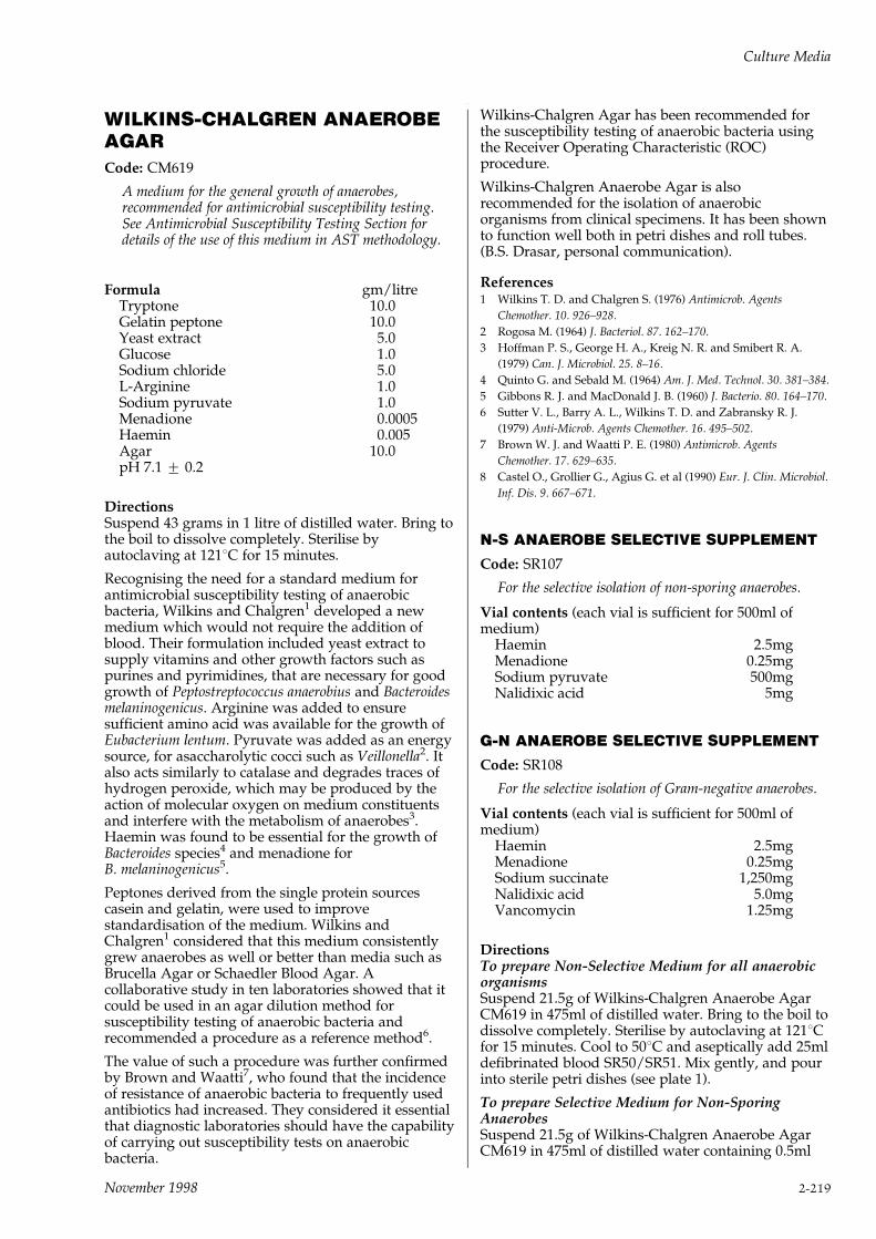
WILKINS-CHALGREN ANAEROBEAGARCode: CM619
A medium for the general growth of anaerobes,recommended for antimicrobial susceptibility testing.See Antimicrobial Susceptibility Testing Section fordetails of the use of this medium in AST methodology.
Formula gm/litreTryptone 10.0Gelatin peptone 10.0Yeast extract 5.0Glucose 1.0Sodium chloride 5.0L-Arginine 1.0Sodium pyruvate 1.0Menadione 0.0005Haemin 0.005Agar 10.0pH 7.1 + 0.2
DirectionsSuspend 43 grams in 1 litre of distilled water. Bring tothe boil to dissolve completely. Sterilise byautoclaving at 1218C for 15 minutes.
Recognising the need for a standard medium forantimicrobial susceptibility testing of anaerobicbacteria, Wilkins and Chalgren1 developed a newmedium which would not require the addition ofblood. Their formulation included yeast extract tosupply vitamins and other growth factors such aspurines and pyrimidines, that are necessary for goodgrowth of Peptostreptococcus anaerobius and Bacteroidesmelaninogenicus. Arginine was added to ensuresufficient amino acid was available for the growth ofEubacterium lentum. Pyruvate was added as an energysource, for asaccharolytic cocci such as Veillonella2. Italso acts similarly to catalase and degrades traces ofhydrogen peroxide, which may be produced by theaction of molecular oxygen on medium constituentsand interfere with the metabolism of anaerobes3.Haemin was found to be essential for the growth ofBacteroides species4 and menadione forB. melaninogenicus5.
Peptones derived from the single protein sourcescasein and gelatin, were used to improvestandardisation of the medium. Wilkins andChalgren1 considered that this medium consistentlygrew anaerobes as well or better than media such asBrucella Agar or Schaedler Blood Agar. Acollaborative study in ten laboratories showed that itcould be used in an agar dilution method forsusceptibility testing of anaerobic bacteria andrecommended a procedure as a reference method6.
The value of such a procedure was further confirmedby Brown and Waatti7, who found that the incidenceof resistance of anaerobic bacteria to frequently usedantibiotics had increased. They considered it essentialthat diagnostic laboratories should have the capabilityof carrying out susceptibility tests on anaerobicbacteria.
Wilkins-Chalgren Agar has been recommended forthe susceptibility testing of anaerobic bacteria usingthe Receiver Operating Characteristic (ROC)procedure.
Wilkins-Chalgren Anaerobe Agar is alsorecommended for the isolation of anaerobicorganisms from clinical specimens. It has been shownto function well both in petri dishes and roll tubes.(B.S. Drasar, personal communication).
References1 Wilkins T. D. and Chalgren S. (1976) Antimicrob. Agents
Chemother. 10. 926±928.
2 Rogosa M. (1964) J. Bacteriol. 87. 162±170.
3 Hoffman P. S., George H. A., Kreig N. R. and Smibert R. A.
(1979) Can. J. Microbiol. 25. 8±16.
4 Quinto G. and Sebald M. (1964) Am. J. Med. Technol. 30. 381±384.
5 Gibbons R. J. and MacDonald J. B. (1960) J. Bacterio. 80. 164±170.
6 Sutter V. L., Barry A. L., Wilkins T. D. and Zabransky R. J.
(1979) Anti-Microb. Agents Chemother. 16. 495±502.
7 Brown W. J. and Waatti P. E. (1980) Antimicrob. Agents
Chemother. 17. 629±635.
8 Castel O., Grollier G., Agius G. et al (1990) Eur. J. Clin. Microbiol.
Inf. Dis. 9. 667±671.
N-S ANAEROBE SELECTIVE SUPPLEMENT
Code: SR107
For the selective isolation of non-sporing anaerobes.
Vial contents (each vial is sufficient for 500ml ofmedium)
Haemin 2.5mgMenadione 0.25mgSodium pyruvate 500mgNalidixic acid 5mg
G-N ANAEROBE SELECTIVE SUPPLEMENT
Code: SR108
For the selective isolation of Gram-negative anaerobes.
Vial contents (each vial is sufficient for 500ml ofmedium)
Haemin 2.5mgMenadione 0.25mgSodium succinate 1,250mgNalidixic acid 5.0mgVancomycin 1.25mg
DirectionsTo prepare Non-Selective Medium for all anaerobicorganismsSuspend 21.5g of Wilkins-Chalgren Anaerobe AgarCM619 in 475ml of distilled water. Bring to the boil todissolve completely. Sterilise by autoclaving at 1218Cfor 15 minutes. Cool to 508C and aseptically add 25mldefibrinated blood SR50/SR51. Mix gently, and pourinto sterile petri dishes (see plate 1).
To prepare Selective Medium for Non-SporingAnaerobesSuspend 21.5g of Wilkins-Chalgren Anaerobe AgarCM619 in 475ml of distilled water containing 0.5ml
Culture Media
November 1998 2-219

`Tween 80'. Bring to the boil to dissolve completelyand sterilise by autoclaving at 1218C for 15 minutes.Cool to 50±558C and aseptically add the contents of 1vial of N-S Anaerobe Supplement SR107 rehydratedwith 10ml of sterile distilled water, together with25ml of defibrinated blood SR50/SR51. Mix gentlyand pour into sterile petri dishes (see plate 2).
To prepare Selective Medium for Gram-NegativeAnaerobesSuspend 21.5g of Wilkins-Chalgren Anaerobe AgarCM619 in 475ml of distilled water. Bring to the boil todissolve completely and sterilise by autoclaving at1218C for 15 minutes. Cool to 50±558C and asepticallyadd the contents of 1 vial of G-N AnaerobeSupplement SR108 rehydrated with 10ml of steriledistilled water, together with 25ml defibrinated bloodSR50/SR51. Mix gently and pour into sterile petridishes (see plate 3).
Wilkins-Chalgren Anaerobe Agar is recommendedbut other media may be used satisfactorily, e.g.Columbia Agar Base CM331 and Blood Agar BaseNo.2 CM271. Sufficient haemin and menadione arecontained in N-S and G-N Supplements to provideadequate levels in these media when used as directed.
NoteUse N-S Supplement with Wilkins-Chalgren Mediumfor NAT medium. Use G-N Supplement withWilkins-Chalgren Medium for NAV Medium1.
DiscussionSelective Medium for Non-Sporing AnaerobesThis medium is referred to in the published literature1
as NAT Medium and is recommended for theisolation of non-sporing anaerobes from clinicalspecimens.
The recovery of non-sporing anaerobes from clinicalmaterial may sometimes prove difficult in specimenscontaining mixtures of aerobic and anaerobic bacteria.A medium which contains nalidixic acid as theselective agent was described by Wren1 for isolatingthese organisms. It was shown to be virtually non-inhibitory to most non-sporing anaerobes whilstretaining good selectivity for these organisms whenpresent in mixed cultures. The medium is particularlyuseful for the recovery of non-sporing Gram-positiveanaerobes since the presence of `Tween 80' stimulatestheir growth2.
Another advantage of this medium is the earliercolonial pigmentation of the Bacteroidesmelaninogenicus group due to the slow lysis of theblood by `Tween 80' during incubation. It is also aless inhibitory medium than aminoglycoside-containing media for non-sporing anaerobes ingeneral.
The NS Anaerobe Supplement SR107 for non-sporinganaerobes contains nalidixic acid as the selectiveagent, together with haemin, menadione and sodiumpyruvate as an additional energy source1,4.
Haemin was found to be essential for the growth ofBacteroides species5 and menadione for B.melaninogenicus.6 Pyruvate, in addition to being anenergy source, acts similarly to catalase and degradestraces of hydrogen peroxide which may be produced
by the action of molecular oxygen on mediaconstituents. Hydrogen peroxide is known to affectthe metabolism of anaerobes7.
Downes et al.8 showed that NAT Medium wassuperior to kanamycin agar (KA) and neomycin agar(NA) in the recovery of all non-clostridial anaerobes.The major superiority was in the recovery ofanaerobic, Gram-positive cocci.
Selective Medium for Gram-Negative AnaerobesThis medium is described9 as NAV Medium and isrecommended for the isolation of Gram-negativeanaerobes from clinical specimens.
NAV Medium is a modification of NAT Medium1 inwhich `Tween 80' and sodium pyruvate have beenreplaced by sodium succinate. Vancomycin has beenadded, thus making the medium totally selective forGram-negative anaerobes.
G-N Anaerobe Supplement SR108 contains nalidixicacid and vancomycin as selective agents; haemin,menadione and sodium succinate as growth factors.Haemin was found to be essential for the growth ofBacteroides species5 and menadione for B.melaninogenicus6. Some Gram-negative anaerobesrequire succinate as a source of energy10.
The recovery of Gram-negative anaerobes on NAVMedium has been shown8 to be superior to that onmedia containing neomycin and kanamycin asselective agents.
In order to isolate the maximum non-sporinganaerobic bacteria from clinical specimens thefollowing scheme must be followed.
SpecimenInoculate on to each of the following media andincubate anaerobically for 48 hours.
Plate 1 Plate 2 Plate 3Wilkins-Chalgren CM619 CM619Anaerobe Agar + `Tween 80' + 5% (v/v)CM619 + 5% (v/v) +5%(v/v) defibrinateddefibrinated defibrinated blood + SR108blood blood + SR107All bacteria capable (NAT Medium) (NAV Medium)of growing under Non-Sporing Gram Gram-negativeanaerobic conditions +ve and Gram -ve anaerobic bacteria
anaerobic bacteria
A non-selective plate is included for attemptedisolation of any strain, in particular B. corrodens whichis sensitive to the selective agents.
Technique1 Prepare supplies of Plate 1 (CM619 + blood) Plate 2
(CM619 + blood + Tween 80 + SR107) and Plate 3(CM619 + blood + SR108) as described in thesection marked Directions.
2 Inoculate the specimens on to plates of eachmedium. Best results are obtained if freshlyprepared plates are used but plates may be storedat 48C for up to 3 days.
3 Incubate the plates anaerobically at 358C for 48hours. The Oxoid Anaerobic System with a GasGenerating Kit BR38 is recommended.Alternatively use Anaerogen AN025A or AN035A.
Culture Media
2-220 November 1998
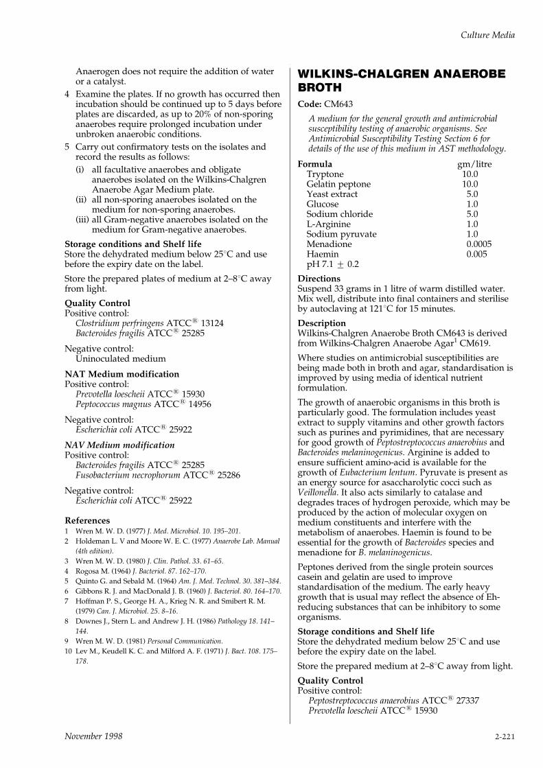
Anaerogen does not require the addition of wateror a catalyst.
4 Examine the plates. If no growth has occurred thenincubation should be continued up to 5 days beforeplates are discarded, as up to 20% of non-sporinganaerobes require prolonged incubation underunbroken anaerobic conditions.
5 Carry out confirmatory tests on the isolates andrecord the results as follows:
(i) all facultative anaerobes and obligateanaerobes isolated on the Wilkins-ChalgrenAnaerobe Agar Medium plate.
(ii) all non-sporing anaerobes isolated on themedium for non-sporing anaerobes.
(iii) all Gram-negative anaerobes isolated on themedium for Gram-negative anaerobes.
Storage conditions and Shelf lifeStore the dehydrated medium below 258C and usebefore the expiry date on the label.
Store the prepared plates of medium at 2±88C awayfrom light.
Quality ControlPositive control:
Clostridium perfringens ATCC1 13124Bacteroides fragilis ATCC1 25285
Negative control:Uninoculated medium
NAT Medium modificationPositive control:
Prevotella loescheii ATCC1 15930Peptococcus magnus ATCC1 14956
Negative control:Escherichia coli ATCC1 25922
NAV Medium modificationPositive control:
Bacteroides fragilis ATCC1 25285Fusobacterium necrophorum ATCC1 25286
Negative control:Escherichia coli ATCC1 25922
References1 Wren M. W. D. (1977) J. Med. Microbiol. 10. 195±201.
2 Holdeman L. V and Moore W. E. C. (1977) Anaerobe Lab. Manual
(4th edition).
3 Wren M. W. D. (1980) J. Clin. Pathol. 33. 61±65.
4 Rogosa M. (1964) J. Bacteriol. 87. 162±170.
5 Quinto G. and Sebald M. (1964) Am. J. Med. Technol. 30. 381±384.
6 Gibbons R. J. and MacDonald J. B. (1960) J. Bacteriol. 80. 164±170.
7 Hoffman P. S., George H. A., Krieg N. R. and Smibert R. M.
(1979) Can. J. Microbiol. 25. 8±16.
8 Downes J., Stern L. and Andrew J. H. (1986) Pathology 18. 141±
144.
9 Wren M. W. D. (1981) Personal Communication.
10 Lev M., Keudell K. C. and Milford A. F. (1971) J. Bact. 108. 175±
178.
WILKINS-CHALGREN ANAEROBEBROTHCode: CM643
A medium for the general growth and antimicrobialsusceptibility testing of anaerobic organisms. SeeAntimicrobial Susceptibility Testing Section 6 fordetails of the use of this medium in AST methodology.
Formula gm/litreTryptone 10.0Gelatin peptone 10.0Yeast extract 5.0Glucose 1.0Sodium chloride 5.0L-Arginine 1.0Sodium pyruvate 1.0Menadione 0.0005Haemin 0.005pH 7.1 + 0.2
DirectionsSuspend 33 grams in 1 litre of warm distilled water.Mix well, distribute into final containers and steriliseby autoclaving at 1218C for 15 minutes.
DescriptionWilkins-Chalgren Anaerobe Broth CM643 is derivedfrom Wilkins-Chalgren Anaerobe Agar1 CM619.
Where studies on antimicrobial susceptibilities arebeing made both in broth and agar, standardisation isimproved by using media of identical nutrientformulation.
The growth of anaerobic organisms in this broth isparticularly good. The formulation includes yeastextract to supply vitamins and other growth factorssuch as purines and pyrimidines, that are necessaryfor good growth of Peptostreptococcus anaerobius andBacteroides melaninogenicus. Arginine is added toensure sufficient amino-acid is available for thegrowth of Eubacterium lentum. Pyruvate is present asan energy source for asaccharolytic cocci such asVeillonella. It also acts similarly to catalase anddegrades traces of hydrogen peroxide, which may beproduced by the action of molecular oxygen onmedium constituents and interfere with themetabolism of anaerobes. Haemin is found to beessential for the growth of Bacteroides species andmenadione for B. melaninogenicus.
Peptones derived from the single protein sourcescasein and gelatin are used to improvestandardisation of the medium. The early heavygrowth that is usual may reflect the absence of Eh-reducing substances that can be inhibitory to someorganisms.
Storage conditions and Shelf lifeStore the dehydrated medium below 258C and usebefore the expiry date on the label.
Store the prepared medium at 2±88C away from light.
Quality ControlPositive control:
Peptostreptococcus anaerobius ATCC1 27337Prevotella loescheii ATCC1 15930
Culture Media
November 1998 2-221

Negative control:Uninoculated medium
Reference1 Wilkins T. D. and Chalgren S. (1976) Antimicrob. Agents
Chemother. 10. 926±928.
WL NUTRIENT AGARCode: CM309
A medium for the determination of the microbiologicalflora in brewing and fermentation which can be madeselective for bacteria with cycloheximide.
Formula gm/litreYeast extract 4.0Tryptone 5.0Glucose 50.0Potassium dihydrogen phosphate 0.55Potassium chloride 0.425Calcium chloride 0.125Magnesium sulphate 0.125Ferric chloride 0.0025Manganese sulphate 0.0025Bromocresol green 0.022Agar 15.0pH 5.5 + 0.2
DirectionsSuspend 75 grams in 1 litre of distilled water. Bring tothe boil to dissolve completely. Sterilise byautoclaving at 1218C for 15 minutes. If required thepH may be adjusted to 6.5 by the addition of 1%sodium bicarbonate solution.
DescriptionWL Nutrient Medium, based on that of Green andGray1 is recommended for the determination of themicrobiological flora in brewing and fermentationprocesses. The medium is suitable for thedifferentiation of `wild' yeasts from brewing yeasts2.
Reliable counts for brewers' yeast are obtained withthe medium at pH 5.5. Adjustment to pH 6.5facilitates the counting of bakers' and distillers'yeasts.
With the addition of cycloheximide 0.004gm/litre, tosuppress yeast growth, the medium becomes selectivefor the bacterial contaminants of yeast cultures.
When making microbial counts with this medium thetime and temperature of incubation will varyaccording to the materials tested and the organismssought. Temperatures of 258C are used for brewingmaterials and 308C for bakers' yeasts.
IncubationTimes can vary from 2 to 14 days. Aerobic oranaerobic incubation conditions will depend on thecharacteristics of the organisms.
Storage conditions and Shelf lifeStore the dehydrated medium below 258C and usebefore the expiry date on the label.
Store the prepared medium at 2±88C.
Quality Controlw/o cycloheximidePositive control:
Saccharomyces cerevisiae ATCC1 9763
Negative control:Uninoculated medium
with cycloheximidePositive control:
Escherichia coli ATCC1 25922
Negative control:S. cerevisiae ATCC1 9763
PrecautionsWhen handling cycloheximide observe theprecautions to be taken under HAZARDS page 2±7.
References1 Green S. R. and Gray P. P. (1950) Wallerstein Lab. Comm. 13. 357.
2 Hall Jean F. (1971) J. Inst. Brewing 77. 513±516.
WL NUTRIENT BROTHCode: CM501
A liquid medium for use in the control of brewing andother fermentation processes.
Formula gm/litreYeast extract 4.0Tryptone 5.0Glucose 50.0Potassium dihydrogen phosphate 0.55Potassium chloride 0.425Calcium chloride 0.125Magnesium sulphate 0.125Ferric chloride 0.0025Manganese sulphate 0.0025Bromocresol green 0.022pH 5.5 + 0.2
DirectionsDissolve 60g in 1 litre of distilled water. Mix well anddistribute into containers. Sterilise by autoclaving at1218C for 15 minutes.
The pH may be raised to 6.5 by the addition of 1%w/v sodium bicarbonate solution. Adding 0.004grams of cycloheximide per litre of broth will formWL Differential Broth. Note the precautions to betaken under HAZARDS page 2±7.
DescriptionWL Nutrient Broth is based on the formulation ofGreen and Gray1 and is used where there areadvantages for broth media e.g. using larger samplesof liquid products or for enrichment cultures withcycloheximide.
Addition of 0.004 grams/litre of cycloheximidesuppresses yeast growth and renders the mediumselective for bacterial contaminants. Adjustment ofthe medium to pH 6.5 facilitates growth of bakers'and distillers' yeasts; the medium at pH 5.5 is usedfor growth of bakers' yeasts.
Culture Media
2-222 November 1998

Storage conditions and Shelf lifeStore the dehydrated medium below 258C and usebefore the expiry date on the label.
Store the prepared medium at 2±88C.
Quality Controlw/o cycloheximidePositive control:
Saccharomyces cerevisiae ATCC1 9763
Negative control:Uninoculated medium
with cycloheximidePositive control:
Escherichia coli ATCC1 25922
Negative control:S. cerevisiae ATCC1 9763
PrecautionsWhen handling cycloheximide observe theprecautions to be taken under HAZARDS page 2±7.
References1 Green S. R. and Gray P. P. (1950) Wallerstein Lab. Comm. 13. 357.
2 Hall Jean F. (1971) J. Inst. Brewing 77. 513±516.
WORT AGARCode: CM247
A medium for the cultivation and enumeration ofyeasts.
Formula gm/litreMalt extract 15.0Peptone 0.78Maltose 12.75Dextrin 2.75Glycerol 2.35Dipotassium phosphate 1.0Ammonium chloride 1.0Agar 15.0pH 4.8 + 0.2
DirectionsSuspend 50g in 1 litre of distilled water and bring tothe boil to dissolve completely. Sterilise byautoclaving at 1218C for 15 minutes.
PROLONGED OR EXCESSIVE HEATING WILLDIMINISH THE GEL STRENGTH OF THE AGAR.
DescriptionWort Agar is a general purpose mycological medium,equivalent to the medium described by Parfitt1 andespecially suitable for the cultivation andenumeration of yeasts. The medium which duplicatesthe composition of natural wort, is of an aciditywhich is optimal for many yeasts but inhibitory tomost bacteria. Parfitt investigated the relative meritsof wort agar and other media for the count of yeastsand moulds in butter, and recommended the use ofdehydrated whey, malt or wort agar for the purpose.Scarr2 employed a modified wort agar ('osmophilicagar') for the examination of sugar products forosmophilic yeasts. Scarr's technique is also used for
the determination of osmophilic yeasts occurring inmaterials used in the manufacture of soft drinks.
TechniqueFor the microbiological examination of butter, makesuitable dilutions in quarter-strength Ringer solution(prepared with Ringer Solution Tablets BR52).Transfer 1ml of each dilution to a separate petri dish;add 15ml of melted Wort Agar, cooled to 458C to488C; mix by rotary movements in a horizontal plane;allow the poured-plates to set (protected from thelight) at room temperature for 30±50 minutes.Incubate in an inverted position, e.g. for 5 days at258C, and count the number of yeasts and mouldcolonies which develop.
For the examination of sugar products for osmophilicyeasts, dissolve dehydrated Wort Agar in a syrupcontaining 35 parts w/w of sucrose and 10 parts w/wof glucose, and autoclave for 20 minutes at 1108C.Inoculate and mix as above. Scarr recommendsincubation at 278c for 3±4 days for Schizosaccharomycesspecies and for 5 days for less common osmophilicyeasts. Colonies on the medium are well defined andnormally opaque.
Storage conditions and Shelf lifeStore the dehydrated medium below 258C and usebefore the expiry date on the label.
Store the prepared medium at 2±88C.
Quality ControlPositive control:
Saccharomyces cerevisiae ATCC1 9763
Negative control:Escherichia coli ATCC1 25922
PrecautionsDo not remelt the solid agar, it will destroy the gel.The surface of the agar is soft but suitable for pouredinocula.
References1 Parfitt E. H. (1933) J. Dairy Sci. 16. 141±147.
2 Scarr M. Pamela (1959) J. Sci. Food Agric. 10(12). 678±681.
Culture Media
November 1998 2-223
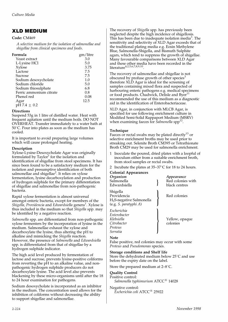
XLD MEDIUMCode: CM469
A selective medium for the isolation of salmonellae andshigellae from clinical specimens and foods.
Formula gm/litreYeast extract 3.0L-Lysine HCl 5.0Xylose 3.75Lactose 7.5Sucrose 7.5Sodium desoxycholate 1.0Sodium chloride 5.0Sodium thiosulphate 6.8Ferric ammonium citrate 0.8Phenol red 0.08Agar 12.5pH 7.4 + 0.2
DirectionsSuspend 53g in 1 litre of distilled water. Heat withfrequent agitation until the medium boils. DO NOTOVERHEAT. Transfer immediately to a water bath at508C. Pour into plates as soon as the medium hascooled.
It is important to avoid preparing large volumeswhich will cause prolonged heating.
DescriptionXylose-Lysine-Desoxycholate Agar was originallyformulated by Taylor1 for the isolation andidentification of shigellae from stool specimens. It hassince been found to be a satisfactory medium for theisolation and presumptive identification of bothsalmonellae and shigellae2. It relies on xylosefermentation, lysine decarboxylation and productionof hydrogen sulphide for the primary differentiationof shigellae and salmonellae from non-pathogenicbacteria.
Rapid xylose fermentation is almost universalamongst enteric bacteria, except for members of theShigella, Providencia and Edwardsiella genera1. Xylose isthus included in the medium so that Shigella spp. maybe identified by a negative reaction.
Salmonella spp. are differentiated from non-pathogenicxylose fermenters by the incorporation of lysine in themedium. Salmonellae exhaust the xylose anddecarboxylate the lysine, thus altering the pH toalkaline and mimicking the Shigella reaction.However, the presence of Salmonella and Edwardsiellaspp. is differentiated from that of shigellae by ahydrogen sulphide indicator.
The high acid level produced by fermentation oflactose and sucrose, prevents lysine-positive coliformsfrom reverting the pH to an alkaline value, and non-pathogenic hydrogen sulphide producers do notdecarboxylate lysine. The acid level also preventsblackening by these micro-organisms until after the 18to 24 hour examination for pathogens.
Sodium desoxycholate is incorporated as an inhibitorin the medium. The concentration used allows for theinhibition of coliforms without decreasing the abilityto support shigellae and salmonellae.
The recovery of Shigella spp. has previously beenneglected despite the high incidence of shigellosis.This has been due to inadequate isolation media3. Thesensitivity and selectivity of XLD Agar exceeds that ofthe traditional plating media e.g. Eosin MethyleneBlue, Salmonella-Shigella, and Bismuth Sulphiteagars, which tend to suppress the growth of shigellae.Many favourable comparisons between XLD Agarand these other media have been recorded in theliterature4,2,5,6,7,8,9,10.
The recovery of salmonellae and shigellae is notobscured by profuse growth of other species3
therefore XLD Agar is ideal for the screening ofsamples containing mixed flora and suspected ofharbouring enteric pathogens e.g. medical specimensor food products. Chadwick, Delisle and Byer11
recommended the use of this medium as a diagnosticaid in the identification of Enterobacteriaceae.
XLD Agar, in conjunction with MLCB Agar, isspecified for use following enrichment culture inModified Semi-Solid Rappaport Medium (MSRV)when examining faeces for Salmonella spp12.
TechniqueFaeces or rectal swabs may be plated directly13 orselective enrichment broths may be used prior tostreaking out. Selenite Broth CM395 or TetrathionateBroth CM29 may be used for salmonella enrichment.
1 Inoculate the poured, dried plates with a loopful ofinoculum either from a suitable enrichment broth,from stool samples or rectal swabs.
2 Incubate the plates at 35±378C for 18 to 24 hours.
Colonial AppearancesOrganism AppearanceSalmonella Red colonies withEdwardsiella black centres
ShigellaProvidencia Red coloniesH2S-negative Salmonella(e.g. S. paratyphi A)
EscherichiaEnterobacterKlebsiella Yellow, opaqueCitrobacter coloniesProteusSerratia
NoteFalse positive, red colonies may occur with someProteus and Pseudomonas species.
Storage conditions and Shelf lifeStore the dehydrated medium below 258C and usebefore the expiry date on the label.
Store the prepared medium at 2±88C.
Quality ControlPositive control:
Salmonella typhimurium ATCC1 14028
Negative control:Escherichia coli ATCC1 25922
}
}
}
Culture Media
2-224 November 1998
References1 Taylor W.I. (1965) Am. J. Clin. Path. 44. 471±475.
2 McCarthy M.D. (1966) N.Z. J. Med. Lab. Technol. 20. 127±131.
3 Isenberg H.D., Kominos S. and Sigeal M. (1969) Appl. Microbiol.
18. 656±659.
4 Taylor W. I. and Harris B. (1965) Am. J. Clin. Path. 44. 476±479.
5 Taylor W. I. and Harris B. (1967) Am. J. Clin. Path. 48. 350±355.
6 Taylor W. I. and Schelhart D. (1967) Am. J. Clin. Path. 48. 356±
362.
7 Taylor W.I. and Schelhart D. (1966) Appl. Microbiol. 16. 1387±
1392.
8 Rollender M.A., Beckford O., Belsky R.D. and Kostroff B. (1969)
Am. J. Clin. Path. 51. 284±286.
9 Taylor W. I. and Schelhart D. (1969) Appl. Microbiol. 18. 393±395.
10 Dunn C. and Martin W.J. (1971) Appl. Microbiol. 22. 17±22.
11 Chadwick P., Delisle G.H. and Byer M. (1974) Can. J. Microbiol.
20. 1653±1664.
12 Aspinall S.T., Hindle M.A. and Hutchinson D.N. (1992) Eur. J.
Clin. Microbiol. Inf. Dis. 11. 936±939.
13 Weissman J.B., Gangarosa E.J., Schmerler A., Marier R.L. and
Lewis J.N. (1975) Lancet I. 1898, 88±90.
Culture Media
November 1998 2-225

YEAST EXTRACT AGARCode: CM19
A nutrient agar for the plate count of organisms inwater.
Formula gm/litreYeast extract 3.0Peptone 5.0Agar 15.0pH 7.2 + 0.2
DirectionsSuspend 23g in 1 litre of distilled water. Bring to theboil to dissolve completely. Sterilise by autoclaving at1218C for 15 minutes.
DescriptionThis medium is made to the formula described byWindle Taylor for the plate count of micro-organismsin water1.
In the UK the usual method of counting heterotrophicbacteria in water is by the poured plate method withYeast Extract Agar. Seperate counts are made of thoseaerobic mesophilic organisms which form visiblecolonies in this medium after 24 hours incubation at358C and those which form colonies after 3 days at20±228C. The two methods give different results2.
The organisms growing under these conditionscomprise bacteria, yeasts and moulds.
Technique1 Prepare appropriate decimal dilutions of the water
sample (with Ringer Solution CM52) and pipette1ml portions of the water and each dilution intoduplicate sterile petri dishes.
2 Add 15ml of Yeast Extract Agar (previously meltedand cooled to 45±508C) to each plate. Mix thecontents by a combination of rapid to-and-froshaking and circular movements lasting over aperiod of 5±10 seconds.
3 Allow to solidify, and incubate duplicate sets ofplates for 24 hours at 378C and 3 days at 20±228Crespectively.
4 Select plates containing 30±300 colonies forcounting. No count should be made on a platecontaining less than 30 colonies unless the platesfrom the undiluted water contain less than thisnumber.
Storage conditions and Shelf lifeStore the dehydrated medium below 258C and usebefore the expiry date on the label.
Store the prepared medium at 2±88C.
Quality ControlPositive control:
Escherichia coli ATCC1 25922
Negative control:Uninoculated medium
References1 Windle Taylor E. (1958) `The Examination of Waters and Water
Supplies', 7th ed., Churchill Ltd., London, pp. 394±398 and 778.
2 DHSS. Report 71 (1982) `The Bacteriological Examination of
Drinking Water Supplies' HMSO. London. pp. 54±55.
YEAST AND MOULD AGARCode: CM920
A medium for the isolation of yeasts and moulds.
Formula gm/litreYeast extract 3.0Malt extract 3.0Peptone 5.0Dextrose 10.0Agar 20.0Final pH 6.2 + 0.2
DirectionsSuspend 41.0 grams of Yeast and Mould Agar CM920in 1 litre of distilled water and bring to the boil todissolve. Sterilise by autoclaving at 1218C for 15minutes. The medium may be rendered selective aftersterilisation by acidifying to pH 4.0 with 12±15mls ofSterile Lactic Acid (Oxoid SR21) after cooling to 508C.Do not reheat after making this addition. Mix welland pour into sterile petri dishes.
DescriptionYeast and Mould Agar is based on the formulationdescribed by Wickerham1,2. The medium isrecommended for the isolation and maintenance ofyeasts and moulds.
Yeast and Mould Agar may be rendered selective bythe addition of acid to reduce the pH of the mediumto pH 4.0. A suitable acid for use is Oxoid SterileLactic Acid (SR21).
Technique1 Prepare Yeast and Mould Agar plates as directed
in the directions for use.
2 Inoculate the medium by surface or poured plateprocedures.
3 Incubate the plates for 48±72 hours at 258C to 308C.
Detection and enumeration of yeasts in the presenceof moulds may be made easier by using a combinedanaerobic/aerobic incubation procedure3.
Reaction after incubationpH 6.2 pH 4.0
Aspergillus niger ATCC1 16404 Good growth Good growthCandida albicans ATCC1 10231 Good growth Good growthSaccharomyces serevisiae ATCC1 9763 Good growth Good growthLactobacillus fermentum ATCC1 9338 Good growth Partial to complete inhibitionEscherichia coli ATCC1 25922 Good growth Partial to complete inhibition
Culture Media
2-226 November 1998
Cultures are initially incubated for 3 days underanaerobic conditions and then for a further 2 daysaerobically. Development of mould colonies isimpeded during the anaerobic phase of incubation.Dimorphic moulds e.g. (mucor spp) may form yeast-like colonies during anaerobic incubation.
TechniqueCultures on plates or membrane filters are incubated for3 days at 258C under strictly anaerobic conditions.Continue incubation of the cultures under aerobicconditions for a further 2 days. Yeast colonies may bevery small immediately following anaerobic incubationbut will increase in size in air. Mould growth maybecome completely unrestricted after 3 days in air.
Storage conditions and Shelf lifeYeast and Mould Agar should be stored tightlycapped in the original container at 108C to 258C.When stored as directed the medium will remainstable until the expiry date printed on the label.
The prepared medium may be stored for up to 2weeks at 2±88C.
Quality ControlYeast and Mould Agar may be tested for performanceusing stable, typical control cultures.
PrecautionsFor in vitro diagnostic use.
Do not use beyond the stated expiry date or if theproduct is caked, discoloured or shows any sign ofdeterioration.
References1 Wickerham L.J. (1951) U.S. Dept. Agric. Tech. Bull. No 1029, 1±19.
2 Wickerham L.J. and Rettger L.F (1939) J. Tropical Med. Hyg. 42.
174±179.
3 De Jong J. and Put H.M.C. (1980) Biology and Activities of Yeasts.
Society for Applied Bacteriology Symposium series No. 9. Skinner
F.A., Passmore S.M. and Davenport R.R. (eds). Academic Press,
London. Pages 289±292.
YERSINIA SELECTIVE AGARBASECode: CM653
A selective medium for Yersinia enterocolitica whenused with Yersinia Selective Supplement SR109(Schiemann CIN Medium).
Formula gm/litreSpecial peptone 20.0Yeast extract 2.0Mannitol 20.0Sodium pyruvate 2.0Sodium chloride 1.0Magnesium sulphate 0.01Sodium desoxycholate 0.5Neutral red 0.03Crystal violet 0.001Agar 12.5pH 7.4 + 0.2
YERSINIA SELECTIVE SUPPLEMENT
Code: SR109
Vial contents (each vial is sufficient for 500ml ofmedium).
Cefsulodin 7.5mgIrgasan 2.0mgNovobiocin 1.25mg
Reconstitute the antibiotic supplement SR109 byaseptically adding 2ml of a sterile 50:50 solution ofethanol and water to one vial and mix gently todissolve the contents completely.
DirectionsSuspend 29.0g in 500ml of distilled water and bringgently to the boil to dissolve completely. Sterilise byautoclaving at 1218C for 15 minutes. Allow to cool to508C and aseptically add the contents of one vial ofYersinia Selective Supplement SR109 reconstituted asdirected. Mix gently and pour into sterile petri dishes.
DescriptionYersinia Selective Medium (CIN Medium) is based onthe formulation of Schiemann1,2 and is recommendedfor the isolation and enumeration of Yersiniaenterocolitica from clinical specimens and food.
Y. enterocolitica is becoming increasingly recognised asa cause of diarrhoeal disease of man. Infection by theorganisms results in diarrhoea, malaise, nausea andfever, plus constant abdominal pain over a period of1±2 days. The organism has also been shown as acause of polyarthritis, mesenteric adenitis andsepticaemia. It is likely that human infections aredirectly or indirectly derived from animal sources andmay be contracted through the ingestion ofcontaminated food. Initially serotypes 0:3 and 0:9were implicated in human infections but since thenother serotypes, mainly 0:5 and 0:8 have also beeninvolved3. It is important to note that incidence ofdisease caused by the various serotypes ofY. enteroccolitica is currently reported to varyconsiderably with geographical location. It is expectedthat with provision of a selective medium, a higherisolation rate will result, and Y. enterocolitica will berecognised as more common and widespread thanpreviously suspected.
Yersinia Selective Agar Base CM653 and the selectivesupplement SR109 have been developed specificallyfor the optimum growth and recovery of Y.enterocolitica after 18±24 hours incubation at 328C.Schiemann2 modified his earlier formulation for CINMedium by replacing Bile Salts with sodiumdesoxycholate (0.5g/l) and by reducing theconcentration of novobiocin from 15 to 2.5mg/l inorder to eliminate the inhibition of some strains ofserotype 0:8.
The typical colonies of Y. enterocolitica will develop asa dark red `bullseye' surrounded by a transparentborder and will vary considerably among serotypes incolony size, smoothness and the ratio of the border tocentre diameter. Most other organisms that arecapable of growing will produce larger colonies(>2mm in diameter) with diffuse pinkish centres andopaque outer zones. Serratia liquefaciens, Citrobacter
Culture Media
November 1998 2-227
freundii and Enterobacter agglomerans may give acolonial morphology resembling Y. enterocolitica.These organisms can be differentiated from Y.enterocolitica by biochemical tests.
Test for growth on Nutrient and MacConkey Agars,test for indole and urease production and for acidreactions from sucrose, cellobiose, amygdalin,melibiose, rhamnose and reffinose. Carry out tests at308C rather than 378C4,5.
Technique for CultureDirect Plate Method1 Pour plates of Yersinia Selective Agar and dry the
surface.
2 Inoculate the plates with a suspension of the food,faeces, etc., to produce single colonies.
3 Incubate at 328C for 24 hours.
Cold Enrichment in Phosphate Buffered Saline6
1 Inoculate food, faeces, etc., into M/15 phosphatebuffered saline.*
2 Hold at 48C for up to 21 days.
3 Periodically sub-culture samples on to plates ofYersinia Selective Agar.
4 Incubate at 328C for 24 hours.
*To prepare an M/15 buffer dissolve one tablet ofOxoid Dulbecco `A' BR14A in 223ml of distilledwater. Distribute into final containers and sterilise byautoclaving at 1158C for 10 minutes.
CIN Agar had been used for isolation of Leptospiraspp7. With enhancement of its nutritional propertiesand addition of 5-fluorouracil to increase selectivity ithas also been used to demonstrate the presence ofArcobacter spp. in ground pork8.
Colonial MorphologyThe typical colonies of Y. enterocolitica will develop adark red `bullseye' surrounded by a transparentborder. The colony size, smoothness and the ratio ofthe border to centre diameter will vary considerablyamong serotypes.
Identification of IsolatesThe presumptive colonies are confirmed as Y.enterocolitica by the biochemical reactions shown inTable 1.
Storage conditions and Shelf lifeStore the dehydrated medium below 258C and usebefore the expiry date on the label.
Store the prepared medium at 2±88C for not morethan 24 hours.
Quality ControlPositive control:
Yersinia enterocolitica ATCC1 27729
Negative control:Escherichia coli ATCC1 25922
PrecautionsSome strains of Y. enterocolitica may grow poorly ornot at all. Other species of Yersinia may grow alongwith some enteric organisms. It is therefore essentialthat full identification tests are carried out on suspectcolonies.
References1 Schiemann D. A. (1979) Can. J. Microbiol. 25. 1298±1304.
2 Swaminathan B., Harmon M. C. and Mehlman I. J. (1982) J.
Appl. Bact. 52. 151±183.
3 Bisset M. L. (1976) J. Clin. Microbiol. 4. 137±144.
4 Swaminathan B., Harmon M. C. and Mehlman I. J. (1982) J.
Appl. Bact. 52. 151±183.
5 Mair N. S. and Fox E. (1986) Yersiniosis: Laboratory Diagnosis,
Clinical Features and Epidemiology. Pub. Hlth Lab. Ser. London.
6 Pai C. H., Sorger S., Lafleur L., Lackman L. and Marks M. I.
(1979) J. Clin. Microbiol. 9. 712±715.
7 Borcyzk A., Rosa S.D. and Lior H. (1991) Abst. Ann. Meet. Am.
Soc. Microbiol. C.267. p.386.
8 Collins C.I., Wesley I.V. and Murano E.A. (1996) J. Food Prot. 59.
448±452.
Table 1
Yersinia enterocolitica
Growth at 48C and on Nutrient/MacConkeyAgars
Motile at 228C
Indole production variable
Urease positive
Ornithine decarboxylase positive
Acid production from sucrose, cellobiose,amygdalin, rhamnose and raffinose
No acid produced from melibiose.
Culture Media
2-228 November 1998
QUALITY CONTROLORGANISMS
OXOID CULTI-LOOPS1
These enable standardised cultures for quality controltesting to be prepared quickly, easily and safely.
Culti-Loops are ideal for:Performance testing of culture media, stains anddiagnostic reagents
Evaluation of bacteriological procedures
Maintenance of stock cultures
Culti-Loops are ready to use, disposablebacteriological loops containing stabilised viablemicro-organisms.
Culti-Loops are recommended for use in theperformance testing of culture media, stains,diagnostic kits and reagents, for the maintenance ofstock cultures and in the evaluation of bacteriologicalprocedures. Each loop is individually packaged in afoil pouch and each can contains ten such loops.
PrecautionsCulti-Loops contain viable micro-organisms andshould be used only by individuals withbacteriological training. Refer to national Guidelinesfor Microbiological Containment Categoryinformation. After use, all loops and packagingshould be placed into an appropriate container andsterilised by autoclaving before their final disposal.Do not place the loops into bunsen burners.
StorageStore Culti-Loops at 2±88C (or frozen forCampylobacter spp.). Remove only the quantity ofloops required for immediate use. Under theseconditions, Culti-Loops will retain their viability untilthe date shown on the foil pouch.
To openCut open the end of the foil packet as indicated on thelabel.
Evidence of deteriorationEach loop should contain an intact dried film. Do notuse the loop if there is any evidence of hydration.
ProcedureThe film in each loop is made from a gelatinformulation and then dried by special processing. Torehydrate the film, the loops must come into contactwith both warmth and moisture.
Direct inoculation of Culti-Loops onto selective mediamay result in slow or absent growth. It is thereforerecommended that where this is observed, inoculationonto non-selective media (such as blood agar) shouldprecede sub-culture onto selective media.
The following two methods may be used forinoculation. Utilise the appropriate method for theselected micro-organism.
Direct Streak MethodThis procedure is recommended for all non-fastidiousmicro-organisms.
1 Warm the appropriate plate medium to 378C.
2 Remove the sheath from the loop.
3 Stab the loop into the medium or lay it flat on thewarm, moist surface. Hold it in this manner for10±15 seconds to allow for absorption of moisture.
4 Streak the plate in the usual manner. As many asfive plates may be streaked with the same loop.
5 Incubate the plates in an appropriate atmosphereand temperature for the optimal growth of theorganism.
Indirect (Broth) MethodThis procedure is recommended for all fastidiousmicro-organisms.
1 Remove the sheath from the loop.
2 Cut off the loop shaft from the handle usingsterilised scissors into a tube containing 0.5 to1.0ml of liquid medium. Use:
a Tryptone Soya Broth (Oxoid CM129) or freshlyprepared Thioglycollate USP (CM391) forbacteria specimens.
b Sterile saline for mycology specimens.
3 Place tube in a 378C incubator just long enough forthe film to dissolve completely out of the loop.Shake the tube gently to suspend the organism.
4 Using the Pasteur pipette, inoculate theappropriate media with several drops and streak inthe usual manner.
5 Incubate the plates in an appropriate atmosphereand temperature for the optimal growth of theorganism.
Most organisms grow in 24±48 hours under theproper conditions. However, some exhibit aconsiderable lag phase and should be incubated foran additional 24 hours.
DisclaimerThose who receive Culti-Loops are responsible fortheir safe storage, handling and use. Oxoid is notliable for damages or injuries resulting from thereceipt and/or use of Culti-Loops. Oxoid is not liablefor damages arising from the misidentification ormisrepresentation of strains.
Reference1 Prier J., Bartola E. and Friedman H. (1973) Quality Control in
Microbiology. University Park Press, Baltimore.
The current range of Culti-Loops may be found in theseparate Oxoid Product List.
Culture Media
November 1998 2-229
OXOID QUANTI-CULTPLUS(TM)
This range features convenient ready-to-use preservedmicro-organisms for use in quality controlprocedures. Each delivers a specific range of colonyforming units (CFUs) and may be used for:
Growth promotion
Bacteriostasis and fungistasis
Testing of antimicrobial preservatives anddisinfectants
Microbial limit tests
Media quality control etc.
UseThis product contains a specified number ofpreserved micro-organisms for use in quality controlprocedures1,2,3. Colony forming units (CFUs) arespecified under defined procedures and growingconditions and cannot be guaranteed under otherconditions. Results may vary with more inhibitory orselective media or with the same medium if of inferiorquality.
DescriptionEach set consists of two vials packaged in a plasticbag. One vial, sealed within a silvery mylar envelope,contains a film of micro-organisms attached to theinside of the cap of the vial. The microbial film can beseen through the red cap as a black area inside theblack `O' ring of the cap. Rehydrating fluid isprovided in the second vial.
PrecautionsQUANTI-CULTPLUS(TM) contains live micro-organisms and should be used only by individualswith microbiological training. Properly disinfect anyspills and sterilise used containers by autoclavingbefore final disposal.
StorageBoth rehydrating fluid and micro-organisms shouldbe stored at refrigerator temperature (2±88C) untiltime of analysis.
Expiration dateThe reagents are stable through the expiration date onthe label when stored as directed.
Evidence of deteriorationEach RED CAP should contain an intact dried film.Do not use if the film has come out of the cap orshows evidence of hydration.
To openThe foil envelope can be cut open with scissors.
To rehydrateCAUTIONTHE REHYDRATED SUSPENSION MUST BE USEDWITHIN 30 MINUTES AFTER THE 10 MINUTEREHYDRATION PERIOD. DO NOT PLAN TO USEA REHYDRATED SAMPLE THROUGHOUT THEWORK DAY OR THE FOLLOWING DAY.PROLONGED HOLDING WILL ADVERSELYEFFECT THE NUMBERS OF VIABLE ORGANISMS.
1 Remove vials from the refrigerator. Place therehydrating fluid vial (blue cap) in a 35 to 378Cincubator to warm.
2 Allow the vial (red cap) containing the microbialfilm (silver envelope) to warm to roomtemperature.
3 Remove the vial (red cap) containing the microbialfilm from the envelope.
4 Remove and discard the blue cap from therehydrating fluid.
5 Remove the red cap containing the micro-organisms and transfer to the rehydrating fluidvial. Tighten cap.
6 Invert this vial, tap to be sure liquid is in contactwith the inside of the cap, and place in 35 to 378Cincubator in an INVERTED position for 10minutes* to dissolve the preserved micro-organisms. Autoclavable polyester racks may beused to hold the vials in an inverted position.Please note that the foam is an efficient insulatingmaterial and will retard warming or cooling ofliquids.
7 Grasp bottom of vial, hold firmly in an invertedposition, tap cap gently to mix suspension and aiddissolution. Excessive vigorous shaking willproduce foam.
*CAUTIONLook at the cap to make certain that all of the micro-organisms are in solution. Undissolved intact blackparticles can be seen through the plastic cap and/orvial. If this happens, reinvert the vial and place backin the incubator and observe closely every 1 to 2minutes for complete dissolution. Undissolvedmicrobial film will cause reduced counts butprolonged heating may also result in incorrect counts.
Inoculation and sample analysis
QUANTI-CULTPLUS(TM) is designed to deliver ten100ml inocula each containing between 10 and 100CFUs from a single source.
QUANTI-CULTPLUS(TM) is suitable for evaluation ofculture media used for the growth promotion and/orbacteriostasis and fungistasis procedures1, othermedia3, or quality control of any quantitativemicrobiological procedure2.
CAUTIONTHE REHYDRATED SUSPENSION MUST BE USEDWITHIN 30 MINUTES AFTER THE 10 MINUTEREHYDRATION PERIOD. DO NOT PLAN TO USEA REHYDRATED SAMPLE THROUGHOUT THEWORK DAY OR THE FOLLOWING DAY.PROLONGED HOLDING WILL ADVERSELYEFFECT THE NUMBERS OF VIABLE ORGANISMS.
Hold the rehydrated suspension at room temperaturethroughout the 30 minute use period. Re-mix samplebetween withdrawals. When inoculating ortransferring to a liquid matrix, it is advisable to havethe receiving liquid pre-warmed to 35±378C.Dispensing into a cold liquid may interfere with evendistribution of the micro-organisms.
Culture Media
2-230 November 1998
References1 United Sates Pharmacopoeia XXII 1990, Mack Publishing Co.
2 Clesceri L.S., Greenberg A.E. & Trussel R.R. eds. 1989. Standard
Methods for the Examination of Water and Wastewater. 17th ed.
A.P.H.A., A.W.W.A. & W.P.C.F.
3 Mehlman I.J. 1984. Appendix I. Culture media. In Bacteriological
Analytical Manual. A.O.A.C. Arlington, VA.
QUANTI-CULTPLUS(TM)
Each tube contains 10 packs (10 tests per pack).Each kit is designed to deliver between 10±100 cfu/0.1ml.
Total diluent volumes per vial is 1.2ml.
NoteThe organisms used in QUANTI-CULTPLUS(TM) arederived from original ATCC stock cultures accordingto the number shown.
QUANTI-CULTPLUS(TM) are manufactured for OxoidLtd by Chrisope Technologies Inc., an FDA approvedcompany.
FDA Registration No: 1625984. QUANTI-CULTPLUS(TM) is a registered trademark of ChrisopeTechnologies Inc, a division of remel.
The current range of Quanti-Cult may be found in theseparate Oxoid Product List.
Culture Media
November 1998 2-231
3PEPTONES, HYDROLYSATES,AGARS & CONSTITUENTS

November 1998
LABORATORYPREPARATIONSOxoid Laboratory Preparations are Culture Mediareagents which are either:
(i) manufactured within Oxoid to specifiedquality performance standards
(ii) manufactured outside for Oxoid to the samehigh standards
(iii) selected from screened buying samples byextensive laboratory testing.
The L-P range includes peptones, proteinhydrolysates, biological extracts, agars and the criticalculture media chemicals such as selective agents, dyesetc.
Products are provided for users who wish to createtheir own media or who wish to supplement existingformulae. It should be stressed, however, that the useof these products will not necessarily reproduce theperformance of listed Oxoid Culture Media, evenused in identical formulae. This is because it isimpossible to produce peptones or hydrolysateswhich can be universally applied to any formulae.
3.1 Peptones, hydrolysates and biologicalextracts.
3.2 Agars.
3.3 Bile, bile salts and derivatives.
3.4 Chemicals for culture media.
For bulk users Oxoid can manufacture laboratorypreparations to meet special requirements.
PEPTONES, HYDROLYSATES ANDBIOLOGICAL EXTRACTS1 Introduction
2 Basic Informationi Biochemistry of Proteins
ii Hydrolysis of Proteins
iii Manufacture of Peptones
iv Quality Assurance
3 Meat Peptones
i Liver Digest Neutralised Code L27
ii Peptone Bacteriological Code L37
Peptone Bacteriological NeutralisedCode L34
iii Mycological Peptone Code L40
iv Tryptose Code L47
v Peptone P Code L49
vi Special Peptone Code L72
vii Proteose Peptone Code L85
4 Vegetable Peptone
i Soya Peptone Code L44
5 Casein Peptones and other milk derivedPeptones
i Peptonised Milk Code L32
ii Casein Hydrolysate (Acid) Code L41
iii Tryptone Code L42
iv Tryptone T Code L43
v Lactalbumin Hydrolysate Code L48
6 Extracts
i Yeast Extract Code L21
ii Lab-Lemco Code L29
iii Malt Extract Code L39
7 References
8 Packaging & Services
9 Table of Analysis
i General Analysis
ii Amino Acid Analysis
10 Uses of Oxoid Peptones, Hydrolysates andBiological Extracts
1 INTRODUCTION
The first time the term `peptone' appeared was inpapers published in 1880 and 1882 by Nageli. He hasbeen credited as the first bacteriologist to discoverthat chemo-organotrophic organisms grow best inculture media containing a partially digested protein.
The problems associated with the production ofprotein hydrolysates were quickly recognised andtheir manufacture became the concern of commercialsuppliers. In fact protein hydrolysate was the firstcomplex culture medium ingredient to be suppliedcommercially. This was the fore-runner of the largerange of commercial culture media now available.
Peptones, Hydrolysates, Agars & Constituents
November 1998 3-1
Oxoid (then the Medical Division of Oxo) started itsinvestigation into the manufacture of peptone in 1924.
The variety of peptones and extracts available reflectsthe differing demands of micro-organisms for aminoacids, peptides and other nutrients. Substrates usedby Oxoid for hydrolysis include: meat, casein,lactalbumin, milk, gelatin, soya and yeast cells.
2 BASIC INFORMATION
Biochemistry of ProteinsProteins are macro-molecules and are fundamental tothe structure and function of all living organisms.Chemically, proteins are made up of one or morechains of alpha-amino-carboxylic acids (amino acids),consecutively linked covalently between the alpha-amino group of one moiety and the alpha carboxylicgroup of the next with the elimination of water. Thislinkage is termed the `peptide bond'. Chains of threeor more amino acids are termed `polypeptides', whilstlarger structures, with an arbitrarily determinedlower molecular weight limit of 5,000 are the proteins.An example of a Peptide and Amino Acid is shown inFigure A.
Only 20 amino acids commonly occur in proteins.They can occupy any position in the protein chainwhich can be at least 80 units long with molecularweights of several millions. The chains are folded in avariety of complex forms and the structures mayincorporate other macro-molecules such ascarbohydrates and lipids.
Hydrolysis of ProteinsThe hydrolysis of proteins, which breaks them downto their constituent amino acids and peptides can beachieved by the use of strong acids, strong bases orproteolytic enzymes such as pepsin, papain andpancreatin (which contains trypsin)1.
Hydrolysis with strong mineral acids, often at hightemperatures and pressures is much used in the foodindustry to produce food flavourings. The mostcommonly used product in microbiology is based onthe hydrolysis of casein. In this process all peptidebonds are attacked and in theory, completebreakdown into component parts could be obtained.However, because the reaction conditions are sosevere, some of the amino acids produced arethemselves destroyed by the process, notablytryptophan which is totally lost. Cystine, serine andthreonine are partially broken down but asparagineand glutamine are converted to their acidic forms.Any vitamins present are largely destroyed. A seriesof reactions may also take place betweencarbohydrates and amino acids (such as the Maillardreactions) which give rise to very dark products oftentoxic to the growth of micro-organisms2. Formicrobiology the amount of hydrolysis is controlledto produce a suitable nitrogen source for bacteria.
Proteolytic enzymes act on proteins under less severeconditions. They will function at much lowertemperatures and at normal pressures and are usuallyspecific to the peptide bond they will attack. Thismeans that the protein is not completely hydrolysedto its constituent amino acids but into polypeptides ofvarying lengths, depending on the frequency of the
specific amino acid linkage. Also, since proteins havea very consistent primary structure, the mixture ofpeptides produced after proteolytic digestion by aspecific enzyme is also consistent.
Enzymes commonly used are papain, pepsin andpancreatin, Figure A.
Pepsin will cut the chain anywhere there is aphenylalanine or leucine bond3.
Papain cuts adjacent to arginine, lysine, phenylalanineand glycine4.
Pancreatin has its action at arginine, lysine, tyrosine,tryptophan, phenylalanine and leucine bonds4.
Raw materials may vary considerably in compositionand the extent to which the protein components havebeen denatured during any processing procedures,therefore the conditions of manufacture must becarefully controlled to minimise the variationsinherent in biological materials and so maintainquality.
More defined protein sources, such as casein andgelatin will give more consistent mixtures of peptideswhen treated with enzymes or acid.
In practical terms, total breakdown of a protein to itsindividual component amino acids is difficult evenwith a mixture of enzymes; the result, even with welldefined proteins such as casein, is a peptonecontaining a chemically undefined mix of peptidesand amino acids.
Manufacture of PeptonesThe manufacturing process is illustrateddiagrammatically in Figure B and the syrup formedcan be stored for long periods at room temperaturebecause the high dissolved solid content inhibitsbacterial contamination. This syrup can be used infermentation processes without drying to a powder.
Quality AssuranceIt is essential that the quality of these products ismaintained at the highest level and lot to lot variationreduced to a minimum by closely following codes ofGood Manufacturing Practice (GMP)5. In order toachieve this several types of analysis are carried outand strict quality control specifications must be metfor a lot to be accepted. A list of average analyses ofhydrolysed products is shown on page 3±12.
To ensure that the product conforms topredetermined specifications tests are carried out andthe following criteria are routinely monitored: clarityand colour, moisture content, pH value, ash residue,chloride, nitrogen content and microbiology.
Clarity and pH ValueThese tests are performed on an autoclaved 2%solution of the final product and are controlled bycomparison with reference materials.
Moisture ContentThe level of moisture should be below 5% to ensureno chemical changes occur if the product is stored athigh ambient temperatures.
Ash ResidueThe ash residue consists mainly of inorganic materialand is estimated after ignition.
Peptones, Hydrolysates, Agars & Constituents
3-2 November 1998
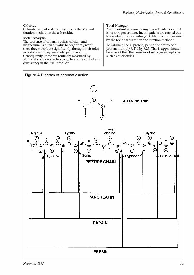
ChlorideChloride content is determined using the Volhardtitration method on the ash residue.
Metal AnalysisThe presence of cations, such as calcium andmagnesium, is often of value to organism growth,since they contribute significantly through their rolesas co-factors in key metabolic pathways.Consequently, these are routinely measured byatomic absorption spectroscopy, to ensure control andconsistency in the final products.
Total NitrogenAn important measure of any hydrolysate or extractis its nitrogen content. Investigations are carried outto ascertain the total nitrogen (TN) which is measuredby the Kjeldhal digestion and titration method6.
To calculate the % protein, peptide or amino acidpresent multiply %TN by 6.25. This is approximatebecause of the other sources of nitrogen in peptonessuch as nucleotides.
Figure A Diagram of enzymatic action
Peptones, Hydrolysates, Agars & Constituents
November 1998 3-3
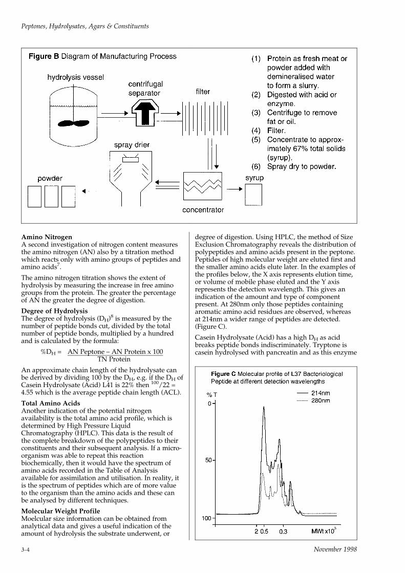
Amino NitrogenA second investigation of nitrogen content measuresthe amino nitrogen (AN) also by a titration methodwhich reacts only with amino groups of peptides andamino acids7.
The amino nitrogen titration shows the extent ofhydrolysis by measuring the increase in free aminogroups from the protein. The greater the percentageof AN the greater the degree of digestion.
Degree of HydrolysisThe degree of hydrolysis (DH)8 is measured by thenumber of peptide bonds cut, divided by the totalnumber of peptide bonds, multiplied by a hundredand is calculated by the formula:
%DH = AN Peptone ± AN Protein x 100TN Protein
An approximate chain length of the hydrolysate canbe derived by dividing 100 by the DH, e.g. if the DH ofCasein Hydrolysate (Acid) L41 is 22% then 100/22 =4.55 which is the average peptide chain length (ACL).
Total Amino AcidsAnother indication of the potential nitrogenavailability is the total amino acid profile, which isdetermined by High Pressure LiquidChromatography (HPLC). This data is the result ofthe complete breakdown of the polypeptides to theirconstituents and their subsequent analysis. If a micro-organism was able to repeat this reactionbiochemically, then it would have the spectrum ofamino acids recorded in the Table of Analysisavailable for assimilation and utilisation. In reality, itis the spectrum of peptides which are of more valueto the organism than the amino acids and these canbe analysed by different techniques.
Molecular Weight ProfileMoelcular size information can be obtained fromanalytical data and gives a useful indication of theamount of hydrolysis the substrate underwent, or
degree of digestion. Using HPLC, the method of SizeExclusion Chromatography reveals the distribution ofpolypeptides and amino acids present in the peptone.Peptides of high molecular weight are eluted first andthe smaller amino acids elute later. In the examples ofthe profiles below, the X axis represents elution time,or volume of mobile phase eluted and the Y axisrepresents the detection wavelength. This gives anindication of the amount and type of componentpresent. At 280nm only those peptides containingaromatic amino acid residues are observed, whereasat 214nm a wider range of peptides are detected.(Figure C).
Casein Hydrolysate (Acid) has a high DH as acidbreaks peptide bonds indiscriminately. Tryptone iscasein hydrolysed with pancreatin and as this enzyme
Peptones, Hydrolysates, Agars & Constituents
3-4 November 1998
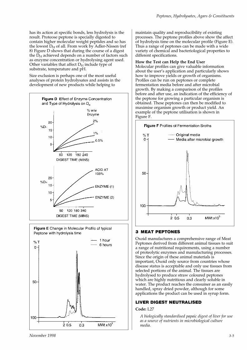
has its action at specific bonds, less hydrolysis is theresult. Proteose peptone is specially digested tocontain higher molecular weight peptides and so hasthe lowest DH of all. From work by Adler-Nissen (ref8) Figure D shows that during the course of a digestthe DH achieved depends on a number of factors suchas enzyme concentration or hydrolysing agent used.Other variables that affect DH include type ofsubstrate, temperature and pH.
Size exclusion is perhaps one of the most usefulanalyses of protein hydrolysates and assists in thedevelopment of new products while helping to
maintain quality and reproducibility of existingprocesses. The peptone profiles above show the affectof hydrolysis time on the molecular profile (Figure E).Thus a range of peptones can be made with a widevariety of chemical and bacteriological properties todifferent specifications.
How the Test can Help the End UserMolecular profiles can give valuable informationabout the user's application and particularly showshow to improve yields or growth of organisms.Profiles can be run on peptones or completefermentation media before and after microbialgrowth. By making a comparison of the profilesbefore and after use, an indication of the efficiency ofthe peptone for growing a particular organism isobtained. These peptones can then be modified tomaximise organism growth or product yield. Anexample of the peptone utilisation is shown inFigure F.
3 MEAT PEPTONES
Oxoid manufactures a comprehensive range of MeatPeptones derived from different animal tissues to suita range of nutritional requirements, using a numberof proteolytic enzymes and manufacturing processes.Since the origin of these animal materials isimportant, Oxoid only source from countries whosedisease status is acceptable and only use tissues fromselected portions of the animal. The tissues arehydrolysed to produce straw coloured peptoneswhich are highly nutritious and clearly soluble inwater. The product reaches the consumer as an easilyhandled, spray dried powder, although for someapplications the product can be used in syrup form.
LIVER DIGEST NEUTRALISED
Code: L27
A biologically standardised papaic digest of liver for useas a source of nutrients in microbiological culturemedia.
Peptones, Hydrolysates, Agars & Constituents
November 1998 3-5
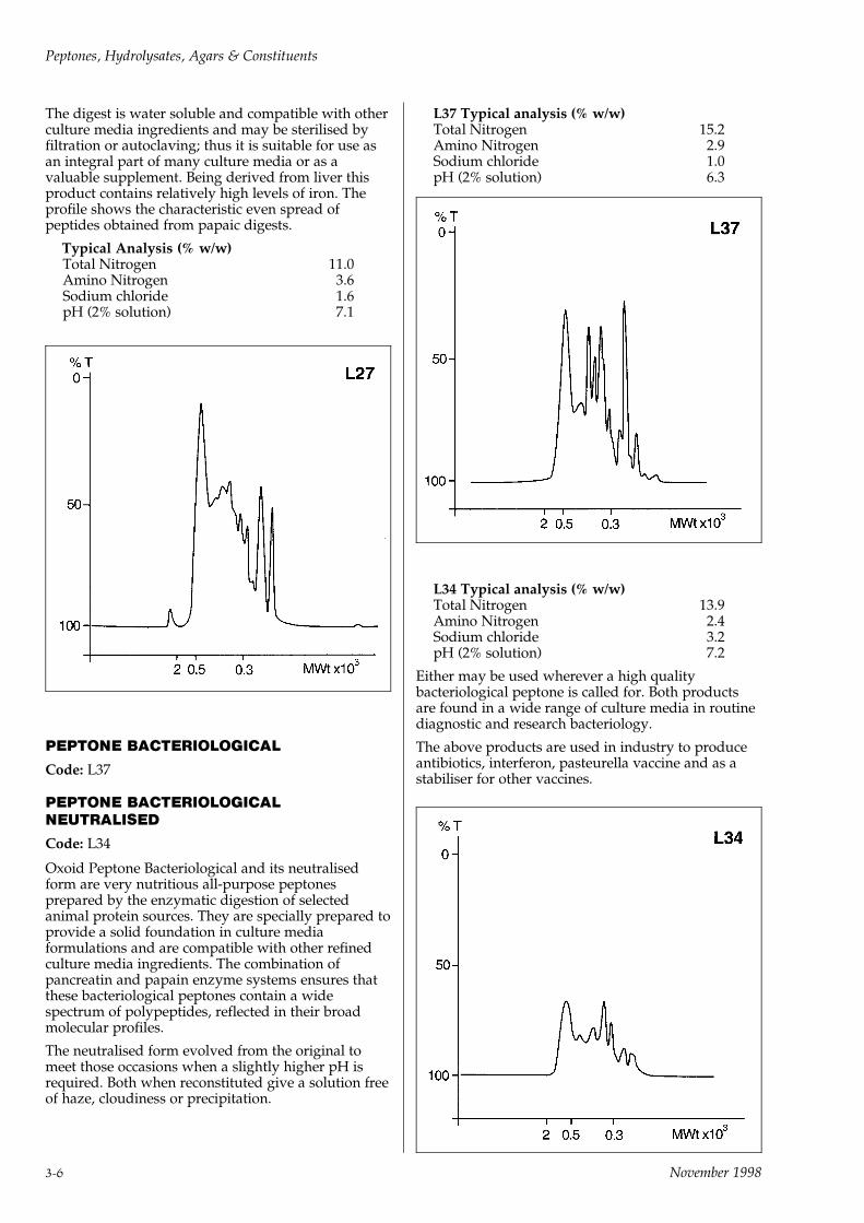
The digest is water soluble and compatible with otherculture media ingredients and may be sterilised byfiltration or autoclaving; thus it is suitable for use asan integral part of many culture media or as avaluable supplement. Being derived from liver thisproduct contains relatively high levels of iron. Theprofile shows the characteristic even spread ofpeptides obtained from papaic digests.
Typical Analysis (% w/w)Total Nitrogen 11.0Amino Nitrogen 3.6Sodium chloride 1.6pH (2% solution) 7.1
PEPTONE BACTERIOLOGICAL
Code: L37
PEPTONE BACTERIOLOGICALNEUTRALISED
Code: L34
Oxoid Peptone Bacteriological and its neutralisedform are very nutritious all-purpose peptonesprepared by the enzymatic digestion of selectedanimal protein sources. They are specially prepared toprovide a solid foundation in culture mediaformulations and are compatible with other refinedculture media ingredients. The combination ofpancreatin and papain enzyme systems ensures thatthese bacteriological peptones contain a widespectrum of polypeptides, reflected in their broadmolecular profiles.
The neutralised form evolved from the original tomeet those occasions when a slightly higher pH isrequired. Both when reconstituted give a solution freeof haze, cloudiness or precipitation.
L37 Typical analysis (% w/w)Total Nitrogen 15.2Amino Nitrogen 2.9Sodium chloride 1.0pH (2% solution) 6.3
L34 Typical analysis (% w/w)Total Nitrogen 13.9Amino Nitrogen 2.4Sodium chloride 3.2pH (2% solution) 7.2
Either may be used wherever a high qualitybacteriological peptone is called for. Both productsare found in a wide range of culture media in routinediagnostic and research bacteriology.
The above products are used in industry to produceantibiotics, interferon, pasteurella vaccine and as astabiliser for other vaccines.
Peptones, Hydrolysates, Agars & Constituents
3-6 November 1998
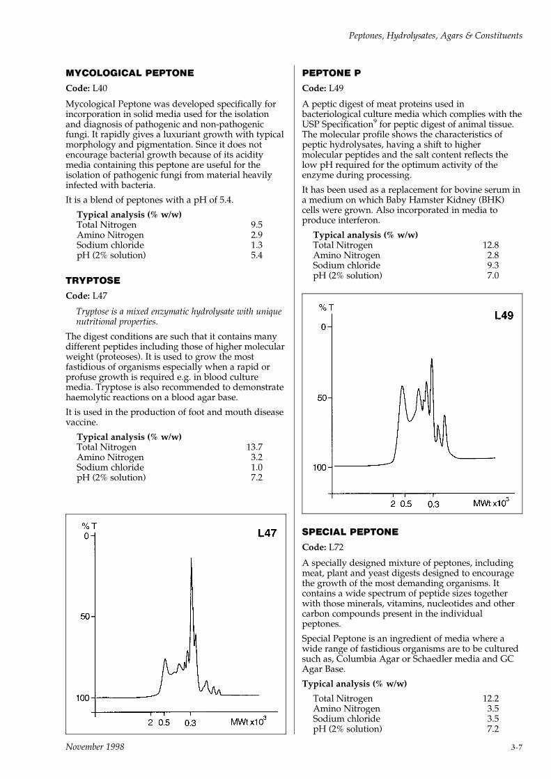
MYCOLOGICAL PEPTONE
Code: L40
MycologicaI Peptone was developed specifically forincorporation in solid media used for the isolationand diagnosis of pathogenic and non-pathogenicfungi. It rapidly gives a luxuriant growth with typicalmorphology and pigmentation. Since it does notencourage bacterial growth because of its aciditymedia containing this peptone are useful for theisolation of pathogenic fungi from material heavilyinfected with bacteria.
It is a blend of peptones with a pH of 5.4.
Typical analysis (% w/w)Total Nitrogen 9.5Amino Nitrogen 2.9Sodium chloride 1.3pH (2% solution) 5.4
TRYPTOSE
Code: L47
Tryptose is a mixed enzymatic hydrolysate with uniquenutritional properties.
The digest conditions are such that it contains manydifferent peptides including those of higher molecularweight (proteoses). It is used to grow the mostfastidious of organisms especially when a rapid orprofuse growth is required e.g. in blood culturemedia. Tryptose is also recommended to demonstratehaemolytic reactions on a blood agar base.
It is used in the production of foot and mouth diseasevaccine.
Typical analysis (% w/w)Total Nitrogen 13.7Amino Nitrogen 3.2Sodium chloride 1.0pH (2% solution) 7.2
PEPTONE P
Code: L49
A peptic digest of meat proteins used inbacteriological culture media which complies with theUSP Specification9 for peptic digest of animal tissue.The molecular profile shows the characteristics ofpeptic hydrolysates, having a shift to highermolecular peptides and the salt content reflects thelow pH required for the optimum activity of theenzyme during processing.
It has been used as a replacement for bovine serum ina medium on which Baby Hamster Kidney (BHK)cells were grown. Also incorporated in media toproduce interferon.
Typical analysis (% w/w)Total Nitrogen 12.8Amino Nitrogen 2.8Sodium chloride 9.3pH (2% solution) 7.0
SPECIAL PEPTONE
Code: L72
A specially designed mixture of peptones, includingmeat, plant and yeast digests designed to encouragethe growth of the most demanding organisms. Itcontains a wide spectrum of peptide sizes togetherwith those minerals, vitamins, nucleotides and othercarbon compounds present in the individualpeptones.
Special Peptone is an ingredient of media where awide range of fastidious organisms are to be culturedsuch as, Columbia Agar or Schaedler media and GCAgar Base.
Typical analysis (% w/w)
Total Nitrogen 12.2Amino Nitrogen 3.5Sodium chloride 3.5pH (2% solution) 7.2
Peptones, Hydrolysates, Agars & Constituents
November 1998 3-7

PROTEOSE PEPTONE
Code: L85
A specialised peptone prepared from a mixture ofpeptones. This product contains proteoses as definedin the United States Pharmacopoeia9. This has beenachieved by carefully controlling manufacturingconditions to achieve a product rich in the highermolecular weight peptides, (e.g. 4000 plus).
Proteose Peptone is especially suitable in media forCorynebacterium diphtheriae toxin, including that forthe Elek reaction for the recognition of toxigenicstrains, as well as in the media for the production oftoxins from staphylococci, clostridia and salmonellae.Media incorporating this peptone are suitable for thecultivation of different bacteria with a wide range ofnutritional requirements, e.g. Neisseria, Staphylococcus,Haemophilus, Salmonella, Pasteurella, Corynebacteriumand Histoplasma species.
It is the peptone used to manufacture diptheriatoxoid, pertussis vaccine and measles vaccine.
Typical analysis (% w/w)Total Nitrogen 13.0Amino Nitrogen 2.2Sodium chloride 8.0pH (2% solution) 7.0
4 VEGETABLE PEPTONE
SOYA PEPTONE
Code: L44
Soya Peptone is obtained by the papain hydrolysis ofsoya flour and complies with the USP specification(ref 9). In additon to its nitrogen constituents, thispeptone has a high carbohydrate content and issuitable for many purposes. The presence of thesugars stachyose, raffinose, sucrose and variousreducing sugars may be of importance in certainapplications. It is widely used in culture media and is
often used for the cultivation of many fastidiousorganisms and where rapid, luxuriant growth isrequired.
Typical analysis (% w/w)Total Nitrogen 9.1Amino Nitrogen 2.3Sodium chloride 0.4pH (2% solution) 7.2
5 CASEIN AND OTHER MILK DERIVEDPEPTONES
Oxoid manufactures a range of casein peptones andmilk derived peptones sourcing the raw materialsfrom countries whose disease status is acceptable.
By careful control of the hydrolysis conditions,products with widely differing physical, chemical andmicrobiological characteristics have been achieved.
Hydrolysis with acid is non specific, attacking allpeptide bonds and degrading proteins andpolypeptides to low chain length peptides and aminoacids. In the analytical data this is shown by a highamino nitrogen (compared to enzymatic digests) andthe presence of high levels of sodium chloride. Thelatter is caused by neutralisation of hydrochloric acidwith sodium hydroxide.
Acid hydrolysis destroys tryptophan and partiallydestroys cystine, serine and threonine. Asparagineand glutamine are converted to their acidic forms andvitamins are destroyed.
Lactalbumin Hydrolysate, Peptonised Milk, Tryptoneand Tryptone T are made by less severe forms ofhydrolysis using pancreatic enzymes on various milkfractions.
PEPTONISED MILK
Code: L32
This is a pancreatic digest of high grade skimmedmilk powder. It constitutes a source of nitrogen morereadily available than milk or milk powder and has ahigh level of carbohydrate. As with milk powder, thecalcium level is relatively high.
The product may be used on its own or in conjunctionwith other ingredients in media for isolation oflactobacilli and bacteriological examination of dairyproducts.
It has a high tryptophan content and is therefore usedin media for testing the indole reaction.
Typical analysis (% w/w)Total Nitrogen 5.3Amino Nitrogen 1.8Sodium chloride 1.6Tryptophan 0.53pH (2% solution) 6.5
Peptones, Hydrolysates, Agars & Constituents
3-8 November 1998

CASEIN HYDROLYSATE (ACID)
Code: L41
An hydrolysate prepared by the reaction of caseinwith hydrochloric acid at high temperature andpressure, followed by neutralisation with sodiumhydroxide. The aggressive hydrolysis conditionsrequire specialised processing to decolorise, toachieve a light coloured peptone. The highavailability of amino acids in their native form isadvantageous in many culture media formulationsand the molecular profile shows a definite shift to thelower molecular spectrum. It has particularcharacteristics which make it compatible for use insensitivity media and those applications where salttolerant organisms are used.
Typical analysis (% w/w)Total Nitrogen 8.2Amino Nitrogen 5.3Sodium chloride 30.2pH (2% solution) 7.0
TRYPTONE
Code: L42
Tryptone is a pancreatic digest of casein. It can beused in any formulation where a pancreatic or trypticdigest of casein is specified and complies with thespecification for pancreatic digest of casein in the U.S.Pharmacopoeia10. Casein is the main protein of milkand is a rich source of amino acid nitrogen. Theprofile shows a broad spread of peaks throughout themolecular weight range characteristic of a pancreaticdigest. This hydrolysate is often mentioned inpublished works, either as a constituent of culturemedia for metabolic or growth studies, or for otherpurposes where high performance and uniformity ofcomposition are of paramount importance. It has ahigh tryptophan content and is therefore used inmedia for testing the indole reaction.
Tryptone can detect `flat-sour' or `sulphide' spoilageorganisms in the canning industry and is also used insterility testing media.
It is a constituent of media used in fermentationprocesses to produce antibiotics, extra-cellularprotein, interferon and diphtheria toxoid.
Typical analysis (% w/w)Total Nitrogen 13.3Amino Nitrogen 3.7Sodium chloride 0.4pH (2% solution) 7.3
TRYPTONE T
Code: L43
This product was developed from Oxoid TryptoneL42 by controlled enzymatic hydrolysis and modifiedby the method of Meuller and Miller11. This producesa lower level of calcium, magnesium and iron than inTryptone which makes it ideal for the production oftoxin by Clostridium tetani.
Typical analysis (% w/w)Total Nitrogen 11.7Amino Nitrogen 3.5Sodium chloride 3.5Calcium 280ppmMagnesium 24ppmIron 3ppmpH (2% solution) 7.0
LACTALBUMIN HYDROLYSATE
Code: L48
After removal of casein from milk, lactalbumin is aprotein extracted from the resulting whey. L48 is apancreatic digest of this protein and contains highlevels of essential amino acids.
It is most commonly used in media for tissue cultureand therefore production of vaccines of viral originincluding foot and mouth disease, polio, dengue,coxsackie B3 and many other viruses.
Peptones, Hydrolysates, Agars & Constituents
November 1998 3-9

Other uses include growth of lactobacilli, sporegrowth of clostridia and in fermentation proceduresfor hormone production.
Typical analysis (% w/w)Total Nitrogen 12.5Amino Nitrogen 5.4Sodium chloride 0.2pH (2% solution) 6.5
6 EXTRACTS
YEAST EXTRACT
Code: L21
This is a dried yeast autolysate which is a good sourceof amino-nitrogen and vitamins, particularly thewater soluble B-complex vitamins. Its addition tomany media or fermentation broths increases theyield of organisms and is recommended where rapidand luxuriant growth is required.
Typical analysis (% w/w)Total Nitrogen 10.9Amino Nitrogen 5.3Sodium chloride 0.3pH (2% solution) 7.0
LAB-LEMCO
Code: L29
Lab-Lemco is a meat extract made from speciallyselected raw materials, adjusted to neutrality anddried to a fine powder. The product has considerableadvantages over conventional meat extracts. Being arefined and clarified extract it can be used with otherrefined ingredients to make culture media whichrequire no filtration. Being only slightly hygroscopicthis product is very easy to handle. Its use eliminatesthe troublesome procedures associated with handlingconventional meat extracts which have a paste-likeconsistency.
It will enhance the growth of many bacteria and istherefore incorporated into a wide range of culturemedia as a solid foundation material.
It is used in fermentation processes.
Typical analysis (% w/w)Total Nitrogen 13.3Amino Nitrogen 2.5Sodium chloride 1.1pH (2% solution) 7.2
MALT EXTRACT
Code: L39
This is prepared by extracting the soluble productsfrom sprouted grain followed by low temperatureevaporation to dryness which conserves thenitrogenous and carbohydrate constituents.
It is recommended for use in media for the growth ofyeast and moulds.
Typical analysis (% w/w)Total Nitrogen 1.1Amino Nitrogen 0.6Sodium chloride 0.1pH (2% solution) 5.6
References1 Haurowitz F. (1963)`The Chemistry & Function of Proteins' 2nd
Edition Academic Press.
2 Einarsson H., Snygg B. G., Ericsson G. (1983) J. Agric. Food Chem.
31. 10.
3 Dixon M., Webb E. C. (1979) `Enzymes'. 3rd Edition Longman, GP
Limited, page 892.
4 Dixon M., Webb E. C. (1979) `Enzymes', 3rd Edition Longman, GP
Limited, page 886.
5 `Guide to Good Pharmaceutical Manufacturing Practice' (1983)
Editor J. Sharp. Her Majesty's Stationery Office.
6 Bradstreet (1965) `The Kjeldahl Method for Organic Nitrogen'
Academic Press, New York.
7 Taylor (1957) Analyst 82. 488.
8 Adler-Nissen J. (1978) Ann. Nutr. Alim. 32. 205±216.
9 United States Pharmacopoeia (1985) 21st Revision p. 1396.
10 United States Pharmacopoeia (1985) 21st Revision pp. 1394±1396.
11 Meuller J. H. and Miller P. A. (1958) J. Bact. 67. 271±277.
7 PACKAGING AND SERVICES
All the products described are in the standard Oxoidrange and are available in the normal sales pack of250 grams or 500 grams. For large users they aresupplied in 2.5K, 5K and 25K containers.
Some products are available in syrup form in 50Kdrums. If supplied in this state, the solution has solidequivalent of between 55% and 67% depending on theproduct. Such a syrup would usually represent theend stage of manufacture immediately before drying.
Lot sizes vary from product to product, between 450Kto 3000K per lot.
Oxoid offer a sampling and reserve system wherebycustomers can reserve a bulk quantity of a productfrom which a sample is sent for them to test and aftersatisfactory results the same lot will be supplied.
Peptones, Hydrolysates, Agars & Constituents
3-10 November 1998
Products not in our normal range, can bemanufactured on request provided a minimum of100K is required. Customers interested in this serviceare asked to state their specifications and discuss thefeasibility of manufacturing any such product withtheir Oxoid contact.
LIVER DESICCATED
Code: L26
Dehydrated whole liver, specially manufactured forthe preparation of infusion media. Liver Desiccated isprepared by the dehydration of fresh ox livers undercarefully controlled conditions designed to ensuremaximum retention of nutritive properties, and isequivalent to five times its weight of fresh liver.
To prepare a liver infusion medium, add 50 grams ofLiver Desiccated to 1 litre of distilled water and allowto infuse (with frequent agitation) for 1 hour at 508C.Boil the mixture for a few minutes to coagulateprotein, strain through 60-mesh stainless steel gauze,add 10 grams of Peptone L34 and 5 grams of sodiumchloride. Adjust the reaction to pH 7.2, boil, strainthrough gauze as above, and sterilise by autoclavingat 1218C for 15 minutes.
8 USES OF OXOID PEPTONES, HYDROLYSATES AND BIOLOGICAL EXTRACTS
Meat Peptones Culture Antibiotic Ferment- Tissue Vaccine Special Notes
Media Manufacture ation Culture Manufacture
Neutral Liver Code L27 .
Bact. Pept. Code L37 . . . .
Bact. Pept. Neut. Code L34 . . . . Gives a clear solution
Mycological Pept. Code L40 . Has low pH
Tryptose Code L47 . . . Foot and mouth vaccine
Peptone P Code L49 . . Interferon productionDemonstrates H2S
Special Peptone Code L72 . Fastidious organisms
Proteose Peptone Code L85 . . Diphtheria toxoid
Vegetable Peptone
Soya peptone Code L44 . . . Antibiotic production
Casein and otherMilk Derived Products
Casein Hydrolysate (Acid)
Code L41 . . . . Antibiotic and vaccines
Peptonised Milk Code L32 . Dairy investigations
Tryptone Code L42 . . . . Diphtheria toxoid,AntibioticsInterferon production
Tryptone T Code L43 . Tetanus toxoidproduction
Lact. Hydrolysate Code L48 . . Spore formation
Hormone production
Extracts
Yeast Extract Code L21 . . . . Increases yield
Lab-Lemco Code L29 . . . . Easy to use meat extract
Malt Extract Code L39 . Yeast and mould media
Peptones, Hydrolysates, Agars & Constituents
November 1998 3-11

TABLE OF ANALYSIS
GENERAL ANALYSIS
Peptones, Hydrolysates, Agars & Constituents
3-12 November 1998
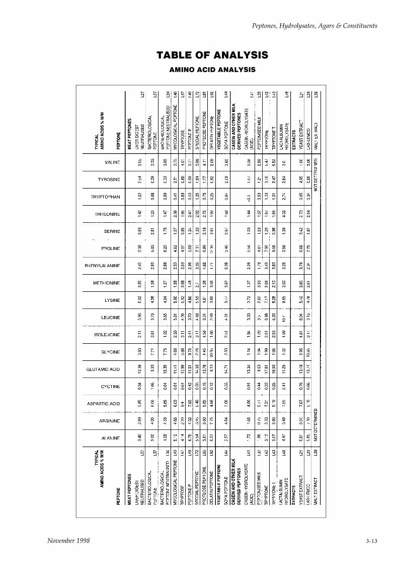
TABLE OF ANALYSIS
AMINO ACID ANALYSIS
Peptones, Hydrolysates, Agars & Constituents
November 1998 3-13
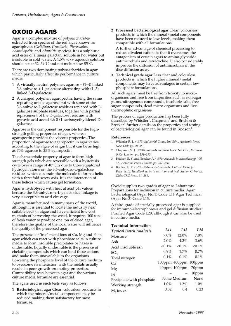
OXOID AGARSAgar is a complex mixture of polysaccharidesextracted from species of the red algae known asagarophytes (Gelidium, Gracilaria, Pterocladia,Acanthopeltis and Ahnfeltia species). It is a sulphuricacid ester of a linear galactan, soluble in hot water butinsoluble in cold water. A 1.5% w/v aqueous solutionshould set at 32±398C and not melt below 858C.
There are two dominating polysaccharides in agarwhich particularly affect its performance in culturemedia.
1 A virtually neutral polymer, agarose ± (1±4) linked3,6-anhydro-a-L-galactose alternating with (1±3)linked b-D-galactose.
2 A charged polymer, agaropectin, having the samerepeating unit as agarose but with some of the3,6-anhydro-L-galactose residues replaced with L-galactose sulphate residues, together with partialreplacement of the D-galactose residues withpyruvic acid acetal 4,6-0-(1-carboxyethylidene)-D-galactose.
Agarose is the component responsible for the high-strength gelling properties of agar, whereasagaropectin provides the viscous properties. Theproportion of agarose to agaropectin in agar variesaccording to the algae of origin but it can be as highas 75% agarose to 25% agaropectin.
The characteristic property of agar to form high-strength gels which are reversible with a hysteresiscycle over a range of 408C is due to three equatorialhydrogen atoms on the 3,6-anhydro-L-galactoseresidues which constrain the molecule to form a helixwith a threefold screw axis. It is the interaction ofthese helices which causes gel formation.
Agar is hydrolysed with heat at acid pH valuesbecause the 3,6-anhydro-a-L-galactoside linkage isvery susceptible to acid cleavage.
Agar is manufactured in many parts of the world,although it is essential to locate the industry nearsuitable beds of algae and have efficient low-costmethods of harvesting the weed. It requires 100 tonsof fresh water to produce one ton of dried agar,therefore the quality of the local water will influencethe quality of the processed agar.
The presence of `free' metal ions of Ca, Mg and Fe inagar which can react with phosphate salts in culturemedia to form insoluble precipitates or hazes isundesirable. Equally undesirable is the presence ofchelating compounds which can bind these cationsand make them unavailable to the organisms.Lowering the phosphate level of the culture mediumto overcome its interaction with the metals usuallyresults in poor growth-promoting properties.Compatibility tests between agar and the variousculture media formulae are essential.
The agars used in such tests vary as follows:
1 Bacteriological agar Clear, colourless products inwhich the mineral/metal components may bereduced making them satisfactory for mostformulae.
2 Processed bacteriological agar Clear, colourlessproducts in which the mineral/metal componentshave been reduced to low levels, making themcompatible with all formulations.
A further advantage of chemical processing toreduce divalent cations is that it overcomes theantagonism of certain agars to amino-glycosideantimicrobials and tetracycline. It also considerablyimproves the diffusion of antimicrobials in thedisc-diffusion assay.
3 Technical grade agar Less clear and colourlessproducts in which the higher mineral/metalcomponents may have advantages in certain low-phosphate formulations.
All such agars must be free from toxicity to micro-organisms and free from impurities such as non-agargums, nitrogenous compounds, insoluble salts, freesugar compounds, dead micro-organisms and livethermophilic organisms.
The process of agar production has been fullydescribed by Whistler1, Chapman2 and Bridson &Brecker3 further details on the properties and testingof bacteriological agar can be found in Bridson4.
References1 Whistler R. L. (1973) Industrial Gums, 2nd Edn., Academic Press,
New York, pp. 29±48.
2 Chapman V. J. (1970) Seaweeds and their Uses. 2nd Edn., Methuen
& Co. London. pp. 151±193.
3 Bridson E. Y. and Brecker A. (1970) Methods in Microbiology. Vol.
3A, Academic Press, London, pp. 257±266.
4 Bridson E. Y. (1978) Natural and Synthetic Culture Media for
Bacteria. In: Handbook series in nutrition and food. Section G. Vol III.
Ohio. CRC Press. 91±281.
Oxoid supplies two grades of agar as LaboratoryPreparations for inclusion in culture media: AgarBacteriological (Agar No.1) Code L11 Agar Technical(Agar No.3) Code L13.
A third grade of specially processed agar is suppliedfor immuno-electrophoresis and gel diffusion studies:Purified Agar Code L28, although it can also be usedin culture media.
Technical Information
Typical Batch Analysis L11 L13 L28
Moisture 7.0% 12.0% 7.0%
Ash 2.0% 4.2% 3.6%
Acid insoluble ash <0.1% <0.1% <0.1%
SO40.9% 1.7% 0.7%
Total nitrogen 0.1% 0.1% 0.1%
Ca 100ppm 400ppm 100ppm
Mg 40ppm 100ppm 70ppm
Fe ± ± 10ppm
Precipitate with phosphate None Medium None
Working strength 1.0% 1.2% 1.0%
Mr index 0.32 0.4 0.23
Peptones, Hydrolysates, Agars & Constituents
3-14 November 1998
The Electroendosmosis value (mr) of an agar gel isdefined as the relative mobility of dextran to that ofcrystalline human albumin. The lower the figure themore suitable the agar for electrophoresis studies.
AGAR BACTERIOLOGICAL (AGAR NO.1)
Code: L11
A processed bacteriological agar of very high workinggel strength (1% w/v) which has low Ca and Mglevels. It is compatible with all culture media and itenables broth and agar formulations of the samemedium to have very similar metal values. Thischaracteristic is especially valuable in antimicrobialMIC studies where differences in mineral/metalcontent can profoundly influence the results. It is alsoa highly satisfactory agar for antimicrobial diffusionstudies (disc diffusion susceptibility tests) because itslow mineral/metal content allows free diffusion ofantimicrobial substances.
AGAR TECHNICAL (AGAR NO.3)
Code: L13
A technical grade, high working gel strength agar(1.2% w/v) suitable for purposes where clarity andcompatibility are not of prime importance or wherethe high mineral/metal content has culturaladvantages. A technical grade agar which conformsto the specifications of the U.S. Pharmacopoeia andAPHA.
PURIFIED AGAR
Code: L28
An agar that has been extensively processed to give alow electroendosmosis factor (mr) enabling theproduct to be used in electrophoresis studies withoutthe high expense of using agarose preparations. It canalso be used for bacteriological culture media whereits special properties are required. An agarrecommended for immuno-electrophoresis and geldiffusion studies.
AGAR TABLETS (OXOID AGAR NO.3)
Code: CM49
DescriptionCompressed tablets of a pre-weighed amount of agarwhich are especially useful for solidifying liquidmedia or for the preparation of semi-solid media.Oxoid Agar Tablets are standardised to produce asatisfactory gel when dissolved in 10ml of liquid. Thiseliminates batch to batch variation and the differencesfound between one type of agar and another. The useof these tablets obviates weighing and filtration thusleading to considerable time saving.
TechniqueTo produce a satisfactory gel for plate cultures andslopes, add 1 tablet to every 10ml liquid medium andautoclave at 1218C for 15 minutes. With very acidmedia it is advisable to use 2 tablets to every 10±15mlof medium. In order to prepare semi-solid agar use 1tablet to every 100ml of liquid medium.
Peptones, Hydrolysates, Agars & Constituents
November 1998 3-15
BILE, BILE SALTS ANDDERIVATIVESBile is a product of the liver and its compositionvaries according to its animal source and its state ofpreservation. It contains bile pigments, bile acids infree and conjugate form, fatty acids, cholesterol,mucin, lecithin, inorganic salts, ethereal sulphates,glycuronic acids, urea and porphyrins. The role of theliver is to detoxify bile acids and it does this byconjugating them to glycine or taurine. A bile salt isthe sodium salt of a conjugated bile acid.
A. BILE DESICCANT
Prepared from freshly collected animal bile (ox orsheep) filtered, concentrated at low temperatures anddried. When reconstituted it should form clearsolutions with a brown-green colour at 10% w/vconcentration. This was the first bile additive used asa selective agent in culture media by MacConkey in1908.
Bile is a nutritive environment for a number of micro-organisms and putrefaction quickly occurs in theabattoir unless it is chilled or chemically preserved.The bacterial spoilage leads to deconjugation thusreleasing varying amounts of free bile acids. As theseacids are more toxic to bacteria than the conjugatesthere is considerable scope for variation in theselectivity of bile in culture media.
B. BILE SALTS
A further refinement in the use of bile is to precipitatethe bile salts with hydrochloric acid and separatethem from the bile pigments and other substances incrude bile.
Although this process yields bile salts, the productobtained may be a mixture of conjugates and free bileacids, depending on the quality of the bile used in theprocess.
The presence of more than 0.02% w/w desoxycholicacid in bile or bile salts indicates a degree of toxicityin the product which may make it unacceptable forcertain media e.g. EE Broth CM317 and MacConkeyAgar CM7.
Although bile-containing media are expected tosuppress Gram-positive organisms and allow onlybile-tolerant Gram-negative organisms to grow, somebile salts (free from bile acids) will allowstaphylococci and streptococci to grow. Particularadvantage is taken of this characteristic in the UnitedKingdom where MacConkey Agar CM7/CM7b isused as a general-purpose medium for clinicalbacteriology. The growth of faecal streptococci andstaphylococci would be looked for on this mediumwhen it is used for the culture of urine, faeces andpurulent material obtained from patients. Thesuppression of swarming growth of Proteus mirabilisin such specimens is a particular advantage of thismedium.
Satisfactory bile salts are presented as a fine, whitepowder which, when reconstituted as a 2% w/vsolution in distilled water, should be a clear, slightly
yellow solution at pH 6.0. When incorporated intoculture media, bile salts should not affect the colour ofthe indicator dyes or their subsequent change incolour. They should not form a surface scum ordeposit in culture media.
C. BILE ACIDS
Bile acids are derived from deconjugated bile salts.Although micro-organisms can deconjugate bilecompounds with great ease, the chemical processrequires high temperatures for long periods at a highpH value. Hydrolysed bile acids are then precipitatedwith acid, washed, neutralised and dried. Thepredominant and most important bile acid is sodiumdesoxycholate which can be separated from the bileacid mixture and used as a relatively pure salt.
Leifson1 showed that the constituent of bile whichhad greatest effect on bacterial growth wasdesoxycholic acid. He created desoxycholate-citrateagar when he discovered that the selectivity ofsodium desoxycholate is enhanced when magnesiumis removed from the medium. The role of sodiumcitrate in the medium is to chelate magnesiumcontributed by the other ingredients. It follows thatthe selectivity of the medium is lowered whenmagnesium is deliberately added to the formulation.This fact is important for the satisfactory growth ofshigellae in desoxycholate media.
The increased selectivity of hydrolysed bile salts (BileSalts No.3 or Bile Salts Mixture) is seen by thelowered concentration in culture media where it isadded at 1.5g/l in place of 5g/l for normal bile salts.It is normal practice, however, to titrate the level intoculture media by measuring the growth ofappropriate test organisms as an indicator.
It can be seen from these variable factors that theproduction of standard bile-containing media is adifficult process which requires much skill andexperience.
BILE SALTS
Code: L55
A standardised bile extract, consisting mainly ofsodium glycocholate and sodium taurocholate, foruse as a selective inhibitory agent in bacteriologicalculture media such as MacConkey Agar CM7 andMacConkey Broth CM5. Bile Salts L55 conforms tobacteriological requirements and batches arestandardised, with respect to inhibitory properties, bythe method of Burman1. It is generally employed inculture media at a concentration of 5 grams per litre.Bile Salts L55 meets the following specification:
Appearance ± a 2% aqueous solution is light strawcoloured, clear and free from extraneous matter.
Reaction ± (2% aqueous solution) ± pH 6 + 0.2.
Minimal Effective Concentration (Burman1) ±0.25% ± 0.5%.
Bacteriological Performance ± MacConkey Broth andAgar made up with L55 at the minimal effectiveconcentration (MEC) support satisfactory growthof bile-tolerant organisms including pathogenic
Peptones, Hydrolysates, Agars & Constituents
3-16 November 1998

staphylococci. Gas production by clostridia inbroths containing 0.5% of L55 is inhibited when theconcentration is raised to 2%, as in Brilliant GreenBile (2%) Broth CM31 thus eliminating falsepositives in the 448C test for Escherichia coli. Duringthe testing of milk for Escherichia coli with BrilliantGreen Bile (2%) Broth, false positives due to milklactobacilli do not occur if the medium containsL55.
Reference1 Burman N. P. (1955) Proc. Soc. Water Treat. Exam. 4. 10.
BILE SALTS NO.3
Code: L56
Oxoid Bile Salts No.3 was developed to meet thedemand for a refined bile salt for use as a selectiveinhibitory agent in bacteriological culture media. Itconsists of a specially modified fraction of bile acidsalts which is effective at less than one-third of theconcentration of bile salts normally quoted. Inselective media such as MacConkey Agar No.3, SSAgar and Violet Red Bile Agars the optimumconcentration of Bile Salts No.3 is 0.15% w/v. In suchmedia there is a very sharp differentiation betweenlactose-fermenters and non-lactose-fermenters ofenteric origin ± permitting the detection of scantynon-lactose-fermenters in the presence of numerouscoliforms.
CHEMICALS FOR CULTUREMEDIAThe following ingredients for culture media aresufficiently defined to be classed as chemical reagentsand would therefore be considered as reproducible.Nevertheless these products are subject to pre-shipment testing as part of a screening process andthe accepted batches tested before release by theQuality laboratory.
Glucose Bacteriological L71Gelatin Bacteriological L8Haemoglobin powder soluble L53Lactose Bacteriological L70Skim milk powder L31Sodium biselenite L121Sodium chloride Bacteriological L5Sodium glutamate L124Sodium thioglycollate L120
GLUCOSE (DEXTROSE)
Code: L71
A special bacteriological grade of anhydrous glucosefor use in culture media. Each batch is testedchromatographically to ensure purity and correctidentity.
GELATIN
Code: L8
Gelatin is a collagenous protein used for thesolidification of culture media and for the detectionand differentiation of certain proteolytic bacteria.Oxoid-Gelatin is a bacteriological grade which hasbeen manufactured and selected specifically for use inculture media. It is readily soluble in water to give aclear solution, free from sulphite and otherpreservatives. A satisfactory firm gel is obtained froma 15% solution, and media containing this proportionof gelatin will withstand short-term autoclaving for 15minutes at 1218C without significant loss of gelstrength.
SOLUBLE HAEMOGLOBIN POWDER
Code: L53
Prepare a 2% w/v solution of Soluble HaemoglobinPowder L53 by adding 250ml of distilled water at508C to 5g of Haemoglobin Powder. Continually stirthe solution during the addition of water. Sterilise byautoclaving at 1218C for 15 minutes.
LACTOSE BACTERIOLOGICAL
Code: L70
A special grade for inclusion in microbiologicalmedia. Each batch is tested chromatographically toensure purity and correct identity.
Peptones, Hydrolysates, Agars & Constituents
November 1998 3-17

SKIM MILK POWDER
Code: L31
The use of ordinary skim milk powder is undesirablein bacteriological media because of the presence ofheat-resistant organisms which give rise to erroneouscultural results. Oxoid Skim Milk Powder is a specialbacteriological grade of spray-dried skim milk freefrom thermophilic organisms.
Average analysis:Moisture 5.0%Ash 8.0%Total Nitrogen 5.3%Reducing Sugars(as lactose monohydrate) 48.0%Ether Soluble Extract 0.25%
Mix the powder to a smooth paste with a smallquantity of distilled water, then gradually add moredistilled water until a 10% w/v mixture is obtained.This is equivalent to fresh milk, and may be sterilisedby autoclaving for 5 minutes at 1218C. Care should betaken not to overheat during sterilisation, otherwisecaramelisation will occur. This product may be usedalone or as a constituent of more complexbacteriological culture media. A 10% `solution' ofSkim Milk Powder, containing 0.001% of bromo-cresol purple forms a highly satisfactory purple milkwhich may be employed for the cultivation of dairyorganisms or for the differentiation of Clostridiumspecies, etc. Media containing skim milk powder areof particular value for diagnostic cultural testsinvolving the fermentation of lactose and digestion orcoagulation of casein.
This product is not always free from antimicrobialresidues. Where antibiotic-free milk powder isspecified in a formulation, tests must be carried out todetermine if it is satisfactory.
SODIUM BISELENITE(Sodium hydrogen selenite)
Code: L121
For use in Oxoid Selenite Broth Base CM395/CM396and Mannitol Selenite Broth Base CM399.CAP SECURELY AFTER USE.
Dissolve 4 grams in 1 litre of distilled water and usethis solution to reconstitute the base medium ortablets.
Toxic by inhalation and if swallowed. Danger ofcumulative effects.
SODIUM CHLORIDE
Code: L5
See also Saline Tablets BR53.
This product is prepared from analytical grade salt toavoid problems associated with additives.
SODIUM GLUTAMATE
Code: L124
For use with Minerals Modified Medium Base(CM607).
SODIUM THIOGLYCOLLATE
Code: L120
For use in bacteriological culture media in order tolower their oxidation-reduction potential and toneutralise mercurial preservatives.
IRRITANT
Irritating to eyes, respiratory system and skin.
Harmful in contact with skin and if swallowed.
Peptones, Hydrolysates, Agars & Constituents
3-18 November 1998

4SELECTIVE SUPPLEMENTS,
STERILE REAGENTS,PREPARED MEDIA & DIP SLIDES

November 1998
SELECTIVE SUPPLEMENTSAND STERILE REAGENTS
SELECTIVE SUPPLEMENTS
Code: SR
Selective Supplements are freeze-dried, accuratepreparations of antimicrobials which are added,normally one vial per 500ml of sterile cooled medium,to base nutrient media to create specific, selectivemedia.
The concepts of antimicrobial selective microbiologyare now widely accepted and have proved theirvalue, particularly in clinical and food microbiologicallaboratories1.
A result of the use of these selective supplements isthat it is possible for non-specialised laboratories toisolate unusual but important organisms fromcontaminated clinical samples or food raw materials.Legionella, Listeria, Campylobacter, Yersinia, Brucella andBordetella species as well as many other `difficult'organisms can be isolated and confident statementsmade about their presence or absence in the materialexamined. Whilst the specialised laboratory can oftenfind their use makes the isolation procedure verymuch simpler than previous methods.
New and improved antimicrobial mixtures continueto be published and will be made available tolaboratories as soon as field trials have proved thevalue of the formulation.
Selection does not lie solely in the antimicrobialsupplement, it is often a combination of specificnutrient base with the supplement. Thus specifiedbase media formulations are paired with specificselective supplements and the results can bepredicted. More difficulty can be expected whereselective supplements are added to unspecified basemedia because some changes in media constituentscan strongly influence the selectivity of theantimicrobials.
The concentration of the antimicrobial mixture in thesupplement is based on the published papers ofworkers who have tested many strains of the selectedorganisms and who have measured their MIC valuesto ensure lack of inhibition. However, across theworld there exists the possibility that strains in othercountries may have MIC values that are different.
If there is doubt about the inhibitory properties ofselective media on local isolates then tests should beset up to make comparative quantitative assessmentson selective and non-selective media. The basenutrient medium can be prepared with and withoutselective supplement. The local strains can then beinoculated on the media in a quantitative way i.e.measured drop inocula. After incubation acomparative count is made. There should be less thanone log number difference between the counts on thetwo media. If a greater reduction in growth occurs onthe selective medium then the volume of anti-microbial solution should be reduced and testedagain. Where a reduction in volume of thesupplement occurs it would be useful to test a mixed
culture of common contaminants to ensure that thealtered level is still selective.
An example of a change in concentration ofantimicrobials can be seen in the description ofClostridium difficile Selective Medium CM601 page77. In this particular case a change in specimenprocessing i.e. an alcohol-shock treatment, helpedreduce contamination but appeared to make theclostridia more susceptible to full strengthantimicrobials. The facility to alter the concentrationsof the selective antimicrobials in the medium is one ofthe many advantages in using freeze-driedantimicrobial supplements.
Full details of the use of the Selective Supplements aregiven in Section 2 Culture Media. The details given inthis section cover the contents and the reconstitutionof the available supplements with directions to theappropriate part of Section 2.
Reference1 Bridson E. Y. (1982) Selective Culture Media ± a renewed activity.
Med. Lab. Sci. 39. 1±2.
GENERAL GUIDELINES TO THEPREPARATION OF SELECTIVE CULTUREMEDIA
The considerations that apply to the preparation ofgeneral purpose media require emphasis whenapplied to Selective culture media.
For optimal performance the medium must besensitive enough to detect the presence of very smallnumbers of organisms which may have been stressedor actually damaged. Different strains of organismsvary in their resistance to selective agents and in somecases the concentration of an agent may exceed or beclose to a level that a very sensitive strain will nottolerate. In such cases it is apparent that a distortedview of the presence and numbers of a givenorganism could be obtained. For this reason mediamust be chosen rationally to match their purpose andthe user should be familiar with the limitations of thedifferent formulations under the conditions of use.
The various components of a selective medium mustbe matched for best performance. For this reason theselective agent and the base must be considered as acompatible `set'. The combination of supplement andbase ensures that the end performance of the mediumwill meet the specification demanded. It is wellknown that identical quantities of selective substanceswill perform differently in different nutrient bases.This is because their disruptive activity in theorganism's metabolism is heavily influenced by thepresence of growth factors, mineral protectivesubstances, pH, etc.
Careful storage of dehydrated media is veryimportant and it does not always receive the attentionit deserves. Manufacturers' instructions regardingstorage in a cool, dry, dark place should be carefullyobserved and once a container has been opened thecontents should be used as soon as possible. Moisturemust be kept out. If not, the more hygroscopic mediamay change in weight, and thus alter the
Selective Supplements, Sterile Reagents
November 1998 4-1
concentration of the components in the final mediumand ± more serious still ± actual physical deteriorationmay occur, particularly if storage is in warmconditions. The effect of this on performance ofselective media would be considerable.
Some selective agents are photo-sensitive andalthough storage should be in the dark the problem isprobably of greater importance when the medium hasbeen reconstituted ready for use.
For best performance, preparation of the selectivemedium needs careful attention to the followingpoints:
1 Use only completely clean equipment. Residuesfrom incomplete cleaning can have a significanteffect.
Care must always be taken to prevent crosscontamination of one medium with another bydirty spatulae, balance pans, pipettes, etc.
2 The choice of water is important. Glass distilledwater is probably best, but de-ionised water issatisfactory. It should be remembered, however,that de-ionisers may harbour microbial flora, someof which may produce inhibitory substances.Tap water should not be used.
3 Weighing and measurement must be preciseotherwise pH and concentrations of theconstituents will be incorrect.
4 Finely powdered products can be scattered as dustand some selective agents could become a healthhazard if inhaled. Selective agents presented invials overcome this objection as the reconstitutionis done by adding the sterile distilled water orother solvent before the cap is removed.
5 The medium must be well mixed to ensure evendistribution, particularly of selective agents whichmay be present in very small amounts. Theaddition of a solution of the selective agent to thebasal medium makes proper distribution morecertain.
6 Carry out the sterilisation procedure meticulously,taking care not to overheat. In most cases a bettermedium will be obtained if the selective agent isadded aseptically after sterilisation.
7 Selective agents, including antibiotics, may belabile especially in solution. Antibiotics are bestkept dry until the time of use. Limited shelf life,unknown potency and cross-contamination are allspecial difficulties associated with antibiotics.
8 Media should be used as soon as possible afterpreparation, but if storage is needed this should bein cool, moist and dark conditions to keep changesin the medium to a minimum.
It was consideration of the special requirements ofselective media detailed above that led to theconception of the Oxoid range of freeze-driedsupplements, which have been carefully standardisedfor use with specified Oxoid dehydrated media. Thishas not only greatly increased convenience for theuser, but has virtually eliminated health hazards tothe laboratory preparations staff.
Oxoid Selective Media have the following features1 Labile and hazardous components are presented in
nitrogen-flushed glass vials.
2 The stability of critical components is assured. Along shelf life (up to two years in most cases) isguaranteed.
3 Each vial contains a measured dose sufficient for astandard volume of medium (usually 500ml). It iseasy to reconstitute.
4 The performance of the medium made with theselected basal medium and its compatiblesupplement will be reproducible.
5 Because the selective agents are not held for longperiods as solutions, but reconstituted immediatelybefore use, they always have optimal activity.
6 A futher advantage of this concept is that someflexibility of concentration is available. Althoughthe vials are designed to be emptied into standardvolumes of medium, there is individual choice ofusing less or more of the selective agent. In somecircumstances there may be advantages inchanging the concentration of the selective agent inorder to yield optimum recovery of the desiredorganism and suppression of other flora.
The benefits of using the Oxoid range ofSelective MediaThese are evident from features described above, butthey may be summed up as follows:
1 More reliable media with greatly reducedlikelihood that labile ingredients will havedeteriorated.
2 The work of the media preparation staff is madesafer and easier.
3 The microbiologist is free to concentrate on endproduct performance, confident of obtainingreproducible results.
SELECTIVE MICROBIOLOGYOxoid freeze-dried, selective and growth supplementsare used to prepare selective, differential or enrichedculture media. They are designed to give optimumperformance when added to the appropriate Oxoidbase media shown in the directions.
All supplements should be stored at 2±88C.
NOTE: before opening supplements containingcycloheximide read the comments under HAZARDSpage 2-7.
Selective Supplements, Sterile Reagents
4-2 November 1998
AMPICILLIN SELECTIVE SUPPLEMENT
Code: SR136
A selective supplement for the isolation of A.hydrophila when used with Aeromonas Medium Base(Ryan) CM833. See Section 2.
Vial contents (each vial is sufficient for 500ml ofmedium)
Ampicillin 2.5mg
DirectionsTo one vial add 2ml of sterile distilled water anddissolve the contents completely. Add the contentsaseptically to 500ml of Aeromonas Agar Base (Ryan)CM833 cooled to 508C. Mix well and pour into petridishes.
ANAEROBE SELECTIVE SUPPLEMENT G-N
Code: SR108
A selective supplement for the isolation of Gram-negative anaerobes when used with Wilkins-ChalgrenAgar CM619. See Section 2.
Vial contents (each vial is sufficient for 500ml ofmedium)
Haemin 2.5mgMenadione 0.25mgSodium succinate 1.25gNalidixic acid 5.0mgVancomycin 1.25mg
DirectionsTo one vial aseptically add 10ml of sterile distilledwater and mix gently to dissolve completely. Avoidfrothing. Add the contents to 500ml of sterile Wilkins-Chalgren Medium, prepared from Wilkins-ChalgrenAnaerobe Agar CM619 plus 5% (v/v) defibrinatedhorse blood SR50 or SR51. Do not add `Tween 80'.Mix gently and pour into sterile petri dishes.
ANAEROBE SELECTIVE SUPPLEMENT N-S
Code: SR107
A selective supplement for the isolation of Non-Sporinganaerobes when used with Wilkins-Chalgren AgarCM 619. See Section 2.
Vial contents (each vial is sufficient for 500ml ofmedium)
Haemin 2.5mgMenadione 0.25mgSodium pyruvate 500.0mgNalidixic acid 5.0mg
DirectionsTo one vial aseptically add 10ml of sterile distilledwater and mix gently to disolve completely. Avoidfrothing. Add the contents to 500ml of sterile Wilkins-Chalgren Medium, prepared from Wilkins-ChalgrenAnaerobe Agar CM619 plus 0.1% (v/v) `Tween 80'and 5% (v/v) defibrinated horse blood SR50 or SR51.Mix gently and pour into sterile petri dishes.
BACILLUS CEREUS SELECTIVESUPPLEMENT
Code: SR99
A selective supplement for the isolation andenumeration of B. cereus when used with Bacilluscereus Selective Agar CM617. See Section 2.
Vial contents (each vial is sufficient for 500ml ofmedium)
Polymyxin B 50,000 IU
DirectionsTo one vial aseptically add 2ml of sterile distilledwater and mix gently to dissolve the contentscompletely. Avoid frothing. Add to this 475ml ofsterile molten Bacillus cereus Agar Base CM617cooled to 508C, together with 25ml of sterile Egg YolkEmulsion SR47. Mix well and pour into sterile petridishes.
BORDETELLA SELECTIVE SUPPLEMENT
Code: SR82
A selective supplement for the isolation of Bordetellaspecies when used with Bordet-Gengou Agar BaseCM267 or Charcoal Agar CM119. See Section 2.
Vial contents (each vial is sufficient for 500ml ofmedium)
Cephalexin 20mg
DirectionsTo one vial add 2ml of sterile distilled water anddissolve the powder completely. Add the contentsaseptically to 500ml of sterile, molten Charcoal AgarCM119, cooled to 508C, together with 10%defibrinated horse blood SR50. Mix well beforepouring into sterile petri dishes.
Bordet-Gengou Agar CM267 may be used instead ofCharcoal Agar. Add the reconstituted contents of onevial to 500ml of Bordet-Gengou Agar cooled to 508C,to which 75±100ml of fresh defibrinated horse bloodSR50 has been added.
The vial contents may also be added to half strengthCharcoal Agar plus 10% defibrinated horse blood(SR50), for use as a transport medium for Bordetellapertussis.
BRUCELLA SELECTIVE SUPPLEMENT
Code: SR83
A selective supplement for the isolation of Brucellaspecies when used with Blood Agar Base No.2CM271, Columbia Agar CM331 or Brucella Agar BasesCM169 or CM691. See Section 2.
Vial contents (each vial is sufficient for 500ml ofmedium)
Polymixin B 2,500 IUBacitracin 12,500 IUCycloheximide 50mgNalidixic acid 2.5mgNystatin 50,000 IUVancomycin 10mg
Selective Supplements, Sterile Reagents
November 1998 4-3
DirectionsAdd 10ml of a 50:50 mixture of methanol and steriledistilled water to form a suspension. Incubate for 10±15 minutes at 378C. Mix thoroughly and immediatelyadd the vial contents to 500ml of sterile nutrientmedium cooled to 508C prepared from BrucellaMedium Base CM169 or CM691, Columbia Agar BaseCM331 or Blood Agar Base No.2 CM271 togetherwith 5±10% v/v inactivated horse serum SR35 and 1±5% w/v sterile solution of glucose. Mix well and pourinto sterile petri dishes.
CAMPYLOBACTER GROWTH SUPPLEMENT
Code: SR84
Supplement for the enhanced growth and aerotoleranceof campylobacter.
Vial contents (each vial is sufficient for 500ml ofmedium)
Sodium pyruvate 0.125gSodium metabisulphite 0.125gFerrous sulphate (hydrated salt) 0.125g
DirectionsTo rehydrate the contents of the vial, aseptically add2ml of sterile distilled water and invert to dissolve.Avoid frothing of solution.
Add the contents of one vial to 500ml of a sterilenutrient medium cooled to 50±558C prepared fromOxoid Columbia Agar CM331, Blood Agar Base No.2CM271, or Campylobacter Agar Base CM689, with5±7% lysed defibrinated horse or sheep blood, and therehydrated contents of one vial of CampylobacterAntibiotic Supplement SR69, SR85 or SR98. Mixgently and pour aseptically into sterile petri dishes.
CAMPYLOBACTER SELECTIVESUPPLEMENT (BLASER-WANG)
Code: SR98
A selective supplement for the isolation ofcampylobacters when used with Blood Agar BaseNo.2 CM271, Columbia Agar CM331 or Brucella AgarBases CM169 or CM691. See Section 2.
Vial contents (each vial is sufficient for 500ml ofmedium)
Vancomycin 5mgPolymixin B 1,250 IUTrimethoprim 2.5mgAmphotericin B 1.0mgCephalothin 7.5mg
DirectionsTo rehydrate the contents of the vial, aseptically add2ml of sterile distilled water and mix gently todissolve. Avoid frothing. Add the contents of one vialto 500ml of sterile nutrient medium cooled to 50±558Cprepared from Brucella Agar Base CM169 or CM691,Columbia Agar CM331 or Blood Agar Base No.2CM271, with 10% sheep blood or 5±7% laked horseblood SR48. Mix gently and pour into sterile petridishes.
CAMPYLOBACTER SELECTIVESUPPLEMENT (BUTZLER)
Code: SR85
A selective supplement for the isolation ofcampylobacters at 358C as well as 438C when used withBlood Agar Base No.2 CM271, Columbia Agar CM331or Brucella Agar Base CM169 or CM691. See Section 2.
Vial contents (each vial is sufficient for 500ml ofmedium)
Bacitracin 12,500 IUCycloheximide 25mgColistin sulphate 5,000 IUCephazolin sodium 7.5mgNovobiocin 2.5mg
DirectionsTo the contents of one vial aseptically add 3ml of50:50 ethanol/water and invert gently to dissolve.Avoid causing frothing. Add the contents of 1 vial to500ml of sterile blood agar cooled to 50±558Cprepared from Columbia Agar CM331, Blood AgarBase No.2 CM169 or CM691 with 5±7% defibrinatedhorse blood. Mix well and pour into sterile petridishes.
CAMPYLOBACTER SELECTIVESUPPLEMENT (PRESTON)
Code: SR117
A selective supplement for the isolation ofcampylobacters when used with Campylobacter AgarBase CM689 or Nutrient Broth Base No.2 CM67. SeeSection 2.
Vial contents (each vial is sufficient for 500ml ofmedium)
Polymixin B 2,500 IURifampicin 5mgTrimethoprim 5mgCycloheximide 50mg
DirectionsTo reconstitute add 2ml of 50:50 acetone/steriledistilled water and mix gently to dissolve completely.Avoid frothing. Add the vial contents to 500ml ofsterile basal medium prepared from CampylobacterAgar Base CM689 and 5% v/v Lysed Horse BloodSR48 or to 500ml of Enrichment Broth prepared fromNutrient Broth No.2 CM67, 5% v/v Lysed HorseBlood SR48 and Campylobacter Growth SupplementSR84.
CAMPYLOBACTER SELECTIVESUPPLEMENT (SKIRROW)
Code: SR69
A selective supplement for the isolation ofcampylobacters when used with Blood Agar Base No.2CM271, Columbia Agar CM331 or Brucella Agar BasesCM169 or CM691. See Section 2.
Selective Supplements, Sterile Reagents
4-4 November 1998
Vial contents (each vial is sufficient for 500ml ofmedium)
Vancomycin 5mgTrimethoprim 2.5mgPolymyxin B 1250 IU
DirectionsTo rehydrate the contents of the vial, aseptically add2ml of sterile distilled water and mix gently todissolve. Avoid frothing. Add the contents of one vialto 500ml of a sterile nutrient medium cooled to50±558C prepared from Brucella Agar Base CM169 orCM691, Columbia Agar CM331 or Blood Agar BaseNo.2 CM271, with 10% sheep blood or 5±7% lakedhorse blood SR48. Mix gently and pour into sterilepetri dishes.
CCDA SELECTIVE SUPPLEMENT
Code: SR155
An improved selective supplement for CampylobacterBlood Free Selective Agar CM739. See Section 2.
Vial contents (each vial is sufficient to supplement500ml of medium)
Cefoperazone 16mgAmphotericin B 5mg
DirectionsTo one vial add 2ml of sterile distilled water and mixgently to dissolve completely. Avoid frothing. Addthe contents to 500ml of sterile Blood FreeCampylobacter Selective Agar Base CM739 cooled to508C. Mix well and pour into sterile petri dishes.
CAMPYLOBACTER SELECTIVESUPPLEMENT (KARMALI)
Code: SR167
Vial contents:Haemin 16mg (equivalent to 32mg/l)Sodium pyruvate 50mg (equivalent to 100mg/l)Cefoperazone 16mg (equivalent to 32mg/l)Vancomycin 10mg (equivalent to 20mg/l)Cycloheximide 50mg (equivalent to 100mg/l)
DirectionsAdd 21.5 grams of Campylobacter Agar Base(Karmali) CM935 to 500mls of distilled water andbring to the boil to dissolve. Sterilise by autoclaving at1218C for 15 minutes. Cool to 508C. Aseptically add 1vial of Campylobacter Selective Supplement (Karmali)SR167 reconstituted with 2ml of sterile distilled water.Mix well and pour into sterile petri dishes.
DescriptionCampylobacter Medium (Karmali) is based on theformulation described by Karmali et al1 and isrecommended for the isolation of Campylobacter jejuniand Campylobacter coli from clinical specimens.
CEFOPERAZONE, AMPHOTERICIN B,TEICOPLANIN SUPPLEMENT (CAT)
Code: SR174
A selective supplement for the isolation of thermophilicCampylobacter spp. and improved recovery ofCampylobacter upsaliensis from faeces.
Vial contentsCAT Supplement Milligrams per litreCefoperazone 8.0Teicoplanin 4.0Amphotericin B 10.0
DirectionsAseptically add 4ml of sterile distilled water to thevial. Mix gently to resuspend the supplement.Prepare 500ml of sterile Blood Free CampylobacterAgar Base CM739 as directed. Cool to 508C andaseptically add one vial of SR174E reconstituted asdirected above. Mix well and pour the resulting CATmedium into sterile petri dishes. Incubate cultures at378C for 48±72 hours in a microaerobic atmosphere.
C-F-C SELECTIVE SUPPLEMENT
Code: SR103
A selective supplement for the isolation of lowtemperature pseudomonads when used withPseudomonas Agar Base CM559. See Section 2.
Vial contents (each vial is sufficient for 500ml ofmedium)
Cetrimide 5.0mgFucidin 5.0mgCephaloridine 25.0mg
DirectionsTo one vial aseptically add 2ml of sterile distilledwater and mix gently to dissolve the contentscompletely. Avoid frothing. Add the contents to500ml of sterile Pseudomonas basal medium, cooledto 50±558C, prepared from Pseudomonas Agar BaseCM559 plus 5ml of glycerol.
C-N SELECTIVE SUPPLEMENT
Code: SR102
A selective supplement for the isolation of Ps.aeruginosa when used with Pseudomonas Agar BaseCM559. See Section 2.
Vial contents (each vial is sufficient for 500ml ofmedium)
Cetrimide 100mgSodium nalidixate 7.5mg
DirectionsTo one vial aseptically add 2ml of sterile distilledwater and mix gently to dissolve the contentscompletely. Avoid frothing. Add the contents to500ml of sterile Pseudomonas basal medium, cooledto 50±558C, prepared from Pseudomonas Agar BaseCM559 plus 5ml of glycerol.
Selective Supplements, Sterile Reagents
November 1998 4-5
CHLORAMPHENICOL SELECTIVESUPPLEMENT
Code: SR78
A selective supplement for the isolation of yeasts andmoulds when used with Rose-Bengal ChloramphenicolAgar CM549, DRBC Agar Base CM727 or Dichloran-Glycerol (DG18) Agar Base CM729. See Section 2.
Vial contents (each vial is sufficient for 500ml ofmedium)
Chloramphenicol 50mg
DirectionsTo one vial add 3ml of acetone and dissolve thepowder completely. Add the contents to 500ml ofRose-Bengal Chloramphenicol Agar Base CM549 orother media requiring addition of chloramphenicol.Sterilise by autoclaving at 1218C for 15 minutes. Coolto 508C. Mix gently and pour into sterile petri dishes.
CLOSTRIDIUM DIFFICILE SELECTIVESUPPLEMENT
Code: SR96
A selective supplement for the isolation of C. difficilewhen used with Clostridium Difficile Agar BaseCM601. See Section 2.
Vial contents (each vial is sufficient for 500ml ofmedium)
D-cycloserine 250mgCefoxitin 8mg
DirectionsTo one vial aseptically add 2ml of sterile distilledwater and mix gently to dissolve the contentscompletely. Add to 500ml of sterile moltenClostridium Difficile Agar Base CM601 cooled to 508Ctogether with 7% v/v Defibrinated Horse Blood SR50.Mix well and pour into sterile petri dishes.
CLOSTRIDIUM DIFFICILE MOXALACTAMNORFLOXACIN (CDMN) SELECTIVESUPPLEMENT
Code: SR173
An alternative more selective supplement.
Vial contentsCDNM Supplement SR173 Milligrams per litreCysteine hydrochloride 500.0Norfloxacin 12.0Moxalactam 32.0
DirectionsAseptically add 2ml of sterile distilled water to eachvial of supplement and mix gently to dissolvecompletely. Avoid frothing. Prepare 500ml of sterileClostridium difficile Agar Base CM601 as directed.Cool to 508C and aseptically add 7% (v/v) ofDefibrinated Horse Blood SR50 and the contents ofone vial of SR173E (reconstituted as directed above).Mix well and pour the resulting CDMN Agar intosterile petri dishes.
CEFIXIME±TELLURITE SUPPLEMENT
Code: SR172
A freeze-dried supplement for use with SorbitolMacConkey Agar, CM813, for the selective isolation ofE. coli O157:H7.
Vial contents:Milligrams per litre
Potassium tellurite 2.5Cefixime 0.05
DirectionsAseptically add 2ml of sterile distilled water to 1 vialof Cefixime-Tellurite Supplement SR172E. Mix gentlyto dissolve the contents completely. Add the vialcontents to 500ml of Sorbitol MacConkey Agarprepared as directed and cooled to 508C. Mix welland pour the medium into petri dishes.
DERMASEL SELECTIVE SUPPLEMENT
Code: SR75
A selective supplement for the isolation of commondermatophyte fungi when used with Dermasel AgarCM539. See Section 2.
Vial contents (each vial is sufficient for 500ml ofmedium)
Cycloheximide 200mgChloramphenicol 25mg
DirectionsTo one vial add 3ml of acetone and mix gently todissolve completely. Add to 500ml of Dermasel AgarCM539. Mix gently and sterilise by autoclaving at1218C for 10 minutes. Avoid overheating at any time.
FRASER SUPPLEMENT
Code: SR156
A selective supplement for the isolation of Listeria spp.when used with Fraser Broth CM895. See Section 2.
Vial contents (each vial is sufficient to supplement500ml of medium)
Ferric ammonium citrate 0.25gNalidixic acid 10.0mgAcriflavine hydrochloride 12.5mg
DirectionsTo one vial add 5ml of ethanol/sterile distilled water(1:1) and mix gently to dissolve. Add the contents to500ml of sterile Fraser Broth Base CM895 cooled to508C. Mix well and distribute into sterile containers.
HALF FRASER SUPPLEMENT
Code: SR166E
A selective supplement for the isolation of Listeria spp.from food and environmental samples when used withFraser Broth CM895.
Vial contents (each vial is sufficient to supplement225ml of medium)
Ferric ammonium citrate 112.50mgNalidixic acid 2.25mgAcriflavine hydrochloride 2.8125mg
Selective Supplements, Sterile Reagents
4-6 November 1998
DirectionsSuspend 14.35 grams of Fraser Broth (CM895) in225ml of distilled water. Sterilise by autoclaving at1218C for 15 minutes. Cool to 508C and asepticallyadd the contents of one vial of Half Fraser SelectiveSupplement (SR166E) reconstituted with 4ml of (1:1)ethanol/sterile distilled water and mixed gently todissolve. Mix well and distribute into final containers.
GARDNERELLA VAGINALIS SELECTIVESUPPLEMENT
Code: SR119
A selective supplement for the isolation of G. vaginaliswhen used with Columbia Agar Base CM331.See Section 2.
Vial contents (each vial is sufficient for 500ml ofmedium)
Gentamicin sulphate 2.0mgNalidixic acid 15mgAmphotericin B 1.0mg
DirectionsTo reconstitute add 2ml of ethanol/sterile distilledwater (1:1) to one vial. Add aseptically the contents ofone vial to 450ml of sterile Columbia Blood Agar BaseCM331 cooled to 508C, and supplement with 50ml ofsterile human, rabbit or horse blood. Mix well. For thesurface inoculation technique pour into sterile petridishes and for the double layer technique place themedium in a water bath at 508C.
GC SELECTIVE SUPPLEMENT
Code: SR56
A selective supplement for the isolation of pathogenicneisseria when used with GC Agar Base CM367.See Section 2.
Vial contents (each vial is sufficient for 500ml ofmedium)
Yeast autolysate 5.0gGlucose 0.75gSodium bicarbonate 0.075gVancomycin 1.5mgColistin methane sulphonate 3.75mgNystatin 6,250 IUTrimethoprim 2.5mg
DirectionsTo one vial add 15ml of sterile distilled water(previously warmed to 378C to 458C) and shakevigorously until the contents are completelydissolved. Add the contents to 485ml of sterileThayer-Martin Medium, cooled to 50±558C, preparedfrom Oxoid GC Agar Base CM367 and 2% (w/v)Soluble Haemoglobin Powder L53.
HELICOBACTER PYLORI SELECTIVESUPPLEMENT (DENT)
Code: SR147
A selective supplement for the isolation of H. pylorifrom clinical specimens.
Vial contents (each vial is sufficient for 500ml ofmedium)
Vancomycin 5.0mgTrimethoprim 2.5mgCefsulodin 2.5mgAmphotericin B 2.5mg
DirectionsTo one vial add 2ml of sterile distilled water anddissolve the powder completely. Avoid frothing. Addthe contents aseptically to 500ml of sterile ColumbiaBlood Agar Base CM331 at 508C. Add 35ml of LakedHorse Blood SR48 and mix well before pouring intosterile petri dishes.
KANAMYCIN SULPHATE SELECTIVESUPPLEMENT
Code: SR92
A selective supplement for the isolation of enterococciwhen used with Kanamycin Aesculin Azide Agar BaseCM591 or Kanamycin Aesculin Broth Base CM771.See Section 2.
Vial contents (each vial is sufficient for 500ml ofmedium)
Kanamycin sulphate 10mg
DirectionsAdd 2ml of sterile distilled water to a vial and mixgently to dissolve completely. Add the contents to500ml of either Kanamycin Azide Agar Base CM591or Kanamycin Azide Broth Base CM771.
LCAT SELECTIVE SUPPLEMENT
Code: SR95
A selective supplement for the isolation of pathogenicneisseria when use with GC Agar Base CM367.See Section 2.
Vial contents (each vial is sufficient for 500ml ofmedium)
Lincomycin 0.5mgColistin sulphate 3.0mgAmphotericin B 0.5mgTrimethoprim 3.25mg
DirectionsTo one vial aseptically add 10ml of sterile distilledwater and mix gently to dissolve the contentscompletely. Avoid frothing. Add the contents to490ml of sterile GC Agar Base CM367, cooled to50±558C, containing 50ml Defibrinated Horse Blood(SR50) lysed with 0.5% (v/v) saponin, and therehydrated contents of one vial of Yeast AutolysateSupplement SR105.
Selective Supplements, Sterile Reagents
November 1998 4-7
LEGIONELLA BCYE GROWTHSUPPLEMENTS
Code: SR110
A growth supplement for the isolation of legionellaewhen used with Legionella CYE Agar Base CM655.See Section 2.
Vial contents (each vial is sufficient for 100ml ofmedium)
ACES Buffer/Potassium hydroxide 1.0gFerric pyrophosphate 0.025gL-cysteine HCl 0.04ga-ketoglutarate 0.1g
DirectionsTo one vial aseptically add 10ml of warm steriledistilled water and mix gently to dissolve the contentscompletely. Avoid frothing. Add the contents to 90mlof sterile Legionella CYE Agar Base CM655 cooled to50±558C. Mix gently and pour into sterile petri dishes.
Code: SR175
Supplement omitting L-cysteine, for presumptiveidentification of Legionella spp.
Vial contents (each vial is sufficient for 100ml ofmedium)
grams per litreACES Buffer/Potassium hydroxide 10Ferric pyrophosphate 0.25L-cysteine hydrochloride Nila-ketoglutarate 1.0
DirectionsPrepare in exactly the same way as for SR110 above.
LEGIONELLA BMPA SELECTIVESUPPLEMENT
Code: SR111
A selective supplement for the isolation of legionellaewhen used with Legionella BCYE Growth SupplementSR110 and Legionella CYE Agar Base CM655.See Section 2.
Vial contents (each vial is sufficient for 100ml ofmedium)
Cefamandole 400mgPolymyxin B 8,000 IUAnisomycin 8mg
DirectionsTo one vial add 2ml of sterile distilled water and mixgently to dissolve the contents completely. Avoidfrothing. Aseptically add the vial contents to 100ml ofBCYE-a Medium, cooled to 508C, prepared fromLegionella CYE Agar Base CM655 and LegionellaBCYE-a Supplement SR110. Mix well and pour intosterile petri dishes.
LEGIONELLA (GVPC) SELECTIVESUPPLEMENT
Code: SR152
A freeze-dried selective supplement for the isolation ofLegionella spp. from environmental water samples,when used with Legionella CYE Agar Base CM655 andLegionella BCYE-a Growth Supplement SR110.See Section 2.
Vial contents (each vial is sufficient to supplement500ml of BCYE-a Medium)
Glycine (Ammonia free) 1.5gVancomycin hydrochloride 0.5mgPolymyxin B sulphate 39600 IUCycloheximide 40.0mg
DirectionsTo one vial aseptically add 10ml of sterile distilledwater and mix gently to dissolve completely. Add thecontents to 500ml of sterile BCYE-a Medium(prepared using Legionella CYE Agar Base CM655 towhich one vial of Legionella BCYE-a GrowthSupplement SR110C has been added), cooled to 50±558C. Mix gently and pour into sterile petri dishes.
LEGIONELLA MWY SELECTIVESUPPLEMENT
Code: SR118
A selective supplement for the isolation of legionellaewhen used with Legionella BCYE Growth SupplementSR110 and Legionella CYE Agar Base CM655.See Section 2.
Vial contents (each vial is sufficient for 100ml ofmedium)
Glycine 0.3gPolymyxin B 5,000 IUAnisomycin 8mgVancomycin 100mgBromothymol blue 1mgBromocresol purple 1mg
DirectionsTo one vial add 2ml of sterile distilled water and mixgently to dissolve the contents completely. Avoidfrothing. Aseptically add the vial contents to 100ml ofBCYE-a Medium, cooled to 508C, prepared fromLegionella CYE Agar Base CM655 and LegionellaBCYE-a Supplement SR110. Mix well and pour intosterile petri dishes.
LISTERIA PRIMARY SELECTIVEENRICHMENT SUPPLEMENT (UVM I)
Code: SR142
A primary selective enrichment supplement for theisolation of listeria when used with Listeria EnrichmentBroth Base (UVM Formulation) CM863. See Section 2.
Vial contents (each vial is sufficient for 500ml ofmedium)
Nalidixic acid 10.0mgAcriflavine 6.0mg
Selective Supplements, Sterile Reagents
4-8 November 1998
DirectionsTo one vial add 2ml of sterile distilled water and mixgently to dissolve the contents completely. Avoidfrothing. Aseptically add the vial contents to 500ml ofsterile Listeria Enrichment Broth Base (UVMFormulation) CM863, below 508C. Mix well and pourrequired volumes into sterile containers.
LISTERIA SECONDARY SELECTIVEENRICHMENT SUPPLEMENT (UVM II)
Code: SR143
A primary selective enrichment supplement for theisolation of listeria when used with Listeria EnrichmentBroth Base (UVM Formulation) CM863. See Section 2.
Vial contents (each vial is sufficient for 500ml ofmedium)
Nalidixic acid 10.0mgAcriflavine 12.5mg
DirectionsTo one vial add 2ml of sterile distilled water and mixgently to dissolve the contents completely. Avoidfrothing. Aseptically add the vial contents to 500ml ofsterile Listeria Enrichment Broth Base (UVMFormulation) CM863, below 508C. Mix well and pourrequired volumes into sterile containers.
LISTERIA SELECTIVE ENRICHMENTSUPPLEMENT
Code: SR141
A selective enrichment supplement for the isolation oflisteria when used with Listeria Enrichment Broth BaseCM862. See Section 2.
Vial contents (each vial is sufficient for 500ml ofmedium)
Nalidixic acid 20.0mgCycloheximide 25.0mgAcriflavine 7.5mg
DirectionsTo one vial add 2ml of sterile distilled water and mixgently to dissolve the contents completely. Avoidfrothing. Aseptically add the vial contents to 500ml ofsterile Listeria Enrichment Broth Base CM862, below508C. Mix well and pour required volumes into sterilecontainers.
LISTERIA SELECTIVE SUPPLEMENT(OXFORD)
Code: SR140
A selective supplement for the isolation of listeria whenused with Listeria Selective Agar Base (OxfordFormulation) CM856. See Section 2.
Vial contents (each vial is sufficient for 500ml ofmedium)
Cycloheximide 200mgColistin sulphate 10mgAcriflavine 2.5mgCefotetan 1.0mgFosfomycin 5.0mg
DirectionsTo one vial add 5ml of ethanol/sterile distilled water(1:1) and mix gently to dissolve the contentscompletely. Avoid frothing. Aseptically add the vialcontents to 500ml of sterilised Listeria Selective AgarBase (Oxford Formulation) CM856, cooled to 508C.Mix well and pour into sterile petri dishes.
MSRV SELECTIVE SUPPLEMENT
Code: SR161
A selective supplement for the detection of motileSalmonella spp when used with MSRV Medium BaseCM910. See Section 2.
Vial contents (each vial is sufficient to supplement500ml of medium)
Novobiocin 10mg
DirectionsTo one vial add 2ml of sterile distilled water and mixgently to dissolve. Avoid frothing. Aseptically addthe vial contents to 500ml of MSRV Medium BaseCM910 cooled to 508C.
MYCOPLASMA SUPPLEMENT ± G
Code: SR59
A selective supplement for the isolation of mycoplasmaswhen used with Mycoplasma Agar or Broth BaseCM401/CM403. See Section 2.
Vial contents (each vial is sufficient for 80ml ofmedium)
Horse serum 20mlYeast extract (25% w/v) 10mlThallous acetate 25mgPenicillin 20,000 IU
DirectionsThe sterile supplement is prepared by asepticallyadding 20ml of sterile distilled water to the vial andmixing gently. Aseptically add the contents of the vialto 80ml of sterilised Mycoplasma Agar or Broth BaseCM401/CM403, previously cooled to 508C.
MYCOPLASMA SUPPLEMENT ± P
Code: SR60
A selective supplement for the isolation of Mycoplasmapneumoniae when used with Mycoplasma Agar BaseCM401. See Section 2.
Vial contents (per vial)Horse serum 6mlYeast extract (25% w/v) 3mlThallous acetate 0.008gGlucose 0.3gPhenol red 0.0012gMethylene blue 0.0003gPenicillin 12,000 IUMycoplasma Broth Base CM403 0.146g
Selective Supplements, Sterile Reagents
November 1998 4-9
DirectionsPrepare the sterile supplement by aseptically adding20ml sterile distilled water to the vial and mix gently.Add 1ml of Mycoplasma Base CM401 withoutsupplements to each of ten small bottles. Sterilise byautoclaving at 1218C for 15 minutes. Allow to set.Aseptically add 2ml of the reconstituted supplementP to each bottle containing agar.
OXYTETRACYCLINE GYE SUPPLEMENT
Code: SR73
A selective supplement for the isolation of yeasts andmoulds when used with Oxytetracycline Glucose YeastExtract Agar Base CM545. See Section 2.
Vial contents (each vial is sufficient for 500ml ofmedium)
Oxytetracycline (in a buffered base) 0.05g
NB: When re-constituted the resultant solution isphoto-sensitive. It is recommended the solution isadded immediately to the prepared agar base. Failureto do so may result in the solution becoming cloudy.
DirectionsTo one vial aseptically add 10ml of sterile distilledwater and dissolve the powder completely. Add thecontents to 500ml of Oxytetracycline-Glucose-YeastExtract Agar Base CM545, cooled to 508C. Mixthoroughly and pour into sterile petri dishes.
PALCAM SELECTIVE SUPPLEMENT
Code: SR150
A selective supplement for the isolation of Listeriamonocytogenes when used with PALCAM Agar BaseCM877. See Section 2.
Vial contents (each vial is sufficient for 500ml ofmedium)
Polymixin B 5mgAcriflavine hydrochloride 2.5mgCeftazidime 10mg
DirectionsTo one vial add 2ml of sterile distilled water and mixgently to dissolve. Avoid frothing. Aseptically addthe vial contents to 500ml of PALCAM Agar BaseCM877 cooled to 508C. Mix well and pour into sterilepetri dishes.
PERFRINGENS (OPSP) SELECTIVESUPPLEMENT A
Code: SR76
A selective supplement for the isolation of C. perfringenswhen used with SR77 and Perfringens Agar Base(OPSP) CM543. See Section 2.
Vial contents (each vial is sufficient for 500ml ofmedium)
Sodium sulphadiazine 50mg
DirectionsTo one vial add 2ml of sterile distilled water anddissolve the powder completely. Add the contentsaseptically to 500ml of Perfringens Agar Base (OPSP)
CM543 at 508C. Mix well before pouring into sterilepetri dishes.
PERFRINGENS (OPSP) SELECTIVESUPPLEMENT B
Code: SR77
A selective supplement for the isolation of C. perfringenswhen used with SR76 and Perfringens Agar Base(OPSP) CM543. See Section 2.
Vial contents (each vial is sufficient for 500ml ofmedium)
Oleandomycin phosphate 0.25mgPolymyxin B 5,000 IU
DirectionsTo one vial add 2ml of sterile distilled water anddissolve the powder completely. Add the contentsaseptically to 500ml of Perfringens Agar Base (OPSP)CM543 at 508C. Mix well before pouring into sterilepetri dishes.
PERFRINGENS (SFP) SELECTIVESUPPLEMENT
Code: SR93
A selective supplement for the isolation of C. perfringenswhen used with Perfringens Agar Base (TSC/SFP)CM587. See Section 2.
Vial contents (each vial is sufficient for 500ml ofmedium)
Kanamycin sulphate 6.0mgPolymyxin B 15,000 IU
DirectionsTo one vial add 2ml of sterile distilled water anddissolve the powder completely. Add the contentsaseptically to 500ml of sterile molten Perfringens AgarBase (TSC/SFP) CM587 cooled to 508C, together with25ml of Egg Yolk Emulsion SR47. Mix well beforepouring into sterile petri dishes.
PERFRINGENS (TSC) SELECTIVESUPPLEMENT
Code: SR88
A selective supplement for the isolation of C. perfringenswhen used with Perfringens Agar Base (TSC/SFP)CM587. See Section 2.
Vial contents (each vial is sufficient for 500ml ofmedium)
D-cycloserine 200mg
DirectionsTo one vial add 2ml of sterile distilled water anddissolve the contents completely. Add the contentsaseptically to 500ml Perfringens Agar Base (TSC/SFP)CM587 at 508C. Mix well before use.
Selective Supplements, Sterile Reagents
4-10 November 1998
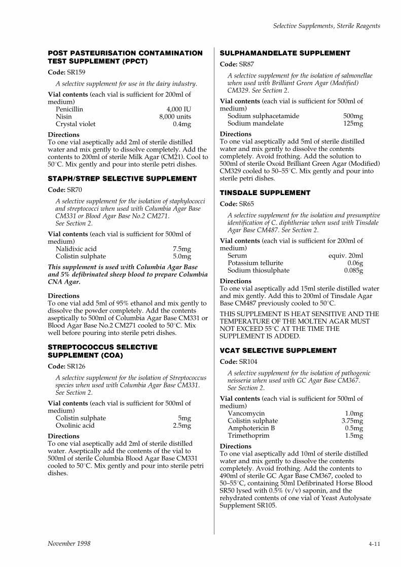
POST PASTEURISATION CONTAMINATIONTEST SUPPLEMENT (PPCT)
Code: SR159
A selective supplement for use in the dairy industry.
Vial contents (each vial is sufficient for 200ml ofmedium)
Penicillin 4,000 IUNisin 8,000 unitsCrystal violet 0.4mg
DirectionsTo one vial aseptically add 2ml of sterile distilledwater and mix gently to dissolve completely. Add thecontents to 200ml of sterile Milk Agar (CM21). Cool to508C. Mix gently and pour into sterile petri dishes.
STAPH/STREP SELECTIVE SUPPLEMENT
Code: SR70
A selective supplement for the isolation of staphylococciand streptococci when used with Columbia Agar BaseCM331 or Blood Agar Base No.2 CM271.See Section 2.
Vial contents (each vial is sufficient for 500ml ofmedium)
Nalidixic acid 7.5mgColistin sulphate 5.0mg
This supplement is used with Columbia Agar Baseand 5% defibrinated sheep blood to prepare ColumbiaCNA Agar.
DirectionsTo one vial add 5ml of 95% ethanol and mix gently todissolve the powder completely. Add the contentsaseptically to 500ml of Columbia Agar Base CM331 orBlood Agar Base No.2 CM271 cooled to 508C. Mixwell before pouring into sterile petri dishes.
STREPTOCOCCUS SELECTIVESUPPLEMENT (COA)
Code: SR126
A selective supplement for the isolation of Streptococcusspecies when used with Columbia Agar Base CM331.See Section 2.
Vial contents (each vial is sufficient for 500ml ofmedium)
Colistin sulphate 5mgOxolinic acid 2.5mg
DirectionsTo one vial aseptically add 2ml of sterile distilledwater. Aseptically add the contents of the vial to500ml of sterile Columbia Blood Agar Base CM331cooled to 508C. Mix gently and pour into sterile petridishes.
SULPHAMANDELATE SUPPLEMENT
Code: SR87
A selective supplement for the isolation of salmonellaewhen used with Brilliant Green Agar (Modified)CM329. See Section 2.
Vial contents (each vial is sufficient for 500ml ofmedium)
Sodium sulphacetamide 500mgSodium mandelate 125mg
DirectionsTo one vial aseptically add 5ml of sterile distilledwater and mix gently to dissolve the contentscompletely. Avoid frothing. Add the solution to500ml of sterile Oxoid Brilliant Green Agar (Modified)CM329 cooled to 50±558C. Mix gently and pour intosterile petri dishes.
TINSDALE SUPPLEMENT
Code: SR65
A selective supplement for the isolation and presumptiveidentification of C. diphtheriae when used with TinsdaleAgar Base CM487. See Section 2.
Vial contents (each vial is sufficient for 200ml ofmedium)
Serum equiv. 20mlPotassium tellurite 0.06gSodium thiosulphate 0.085g
DirectionsTo one vial aseptically add 15ml sterile distilled waterand mix gently. Add this to 200ml of Tinsdale AgarBase CM487 previously cooled to 508C.
THIS SUPPLEMENT IS HEAT SENSITIVE AND THETEMPERATURE OF THE MOLTEN AGAR MUSTNOT EXCEED 558C AT THE TIME THESUPPLEMENT IS ADDED.
VCAT SELECTIVE SUPPLEMENT
Code: SR104
A selective supplement for the isolation of pathogenicneisseria when used with GC Agar Base CM367.See Section 2.
Vial contents (each vial is sufficient for 500ml ofmedium)
Vancomycin 1.0mgColistin sulphate 3.75mgAmphotericin B 0.5mgTrimethoprim 1.5mg
DirectionsTo one vial aseptically add 10ml of sterile distilledwater and mix gently to dissolve the contentscompletely. Avoid frothing. Add the contents to490ml of sterile GC Agar Base CM367, cooled to50±558C, containing 50ml Defibrinated Horse BloodSR50 lysed with 0.5% (v/v) saponin, and therehydrated contents of one vial of Yeast AutolysateSupplement SR105.
Selective Supplements, Sterile Reagents
November 1998 4-11
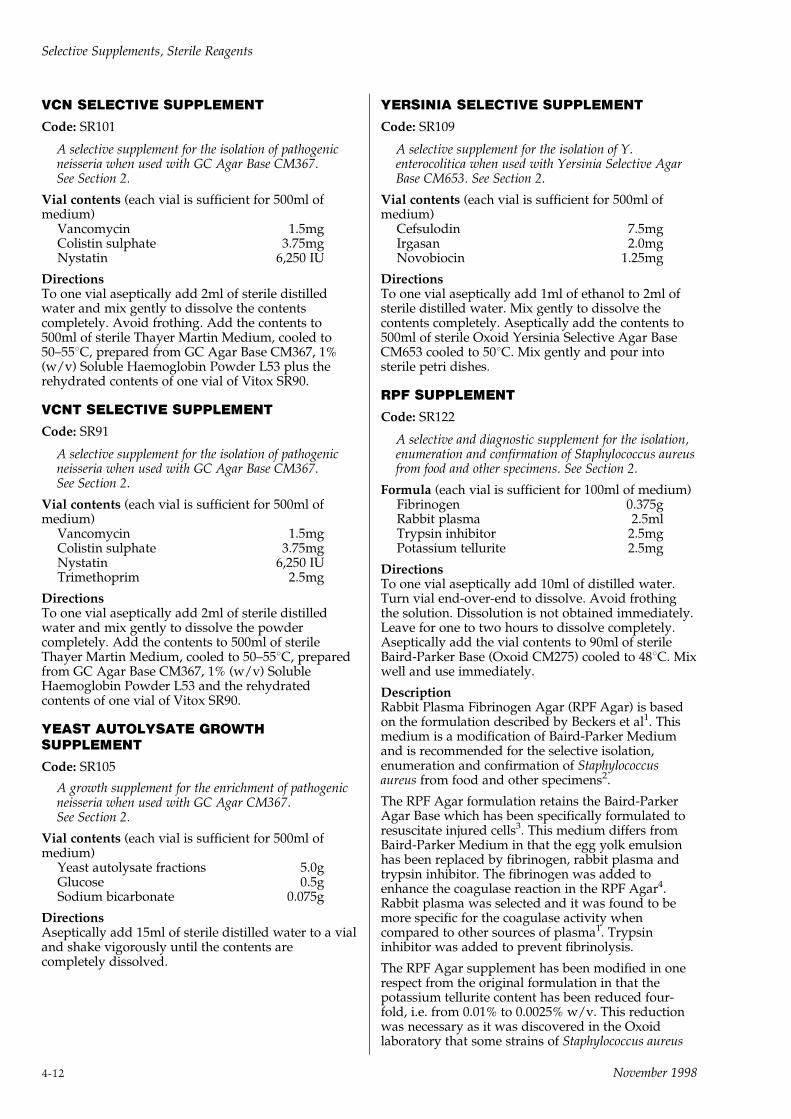
VCN SELECTIVE SUPPLEMENT
Code: SR101
A selective supplement for the isolation of pathogenicneisseria when used with GC Agar Base CM367.See Section 2.
Vial contents (each vial is sufficient for 500ml ofmedium)
Vancomycin 1.5mgColistin sulphate 3.75mgNystatin 6,250 IU
DirectionsTo one vial aseptically add 2ml of sterile distilledwater and mix gently to dissolve the contentscompletely. Avoid frothing. Add the contents to500ml of sterile Thayer Martin Medium, cooled to50±558C, prepared from GC Agar Base CM367, 1%(w/v) Soluble Haemoglobin Powder L53 plus therehydrated contents of one vial of Vitox SR90.
VCNT SELECTIVE SUPPLEMENT
Code: SR91
A selective supplement for the isolation of pathogenicneisseria when used with GC Agar Base CM367.See Section 2.
Vial contents (each vial is sufficient for 500ml ofmedium)
Vancomycin 1.5mgColistin sulphate 3.75mgNystatin 6,250 IUTrimethoprim 2.5mg
DirectionsTo one vial aseptically add 2ml of sterile distilledwater and mix gently to dissolve the powdercompletely. Add the contents to 500ml of sterileThayer Martin Medium, cooled to 50±558C, preparedfrom GC Agar Base CM367, 1% (w/v) SolubleHaemoglobin Powder L53 and the rehydratedcontents of one vial of Vitox SR90.
YEAST AUTOLYSATE GROWTHSUPPLEMENT
Code: SR105
A growth supplement for the enrichment of pathogenicneisseria when used with GC Agar CM367.See Section 2.
Vial contents (each vial is sufficient for 500ml ofmedium)
Yeast autolysate fractions 5.0gGlucose 0.5gSodium bicarbonate 0.075g
DirectionsAseptically add 15ml of sterile distilled water to a vialand shake vigorously until the contents arecompletely dissolved.
YERSINIA SELECTIVE SUPPLEMENT
Code: SR109
A selective supplement for the isolation of Y.enterocolitica when used with Yersinia Selective AgarBase CM653. See Section 2.
Vial contents (each vial is sufficient for 500ml ofmedium)
Cefsulodin 7.5mgIrgasan 2.0mgNovobiocin 1.25mg
DirectionsTo one vial aseptically add 1ml of ethanol to 2ml ofsterile distilled water. Mix gently to dissolve thecontents completely. Aseptically add the contents to500ml of sterile Oxoid Yersinia Selective Agar BaseCM653 cooled to 508C. Mix gently and pour intosterile petri dishes.
RPF SUPPLEMENT
Code: SR122
A selective and diagnostic supplement for the isolation,enumeration and confirmation of Staphylococcus aureusfrom food and other specimens. See Section 2.
Formula (each vial is sufficient for 100ml of medium)Fibrinogen 0.375gRabbit plasma 2.5mlTrypsin inhibitor 2.5mgPotassium tellurite 2.5mg
DirectionsTo one vial aseptically add 10ml of distilled water.Turn vial end-over-end to dissolve. Avoid frothingthe solution. Dissolution is not obtained immediately.Leave for one to two hours to dissolve completely.Aseptically add the vial contents to 90ml of sterileBaird-Parker Base (Oxoid CM275) cooled to 488C. Mixwell and use immediately.
DescriptionRabbit Plasma Fibrinogen Agar (RPF Agar) is basedon the formulation described by Beckers et al1. Thismedium is a modification of Baird-Parker Mediumand is recommended for the selective isolation,enumeration and confirmation of Staphylococcusaureus from food and other specimens2.
The RPF Agar formulation retains the Baird-ParkerAgar Base which has been specifically formulated toresuscitate injured cells3. This medium differs fromBaird-Parker Medium in that the egg yolk emulsionhas been replaced by fibrinogen, rabbit plasma andtrypsin inhibitor. The fibrinogen was added toenhance the coagulase reaction in the RPF Agar4.Rabbit plasma was selected and it was found to bemore specific for the coagulase activity whencompared to other sources of plasma1. Trypsininhibitor was added to prevent fibrinolysis.
The RPF Agar supplement has been modified in onerespect from the original formulation in that thepotassium tellurite content has been reduced four-fold, i.e. from 0.01% to 0.0025% w/v. This reductionwas necessary as it was discovered in the Oxoidlaboratory that some strains of Staphylococcus aureus
Selective Supplements, Sterile Reagents
4-12 November 1998
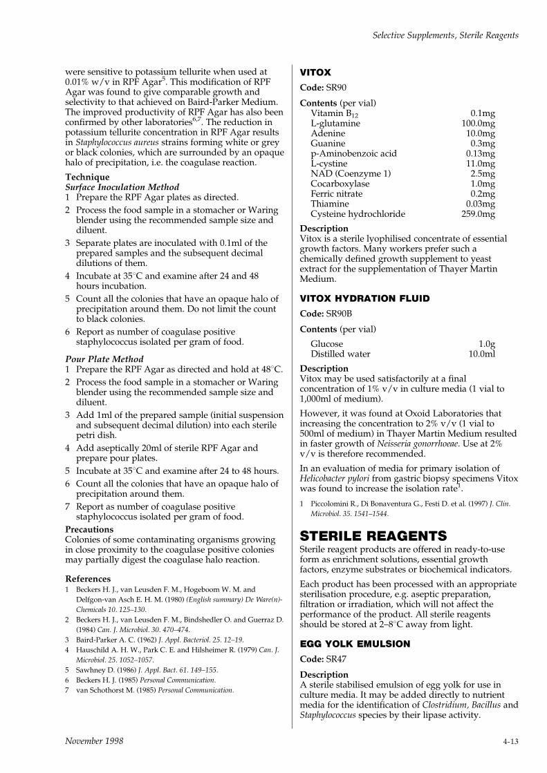
were sensitive to potassium tellurite when used at0.01% w/v in RPF Agar5. This modification of RPFAgar was found to give comparable growth andselectivity to that achieved on Baird-Parker Medium.The improved productivity of RPF Agar has also beenconfirmed by other laboratories6,7. The reduction inpotassium tellurite concentration in RPF Agar resultsin Staphylococcus aureus strains forming white or greyor black colonies, which are surrounded by an opaquehalo of precipitation, i.e. the coagulase reaction.
TechniqueSurface Inoculation Method1 Prepare the RPF Agar plates as directed.
2 Process the food sample in a stomacher or Waringblender using the recommended sample size anddiluent.
3 Separate plates are inoculated with 0.1ml of theprepared samples and the subsequent decimaldilutions of them.
4 Incubate at 358C and examine after 24 and 48hours incubation.
5 Count all the colonies that have an opaque halo ofprecipitation around them. Do not limit the countto black colonies.
6 Report as number of coagulase positivestaphylococcus isolated per gram of food.
Pour Plate Method1 Prepare the RPF Agar as directed and hold at 488C.
2 Process the food sample in a stomacher or Waringblender using the recommended sample size anddiluent.
3 Add 1ml of the prepared sample (initial suspensionand subsequent decimal dilution) into each sterilepetri dish.
4 Add aseptically 20ml of sterile RPF Agar andprepare pour plates.
5 Incubate at 358C and examine after 24 to 48 hours.
6 Count all the colonies that have an opaque halo ofprecipitation around them.
7 Report as number of coagulase positivestaphylococcus isolated per gram of food.
PrecautionsColonies of some contaminating organisms growingin close proximity to the coagulase positive coloniesmay partially digest the coagulase halo reaction.
References1 Beckers H. J., van Leusden F. M., Hogeboom W. M. and
Delfgon-van Asch E. H. M. (1980) (English summary) De Ware(n)-
Chemicals 10. 125±130.
2 Beckers H. J., van Leusden F. M., Bindshedler O. and Guerraz D.
(1984) Can. J. Microbiol. 30. 470±474.
3 Baird-Parker A. C. (1962) J. Appl. Bacteriol. 25. 12±19.
4 Hauschild A. H. W., Park C. E. and Hilsheimer R. (1979) Can. J.
Microbiol. 25. 1052±1057.
5 Sawhney D. (1986) J. Appl. Bact. 61. 149±155.
6 Beckers H. J. (1985) Personal Communication.
7 van Schothorst M. (1985) Personal Communication.
VITOX
Code: SR90
Contents (per vial)Vitamin B12 0.1mgL-glutamine 100.0mgAdenine 10.0mgGuanine 0.3mgp-Aminobenzoic acid 0.13mgL-cystine 11.0mgNAD (Coenzyme 1) 2.5mgCocarboxylase 1.0mgFerric nitrate 0.2mgThiamine 0.03mgCysteine hydrochloride 259.0mg
DescriptionVitox is a sterile lyophilised concentrate of essentialgrowth factors. Many workers prefer such achemically defined growth supplement to yeastextract for the supplementation of Thayer MartinMedium.
VITOX HYDRATION FLUID
Code: SR90B
Contents (per vial)
Glucose 1.0gDistilled water 10.0ml
DescriptionVitox may be used satisfactorily at a finalconcentration of 1% v/v in culture media (1 vial to1,000ml of medium).
However, it was found at Oxoid Laboratories thatincreasing the concentration to 2% v/v (1 vial to500ml of medium) in Thayer Martin Medium resultedin faster growth of Neisseria gonorrhoeae. Use at 2%v/v is therefore recommended.
In an evaluation of media for primary isolation ofHelicobacter pylori from gastric biopsy specimens Vitoxwas found to increase the isolation rate1.
1 Piccolomini R., Di Bonaventura G., Festi D. et al. (1997) J. Clin.
Microbiol. 35. 1541±1544.
STERILE REAGENTSSterile reagent products are offered in ready-to-useform as enrichment solutions, essential growthfactors, enzyme substrates or biochemical indicators.
Each product has been processed with an appropriatesterilisation procedure, e.g. aseptic preparation,filtration or irradiation, which will not affect theperformance of the product. All sterile reagentsshould be stored at 2±88C away from light.
EGG YOLK EMULSION
Code: SR47
DescriptionA sterile stabilised emulsion of egg yolk for use inculture media. It may be added directly to nutrientmedia for the identification of Clostridium, Bacillus andStaphylococcus species by their lipase activity.
Selective Supplements, Sterile Reagents
November 1998 4-13

TechniqueExamination of Bacteria for LecithinaseFor demonstration of lecithinase activity (especially inthe investigation of `bitty cream' conditions) add 0.5or 1.0ml of the emulsion to 10ml of sterile Blood AgarBase CM55 or Nutrient Broth No.2 CM67 ± in bothcases to clear the medium, raise the final saltconcentration by the addition of 1% of sodiumchloride. After incubation for up to 5 days at 358C,lecithinase-producers render the broth opalescent,whilst, on the solid medium, their colonies aresurrounded by zones of opacity.
EGG YOLK TELLURITE EMULSION
Code: SR54
DescriptionA sterile emulsion of egg yolk containing potassiumtellurite for use in Baird-Parker Medium CM275.Baird-Parker Medium is widely used in the foodindustry for the detection of pathogenicstaphylococci. Baird-Parker plates incorporating EggYolk Tellurite Emulsion should be protected frommoisture loss by enclosure in plastic or other vapourproof packaging.
DirectionsAdd 50ml to 1 litre of Baird-Parker Medium CM275.(50ml Egg Yolk Tellurite Emulsion contains theequivalent of 3ml of 3.5% potassium tellurite. This isthe amount recommended for 1 litre of Baird-ParkerMedium, i.e. concentration in SR54 is 0.21% w/v.Final concentration in Baird-Parker Medium is 0.01%w/v.)
FILDES PEPTIC DIGEST OF BLOOD
Code: SR46
DescriptionFildes Extract is prepared by the action of the enzymepepsin on defibrinated horse blood at optimumtemperature and pH value, as described by Fildes1. Itis a rich source of growth factors, including haematinand coenzyme, derived from the blood cells fromwhich it is prepared. As some of the growth factorsare thermolabile it should not be heated above 558C.
The extract is supplied in sterile screw-capped bottles.Add to the appropriate medium only after themedium has been sterilised.
TechniqueFildes Extract is recommended for the preparation ofmany culture media among which are the followingexamples:
Nagler MediumMelt 100ml sterile nutrient agar (Blood Agar BaseCM55 is recommended) and cool to 508C. Add 5ml ofFildes Extract and 20ml sterile serum or plasma. Pourplates and dry. Concentrated Egg Yolk EmulsionSR47 may be used in 5% concentration.
Fildes Broth (Fildes Peptic Blood Broth)Add 5ml of Fildes Extract to 100ml of sterile NutrientBroth No.2 CM67 or other liquid nutrient media.
Fildes Agar (Fildes Peptic Blood Agar)Melt 100ml of nutrient agar or Blood Agar BaseCM55, cool to 508C and add 5ml of Fildes Extract.
Fildes Broth and Agar, which are transparent andhave the colour of nutrient broth or agar, give copiousgrowths of Haemophilus influenzae and are admirablysuited for the primary isolation of this organism.
Lactose Egg-Yolk Milk Agar1,2 ± a medium for theidentification of anaerobes which, in addition toserving as a half antitoxin-Nagler plate, alsodemonstrates lactose fermentation and proteolysis.Egg Yolk Emulsion SR47 is recommended for use inthe preparation of the medium.
References1 Willis A. T. and Hobbs G. (1959) J. Path. Bact. 77. 511±521.
2 Willis A. T. (1977) Anaerobic Bacteriology 3rd Edn. Butterworths,
London.
TOMATO JUICE
Code: SR32
DescriptionThis product is the juice of ripe tomatoes, clarifiedand sterilised by filtration. It has a pH of 4.1 (approx)and 100ml of SR32 is equivalent to 227 grams oftomato. Tomato juice can be added to nutrient mediaas a specific growth stimulant for lactic acid bacteria.
STERILE SOLUTIONS OFCHEMICAL PRODUCTS FORCULTURE MEDIA
LACTIC ACID 10%
Code: SR21
POTASSIUM LACTATE 50%
Code: SR37
POTASSIUM TELLURITE 3.5%
Code: SR30
UREA 40%
Code: SR20
FREEZE-DRIED STERILEREAGENTS FORBACTERIOLOGICAL PURPOSES
BROAD SPECTRUM BETA-LACTAMASEMIXTURE
Code: SR113
Broad Spectrum Beta-Lactamase Mixture SR113 is amixture of beta-lactamase (E.C.3.5.2.6.) from Bacilluscereus1 569/H9.
The enzymes are presented as a sterile freeze-driedpowder containing buffer and zinc salts. Each vialcontains minima of 500 units beta-lactamase I and 50
Selective Supplements, Sterile Reagents
4-14 November 1998
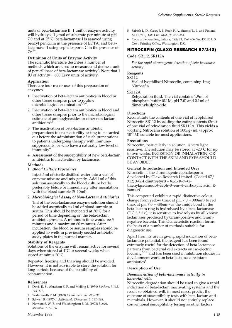
units of beta-lactamase II. 1 unit of enzyme activitywill hydrolyse 1 mmol of substrate per minute at pH7.0 and at 258C; beta-lactamase I is assayed usingbenzyl penicillin in the presence of EDTA, and beta-lactamase II using cephalosporin C in the presence ofZn2+.
Definition of Units of Enzyme ActivityThe scientific literature describes a number ofmethods which are used to measure and define a unitof penicillinase of beta-lactamase activity2. Note that 1IU of activity = 600 Levy units of activity.
ApplicationThere are four major uses of this preparation ofenzymes.
1 Inactivation of beta-lactam antibiotics in blood orother tissue samples prior to routinemicrobiological examination2,3,4.
2 Inactivation of beta-lactam antibiotics in blood andother tissue samples prior to the microbiologicalestimate of aminoglycosides or other non-lactamantibiotics4,5.
3 The inactivation of beta-lactam antibioticpreparations to enable sterility testing to be carriedout before the administration of such preparationsto patients undergoing therapy with immuno-suppressants, or who have a naturally low level ofimmunity6.
4 Assessment of the susceptibility of new beta-lactamantibiotics to inactivation by lactamase.
Methods1 Blood Culture Procedures
Inject 5ml of sterile distilled water into a vial ofenzyme mixture and mix gently. Add 1ml of thissolution aseptically to the blood culture bottle,preferably before or immediately after inoculationwith the blood sample (5±10ml).
2 Microbiological Assay of Non-Lactam Antibiotics
1ml of the beta-lactamase enzyme solution shouldbe added aseptically to 1ml of blood sample orserum. This should be incubated at 308C for aperiod of time depending on the beta-lactamantibiotic present. A minimum time would be 5minutes and a maximum 60 minutes. Afterincubation, the blood or serum samples should beapplied to wells in previously seeded antibioticassay plates in the normal manner.
Stability of ReagentsSolutions of the enzyme will remain active for severaldays when stored at 48C or several weeks whenstored at minus 208C.
Repeated freezing and thawing should be avoided.However, it is not advisable to store the solution forlong periods because of the possibility ofcontamination.
References1 Davis R. B., Abraham E. P. and Melling J. (1974) Biochem. J. 143.
115±127.
2 Waterworth P. M. (1973) J. Clin. Path. 26. 596±598.
3 Selwyn S. (1977) J. Antimicrob. Chemother. 3. 161±168.
4 Newson S. W. B. and Walshingham B. M. (1973) J. Med.
Microbiol. 6. 59±66.
5 Sabath L. D., Casey J. I., Ruch P. A., Stumpf L. L. and Finland
M. (1971) J. Lab. Clin. Med. 78. 457±463.
6 Code of Federal Regulations, Title 21, Part 436, Sec.436.20 U.S.
Govt. Printing Office, Washington, D.C.
NITROCEFIN (GLAXO RESEARCH 87/312)
Code: SR112, SR112A
For the rapid chromogenic detection of beta-lactamaseactivity.
ReagentsSR112
Vial of lyophilised Nitrocefin, containing 1mgNitrocefin.
SR112ARehydration fluid. The vial contains 1.9ml ofphosphate buffer (0.1M, pH 7.0) and 0.1ml ofdimethylsulphoxide.
DirectionsReconstitute the contents of one vial of lyophilisedNitrocefin SR112 by adding the entire contents (2ml)of one vial of rehydration fluid SR112A. This yields aworking Nitrocefin solution of 500mg/ml, (approx10-3 M) suitable for most applications.
PrecautionsNitrocefin, particularly in solution, is very lightsensitive. The solution may be stored at ±208C for upto two weeks. INGESTION OR INHALATION, ORCONTACT WITH THE SKIN AND EYES SHOULDBE AVOIDED.
General Introduction and Intended UsesNitrocefin is the chromogenic cephalosporindeveloped by Glaxo Research Limited. (Coded 87/312; 3-(2,4 dinitrostyrl) ± (6R,7R±7±(2-thienylacetamido)±ceph±3±em±4±carboxylic acid, E-isomer)1.
This compound exhibits a rapid distinctive colourchange from yellow (max at pH 7.0 = 390nm) to red(max at pH 7.0 = 486nm) as the amide bond in thebeta-lactam ring is hydrolysed by a beta-lactamase(E.C 3.5.2.6); it is sensitive to hydrolysis by all knownlactamases produced by Gram-positive and Gram-negative bacteria. This characteristic reaction formsthe basis of a number of methods suitable fordiagnostic use.
Apart from its use in giving rapid indication of beta-lactamase potential, the reagent has been foundextremely useful for the detection of beta-lactamasepatterns from bacterial cell extracts on iso-electricfocusing2,3,4 and has been used in inhibition studies indevelopment work on beta-lactamase resistantantibiotics5.
Description of Use
Demonstration of beta-lactamase activity inbacterial cells.Nitrocefin degradation should be used to give a rapidindication of beta-lactam inactivating systems and theresult so obtained will, in most cases, predict theoutcome of susceptibility tests with beta-lactam anti-microbials. However, it should not entirely replaceconventional susceptibility testing as other factors
Selective Supplements, Sterile Reagents
November 1998 4-15

also influence the results of such tests, and onoccasion intrinsic resistance to beta-lactamantimicrobials has not been correlated withproduction of beta-lactamase6.
Nitrocefin degradation has been found to be highlyefficient in detecting beta-lactamase producingisolates of Neisseria gonorrhoeae7,8, Haemophilusinfluenzae7,9,10,11 and staphylococci10,11.
Excellent results have also been obtained with certainanaerobic bacteria, notably with Bacteroidesspecies13,14,15. It should be emphasised that theefficacy of the Nitrocefin tests in predicting the beta-lactam susceptibilities of other micro-organisms is atpresent unproven.
Another chromogenic cephalosporin, PADAC(Hoechst-Roussel) was not as effective as Nitrocefin indetecting staphylococcal beta-lactamase12.
TechniqueRehydrate the Nitrocefin as directed, and use thissolution in the following ways:
1 Direct Plate Method1
Add one drop of the Nitrocefin solution on to thesurface of the colony. If the isolate is a high beta-lactamase producer then the colony and thesurrounding area will quickly turn red.
To detect a weak beta-lactamase producer the plateshould then be incubated for 30 minutes beforebeing reported as negative.
2 Slide Method1
Add one drop of the Nitrocefin solution on to aclean glass slide. Using a sterile loop, pick onecolony from the plate and emulsify into theNitrocefin drop. Report as positive if the colourchanges from yellow to red within 30 minutes(protect the slide from desiccation during thewaiting period).
3 Broth Method1
Add four drops of Nitrocefin solution to 1ml of thegrown culture. Report as positive if the colourchanges to red within 30 minutes.
4 Broken Cell Method1
Sonicate 1ml of the culture in order to break openthe cells. Add 4 drops of Nitrocefin solution.Report as positive if the colour changes to redwithin 30 minutes.
5 Paper Disc Spot Test10
A Whatman No.1 filter paper disc (diameter 7cm)is placed in a petri dish and impregnated withNitrocefin solution (0±5ml). This impregnatedpaper is generally usable for one day, but shouldbe kept away from light to avoid spontaneousdegradation. An isolated colony is applied to theimpregnated paper with a loop; a pink to redreaction developing within 15 minutes indicatesbeta-lactamase presence.
Detection of beta-lactamase activity on gelsMethods for preparing extracts containing the beta-lactamase activities from bacterial cells and thetechnique for analytical iso-electric focusing havebeen described by Matthew et al.2 The developed gels
are stained by applying Whatman No.54 paperimpregnated with the Nitrocefin solution2. Focusedbands in the gel with beta-lactamase activity appearpink on a yellow background.
Determination of beta-lactamase activity byspectrophotometric assayThe working solution of Nitrocefin (500mg/ml) isdiluted tenfold in buffer (0.1M phosphate; 1mMEDTA, pH 7.0). Spectrophotometric assays for beta-lactamase are carried out measuring changes inwavelength at 486nm. The molar extinction coefficientof Nitrocefin at this wavelength is 20,500.
Test samples of the finished product for performancewith control cultures.
References1 O'Callaghan C. H., Morris A., Kirby S. M. and Shingler A. H.
(1972) Antimicrob. Ag. & Chemother. 1. 283±288.
2 Mathew M., Harris A. M., Marshall M. J. and Ross G. W. (1975)
J. Gen. Microbiol. 88. 169±178.
3 Sparks J. and Ross G. W. (1981) J. Med. Microbiol. 15. p. iv.
4 King A., Shannon K. and Phillips I. (1980) Antimicrob. Ag. &
Chemother. 17. 165±169.
5 Guay R., Letarte R., Pechere J. C. and Roy B. (1980) IRCS Med.
Science 8. 209.
6 Markowitz S. M. (1980) Antimicrob. Ag. & Chemother. 6. 80±83.
7 Shannon K. and Phillips I. (1980) J. Antimicrob. Chemother. 6.
617±621.
8 Sng E. H., Yeo K. L., Rajan V. S. and Lim A. L. (1980) Br. J.
Vener. Dis. 56. 311±313.
9 Bell S. M. and Plowman D. (1980) Lancet i. 279.
10 Montgomery K., Raymundo L. and Drew W. L. (1979) J. Clin.
Micro. 9. 205±207.
11 Lucas T. J. (1979) J. Clin. Pathol. 32. 1061±1065.
12 Anhalt J. P. and Nelson R. (1982) Antimicrob. Ag. & Chemother.
21. 993±994.
13 Gabay E. L., Sutter V. L. and Finegold S. M. (1981) J. Antimicrob.
Chemother. 8. 413±416.
14 Timewell R., Taylor E. and Phillips I. (1981) J. Antimicrob.
Chemother. 7. 137±146.
15 Bourault A. M. and Rosenblatt J. E. (1979) J. Clin. Micro. 9. 654±
656.
PENASE
Code: SR129
569/H9 Lactamase active against a range of penicillins.
Materials SuppliedPenase SR129 is a Bacillus cereus 569/H9 lactamase(E.C.5.2.6) presented as a sterile freeze-dried powdercontaining buffer salts. Each vial contains 3,300 IU ofactivity (1 unit of enzyme activity will hydrolyse 1.0mmol of benzylpenicillin to benzylpenicilloic acid perminute pH 7.0 and at 258C). The preparation willsuccessfully inactivate a range of penicillins1.
Definition of Units of Enzyme ActivityThe scientific literature describes a number ofmethods which are used to measure and define a unitof penicillinase activity2. Note that 1 IU of activity =600 Levy units of activity.
ApplicationThe major use of this enzyme preparation is for theinactivation of susceptible beta-lactam antibiotic
Selective Supplements, Sterile Reagents
4-16 November 1998

preparations to enable sterility testing to be carriedout3.
The preparation may also be used for inactivation ofsusceptible antibiotics in blood or other tissuesamples prior to routine microbiologicalexamination2,4,5, and inactivation of susceptibleantibiotics in blood and other tissue samples prior tothe microbiological estimation of aminoglycosides orother non beta-lactam antibiotics6.
Methods1 Sterility Testing of Penicillin Products3
The product is rehydrated by adding 5ml of steriledistilled water to a vial of enzyme with gentlemixing. The resulting solution will contain 660 IUof activity/ml.
The sterility of penicillin products with respect tobacterial contamination is determined by adding300mg or less of the test sample to sterile FluidThioglycollate Medium CM173 or other prescribedmedia to which a suitable amount of Penasesolution has been added aseptically when thetemperature has fallen below 508C. Tubes (35mm x200mm) containing 90±100ml medium areincubated for 7 days at 308C.
The sterility of penicillin products with respect toyeast and moulds is accomplished by adding thetest sample to Sabouraud Liquid Medium CM147and incubating for 7 days at 20±258C.
2 Blood Culture Procedures0.1ml of the reconstituted solution should be addedaseptically to blood culture broths, preferablybefore but otherwise immediately after inoculationwith the blood sample.
Stability of ReagentsSolutions of the enzyme will remain active for severaldays when stored at 48C or several weeks whenstored at minus 208C. Repeated freezing and thawingshould be avoided. However, it is not advisable tostore the solution for long periods because of thepossibility of contamination.
References1 Melling J. (1979) `Antibiotic-Inactivating Enzymes' Ed. Wiseman A.,
`Topics in Enzyme and Fementation Technology', Vol.2. 153±199.
Publishers Ellis Horwood Ltd., Chichester.
2 Newson S. W. B. and Walshingham B. M. (1973) J. Med.
Microbiol. 6. 59±66.
3 Code of Federal Regulations, Title 21, Part 436, Sec. 436.20. U.S.
Govt. Printing Office, Washington, DC.
4 Waterworth P. M. (1973) J. Clin. Path. 26. 596±598.
5 Selwyn S. (1977) J. Antimicrob. Chemother. 3. 161±168.
6 Sabath L. D., Casey J. I., Ruch P. A., Stumpf L. L. and Finland
M. (1971) J. Lab. Clin. Med. 78. 457±463.
SPUTASOL
Code: SR89
Formula (per vial)
Dithiothreitol 0.1gSodium chloride 0.78gPotassium chloride 0.02gDisodium hydrogen phosphate 0.112gPotassium dihydrogen phosphate 0.02gpH 7.4 + 0.2
DirectionsTo one vial aseptically add 5ml of sterile distilledwater and mix gently to dissolve the contentscompletely. Aseptically add the contents to 95ml ofsterile distilled water. Use immediately or store at 48Cfor 48 hours only.
DescriptionSputum generally consists of inflammatory exudatefrom the lower respiratory tract mixed with saliva.
Mulder1 recognised the problem of interpreting thesignificance of growth from sputum and suggested.rinsing it in saline before culture to remove the saliva.May2 showed that bacteria are often unevenlydistributed in the sputum of patients suffering fromchronic bronchitis and that single cultures may fail toreveal all the bacterial species present.
The introduction by Rawlins3 of a method for thehomogenisation of sputum before culture overcamethe variations present in any method that is based onthe examination of small proportions ofheterogeneous material. It enables the bacteria in thesputum to be distributed evenly throughout thespecimen after digestion. Dixon and Muller4 in anattempt to distinguish between contaminants andbronchial pathogens, suggested a semi-quantitativeanalysis by diluting the digested sputum down to10-4.
Dithiothreitol, Cleland's Reagent5, has been evaluatedas a sputum liquefying agent6. It was found the mosteffective of a group of agents tested containing asulphydryl group.
A 0.1m solution of dithiothreitol was found to achievea significantly greater decrease in sputum viscositythan 1.2M N-acetyl cysteine for use prior to sputumculture.
The use of dithiothreitol instead of N-acetyl cysteineto digest sputum before decontamination has beenshown7 to yield a higher number of acid-fast bacilliwhen smears are stained by the Ziehl-Neelsenmethod. After culture and incubation for three weeksit was reported that in general the number and size ofcolonies isolated using dithiothreitol as a liquefyingagent was greater than that using N-acetyl cysteine.
TechniqueThe procedure for the routine liquefaction of sputumis as follows:
1 The sputum is expectorated into a sterile Universalcontainer or other wide mouthed screw-cappedbottle.
2 Add approximately 5 times the volume of 0.85%saline and agitate to free the sputum from adherentsaliva. Remove the saline with a sterile Pasteurpipette.
3 To the washed sputum, add an equal volume ofSputasol solution.
4 Shake the mixture well, place in a 378C water bathand incubate, with periodic shaking, untilliquifaction is complete.
5 Inoculate on to a suitable culture medium. For thetotal cell count, place a drop of the liquefiedsputum in a haemocytometer for enumeration. For
Selective Supplements, Sterile Reagents
November 1998 4-17
a differential cell count, fix a dried smear in methylalcohol and stain with haematoxylin and eosin orwith Lieshmann stain.
Reconstituted solutions of Sputasol, if kept sterile, arestable for at least 48 hours stored at 2±88C.
An investigation into the survival of respiratorypathogens in specimens that had been stored for 48hours at 48C following homogenisation usingSputasol, showed that the organisms remained viableand, when necessary, treated specimens could besuccesfully re-cultured8.
References1 Mulder J. (1938) Acta. Med. Scand. 94. 98.
2 May J. R. (1952) Lancet 20.12.52. 1206±1207.
3 Rawlins G. A. (1955) J. Med. Lab. Technol. 13. 133±143.
4 Dixon J. M. S. and Miller D. C. (1965) Lancet ii, 1046±1048.
5 Cleland W. W. (1964) Biochemistry 3. 480±482.
6 Hirsch S. R., Zastrow J. E. and Kory R. C. (1969) J. Lab. & Clin.
Med. 74. 346±353.
7 Shah R. R. and Dye W. E. (1965) Amer. Rev. Resp. Dis. 94. 454.
8 Could F. K., Freeman R., Hudson S. et al (1996) J. Clin. Pathol.
49. 684±686.
TTC SOLUTION (0.05%)
Code: SR148
TTC Solution is supplied as 5ml of filter-sterilisedaqueous solution of tri-phenyltetrazolium chloride(TTC). It is used to supplement Tergitol-7 AgarCM793 for the enumeration of coliforms in food andwater supplies.
READY PREPARED MEDIA ANDDIP SLIDESCoded PM, R and DS
These products are supplied in a form which is readyfor inoculation and incubation.
Storage conditions and Shelf lifePM and R products should be stored at 2±88C. DSproducts can be stored at low room temperature. Allproducts should be used before their expiry dateshown on the label.
Quality ControlThese products have been prepared and tested tohigh standards. Microbiological control tests, usingappropriate organisms, are made and the product isreleased only if it satisfies these tests. If the medium isstored as directed and used within the expiry date,then further tests are not required. However, if it is tobe used for purposes other than those describedbelow, then it would be wise to make suitable controltests to ensure growth of the expected organism.
PrecautionsAs with all culture media, these products should beused by trained staff only, in suitable laboratories anddisposed of safely by autoclaving at 1218C for at least20 minutes.
Mycobacterium tuberculosis (incl. M. bovis) is classifiedas a hazardous organism, specimens and culturesmust be processed in contained laboratories, using
approved protective cabinets. Staff should bespecially trained, tested for adequate immunity andmedically examined at appropriate intervals.
ACTIDIONE1 AGAR
Code: PM118
Formula gm/litreYeast extract 4.0Tryptone 5.0Glucose 50.0Potassium dihydrogen phosphate 0.55Potassium chloride 0.425Calcium chloride 0.125Magnesium sulphate 0.125Ferric chloride 0.0025Manganese sulphate 0.0025Bromocresol green 0.022Cycloheximide 0.01Agar 15.0Distilled water to 1 litrepH 5.5 + 0.2
DescriptionActidione1 (cycloheximide) at a concentration of0.001% w/v permits the growth of bacteria butinhibits the growth of most yeasts and moulds exceptdermatophytes. Media containing this antibiotic areinvaluable for the enumeration and detection ofbacteria in specimens containing large numbers ofyeasts and moulds. For example, the medium is usedfor the estimation of bacterial contamination ofpitching yeast. `Actidione' Agar with added penicillinand streptomycin is also valuable as a selectivemedium for the isolation of dermatophytes.
The Oxoid medium, based on that of Green andGray1, may be used for microbiological investigationsduring brewing and baking. As an approximateguide, incubation may be at 258C or 308C for up to 14days, according to the flora present. Green and Grayemployed their medium at two different reactions,pH 5.5 and pH 6.5, the latter may be attained byadding approximately 16ml of sterile 1.5% sodiumcarbonate to each litre of molten medium.
Reference1 Green S. R. and Gray P. P. (1950) Wallerstein Lab. Communication
13. 357.
SELENITE BROTH
Code: R39
Formula gm/litrePeptone 5.0Lactose 4.0Sodium biselenite 4.0Sodium phosphate 10.0Distilled water to 1 litre.pH 7.1 + 0.2
DescriptionSelenite Broth in ready-to-use form is prepared fromSelenite Broth Base CM395 with Sodium BiseleniteL121.
It is a modification of the original Leifson1 selenitebroth F, used as an enrichment broth for the isolation
Selective Supplements, Sterile Reagents
4-18 November 1998
of salmonellae from samples of faeces, urine or othersuspected material.
Incubation at 438C will improve the selectivity of themedium when large numbers of contaminatingbacteria are present2.
References1 Leifson E. (1936) Am. J. Hyg. 24. 423±432.
2 Fricker C. R. (1984) Z. Bakt. Micro. Hyg. Abt. I. Orig. B. 179.
170±178.
TRIBUTYRIN AGAR
Code: PM4
Formula gm/litrePeptone 5.0Yeast extract 3.0Tributyrin (glyceryl tributyrate) 10.0Agar 15.0Distilled water to 1 litrepH 7.5 + 0.2
DescriptionTributyrin Agar which consists of a stablehomogenate of nutrient agar and tributyrin (glyceryltributyrate) is used for the detection and enumerationof lipolytic (fat-splitting) bacteria and moulds.
The Oxoid medium is also frequently used for thedetection of the lipolytic activity of specific organismssuch as staphyloccoci, clostridia, pseudomonads andflavobacteria1,2,3,4,5,6.
TechniqueFor use, it is normally sufficient to prepare 1/10,1/100, 1/1,000 and 1/10,000 homogenates of themelted butter in quarter strength Ringer Solution and,within 10 minutes, to transfer 1ml quantities of eachdilution to separate sterile petri dishes. Add 10ml ofthe medium, melted and cooled to 45±488C, mix, andincubate at 308C for three days. After incubation, themedium appears opaque but lipolytic colonies aresurrounded by a zone of clear medium.
References1 Davey B. B. and Turner, Myfanwy (1961) J. Appl. Bact. 24(1).
78±82.
2 Hayes P. R. (1963) J. Gen. Microbiol. 30(1). 1±19.
3 Innes A. G. (1956) J. Appl. Bact. 19(1). 39±45.
4 Tanner F. W. (1944) `The Microbiology of Foods', 2nd ed., Garrard
Press, Illinois, pp. 474±479.
5 Willis A. T. (1960) J. Path. Bact. 80(2). 379±390.
6 Willis A. T. and Gowland G. (1965) Nature 187(4735). 432±433.
TRICHOMONAS MEDIUM NO.2
Code: R27
Formula gm/litreLiver digest 18.0Glucose 20.0Chloramphenicol 0.125Tryptone soya broth CM129 30.0Horse serum SR35 250mlCalcium pantothenate 0.5% w/v 1mlDistilled water 1,000mlpH 6.2 + 0.2
DescriptionTrichomonas Medium No.2 is a modification of themedia described by Bushby and Copp1 and Squiresand McFadzean2 for the isolation of Trichomonasvaginalis.
Clinical trials have established that TrichomonasMedium No.2 is very successful for the culture ofTrichomonas vaginalis from primary isolates. Acombination of laboratory culture and slideexamination provides the best chance of establishingthe presence of this pathogen.
Trichomonas Medium No.2 contains both serum andantibiotic and is therefore ready for immediateinoculation. It will store at 48C for several monthswithout deterioration.
Technique1 Inoculate the medium directly with vaginal swab,
sub-preputial scrape, urethral scrape, prostaticfluid, or the deposit from lightly centrifuged urine3.
2 Incubate at 348C4.
3 Examine microscopically every 24 hours.
4 Incubate microscopically negative cultures up tofive days.
References1 Bushby S. R. M. and Copp F. C. (1955) J. Pharm. Pharmacol. 7.
112±117.
2 Squires S. and McFadzean J. A. (1962) Brit. J. Vener. Dis. 38. 218±
219.
3 Stenton P. (1957) J. Med. Lab. Technol. 14. 228±230.
4 Thomas Patricia M. (1964) J. Med. Lab. Technol. 2. 46±50.
Selective Supplements, Sterile Reagents
November 1998 4-19
LOWENSTEIN-JENSEN MEDIUM
Code: PM1
Formula gm/litrePotassium dihydrogen phosphate 2.4Magnesium sulphate 0.24Magnesium citrate 0.6Asparagine 3.6Glycerol 12.0mlPotato starch 30.0Fresh egg mixture 1,000mlMalachite green 0.4Distilled water to 1640ml
DescriptionAn inspissated egg medium for the cultivation anddifferentiation of Mycobacterum species, especially M.tuberculosis and M. bovis. Growth of M. tuberculosis isenhanced by the incorporation of glycerol, whilst thegrowth of M. bovis and certain other mycobacteria isnot enhanced and may even be inhibited. The Oxoidmedium corresponds to the modification of theLowenstein1 medium developed by Jensen2.
TechniqueThe specimen is inoculated directly on to the surfaceof the medium; material which is contaminated withother micro-organisms, or which contains fewmycobacteria, may be subjected to one of thenumerous concentration methods before culturing.
It is preferable to inoculate duplicate slopes ofmedium with each specimen. Incubate at 358C andexamine after 10 to 14 days, and then every weekuntil the culture is discarded after a total of 8 weeks
incubation. Mycobacterium tuberculosis grows well toproduce large dry, heaped-up yellowish colonies,whilst M. bovis fails to grow or is only able to producesmall, `colourless', discrete colonies. For thecultivation of glycerol-sensitive mycobacteria, such assome strains of M. bovis, this medium is available asLowenstein-Jensen Medium (without glycerol) PM2.
References1 Lowenstein E. (1931) Zent. f. Bakt., Parasitenk. u. Infektionskr. I.
Abt. Orig. 120. 127±129.
2 Lowenstein E. (1932) Zent. f. Bakt., Parasitenk. u. Infektionskr. I.
Abt. Orig. 125. 222±239.
ACID EGG MEDIUM
Code: PM1a
Formula gm/litrePotassium dihydrogen phosphate 12.3Magnesium sulphate 0.3Glycerol 12.0mlPotato starch 30.0Fresh egg mixture 1000mlMalachite green 0.4Penicillin G 100,000 IUDistilled water to 1640ml
DescriptionA solid medium for use in a simplified method ofMycobacterium tuberculosis culture, described byMarks1. The method entails the use of an acid eggmedium which may be inoculated with an alkalinesputum homogenate which has been neitherneutralised nor centrifuged. This saves time,
METHODS AND MEDIUM FOR THE CULTURE OF TUBERCLE BACILLI(F. Zaher and J. Marks1)
MEDIA FOR THE ISOLATION OF TUBERCLE BACILLI
Method Digestion Technique Reaction of Zaher & Marks NewInoculum Media1 and Codes New Oxoid Media
Simple Mix specimen 1:1 with 4% NaOHfor digestion. No centrifugingbefore inoculation.
HighlyalkalinepH
Acid Egg Medium 1a
Acid Egg Medium 1b
Modified Acid EggMedium PM95Modified Pyruvic AcidEgg Medium PM96
StandardReference
Mix specimen 1:1 with 4% NaOHfor digestion then further dilutetotal volume 1:4 in distilled water.Centrifuge before inoculation.
ModeratelyalkalinepH
Acid Egg Medium 2a
Acid Egg Medium 2b
Standard Ref Acid EggMedium PM99Standard Ref PyruvicAcid Egg Medium PM100
Petroff Mix specimen 1:1 with 3% or 4%NaOH for digestion then centrifugeand neutralise before inoculation.
Neutral toslightlyacid pH
Simplified L-JMedium Marks 2a(without HCl).Simplified L-JMedium Marks 2b(without HCl).
Simplified Lowenstein-Jensen PM97
Simplified, PyruvateLowenstein-Jensen PM98
SulphuricAcid
Variable technique utilising H2SO4
concentrations of 2±5% for timeperiods 2±30 minutes dependent onspecimen and degree ofcontamination. Used for specimenswith small amounts of solids aftercentrifugation e.g. urines, CSF,pleural effusion.
Highlyacid pH
Simplified L-JMedium Marks 2a(without HCl)Simplified L-JMedium Marks 2b(without HCl)
Simplified Lowenstein-Jensen PM97
Simplified, PyruvateLowenstein-Jensen PM98
Selective Supplements, Sterile Reagents
4-20 November 1998
eliminates the need for an expensive centrifuge andreduces the risk of disseminating tubercle bacilli. Thevolume of medium (4.5ml) was designed to neutralisethe alkali in 0.2ml of homogenate and this ratioshould not be altered. Marks considered that hismethod was considerably more sensitive, andpossessed a much lower contamination rate thanthose described by the International Union againstTuberculosis2 and by Nassau3 and summarises `Withthe conditions recommended, this method isconsidered to be 99% as effective as a full-scalemethod with film-positive specimens and 98% withfilm-negative specimens'.
Technique (Marks1)1 Distribute 2ml amounts of sterile 4% NaOH into
sterile 1oz. universal containers.
2 With a sterile plugged glass tube, pipette 2mlamounts of sputum into the NaOH, avoidingcontact with the top half of the bottles. With scantyspecimens, first add sterile distilled water to theNaOH equivalent to the volume deficiency.
3 Shake the bottles briefly in a rack, avoidingcontamination of the caps, and place in a 358C dryincubator, shake for 20 seconds after 7�, 15 and (ifnecessary) 22�, minutes. Inspect the mixtures after15 and 22�, minutes, and each time immediatelyinoculate any which have become watery (ignorevery small particles). Inoculate after 30 minutes'incubation, whatever the consistency. There is thusa minimum of 15 and a maximum of 30 minutesincubation during homogenisation.
4 On their withdrawal from the incubator, inoculatethe alkaline homogenates directly into the AcidEgg Medium. For pipettes use sterile plugged glasstubes with an internal diameter of 4mm andmarked corresponding to 0.2ml before sterilisation.
Inoculate the Acid Egg Medium with 0.2ml ofhomogenate and tilt so that the inoculum runs overthe entire surface of the slope.
5 Incubate at 358C and examine every week until theculture is discarded after a total of 8 weeks'incubation.
References1 Marks J. (1959) Mon. Bull. Min. Hlth Pub. Hlth Lab. Serv. 18.
81±86.
2 Report (1955) Bull. Int. Union Tuberc. 25. 89.
3 Nassau E. (1958) Tubercle 39. 18±23.
PYRUVIC ACID EGG MEDIUM
Code: PM2A
Formula gm/litreFresh egg mixture 1000mlPyruvic acid 3.0 `Potassium dihydrogen phosphate 11.4Disodium hydrogen phosphate 6.0Magnesium sulphate 0.3Malachite green 0.125Penicillin G 100,000 IUDistilled water to 1640ml
DescriptionStonebrink1,2, demonstrated the value of pyruvic acidin the cultivation of mycobacteria and presentedevidence of enhanced growth of Myco. bovis anddysgonic strains of Myco. tuberculosis.
Marks3 extended this work and confirmed thefindings of Stonebrink, especially in the presence ofreduced malachite green concentration. The Oxoidmedium is made to Marks' formula.
Although Pyruvic Acid Egg Medium is superior toglycerol medium, Marks comments that the use ofboth media is to be recommended. This is becausepyruvic acid may discourage the growth of certainmycobacteria, especially Myco. kansasii.
Hughes4 confirmed Marks' comment that PyruvicEgg Acid Medium is superior to glycerol, inasmuchas 5% of his isolated strains grew on pyruvatemedium only.
Cruickshank5 recommended the use of Pyruvic AcidEgg Medium for the isolation of human strains thatare drug-resistant and difficult to grow.
References1 Stonebrink B. (1958) Acta Tuberc. Scand. 35. 67±74.
2 Stonebrink B. (1961) Selected Papers of the Royal Netherlands
Tuberculosis Association 2. 1±22.
3 Marks J. (1963) Mon. Bull. Min. Hlth and PHLS 22. 150±152.
4 Hughes M. H. (1966) J. Clin. Path. 19. 73±76.
5 Cruickshank R. (1965) `Medical Microbiology', 11th ed., p. 754,
Livingstone, Edinburgh.
IntroductionFollowing studies of the constitution of egg mediaused for the culture of tubercle bacilli (Zaher, 19772) itis possible to recommend a number of improvements.These will be presented in relation to current methodsof preparing specimens for culture because a surveymade by the authors has shown that laboratories notinfrequently use media unsuitable for their inocula.
Materials and Methods`Simple Method' for sputum culture (Marks3)This method was devised for laboratories withlimited resources and is not as efficient as the full-scale method described next. Nevertheless it is widelyused in Britain because it reduces work and hazard.Equal volumes of sputum and 4% NaOH areincubated at 358C for 15 to 30 minutes, depending onthe rate of liquefaction, and precisely 0.2ml isinoculated onto each slope. The medium nowrecommended for this method will be called Acid EggMedium, No.1; it is prepared in a glycerol version (a)and a pyruvate version (b).
Acid Egg Medium No.1a: 6.3g KH2PO4, 0.3gMgSO4.7H2O, 12.0ml glycerol are dissolved in 600mldistilled water and autoclaved at 1108C for 15minutes. To this solution the following are thenadded aseptically: 1100ml whole egg (mixed andstrained through gauze), 32ml N HCl, 11ml malachitegreen (2% w/v) and 100 000 IU of sodium penicillin.
The medium is dispensed in volumes within thelimits 4.5 to 5.0ml in 28ml bottles and inspissated by asingle heating. The oven is brought to 758C and held
Selective Supplements, Sterile Reagents
November 1998 4-21
between 75 and 858C for 45 minutes. The high eggcontent is needed to provide a firm slope.
Acid Egg Medium No.1b: This is prepared as above,but the 12.0ml glycerol are replaced with 7.0g ofsodium pyruvate.
Reference Laboratory method for sputum (Marks andThomas4)Two ml of 4% NaOH are made up to 4ml withsputum or in the case of scanty specimens, withsputum and sterile water. After incubation for 15 to30 minutes as above, the treatment bottle is held at anangle and 16ml of sterile water poured in from a 28ml(universal) container. Centrifugation and decantingfollow and the deposit is divided between two slopesand a film. The medium recommended for thismethod will be called Acid Egg Medium No.2; it is alsomade in glycerol (a) and pyruvate (b) versions.
Acid Egg Medium No.2a contains 2.4g KH2PO4, 0.3gMgSO4.7H20, 12.0ml glycerol and 600ml distilledwater. The ingredients are dissolved in the waterautoclaved at 1108C for 15 minutes. To this solutionthe following are then added aseptically: 1000mlwhole egg (mixed and strained through gauze), 40mlN HCl, 10ml malachite green (2% w/v) and 100 000IU of sodium penicillin. The medium is distributedand inspissated as for Acid Egg Medium No.1. In thepyruvate version (2b) the 12.0ml glycerol are replacedby 7.0g sodium pyruvate.
Petroff's method and modificationsIn the classical method of Petroff and in itsmodifications, sputum is incubated with an equalvolume of 3% or 4% NaOH. The mixture is thenneutralised, centrifuged and the deposit inoculated.Some workers centrifuge first, with or withoutpreliminary dilution, and neutralise the deposit. Themedium recommended for this method is calledSimplified Lowenstein-Jensen (L-J) and it is made inglycerol and pyruvate versions.
Simplified L-J Medium This medium has the sameformula as Acid Egg Medium No.2 except that thehydrochloric acid is omitted and the volume ofmalachite green solution is increased to 20ml.
Sulphuric acid treatmentApart from sputum and the deposit of gastriccontents, most specimens are best treated withsulphuric acid as described by Marks5. However, theReference Laboratory has now reduced the maximumreagent concentration of 5% to 4% (v/v).
Treatment is followed by dilution, centrifugation anddecanting which leaves a moderately acid deposit forinoculation. The medium recommended for thismethod is Simplified L-J Medium.
ResultsThe preliminary experiments which led to theimprovements of medium formulation is citedelsewhere (Zaher 1977). However, the results of threefinal trials are presented here. The `simple method' ofsputum culture used to compare Acid Egg MediumNo.1 with the `acid egg medium' originally devisedfor this method of culture. Glycerol and pyruvateversions of both media were used in parallel for 50specimens selected as likely positives. The new
formula gave earlier or heavier growth in 25 casesand the original medium in 3 cases, a significantadvantage of the new medium (P<0.001); growth wasequal in 14 cases and absent in the remainder.
The Reference Laboratory's method for sputum wasused to compare Acid Egg Medium No.2b with thepyruvate version of the method's original `bufferedegg medium'. This trial was conducted on unselectedroutine specimens. The new formula gave earlier orheavier growth in 102 cases and the original mediumin 23 cases, which is again a significant advantage tothe new medium (P<0.001); growth was equal in 62cases. All the media in these two trials were given thesame low content of malachite green (approximately0.0125%) as the object was to isolate the effect ofphosphate concentration.
The third trial compared 0.0125% and 0.025%malachite green in Acid Egg Medium No.2b usingunselected specimens. With the weaker dye, growthwas earlier or heavier in 32 cases and with thestronger dye in 7 cases, a significant advantage to thelower concentration of malachite green in thismedium (P<0.001); growth was equal in 26 cases.
DiscussionFour procedures for preparing specimens for cultureare described above and these provide respectivelyinocula which are (1) highly alkaline, (2) moderatelyalkaline, (3) neutral or slightly acid (by over-shoot)and (4) moderately acid. Lowenstein-Jensen Mediumand its simplified versions are suitable for the inoculain groups (3) and (4) which vary from neutral tomoderately acid, but the pH of this medium rises toan unfavourable level with alkaline inocula. Themedia originally recommended for the methodsproviding alkaline inocula depended on phosphatebuffer for control of the pH.
Experiments preliminary to the present work showedthat the concentrations of phosphate used weredetrimentally high. In the new media, phosphate hasbeen partially replaced by HCl. Malachite green ismore antibacterial at lower pH levels and the presentwork has confirmed that lower concentrations thanusual should be used in acid egg medium.
The Simplified L-J Medium described above is based onearlier work aimed at simplifying Lowenstein-JensenMedium (Marks and Thomas4, Stonebrink (1961)6)and has not been subjected to further trials. Credit forthe introduction of pyruvate medium is due toStonebrink6. We advise that most types of specimenshould be cultured on both glycerol and pyruvatemedium as tubercle bacilli vary in their preference.
References1 Zaher F. and Marks J. (1977) Tubercle 58. 143±145. Reproduced by
permission of the Authors and the Editor and Publisher of Tubercle.
2 Zaher F. (1977) Factors promoting the growth of tubercle bacilli in
artificial culture. M.Sc. Thesis, University of Wales.
3 Marks J. (1959) A simple method for the cultivation of tubercle bacilli.
Monthly Bulletin of the Ministry of Health and the PHLS, 18. 81.
4 Marks J. and Thomas C. H. H. (1958) Notes on the cultivation of
tubercle bacilli. Monthly Bulletin of the Ministry of Health and the
PHLS, 17. 194.
5 Marks J. (1972) Endiong the routine guinea-pig test. Tubercle, 53. 31.
Selective Supplements, Sterile Reagents
4-22 November 1998

6 Stonebrink B. (1961) A new medium for the cultivation of
Mycobacterium tuberculosis. Selected Papers. The Royal Netherlands
Tuberculosis Association, 2. 1±22.
THE OXOID DIP SLIDECode: DS
Kass1 defined significant bacteriuria on the basis ofcolony counts of carefully collected, freshly voided,total specimens of urine obtained from populations ofasymptomatic women. He found that, on the basis ofthe counts obtained, it was possible to distinguishbetween infected and contaminated urine. When thecount was more than 105 organisms per ml in a singlespecimen, the probability was about 80 per cent thatinfection was present. If the observation was repeatedin a further specimen, the probability of infection wasgreater than 90 per cent.
Since these original observations it has becomeapparent that when carefully collected, mid-streamurine samples are obtained from symptomaticpatients, the colony counts required for the diagnosisof urinary tract infection are lower than those definedby Kass for asymptomatic populations2,3. A count ofmore than 102 organisms per ml can now be regardedas diagnostic in a woman with acute symptoms ofurinary tract infection, while in men the diagnosticcount is 103 organisms per ml. In most circumstances,however, the counts obtained will be greater than 105
organisms per ml.
OXOID DIP SLIDE REGRESSION SLOPE
Since the original observations by Kass many culturaland chemical methods have been devised to measurethe bacterial population of urine.
All these methods depend on freshly voided mid-stream urine promptly delivered to the bacteriologylaboratory for examination, either immediately orafter brief refrigerated storage.
Mackey and Sandys4 devised the method of filling aspoon with a solid medium and using this as a dip-inoculum transport medium, thus avoiding erroneouscounts that may result from bacterial growth in theurine between collection and examination in thelaboratory.
The `Dip Slide' was a logical development of thismethod5,6. By use of a microscope slide coated oneach side with a different culture medium larger areasof medium were available to obtain a semi-quantitative measure of the bacterial count.
Oxoid investigated the dip slide and concluded thatthough the basic principle was satisfactory, usefulchanges could be made in the design. The difficulty ofhandling microscope dip slides withoutcontaminating the media, the relatively thin layers ofthe coating agar and the uneven draining of the urineon to a paper disc in contact with the bottom of theslide, were disadvantages not evident with the dipspoon.
The principle is simple. An agar-coated slide isprovided, attached to the cap of a screw-capped
container. By dipping the slide into freshly voidedurine, reliable counts can be obtained. These show thenumber of organisms present in the urine at the timeof voiding.
The method has the following advantages:
1 The Dip Slide technique is the simplest, most rapidand most reliable method for determining thepresence or absence of urinary tract infection.
2 Counts on a Dip Slide have been shown tocorrelate closely with counts made by carefulquantitative methods in the laboratory6,8,9,10.
3 Since the slide is dipped into freshly voided urine,the counts obtained reflect the number oforganisms present at the time the urine waspassed. It is well-known that, as a result of itsgrowth supporting properties, urine held for sometime at room temperature before examinationcannot be relied upon to yield reliable colonycounts.
4 It is usually a simple matter for the practised eye,when the Dip Slide is used, to distinguish betweenurinary infection and contamination from othersources.
5 The time required by the medical laboratoryscientist to examine each specimen is minimal.Screening of large numbers of specimens can beachieved almost at a glance.
6 The Dip Slide inoculation technique is sufficientlysimple that it can be carried out at home by thepatient.
7 The Dip Slide technique can be used equally for theexamination of urine from infants and children.
METHOD OF USE
The object is to wet both sides of the Oxoid Dip Slidewith a mid-stream specimen of urine. This is done bycollecting a mid-stream specimen in a sterile containerand then dipping the slide into the urine. Afterdipping, the slide is held vertically for a moment toallow any surplus urine to drain from its lower end,after which the slide is returned into its sterilecontainer.
Arniel11,12 devised a `dip-stream' technique that isparticularly suitable for children but can equally beused with adults. The patient commences micturitionand, when the stream is well-established, the Dip Slideis held into it sufficiently to wet each side thoroughly.The slide is then allowed to drain briefly and isreturned into its sterile container. The Dip Slide dip-stream technique is completely reliable andinexpensive6,7 and can be used for large-scale screening.
With both methods care must be taken to ensure thatthe agar surface is not touched and patients must becarefully instructed about the technique they shouldfollow13. It is helpful if the vulva are held apartduring the collection of mid-stream urine, butcleansing is not important. If it is done for any reason,antibacterial soaps and antiseptics must be avoided.
The patient's name and details of the date and time ofcollection are noted on the label and the containerwith the Dip Slide is either posted or taken to the
Selective Supplements, Sterile Reagents
November 1998 4-23
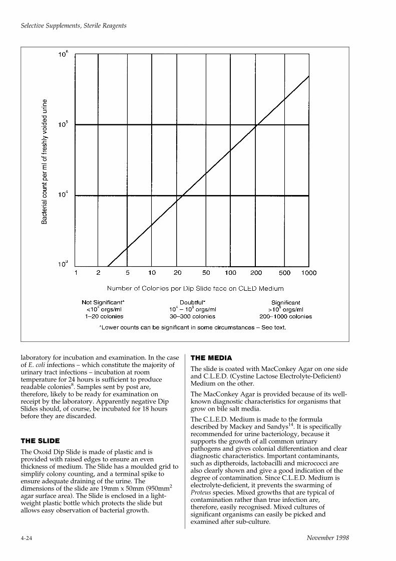
laboratory for incubation and examination. In the caseof E. coli infections ± which constitute the majority ofurinary tract infections ± incubation at roomtemperature for 24 hours is sufficient to producereadable colonies8. Samples sent by post are,therefore, likely to be ready for examination onreceipt by the laboratory. Apparently negative DipSlides should, of course, be incubated for 18 hoursbefore they are discarded.
THE SLIDE
The Oxoid Dip Slide is made of plastic and isprovided with raised edges to ensure an eventhickness of medium. The Slide has a moulded grid tosimplify colony counting, and a terminal spike toensure adequate draining of the urine. Thedimensions of the slide are 19mm x 50mm (950mm2
agar surface area). The Slide is enclosed in a light-weight plastic bottle which protects the slide butallows easy observation of bacterial growth.
THE MEDIA
The slide is coated with MacConkey Agar on one sideand C.L.E.D. (Cystine Lactose Electrolyte-Deficient)Medium on the other.
The MacConkey Agar is provided because of its well-known diagnostic characteristics for organisms thatgrow on bile salt media.
The C.L.E.D. Medium is made to the formuladescribed by Mackey and Sandys14. It is specificallyrecommended for urine bacteriology, because itsupports the growth of all common urinarypathogens and gives colonial differentiation and cleardiagnostic characteristics. Important contaminants,such as diptheroids, lactobacilli and micrococci arealso clearly shown and give a good indication of thedegree of contamination. Since C.L.E.D. Medium iselectrolyte-deficient, it prevents the swarming ofProteus species. Mixed growths that are typical ofcontamination rather than true infection are,therefore, easily recognised. Mixed cultures ofsignificant organisms can easily be picked andexamined after sub-culture.
Selective Supplements, Sterile Reagents
4-24 November 1998

INTERPRETATION OF DIP SLIDE RESULTS
After incubation, the slide is examined for evidence ofbacterial growth. On the reliable assumption that eachcolony represents a single bacterial cell in the urinesample, there is a correlation between the number ofbacteria in the specimen and the number of colonieson the slide.
Until recently it was assumed that contaminants werepresent in counts of less than 104 organisms per ml,while in true infection the counts exceed 105 per ml.In calibration experiments 104 organisms per ml giveabout 30 colonies on the slide, while 105 organismsgive about 300 colonies. The Dip Slide, therefore,provides maximum discrimination over the usualrange of bacterial counts found in urinary tractinfections.
When the results of the Dip Slide test are read, thecolony count and purity of growth should be assessedfrom the C.L.E.D. Medium, which is initially green,and not from the MacConkey Medium which isinitially red. The MacConkey Medium is providedonly to assist in the identification of the organismspresent. However, since in community-acquiredinfection generally and in children the overwhelmingmajority of urinary tract infection is due to E. coli,reading of the MacConkey Medium alone gives a lowlevel of false-positive results.
When, as is often the case, the bacterial count is veryhigh, the surface of the medium may be covered by aconfluent of growth that is easily overlooked unlessthe slide is examined in reflected light and comparedwith an uninoculated slide. Confluent growth showsup as the absence of a reflection and by this meansthe observer should also be able to pick out verysmall colonies that would otherwise be overlooked.
The bacterial content of urine is normally expressedas follows:
Positive ± more than 100,000 bacteria per 1ml of urine(105 organisms/ml)
Doubtful ± between 10,000 and 100,000 bacteria per1ml of urine (104±105 organisms/ml)
Normal ± less than 10,000 bacteria per 1ml of urine(104 organisms/ml)
The above figures apply to a single type of organismgrown on an appropriate medium such as C.L.E.D.
While these figures represent useful ranges ofbacterial numbers, it should be recalled that undersome circumstances counts as low as 102 organismsper ml may be significant.
If the growth obtained consists of more than one type,this almost certainly represents contamination and,depending on the circumstances, the test may have tobe repeated.
References1 Kass E. H. (1956) Trans. Assoc. of American Physicians 69. 56±64.
2 Stamm W.E., Counts G.W., Running K.R., Fihn S., Turck M. &
Holmes K.K. (1982) New Eng. J. Med. 307. 463±468.
3 Lipsky B.A. (1989) Ann. Int. Med. 110. 138±150.
4 Mackey J. P. & Sandys G. H. (1965) Br. Med. J. 2. 1286±1288.
5 Guttmann D. & Naylor G. R. E. (1967) Br. Med. J. 3. 343±345.
6 Naylor G. R. E. & Guttmann D. (1967) J. Hyg. Camb. 65. 367±371.
7 Robins D. G., Rogers K. S., White R. H. R. & Osman M. S. (1975)
Lancet. I. 476±478.
8 Arneil G. C., McAllister T.A. & Kay P. (1970) Lancet. 1. 119±121.
9 Dove G. A., Baile A. J., Gower P. E., Roberts A. P. & de
Wardener H.E. (1972) Lancet. 2. 1281±1283.
10 Grob P. R. (1972) Update, October 749±752.
11 Arneil G. C. (1972) Update, November 1115.
12 Arneil G. C., McAllister T.A. & Kay P. (1973) Lancet. I. 94.
13 Savage D. C. L., Wilson M. I., Ross E. M. & Fee W. M. (1969) Br.
Med. J. 3. 75±80.
14 Mackey J.P. & Sandys G.H. (1966) Br. Med. J. 1. 1173.
Selective Supplements, Sterile Reagents
November 1998 4-25


5BIOCHEMICAL REAGENTS AND
DIAGNOSTIC DISCS

November 1998

BIOCHEMICAL REAGENTSAND DIAGNOSTIC DISCS
BIOCHEMICAL REAGENTSCode: BR
These products are a diverse mixture of biological andchemical reagents used in microbiology. There are nogeneral rules for these reagents and the storageconditions given for each product should be followed.Store as directed and use in strict order of the batch/lot number to ensure proper stock rotation.Identification reagents should be checked with knownstrains of positive and negative organisms to monitorthe performance of the reagents.
Biochemical reagents are classified under thefollowing general headings:
Biological IndicatorBacillus stearothermophilus Spore Strips BR23
Buffer SaltsBarbitone Complement Fixation Diluent BR16*Phosphate Buffered Saline (Dulbecco A) BR14a
Culture Medium IndicatorBasic fuchsin (for use with Endo Agar Base CM479)BR50*
Identification TestsBeta-lactamase Detection Papers BR41Beta-lactamase Detection Sticks BR66MUG Reagent BR71Oxidase Detection Sticks BR64
Ringer-Saline TabletsCalgon Ringer Solution BR49Ringer Solution BR52Thiosulphate Ringer BR48Saline Tablets BR53
*Hazchem precautionsBarbitione is a registered narcotic drug in the UK andmany other countries. The strict regulations whichapply to this drug about licences to send and receivethe product, about storage conditions and recordkeeping must be observed by sender and receiver.
Basic fuchsin is closely related to known carcinogeniccompounds. It should be handled with care to avoidinhaling the powder or staining the skin.
BARBITONE CFT DILUENTTABLETSCode: BR16
Formula gm/litreBarbitone 0.575Sodium chloride 8.5Magnesium chloride 0.168Calcium chloride 0.028Barbitone soluble 0.185pH 7.2 + 0.2
DirectionsDissolve 1 tablet in 100ml of warm distilled water.
DescriptionComplement fixation tests are extensively employedin the diagnosis of many different diseases, includinginfections due to viruses as well as bacteria.
These tablets are intended for the simple andconvenient preparation of the special diluent for usein complement fixation tests. This diluent, amodification of that described by Whillans1, is similarto the diluent employed by Mayer et al2. It is superiorto the physiological saline formerly used because itcontains calcium and magnesium, and is of the correctpH for optimum results.
Mayer et al.2 investigated the effect of cations on thehaemolytic function of complement, by the additionof a number of substances to the veronal bufferedsaline base. They were able to conclude:
i Some divalent cations, especially Mg++, areessential for the haemolytic action of complement.
ii The haemolytic system does not contain sufficientMg++ for optimal haemolytic activity, so that amarked enhancement can be obtained by theaddition of extra Mg++. The enhancing effect oftissue fluids can be ascribed to their contribution ofMg++.
iii The anticomplementary effect of some substancescan be overcome by the addition of extra Mg++.
iv Ca++ may also be essential to the haemolyticprocess but its action is much less pronounced thanthat of Mg++.
The use of Oxoid Complement Fixation Test DiluentTablets in a description of complement fixation testsfor virus diseases was recommended3,4.
PrecautionBarbitone is a registered narcotic drug and subject tocontrol regulations on transport, storage and records.See Hazchem precaution (page 2±7).
References1 Whillans D. (1950) J. Clin. Path. 3. 57.
2 Mayer M. M. et al (1946) J. Exp. Med. 84. 535±548.
3 Bradstreet C. M. Patricia and Taylor C. E. D. (1962) Mon. Bull.
Min. Hlth Pub. Hlth Lab. Serv. 21. 96±104.
4 Fulton F. and Dumbell K. R. (1949) J. Gen. Microbiol. 3. 97.
Biochemical Reagents, Diagnostic Discs
November 1998 5-1
(BASIC) FUCHSIN
Code: BR50
DescriptionThis dye has been selected for use in Endo Agar BaseCM479. It should be dissolved at 10% w/v in 95%ethyl alcohol. For each litre of medium add theappropriate volume of alcoholic solution shown onthe label (usually 8ml per litre). The magenta dyes areclosely related to known carcinogenic substances;therefore basic fuchsin should be handled with care,avoiding inhaling the powder or staining the skinwith dye.
MUG REAGENTCode: BR71
A fluorescent agent for the detection of Escherichia coli.
Vial content4-methylumbelliferyl-b-D-glucuronide 50mg
DirectionsAdd 2ml of distilled water to a vial and invert gentlyuntil completely dissolved. Add the vial contents tothe following volumes of suggested media, beforesterilisation.
Medium Final conc. of Number of vialsMUG per litre per litre
Violet Red BileAgar CM107 100mg 2MacConkey AgarNo. 3 CM115 100mg 2Brilliant Green Bile(2%) Broth CM31 50mg 1MacConkey BrothPurple CM5a 50mg 1Lauryl TryptoseBroth CM451 50mg 1
Other media can be used, consult Oxoid for furtheradvice.
DescriptionOxoid MUG reagent is a lyophilised presentation ofthe substrate 4-methylumbelliferyl-b-D-glucuronide in50mg quantities. The incorporation of MUG reagentinto culture media is reported to improve thesensitivity and specificity of Esch. coli detection1,2,3,4,5.This improved sensitivity is mainly due to thedetection of anaerogenic strains of Esch. coli whenpresent in mixed cultures. The sensitivity reported forvarious media varies from 59% to 85.8%6.
MUG reagent is cleaved by the enzyme glucuronidaseto release an end product 4-methylumbelliferonewhich produces a visible green/blue fluorescenceunder long wave ultra-violet light (366nm). Theaddition of MUG reagent to culture media providesanother criterion by which to determine the presenceof Esch. coli in food and environmental samples.
Alkaline pH increases the intensity of fluorescence.Maddocks and Greenan7 adjusted the pH of theircultures with sodium hydroxide to maximise lightoutput in their investigations of MUG hydrolysis asan alternative to conventional biochemical tests for
identifying bacteria. The acidification of the agarsurrounding E. coli colonies on lactose-based mediadiminishes the discrimination of MUG-hydrolysingcolonies8. Freir and Hartman9 exposed membranefilter cultures to ammonia vapour to enhancefluorescence. A phosphate-buffered MUG agar wasused by Entis and Boleszczuk to minimise pH fall inan improved 24 hour hydrophobic grid membranefilter method for coliform and E. coli enumeration10.
MUG has been included in media used withmembrane filters in methods for enumeration of E.coli in foods, water and sewage11,12. Conditions thataffect the fluorescence intensity of MUG wereinvestigated by Villari, Iannuzzo and Torre andrecommendations made for its optimum use13.
TechniqueFollow the method and procedure relevant to thesample and the selected medium. Uninoculated tubesor agar plates should be used as controls. (SeePrecaution.)
After incubation detect glucuronidase activity byexamining the microbial growth under UV light(366nm).
The presence of blue/green fluorescence indicatesglucuronidase activity.
Report fluorescence as presumptive presence of Esch.coli and confirm by further biochemical tests.
Storage and stabilityShould be stored at 2±88C. When stored as directedthe unopened vial is stable until the expiry date onthe label.
Quality controlPositive control:
Escherichia coli ATCC1 25922
Negative control:Proteus mirabilis ATCC1 10975
PrecautionsThe presence of endogenous glucuronidase inshellfish samples may result in false positivefluorescence. Test tubes used in the MPN methodshould be checked under UV light to ensure the glassdoes not fluoresce. To avoid false positiveflourescence the source of long wave UV light mustnot exceed 6 watts.
References1 Feng P. C. S. and Hartman P. A. (1982) Appl. Environ. Microbiol.
43. 1320±1329.
2 Harsen W. and Yourassowsky (1984) J. Clin. Microbiol. 20. 1177±
1179.
3 Le Uinor L., Buissieve J., Novel G. and Novel M. (1978) Ann.
Microbiol. (Paris) 129B. 155±165.
4 Kilan M. and Bulow P. (1976) Acta Pathol. Microbiol. Scand. sect B.
84. 245±251.
5 Kilan M. and Bulow P. (1979) Acta Pathol. Microbiol. Scand. sect B
87. 271±276.
6 Heizmon H. (1988) J. Clin. Microbiol. 26. 2682±2684.
7 Maddocks J. L. and Greenan M. J. (1975) J. Clin. Pathol. 28. 686±
687.
8 Frampton E. W. and Restaino L. (1993) J. Appl. Bact. 74. 223±233.
Biochemical Reagents, Diagnostic Discs
5-2 November 1998
9 Freir T. A. and Hartman P. A. (1987) Appl. Env. Microbiol. 53.
1246±1250.
10 Entis P. and Boleszczuk P. (1990) J. Food Prot. 53. 948±952.
11 Freir T. A. and Hartman P. A. (1987) Appl. Env. Microbiol. 53.
1246±1250.
12 Shadix L. C., Dunningan M. E. and Rice E. W. (1993) Can. J.
Microbiol. 39. 1066±1070.
13 Villari P., Iannuzzo M. and Torre I. (1997) Lett. Appl. Microbiol.
24. 286±290.
PHOSPHATE BUFFERED SALINECode: BR14a (Dulbecco `A' Tablets)
Formula gm/litreSodium chloride 8.0Potassium chloride 0.2Disodium hydrogen phosphate 1.15`Potassium dihydrogen phosphate 0.2pH 7.3
DirectionsDissolve 10 tablets in 1 litre of distilled water andautoclave for 10 minutes at 1158C. The solution willbe quite free from insoluble matter.
DescriptionThis balanced salt solution is issued to meet therequirements of those tissue culture workers who usethe Dulbecco Solution with and without calcium andmagnesium. Oxoid Dulbecco `A' Solutioncorresponds to the original formulation of Dulbeccoand Vogt1 except that calcium and magnesium areomitted and may be added separately.
An example of the use of Solution `A' is in thesuspension of cells from a monolayer culture2. Cellsgrowing in monolayer on a glass surface may bebrought into suspension by a number of methods,including the application of chelating agents such asVersene (diamino-ethane-tetra-acetic acid) or its salts.Chelating agents bind divalent cations, such ascalcium and magnesium ions, and it is probably thiseffect which causes the cells to become detached fromthe glass. Dulbecco `A' Solution is therefore used torinse the sheet of cells free from growth mediumbefore application of the chelating agent ± this lattersolution may consist of Dulbecco `A' Solutioncontaining 0.02% w/v of sodium versenate and 0.02%w/v of glucose.
MolarityThe molarity of a solution of phosphate-bufferedsaline is expressed as the number of moles of solute in1 litre and is associated with each species in amixture.
Molarity figures given are for the individualcomponents of the formula.
Sodium choloride 0.16mPotassium chloride 0.003mDisodium hydrogen phosphate 0.008mPotassium dihydrogen phosphate 0.001m
Total 0.172
References1 Dulbecco and Vogt (1954) J. Exp. Med. 99. 167±182.
2 Paul J. (1965) `Cell and Tissue Culture' 3rd ed, Livingstone Ltd.,
London.
BETA-LACTAMASE DETECTIONPAPERSCode: BR41
DescriptionWith the emergence of ampicillin resistance inHaemophilus influenzae and Neisseria gonorrhoeae, it hasbecome desirable to have a rapid method fordetecting it. This test detects the enzyme beta-lactamase in those strains of H. influenzae and N.gonorrhoeae which produce it. The reagents are stableand the test is rapid ± not requiring incubation.
TechniquePlace one drop of distilled water on a cleanmicroscope slide and cover with a test paper so thatthe paper is moist but not over-saturated.
Using a wire loop or a wooden stick pick one or morecolonies and streak onto the dampened strip.
The presence of a beta-lactamase-producing strain isindicated by the streaked portion of the stripchanging colour from violet to yellow after 5 minutesapproximately. Known positive and negative controlsshould be applied to each strip.
The colour remains stable after the papers have dried.
Please note that the method is only suitable fordetection of beta-lactamase which becomesdissociated from the cell, or, if remaining cellassociated, is in a readily accessible site. It is notsuitable for species where induction is necessary forenzyme elaboration or where the enzyme remainsstrongly cell-bound.
DiscussionConventional diffusion tests for antibioticsusceptibility do not usually yield a result in less than48 hours from the time the specimen is collected andare consequently often not sufficiently rapid fordetection of ampicillin resistance in H. influenzae.Furthermore, because of the marked influence of bothinoculum size and medium composition on theapparent susceptibility of H. influenzae, resultsobtainable from such tests provide a less reliableindication of antibiotic susceptibility in vivo than formost other species encountered5,6,7. Ampicillinresistance in H. influenzae and N. gonorrhoeae is due toan acquired capacity to elaborate a beta-lactamase.Intrinsic resistance is uncommon and induction is notrequired8,9. The enzyme remains associated with thecell but occupies a readily accessible site and for thisreason is rapidly activated when a dense suspensionof intact bacteria is mixed with penicillins. Tests haveshown that the beta-lactamase of cell suspensions isrelatively stable and that rapid hydrolysis ofpenicillins occurs at 258C over a broad pH range10.
Several tests have been described which allowrecognition of bacteria that produce beta-lactamase assoon as growth is apparent in isolation media11,12,13,14.They are not entirely satisfactory because the methods
Biochemical Reagents, Diagnostic Discs
November 1998 5-3

utilise unstable reagents with a short shelf life. Also,because they require incubation, they are slow.
In contrast the method used for testing for beta-lactamase with Oxoid Detection Papers utilises asystem with a greater inate stability. The test is alsofast; a result may be obtained in 3±5 minutes from asingle colony picked as soon as there is visible growthon the culture plate.
Oxoid Beta-Lactamase Detection Papers areimpregnated with benzyl-penicillin and a pHindicator15. The method utilises the resulting drop inpH of benzyl-penicillin upon hydrolysis by a beta-lactamase to record a change in colour of the indicatordye from violet to yellow.
The test may be used for routine surveillance for beta-lactamase-producing strains of H. influenzae and N.gonorrhoeae, giving full agreement with thechromogenic cephalosporin substrate method15.
The method is not suitable for detection of beta-lactamase enzymes from other species whereinduction is necessary for enzyme elaboration orwhere the enzyme remains cell-bound.
Conventional antibiotic disc sensitivity testing of H.influenzae with ampicillin under adequately controlledconditions will ensure the detection of rare ampicillin-resistant non-beta-lactamase-producing strains.
References1 Akomas W. J., McReynolds J. W., Mock C. R. and Bailey D. W.
(1974) Lancet 1. 313.
2 Clyne A. B. and Harper I. A. (1974) Lancet 1. 453±454
3 Phillips C. W., Aller R. D. and Cohen S. N. (1976) Lancet 2. 960.
4 Percival A., Rowlands J., Corkhill J. E., Alergant C. D., Arya O.
P., Rees E. and Annel E. H. (1976) Lancet 2. 1379±1382.
5 McLinn S. E., Nelson J. D. and Haltalin K. C. (1970) Pediatrics 45.
827±838.
6 Marks M. I. and Weinmaster G. (1975) Antimicrob. Agents
Chemother. 8. 657±663.
7 Hodge W., Ciak J., Tramont E. C. (1978) J. Clin. Microbiol. 7. 102±
103.
8 Thornsberry C., Baker C. N., Kirven L. A., Swenson J. M. (1976)
Antimicrob. Agents Chemother. 9. 70.
9 Farrar W. E. Jr. and O'Dell N.M. (1974) Antimicrob. Agents
Chemother. 6. 625±629.
10 Catlin W. B. (1975) Antimicrob. Agents Chemother. 7. 265±270
11 Schierfele E. W., Syriopoulou V. P., Harding A. L., Emerson B. B.
and Smith A. L. (1976) Pediatrics 58. 382±387.
12 Rosen I. G., Jacobson J. and Rudderman R. (1972) Appl.
Microbiol. 23. 649.
13 Odugbemi T. O., Hafiz S. and McEntegart M. G. (1977) B. M. J.
2. 500.
14 Weissfeld A. S., Sanner G. D., Childress J. R., Dyckman J. D.,
Huber T. W. and Williams R. P. (1977) Antimicrob. Agents
Chemother. 12. 703±706.
15 Slack M. P. E., Wheldon D. B. and Turk D. C. (1977) Lancet 2.
906.
16 O'Callaghan C. H., Morris A., Kirby S. M. and Shindler A. H.
(1972) Antimicrob. Agents Chemother. 1. 283±288.
BETA-LACTAMASEIDENTIFICATION STICKSCode: BR66
Oxoid Identification Sticks, Beta-Lactamase are usedfor the detection of beta-lactamases produced byGram-positive and Gram-negative bacteria. The tipsof the sticks are impregnated with Nitrocefin, achromogenic cephalosporin (Glaxo Research 87/312).The sticks are convenient to use and overcome thenecessity for preparing fresh reagents daily.
FormulaOne end of each stick is impregnated with a solutionof Nitrocefin, phosphate buffer anddimethylsulphoxide. The opposite end is colouredblack to identify the correct end for handling. Thecolour also identifies the product.
General Introduction and Intended UsesNitrocefin is the chromogenic cephalosporindeveloped by Glaxo Research Limited coded 87/312;3 ± (2,4 dinitrostyrl) ± 6R, 7R) ± 7 ±(2 thienylacetamido)-ceph-3-em-4 carboxylic acid,E-isomer. This compound exhibits a rapid distinctivecolour change from yellow to red as the amide bondin the beta-lactam ring is hydrolysed by a beta-lactamase (E.C.3.5.2.6.); it is sensitive to hydrolysis byall known lactamases produced by Gram-positive andGram-negative bacteria.
Demonstration of Beta-Lactamase Activity byBacterial CellsNitrocefin hydrolysis has been found to be highlyefficient in detecting beta-lactamase producingisolates of Neisseria gonorrhoeae,1,2,3 Haemophilusinfluenzae,2,4,5,6 staphylococci5,6 andbacteroides7,8,9,10,11. It should be emphasised that theefficiency of Nitrocefin tests in predicting the beta-lactam susceptibilities of other micro-organisms is atpresent unproven. In a positive reaction the beta-lactamase enzymes hydrolyse the amide bond in thebeta-lactam ring. This is detected by a colour changeon the end of the stick from yellow to pink/red.
Lactamase InductionIt should be noted that some organisms will notexhibit lactamase unless the enzyme has been inducedby exposure to a beta-lactam antimicrobial. In suchcircumstances the organism should be tested fromgrowth adjacent to beta-lactam antimicrobial discs orfrom agar containing beta-lactams.
Nitrocefin hydrolysis will give a rapid indication ofbeta-lactamase activity and the result so obtainedwill, in most cases, predict the outcome ofsusceptibility tests with these antimicrobials.However, it should not entirely replace conventionalsusceptibility testing as other factors also influencethe results of such tests.
Technique1 Remove the container from the freezer and allow it
to reach room temperature.
2 Select a well separated representative colony fromthe primary isolation medium.
3 Remove one stick (colour coded black) from thecontainer and holding the coloured end, touch the
Biochemical Reagents, Diagnostic Discs
5-4 November 1998

colony with the impregnated tip of the stick, rotatethe stick, picking up a small mass of cells.
4 Place the inoculated tip of the stick between the lidand the base of the inverted plate.
5 The reaction requires moisture. The inoculatedtip of the stick should be placed in the moisturecondensate in the lid. If condensate is notavailable in the inverted lid add one or two dropsof distilled water to the lid and moisten the tip ofthe stick.
6 Examine the reagent impregnated tip of the stickfor up to five minutes and, if negative re-examineafter fifteen minutes.
Note: Some staphylococci may take up to 1 hourbefore reaction shows a colour change.
7 A positive reaction is shown by the development ofa pink/red colour. No colour change is observedwith organisms that do not produce beta-lactamase. To ensure correct reading the colour ofthe stick should be compared to an unused stick.
CautionOrganisms producing pigmented colonies i.e. Staph.citreus, may give false positive results. It isrecommended therefore that when pigmentedcolonies are to be tested Nitrocefin in solution ± codeSR112, should be used.
Quality ControlReference strains should be tested to control theproduct at appropriate intervals.
Positive control:Beta-lactamase producing staphylococcus
Negative controls:Non beta-lactamase producing staphylococcus
Storage TemperatureStore below ±108C.
References1 O'Callaghan C. H., Morris A., Kirby S. M. and Shingler A. H.
(1972) Antimicrob. Ag. & Chemother. 1. 283±288.
2 Shannon K. and Phillips I. (1980) J. Antimicrob. Chemother. 6.
617±621.
3 Sng E. H., Yeo K. L., Rajan V. S. and Lim A. L. (1980) Br. J.
Vener. Dis. 56.311±313.
4 Bell S. M. and Plowman D. (1980) Lancet i. 279.
5 Montgomery K., Raymundo L. and Drew W. L. (1979) J. Clin.
Micro. (9) 205±207.
6 Lucas T. J. (1979) C. Clin. Pathol. 32. 1061±1065.
7 Gabay E. L., Sutter V. L. and Finegold S. M. (1981) J. Antimicrob.
Chemother. 8. 413±416.
8 Timewell R., Taylor E. and Phillips I. (1981) J. Antimicrob.
Chemother. 7. 137±146.
9 Bourgault A. M. and Rosenblatt J. E. (1979) J. Clin. Micro. 9. 654±
656.
10 Lee D. T. F. and Rosenblatt J. E. (1983) Diagn. Microbial. Infect.
Dis. 1. 173±175.
11 Markowitz S. M. (1980) Antimicrob. Ag. & Chemother. 17. 80±83.
OXIDASE IDENTIFICATIONSTICKSCode: BR64
A convenient and stable presentation of oxidasereagent for the detection of oxidase-positive bacteria.
The enzyme cytochrome oxidase is produced bymany organisms including Neisseria and Pseudomonasspecies and the `Oxidase Test' is an important andcommonly used reaction for the screening andpresumptive identification of microbial cultures.Unfortunately, the reagent used is unstable insolution and needs frequent preparation for reliableresults to be obtained.
Oxidase Identification Sticks utilise a dry reagentspecially stabilised to give it a long life. Theytherefore overcome the necessity for daily preparationof fresh reagent and are very convenient to use.
FormulaThe tip of each stick is impregnated with a solution ofN,N-dimethyl-p-phenylenediamine oxalate, ascorbicacid and a-napthol. The other end is coloured red foridentification and to ensure that the correct end isheld.
DescriptionThe Oxidase test is an important differentialprocedure which should be performed on all Gram-negative bacteria that are to be identified. TheOxidase reaction, based upon the ability of certainbacteria to produce indophenol blue from theoxidation of dimethyl-p-phenylenediamine anda-napthol was introduced by Gordon and McLeod1 toaid in the identification of gonococci. Its wider useoriginated with the test devised by Kovacs2, todistinguish Pseudomonas species from enteric bacteria.Kovacs smeared bacterial growth on filter-paperimpregnated with 1% w/v aqueous tetramethyl-p-phenylenediamine dihydrochloride solution. Steel3
found Kovacs' method too sensitive, with somestaphylococci giving weak or delayed reactions. Moreuseful results were obtained by the method describedby Gaby and Hadley4 using N,N-dimethyl-p-phenylenediamine oxalate where all staphylococciwere oxidase negative.
Barry and Bernsohn5 confirmed the observations ofCarpenter et al6 that dried crystals of the oxalate saltdimethyl-p-phenylenediamine have a longer shelf lifethan the tetra-methyl-p-phenylenediaminedihydrochloride. The loss of activity of the oxidasereagents is caused by autoxidation which may beretarded by the addition of 0.1% ascorbic acid3. Filterpaper strips impregnated with the oxidase reagentsand their use have been described by Rogers7 and byBarry and Bernsohn5. However, the Oxoid oxidasestick, impregnated with oxidase reagents described byGaby and Hadley4, is a much more convenienttechnique to use. The colony under test is touchedwith the impregnated end of the stick so that somemicrobial mass is picked from the colony. The use ofthe stick also overcomes the problems of ironoxidation of the reagent associated with nichromewire loops.
Biochemical Reagents, Diagnostic Discs
November 1998 5-5
In a positive reaction the enzyme cytochrome oxidasecombines with N,N-dimethyl-p-phenylenediamineoxalate and a-napthol to form the dye indophenolblue.
Technique1 Remove the container from the refrigerator and
allow it to stand for 5 minutes at roomtemperature.
2 Choose a well separated representative colony onthe primary isolation medium.
3 Remove one stick (colour coded red) from thecontainer and holding it by the coloured end, touchthe colony with the impregnated end of the stick.Rotate the stick, picking off a small mass of cells.
4 Place the stick between the lid and the base of theinverted plate.
5 Examine the impregnated end of the stick after 30seconds. If a colour change has not occurredexamine again after 3 minutes.
6 A positive reaction is shown by the development ofa blue-purple colour. No colour change is observedwith organisms that are oxidase negative.
Main UsesTo differentiate Neisseria (oxidase positive) from otherGram-negative diplococci.
To differentiate strains of Pseudomonas species(pigmented and non-pigmented) from other entericorganisms.
To differentiate Aeromonas hydrophila (oxidasepositive) from Escherichia coli (negative).
To differentiate Plesiomonas shigelloides (oxidasepositive) from Shigella sonnei (negative).
Quality ControlReference strains should be tested to control theproduct at appropriate intervals.
Positive Control:Pseudomonas aeruginosa ATCC1 27853
Negative Control:Staphylococcus aureus ATCC1 25923
PrecautionCytochrome oxidase production may be inhibited byacid production and false negative reactions may begiven by Vibrio, Aeromonas and Plesiomonas specieswhen growing on a medium containing a fermentablecarbohydrate e.g. McConkey Agar and TCBS Agar.However, well isolated non-fermenting colonies canbe tested. Colonies picked from media containingnitrate may give unreliable results.
Storage TemperatureStore at 2 to 88C.
References1 Gordon J. and McLeod J. W. (1928) J. Path. Bact. 31. 185.
2 Kovacs W. (1956) Nature Lond. 178. 703.
3 Steel K. J. (1962) J. Appl. Bact. 25. 445±447.
4 Gaby W. L. and Hadley C. (1957) J. Bact. 74. 356±358.
5 Barry A. L. and Bernsohn K. L. (1969) Appl. Micro. 17. 933±934.
6 Carpenter C. M., Suhrland L. G. and Morrison M. (1947) Science
105. 649±650.
7 Rogers K. G. (1963) Lancet ii. 686.
`CALGON' RINGER TABLETSCode: BR49
Formula gm/litreSodium chloride 2.25Potassium chloride 0.105Calcium chloride 6H2O 0.12Sodium bicarbonate 0.05Sodium hexametaphosphate ('Calgon') 10.0pH 7.0
One tablet makes 10ml of quarter-strength RingerSolution containing 1% of `Calgon'. (Sodiumhexametaphosphate.)
DirectionsTo prepare a solution containing 1% sodiumhexametaphosphate in quarter-strength RingerSolution, dissolve 1 tablet in 10ml of distilled water.Sterilise by autoclaving at 1218C for 15 minutes.
DescriptionHiggins1 showed that bacteriological swabs preparedfrom calcium alginate wool will dissolve completelyin `Calgon' Ringer Solution, thus releasing all theorganisms taken up on the swab and giving a moreaccurate quantitative recovery. She concluded that theuse of this material in place of cotton-wool for thepreparation of swabs, seemed justified in quantitativework since the recovery of organisms was muchgreater.
This principle was successfully applied to thebacteriological examination of tableware2 and also ofcrockery and kitchen utensils3.
Trimarchi4 found that calcium alginate swabs weresuperior to raw cotton swabs for the bacteriologicalexamination of eating utensils.
TechniquePrepare swabs, using approximately 25±50 milligramsof alginate wool to each wooden applicator. Sterilise,in plugged tubes, by autoclaving at 1218C for 15minutes. After swabbing, aseptically break off theswab end and drop into a screw-top bottle containing10ml of sterile `Calgon' Ringer Solution. Close thebottle and shake vigorously for 10±30 minutes todissolve the alginate wool completely. The suspensionmay then be examined quantitatively or qualitativelyin the usual manner.
References1 Higgins M. (1950) Mon. Bull. Min. Hlth Pub. Hlth Lab Serv. 9.
50±51.
2 Higgins M. and Hobbs B. C. (1950) Ibid. 38±49.
3 Higgins M. (1950) Ibid. 52±53.
4 Trimarchi G. (1959) Igiene Moderna 52. 95±111.
Biochemical Reagents, Diagnostic Discs
5-6 November 1998
RINGER SOLUTION ± 1/4STRENGTH RINGER SOLUTIONTABLETSCode: BR52
Formula gm/litreSodium chloride 2.25Potassium chloride 0.105Calcium chloride 6H2O 0.12Sodium bicarbonate 0.05pH 7.0
One tablet makes 500ml of quarter-strength Ringersolution.
DirectionsTo prepare quarter-strength Ringer Solution, dissolve1 tablet in 500ml of distilled water. Sterilise byautoclaving at 1218C for 15 minutes.
NoteIt is our experience that providing the quality of thewater and glassware used in preparation conform tothe specifications laid out in the Oxoid Manual(Section 2 page 4), under ``General Guide to the use ofOxoid culture media''; a pH of 7.0 + 0.2 prior toautoclaving can be considered confirmation of theintegrity of the product. Variation of pH out ofspecification after autoclaving is most likely to becaused by factors originating from sources other thanthe product e.g. water, glassware and autoclaving.
DescriptionThese tablets are prepared according to the formula inthe Dept. of Health & Social Security publicationMemo. 139/Foods1. They dissolve readily in water togive a solution which does not precipitate whensterilised by autoclaving.
The sterile solution is used as an isotonic diluent forboth bacterial cells and bacteriological specimens, oras a rinse during the bacteriological examination ofplant and apparatus.
TechniqueOxoid quarter-strength Ringer Solution may be usedfor the decimal dilution of milk and dairy productsamples, e.g. prior to inoculation into MacConkeyBroth for the presumptive coliform test, themaceration and suspension of solid food and otherspecimens before bacteriological examination; thepreparation of suspensions and serial dilutions frompure cultures of bacteria; the examination of dairyplant and apparatus by the swab or rinse method2.
See also Thiosulphate Ringer Tablets BR48 and`Calgon' Ringer Tablets BR49.
References1 Dept. of Health & Social Security (1937) Memo. 139/Foods.
2 Davis J. G. (1956) `Laboratory Control of Dairy Plant'. Dairy
lndustries Ltd., London.
THIOSULPHATE RINGER TABLETSCode: BR48
Formula gm/litreSodium chloride 2.15Potassium chloride 0.075Calcium chloride 6H2O 0.12Sodium thiosulphate 5H2O 0.5pH 6.6
One tablet makes 500ml of quarter-strength Ringercontaining 0.05% of sodium thiosulphate.
DirectionsTo prepare quarter-strength Ringer Solutioncontaining thiosulphate, dissolve 1 tablet in 500mldistilled water. Sterilise by autoclaving at 1218C for 15minutes.
DescriptionOxoid Thiosulphate Ringer Tablets are formulated toprovide, after the addition of distilled water, a RingerSolution (British Pharmacopoeia1) containing 0.05% ofsodium thiosulphate hydrate. Use of the B.P. Ringerinstead of the Ministry of Health Ringer ensureschemical stability without altering the physiologicalproperties of the diluent. The purpose of the sodiumthiosulphate is to neutralise residual chlorine presentin bacteriological samples and rinses as a result of theprevious use of gaseous chlorine or chlorine-containing disinfectants. The thiosulphateimmediately terminates the bactericidal action of thechlorine in the sample ± so that subsequentbacteriological examination more accurately indicatesthe pollution of the sample at the time of collection.Sterile Thiosulphate Ringer Solution is consequentlyemployed for rinse water samples taken for sanitarytests (from equipment and pipe lines, etc.) where achlorine-containing cleansing fluid has been used.Davis used a Ringer Solution with added sodiumthiosulphate, as a rinse after the use of sodiumhypochlorite2. Egdell et al.3 employed a ThiosulphateRinger Solution in their bacteriological studies ofimmersion cleaned milking equipment.
Technique100ml of prepared solution will completely neutralise7mg of chlorine. Oxoid Thiosulphate Ringer Solutionmay be used for assessing the cleanliness and sterilityof dairy plant, after hypochlorite solution has beenused for disinfection purposes (see Davis2). Themethods described by Davis rely on swabbing orrinsing of apparatus or milk bottles. In reference tothe swab method, see `Calgon' Ringer Tablets.
References1 British Pharmacopoeia (1953) p 501.
2 Davis J. G. (1956) `Laboratory Control of Dairy Plant'. Dairy
Industries Ltd., London.
3 Egdell J. W., Lomax K. L., Adams R. P. and Aitken M. J. (1958) J.
Appl. Bact. 21(1). 109±117.
Biochemical Reagents, Diagnostic Discs
November 1998 5-7

SALINE TABLETS(FOR LABORATORY USE ONLY)
Code: BR53
Dissolve 1 tablet in 500ml of distilled water in orderto obtain 0.85% ('normal', physiological, or isotonic)saline.
DIAGNOSTIC DISCSCode: DD
These products are, as the name implies, 6mm paperdiscs impregnated with compounds which aid theidentification of micro-organisms when placed onagar surfaces or into suspensions of organisms.
StorageAll DD products should be stored at 2±88C except Vfactor discs DD4 and XV factor discs DD5 which mustbe stored at ±208C to preserve the labile co-enzyme1.
Discs should not be used beyond the expiry dateshown on the label and containers should be used instrict batch/lot number order to ensure good stockrotation.
Moisture ProtectionCartridges of discs should be kept in moisture-proofcontainers with desiccant sachets and allowed toreach room temperature before opening. Discs incartridges are protected from moisture but careshould be taken not to open them before they havereached room temperature. This is especiallyimportant when cartridges are taken from ±208Cstorage.
Quality ControlRoutine tests should be made using known strains ofpositive and negative-reaction organisms to confirmthe activity of the discs and to control the otherfactors in the test.
ANAEROBIC GRAM NEGATIVE BACTERIA
`AN-IDENT' DISCS
Code: DD6
To provide a rapid and simple method for presumptiveidentification of anaerobic gram negative bacteria.
Disc ContentsThe set consists of one cartridge of each of theantibiotic discs packed with a desiccant sachet in asecuritainer. Each cartridge contains 50 discs. `An-ident' discs should be stored according to theinstructions on the container.
Antibiotic levels on the discs are as follows:
CodedErythromycin 60mg E DDRifampicin 15mg RD DDColistin 10mg CT DDPenicillin 2 IU P DDKanamycin 1000mg K DDVancomycin 5mg VA DD
The set should be arranged in the dispenser in theabove order to coincide with the order given in the
identification table. It is primarily designed to alloweasy identification of Bacteroides fragilis, the speciesmost commonly isolated in the routine hospitallaboratory.
Description`An-ident' discs provide a rapid and simple methodfor presumptive identification of anaerobic Gramnegative bacteria. They are not suitable for antibioticsusceptibility testing to assess the clinicaleffectiveness of the various antibiotics.
Sutter and Finegold1 proposed a method for theidentification of Gram negative anaerobic bacillibased on differences in response to a number ofantibiotics. Using carefully chosen levels of colistin,erythromycin, kanamycin, neomycin, penicillin andrifampicin with disc diffusion sensitivity testingtechniques they showed that these bacteria could beplaced fairly consistently into five groups. Othersimple biochemical tests and cultural characteristicswere used for further identification. In later worksummarised by Finegold2 5mg vancomycin discs weresubstituted for neomycin.
The Oxoid `An-ident' set makes available these discsin a cartridge presentation suitable for use either inthe Oxoid Dispenser or with Oxoid Ejectors. Ejectorscan be used independently of the dispenser.
TechniqueThe technique as recommended by Leigh andSimmons3 is as follows:An actively growing culture in ThioglycollateMedium CM173 or Schaedler Broth CM497 is used asan inoculum. Using a sterile swab, spread theorganism uniformly across the surface of a blood agarplate. Alternatively a flood-plate may be preparedfrom a suspension of several colonies inThioglycollate Broth.
Discs of each antibiotic are then applied and the plateincubated anaerobically at 358C for 24±48 hours. TheOxoid Gas Generating Kit provides a convenient wayof achieving anaerobiosis and ensuring that adequatelevels of CO2 are present in the anaerobic jar. Strainsof Bacteriodes fragilis, Fusobacterium fusiforme,Sphaerophorus necrophorus and S. varius are used ascontrols. The diameters of inhibition zones aremeasured in millimetres; less than 10mm isconsidered resistant; equal to or greater than 10mmconsidered sensitive.
Quality ControlSee under Technique.
References1 Sutter V. L. and Finegold S. M. (1971) Applied Microbiology 21.
13±20.
2 Finegold S. M. (1977) Paper presented at the IMLS meeting,
Liverpool, August 1977.
3 Leigh D. A. and Simmons K. (1977) J. Clin. Path. 30. 991±992.
Biochemical Reagents, Diagnostic Discs
5-8 November 1998
ANAEROBIC GRAM POSITIVE COCCI
SPS DISCS
Code: DD16
Sodium polyanethol sulphonate discs for thepresumptive identification of Peptostreptococcusanaerobius.
Contents1 cartridge. Each contains 50 discs.
Disc ContentSPS 1mg
DescriptionWideman et al.1 reported that Peptostreptococcusanaerobius may account for one-fifth to one-third of allGram-positive cocci encountered in clinical specimensand confirmed that all strains of Peptostreptococcusanaerobius are totally inhibited by sodium polyanetholsulphonate (SPS) as described previously by Graves etal.2. It has been recommended1 that the SPS discmethod should be used as a rapid and simple methodfor the presumptive identification of Peptostreptococcusanaerobius. By this method all strains ofPeptostreptococcus anaerobius give inhibition zones of12 to 30mm. Peptostreptococcus micros and Peptococcusprevotii may give zones that overlap the zone sizerange obtained with Peptostreptococcus anaerobius.
SPS Disc Sensitivity of Clinical Isolates1
SPS DiscInhibition
ZoneOrganism Diameter
(Range mm)Peptostreptococcus anaerobius 18±27P. micros 6±12a
P. parvulus No zoneP. productus No zonePeptococcus asaccharolyticus No zoneP. magnus No zoneb
P. prevotii 6/16c
P. saccharolyticus No zoneP. variabilis No zonePeptococcus species No zoneAcidaminococcus fermentans No zone`Gaffkya anaerobia' No zoneStreptococcus intermedius No zoneS. morbillorum No zoneStreptococcus species No zoneVeillonella alcalescens No zoneV. parvula No zoneaTwo isolates had zone diameters of 10mm and oneisolate had a 12mm zone diameter.bOne isolate had a 17mm zone of diminished growth.cOne isolate had a zone of 16mm.
Technique1 Prepare Wilkins-Chalgren Anaerobe Agar CM619
plates as directed.
2 Adjust the 18±24 hour culture of the test organismto give 0.5 of the McFarland No.1 nephelometerstandard.
3 Evenly inoculate the surface of a Wilkins-ChalgrenAnaerobe Agar plate with the culture under test.
4 Place one DD16 on each plate.
5 Incubate anaerobically at 358C for 48 hours.
6 Observe zones of inhibition.
Quality ControlUse known strains of P. anaerobius to monitor theperformance of the discs.
References1 Wideman P. A., Vargo V. L., Citronbaum D. and Finegold S. M.
(1976) J. Clin. Micro. 4. 330±333.
2 Graves M. H., Morello J. A. and Kocka F. E. (1974) Appl.
Microbiol. 27. 1131±1133.
ENTEROBACTERIACEAE
ONPG DISCS
Code: DD13
For the rapid detection of beta-galactosidase activity inmicro-organisms.
Contents1 cartridge. Each contains 50 discs.
Disc ContentsEach disc is impregnated with phosphate bufferedO-nitrophenyl-b-D-galacto-pyranoside (ONPG).
DescriptionLactose fermentation is a classical identification testfor many organisms. It is normally demonstrated byacid production after the disaccharide has beencleaved into galactose and glucose by the enzymebeta-galactosidase. However, it is essential for thelactose to be conveyed into the cell by a specificgalactoside-permease enzyme before such cleavage.
The role of these two essential enzymes is importantin the classification of micro-organisms into:
1 Active lactose fermenters (taking 18±24 hours)possessing both permease and galactosidase P+G+.
2 Delayed lactose fermenters (taking longer than 24hours) lacking permease but possessinggalactosidase: P± G+.
3 Non-lactose fermenters lacking both permease andgalactosidase: P± G.
For the ONPG test1 a synthetic galactoside (ortho-nitrophenyl-b-D-galacto-pyranose) is substituted forlactose. It is hydrolysed in the same way as lactosebut the ortho-nitrophenol is chromogenic and whencleaved off in alkaline solution it produces a yellowsolution:
Bacterium + ONPG hydrolysed O-nitrophenol(colourless) b-galactosidase (yellow)
This test is independent of an induced or constitutivepermease enzyme and can be very rapid.
The ONPG test can be used to:
1 Differentiate late lactose fermenters (P± G+) fromnon-lactose fermenters (P± G±).
(a) Citrobacter (+) and Salm. arizonae (+) fromSalmonella (±).
(b) Escherichia coli (+) from Shigella sonnei (±).
2 Aid in species differentiation.
Biochemical Reagents, Diagnostic Discs
November 1998 5-9
(a) Pseudomonas cepacia (+) and Ps. maltophila (+)from other Pseudomonas species.
(b) Neisseria lactamica (+) from other Neisseria (±).
Technique1 Place one disc into a sterile tube.
2 Add 0.1ml of sterile 0.88% sodium chloride(physiological saline).
3 Pick the colony under test with a sterile loop andemulsify it in the tube containing the disc andphysiological saline.
4 Incubate at 358C.
5 Examine at hourly intervals for up to 6 hours todetect active lactose fermenters.
6 Organisms that are negative after 6 hours shouldbe incubated for up to 24 hours to detect the latelactose fermenters.
Interpretation of ResultsColourless ONPG NegativeYellow ONPG Positive
The reaction speed depends on the size of inoculum.
Quality ControlUse known positive and negative beta-galactosidaseproducing organisms to monitor the disc reactions.
Reference1 Lowe G. H. (1962) J. Med. Lab. Technol. 19. 21±25.
GARDNERELLA VAGINALIS
METRONIDAZOLE DIAGNOSTIC DISCS(50MG)
Code: DD8
SULPHONAMIDE DIAGNOSTIC DISCS(1000MG)
Code: DD11
An aid in the identification of Gardnerella vaginalis.
ContentsDD8. 1 cartridge. Each contains 50 discs.DD11. 1 cartridge. Each contains 50 discs.
Disc ContentsDD8 Metronidazole 50mgDD11 Sulphonamide 1000mg
DescriptionSmith and Dunkelberg1 reported that metronidazole,previously thought to inhibit obligately anaerobicbacteria only was found in vitro to inhibit the growthof facultatively anaerobic strains of Gardnerellavaginalis when using the agar diffusion method.
Small pleomorphic Gram negative rods associatedwith `non-specific' bacterial vaginitis were recognisedby Gardner and Dukes2 as the aetiologic agent. Theynamed the organism Haemophilus vaginalis. However,in obvious conflict with the accepted definition of thegenus Haemophilus it does not have a requirement forHaemin (X factor), Nicotinamide AdenineDinucleotide (NAD or V factor) or any other co-enzyme-like growth factor. On the basis of
microscopic morphology, Zinnemann and Turner3
argued that it should be reclassified in the genusCorynebacterium and suggested the nameCorynebacterium vaginale.
Taxonomic studies4,5 have led to the naming of a newgenus Gardnerella for inclusion of the organismspreviously classified as H. vaginalis or C. vaginale withthe type species G. vaginalis.
Bailey et al.6 have recommended that thesusceptibility and resistance to metronidazole andsulphonamide in conjunction with fermentation testsshould be used as an aid in the separation of G.vaginalis from other possibly unrecognised biotypes ofG. vaginalis or other vaginal bacteria thatpresumptively resemble the organism. Theyrecommended a content of 50mg metronidazole perdisc.
Bacterial group or speciesSusceptibility to disc of
sulphonamide metronidazole(1mg) (50mg)
G. vaginalis R SBifodobacteria and G. vaginalis-like organisms S SStreptococci R RLactobacilli R R
S: Susceptible R: Resistant
It has been shown7 that in the treatment of G. vaginalis± associated vaginitis with metronidazole, thehydroxy metabolite may contribute a significantantimicrobial effect, in view of its excellent activity invitro. The diagnosis and treatment of non-specificvaginitis has been reviewed8.
TechniqueInoculate the isolation medium with the specimenand place a 50mg metronidazole disc and a 1000mgsulphonamide disc on an area of the plate whereheavy, but not confluent, growth can be expected.Smith and Dunkelberg1 incubated at 358C in aircontaining approximately 8% carbon dioxide, butmore recently Ralph et al.9 in a study of MICs of anumber of antibiotics for G. vaginalis reported thatsusceptibility to metronidazole was significantlyincreased by incubation in an anaerobic atmosphereand more reliable results may be expected byincubation under these conditions.
G. vaginalis is best isolated on Columbia Agar CM331with Gardnerella vaginalis Supplement SR119. SeeSection 2.
Quality ControlUse known strains of G. vaginalis to monitor theperformance of the discs.
References1 Smith R. F. and Dunkelberg W. E. (1977) Sex. Trans. Dis. 4.
20±21.
2 Gardner H. L. and Dukes C. D. (1955) Am. J. Obstet. Gynecol. 60.
962±976.
3 Zinnemann K. and Turner G. C. (1963) J. Pathol. Bacteriol. 85.
213±219.
4 Piot P., Van Dyke E., Goodfellow M. and Falkow S. (1980) J.
Gen. Microbiol. 119. 373±396.
Biochemical Reagents, Diagnostic Discs
5-10 November 1998
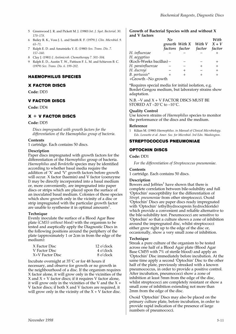
5 Greenwood J. R. and Pickett M. J. (1980) Int. J. Syst. Becteriol. 30.
170±178.
6 Bailey R. K., Voss J. L. and Smith R. F. (1979) J. Clin. Microbiol. 9.
65±71.
7 Ralph E. D. and Amatnieks Y. E. (1980) Sex. Trans. Dis. 7.
157±160.
8 Clay J. (1981) J. Antimicrob. Chemotherapy 7. 501±504.
9 Ralph E. D., Austin T. W., Pattison F. L. M. and Schieven B. C.
(1979) Sex. Trans. Dis. 6. 199±202.
HAEMOPHILUS SPECIES
X FACTOR DISCS
Code: DD3
V FACTOR DISCS
Code: DD4
X + V FACTOR DISCS
Code: DD5
Discs impregnated with growth factors for thedifferentiation of the Haemophilus group of bacteria.
Contents1 cartridge. Each contains 50 discs.
DescriptionPaper discs impregnated with growth factors for thedifferentiation of the Haemophilus group of bacteria.Haemophilus and Bordetella species may be identifiedaccording to whether basal media require theaddition of `X' and `V' growth factors before growthwill occur. X factor (haemin) and V factor (coenzymeI) may be directly incorporated into a basal mediumor, more conveniently, are impregnated into paperdiscs or strips which are placed upon the surface ofan inoculated basal medium. Colonies of those specieswhich show growth only in the vicinity of a disc orstrip impregnated with the particular growth factorare unable to synthesise it in optimal amounts1.
TechniqueEvenly inoculate the surface of a Blood Agar Baseplate (CM55 without blood) with the organism to betested and aseptically apply the Diagnostic Discs inthe following positions around the periphery of theplate (approximately 1 or 2cm in from the edge of themedium):
X Factor Disc 12 o'clockV Factor Disc 4 o'clockX+V Factor Disc 8 o'clock
Incubate overnight at 358C or for 48 hours ifnecessary, and observe for growth or no growth inthe neighbourhood of a disc. If the organism requiresX factor alone, it will grow only in the vicinities of theX and X + V factor discs; if it requires V factor alone,it will grow only in the vicinities of the V and the X +V factor discs; if both X and V factors are required, itwill grow only in the vicinity of the X + V factor disc.
Growth of Bacterial Species with and without Xand V factors
No Withgrowth With X With V X + Vfactors factor factor factor
H. influenzae ± ± ± +H. aegyptius(Koch-Weeks bacillus) ± ± ± +H. parainfluenzae ± ± + +H. ducreyi ± + ± +B. pertussis* + + + ++Growth ±No growth
*Requires special media for initial isolation, e.g.Bordet-Gengou medium, but laboratory strains showadaptation.
N.B. ±V and X + V FACTOR DISCS MUST BESTORED AT ±208C to ±108C.
Quality ControlUse known strains of Haemophilus species to monitorthe performance of the discs and the medium.
Reference1 Kilian M. (1980) Haemophilus. in Manual of Clinical Microbiology.
Eds. Lennette et al. Amer. Soc. for Microbiol. 3rd Edn. Washington.
STREPTOCOCCUS PNEUMONIAE
OPTOCHIN DISCS
Code: DD1
For the differentiation of Streptococcus pneumoniae.
Contents1 cartridge. Each contains 50 discs.
DescriptionBowers and Jeffries1 have shown that there iscomplete correlation between bile-solubility and full`Optochin' susceptibility for the differentiation ofStrept. pneumoniae from other streptococci. Oxoid`Optochin' Discs are paper discs ready impregnatedwith `Optochin' (ethylhydrocuprein hydrochloride)which provide a convenient and reliable alternative tothe bile-solubility test. Pneumococci are sensitive to`Optochin' so that a culture shows a zone of inhibitionaround the impregnated disc, whilst streptococcieither grow right up to the edge of the disc or,occasionally, show a very small zone of inhibition.
TechniqueStreak a pure culture of the organism to be testedacross one half of a Blood Agar plate (Blood AgarBase CM55 with 7% of sterile blood) and apply an`Optochin' Disc immediately before incubation. At thesame time apply a second `Optochin' Disc to the otherhalf of the plate, previously streaked with a knownpneumococcus, in order to provide a positive control.After incubation, pneumococci show a zone ofinhibition at least 5mm from the edge of the disc,whilst streptococci are completely resistant or show asmall zone of inhibition extending not more than2mm from the edge of the disc.
Oxoid `Optochin' Discs may also be placed on theprimary culture plate, before incubation, in order toprovide rapid indication of the presence of largenumbers of pneumococci.
Biochemical Reagents, Diagnostic Discs
November 1998 5-11
Quality ControlSee under Technique.
Reference1 Bowers E. F. and Jeffries L. R. (1955) J. Clin. Path. 8. 58.
STREPTOCOCCUS PYOGENES (GROUP A)
BACITRACIN DISCS
Code: DD2
For the differentiation of Lancefield Group A from otherbeta-haemolytic streptococci.
Contents1 cartridge. Each contains 50 discs.
DescriptionSterile paper discs each containing 0.05 units ofbacitracin, for the differentiation of Lancefield GroupA from other beta-haemolytic streptococci. Whenused as a screening test prior to serological grouping,Bacitracin Discs effect considerable economies in time,labour and materials.
Maxted1 showed that Group A streptococci weremore sensitive to bacitracin than beta-haemolyticstrains of other groups, and that bacitracin mighttherefore be used as a rapid diagnostic agent forGroup A streptoccoci. He found that only 1.7% of2,386 bacitracin sensitive strains proved not to beGroup A, and only 2.5% of 851 resistant strains wereGroup A. Levinson and Frank2 who employedimpregnated paper discs for the same purpose,observed that 93.2% of 866 sensitive beta-haemolyticstreptoccoci belonged to Group A. Streamer et al.3
compared bacitracin disc, fluorescent antibody andLancefield precipitin techniques for the identificationof Group A streptococci. The bacitracin disc techniquewas considered to be the simplest and most practicalfor the routine clinical laboratory.
The use of bacitracin sensitivity testing is notrestricted to the differentiation of beta-haemolyticstreptococci. Guthof4 found sensitivity to optochin,bacitracin, and furacin to be a useful test fordistinguishing Aerococcus viridans and Streptococcusmilleri from enterococci and mitis streptococci.
TechniqueA. Pure Cultures
1 Evenly inoculate the surface of a Blood Agar plate(Blood Agar Base No.2 CM271 with 7% SterileOxalated Horse Blood SR49) with a pure culture ofthe beta-haemolytic streptocccus to be tested.
2 Aseptically place a Bacitracin Disc on theinoculated surface.
3 Incubate for 18 to 24 hours at 358C.
4 Examine for the presence of a zone of inhibitionaround the Bacitracin Disc. A zone indicates thatthe streptococcus is presumptively Group A ± ifdesired, further confirmation is obtained byserological grouping.
B. Clinical Material
1 Inoculate a Blood Agar plate with the throat swabor other material. Spread the inoculum so as to
ensure that discrete colonies occur on some portionof the plate ± so that the species in a mixed growthmay be determined.
2 Aseptically place a Bacitracin Disc on thesecondary area of inoculation.
3 Incubate for 18 to 24 hours at 358C.
4 Examine for zones of inhibition. Organisms otherthan beta-haemolytic streptococci are sensitive tobacitracin, so that the presence of a zone ofinhibition does not necessarily indicate LancefieldGroup A streptococci. It is possible to selectpresumptive Group A streptococci if attention isgiven to colonial morphology. Furtherconfirmation is obtained by serological grouping.
Bacitracin Discs DD2 are not recommended forroutine testing of the sensitivity of organisms toBacitracin. For this purpose it is advisable toemploy Bacitracin Sensitivity Discs at a level of 10units.
Quality ControlUse known Group A and non-Group A streptococcito monitor the accuracy of the discs and inoculum.
References1 Maxted W. R. (1953) J. Clin. Path. 6. 224±226.
2 Levinson M. L. and Frank P. F. (1955) J. Bact. 69. 284±287.
3 Streamer C. W. et al. (1962) Amer. J. Dis. Children 104. 157±160.
4 Guthof O. (1960) Ztschr. f. Hyg. u. Infektionskr. 146. 425±432.
VIBRIO SPECIES
0129 DISCS
Codes: DD14 and DD15
For the differentiation of vibrios from other Gram-negative rods.
ContentsDD14 1 cartridge. Each contains 50 discs.Disc content 0129 10mg per disc.
DescriptionThe sensitivity of vibrios to the vibrio static agent0129 (2,4-Diamino-6,7-di-iso-propylpteridinephosphate) has long been recognised1. This test is ofgreat value in differentiating vibrios from otherGram-negative rods and particularly fromaeromonads, which are characteristically resistant to0129. The degree of sensitivity of vibrios to 0129 canalso be used as a diagnostic feature in differentiationof Vibrio species (see Table 1).
Negative 0129 tests must be interpreted with caution.Strains of V. cholerae resistant to 0129 andtrimethoprim have been reported2. Both plasmids andtransposons appear to be responsible3.
Specialised sensitivity testing media which containlow levels of sodium chloride should not be used asthese may give poor growth of halophilic vibrios andmay also give false positive results withEnterobacteriaceae4. Blood Agar Base No.2 CM271can be recommended for such testing.
Biochemical Reagents, Diagnostic Discs
5-12 November 1998
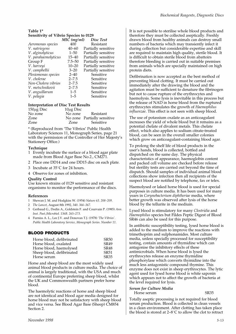
Table 1*Sensitivity of Vibrio Species to 0129
MIC (mg/ml) Disc TestAeromonas species 400 ResistantV. natriegens 40±60 Partially sensitiveV. alginolyticus 1±50 Partially sensitiveV. parahaemolyticus 15±40 Partially sensitiveGroup F 7.5±50 Partially sensitiveV. harveyi 10±20 Partially sensitiveV. campbellii 3±20 Partially sensitivePlesiomonas species 2±40 SensitiveV. cholerae 2±7.5 SensitiveNon-Cholera vibrios 2±7.5 SensitiveV. metschnikovii 2±7.5 SensitiveV. anguillarum 1±5 SensitiveV. pelagia 1±5 Sensitive
Interpretation of Disc Test Results150mg Disc 10mg DiscNo zone No zone ResistantZone No zone Partially sensitiveZone Zone Sensitive
* (Reproduced from `The Vibrios' Public HealthLaboratory Sciences 11, Monograph Series, page 21with the permission of the Controller of Her Majesty'sStationery Office.)
Technique1 Evenly incubate the surface of a blood agar plate
made from Blood Agar Base No.2., CM271.
2 Place one DD14 and one DD15 disc on each plate.
3 Incubate at 358C for 24 hours.
4 Observe for zones of inhibition.
Quality ControlUse known strains of 0129 sensitive and resistantorganisms to monitor the performance of the discs.
References1 Shewan J. M. and Hodgkiss W. (1954) Nature 63. 208±209.
2 The Lancet, August 8th 1992, 340. 366±367.
3 Gerbaud G., Dodin A., Goldstein F. and Courvain P. (1985) Ann.
Inst. Past./Microbiol. 136B. 265±273.
4 Furniss A. L., Lee J.V. and Donovan T.J. (1978) `The Vibrios'.
Public Health Laboratory Service, Monograph Series. Number 11.
BLOOD PRODUCTS
Horse blood, defibrinated SR50Horse blood, oxalated SR49Horse blood, haemolysed SR48Sheep blood, defibrinated SR51Horse serum SR35
Horse and sheep blood are the most widely usedanimal blood products in culture media. The choice ofanimal is largely traditional, with the USA and muchof continental Europe preferring sheep blood, whilstthe UK and Commonwealth partners prefer horseblood.
The haemolytic reactions of horse and sheep bloodare not identical and blood agar media designed forhorse blood may not be satisfactory with sheep bloodand vice versa. See Blood Agar Base (Sheep) CM854Section 2.
It is not possible to sterilise whole blood products andtherefore they must be collected aseptically. Freshlydrawn blood from healthy animals can destroy smallnumbers of bacteria which may transiently infect itduring collection but considerable expertise and skillare required to maintain high quality, sterile blood. Itis difficult to obtain sterile blood from abattoirstherefore bleeding is carried out in suitable premisesfrom animals which are specially maintained on highprotein diets.
Defibrination is now accepted as the best method ofpreventing blood clotting. It must be carried outimmediately after the drawing the blood and theagitation must be sufficient to denature the fibrinogenbut not to cause rupture of the erythrocytes andhaemolysis. Some lysis is inevitable in this process butthe release of NAD in horse blood from the rupturederythrocytes stimulates the growth of Haemophilusinfluenzae. This effect is not seen with sheep blood.
The use of potassium oxalate as an anticoagulantincreases the yield of whole blood but it remains as apotential chelate of divalent metals. This chelateeffect, which also applies to sodium citrate-treatedblood, can be seen in the overall smaller colonieswhich grow on anticoagulant-containing blood agar.
To prolong the shelf life of blood products in theuser's hands, blood is collected, bottled anddispatched on the same day. The physicalcharacteristics of appearance, haemoglobin contentand packed cell volume are checked before releasebut sterility tests are carried out beyond the time ofdispatch. Should samples of individual animal bloodcollections show infection then all recipients of thesuspect blood are notified by telephone, fax or telex.
Haemolysed or laked horse blood is used for specialpurposes in culture media. It has been used for manyyears in Corynebacterium diphtheriae media, wherebetter growth was observed after lysis of the horseblood by the tellurite in the medium.
Lysed blood is stimulatory for many Clostridia andHaemophilus species but Fildes Peptic Digest of BloodSR46 can also be used for this purpose.
In antibiotic susceptibility testing, lysed horse blood isadded to the medium to improve the reactions withtrimethoprim and sulphonamides. Most culturemedia, unless specially processed for susceptibilitytesting, contain amounts of thymidine which canantagonise the inhibitory effects of theseantimicrobials. When horse blood is lysed theerythrocytes release an enzyme thymidinephosphorylase which converts thymidine into themuch less antagonistic compound thymine. Thisenzyme does not exist in sheep erythrocytes. The lyticagent used for lysed horse blood is white saponinwhich appears not to affect the growth of bacteria atthe level required for lysis.
Serum for Culture MediaHorse serum SR35
Totally aseptic processing is not required for bloodserum production. Blood is collected in clean vesselsin a clean environment. After clotting has taken placethe blood is stored at 2±88C to allow the clot to retract
Biochemical Reagents, Diagnostic Discs
November 1998 5-13

and the serum separate. The separated serum is thenfilter-sterilised and filled out into sterile bottles. Allsera should be stored at 2±88C but for longer termstorage it is best frozen at ±208C.
Serum can become cloudy on storage caused by lipaseenzymes and lipid changes. Heating the serum to568C for 30 minutes (inactivation) will usuallyovercome this problem.
Sheep Erythrocytes for Immunological Reactions
Sheep blood (in formaldehyde) SR52Sheep blood (in Alsever solution) SR53
After washing the sheep erythrocytes, by centrifugingwith physiological saline until the supernatant salineis free from haemolysis, the washed cells may be usedfor any immunological/serological test where sheepcell antigens are required e.g. Rose-Waaler, PaulBunnell and various virological complement fixationtests.
The sheep blood should be stored at 2±88C and usedbefore the expiry date on the label.
Biochemical Reagents, Diagnostic Discs
5-14 November 1998
6ANTIMICROBIAL
SUSCEPTIBILITY TESTING

November 1998
ANTIMICROBIAL SUSCEPTIBILITYTESTING (AST)The Oxoid system of AST is the disc diffusion methodwhich although developed in 19471 remains as themost widely used test. Performed with care, usingadequate controls, it is as accurate as more costly andcomplicated tests.
In common with all other methods it cannot mimicthe in vivo environment but it does uniquely show theeffect of a changing antimicrobial concentration (firstrising and then falling) on an increasing microbialpopulation. The minimum inhibitory concentration(MIC) of the antimicrobial appears at the edge of thezone of inhibition which represents the interaction ofa critical concentration of antimicrobial at a criticaltime on a critical microbial population2.
In October 1997 Oxoid aura was launched. Thisenhances the AST system by using an accurate andobjective electronic zone size measurement facilitylinked to unique species specific interpretativesoftware.
Oxoid aura ensures high reproducibility and betterseparation of Susceptible, Intermediate, Resistant(S,I,R) categories with the facility to store and processdata for future comparison and epidemiologicalstudies. (See page 6-3.)
The methods are simple but the above interactions arevery complex and the accuracy and precision of thetest is based on the following critical components:
1 CULTURE MEDIUMThe medium chosen for the test must have beenmanufactured and tested specifically for AST. Oxoidmanufactures and recommends the following agarmedia:
i Diagnostic Sensitivity Test Agar (DST Agar)CM261 which was the first medium specificallydesigned for this test. It is still very popular but ithas been modified to reduce its thymidine contentand it is therefore less useful as a diagnosticmedium.
ii HR Medium CM845 ± a chemically definedmedium for susceptibility testing with antifungalagents.
iii HTM (Haemophilus Test Medium) CM898 wasspecifically formulated for the susceptibility testingof Haemophilus influenzae. The medium is based onMueller-Hinton Agar to which is added asupplement (SR158). The supplement containsNAD and Haematin.
iv Iso-Sensitest Agar CM471 was developed as a semi-synthetic AST medium in which undefined proteinhydrolysates were reduced to the minimum levelwhich would allow optimum growth of a widerange of organisms and the `free' cation strengthadjusted to give correct MIC results. Its popularityrests on its reproducible performance.
v Mueller-Hinton Agar CM337 was originallyformulated to grow pathogenic neisseria and it wasadopted for use in the Bauer-Kirby test4. Its
variable performance was strongly criticised butfollowing agreement between culture mediamanufacturers and representatives of NCCLS andFDA a uniform medium is now produced whichmust meet the NCCLS specifications5. ThereforeMueller-Hinton medium suitable for AST willcarry a statement on the label that it meets NCCLSstandards.
vi Sensitest Agar CM409 the first semi-defined ASTmedium which remains popular with laboratorieswhich have designed methods around this specificformulation3.
viiWilkins-Chalgren Anaerobic Agar CM619 wasdeveloped as a medium for the growth ofanaerobic bacteria. Its relative lack of inhibitoryaction towards anaerobes made it the medium ofchoice for anaerobic agar dilution tests6.
Anaerobic disc diffusion tests can be carried out but itis essential to include adequate controls which canmonitor the effect of anaerobiosis on antimicrobials.
All the above media have broth versions of theirformulations (except DST and H.T.M.). This allowsdilution or pre-enrichment to be carried out in thesame medium. It is also helpful when carrying outMIC tests in broth to standardise disc diffusion zonesto establish the breakpoint categories of S,I,R.
Many of the constituents used in culture media affectthe precision and accuracy of AST results. The agarmust allow free diffusion of the antimicrobial fromthe disc. Variations in pH and ionic strength willcause differences in zone sizes. Blood can reduce thezone size of highly protein-bound antimicrobials e.g.fusidic acid. Changes in `free' electrolyte content willaffect aminoglycosides, tetracyclines and polymyxins.Glucose will enlarge the zones of antimicrobialsagainst organisms which are adversely affected by afall in pH following fermentation of the sugar.Thymidine and thymine levels have to be monitoredand reduced, if necessary, to prevent antagonism oftrimethoprim and sulphonamides. All AST mediamust be specifically tested with critical `drug-bug'combinations to measure their performance and toensure that they meet the quality specifications. Thelatter tests are carried out by the manufacturer,especially to ensure that the medium conforms toregulatory standards, they should also be carried outin an abbreviated form in the user laboratory tomonitor the performance of all of the components ofAST.
2 ANTIMICROBIAL DISCSThe paper discs used in the diffusion method aremade from paper which conforms to the WHO7 andFDA8 standards.
Impregnation of the discs ensures that accuratelyprepared solutions of antimicrobials are uniformlyspread across the paper. The drying procedures useddo not affect this uniform distribution or causedeterioration in activity of the antimicrobial.
When each cartridge of 50 discs is sealed togetherwith a silica gel capsule in a foil-covered see-throughblister, the discs contain less than 2% moisture and
Antimicrobial Susceptibility Testing
November 1998 6-1
are stored at low temperatures. Shelf-life studies ofthe discs in their packaging demonstrate that theymeet the stated storage life printed on the labels.
Finally, samples are taken from every batch/lotmanufactured and the discs tested microbiologicallyto confirm that the antimicrobial content lies within80±120% of the stated content on the label.
StorageDiscs must be stored at ±208C if kept for long periods.Storage at 2±88C is suitable for discs currently beingused or to be used very soon. Discs should bereturned to the refrigerator as quickly as possible afteruse. The most common cause of moisture reaching thediscs and causing destruction of labile antimicrobialsis condensation of warm laboratory air on cold discsremoved from the refrigerator.
It is important to allow the cartridge blister pack toreach room temperature before exposing the discs, aperiod of one hour is generally sufficient. Use discs inorder of expiry date, which is valid only for unopenedblisters stored under proper conditions.
3 INOCULUMOne of the very critical factors for accuracy andprecision in disc diffusion tests is the inoculumpreparation. It is therefore important to use atechnique which will always yield a uniformsuspension of the correct number of organisms,105±106 orgs/ml.
Various techniques are described in whichsuspensions of pre-grown organisms are prepared orsmall inocula are incubated for fixed periods of time.It is important that more than one colony is sampled(4±10 cols) to ensure a representative sample of theorganism has been taken.
Some form of standardisation of the final suspensionis necessary and it should be noted that differentorganisms will display different opacities of solutionto yield a dense but not confluent growth.
To complement the newly launched Oxoid auraSystem (see page 6-3) Oxoid have launched the OxoidTurbidometer. This instrument provides the inoculumdensity standardisation necessary to ensure accuratereproducible results.
4 OTHER FACTORS INFLUENCINGTHE RESULTS1 Temperature of incubation ± the incubators
should be checked for satisfactory performanceand their recording thermometers should show airtemperatures of 35±378C with fluctuations of notmore than 28C. Agar plates should not be placed inhigh stacks because the middle plates will takelonger to reach the incubator temperature and thisdelay could cause overlarge zones.
2 Pre-incubation and pre-diffusion conditions ± aroutine procedure should be established so thatinoculated plates have discs applied not later than15 minutes after inoculation. This prevents a pre-incubation of organisms before the antimicrobial
discs are applied. Similarly once the discs havebeen applied, plates should be placed in theincubator within a 15 minute interval, to preventpre-diffusion of the antimicrobial at roomtemperature.
3 Uniformity of agar depth ± plates should bepoured on levelled surfaces, using dishes with flatbottoms, to ensure a uniform depth of agar(3±4mm in depth).
4 Application of discs ± it is essential that discs arein intimate contact with a moist agar surface.Therefore either use dispensers which have atamping action on the discs or press themseparately after application. Do not overdry theagar surface before applying the discs. Do notattempt to place too many discs on the agarsurface; six 6mm discs are ideal for a 90mm dish.Some workers apply up to 12 discs onto a 150mmdish.
5 Incubation period ± ensure uniform times ofincubation for the plates, 16±18 hours at 35±378C isusually satisfactory.
6 Interpretation of zone sizes ± after incubation theplates are removed and the zones of inhibitionnoted and measured. (See Oxoid aurapage 6-3.)
The diameter of the zone (including the diameter ofthe disc) is measured to the nearest millimetre,using calipers. If the NCCLS Bauer-Kirby methodis being used then the zone diameters can becompared with the current NCCLS Standardswhich can be obtained from the NationalCommittee for Clinical Laboratory Standards,NCCLS Publications, Villanova, Pa. USA. Thisstandard should not be used unless the test iscarried out following the described methodologyon Mueller-Hinton medium with the appropriatediscs.
Where other systems are used the zone sizebreakpoints should have been determined usingMIC/zone comparative tests, following theparticular methodology chosen.
7 Control cultures ± it is essential that eachlaboratory maintains adequate control over ASTmethods by testing reference cultures at regularintervals. Daily tests may be required if new mediaand discs are constantly being used.
The following reference strains are used in theNCCLS test method.
Staphylococcus aureus ATCC1 29213
Enterococcus faecalis ATCC1 29212
Escherichia coli ATCC1 25922
Pseudomonas aeruginosa ATCC1 27853
Escherichia coli ATCC1 35218 for testing clavulanicacid compounds.
Records of the results obtained with these referencestrains should be maintained in log books and anydeviation of zone sizes from the range acceptedshould be investigated.
Antimicrobial Susceptibility Testing
6-2 November 1998

References1 Bondi A., Spauling E. H., Smith E. D. & Dietz C. C. (1947) Amer.
J. Med. Sci. 214. 221±225.
2 Cooper H. E. (1964) The theory of antibiotic zones. In Kavanagh F.
(Ed) Analytical Microbiology. Academic Press. New York. 1±86.
3 Bell S. M. (1975) Pathology. 7. l-48 (Suppl).
4 Bauer A. W., Kirby W. M. M., Sherris J. C. & Turck M. (1966)
Amer. J. Clin. Path. 45. 493±496.
5 Pollock H. M. et al. (1986) J. Clin. Microbiol. 24. 1±6.
6 Nat. Comm. Clin. Lab. Standards (1979) Proposed reference
dilution procedure for AST of anaerobic bacteria. NCCLS PSM-
11. Villanova. Pa. USA.
7 WHO (1977) Tech. Rep. Ser. No. 610.
8 FDA (1978) Codes of Fed. Rebs. 21. Part 460.
NoteThe list of antimicrobial discs manufactured, theagents, generic and proprietary names, the symbolsand range of levels available are supplied in theOxoid Product List and the list is updated annually.
Antimicrobial SusceptibilityDiscsDiscs in routine use should be stored at 2 to 88C.Longer term storage should be at ±208C. After coldstorage allow discs to reach room temperature beforeopening storage containers. Discs are presented incartridges for dispensing either individually withOxoid ejectors or with the Oxoid Dispenser System.There are 50 discs per cartridge, 5 cartridges per pack.
Cartridge packsEach cartridge is individually sealed together with asilica gel capsule in a foil-covered see-through blister.Designed to allow the microbiologist better controland storage of the discs in use.
THE OXOID DISC DISPENSER MKIII
An enhanced system for antimicrobial susceptibilitytesting incorporating the new ergonomically designedDisc Dispenser. It has a simple one-handed operationand is fully height-adjustable to cater for variousdepths of media. The cover and base of the dispenserare fully interlocked to prevent the ingress ofmoisture: cartridges `click' positively into their correctlocations and a plastic skirt ensures that each agarplate is precisely centred every time the dispenser isused.
Product Pack size Order CodeDisc Dispenser Mk III (90mm)for 90mm petri dishesDisc Dispenser Mk III (90mm) 1 ST6090for 6 cartridgesDisc Dispenser Mk III (90mm) 1 ST8090for 8 cartridgesDisc Dispenser Mk III (100mm)for 100mm petri dishes
Disc Dispenser Mk III (100mm) 1 ST6100for 6 cartridgesDisc Dispenser Mk III (100mm) 1 ST8100for 8 cartridgesDisc Dispenser (150mm) 1 ST1215for 12 cartridges
Ejector 6 HP053Afor using cartridges individuallyReplacement Dessicant Pack 1 pack ST9100
Antimicrobial SusceptibilityTesting MediaMedia for Antimicrobial Susceptibility Testing (codedCM) should be stored in their closed containers at aneven temperature in a cool, dry place, away fromdirect light.
Product Pack Size Order CodeDiagnostic SensitivityTestAgar Base (DST Agar) 500g CM261BHR Medium 100g CM845AHaemophilus Test Medium (HTM)HTM Supplement 10 vials SR158EHTM Base 500g CM898B`Iso-Sensitest' Agar 500g CM471B`lso-Sensitest' Broth 500g CM473BMueller-Hinton Agar 500g CM337BMueller-Hinton Broth 500g CM405B`Sensitest' Agar 500g CM409BWilkins-Chalgren Anaerobe Agar 500g CM619BWilkins-Chalgren Anaerobe Broth 500g CM643B
OXOID auraThe importance of standardisation.
Antimicrobial Susceptibility Testing by the discdiffusion method is simple, flexible, informative andcost-effective1.
However, a recent survey demonstrated that, betweenlaboratories, there is ``considerable variation in themedium used, the method of inoculum preparation andapplication, disc concentrations, incubation conditionsand the basis of interpretation of zone sizes''2.
It is generally accepted that there is a need for greaterstandardisation of methodology and interpretation ofresults to ensure accurate, reproducible results, withinand between laboratories.
The Oxoid aura database enables clinical isolates to becompared to an appropriate standard for everyantibiotic and concentration in the system. Thecomparison is performed at species level andautomatically generates interpretation of the result.
Interpretations are based on the principles set forth bythe Swedish Reference Group of Antibiotics (SRGA)and its sub-committee on Methodology (SRGA-M)3.The value and function of this system has beenproven over many years.
The Complete Antimicrobial Susceptibility Testing(AST) System.
Accurate, reproducible AST results depend on thefollowing:
. High quality products used in a definedmethodology
. Standardised inoculum density
. Precise zone size measurement (aura)
. Accurate and meaningful interpretation of results(aura)
Antimicrobial Susceptibility Testing
November 1998 6-3

All of this is achievable using the complete OxoidAST System:
Components of the System:
Specialist culture media Range includes Mueller-Hinton Agar and Iso-Sensitest Agar.
AntimicrobialSusceptibility Discs
See Product List for fullrange
Disc Dispensers 6, 8 and 12 Place
Turbidometer Standardised inoculumdensity
Interpretative database Performed at specieslevel ± generatesinterpretation of theresult
References1 Bridson E. (1996) Antimicrobial Susceptibility Testing ± the disc
diffusion method. Oxoid, Basingstoke, UK. folio number 547.
2 Andrews J. M. et al (1996) Antimicrob. Chemother. 37: 187±204.
3 Report from the Swedish Reference Group of Antibiotics and its
Methodology Subcommittee (SRGA-M), 1991.
In addition to Culture Media, Susceptibility Discs,Dispensers and the Oxoid Turbidometer the fullOxoid aura system is as follows:
Product Description ProductCode
aura Callipers AU101Aaura Barcode Labels (1±50,000) AU102Aaura Barcode Labels (50,000±100,000) AU103Aaura Barcode Reader AU104Aaura System Software AU106A
Oxoid Turbidometer (UK) AU107AOxoid Turbidometer (International) AU108A
Separate literature available.
Antimicrobial Susceptibility Testing
6-4 November 1998

7ANAEROBIC SYSTEMS

November 1998
ANAEROBIC BACTERlOLOGY
THE ROLE OF OXYGEN
The fact that life almost certainly evolved in anenvironment which lacked oxygen was not originallyappreciated. At the time of Pasteur there was noprecedent that life could exist in the absence ofoxygen. When, in 1861, Pasteur declared that someorganisms could exist without oxygen and appearedto die in its presence, the theory of the toxicity ofoxygen was born.
The beginning of this theory was Pasteur'sobservation, looking down a microscope, that rapidlymoving bacteria in a fermenting sugar solution ceasedmoving at the periphery of the drop, whilst remainingactively motile in the centre. Such organisms he called`anaerobies' as opposed to `aerobies' who dependedon oxygen for their survival.
ANAEROBIC BACTERIA
Pasteur's fermentation studies on yeasts showed thatthese organisms could multiply with or without airand changed their metabolism of sugar to suit thegaseous environment. Thus facultative anaerobes,which could adapt and survive, were distinguishedfrom strict anaerobes and from strict aerobes2. In 1893Veillon3 isolated a micrococcus which was strictlyanaerobic (obligate anaerobe). Later, in 1897, Veillonand Zuber4 described various pathological conditionswhere anaerobic bacteria could be the cause of theinfective condition.
Further work, between 1898±1902, linked anaerobesto several human pathologies including empyaema,female genital infections, gangrene of the lung etc.
The anaerobic bacteriology of botulism, gas gangreneand tetanus soon became familiar to cliniciansbecause the causative organisms, members of theClostridium genus, were relatively easy to cultivate.Other anaerobes remained obscure, even though theywere responsible for common supperative conditions.The reason for their obscurity was the cumbersomeand difficult techniques required for their cultivation.
Anaerobic methods in liquid or semi-solid mediaMethods to cultivate anaerobic organisms in liquidmedia were quickly developed because simple boilingexpelled most of the dissolved oxygen. The additionof a heated iron strip or meat granules to the brothmaintained the low oxygen levels.
A small amount of agar (0.2% w/v) in the broth wasalso effective by increasing the viscosity of the liquidand slowing down the re-absorption of air in thecooled medium.
THEORY OF OXYGEN TOXICITY
The addition to media of Eh reducing agents, such asthioglycollate or cysteine which contain sulphydralgroups, is protective to anaerobic organisms undercertain circumstances. The oxidation-reductionpotential (Eh) of an environment or medium ismeasured in mV and expressed as the tendency eitherto accept electrons (become reduced) or donateelectrons (become oxidised). The Eh of normal tissue
is +150mV but in the colon it can be as low as±250mV.
The question of limiting levels of Eh which allowanaerobes to grow has not been determined becauseof the relative influence of oxygen and Eh. It appearsthat the presence of oxygen is of greater importancebecause it has been shown that the growth ofanaerobes was inhibited in the presence of air, eventhough the Eh was ±50mV. In the absence of air,anaerobic organisms grew in spite of positive Ehvalues6.
The toxicity of oxygen occurs because of the radicalsformed by electron capture along reductionpathways: superoxide O±
2, singlet oxygen O22 as well
as hydroxyls and peroxides. The enzymes catalase,peroxidase and superoxide dismutase are protectivesystems against these toxic radicals7. Aerobicorganisms and facultative anaerobes have efficientsystems of enzymes which can neutralise such toxicradicals; obligate anaerobes lack such protectivesystems.
Furthermore the addition of protective substances toculture media can overcome oxygen toxicityproduced biologically by the organisms or chemicallyin the medium and help protect vunerable organisms.Examples of such substances are blood, charcoal, ironsalts, pyruvate, cysteine.
Anaerobic methods for solid mediaAnaerobic bacteria can be grown in solid agar mediaby making deep stabs in tubes of agar or adding asuspension of the organism to molten, cooled agarbefore pouring butts or plates of agar media. Thelatter method usually requires an agar overlay toensure anaerobic growth.
However, submerged colonies of bacteria are difficultto access and investigate further. It would not havebeen possible to make good progress in anaerobicbacteriology without the ability to produce separatecolonies of organisms on the surface of agar plates. Toachieve this goal, systems of anaerobic cultivation foragar plates had to be created.
Metal or glass jars with gas-tight lids that had valvesfitted to them were developed. Air could beevacuated from the jar through a valve and oxygen-free gas flushed through the jar. A more efficientmethod came with the use of a palladium catalystfixed to the underside of the lid and hydrogen gasadded to the jar. The necessity to heat the catalyst andthe potentially violent combination of oxygen andhydrogen to form water, gave an ever present risk ofexplosions.
It was the development of the `cold' catalyst, in aflameproof capsule, and the replacement of glass withpolycarbonate plastic which made the process muchsafer. A further development was the replacement ofgas cylinders by foil sachets of chemicals which, whenactivated with water, produced hydrogen in sufficientquantities to reduce the oxygen level in the jars below1% v/v. These sachets also supply carbon dioxide at alevel of 5±8% in the jar to improve the growth ofmany anaerobes. Further modifications of thechemicals used in the sachets enabled atmospheres to
Anaerobic Systems
November 1998 7-1

be created which would support micro-aerophilicorganisms (about 6% oxygen in the jar) or capnoeicorganisms (about 10% CO2).
Anaerobic indicators will show whether the redoxpotential (Eh) in the jar has been reduced below±50mV, which is the level required to change theresazurin indicator from pink to white.
Collection and transport of anaerobic specimensIt must be emphasised that in spite of every caretaken in cultivation, poorly collected and transportedsamples will yield poor or negative results.
Samples for anaerobic investigation should becollected with care and protected from air. Swabs willnot be suitable, unless they are specially treated8 orplaced in Stuarts Transport Medium9. Generoussamples of pus or fluids do not need anaerobictransport but they should not be unduly delayedbefore examination10.
It was Dack who said 50 years ago that it should bepossible to isolate and study anaerobes in a relativelyconvenient, routine fashion using `modernapparatus'11. Much later and armed with thecomponents described below, anaerobic bacteriologyis within reach of all microbiological laboratories.
References1 Halliwell B. (1984) Med. Lab. Sci. 41. 157±171.
2 Dubos R. (1988) in Pasteur and Modern Science. Sci. Tech. Publ.
Madison USA. pp. 76±79.
3 Veillon A. (1893) C. R. Soc. Biol (Paris) 45. 807±809.
4 Veillon A. and Zuber A. (1897) C. R. Soc. Biol (Paris) 49. 253±
389.
5 Finegold S.M. et al. (1975) Ann. lnt. Med. 83. 375±389.
6 Walden W.C. and Hentges D.I. (1975) Appl. Microbiol. 30. 781±
785.
7 McCord J.M. Keele B.B. and Fridovich I. (1971) Proc. Natl. Acad.
Sci. USA. 68. 1024±1027.
8 Smith L.L. and Ferguson J.R. (1977) Med. Lab. Sci. 34. 247±258.
9 Yrios J.M. et al. (1975) J. Clin. Microbiol. 1. 196±200
10 Bartlett J.G., Sullivan-Sigler N., Louie T.J. and Gorbach S.L.
(1976) J. Clin. Microbiol. 3. 133±136.
11 Dack G.M. (1940) Bact. Rev. 4. 227±259.
ANAEROBIC SYSTEMS
For some time now Oxoid has been marketing twodistinctly different systems used for growing bacteriawhich require special atmospheres includinganaerobic organisms, microaerophilic organisms andCO2 dependent organisms.
The original Oxoid system consisted of an anaerobicjar of advanced design (Code HP11) together with agas generating kit for anaerobes (BR38), a CO2 gasgenerating kit (BR39) and gas generating kits forCampylobacter (BR56 and BR60).
In the case of BR38, BR56 and BR60 water is added tothe sachets to initiate a reaction and the production ofhydrogen and CO2. A low temperature catalyst(BR42) is required to be used in the anaerobic jar oneach occasion.
THE OXOID ANAEROBIC JAR HP11
A 3.4 litre capacity Anaerobic Jar of advanced designthat gives great flexibility in use by coping equallywell with Gas Generating Envelopes or GasCylinders.
Both Jar and Lid are of robust construction and usedwith the Low Temperature Catalyst BR42 ensureunprecedented protection to operator and equipment.
The Oxoid Anaerobic Jar has a number of noveldesign features and for greater convenience in use it issupplied with a corrosion-resistant plate carrier thatgreatly reduces the time and effort needed to load thejar. A test tube carrier is available as an optional extraminimising the risk of spillage of broth cultures.
Anaerobiosis is achieved rapidly, safely andefficiently using the Gas Generating Kit BR38 orhydrogen obtained from cylinders.
The Oxoid Anaerobic Jar may also be used for theisolation of microaerophilic and CO2-dependentorganisms by using the Campylobacter GasGenerating Kit BR56 or the CO2 Gas Generating KitBR39.
Catalytic activity may be checked both by thepressure gauge for an immediate indication ofefficiency and by the Anaerobic Indicator BR55 whichprovides supporting evidence of the pressurechanges. These checks ensure that the absence ofgrowth does not reflect poor anaerobic incubations.
The Oxoid Anaerobic Jar HP11 is part of the completeOxoid Anaerobic System consisting of:. 3.4 Litre Anaerobic Jar of advanced design.
. Gas Generating Kit which is superior in design toany other on the market.
. A new safe low temperature catalyst.
. An Anaerobic Indicator that is reliable and reactsfaster than any other equivalent products.
OXOID GAS GENERATING KIT BR38
The Oxoid Gas Generating Kit BR38 is a laminatedfoil envelope presented with an inner containerholding tablets of sodium borohydride, sodiumbicarbonate and tartaric acid. The addition of water tothe envelope activates the system causing hydrogenand carbon dioxide to be produced.
Gas production takes place smoothly andreproducibly because the porous membrane of theinner container regulates the passage of waterinwards and gas outwards.
Each individual Gas Generating Kit when activatedwith water evolves sufficient hydrogen for thecatalytic removal of oxygen present in the jar andleaves the final internal pressure approximating tothat of the atmosphere.
Carbon dioxide is also evolved to give a finalconcentration of 10% v/v in the atmosphere.
Gas generation is completed within 30 minutes andbecause the resultant solution is acid it does notreabsorb the carbon dioxide so necessary for thegrowth of fastidious anaerobes.
Anaerobic Systems
7-2 November 1998

Anaerobic Systems
November 1998 7-3
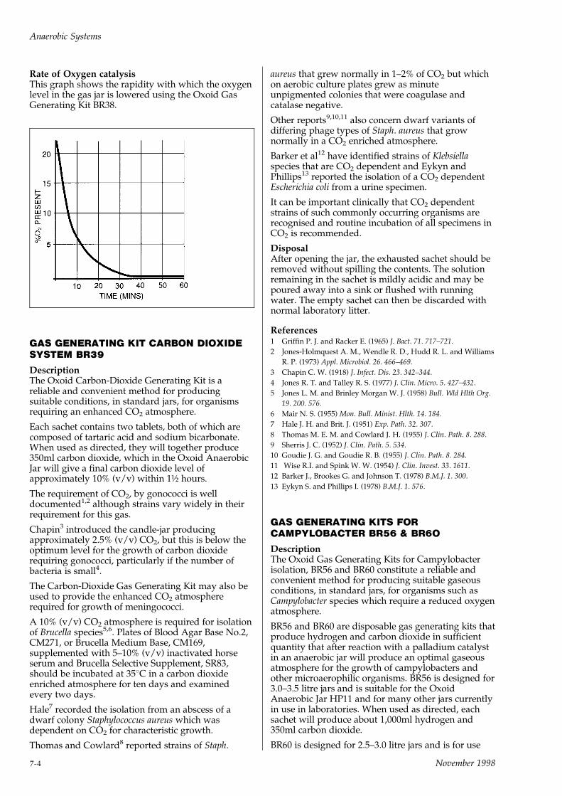
Rate of Oxygen catalysisThis graph shows the rapidity with which the oxygenlevel in the gas jar is lowered using the Oxoid GasGenerating Kit BR38.
GAS GENERATING KIT CARBON DIOXIDESYSTEM BR39
DescriptionThe Oxoid Carbon-Dioxide Generating Kit is areliable and convenient method for producingsuitable conditions, in standard jars, for organismsrequiring an enhanced CO2 atmosphere.
Each sachet contains two tablets, both of which arecomposed of tartaric acid and sodium bicarbonate.When used as directed, they will together produce350ml carbon dioxide, which in the Oxoid AnaerobicJar will give a final carbon dioxide level ofapproximately 10% (v/v) within 1� hours.
The requirement of CO2, by gonococci is welldocumented1,2 although strains vary widely in theirrequirement for this gas.
Chapin3 introduced the candle-jar producingapproximately 2.5% (v/v) CO2, but this is below theoptimum level for the growth of carbon dioxiderequiring gonococci, particularly if the number ofbacteria is small4.
The Carbon-Dioxide Gas Generating Kit may also beused to provide the enhanced CO2 atmosphererequired for growth of meningococci.
A 10% (v/v) CO2 atmosphere is required for isolationof Brucella species5,6. Plates of Blood Agar Base No.2,CM271, or Brucella Medium Base, CM169,supplemented with 5±10% (v/v) inactivated horseserum and Brucella Selective Supplement, SR83,should be incubated at 358C in a carbon dioxideenriched atmosphere for ten days and examinedevery two days.
Hale7 recorded the isolation from an abscess of adwarf colony Staphylococcus aureus which wasdependent on CO2 for characteristic growth.
Thomas and Cowlard8 reported strains of Staph.
aureus that grew normally in 1±2% of CO2 but whichon aerobic culture plates grew as minuteunpigmented colonies that were coagulase andcatalase negative.
Other reports9,10,11 also concern dwarf variants ofdiffering phage types of Staph. aureus that grownormally in a CO2 enriched atmosphere.
Barker et al12 have identified strains of Klebsiellaspecies that are CO2 dependent and Eykyn andPhillips13 reported the isolation of a CO2 dependentEscherichia coli from a urine specimen.
It can be important clinically that CO2 dependentstrains of such commonly occurring organisms arerecognised and routine incubation of all specimens inCO2 is recommended.
DisposalAfter opening the jar, the exhausted sachet should beremoved without spilling the contents. The solutionremaining in the sachet is mildly acidic and may bepoured away into a sink or flushed with runningwater. The empty sachet can then be discarded withnormal laboratory litter.
References1 Griffin P. J. and Racker E. (1965) J. Bact. 71. 717±721.
2 Jones-Holmquest A. M., Wendle R. D., Hudd R. L. and Williams
R. P. (1973) Appl. Microbiol. 26. 466±469.
3 Chapin C. W. (1918) J. Infect. Dis. 23. 342±344.
4 Jones R. T. and Talley R. S. (1977) J. Clin. Micro. 5. 427±432.
5 Jones L. M. and Brinley Morgan W. J. (1958) Bull. Wld Hlth Org.
19. 200. 576.
6 Mair N. S. (1955) Mon. Bull. Minist. Hlth. 14. 184.
7 Hale J. H. and Brit. J. (1951) Exp. Path. 32. 307.
8 Thomas M. E. M. and Cowlard J. H. (1955) J. Clin. Path. 8. 288.
9 Sherris J. C. (1952) J. Clin. Path. 5. 534.
10 Goudie J. G. and Goudie R. B. (1955) J. Clin. Path. 8. 284.
11 Wise R.I. and Spink W. W. (1954) J. Clin. Invest. 33. 1611.
12 Barker J., Brookes G. and Johnson T. (1978) B.M.J. 1. 300.
13 Eykyn S. and Phillips I. (1978) B.M.J. 1. 576.
GAS GENERATING KITS FORCAMPYLOBACTER BR56 & BR6O
DescriptionThe Oxoid Gas Generating Kits for Campylobacterisolation, BR56 and BR60 constitute a reliable andconvenient method for producing suitable gaseousconditions, in standard jars, for organisms such asCampylobacter species which require a reduced oxygenatmosphere.
BR56 and BR60 are disposable gas generating kits thatproduce hydrogen and carbon dioxide in sufficientquantity that after reaction with a palladium catalystin an anaerobic jar will produce an optimal gaseousatmosphere for the growth of campylobacters andother microaerophilic organisms. BR56 is designed for3.0±3.5 litre jars and is suitable for the OxoidAnaerobic Jar HP11 and for many other jars currentlyin use in laboratories. When used as directed, eachsachet will produce about 1,000ml hydrogen and350ml carbon dioxide.
BR60 is designed for 2.5±3.0 litre jars and is for use
Anaerobic Systems
7-4 November 1998
with the small lightweight plastic anaerobic jar. Whenused as directed, each sachet will produce about700ml hydrogen and 250ml carbon dioxide.
In each case a residual concentration of about 6%oxygen and 10% carbon dioxide in the jar is obtainedin under 30 minutes.
Some variation in the oxygen level in each jar willoccur, depending on the number of inoculated plates.
Use of an Anaerobic Indicator to check the efficiencyof catalysis is inappropriate but an active catalyst willproduce pressure changes which can be observedfrom the gauge fitted to the Oxoid Anaerobic Jar.After an initial pressure increase of approximately 0.1bar, catalytic activity will lead to a pressure reductionto zero or even ± 0.05 bar. Should the catalyst beinactive the pressure will rise to approximately 0.2bar and be maintained.
Campylobacter species require some oxygen yet areinhibited by the amount in air. The preferred level ofoxygen required for growth has been reported to bebetween 5 and 7%. This exacting level, together with acarbon dioxide requirement, has made isolation ofthese organisms from human and animal sources acomplicated procedure1.
Alternative methods for achieving the specialatmosphere may be hazardous and give a widevariation in the residual oxygen level in the jarleading to erratic recovery of Campylobacter species.
The Gas Generating Kit for Campylobacter provides areproducible atmosphere containing approximately6% of oxygen and 10% of carbon dioxide.
An evaluation which compared different methods forproducing microaerobic atmospheres confirmed theeffectiveness of gas generating envelopes2.
By using media made selective by the addition ofCampylobacter Selective Supplements maximumrecovery of the majority of Campylobacter strains willbe achieved.
Directions1 Cut off the corner of a sachet as indicated by the
broken line. Avoid folding or crushing the sachet.
2 Add a measured 10ml of water.
3 Immediately stand the sachet upright in theanaerobic jar fitted with an active catalyst and closethe lid according to the manufacturer'sinstructions. (If preferred, water may be added tosachets already located in the jar.) The use of theOxoid catalyst is recommended, because of its highefficiency, combined with inbuilt safety features.Activity of the catalyst may be prolonged byensuring that it is dry on each occasion it is used.Heating to 1608C for 90 minutes after each use isrecommended.
4 Disposal. After opening the jar, the exhaustedsachet should be removed without spilling thecontents. The solution left in the sachet is mildlyacid and may be poured away into a sink andflushed with running water. The empty sachet canthen be discarded with normal laboratory litter.
WarningGas Generating Kits activated but not in gas jarsshould be kept away from unguarded flames andsparks. Once the reaction has subsided (after about 30minutes) the sachet can be discarded as above.
Reference1 Butzler I. P. and Skirrow M. (1979) Clinics in Gastroenterology 8.
737±765.
2 Bolton F. J., Wareing D. R. A. and Sails A. D. (1997) Eur. J. Clin.
Microbiol. Inf. Dis. 16. 839±842.
THE OXOID ANAEROBIC CATALYST BR42
The Oxoid low temperature Catalyst BR42 is ofpatented design1 and is a safer and more efficientversion of the cold catalytic devices which are useduniversally to create low oxygen atmospheres withinanaerobic gas jars.
Each Oxoid Catalyst contains 4 grams of palladium-coated pellets and is suitable for use in anaerobic jarsup to 3.5 litres effective volume. This follows therecommendation of the United Kingdom Departmentof Health and Social Security2 that there should be notless than 1 gram of catalyst for each litre volume ofanaerobic jar. The extra large charge of activatedpalladium in the Oxoid Catalyst is wrapped in aspecial foil and enclosed in a large surface areacapsule woven from very fine stainless steel wire.
The graph (right) shows the difference in operatingtemperature between a conventional catalyst(unprotected pellets enclosed in wire gauze) and theOxoid Catalyst. The description `safe' applied to theOxoid Catalyst refers to the reduction in risk of anexplosion in the Oxoid Anaerobic Jar. Explosions inAnaerobic Systems although rare, can occur whenhydrogen and oxygen are present in criticalproportions. Such explosions are triggered byextremely hot catalyst envelope temperature or byglowing particles of catalyst which have becomedetached. The Oxoid Catalyst design protects thecustomers from both of these possibilities.
How the Oxoid Anaerobic Catalyst BR42 helps themicrobiologist
Features1 Maximum working temperature is approximately
half that of other catalysts.
2 Double wrapped in perforated aluminium foil andfine stainless steel mesh.
3 Four grams of catalyst (more than 1 gram/litreairspace) is provided.
Benefits1 Safety in use because the maximum operating
temperature is lower than the flash point ofhydrogen gas.
2 Acts as an efficient heat sink
3 Method of wrapping minimises the likelihood ofsmall particles falling into jars thus ensuring extrasafety in use.
4 Helps to create anaerobic conditions quickly.
Anaerobic Systems
November 1998 7-5
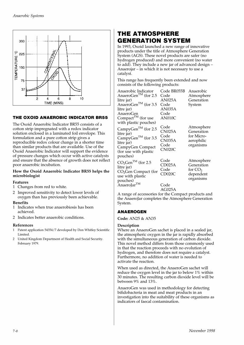
THE OXOID ANAEROBIC INDICATOR BR55
The Oxoid Anaerobic Indicator BR55 consists of acotton strip impregnated with a redox indicatorsolution enclosed in a laminated foil envelope. Thisformulation and a pure cotton strip gives areproducible redox colour change in a shorter timethan similar products that are available. Use of theOxoid Anaerobic Indicator will support the evidenceof pressure changes which occur with active catalystsand ensure that the absence of growth does not reflectpoor anaerobic incubation.
How the Oxoid Anaerobic Indicator BR55 helps themicrobiologist
Features1 Changes from red to white.
2 Improved sensitivity to detect lower levels ofoxygen than has previously been achievable.
Benefits1 Indicates when true anaerobiosis has been
achieved.
2 Indicates better anaerobic conditions.
References1 Patent application 54354/7 developed by Don Whitley Scientific
Limited.
2 United Kingdom Department of Health and Social Security.
February 1979.
THE ATMOSPHEREGENERATION SYSTEMIn 1993, Oxoid launched a new range of innovativeproducts under the title of Atmosphere GenerationSystem (AGS). These novel products are safer (nohydrogen produced) and more convenient (no waterto add). They include a new jar of advanced design ±Anaerojar ± in which it is not necessary to use acatalyst.
This range has frequently been extended and nowconsists of the following products:
Anaerobic IndicatorAnaeroGenTM (for 2.5litre jar)AnaeroGenTM (for 3.5litre jar)AnaeroGenCompactTM (for usewith plastic pouches)
Code BR055BCodeAN025ACodeAN035ACodeAN010C
AnaerobicAtmosphereGenerationSystem
CampyGenTM (for 2.5litre jar)CampyGenTM (for 3.5litre jar)CampyGen Compact(for use with plasticpouches)
CodeCN025ACodeCN035ACodeCN020C
AtmosphereGenerationfor Micro-aerophilicorganisms
CO2GenTM (for 2.5litre jar)CO2Gen Compact (foruse with plasticpouches)
CodeCD025ACodeCD020C
AtmosphereGenerationfor CO2
dependentorganisms
AnaeroJarTM CodeAG025A
A range of accessories for the Compact products andthe AnaeroJar completes the Atmosphere GenerationSystem.
ANAEROGEN
Code: AN25 & AN35
DescriptionWhere an AnaeroGen sachet is placed in a sealed jar,the atmospheric oxygen in the jar is rapidly absorbedwith the simultaneous generation of carbon dioxide.This novel method differs from those commonly usedin that the reaction proceeds with no evolution ofhydrogen, and therefore does not require a catalyst.Furthermore, no addition of water is needed toactivate the reaction.
When used as directed, the AnaeroGen sachet willreduce the oxygen level in the jar to below 1% within30 minutes. The resulting carbon dioxide level will bebetween 9% and 13%.
AnaeroGen was used in methodology for detectingbifidobacteria in meat and meat products in aninvestigation into the suitability of these organisms asindicators of faecal contamination.
Anaerobic Systems
7-6 November 1998

ComponentsEach box contains:
10 AnaeroGen paper sachets which areindividually foil packed.
1 Product Insert.
The active component within each AnaeroGen sachetis ascorbic acid.
Precautions
This product is for in-vitro use only.
As soon as the AnaeroGen paper sachet is exposed tothe air, the reaction will start. It is therefore essentialthat the paper sachet is placed in the jar and the jarsealed within one minute.
The reaction of the ascorbic acid with oxygen isexothermic. However, the temperature of theAnaeroGen paper sachet will not exceed 658C.
StorageStore at 2±258C. Under these conditions, theAnaeroGen sachets will retain their reactivity until theexpiry date declared on the outer box and on the foilsachet.
DirectionsAN35 is designed for use in 3.5 litre jars. It istherefore suitable for the Oxoid Anaerobic Jar HP11and for other jars of similar capacity.
AN25 is designed for use in 2.5 litre jars such as thenew Oxoid AnaeroJar AG25 and other jars of similarcapacity.
1 Place the inoculated media plates in theappropriate anaerobic jar. Disposable plastic petridishes should be of the vented variety to aid gastransfer between the interior and exterior of theplates.
2 Tear open an AnaeroGen foil sachet at the tear-nickindicated, and remove the AnaeroGen paper sachetfrom within.
3 Immediately place the AnaeroGen paper sachet inthe appropriate clip on the plate carrier within the jar.
N.B. The AnaeroGen paper sachet will becomewarm to touch on exposure to air.
4 Close the jar lid immediately.
N.B. The time taken between opening the foilsachet and sealing the jar should not exceed 1minute. Extended exposure will result in loss ofreactivity, and full anaerobic conditions may not beachieved in the jar.
5 After the appropriate incubation period remove theplates and examine for the presence of anaerobes.If the plates require re-incubation then a freshAnaeroGen sachet must be used following steps2±5 described above.
6 After incubation, the exhausted AnaeroGen sachetshould be discarded with the appropriatelaboratory waste.
Control TestingIt is recommended that an OXOID AnaerobicIndicator (BR55) is also used in the jar as a visualcheck that anaerobic conditions have been achievedand maintained.
The user should check their Anaerobic systemperiodically for its ability to provide adequateconditions for the growth of appropriate bacteria. Thefollowing strains are recommended:
Clostridium novyii ATCC1 9690 growth
Micrococcus luteus ATCC1 9341 no growth
DisposalOn removal from the jar after incubation, theAnaeroGen paper sachet will retain a small amount ofreactivity and will warm up. The sachets shouldtherefore be allowed to cool at room temperatureprior to disposal alongside the appropriate laboratorywaste.
Reference1 Beerens H. (1998) Int. J. Food Microbiol. 40. 203±207.
ANAEROGENTM COMPACT
Code: AN010C
AnaeroGen Compact is a simple system for theanaerobic incubation of up to 4 petri dishes or anidentification panel.
DescriptionThe system consists of a plastic pouch and a papergas generating sachet. The paper sachet containsascorbic acid and activated carbon which react oncontact with air. Oxygen is rapidly absorbed andcarbon dioxide is produced. When the paper sachet isplaced in a sealed plastic pouch, this reaction willcreate ideal atmospheric conditions for the growth ofanaerobes. It proceeds with no evolution of hydrogen,and therefore does not require a catalyst. No additionof water is necessary to activate the reaction. Thisgives the system many advantages over thecommonly used borohydride systems with increasedsafety and convenience.
When used as directed, the AnaeroGen Compactsachet will reduce the oxygen content in the pouch tobelow 1% within 30 minutes. The resulting carbondioxide content will be between 8% and 14%. Thelevel of carbon dioxide will depend on how manyplates are placed in the pouch. AnaeroGen Compacthas been designed for use with 1±4 plates.
ComponentsEach box contains:
10 AnaeroGen Compact paper sachets, individuallywrapped in aluminium foil
10 Plastic Pouches
1 Product Insert
The active components within each AnaeroGenCompact sachet are ascorbic acid and activatedcarbon.
Materials Required but not ProvidedAnaeroGen Compact Sealing Clips (AN005C).
PrecautionsThis product is for in vitro use only.
The AnaeroGen Compact paper sachet will becomeactive on contact with air. It it therefore essential thatthe paper sachet is placed into the pouch and the
Anaerobic Systems
November 1998 7-7

pouch sealed within one minute.
The reaction of the ascorbic acid with oxygen isexothermic. However, the temperature of theAnaeroGen Compact paper sachet will not exceed658C.
This temperature will only be maintained whileanaerobic conditions are being achieved. Once theoxygen in the pouch has been absorbed, thetemperature within the pouch will return to ambienttemperature.
StorageStore at 2±258C. Under these conditions, theAnaeroGen Compact sachets will retain their activityuntil the expiry date declared on the outer box and onthe foil wrapped sachet.
Directions1 Place the inoculated media plates or identification
panel in the plastic pouch provided. Disposableplastic petri dishes should be of the vented varietyto aid gas transfer between the interior and exteriorof the plates.
2 Tear open an AnaeroGen Compact foil sachet atthe tear-nick indicated. Remove the AnaeroGenCompact paper sachet from within.
3 Immediately place the AnaeroGen Compact papersachet in the plastic pouch.
N.B. The AnaeroGen Compact paper sachet willbecome warm to the touch on exposure to air.
4 Expel excess air from the plastic pouch. Seal theplastic pouch immediately with the AnaeroGenCompact clip (AN005C).
N.B. the time taken between opening the foil sachetand sealing the plastic pouch should not exceed1 minute. Extended exposure will result in loss ofreactivity, and full anaerobic conditions may not beachieved in the pouch.
5 Incubate appropriately.
6 After the incubation period remove the plates or IDpanel and examine for the presence of colonies orbiochemical reaction. If the plates require re-incubation then a fresh AnaeroGen Compact sachetmust be used following steps 2±5 described above.
N.B. The plates may be initially inspected throughthe transparent plastic pouch. If the bag is notopened, a fresh AnaeroGen Compact sachet is notrequired for re-incubation.
7 After incubation, the exhausted AnaeroGenCompact paper sachet and plastic pouch should besterilised and discarded with the non-hazardouslaboratory waste.
Control TestingIt is recommended that OXOID Anaerobic Indicator(BR055B) is also used in the plastic pouch as a visualcheck that anaerobic conditions have been achievedand maintained.
The user should check their anaerobic techniqueperiodically for its ability to provide adequateconditions for the growth of anaerobic bacteria. Thefollowing strains are recommended:
Clostridium novyii ATCC1 9690 growth
Micrococcus luteus ATCC1 9341 no growth
DisposalOn removal from the pouch after incubation, theAnaeroGen Compact paper sachet will retain a smallamount of activity and become warm. The sachetsshould be allowed to cool to room temperature priorto sterilisation and disposal with the non-hazardouslaboratory waste.
ANAEROJAR
Code: AG25
DescriptionThe 2.5 litre Oxoid AnaeroJar is an important additionto the Oxoid range of Atmosphere GenerationProducts. The jar is designed for use with the 2.5 litreAnaeroGen/CampyGen sachet.
Important features include:
. No catalyst required.
. Polycarbonate base which is secured to the lid by 4clips.
These clips are designed to allow venting in theunlikely event of a positive pressure build-upoccurring i.e. by allowing lid to lift and reseal tomaintain correct conditions.
. A carrying handle for the safe transportation of thejar from bench to incubator.
. Vacuum Relief Screw to overcome any vacuumwhich may occasionally occur.
Operating InstructionsNoteBefore use check:
a. `O' ring is correctly seated
b. The vacuum relief screw is in the closed position.
1 Place inoculated plates into the plate carrier.Disposable plastic petri dishes should be of thevented variety to aid gas transfer between interiorand exterior of the dishes.
2 When using the anaerobic system (i.e. AN25)prepare the Oxoid Anaeraobic Indicator (BR55) bycutting and exposing 10mm of the fabric strip,insert into the smaller, upper clip on the dishcarrier.
3 Lower the carrier into the polycarbonate base.
4 Tear open an AnaeroGen/CampyGen/CO2Gensachet at the tear-nick indicated, and remove thepaper sachet from within.
5 Immediately place the paper sachet in theappropriate clip in the plate carrier within the jar(see Technical insert).
6 Having inserted the sachet into the carrierimmediately place the lid on the jar, making surethe `O' ring is in place. Secure the clips with fingersshown in figure1. Repeat this process with each ofthe four clips to properly secure the lid.
7 Use carrying handle situated on the lid to transportjar to the incubator.
8 The anaerobic indicator will change from pink towhite giving a visual indication of anaerobiosis.
Anaerobic Systems
7-8 November 1998
9 Remove jar after the appropriate incubation periodand open lid by carefully depressing the clips torelease the jar lid from the base. Excessive downwardpressure on the clips should be avoided.
10 Occasionally a slight vacuum may occur afteranaerobiosis producing a negative pressure,resulting in resistance to the removal of the lid (afterrelease of the clips). This is overcome by placing anappropriate object such as a small coin into the screwand turning anticlockwise allowing inlet of air. It isimportant, however, to ensure the valve is resealed,by turning clockwise, prior to further use.
Precautions1 THE JAR IS DESIGNED TO BE USED WITH
ANAEROGEN/CAMPYGEN/CO2GEN ANDMUST NOT BE USED WITH GAS GENERATINGSYSTEMS REQUIRING THE USE OF CATALYST(BR38), WHICH WITHOUT CATALYST WOULDRESULT IN A POTENTIALLY EXPLOSIVE H2/O2
GAS MIXTURE.
2 The jar should not be autoclaved.
Cleaning and DisinfectionNote
Disposable gloves should be worn throughout thefollowing operations.
Internal surface should be cleaned and disinfectedwith a compatible proprietary disinfectant madeup to manufacturer's recommended instructions.
Disinfectants such as sodium hypochlorite,phenolic compounds, methyl alcohol andchloroform should be avoided as they will damagethe surface of the jar.
It is imperative the jar is properly disinfected if it isnecessary to return it to Oxoid.
Routine Maintenance and Checking1 Lid and outer surface can be cleaned and dried
with a soft tissue.
2 Regularly check integrity of the `O' ring. Replace ifthere are any signs of deterioration such assplitting. Do not allow grease/organic solvents tocome into contact.
3 Ensure that the jar is dry before use. Store in asuitable environment as excess moisture mayquench reaction. The appearance of condensationduring use is normal.
CAMPYGEN
Code: CN25 & CN35
DescriptionWhen a CampyGen sachet is placed in a sealed jar,the atmospheric oxygen in the jar is rapidly absorbedwith the simultaneous generation of carbon dioxide,producing the appropriate microaerobic conditions.This novel method differs from those commonly usedin that the reaction proceeds with no evolution ofhydrogen, and therefore does not require a catalyst.Furthermore, no addition of water is needed toactivate the reaction.
An evaluation which compared CampyGen with theevacuation/replacement method and gas generatingenvelopes showed CampyGen to be effective.
ComponentsEach box contains:
10 CampyGen paper sachets which areindividually foil packed.
1 Product Insert
The active component within each CampyGen sachetis ascorbic acid.
PrecautionsThis product is for in vitro use only
As soon as the CampyGen paper sachet is exposed toair, the reaction will start. It is therefore essential thatthe paper sachet is placed in the jar and the jar sealedwithin one minute.
The reaction of the ascorbic acid with oxygen isexothermic. However, the temperature of theCampyGen paper sachet will not exceed 658C.
StorageStore at 2±258C. Under these conditions, the CampyGensachets will retain their reactivity until the expiry datedeclared on the outer box and on the foil sachet.
DirectionsCN25 is designed for use in 2.5 litre jars including thenew Oxoid AnaeroJar.
CN35 is designed for use in 3.5 litre jars.
1 Place the inoculated media plates in theappropriate jar. Disposable plastic petri dishesshould be of the vented variety to aid gas transferbetween the interior and exterior of the plates.
2 Tear open the CampyGen foil sachet at the tear-nick indicated, and remove the CampyGen papersachet from within.
3 Immediately place the CampyGen paper sachet inthe appropriate clip on the plate carrier within thejar.
N.B. The CampyGen paper sachet will becomewarm to the touch on exposure to air.
4 Close the jar lid immediately.
N.B. The time taken between opening the foilsachet and sealing the jar should not exceed 1minute. Extended exposure will result in loss ofreactivity, and microaerobic conditions may not beachieved in the jar.
5 After the appropriate incubation period remove theplates and examine for the presence ofCampylobacter. If the plates require re-incubationthen a fresh CampyGen sachet must be usedfollowing steps 2±5 described above.
6 After incubation, the exhausted CampyGen sachetshould be discarded with the appropriatelaboratory waste.
Control TestingThe user should check their microaerobic systemperiodically for its ability to provide adequateconditions for the growth of appropriate bacteria. Thefollowing strain can be used for this purpose.
Campylobacter jejuni ATCC1 33291
Anaerobic Systems
November 1998 7-9
DisposalOn removal from the jar after incubation, theCampyGen paper sachet may retain a small amountof reactivity and will warm up. The sachets shouldtherefore be allowed to cool to room temperature onan inert surface prior to disposal with the laboratorywaste.
Reference1 Bolton F. J., Wareing D. R. A. and Sails A. D. (1997) Eur. J. Clin.
Microbiol. Inf. Dis. 16. 839±842.
CAMPYGEN COMPACT
Code: CN020C
DescriptionCampyGen Compact for 1 or 2 petri dishes, is asimple system for generating microaerobic conditions.The system consists of a plastic pouch and sealing clipand a paper gas generating sachet. The paper sachetcontains ascorbic acid which reacts on contact with airto produce the microaerobic conditions for the growthof microaerophilic organisms.
Components20 CampyGen Compact paper sachets,individually wrapped in foil
1 product leaflet
Materials Required but not ProvidedSealing Clips (AN005C)
Plastic Pouches (AG020C).
PrecautionsThis product is for in vitro use only.
The CampyGen Compact paper sachet will becomeactive on contact with air. It is essential that theplastic pouch is sealed within one minute of exposingthe paper sachet to the air.
The reaction of ascorbic acid with oxygen isexothermic. However, the temperature of theCampyGen Compact paper sachet will not exceed658C.
StorageStore at 2±258C. Under these conditions, theCampyGen Compact sachets will retain their activityuntil the expiry date given on the outer box and onthe foil wrap of the sachets.
Directions1 Place 2 inoculated plates in a plastic pouch.
Disposable plastic petri dishes should be of thevented variety to aid gas transfer between theinterior and exterior of the plates. If only one plateis to be inoculated, an uninoculated plate shouldalso be placed in the plastic pouch to preventfurther activity as the volume of O2 and CO2 iscritical.
2 Tear open a CampyGen Compact foil sachet at thetear-nick indicated. Remove the CampyGenCompact paper sachet from within.
3 Immediately place the paper sachet in the plasticpouch with the plates.
N.B. The paper sachet will become warm to thetouch on exposure to air.
4 Expel excess air from the plastic pouch. Seal theplastic pouch immediately with a sealing clip. Thetime taken between opening the foil sachet andsealing the plastic pouch should not exceed 1minute.
5 Incubate appropriately.
6 After the incubation period, remove the plates andexamine for the presence of colonies. If the platesrequire re-incubation, a fresh CampyGen Compactsachet must be used following steps 2±5 describedabove.
N.B. The plates may be initially inspected throughthe transparent plastic pouch. If the bag is opened,a fresh CampyGen Compact sachet is required forre-incubation.
7 After incubation, the exhausted CampyGenCompact paper sachet and plastic pouch should besterilised and discarded with the non-hazardouslaboratory waste.
Control TestingThe user should check their technique periodically fortheir ability to provide adequate conditions for thegrowth of microaerophilic bacteria. Campylobacterjejuni (ATCC1 33291) may be used for this purpose.
DisposalOn removal from the pouch after incubation, theCampyGen Compact paper sachet will retain a smallamount of activity and become warm. The sachetsshould be allowed to cool to room temperature priorto sterilisation and disposal with the non-hazardouslaboratory waste.
CO2GEN
Code: CD025A
DescriptionCO2Gen is designed for the generation of a carbondioxide-rich atmosphere within a gas jar. The papersachet contains ascorbic acid which reacts on contactwith air to produce a level of approximately 6%carbon dioxide within a 2.5 litre gas jar such as theOxoid AnaeroJar (AG025A). The final concentrationof oxygen is 15%.
Components10 CO2Gen paper sachets, individually wrapped infoil
1 product leaflet
Materials Required but not Provided2.5 litre gas jar (Oxoid AnaeroJar AG025A).
PrecautionsThis product is for in vitro use only.
The CO2Gen paper sachet will become active oncontact with air. It is therefore essential that the papersachet is placed in the jar and the jar sealed withinone minute.
The reaction of the ascorbic acid with oxygen isexothermic. However, the temperature of the CO2Genpaper sachet will not exceed 658C.
Anaerobic Systems
7-10 November 1998
StorageStore at 2±258C. Under these conditions, the CO2Gensachets will retain their activity until the expiry dategiven on the outer box and on the foil wrap of thesachets.
Directions1 Place inoculated media plates in a 2.5 litre gas jar.
Do not use a 3.5 litre jar. Disposable plastic petridishes should be of the vented variety to aid gastransfer between the interior and exterior of theplates.
2 Tear open a CO2Gen foil sachet at the tear-nickindicated. Remove the sachet from within.
3 Immediately place the paper sachet in the 2.5 litregas jar.
N.B. The paper sachet will become warm to thetouch on exposure to air.
4 Seal the jar immediately. The time taken betweenopening the foil sachet and sealing the jar shouldnot exceed 1 minute.
5 Incubate appropriately.
6 After the incubation period, remove the plates andexamine for the presence of colonies. If the platesrequire re-incubation, a fresh CO2Gen sachet mustbe used following steps 2±5 described above.
7 After incubation, the exhausted CO2Gen papersachet should be sterilised and discarded with thenon-hazardous laboratory waste.
DisposalOn removal from the jar after incubation, the CO2Genpaper sachet will retain a small amount of activityand become warm. The sachets should be allowed tocool to room temperature prior to sterilisation anddisposal with the non-hazardous laboratory waste.
CO2GEN COMPACT
Code: CD020C
DescriptionCO2Gen Compact is a simple system for the generationof a carbon dioxide-enriched atmosphere for theincubation of 2 petri dishes. The system consists of aplastic pouch and sealing clip and a paper gasgenerating sachet. The paper sachet contains ascorbicacid which reacts on contact with air to produce anatmosphere which contains approximately 6% carbondioxide. The final concentration of oxygen is 15%.
Components20 CO2Gen Compact paper sachets, individuallywrapped in foil
1 product leaflet
Materials Required but not ProvidedSealing Clips (AN005C)
Plastic Pouches (AG020C)
PrecautionsThis product is for in vitro use only.
The CO2Gen Compact paper sachet will becomeactive on contact with air. It is therefore essential thatthe paper sachet is placed in the pouch and the pouchsealed within one minute.
The reaction of the ascorbic acid with oxygen isexothermic. However, the temperature of the CO2GenCompact paper sachet will not exceed 658C.
StorageStore at 2±258C. Under these conditions, the CO2GenCompact sachets will retain their activity until theexpiry date given on the outer box and on the foilwrap of the sachets.
Directions1 Place 2 inoculated media plates in a plastic pouch.
Disposable plastic petri dishes should be of thevented variety to aid gas transfer between theinterior and exterior of the plates. If only one plateis to be inoculated, an uninoculated plate shouldalso be placed in the plastic pouch.
2 Tear open a CO2Gen Compact foil sachet at thetear-nick indicated. Remove the CO2Gen Compactpaper sachet from within.
3 Immediately place the paper sachet in the plasticpouch.
N.B. The paper sachet will become warm to thetouch on exposure to air.
4 Expel excess air from the plastic pouch. Seal theplastic pouch immediately with a sealing clip. Thetime taken between opening the foil sachet andsealing the plastic pouch should not exceed 1 minute.
5 Incubate appropriately.
6 After the incubation period, remove the plates andexamine for the presence of colonies. If the platesrequire re-incubation, a fresh CO2Gen Compactsachet must be used following steps 2±5 describedabove.
N.B. The plates may be initially inspected throughthe transparent plastic pouch. If the bag is notopened, a fresh CO2Gen Compact sachet is notrequired for re-incubation.
7 After incubation, the exhausted CO2Gen Compactpaper sachet and plastic pouch should be sterilisedand discarded with the non-hazardous laboratorywaste.
DisposalOn removal from the pouch after incubation, theCO2Gen Compact paper sachet will retain a smallamount of activity and become warm. The sachetsshould be allowed to cool to room temperature priorto sterilisation and disposal with the non-hazardouslaboratory waste.
NOTE
In addition Oxoid manufacture a wide range of mediafor the transport, culture, selective isolation andsusceptibility testing of anaerobic bacteria.
The Microbiology Laboratory that is equipped withOxoid Anaerobic Systems will have:. Highly flexible systems that can cope equally well
with Gas Generating Envelopes or Gas Cylinders.
. Effective systems that will provide rapidproduction of the atmosphere within the jar andensure growth even of those anaerobes which mayhave been damaged in transit to the laboratory.
Anaerobic Systems
November 1998 7-11
. The safest Anaerobic Systems.
. Advanced systems which contain all the facilitiesrecommended by leading microbiologists in thefield of anaerobic bacteriology.
. The best systems to ensure high isolation rates ofeven the most fastidious anaerobes.
Anaerobic Systems
7-12 November 1998

8BLOOD CULTURE SYSTEMS

November 1998

BLOOD CULTURE SYSTEMS
INTRODUCTION
The rapid and reliable detection of organisms in theblood of patients is important to help guide theoptimal treatment of septicaemia and infectiveendocarditis. It also aids in the diagnosis of infectivecases of pyrexia of unknown origin. The importanceof the detection, isolation, identification andantimicrobial susceptibility testing of these micro-organisms is underscored by the correlation betweenthe appropriateness of antimicrobial therapy and theoutcome of the septic episode. Microbiologists must,therefore, select the optimal procedures for the rapiddetection and isolation of the etiological agent.
Blood culture systems have diversified since the 1960swhen mainly ``home made'' systems were the onlyoption. Commercially prepared broth cultures, withsodium polyanetholesulfonate added to counteractthe effect of phagocytes and complement in bloodwere the first innovation. Then the use of biphasicmedia was extended from Castenada's method for theisolation of Brucella sp., to routine culture of otherbacteria and fungi. Semi-automated methodsfollowed with manometric, radiometric, infraredspectronomy, CO2 detection, bioluminescence,electrical impedance and fluorescent detectionmethods being employed.
SepticaemiaThe terminology for the presence of micro-organismsin the blood uses the name of the organism plus thesuffix `aemia': bacteraemia, viraemia, fungaemia etc.Confusion can arise by the interchangeable use ofbacteraemia and septicaemia. Bacteraemia is thepresence of organisms in the blood stream. However,septicaemia implies the multiplication of organismswith clinical symptoms and signs of disease1,2.
The most frequently isolated bacteria are Gram-negative bacilli, followed by pyogenic cocci (e.g.staphylococci, streptococci) and anaerobes3,4. Virusesand fungi may also be transported in the bloodstream to seed other organs. Some protozoa areespecially adapted to life in the blood circulationsystem and carry out parts of their life cycle within it(e.g. agents of malaria, leishmaniasis andtrypanosomiasis).
In normal circumstances the blood stream is arelatively inhospitable part of the body for micro-organisms. Blood contains many powerfulantimicrobial systems, including leucocytes,immunoglobulins and complement. These chemicalbarriers act within the moving blood stream, e.g.antibiotics and complement can attach to circulatingbacteria. Conversely, cellular defences, such asneutrophils, act efficiently only after the blood hasdelivered them to a suitable site in another tissue. Theblood stream can carry opsonised bacteria to thespleen, liver or bone marrow. Reticulo-endothelialcells lining these channels can effectively remove vastnumbers of opsonised bacteria from the circulation.
In a normal immunocompetent person the blood willnormally be sterile. However, bacterial `normal flora'can `spill over' mechanical barriers such as skin or
mucosa. These organisms are usually non-pathogenicto the host and are quickly cleared away without anyill effect.
Establishment of a clinically significant blood streaminfection requires one or more of the followingconditions:
1 Introduction of a large enough inoculum ofbacteria to overwhelm normal defences.
2 Pre-existing impairment of defence mechanisms.
3 Adaptation of the invading organisms to survive inthe blood.
Modern therapeutic and diagnostic techniques, whilstgiving enormous advantages to their specialist areas,unfortunately can facilitate entry of bacteria into theblood stream and render the host less able to removeorganisms. The following factors may weaken orinterfere with host defences.
Invasive techniquesCatheters of all kinds, and other medical devices,often by-pass natural host barriers and permitbacteria to enter the blood stream.
Immunosuppressive therapy and diseasesDrugs given to combat malignancies, inflammatorydiseases and for organ transplantation are being givento an even larger number of patients. Patients withthe Human Immunodeficiency Virus (HIV) have littleor no resistance to bacterial infection.
Supportive measuresPatients are kept alive for long periods despite severeincurable underlying disease.
AntibioticsHigh doses and use of multiple agents promote theselection of resistant organisms.
The clinical manifestation of septicaemia are usuallydue to toxic bacterial products, the host response, orboth. With Gram-negative septicaemia the majorproblem is endotoxin, this is the lipopolysaccharide ofthe outer membrane of these organisms5. The lipid Aportion is responsible for a chain of reactions,activation of other molecules including TNF, IL-1 andcomplement which contribute to the toxic shockexperienced in patients with severe Gram-negativesepticaemia. Gram-positive organisms involve a lesserlikelihood of shock, but some do produce endotoxinswhich, in the case of Staphylococcus aureus have beenimplicated in producing the manifestations of toxicshock1,5,6.
Studies have indicated that there is a correlationbetween appropriate antimicrobial therapy andoutcome of the septic episode7,8. Although manyfactors influence the initial selection of anantimicrobial agent, such as underlying disease orconditions, immune status and probable cause ofinfection, a successful outcome is enhanced by use ofantimicrobial agents to which the causative organismis susceptible9. The clinical microbiology laboratory'sability to rapidly detect, isolate, identify and providean antimicrobial susceptibility pattern is obviously ofthe utmost importance10.
Blood Cultures
November 1998 8-1
EndocarditisInfective endocarditis is the disease caused byinfection of the endothelial surface of the heart, mostoften located on one of the valves. Before antibiotictherapy was available endocarditis was described asacute, sub-acute or chronic, defined by the length oftime from onset to death. Predisposition to infectiveendocarditis in the past was mainly chronic rheumaticheart disease. This is now much less prevalent but hasbeen replaced by other cardiac conditions: congenitalheart disease, mitral valve prolapse and degenerativevalvular disease in the elderly.
Infective endocarditis is often caused by members ofthe normal oropharyngeal flora. The organisms enterthe blood stream through minor abrasions caused bytoothbrushing, flossing etc. About 1 in every 5patients with an endocarditis caused by an alphahaemolytic streptococcus have had dental workshortly before the onset of the disease. Lesscommonly organisms from faecal and vaginal tractsmay enter the blood stream e.g. Enterococcus spp.
Staph. aureus is the most common cause of acuteendocarditis in patients with normal heart valves,other organisms include groups B and D streptococci,pneumococci and gonococci.
Infective endocarditis is almost always preceded bybacteraemia, except when it is caused by thecontamination of prosthetic valves at time of surgery.Staph. epidermidis is the most frequent infectingorganism. Gram-positive organisms are the mostcommon cause of endocarditis as they tend to adheremore easily to collagen, fibrin, platelets and heartvalve surfaces. Underlying valvular diseasecontributes to bacterial colonisation in several ways:
Damaged or incompetent valves cause eddies andturbulence, which leads to deposits of fibrin andplatelets on exposed collagen.
On a damaged endocardium bacteria may shelter invegetations within which they can freely multiply.They may re-seed the blood stream, leading to acontinuous bacteraemia and possible metastaticinfections in other sites.
The key to diagnosis of infective endocarditis is bloodculture. This is essential as the aetiology andsubsequently the antimicrobial susceptibility patternof each case vary considerably. High doses ofbactericidal antibiotics effective against the infectiousagent for a prolonged length of time is the usualregime. Therefore the rapid isolation, identificationand susceptibility pattern of the causative organism isof optimal importance1.
Bacteraemia in children and neonatesBlood culture specimens from young children are notnecessarily processed optimally when cultured in thesame fashion as specimens from older patients11.Blood stream infections and therefore blood culturespecimens differ in children in four areas; firstly, theconcentration of micro-organisms in blood is usuallyhigher than that found in adults; secondly, thevolume of blood is, by necessity, considerably lessfrom children; thirdly, some micro-organisms, (e.g.Haemophilus influenzae type b, Streptococcus
pneumoniae, Neisseria meningitidis cause more frequentinfection in children than adults12. Finally,polymicrobial or anaerobic bacteraemia is much rarerin children than in adults11.
Most neonatal patients have only one blood sampledrawn with a volume ranging from 0.5±1.5ml. It isimportant to use a blood culture system that providesa sensitive method for the detection of neonatalsepticaemia13.
Collection of SpecimensThe physical collection of blood can pose manyproblems. The patient's skin must be adequatelydisinfected to avoid members of the normal skin florabeing incorporated into the culture system. This isespecially important now that these bacteria are beingisolated in increasing numbers, as infectious agents,from immunocompromised patients. Separate swabscontaining 70% ethanol or isopropyl alcohol, to cleanthe patient and the rubber caps of the culture bottleshave been recommended14.
It has been shown that trained teams of phlebotomystaff can reduce contamination rates to 2±3% inadults, as opposed to using medical or nursing staff14.Contamination rates are higher in children, this couldbe due to uncooperative patients and the skin of suchpatients with profuse respiratory secretions anddiarrhoea is likely to harbour high bacterial counts.One problem with phlebotomy teams is cost ± towhom are they responsible and who foots the salarybill? The cost must be weighed against the incidencesand the cost of improperly collected specimens,excessive collection of blood and contaminatedbottles15.
With an increasing number of patients withtemporary or permanent intra-vascular lines thatoften serve as a major access site to blood for a varietyof therapeutic and monitoring purposes, there hasbeen an increase in blood collection for culture fromthese sites. Contamination of blood, e.g. by Staph.epidermidis, may occur when drawn through theselines, contamination has also been shown when thecollection is distributed into other testing systems, e.g.ESR bottles, before inoculation into the blood culturebottles. It is recommended that blood for culture isalways collected separately14.
It has been shown that differential quantitativecultures of blood drawn simultaneously from asuspected infected intra-vascular device and aperipheral vein, is accurate in predicting catheter-related sepsis. Usually a four fold increase betweenthe two blood samples is seen13,16.
The timing of blood collection in endocarditis isprobably unimportant, but in most other conditionsbacteraemia is intermittent, being related to the feversand rigors which occur 30±60 minutes after entry ofthe organisms into the blood stream. Ideally, culturesshould be taken before antimicrobial therapy hasstarted. Up to three sets are usually adequate toestablish septicaemia8.
A series of studies have shown that in bacteraemicadults the probability of blood cultures being positiveincreases markedly when larger volumes of blood are
Blood Cultures
8-2 November 1998

cultured8,17. Culturing 10ml instead of 5ml canincrease the isolation rate by about 15%, from 20mlthe rate was 35% greater than 5ml14. When acommercial blood culture system is used themanufacturer's recommendations should be followed,but with some systems this could involve culturing aninadequate amount of blood. With children, becausethe total blood volume is much less than in adults, itis not feasible to culture large volumes of blood.However, many colony counts in children tend to behigher than in adults and satisfactory results can beobtained when 1±5ml of blood are cultured.
Contributed by Mrs Alison Elizabeth Eyre FIBMS., MSc.
First published in the Newsletter of the British Society ofMicrobial Technology.
References1 Durack D. (1989) Blood and Circulation in: Mechanisms of Microbial
Disease. Eds Schaechter M., Medoff G., Schlessinger D., Williams
and Wilkins, Baltimore, pp 710±722.
2 Sprung C.L. (1991) Definitions of sepsis ± have we reached a
consensus? Critical Care Medicine 19, 849±851.
3 Weinstein M.P., Barth-Reller L., Murphy J.R., Lichtenstein K.A.
(1983). The clinical significance of positive blood cultures: a
comprehensive analysis of 500 episodes of bacteraemia and fungemia
in adults. Review of Infectious Diseases 5, 35±53.
4 Graves S., Sinikas V., Hellyar A. (1992) Positive blood cultures in a
large city hospital. Australian Microbiologist 13, 159.
5 Mileski W.J. (1991) Sepsis. What is it and how to recognize it.
Surgical Critical Care 71, 749±764.
6 Bone R.C. (1991) The pathogenesis of sepsis. Annals of Internal
Medicine 115, 457±469.
7 Shanson D.C. (1989) Modern blood culture techniques and other
methods for detecting microbes in the blood. In ± septicaemia and
endocarditis (Shanson, D.C. Editor) 76±102. Oxford University Press.
8 Shanson D.C., Dryden M.S. (1988) Comparison of methods for
isolating Mycobacterium avium-intracellulare from blood of patients
with AIDS. Journal of Clinical Pathology 41, 687±690.
9 Washington J.A. (1989) Blood cultures: An overview. European
Journal of Clinical Microbiology 8, 803±806.
10 Welby P.L., Zusag T.M., Storch G.A. (1992) Comparison of the
BACTEC Peds plus pediatric blood culture vial with Roche pediatric
Septi-Chek for blood cultures from pediatric patients. Journal of
Clinical Pathology 30, 1361±1362.
11 Campos J.M. (1898) Detection of blood stream infections in children.
European Journal of Clinical Microbiology and Infectious Diseases 9,
815±824.
12 Klein J.O. (1990) Bacteriology of neonatal sepsis. The Pediatric
Infectious Diseases Journal 9, 778.
13 Ascher D.P., Shoupe B.A., Robb D.A. (1992) Comparison of
standard and quantitative blood cultures in the evaluation of children
with suspected central venous line sepsis. Diagnostic Microbiology of
Infectious Diseases 15, 499±503.
14 Ackerman V.P., Pritchard R.C. (1987) Blood culture techniques. A
survey in Australian laboratories. Pathology 19, 265±273.
15 Bates D.W., Goldman L., Lee T.H. (1991) Contaminant blood
cultures and resource utilization. JAMA 3, 365±369.
16 Capderila J.A., Planes A.M., Palomar M., Grasser I., Almirante
B., Pahissa A., Crespo E., Martinez-Vazquez J.M. (1992) Value of
differential quantitative blood cultures in the diagnosis of catheter-
related sepsis. European Journal of Clinical Microbiology and
Infectious Diseases 5, 403±407.
17 Wilson M.L., Mirrett S., Weinstein M.P., Reimer L.G., Barth-
Reller L. (1993) Recovery of clinically important microorganisms
from BacT/Alert blood culture system does not require testing for
seven days. Diagnostic Microbiology and Infectious Diseases 16,
31±34.
18 Becton Dickinson Diagnostic Section, Between Towns Road,
Cowley.
19 McGowan J.E., Metchock B.G. (1992) Determination of growth
value thresholds for BACTEC PLUS aerobic blood culture vials.
Journal of Clinical Microbiology 30, 771±774.
20 Weinstein M.P., Mirrett S., Wilson M.L., Harrell L.J., Stratton
C.W., Barth-Reller L. Controlled evaluation of BACTEC plus 26 and
Roche Septi-Chek aerobic blood culture bottles. Journal of Clinical
Microbiology 29, 879±882.
21 Hubbard M., Chong K., Eiess-Levy E. (1992) An evaluation of
Bactec 860 and 660 automated blood culture systems. Australian
Microbiologist 13, 158.
22 Marcelis L., Verhaegen J., Vandeven J., Bosman A., Verbist L.
(1992) Evaluation of BACTEC high blood volume resin media.
Diagnostic Microbiology of Infectious Diseases 15, 385±391.
OXOID SIGNAL BLOOD CULTURE SYSTEM
The following section describes briefly the OxoidSIGNAL Blood Culture System, the principles of itsfunction and the equipment required for its optimalperformance. For full details of the usage of theSystem the product insert should be consulted.
Principle of the TestBlood samples are collected from patients, using strictaseptic technique and sterile equipment. The samplesare inoculated into the blood culture bottles andmixed with the medium.
The formulation of the medium encourages thegrowth of aerobic, anaerobic and micro-aerophilicorganisms. The medium is also designed to createpressure in the sealed bottle when organisms aregrowing.
The detection of positive pressure is by means of agrowth indicator device which is connected to thebottle after the blood sample is added. A positivepressure in the bottle displaces a quantity of blood/broth mixture into the chamber as a sign of microbialactivity.2,3,4,5
A positive result is indicated when the blood/brothmixture rises above the green locking sleeve of thegrowth indicator device.
Medium CompositionTypical formulation (European Patent 0124193 Al)
gm/litreTryptone Soya Broth 10.0Gelatin peptone 10.0Yeast extract 5.0Meat extract 5.0Sodium chloride 8.0Potassium nitrate 2.0Glucose 1.0L-arginine 1.0Sodium pyruvate 1.0Gelatin 1.0Sodium thioglycollate 0.5Cysteine HCl 0.4Sodium bicarbonate 0.4Phosphate buffer 0.3Sodium polyanethol sulphonate 0.3Dithiothreitol 0.2
Blood Cultures
November 1998 8-3
Adenine sulphate 0.01Sodium succinate 0.01Ammonium chloride 0.008Magnesium sulphate 0.008Menadione 0.005pH 7.0
Sodium polyanethol sulphonate (SPS), 0.03% is addedbecause it inhibits clotting,6 neutralises thebactericidal effect of human serum7, preventsphagocytosis8 and partially inactivates certainantibiotics (streptomycin, kanamycin, gentamicin andpolymyxin B).9,10 SPS may be inhibitory to somestrains of Peptostreptococcus anaerobius, Neisseriameningitidis and N. gonorrhoeae; therefore gelatin isadded to the medium to neutralise this inhibition.11,12
When human blood is added to this medium, CO2
produced can be detected at 2.5 to 5% v/v in thebottle head-space.5
Materials required but not provided1 Sterile syringe or other means of obtaining blood.
2 Alcohol solutions, or other suitable skindisinfection material.
3 Culture media and other equipment forsubcultures.
4 Incubator equipment to maintain 368 + 18C.
5 Orbital shaker (for optimal results). Available fromOxoid Ltd, Code BC 301 (110 to 120 volt), BC 302(220 to 240 volt)
or
6 Incubator shaker BC 107 (110 to 120 volt), BC 108(220 to 240 volt). Allows continuous visualmonitoring of the system.
Components of the System1 A sealed blood culture bottle containing 80ml of
broth medium. 20 bottles per pack, product codeBC 102.
2 A sterile growth indicator device which is ventedthrough a 0.2 micron hydrophobic membrane. 20per pack, product code BC 101.
Method of Use (for full details see current productinsert)A. Inoculation Procedure1 Examine the bottle of broth before taking the blood
sample and discard it if any evidence ofcontamination can be seen.
2 Prepare the bottle for inoculation before taking theblood sample. Remove the green plastic `flip-off'cap and disinfect the exposed part of the rubberstopper.
3 Aseptically inject a maximum volume of 10ml ofblood through the central ring of the rubberstopper. (The partial vacuum in the bottle willaccept 12ml of blood.)
4 Thoroughly mix the blood with the broth in thebottle.
5 Write the patient's name and identification detailson the bottle label.
6 Immediately transfer the inoculated blood culturebottle to the laboratory. In the event of thelaboratory being closed or transportation being
delayed, the bottle should be incubated at 368 +18C, and the `Laboratory Procedure', detailedbelow, carried out at the earliest opportunity(within 24 hours).
B. Laboratory Procedure1 Place the inoculated bottle in an incubator at 368 +
18C for approximately 1 hour.
2 Remove from the incubator and place the bottle inan incubation tray (BC 104).
3 Remove the growth indicator device from its sterilepackage and ensure that the needle and cap arefully tightened. (Hold the clear plastic body of thedevice with the covered needle pointingdownwards. Tighten the needle by turning theneedle cover anti-clockwise. Tighten the cap byturning it clockwise.)
4 Disinfect the rubber stopper of the bottle byswabbing, e.g. with alcohol.
5 Slide the plastic shield from the needle. Do nottouch the needle.
6 Aseptically insert the needle through the centre ofthe rubber stopper. Push the needle shaft as far asit will go through the rubber stopper.
7 Slide the green locking sleeve of the growthindicator device downwards until it fully locks onto the neck of the blood culture bottle. Press downthe chamber to ensure full contact with the rubberseal of the bottle.
8 For optimal results shake the system forapproximately 24 hours at 150 orbits/minute,using a shaker placed in the incubator, or a benchtop integrated shaker/incubator, at 368 + 18C. (Ifuse of a shaker in the first 24 hours is impossiblethe system should be manually shaken as often aspossible (at least 4 times) during this period.)
9 Examination of the system for a positive resultshould be carried out at least twice daily.
10 At the end of the 24 hour period, remove thesystem from the shaking apparatus and place onthe shelf of an incubator preset at 368 + 18C.
11 Examine the system on the incubator shelf twicedaily and if positive remove for furtherexamination. Vigorously agitate the negativesystems to resuspend the erythrocytes in the brothand return to the incubator shelf. A totalincubation period of at least 7 days isrecommended.
12 POSITIVES ± mix the contents of the chamber,unscrew the green cap and aseptically remove asample of blood/broth mixture for subculture,microscopy and susceptibility testing. The vent inthe cap contains a 0.2 micron hydrophobicmembrane which ensures that the chamber is notunder pressure. After sampling replace the cap onthe chamber.
A POSITIVE BLOOD CULTURE, INDICATINGGROWTH OF MICRO-ORGANISMS ISRECOGNISED BY THE APPEARANCE OF THEBLOOD/BROTH MIXTURE IN THETRANSPARENT GROWTH INDICATOR DEVICEABOVE THE LEVEL OF THE GREEN LOCKINGSLEEVE.
Blood Cultures
8-4 November 1998
Quality AssuranceThe following organisms are used by Oxoid as part ofthe quality assurance of the product. The totalinoculum challenge for each test organism per bottleis 10 to 50 colony forming units (CFU's).
NTCC No. ATCC No.Bacillus cereus 7464 10876Bacteroides fragilis 9343 25285Clostridium novyi 27606Escherichia coli 10418 10536Fusobacterium nucleatum 10562 10953Haemophilus influenzae 4560 19418Klebsiella pneumoniae 11228 29665Neisseria meningitidis 10025 13077Peptostreptococcus anaerobius 11460 27337Pseudomonas aeruginosa 10662 25668Staphylococcus aureus 6571 9144Staphylococcus epidermidis 14990Streptococcus pneumoniae 6303Streptococcus mutans 10449 25175Candida albicans (NCPF3179) 10231
User Quality Assurance1 Examine the bottles of broth for turbidity and/or
change of colour before adding any blood. Discardany bottles showing abnormal characteristics.
2 If further user quality control is required, it isrecommended that 3 aerobes and 1 anaerobe fromthe above list be used.
References1 Finegold S. M. and Martin W. J. (1982) Diagnostic Microbiology
6th Edn. Published C. V Mosby Co. St Louis. p.42.
2 European Patent No. EP 0124 193A1.
3 Hinder S. M., Sawhney D. and Swaine D. 2nd European
Congress of Clinical Microbiology 1985, Abstract 12/2.
4 King A., Bone G. and Phillips I. 2nd European Congress of
Clinical Microbiology 1985, Abstract 12/4.
5 King A., Bone G. and Phillips I. (1986) J. Clin. Pathol. 39. 661±
665.
6 Sawhney D., Hinder S., Swaine D. and Bridson E. Y. (1986) J.
Clin. Pathol. 39. 1259±1263.
7 Van Haebler T. and Miles A. A. (1938) J. Path. Bact. 46. 245±252.
8 Lowrance B. L. and Traub W. H. (1969) Appl. Microbiol. 17. 839±
842.
9 Rosner R. (1972) Amer. J. Clin. Path. 57. 220±227.
10 Traub W. H. (1969) Experientia 25. 206±207.
11 Traub W. H. and Lowrance B. L. (1969) Experientia 24. 1184-
1185.
12 Eng J. and Holten E. (1977) J. Clin. Microbiol. 6. 1±3.
13 Wilkins T. D. and West S. E. H. (1976) J. Clin. Microbiol. 3. 393±
396.
14 A Weinstein M. P., Mirrett S. and Reller L. B. (1988) J. Clin.
Microbiol. 5. 962±964.
15 A Weinstein M. P., Reller L. B., Mirrett S. and Reimer L. G.
(1987) 27th ICAAC Meeting. Abstract 198.
16 Weinstein M. P., Mirrett S., Reimer L. G. and Reller L. B. (1989)
J. Clin. Microbiol. 3. 427±430.
17 Weinstein M. P., Mirrett S., Reimer L. G. and Reller L. B. (1988)
Poster Presentation, 28th ICAAC Meeting, Los Angeles.
18 Rene P. and Lavallee J. (1987) 27th ICAAC Meeting Abstract
199.
19 Clayton P., Mitchell C. J. and Swan R. A. (1987) 3rd European
Congress of Clinical Microbiology. Hague, Holland. Abstract 451.
20 Statham G. B., Barratt A. I., Wilson J. A. and Gray J. (1987) 3rd
European Congress of Clinical Microbiology. Hague, Holland.
Abstract 445.
21 Daley D., Tomlinson P., Monro R. (1987) Poster No. l'222. 8
Australian Microbiologist.
22 Schmideder H. (1987) Poster Presentation, Symposium on
``Rapid Methods and Automation in Microbiology and
Immunology'', Florence.
23 Rohner P. and Auckenthaler R. (1989) Eur. J. Clin. Microbiol.
Infec. Dis. 8. 150±153.
OXOID and OXOID SIGNAL are trademarks.
ISOLATOR* 1.5 TUBES
Intended useThe ISOLATOR 1.5 Tube is intended for the collectionof small volume, paediatric blood samples to be usedfor isolation of micro-organisms. The blood sample istransferred from the tube directly to conventionalagar growth media for the purpose of isolation andidentification of micro-organisms.
Principles of the testThe ISOLATOR 1.5 Tube contains agents which lyseleucocytes and erythrocytes in blood, and blockcoagulation.
The specific agents used in the tube are:
Purified Saponin, an effective and rapid cell lysingagent, non-toxic to micro-organisms.
Polypropylene Glycol to block the foamingtendency of Saponin.
Sodium Polyanetholsulphonate (SPS) which acts asan anticoagulant, neutralises the bactericidalproperties of blood and inhibits phagocytosis.
ReagentsEach ISOLATOR 1.5 Tube contains the followingreagents in aqueous solution (content prior tosterilisation).
Polypropylene Glycol 8 millimetres/litreSodium
Polyanetholsulphonate 9.6 grams/litrePurified Saponin 40 grams/litre
The internal components of the tube are sterile.
PrecautionsUsed tubes, syringes and needles contain humanbody fluids. All materials should be handled as ifthey are capable of transmitting disease. Handle withappropriate care. Autoclave all used materials beforediscarding.
Tube reagents can cause transient eye irritation. In theevent of contact with the eyes, flush with copiousamounts of water and seek medical advice.
Care must be exercised to avoid injury when needlesare used.
The ISOLATOR 1.5 Tube is not intended for thetransportation of specimens through the mail.
FOR IN VITRO DIAGNOSTIC USE ONLY
Storage InstructionsThe tubes can be stored between 28C and 408C; roomtemperature (258C) is recommended. Turbidity within
Blood Cultures
November 1998 8-5

the solution in the tube is normal. Tubes in use shouldbe at 208C to 308C to ensure proper mixing of theblood with the reagents at the time of collection.
Specimen Collection and Preparation1 Open a needle cartridge. Twist to break the
tamper-evident seal. Remove cap, exposing therear end of the needle and threaded hub. Do notremove front needle cover.
2 Assemble needle and holder. Thread needle intoholder until firmly seated. Take care not to touchthe needle valve to the holder.
3 Use an appropriate disinfectant (e.g. 10% PVPiodine solution) for disinfecting the stopper of theISOLATOR 1.5 Tube. Do not allow the iodinesolution to pool on the stopper. Pooling couldresult in the introduction of disinfectant into thetube; this may interfere with the recovery of micro-organisms.
4 Allow the disinfectant to dry completely. Insert thestopper of the ISOLATOR 1.5 Tube into the holder.Advance the tube straight onto the needle but nofurther than the guideline on the holder.
Blood Drawing Procedure1 Apply a tourniquet and select a venipuncture site.
Loosen the tourniquet, double cleanse and disinfectthe site with an appropriate agent (e.g. alcohol andPVP iodine). Allow the disinfectant to dry for atleast one minute. Always collect the ISOLATORspecimen before collecting the other specimens toavoid contaminating the blood culture.
2 Reapply the tourniquet. Remove the needle cover.Perform venipuncture with the patient's arm orother venipuncture site in a downward position.During venipuncture hold the tube/needleassembly so that the needle is elevated relative tothe bottom of the tube.
During the collection procedure, do not permitcontents of the tube to contact the stopper in orderto avoid the possibility of backflow of reagentsfrom the tube with the attendant possibility ofadverse patient reaction.
Push the evacuated tube to the end of the tubeholder or until blood flow is visible. When bloodflows into the tube, remove the tourniquet.
3 Immediately remove the tube when fill is completeand flow has ceased (approximately 1.5ml).
4 When sampling is completed, remove the needle/holder assembly with the last tube. Apply and holda dry sterile compress to the venipuncture site.Elevate the arm.
5 Remove the tube from the needle/holder assembly.Immediately mix the collection tube to preventcoagulation and to initiate red blood cell lysis bygently inverting the tube four or five times.Incomplete mixing will result in blood clotting inthe tube.
6 Handle and discard used needle in a suitablemanner.
7 Label the specimen appropriately.
Alternative needle/syringe method for specimencollection1 Assemble a sterile needle onto a 3ml syringe or use
sterile needle/syringe combinations. Loosen but donot remove the needle shield.
2 Use 10% PVP iodine solution for disinfecting thestopper of the tube.
3 Prepare the venipuncture site as previouslydescribed.
4 Remove needle shield and perform venipuncture.Collect 1.6ml of blood.
5 Add 1.5ml of the blood to the ISOLATOR 1.5 Tubeby puncturing the stopper with the needle. Do notforce the blood into the tube. This may cause thetop to pop off the tube.
6 After removal of the needle from the tube,immediately mix the blood with the reagents in thetube by gently inverting four or five times.
7 Replace the protective cover onto the needle anddiscard in a suitable manner.
Specimen Processing1 Specimens should be processed as soon as they are
received in the laboratory. Immediate processing ofthe ISOLATOR 1.5 Tube results in faster isolation,minimises antimicrobial effects of blood, maximisesthe opportunity for polymicrobial isolation andmay provide valuable quantitative information.
Specimens may be held in ISOLATOR 1.5 Tubes forup to 16 hours at room temperature without adverseeffect on the recovery of micro-organisms. Specimensobtained from patients on antimicrobialchemotherapy should be processed immediately.Colony counts will not reflect the colony formingunits per millilitre of blood if the specimen is held inthe tube for more than 4 hours.
Do not refrigerate specimens collected in ISOLATOR1.5 Tubes. The recovery of cold-sensitive organismssuch as Neisseria gonorrhoeae may be dramaticallydecreased.
2 Vigorously mix the contents of the tube. A Vortex-type mixer (highest setting for 5±10 seconds) isrecommended.
3 Disinfect the stopper with an appropriatedisinfectant. Allow to dry for one minute.
4 Using a 3ml syringe enter the upright tube at anangle so that the needle emerges from the bottomof the stopper between the wall of the tube and theside of the stopper. Tilt (do not invert) the tube to ahorizontal position and collect the blood. Be sure toremove the blood that may have accumulated inthe base of the stopper.
Expel any air in the syringe into the tube andremove the needle/syringe. Discard the tube.
5 Divide the lysate evenly among the primaryisolation media, using a maximum of 0.35ml perplate. Suggested culture media and growthconditions are shown in Table 1.
6 Keeping the lids of the plates as low as possible,position the needle over the medium (don't touchthe agar with the needle).
7 Dispense up to 0.35ml of inoculum in a straight
Blood Cultures
8-6 November 1998
line across the surface of the plate, avoiding theedge of the agar. Discard the needle and syringeappropriately.
8 Raising the plate cover only far enough to admit along sterile disposable or wire loop, cross-streakthe inoculum starting at the top and proceeding tothe bottom of the inoculum line (do not streak toedge of the agar). Rotate the plate 90 degrees andstreak parallel to the original inoculum line. Rotateplate 45 degrees and streak a third time to ensuremaximum distribution of the inoculum. Do notsterilise the loop between plates.
9 After plating and streaking, either appropriatelydiscard the disposable loop, or sterilise theinoculating loop.
10 Plates should be placed under appropriateincubation conditions as soon as possible afterinoculation to optimise the isolation of fastidiousand anaerobic micro-organisms.
11 Incubate aerobic plates upright for the first 24hours, and anaerobic plates upright for the first 48hours. Thereafter incubate all plates inverted.
12 Examine all plates daily until discarded. Evenwhen growth appears early, the plates should bereincubated to check for a second organism whichmay grow later. Plates should be examined withlids in place whenever possible. If a lid must beremoved due to condensation or to better visualisecolonies, do not remove the lid completely; raise itonly high enough to examine the area in question.
Table 1.Suggested culture media and growth conditions.
No Medium Incubation conditions Discard
1 Blood Agar Anaerobic, 358C±378C 6 days1±4 Chocolate Agar 5% CO2, 358C±378C 4 days1 Sab Dext Agar Aerobic, 228C±308C 8 days
Plates should be pre-dried at least overnight at roomtemperature. This enhances absorption of theinoculum and reduces condensation on the plate lid.
Interpretation of Results1 If a colony appears only within the area inoculated,
it should be considered a significant positiveculture regardless of genus or species. Whilecolony counts in paediatric blood cultures aregenerally higher than those found in adults, it isnot uncommon for the counts to be low (<10 cfu/ml) during episodes of bacteraemia associated withupper respiratory tract infections or occurring afterantimicrobial therapy.
2 If colonies appear on both the inoculated area andoutside the inoculated area, consider the colonywithin the inoculated area as a positive culture andthe one outside as a contaminant.
3 If a colony appears only outside the inoculatedarea, it may be considered a plate contaminant.
Clinical SignificanceThe clinical significance of a micro-organism isolatedfrom a patient's blood should be determined by thePhysician, taking into consideration the patient'shistory, clinical status, repetitive cultures and otherpertinent laboratory findings.
ISOLATOR* 10 TUBES
Intended useThe ISOLATOR 10 Tube is intended for the collectionand concentration of micro-organisms from blood andother body fluids. The tube is used by clinicallaboratories to concentrate micro-organisms beforetransfer to conventional agar media for isolation andidentification.
Principles of the testThe ISOLATOR 10 Tube contains agents which lyseleucocytes and erythrocytes in blood, and blockcoagulation.
The specific agents used in the tube are:
Purified Saponin, an effective and rapid cell lysingagent, non-toxic to micro-organisms.
Polypropylene Glycol to block the foamingtendency of Saponin.
Sodium Polyanetholsulphonate (SPS) which acts asan anticoagulant, neutralises the bactericidalproperties of blood and inhibits phagocytosis.
ReagentsEach ISOLATOR 10 Tube contains the followingreagents in aqueous solution (content prior tosterilisation).
Polypropylene Glycol 8 millimetres/litreSodium
Polyanetholsulphonate 15.3 grams/litrePurified Saponin 28 grams/litre
The internal components of the tube are sterile.
PrecautionsUsed tubes, syringes and needles contain humanbody fluids. All materials should be handled as ifthey are capable of transmitting disease. Handle withappropriate care. Autoclave all used materials beforediscarding.
Tube reagents can cause transient eye irritation. In theevent of contact with the eyes, flush with copiousamounts of water and seek medical advice.
Care must be exercised to avoid injury when needlesare used.
The ISOLATOR 10 Tube is not intended for thetransportation of specimens through the mail.
FOR IN VITRO DIAGNOSTIC USE ONLY
Storage InstructionsThe tubes can be stored between 48C and 408C; roomtemperature (258C) is recommended. Turbidity withinthe solution in the tube is normal. Tubes in use shouldbe at 208C to 308C to ensure proper mixing of theblood with the reagents at the time of collection.
Specimen Collection and Preparation1 Open a needle cartridge. Twist to break the
tamper-evident seal. Remove cap, exposing therear end of the needle and threaded hub. Do notremove front needle cover.
2 Assemble needle and holder. Thread needle intoholder until firmly seated. Take care not to touchthe needle valve to the holder.
Blood Cultures
November 1998 8-7
3 Use an appropriate disinfectant (e.g. 10% PVPiodine solution) for disinfecting the stopper of theISOLATOR 10 Tube. Do not allow the iodinesolution to pool on the stopper. Pooling couldresult in the introduction of disinfectant into thetube; this may interfere with the recovery of micro-organisms.
4 Allow the disinfectant to dry completely. Insert thestopper of the ISOLATOR 10 Tube into the holder.Advance the tube straight onto the needle but nofurther than the guideline on the holder.
Blood Drawing Procedure1 Apply a tourniquet and select a venipuncture site.
Loosen the tourniquet, double cleanse and disinfectthe site with an appropriate agent (e.g. alcohol andPVP iodine). Allow the disinfectant to dry for atleast one minute. Always collect the ISOLATORspecimen before collecting the other specimens toavoid contaminating the blood culture.
2 Reapply the tourniquet. Remove the needle cover.Perform venipuncture with the patient's arm orother venipuncture site in a downward position.During venipuncture hold the tube/needleassembly so that the needle is elevated relative tothe bottom of the tube.
During the collection procedure, do not permitcontents of the tube to contact the stopper in orderto avoid the possibility of backflow of reagentsfrom the tube with the attendant possibility ofadverse patient reaction.
Push the evacuated tube to the end of the tubeholder or until blood flow is visible. When bloodflows into the tube, remove the tourniquet.
3 Immediately remove the tube when fill is completeand flow has ceased (approximately 10ml).
4 When sampling is completed, remove the needle/holder assembly with the last tube. Apply and holda dry sterile compress to the venipuncture site.Elevate the arm.
5 Remove the tube from the needle/holder assembly.Immediately mix the collection tube to preventcoagulation and to initiate red blood cell lysis bygently inverting the tube four or five times.Incomplete mixing will result in blood clotting inthe tube.
6 Handle and discard used needle in a suitablemanner.
7 Label the specimen appropriately.
Alternative needle/syringe method for specimencollection1 Assemble a sterile needle onto a 20ml syringe or
use sterile needle/syringe combinations. Loosenbut do not remove the needle shield.
2 Use 10% PVP iodine solution for disinfecting thestopper of the tube.
3 Prepare the venipuncture site as previouslydescribed.
4 Remove needle shield and perform venipuncture.Collect 11ml of blood.
5 Add 10ml of the blood to the ISOLATOR 10 Tubeby puncturing the stopper with the needle. Do not
force the blood into the tube. This may cause thetop to pop off the tube.
6 After removal of the needle from the tube,immediately mix the the blood with the reagents inthe tube by gently inverting four or five times.
7 Replace the protective cover onto the needle anddiscard in a suitable manner.
Specimen Processing1 Specimens should be processed as soon as they are
received in the laboratory. Immediate processing ofthe ISOLATOR 10 Tube results in faster isolation,minimises antimicrobial effects of blood, maximisesthe opportunity for polymicrobial isolation andmay provide valuable quantitative information.
Specimens may be held in ISOLATOR 10 Tubes forup to 16 hours at room temperature without adverseeffect on the recovery of micro-organisms. Specimensobtained from patients on antimicrobialchemotherapy should be processed immediately.Colony counts will not reflect the colony formingunits per millilitre of blood if the specimen is held inthe tube for more than 4 hours.
Do not refrigerate specimens collected in ISOLATOR10 Tubes. The recovery of cold-sensitive organismssuch as Neisseria gonorrhoeae may be dramaticallydecreased.
2 Place the tube into an adaptor in a fixed angle rotor(35 degree) in a suitable centrifuge. Use only theunique adaptors intended for the ISOLATOR 10Tubes. Centrifuge at 3000xg for 30 minutes. Thecentrifuge brake should not be used; this coulddisturb the concentrate and decrease recovery ofmicro-organisms. Be careful when removing thetube from the centrifuge to avoid mixing thesupernatant fluid and the microbial concentrate.
3 The proper orientation of the adaptor is critical forthe centrifugation of ISOLATOR 10 Tubes withoutbreakage or leakage. Putting the adaptor in upsidedown or spinning it empty may cause it to deform.It is therefore important to properly orientate theadaptor in the rotor and to remove adaptors thatare not being used before centrifugation. Periodicapplication of a light lubricant inside the adaptorwill make tube insertion and removal easier.
4 Following centrifugation, carefully remove eachISOLATOR 10 Tube from its adaptor and place inthe ISOSTAT rack. A slight clockwise twist willfacilitate insertion of the tube into the rack. Be surethat tubes are firmly seated and vertically aligned,to avoid breakage while applying the cap.
5 Disinfect the stopper with an appropriatedisinfectant. Do not allow the disinfectant to poolin the stopper cavity. Allow to dry for one minute.
6 Place the rack on the base of the ISOSTAT Press.
7 Remove an ISOSTAT Cap by pushing the base ofthe cap out through the sterile pack. To avoidcontamination, handle by the sides only. Do nottouch the top of the cap or the tip of the internalspike.
Place a cap over the stopper of each ISOLATOR 10Tube. If more than one tube is being processed,
Blood Cultures
8-8 November 1998

position caps on all tubes in the rack beforeproceeding to the next step.
8 Position a tube with its cap under the press head.Gently pull the handle of the press down as far aspossible and hold down for five seconds. The spikewill penetrate the stopper and the cap will befirmly seated on top of the tube. Return the handleto the upright position.
If more than one tube is being processed, rotate therack to position the next tube. Press the cap ontothis tube, and continue until caps have beenpressed onto each tube in the rack. Carefully movethe rack of tubes from the press to the work area.
9 Open the heat seal at the top of a pack of ISOSTATsupernatant pipettes, then pull apart the zipperedseal. Remove a supernatant pipette from the pack.To avoid contamination, handle pipettes by thebulb only. Do not touch the pipette stem.
The pack may be reclosed by pressing the edges ofthe zippered seal together.
10 Squeeze the bulb of the ISOSTAT supernatantpipette to collapse it and to provide a vacuum forsupernatant withdrawal. Do this before insertingthe stem of the pipette into the ISOLATOR 10Tube.
Do not squeeze the pipette bulb after insertion ofthe pipette stem into the tube. Bubbling maydisturb the microbial concentrate and result in thedecreased recovery of micro-organisms. If thisoccurs the ISOLATOR 10 Tube should becentrifuged again. The ISOSTAT cap must beremoved prior to centrifugation.
11 Carefully insert the stem of the supernatant pipetteinto the ISOLATOR 10 Tube through themembrane of the ISOSTAT cap while maintainingpressure on the bulb.
Insert the pipette into the tube as far as possible;the base of the bulb must rest on the cap.
Release the bulb and allow the supernatant fluid tobe drawn into the pipette. Repeat this procedurewith the remaining tubes in the rack, using a newpipette for each tube. Confirm that air has enteredthe pipettes indicating that all the supernatant fluidhas been withdrawn.
12 When the supernatant fluid has been withdrawnfrom all ISOLATOR 10 Tubes, remove and discardthe pipettes into an appropriate receptacle forcontaminated waste.
13 Open the heat seal at the top of the pack ofISOSTAT concentrate pipettes, then pull apart thezippered seal. Remove a concentrate pipette fromthe pack.
To avoid contamination, handle pipettes by thebulb only. Do not touch the pipette stem. The packmay be reclosed by pressing the edges of thezippered seal together.
14 Remove the first ISOLATOR 10 Tube from the rackand vigorously mix the contents for 5±10 secondsin order to achieve a homogeneous emulsion. Avortex-type mixer (highest setting) isrecommended.
15 Squeeze the bulb of the concentrate pipette to
collapse it and to provide a vacuum for concentratewithdrawal. Do this before inserting the stem ofthe pipette into the tube.
Carefully insert the stem of the concentrate pipetteinto the ISOLATOR 10 Tube through themembrane in the ISOSTAT cap while maintainingpressure on the bulb.
Insert the pipette into the tube so that the tipreaches the bottom. It may be necessary tomanipulate both pipette and tube to properlyorientate the pipette tip.
Gradually release pressure on the bulb and allowthe concentrate to be drawn into the pipette. Aslow controlled release of the bulb is necessary toachieve maximum recovery of concentrate.
16 Immediately remove the pipette and use it todistribute the concentrate evenly onto the selectedagar media. Keeping the lids of the plates as low aspossible, dispense the concentrate in a straight lineacross the surface of the agar. Keep the inoculumaway from the edge of the plate. For suggestedculture media and growth conditions see Table 2.
17 Using the tip of the concentrate pipette, streakthrough the concentrate, making about 15 to 20passes perpendicular to the original inoculum line.Streak lines should be kept away from the edges ofthe plate.
18 Discard used pipettes and ISOLATOR 10 Tubesinto an appropriate receptacle for contaminatedwaste.
19 Plates should be placed under appropriateincubation conditions as soon as possible afterinoculation to optimise the isolation of fastidiousand anaerobic micro-organisms.
20 Incubate aerobic plates upright for the first 24hours, and anaerobic plates upright for the first 48hours. Thereafter incubate all plates inverted.
21 Examine all plates daily until discarded. Evenwhen growth appears early, the plates should bereincubated to check for a second organism whichmay grow later. Plates should be examined withlids in place whenever possible. If a lid must beremoved due to condensation or to better visualisecolonies, do not remove the lid completely; raise itonly high enough to examine the area in question.
Table 1.Suggested culture media and growth conditions.
No Medium Incubation conditions Discard
1 Blood Agar Anaerobic, 358C±378C 6 days2 Chocolate Agar 5% CO2, 358C±378C 4 days1 Sab Dext Agar Aerobic, 228C±308C 8 days
Plates should be pre-dried at least overnight at roomtemperature. This enhances absorption of theinoculum and reduces condensation on the plate lid.
Interpretation of Results1 If a colony appears only within the area inoculated,
it should be considered a significant positiveculture regardless of genus or species. In adults,bacteraemia at a level of one colony forming unitor less per millilitre of blood is common. Thereafter
Blood Cultures
November 1998 8-9

the recovery of a single colony on the streak withthe ISOLATOR system can be significant.
2 If colonies appear on both the inoculated area andoutside the inoculated area, consider the colonywithin the inoculated area as a positive culture andthe one outside as a contaminant.
3 If a colony appears only outside the inoculatedarea, it may be considered a plate contaminant.
Clinical SignificanceThe clinical significance of a micro-organism isolatedfrom a patient's blood should be determined by thephysician, taking into consideration the patient'shistory, clinical status, repetitive cultures and otherpertinent laboratory findings.
* Isolator is a trademark of Carter-Wallace, Inc., NewYork, N.Y. 10105 USA
Blood Cultures
8-10 November 1998
9DIAGNOSTIC KITS AND
REAGENTS,RAPID FOOD TESTS

November 1998

DIAGNOSTIC KITS ANDREAGENTS, RAPID FOOD TESTS
INTRODUCTION
Oxoid diagnostic kits are used for the identification ofbacteria growing on culture media and for the directdetection of specific antibody produced due tomicrobial infection.
All of these products are based on particleagglutination with simple visual reading. The testscan be performed easily on single or batched samplesand the provision of appropriate controls ensures thereliability of results.
The tests are rapid and offer the benefit of minimalsample preparation with actual assay times ofbetween only 20 seconds for the Staphytect kit up toeight minutes for the VDRL kit.
Three particle types are used for the assays; stabilisedsheep red blood cells, carbon particles and bluecoloured latex particles. All of these formats givehighly visible reactions when viewed against thesupplied, high quality white disposable reactioncards. Most of the tests are based upon antibody/antigen reactions, which are made visible by thepresence of the coloured particles.
Culture Confirmation Tests for the Identification ofPlate IsolatesAs an example, the test reagent in the E. coli 0157 kit(DR620) consists of a suspension of sub-micron bluelatex particles which have been sensitised with rabbitantibodies that react specifically with thelipopolysaccharide found on E. coli strains of theserotype 0157. When a portion of a colony of E. coli0157 is emulsified with the latex reagent, the antibodypresent on the latex surface binds the LPS on the cellsand also any antigen fragments present. This bindingcauses considerable cross-linking of the latex particlesto produce large clumps. The clumps, when of asufficient size become visible to the naked eye duringthe rocking of the card. In addition to the presence ofthe clumps the latex particles are removed from theliquid phase causing a noticeable (and oftencomplete) clearing of the smooth blue background. Ina negative reaction (when the organism does notcarry the 0157 antigen) the latex particles do not bindto the bacteria and therefore no cross-linking occursand the blue suspension remains smooth.
Other culture confirmation test kits currentlyavailable are listed below.
Direct Tests for Detection of Specific AntibodyThe Helicobacter pylori latex kit (DR700) is an exampleof a test that may be used to test a specimen directly.It utilises antigen-sensitised blue latex particles todetect circulating antibodies present in patients whoare infected with the H. pylori bacterium. Specificantibodies if present will cross-link the latex particlescausing visible agglutination.
Other direct test kits currently available are listedbelow.
All Oxoid diagnostic kits feature ready to usereagents, clear multi-language labelling andinstructions and a work station format for ease of useand storage. The disposable test cards areinterchangeable within all of the kits except the VDRLkit and all card types are available separately ifrequired. Individual reagents are also available,where stated to enable replenishment of kits or formore limited testing.
New products are regularly being introduced into theDiagnostic Reagent range to cover an ever widerrange of analytes.
CAMPYLOBACTER TEST KIT
Code: DR150
The Oxoid Campylobacter Test Kit is a latexagglutination test for the identification ofenteropathogenic Campylobacter spp. from solid culturemedia.
IntroductionCampylobacters are helical, or curved, Gram-negative, oxidase positive rod-shaped bacteria.1 Theyhave been isolated from the environment as well asfrom humans and animals. The adoption ofCampylobacter culture in laboratory routines forinvestigating enteritis has shown Campylobacter spp.to be the leading cause of diarrhoeal disease. Infectionhas been associated with the consumption ofcontaminated water and foods, particularly poultryand unpasteurised milk.
Current methods for the isolation and culture ofCampylobacters have been recently reviewed.2
The Dryspot Campylobacter test reagent consists ofblue latex particles sensitised with rabbit antibodyreactive with selected Campylobacter cell surfaceantigens. The control reagent consists of blue latexparticles sensitised with rabbit antibody not reactivewith Campylobacters.
The latex reagents are dried onto reaction cards.When a Campylobacter extract is mixed with the testreagent, agglutination occurs due to cross-linking oflatex-bound antibody and Campylobacter antigens. Ifthe extract does not contain recognisedCampylobacter antigens agglutination will not occurand the result will be negative.
The Oxoid Dryspot Campylobacter Test includesantigen extraction reagents and a positive controlantigen preparation.
Components of the KitDR151M Dryspot Campylobacter Reagent Cards
Test areas: Blue latex particles sensitised withrabbit antibody reactive with selectedCampylobacter cell surface antigens.
Control areas: Blue latex particles sensitised withrabbit antibody not reactive with Campylobacters.
10 foil-sealed plastic trays each containing fivereaction cards and a desiccant pouch. Each cardhas a test and a control area. 50 tests in total.
Diagnostic Kits and Reagents, Rapid Food Tests
November 1998 9-1

DR152M Extraction Reagent 1A solution of acetic acid (1.2m)
DR153M Extraction Reagent 2A neutralising reagent of Tris buffer containing0.09% sodium azide as a preservative.
DR154M Positive Control ReagentConsists of a neutralised acid extract of appropriateCampylobacter organisms in buffer containing0.09% sodium azide as a preservative.
DR699M Paddle Pastettes
DR155M Storage Bag
Instructions for use.
Materials Required but not ProvidedTimer
Sterile Loop (5ml calibrated)
12 x 75mm test tubes
A suitable laboratory disinfectant.
For full procedure please see product insert.
References1 Cowan S. T. and Steel K. J. (1965) Characters of Gram-negative
bacteria. In Manual for the identification of medical bacteria. Barrow
G. I. and Feltham R. K. A. (ed.) Third Edition. Cambridge
University Press. Cambridge, U.K.
2 Corry J. E. L., Post D. E., Colin P. et al. (1995) Culture media for
the isolation of campylobacters. Int. J. Food Microbiol. 26.
43±76.
E. COLI 0157 LATEX TEST
Code: DR620
A latex agglutination test for the identification of E. coliSerogroup 0157.
Certain strains of Escherichia coli have recently beenimplicated in some cases of haemorrhagic colitis (HC)and haemolytic uraemic syndrome (HUS).
It has been shown that these strains produce a vero-cytotoxin (VT). The E. coli serotype most frequentlyisolated from HC and HUS cases is 0157:H7. Isolationof this serotype from a diarrhoeal stool, especiallywith blood, is indicative of a verocytotoxin-producingstrain1,2,3,4,5,6,7,8.
The Oxoid E. coli 0157 Latex Test will demonstrate byslide agglutination E. coli strains possessing the 0157antigen. The test is best used in conjunction withSorbitol MacConkey Agar (Oxoid CM813). E. coli0157:H7 strains do not ferment sorbitol and thereforegive colourless colonies on this medium. The majorityof E. coli isolates do ferment sorbitol and givecharacteristic pink colonies.
Sorbitol MacConkey Agar should be used as theprimary screen. Non-sorbitol-fermenting colonies canthen be tested with the latex reagents, to determine ifthe isolate belongs to the 0157 serogroup andtherefore a potential VT-producing strain.
References1 Borczyk A., Lior H. and Crebin B. (1957) Int. J. Food Microbiol. 4.
347±349.
2 Konowalchuk J., Speirs J. and Stavric S. (1977) Infect. Immune 18.
775±779.
3 Scotland S., Day N. and Rowe B. (1980) FEMS Microbiol. Lett. 7.
15±17.
4 Centers for Disease Control (1982) Morbid Mortal Weekly 31. 580±
585.
5 Karmali M., Steel B., Petric M. and Lim C. (1983) Lancet i. 619±
620.
6 Johnson W., Lior H. and Bezanson (1983) Lancet i. 76.
7 March S. and Tarnam (1986) J. Clin. Microbiol. 23. 869±872.
8 Krishnan C., Fitzgerald V., Dakin S. and Behme R. (1987) J. Clin.
Microbiol. 25. 1043±1047.
INFECTIOUS MONONUCLEOSIS KIT
Code: DR680
The Oxoid Infectious Mononucleosis Kit is a simple,two-minute latex agglutination test for the detection ofthe heterophile antibody associated with infectiousmononucleosis in serum and plasma.
Infectious mononucleosis (glandular fever) is an acuteinfectious disease caused by the Epstein-Barr virusand primarily affects lymphoid tissue. It ischaracterised by the appearance of enlarged and oftentender lymph nodes, enlarged spleen, and abnormallymphocytes in blood. Patients usually, but notalways, develop a transient heterophile antibodyresponse.
The detection of heterophile antibodies to infectiousmononucleosis by the agglutination of sheep redblood cells was first reported by Paul and Bunnell1.Subsequent work by Davidsohn,2,3 Lee3 and Beer4
showed the need for differential absorption of sera toremove non-infectious mononucleosis heterophileantibodies. Fletcher and Woolfolk5 showed thatantigens obtained from the membranes of bovineerythrocytes were more effective in combining withthe infectious mononucleosis heterophile antibodiesthan those antigens obtained from either sheep orhorse erythrocytes6,7.
References1 Paul J. R. and Bunnell W. N. The presence of heterophile
antibodies in infectious mononucleosis. Am. J. Med. Sci. 1932;
183. 90±104.
2 Davidsohn I. Serologic diagnosis of infectious mononucleosis.
JAMA 1937: 108. 289±295.
3 Davidsohn I. and Slaby R. Horse agglutinins in infectious
mononucleosis. Am. J. Clin. Path. 1968: 49. 3±11.
4 Beer P. The heterophile antibodies in infectious mononucleosis
and after injection of serum. J. Clin. Invest. 1935: 15. 591±599.
5 Fletcher M. A and Woolfolk B. J. Immunochemical studies of
infectious mononucleosis. Isolation and Characterisation of
heterophile antigens from hemoglobin-free stroma. J. Immunol.
1971: 107. 842±853.
6 Data on file at Oxoid Limited.
7 Henle G. E Horwitz C. A Hum. Pathol. 1974: 5. 551±565.
Paddle Pastettes is a registered trademark of AlphaLaboratories.
Diagnostic Kits and Reagents, Rapid Food Tests
9-2 November 1998

LEGIONELLA LATEX TEST
Code: DR800
The Oxoid Legionella Latex Test is a latexagglutination test for the identification ofpredominant Legionella species grown on plate mediafrom patients with suspected Legionellosis or fromenvironmental sources. The Oxoid Legionella LatexTest allows separate identification of Legionellapneumophila serogroup 1 and serogroups 2±14 anddetection of seven other Legionella species which havebeen implicated in human disease.
IntroductionLegionnaires' disease named after the outbreak in1976 at the American Legion Convention inPhiladelphia, is caused by Legionella pneumophila andother Legionella species. It is characterised as an acutefebrile respiratory illness ranging in severity frommild illness to fatal pneumonia. Since that time, it hasbeen recognised that the disease occurs in bothepidemic and endemic form and that the sporadiccases are not readily differentiated from otherrespiratory infections by clinical symptons. It isestimated that worldwide about 25,000 cases ofLegionella infections occur annually. Known riskfactors include immunosuppression, cigarettesmoking, alcohol consumption and concomitantpulmonary disease. The mortality rate, which can beas high as 25% in untreated immunosuppressedpatients, can be lowered if the disease is diagnosedrapidly and appropriate antimicrobial therapy startedearlier.
Legionella pneumophila has been shown to be a majorcause of both pneumonia and an acute self limitingfebrile disease called Pontiac Fever. L. pneumophilastrains and other Legionella species are isolated frompatients with pneumonia and from the environment(mainly water).
Rare isolations have also been made in cases otherthan pneumonia, such as wound abscesses. The majorreservoir of Legionella species appears to be freshwater sites, air-conditioning units and various waterplumbing fixtures.
L. pneumophila is the most common cause ofLegionnaires' disease. At present, 14 differentserotypes exist of which L. pneumophila serogroup 1accounts for 90% of cases.
The Oxoid Legionella Latex Test uses antibodysensitised blue latex particles which will agglutinatein the presence of specific Legionella cell wallantigens to form visible clumps. This provides a fastand simple screening procedure for predominantpathogenic Legionella species and serotypes1,2.
Components of the KitDR801 Legionella pneumophila serogroup 1 TestReagent
Consists of blue latex particles sensitised withspecific rabbit antibody reactive with Legionellapneumophila serogroup 1 antigen. Each kit containssufficient reagent for 50 tests.
DR802 Legionella pneumophila serogroup 2±14 TestReagent
Consists of blue latex particles sensitised withspecific rabbit antibody reactive with Legionellapneumophila serogroup 2±14 antigen. Each kitcontains sufficient reagent for 50 tests.
DR803 Legionella species Test ReagentConsists of blue latex particles sensitised withspecific rabbit antibody reactive with the followingspecies and serotypes:
L. longbeachae 1 & 2L. bozemanii 1 & 2L. dumoffiiL. gormaniiL. jordanisL. micdadeiL. anisa
Each kit contains sufficient reagent for 50 tests.
DR804 Positive Control SuspensionA polyvalent suspension of Legionella cells inbuffer, sufficient for 25 tests.
DR805 Negative Control SuspensionA suspension of L. spiritensis cells in buffer non-reactive with the test reagents sufficient for 25 tests.
DR806 Control LatexConsists of blue latex particles sensitised with non-reactive rabbit globulin. Each kit contains sufficientreagent for 50 tests.
DR807 Suspension BufferA phosphate buffered saline solution. pH 7.3.
DR500 Reaction CardsThere are 50 disposable reaction cards provided inthe kit.
Instruction leaflet
Materials Required but not Provided:Microbiological loop and bunsen burner.
0.85% saline (for optional tube method).
Suitable laboratory disinfectant e.g. Sodiumhypochlorite solution > 1.3% w/v.
For full procedure please see product insert.
References1 Sedgwick A.K., & Tilton R.C. (1983) J. Clin. Microbiol. 17: 365±
368.
2 Ciesielski C.A., Blaser M.J. & Wang W.L. (1986) Infect. Immun.
51: 397±404.
3 Dennis P.J.L. (1988) Isolation of Legionella from Environmental
Specimens p.31±44. In Harrison T.G. and Taylor A.G. (eds). A
Laboratory Manual for Legionella: John Wiley & Sons Ltd.
Chichester, UK.
4 Dournon E. (1988) Isolation of Legionellae from Clinical Specimen
p.13±30. In Harrison T.G. and Taylor A.G. (eds). A Laboratory
Manual for Legionella. John Wiley & Sons Ltd., Chichester, U.K.
5 Cowan and Steel's Manual for the Identification of Medical Bacteria
3rd Ed. Barrow, G.I., and Feltham R.K.A., (eds) (1993) p161±163.
University Press, London.
6 Harrison T.G. and Taylor A.G. (1988). Identification of Legionella
by Serological Methods. In Harrison T.G. and Taylor A.G. (eds). A
Laboratory Manual for Legionella: John Wiley & Sons Ltd.,
Chichester, UK.
7 Data on file Oxoid Ltd.
Diagnostic Kits and Reagents, Rapid Food Tests
November 1998 9-3
OXOID-PYR
Code: DR580
Oxoid-PYR is a rapid colorimetric test for thedetermination of PYRase activity in streptococcalorganisms.
IntroductionThe Oxoid-PYR test for the detection of PYRase instreptococcal organisms offers a rapid diagnosticalternative to time-consuming culture methods suchas growth in 6.5% NaCl. PYRase activity is indicativeof enterococci or Group A streptococci. In associationwith serological grouping this test will provideidentification of enterococci or Group A streptococci.All enterococci are PYRase positive. Group Dstreptococci (Streptococcus bovis, previously callednon-enterococci) are negative.
The Oxoid-PYR test utilises test cards impregnatedwith substrate for the detection of PYRase activity.The enzymatic hydrolysis of this substrate byenterococci or Group A streptococci produces a redcolour upon the addition of the colour developer.
Components of the KitEach Oxoid-PYR kit contains the following reagentssufficient for 50 tests:
Test cards50 test cards each containing filter paperimpregnated with L-pyroglutamic acid.
Buffer1 dropper bottle containing 28ml of saline solutionwith 0.1% sodium azide.
Colour Developer1 dropper bottle containing 14ml of P-Dimethylaminocinnamaldehyde.
Materials Required but not Provided:Wooden mixing sticks or microbiological loop.
For full procedure please see product insert.
References1 Facklam R.R. and Washing J.A. Streptococci and Aerococci in:
Lunette I.H., Bales A., Hauler W.J. Jr and Shadowy H.J. (1991).
Manual of Clinical Microbiology, 5th ed. Amer. Soc. for Microb.,
Washington, D.C. Pp.238±258.
2 Facklam R.R. (1972) Recognition of Group D Streptococcal species of
human origin by biochemical and physiological tests. Appl. Microbiol.
23: 1131±1139.
3 Ellner P.D., Williams D.A., Hosmer M.E. and Cohenford M.
(1985) Preliminary Evaluation of rapid colorimetric method for the
presumptive identification of Group A streptococci and enterococci. J.
Clin. Microb. 22:880±881.
4 Bosley G.S., Gacklam R.R. and Grossman D. (1983) Rapid
identification of enterococci. J. Clin. Microb. 18: 1275±1277.
5 Facklam R.R., Thacker L.G., Fox B. and Eriques L. (1982).
Presumptive identification of streptococci with a new test system. J.
Clin. Microbiol. 15: 978±990.
6 Edberg S.C., Gram K. Bottenbley C.J. and Singer J.M. (1976)
Rapid spot test for determination of esculin hydrolysis. J. Clin.
Microbiol. 4:180.
7 Facklam R.R., Padula J.F., Thacker L.G., Wortham E.C. and
Sconyers B.J. (1974) Presumptive identification of Group A, B and D
streptococci. Appl. Micro. 27: 107±109.
8 Facklam R.R. (1973) Comparison of several laboratory media for
presumptive identification of enterococci and Group D streptococci.
Appl. Micro. 26: 138±141.
9 Trepta R.W. and Edberg S.C. (1987) Esculinase (beta-glucosidase)
for the rapid estimation of activity in bacteria utilising a hydrolysable
substrate, p-nitrophenyl-beta-D-glucopyranoside. Antonia. Van
Leeuwnehoek. 53: 173±277.
10 Dealler S.F., Cambell L., Kerr K.G., McGoldrick K.G., Flannigan
K.A. and Hawkey P.M. (1989) Reliability five minute test strip
method for identification of Streptococcus pyrogenes. Eur. J. Clin.
Microbiol. Rev. 8: 308±310.
11 Murray B.E. (1990) The life and times of the Enterococcus. Clin.
Micro. Rev. 3: 46±65.
STAPHYTECT
Code: DR650
Staphytect is a latex slide agglutination test1 for thedifferentiation of staphylococci which possess Clumpingfactor and/or Protein A from those that do not.
Principle of the TestTraditionally differentiation between coagulasepositive and negative staphylococci has beenperformed with the tube coagulase test that detectsextracellular staphylocoagulase or the slide coagulasetest that detects the clumping factor (boundcoagulase) present on the bacterial cell surface.Several other differentation tests are also availableincluding the passive haemagglutination test (OxoidStaphylase) and the DNase test.
It has been reported that approximately 97% ofhuman strains of Staphylococcus aureus possess bothbound coagulase and extracellular staphylocoagulase.
Protein A is found on the cell surface of about 95% ofhuman strains of S. aureus and has the ability to bindthe Fc portion of immunoglobulin G (IgG)2.
The Staphytect test consists of blue latex particlescoated with both human fibrinogen and IgG. Onmixing the latex reagent with colonies ofstaphylococci which have clumping factor or ProteinA present on the bacterial cell surface cross linkingwill occur giving visible agglutination of the latexparticles. Such agglutination will occur notably withS. aureus. Agglutination may also occur with otherspecies which possess clumping factor or Protein Asuch as Staphylococcus hyicus and Staphylococcusintermedius. If neither clumping factor nor Protein Aare present no agglutination will occur and the resultwill be regarded as negative. The most frequentcoagulase and Protein A negative isolates ofstaphylococci are Staphylococcus epidermidis.
Components of the KitDR651 Staphytect Test Reagent
Blue latex particles coated with both humanfibrinogen and IgG. The human plasma proteinsused in the manufacture of the reagent have beentested for the presence of the antibody HIV(Human Immunodeficiency Virus) and HBsAg(Hepatitis B surface antigen) and found to be non-reactive.
DR652 Staphytect Control ReagentBlue unsensitised latex particles.
Diagnostic Kits and Reagents, Rapid Food Tests
9-4 November 1998
DR500 Reaction cardsDisposable reaction cards are provided in each kit.Each card may be used for 6 tests. If fewer tests areperformed the card may be cut with scissors andthe unused rings saved for later use.
Instruction Leaflet
Materials Required but not Provided:Timer
Microbiological loop and bunsen burner
Disinfectant
For full procedure please see product insert.
References1 Essers L. and Radebold K. (1980) J. Clin. Microbiol. 12. 641±643.
2 Taussig M.J. (1984) Processes in Pathology and Microbiology 2nd
Ed. 520±530.
3 Kloos W.E. and Smith P.B. (1980) Manual of Clinical Microbiology
3rd Ed. 83±87.
4 Philips W.E., Kloos W.E. (1981) J. Clin. Microbiology 14. 671±673.
5 Myhre E.B. and Kuusela P., (1983) Inf. Imm. 40. 29±34.
6 Runehagen A., Schonbeck C., Heddneru, Hessel B. and Kronvall
G. (1981) Acta Path. Microbiol. Scand. Sect B. 89. 49±55.
STAPHYLASE TEST
Code: DR595
A rapid slide identification test for Staphylococcusaureus.
The generally accepted identifying characteristic ofStaphylococcus aureus is the ability to producecoagulase. The presense of this coagulase (or`clumping factor') may be detected in a number ofways. The Oxoid Staphylase Test detects the presenceof coagulase through clumping of fibrinogen-sensitised sheep red blood cells.1,2 The specificity ofthe reaction is ensured by a simultaneous test with acontrol reagent (unsensitised sheep red blood cells),when of course no clumping reaction should beobserved.
References1 Flandrois J. P and Carret G. (1981) Zbl. Bakt. Hyg. Orig. A251,
171±176.
2 Duthie E. S. (1955) J. Gen. Microbiol. 13. 383±393.
STREPTOCOCCAL GROUPING KIT
Code: DR585
A latex agglutination test for the identification ofstreptococcal groups A, B, C, D, F, and G.
Lancefield1 showed that the majority of pathogenicstreptococci possess specific carbohydrate antigens,which permit the classification of streptococci intogroups. These streptococcal group antigens can beextracted from the cells and their presencedemonstrated with latex particles previously coatedwith group-specific antibodies. These latex particleswill agglutinate in the presence of homologousantigen, but will remain in smooth suspension in theabsence of such antigen. The Oxoid StreptococcalGrouping Kit is such a latex agglutination test for theidentification of the streptococcal group, and reagentsare provided for groups A, B, C, D, F and G. The useof a new enzymatic extraction procedure considerablyshortens the time required for antigen extraction andmuch improves the antigen yield, particularly forGroup D streptococci2,3,4,5,6,7.
British patent application 8414273.
References1 Lancefield R. C. (1938) Proc. Soc. Exp. Bio. Med. 38. 473.
2 Facklam R. R. (1980) Manual of Clinical Microbiology, Third
Edition, American Society for Microbiology, Washington, D.C., pp.
88±110.
3 McIllmurray M. B. (1984) Lancet i. 1353.
4 Birch B. R., Keaney M. G. L. and Ganguli L. A. (1984) Lancet i
856±857.
5 Facklam R. R., and Carey R. B. (1985) in `Manual of Clinical
Microbiology'. 4th edition, Eds, Lennette E.H., Balows A., Hausler
W.J., Shadomy H.J., Amer. Soc. for Microbiol., Washington D.C.,
pp.154±175.
6 Kloos W. E., and Jorgensen J. H. (1985) in `Manual of Clinical
Microbiology' 4th Edition, pp.143±153.
7 Bortolussi R., Schlech W. F. and Albritton W. L. (1985) in
`Manual of Clinical Microbiology'. 4th Edition, pp.205±208.
STREPTOCOCCAL GROUPING KIT ANTIGENEXTRACTION BY NITROUS ACID
Code: DR575
This kit is a streptococcal grouping test utilising nitrousacid extraction for the rapid identification of bhaemolytic streptococci of Lancefield typesA, B, C, F & G.
Introductionb-haemolytic streptococci can be differentiated intoLancefield groups based on specific carbohydrateantigens1. Differentiation is necessary for clinicaltreatment and for epidemiological purposes2. Forextraction of the group specific antigen prior togrouping a variety of methods have been usedincluding hot acid1, hot formamide3 and enzymeextraction methods4,5. The Oxoid StreptococcalGrouping Kit (DR585) utilises an enzyme extractiontechnique. This requires a 10 minute incubationperiod and efficiently extracts streptococci ofLancefield Groups A, B, C, D, F and G6. The OxoidNitrous Acid Kit is based on modified nitrous
Diagnostic Kits and Reagents, Rapid Food Tests
November 1998 9-5
reagents6,7, which will rapidly extract the groupantigens without the requirement for any incubation.However, the group D antigen is not extracted asefficiently and therefore an alternative method mustbe used to confirm suspected group D streptococci.
ProcedureThe test is intended for use with streptococci coloniesthat are b-haemolytic on blood agar. The groupspecific antigens are extracted from streptococci byusing an instant room temperature nitrous acidextraction procedure. The extract is then neutralisedand the antigens are identified by agglutination.
Components of the KitDR576 Extraction Reagent 1
One bottle containing 8ml of sodium nitratesolution with a pH indicator. The reagent contains0.095% sodium azide.
DR577 Extraction Reagent 2One bottle containing 8ml of 0.4N hydrochloricacid.
DR578 Extraction Reagent 3One bottle containing 8ml of a neutralising solutionwith 0.095% sodium azide.
DR586 Latex Grouping Reagent A
DR587 Latex Grouping Reagent B
DR588 Latex Grouping Reagent C
DR590 Latex Grouping Reagent F
DR591 Latex Grouping Reagent G
DR592 Polyvalent Positive Control
DR500 Disposable Reaction Cards
Instruction Leaflet
Material Required but not Provided in the kit:Microbiological loop
12 x 75mm test tubes
Pasteur pipette
Suitable laboratory disinfectant
PrecautionsThis product is for in vitro diagnostic use only.DO NOT FREEZE.
Reagents contain 0.095% sodium azide as apreservative. Sodium axide is toxic and may reactwith lead or copper plumbing to produce metalazides which are explosive by contact detonation. Toprevent azide accumulation in plumbing flush withcopious amounts of water immediately after wastedisposal.
Specimen materials may contain pathogenicorganisms, handle with the appropriate precautions.The extraction procedure may not kill bacteriatherefore the extract must be handled with the sameprecautions.
Extraction Reagents 1, 2 contain a mild irritant and aweak acid respectively. Avoid direct contact bywearing suitable protective equipment. If the materialcomes into contact with the skin, mucous membranesor eyes immediately wash the area by rinsing withplenty of water.
StorageThis kit must be stored 2±88C. Under these conditionsthe reagents will retain their reactivity until the expirydate shown on the kit box.
For full procedure please see product insert.
References1 Lancefield R.C. (1938) Proc. Soc.Exp. Biol. 38: 473±478.
2 Facklam R.R. and Washington J.A. II. (1991) Streptococcus and
Related catalase-Negative Gram-Positive Cocci. p.238±257. In A.
Balows, W. Hausler, K.L. Herrmman, H.D., Isenberg and H.J.
Shadomy (Ed) Manual of Clinical Microbiology, 5th Ed. American
Society for Microbiology, Washington, D.C. Pages 238±257.
3 Fuller A.T. (1938) Brit. J. Exp. Path. 19: 130±139.
4 Maxted W.R. (1948) Lancet, ii. 255±256.
5 Ederer G.M., Herman M.M., Bruce. R., Matsen J.M. and
Chapman S.S. (1972) Appl. Microbiol. 23: 285±288.
6 Data on File, Oxoid.
7 El Kholy A., Wannamaker L.W. and Krause R.M. (1974)
Appl.Microbiol., 28: 836±839.
8 Birch B.R., Keaney M.G.L. and Ganguli L.A. (1984) J. Clin.
Pathol. 37: 1289±1292.
9 Harvey C.L. and McIllmurray M.B. (1984) Eur. J. Clin. Microbiol.
3: 526±530.
10 Hopfer R.L., Pinzon R., Wenglar M. and Rolston K.V.I. (1985)
J.Clin.Microbiol., 22: 677±679.
11 Lee P-C. and Wetherall B.L. (1987) J. Clin. Microbiol. ``Cross-
Reaction between Streptococcus pneumoniae and Group C
Streptococcal Latex Reagent''. 25: 152±153.
12 Lawrence J., Yajko D.M. and Hadley W.K. 1985 J. Clin. Microbiol.
22: 772±777.
TPHA TEST
Code: DR530
Treponema pallidum haemagglutination test (TPHA)for the serodiagnosis of Syphilis.
IntroductionSyphilis is a sexually transmitted disease. Thecausative organism is Treponema pallidum, aspirochaete which cannot be grown on culture mediaor in tissue culture. Diagnosis of infection is normallyby the detection of antibody specific for Treponemapallidum in the patient's blood or CSF.
Detection of the antibody becomes possible after 3±4weeks following infection. Detectable levels mayremain for long periods after treatment. Two groupsof antibodies are formed in response to infection:
1 Antibodies reactive with non-treponemal antigens(reagin antibodies)
Reagin antibodies are normally found in the activedisease. They are detected by the VDRL/CarbonAntigen and RPR tests (Oxoid VDRL CarbonAntigen Test (DR525M)). Reagin antibodies levelssubside after successful treatment1.
The non-treponemal antibodies may arise forreasons other than syphilitic infection. Positivetests for these should therefore be confirmed by atest for the specific antibodies.
2 Antibodies reactive with the specific antigens of T.pallidum:
Specific antibodies persist long after the infectionhas been successfully treated.
Diagnostic Kits and Reagents, Rapid Food Tests
9-6 November 1998
The TPHA test will detect these antibodies. TheOxoid TPHA test is a sensitive passivehaemagglutination test specifically for thedetection of antibodies to Treponema pallidum.
Components of the Kit (DR530)DR531 Test Cell Suspension
2 bottles each containing 8.5ml of antigen coatedformolised tanned fowl erythrocytes. The dropperbottle will dispense 75ml drops. Each kit containssufficient suspension for 200 tests.
DR532 Control Cell Suspension2 bottles each containing 8.5ml of uncoatedformolised tanned fowl erythrocytes. The dropperbottle will dispense 75ml drops. Each kit containssufficient suspension for 200 tests.
DR533 Diluent Buffer2 bottles each containing 20ml of buffer.
DR534 Positive Control Serum1 bottle containing 2ml of pre-diluted (1/20) serum,positive for antibodies to T. pallidum. The serumshould cause agglutination in the screening testand remain positive to a serum dilution of 1/2560plus or minus one doubling dilution in thequantitative test.
DR535 Negative Control Serum1 bottle containing 2ml of pre-diluted (1/20) serumnegative for antibodies to T. pallidum.
The human sera used in the manufacture of thecontrols have been shown to be negative forHBsAG (Hepatitis B surface antigen), Hepatitis Cand HIV 1 and 2 antibodies by FDA approvedtests.
Instruction LeafletThe following materials are required but notprovided.
U-well microtitration plate.
Micropipettes and tips to deliver 25 and 100mlvolumes.
Suitable laboratory disinfectant.
For full procedure please see product insert.
References1 Garner M. F., Backhouse J. L., Daskalopoulos G. and Walsh J. L.
(1973) J. Clin Path. 26. 258±260.
2 Rathlev T. (1965) W.H.O. VDT/RES/77 65.
3 Rathlev T. (1976) Brit. J. Vener. Dis. 43. 181.
4 Tomizawa T., Kasamatsu S. and Yamaya S.-I. (1969) Jap. J. Med.
Sci. Biol. 22. 341±350.
5 Sequeria P. J. L. and Eldridge A. E. (1973) Brit. J. Vener. Dis. 49.
242±248.
6 Cox P. M., Logan L. C. and Norins L. C. (1969) Appl. Microbiol.
18. 485±489.
7 Johnston N. A. (1972). Brit. J. Vener. Dis. 48. 474±478.
8 Uete T., Fukazawa S., Ogi K. and Takeuchi Y. (1971) Brit. J.
Vener. Dis. 47. 73±76.
9 Young H., Henrichsen C. and Robertson D. H. H. (1974) Brit. J.
Vener. Dis. 50. 341±346.
10 Coffey E. M., Bradford L. L., Naritomi L. S. and Wood R. M.
(1972) Appl. Microbiol. 24. 26±30.
11 Dyckman J. D., Storms S. and Huber T. W. (1980) J. Clin.
Microbiol. 12. 629±630.
VDRL TEST KIT
Code: DR525
A test kit for the detection and quantitative assessmentof reagin antibodies in syphilis screening.
The Oxoid VDRL Test Kit is a macroscopic non-treponemal flocculation test for use in the detectionand quantification of reagin antibodies. Apresumptive diagnosis of syphilis can be made whenthese antibodies are detected in serum. The Card Testuses a modified form of the VDRL antigen1
containing micro-particulate carbon to improve thereading of results.
The test can be performed using unheated serum orplasma. The VDRL Carbon Antigen Suspension canalso be used on both single and multi-channel Auto-Analyser equipment2,3,4,5,6.
VDRL ANTIGEN SUSPENSION
Code: DR526
Oxoid VDRL Carbon Antigen Suspension is used forthe detection of reagin antibodies, which indicate aserological diagnosis of syphilis. It is a modified formof VDRL antigen utilising micro-particulate carbon toenhance the visual reading of results. The product issuitable for use on both single and multi-channelAnto-Analyser equipment and for a manual slidetest2,3,4,5,6.
VDRL CARBON ANTIGEN SUSPENSION
Code: DR520
Oxoid VDRL Carbon Antigen Suspension is used forthe detection of reagin antibodies, which indicate aserological diagnosis of syphilis. It is a modified formof the VDRL antigen utilising micro-particulatecarbon to enhance the visual reading of results. Theproduct is suitable for use on both single and multi-channel Auto-Analyser equipment and for a manualslide test2,3,4,5,6.
References1 Portnoy J., Brewer J. H. and Harris A. D. (1962) US Public Health
Report 77. 645.
2 McGrew B. E., Stout G. W. and Falcone V. H. (1968) Am. J. Med.
Tech. 34. 634.
3 McGrew B. E., Ducross M. J. F., Stout G. W. and Falcone V. H.
(1968) Am. J. Clin. Pathol. 59. 52.
4 Norins L. C. (1968) Automation in Analytical Chemistry,
Technicon Symposium 1967, 1 157 New York Mediad.
5 Stevens R. W. and Stroebel E. (1970) Am. J. Clin. Pathol. 53. 32.
6 Stout G. W. McGrew B. E. and Falcone, V. H. (1968) J. Conf.
Public Health Lab. Directors 2. 67.
Diagnostic Kits and Reagents, Rapid Food Tests
November 1998 9-7
DRYSPOT RANGE
In 1997 the Oxoid Dryspot range was launched. Thenew range consists of easy-to-use latex slideagglutination tests using dry reagent technology calledDryspot.
The Dryspot range is characterised by the use ofsensitive blue latex reagents which are dried on to thesurface of specially designed reaction cards or teststicks. This convenient format replaces bottles of wetreagents, eliminating the need for refrigeration anddispensing of reagents.
The reaction cards and test sticks are packed in re-sealable moisture-proof pouches which may be storedat room temperature thus easing demands onrefrigerator space. A further significant benefit is thatall kits have a shelf life of two years.
DRYSPOT E. COLI O157 LATEX TEST
Code: DR120M
A latex slide agglutination test for the identification ofE. coli Serogroup O157.
The Oxoid Dryspot E. coli. O157 Latex Test willdemonstrate by slide agglutination E. coli strainspossessing the O157 antigen. The test is best used inconjunction with Sorbitol MacConkey Agar (OxoidCM813). E. coli O157:H7 strains do not fermentsorbitol and therefore give colourless colonies on thismedium. The majority of E. coli isolates do fermentsorbitol and give characteristic pink colonies.
Sorbitol MacConkey Agar should be used as theprimary screen. Non-sorbitol-fermenting colonies canthen be tested with the latex reagents, to determine ifthe isolate belongs to the O157 serogroup andtherefore a potential VT-producing strain.
Contents of the KitDryspot E. coli O157 Reagent Cards.
Blue latex particles coated with polyclonal antibodiesspecific to the O157 antigen of E. coli.
4 pouches each containing 10 cards plus a moistureabsorbent sachet.
3 test and 3 control reaction areas on each card, 120tests in total.
1 pouch containing 3 positive control strips (10 sticks± pink spots) and 3 negative control strips (10 sticks ±green spots).
Plastic pouch clip for sealing of opened pouches.
Instruction leaflet.
DRYSPOT IM
Code: DR180M
A latex slide agglutination screening test for thedetection of infectious mononucleosis (IM) heterophileantibody in serum or plasma.
IntroductionDryspot IM is a simple two minute latexagglutination test for the detection of heterophileantibody associated with infectious mononucleosis
(IM) in serum and plasma.
IM syndrome is caused by Epstein-Barr virus (EBV)and usually presents symptoms including fatigue,pharyngitis, malaise, fever, jaundice,lymphadenopathy and splenomegaly1,2. Since thesesymptoms are not specific for IM, serological evidenceis necessary to confirm all suspected cases3,4.Antigens obtained from the membrane of bovineerythrocytes have been shown to be more effectivethan those obtained from other species5, in detectionof heterophile antibody.
In the Dryspot IM test, purified antigen from bovinered cell membranes is used to coat latex particleswhich are presented as dry spots on the surface of areaction card.
Contents of the KitDryspot IM test reagent cards.
Blue latex particles sensitised with purified bovineantigen.
2 pouches each containing 10 cards and a moistureabsorbent sachet.
3 test reaction areas on each card, 60 tests in total.
Positive control strips (10 sticks ± pink spots)
Pink-dyed rabbit antiserum containing specificantibody reactive with the test latex.
Negative control strips (10 sticks ± green spots)
Green-dyed rabbit serum not reactive with the testlatex.
Paddle pastettes.
Plastic pouch clip for sealing of open pouches.
Instruction leaflet
Materials required but not provided:
Timer
A suitable laboratory disinfectant.
Additional items required for optional semi-quantitativeassay:
Test tube (12 x 75mm)
Pipettes (for delivery of 0.5 ml)
Saline (0.9%)
References1 Lennette E.T. and Henle W. (1997) Clinical and Serological
Features, Lab. Management. 25.23±28.
2 Cook L., Modget J. and Willis D. et al. (1997) J. Clin. Microbiol.
25(12). 2391±2394.
3 Davidsohn I. (1937) JAMA. 108. 289±295.
4 Sumaya C.V. (1986) Diagnostics Factors. Lab. Management. 24.
37±46.
5 Fletcher M.A. and Woolfolk B.J. (1971) J. Immunol. 107. 842±853.
DRYSPOT STAPHYTECT PLUS
Code: DR100M
A latex slide agglutination test for the differentiation ofclinically important staphylococci, including MRSA.
Diagnostic Kits and Reagents, Rapid Food Tests
9-8 November 1998

IntroductionDryspot Staphytect Plus is a latex slide agglutinationtest1 for the differentiation of staphylococci whichpossess clumping factor, Protein A and certaincapsular polysaccharides, found in MRSA strains,from those which do not.
It has been reported that approximately 97% ofhuman strains of Staphylococcus aureus possess bothbound coagulase and extracellularstaphylocoagulase.
Protein A is found on the cell surface of about 95% ofhuman strains of S. aureus and has the ability to bindthe Fc portion of immunoglobulin G (IgG)2.
Dryspot Staphytect Plus is designed to identify thesemarkers by using blue latex particles coated with bothporcine fibrinogen and rabbit IgG, including specificpolyclonal antibodies raised against capsularpolysaccharides of S. aureus3.4.
Components of the KitDR101M Dryspot Staphytect Plus Reagent Cards
Blue latex particles coated with both porcinefibrinogen and rabbit IgG together with specificpolyclonal antibodies raised against capsularpolysaccharide of S. aureus.
4 pouches each containing 10 cards and a moistureabsorbent sachet.
3 test and 3 control reaction areas on each card, 120tests in total.
Plastic pouch clip for sealing of opened pouches
Instruction leaflet
Materials required but not provided:
Saline (0.9%)
Timer
Pipette or dropper (50ml)
Microbiological Loop
A suitable laboratory disinfectant
References1 Essers L. and Radebold, K. (1980) J.Clin. Microbiol. 12. 641±643.
2 Taussig M.J. (1984) Processes in Pathology and Microbiology 2nd
Ed. 520±530.
3 Fournier J.M., Bouvet A., Boutonnier A., Audurier A., Goldstein
F., Pierre J., Bure A., Lebrun L., and Hochkeppel, H.K. (1987)
J.Clin.Microbiol. 25. 1932±1933.
4 Karakawa W.W., Fournier J.M., Vann W.F., Arbeit R.,
Schneerson R.S. and Robbins, J.B. (1985) J.Clin.Microbiol. 22. 445±
447.
DRYSPOT STREPTOCOCCAL GROUPING KIT
Code: DR400M
A grouping kit for the rapid differentiation of b-haemolytic streptococci types A, B, C, D, F and G.
IntroductionThe differentiation of b-haemolytic streptococci intoLancefield types A, B, C, D, F and G can be achievedfor clinical or epidemiological purposes1 usingspecific carbohydrate antigens2.
Various methods have been used to extract the groupspecific antigen prior to grouping. These include hotacid2, hot formamide3 and enzyme extractionmethods4,5.
The Dryspot Streptococcal Grouping Kit can be usedwith an enzyme extraction technique (OxoidDR593G). This requires a 10 minute incubation periodand efficiently extracts antigens from group A, B, C,D, F and G streptococci6. Alternatively, it may beused with nitrous acid reagents (Oxoid DR574M)which will instantly extract the group antigens(except for group D) without the need for incubation.
Components of the KitDryspot test reagent sticks.
Blue latex particles sensitised with rabbit antibodyto appropriate group specific antigen (Lancefieldtypes A, B, C, D, F and G).
6 pouches each containing 6 strips of 10 testreagent sticks.
60 tests in total for each group.
6 positive control sticks (10 sticks ± pink spots)
60 white disposable reaction cards.
Plastic pouch clip for sealing of opened pouches.
Oxoid Extraction Enzyme Reagent (DR593G).
Instruction leaflet.
Materials required but not provided:
Microbiological loop
12±35mm test tubes
Pasteur pipettes
Suitable laboratory disinfectant
Saline (0.9%)
Distilled or deionised water
Timer
References1 Facklam R.R. and Washington J.A.H. (1991) Streptococcus and
Related Catalase-Negative Gram-Positive Cocci. In Manual of
Clinical Microbiology 5th Edition. 237±257. A. Balows, W.
Hausler, K.I. Kerrman, H.D. Isenberg and H.J. Shadony (ed.).
American Society for Microbiology. Washington D.C.
2 Lancefield R.C. (1938) Proc. Soc. Exp. Biol. 38. 473±478.
3 Fuller A.T. (1938) Brit. J. Esp. Path. 130±139.
4 Maxted W.R. (1948) Lancet. ii. 255±256.
5 Ederer G.M., Herman M.M. and Brice R. et al. (1972) App.
Microbiol. 23. 285±288.
6 Data on file, Oxoid.
Diagnostic Kits and Reagents, Rapid Food Tests
November 1998 9-9

RAPID FOOD TESTS
(AOAC and AFNOR approved)
OXOID LISTERIA RAPID TEST
Code: FT401
Intended UseThe Oxoid Listeria Rapid Test is designed for thedetection of Listeria species in foods andenvironmental samples within 43 hours. The testprotocol allows for the availability of the result twoworking days after the sample is received in thetesting laboratory. The procedure uses two carefullyselected enrichment steps for the maximum recoveryand growth of Listeria followed by an immunoassayin the ClearviewTM format. This simple system gives aclear visual result 20 minutes after the addition of theheated and cooled sample to the test device with nofurther manipulations being required.
IntroductionListeria is a genus of Gram positive, non-sporingbacilli with a DNA G+C content of 36±38%. Theyhave up to 6 peritrichous flagella and are motile whengrown at 308C or below. They are aerobic andfacultatively anaerobic, catalase positive and oxidasenegative. The genus comprises six species, L.monocytogenes, L. ivanovii, L. innocua, L. welshimeri, L.seeligeri and L. grayi subsp. Grayi and L. Grayi subsp.murrayi. All Listeria, except L. grayi, variously share 4flagella antigens A, B, C, and D, of which flagellaantigen B is common1.
Pathogenic and non-pathogenic Listeria areubiquitous in nature and can be isolated from soil,vegetables and natural waters as well as from healthyanimals and man. They are able to grow over atemperature range of 1±458C. Consequently, L.monocytogenes is a food poisoning risk to susceptibleindividuals if present in foods that are subsequentlystored at these temperatures for sufficient time for theorganism to grow to infectious levels before ingestion.Clinical symptoms include flu-like illness,spontaneous abortion, still birth, meningitis,pneumonitis, septicaemia and endocarditis. Listeriamonocytogenes infections mainly occur in neonates,pregnant women, the elderly andimmunocompromised individuals.
Test Principle1 Enrichment Broth SystemCulture of the test sample is in two sequentialenrichments, taking 42 hours. Any Listeria organismspresent in the food or environmental sample areselectively enriched using growth conditions whichare optimal for flagella expression.
2 Antigen ExtractionThe second enrichment media is heated at 808C for 20minutes to extract the flagella antigen.
3 ClearviewTM Listeria DeviceThe Clearview Listeria device contains specificmonoclonal antibodies to the B flagella antigen2 thatis common to the Listeria species indicated earlier.
The extracted antigen is added onto a pad in the
Sample Window. This contains blue latex labelledwith antibody. The extract rehydrates the complexand the specific antigen reacts, if present, with theantibody.
The complex moves through the pad by capillaryaction to a test strip containing an immobilised line ofantibody midway along the Result Window.
A further reaction between antigen/latex complexand the fixed antibody results in a blue line in theResult Window.
If no flagella antigen is present the Result Windowwill remain clear.
The Clearview Listeria device also provides anintegral control feature. The appearance of a blue linein the Control Window shows the test has beencarried out correctly.
Components of the KitOxoid SR166M Half Fraser Supplement: 50 vials
Half Fraser Supplement is used in conjunction withFraser Broth (CM895), it is modified by theaddition of only half the level of selective agentsnormally found in Fraser Supplement. Each vial issufficient for 225ml of broth.
Clearview Listeria Test Units: 50
Positive Clearview Control: 3 vialsNon-viable Listeria monocytogenes suspension.
Instruction leaflet
Materials Required but not Provided:Oxoid Fraser Broth, (FB) CM895
Oxoid Buffered Listeria Enrichment Broth,(B.L.E.B.) CM 897
B.L.E.B. Selective Enrichment Supplement, SR141E
Incubator at 30 + 28C
Water bath at 80 + 28C
Glass test tubes of 5±8ml capacity
Sterile water/ethanol mixture 1:1, (v/v).
For full procedure please see product insert.
References1 Seeliger H.P.R. and Jones D. Genus Listeria Pirie. In: Sneath
P.H.A., Mair H.S., Sharp N.E. and Holt J.G. (Eds): Bergey's
Manual of Systematic Bateriology. William & Wilkin Co. Baltimore,
1986.
2 Parry S.H., Briggs T., Blades J.A., Garni M & Piron J. (1993) A
rapid Clearview Immunoassay for detection of Listeria. 7th.
International Congress on Rapid Methods and Automation in
Microbiology and Immunology.
3 Holbrook R., Briggs T.A., Anderson J.A., & Sheard P.N. (1993)
Detection of Listeria species in foods in 43 hours using enrichment
and the Listeria ClearviewTM Immunoassay. 7th. International
Congress on Rapid Methods and Automation in Microbiology and
Immunology.
4 Holbrook R., Briggs T., Anderson J., Blades J. & Sheard P.N.
(1994) A 43 hour Test for Detecting Listeria in Foods using the
Unipath Clearview Immunoassay. 81st Annual Meeting of the
International Association of Milk, Food and Environmental
Sanitarians, Inc., San Antonio.
Diagnostic Kits and Reagents, Rapid Food Tests
9-10 November 1998
SALMONELLA RAPID TEST
Code: FT201
IntroductionThe Oxoid Salmonella Rapid Test is for thepresumptive detection of motile salmonellae in rawfood materials, finished food products and factoryenvironmental samples.
PrinciplePre-enrichment of a homogenised sample in suitablemedium is followed by inoculation of a culture vesselcontaining a Salmonella Elective Medium and twotubes. Each tube contains a selective medium and anupper indicator medium separated by a porouspartition. Salmonellae migrate actively through thelower selective media to the upper indicator mediawhere their presence is indicated by a colour change.
Components of the Kit50 Culture vessels
Each culture vessel contains two tubes.Tube A (Blue Cap) contains modified Rappaport-Vassiliadis Medium as the selective medium andmodified Lysine Iron Cystine Neutral Red Mediumas the indicator medium.
Tube B (Red Cap) contains modified Lysine IronDesoxycholate Medium as the selective mediumand modified Brilliant Green Medium as theindicator medium.
50 Novobiocin Discs (Code FT207).Each disc contains 1.8mg of novobiocin.
2 Syringes and needles
1 Spanner (Code FT202)
50 Labels
1 Instruction leaflet
Materials Required but not Supplied:Pre-enrichment medium
Sterile distilled water
Salmonella Rapid Test Elective Medium (SRTEM)(Code CM857)
Pipettes
Salmonella Latex Test (Code FT203)
Vortex mixer
Incubators (358C or 378C and 418C)
Preparation of the test materialTest samples are pre-enriched in a suitable medium,for example by homogenisation of a 1 in 10 dilution ofthe sample in Buffered Peptone Water (Code CM509)or other recommended media. Incubate the pre-enrichment culture at 358C for 18 hours.
For full procedure please see product insert.
References1 Compendium of Methods for the Microbiological Examination of
Foods, 2nd Ed. (1984), American Public Health Association Inc,
Ed. M.L. Speck.
2 F.D.A. Bacteriological Analytical Manual, 6th Ed. (1984). Published
by Association Official Analytical Chemists.
3 Microorganisms in Food 1, (1978), 2nd Ed., International
Commission on Microbiological Specifications for Foods,
University of Toronto Press.
4 Personal Communication from Dr. A.C. Baird-Parker.
5 Rapid Detection of Salmonellae in Foods ± A Convenient two d
Procedure. Letters in Applied Microbiol., 1989, 8, 139±142. R.
Holbrook, J.M. Anderson, A.C. Baird-Parker, L.M. Dodds, D.
Sawhney, S.H. Stutchbury and D. Swaine.
6 Comparative Evaluation of the Oxoid Salmonella Rapid Test with
three other Rapid Salmonella Methods. Letters in Applied
Microbiol., 1989, 9, 161±164. R. Holbrook, J.M. Anderson, A.
Baird-Parker and S.H. Stuchbury.
S.P.R.I.N.T.
Oxoid S.P.R.I.N.T. (Simple Pre-enrichment and RapidIsolation New Technology) Salmonella Kit is a rapid 24-hour enrichment method for the isolation of Salmonellafrom foods.
The kit allows traditional pre-enrichment in BufferedPeptone Water (BPW) and selective enrichment inRappaport-Vassiliadis (RV) to be performed in one24-hour step. The kit contains the following:
S.P.R.I.N.T. Salmonella Recovery SupplementThe supplement allows all damaged Salmonella torecover within 6 hours reducing the time requiredin S.P.R.I.N.T. Salmonella Enrichment Broth.
S.P.R.I.N.T. Salmonella Time Release CapsulesThese burst open after approximately 6 hoursreleasing a mixture of selective agents into theEnrichment Broth.
Product Pack Size Order CodeS.P.R.I.N.T. Salmonella Kit 20 tests FT601AS.P.R.I.N.T. Salmonella
Enrichment Broth 500g CM966Ban enrichment broth specifically formulated for use withS.P.R.I.N.T. Salmonella Kit.
Novobiocin Supplement 10 vials SR181E for use withheavily contaminated samples.
Diagnostic Kits and Reagents, Rapid Food Tests
November 1998 9-11

10TOXIN DETECTION KITS

November 1998
TOXlN DETECTION KITSThese products detect a wide variety of bacterialtoxins and enterotoxins in food, faecal or culturesamples.
The kits are simple to operate but are reliable for toxindetection in the majority of laboratories wheresophisticated equipment or special skills in chemistryare not available.
Both industrial and clinical applications benefit fromtheir use. Improved quality control of foods and rawmaterials, better detection of food poisoningoutbreaks and rapid clinical diagnosis are examples ofthese benefits. The ability to carry out such tests in thelaboratory, without sending them to externalreference laboratories, is an additional benefit.
RPLA BACTERIAL TOXIN DETECTION
Bacterial enterotoxins can be detected by performinga simple dilution assay, using reverse passive latexagglutination (RPLA). In a standard agglutinationassay, soluble antibody reacts with particulate antigensuch as bacterial cells. However, in a REVERSEDagglutination assay the antibody, which is attached toparticles, reacts with the soluble antigen. The particles(in this case, latex) do not themselves play a part inthe reaction and they are therefore PASSIVE. Thecross-linking of the latex particles by the specificantigen/antibody reaction results in the visibleLATEX AGGLUTINATION reaction. The test isperformed in a microtitre plate, using antitoxin-coated latex particles which agglutinate in thepresence of toxin to give clear visual readings, after 20to 24 hours incubation at room temperature.
Enterotoxins of Staphylococcus aureus, Vibrio cholerae,Escherichia coli, Clostridium perfringens and Bacilluscereus can be detected with these tests by anylaboratory.
Escherichia coli produces two types of enterotoxin: aheat-labile (LT) and a heat-stable enterotoxin (ST).The former may be isolated and identified using theRPLA test described above; the latter, however, bymeans of the E. coli ST EIA kit, a competitive enzymeimmunoassay. Both kits provide a rapid and effectivemeans of detection.
SET-RPLA
Code: TD900
A kit for the detection of staphylococcal enterotoxins A,B, C and D in food samples or culture filtrates byRPLA.
Staphylococcal enterotoxin food poisoning is causedby the growth of Staph. aureus in the food but itcannot be accurately diagnosed by bacterialcultivation. Some strains of Staph. aureus are nottoxin-producers and some subsequent food treatmentmay destroy the staphylococci without affecting thepre-formed toxin. By direct testing for the toxin in thesuspected food, the specific cause of food poisoningcan be identified. Toxin-producing strains of Staph.aureus isolated in the food preparation environment
can also be identified by their specific toxin type andlinked with the food sample.
VET-RPLA
Code: TD920
A kit for the detection of Vibrio cholerae enterotoxin andEscherichia coli heat-labile (LT) enterotoxin in culturefiltrates by RPLA.
Enterotoxigenic E. coli (ETEC) strains are a commoncause of diarrhoea in developing countries and oftravellers moving between countries. The LT toxincan easily be detected in culture filtrates of thesuspected strains. V. cholerae enterotoxin (CT) isantigenically similar to LT toxin and can be detectedmore easily because of the higher level of toxinproduced in culture filtrates.
[See E. coli ST EIA (Code TD700) for the detection ofST enterotoxin]
PET-RPLA
Code: TD930
A kit for the detection of Clostridium perfringensenterotoxin in faecal samples or culture filtrates byRPLA.
The detection of Cl. perfringens toxin in faecal sampleswill, in the absence of other definitive evidence, showthe specific cause of food poisoning from foods inwhich the organism has grown. The test also detectsthe minority of toxin-producing strains of Cl.perfringens which may be isolated from the foodenvironment and the victim.
TST-RPLA
Code: TD940
A kit for the detection of staphylococcal toxic shock toxinin culture filtrates by RPLA.
The syndrome of staphylococcal toxic shock has beenassociated with certain toxin-producing strains ofStaph. aureus.
Detection of these strains of staphylococci can bereliably carried out using this test.
BCET-RPLA
Code: TD950
A kit for the detection of Bacillus cereus enterotoxin(diarrhoeal type) in foods and culture filtrates by RPLA.
Diarrhoeal food poisoning caused by B. cereusenterotoxin can be diagnosed directly by detection ofthe specific toxin in the suspected food or indirectlyfrom culture filtrates of B. cereus isolated from thefood environment or patient.
The acute onset ``emetic-syndrome'' toxin associatedwith cooked rice will not be detected with this kit.
Toxin Detection Kits
November 1998 10-1
E. COLI ST EIA
Code: TD700
A kit for the detection of E. coli heat-stable (ST)enterotoxin in culture filtrates or supernatants byenzyme immunoassay (EIA).
Enterotoxigenic E. coli (ETEC) are a common cause ofinfantile diarrhoea in developing countries and acause of traveller's diarrhoea. These ETEC strainsmay produce heat-labile enterotoxin (LT) or heat-stable enterotoxin (ST) or both of these. This kittogether with VET-RPLA (Code TD920) willdetermine the toxin production of any suspectedETEC strain.
The EIA test may be carried out on culture filtrates orsupernatants. Performed in microtitre wells, the test isa competitive EIA technique with solid-phase toxincoated on the wells. The toxin used is a pure syntheticE. coli heat-stable preparation. The antibody-enzymeconjugate is composed of a monoclonal antibodylinked to horseradish peroxidase. This conjugate willbind to the solid-phase toxin or to the toxin in theculture fluid. The presence of toxin in the culture fluidwill compete with the solid-phase toxin and thusreduce the solid-phase binding of conjugate. Afterwashing the well, such competition will result in areduced colour intensity on addition of the substrate,which is clearly distinguished from a colourfulnegative result.
VTEC-RPLA
Code: D960
A reverse passive latex agglutination test for thedetection of verocytotoxins VT1 and VT2 produced byEscherichia coli cultured from food and faecal samples.
INTRODUCTION
Verocytotoxin producing E. coli (VTEC) aretransmitted through food, water and person-to-person contact, and are known to cause a range ofillnesses from self-limiting watery diarrhoea andhaemorrhagic colitis to haemolytic uraemic syndrome(HUS) and thrombotic thrombocytopenic purpura.These illnesses can be fatal, making the increasingincidence of VTEC contamination a cause forwidespread concern.
Unlike other tests which detect the presence of strainssuch as E. coli O157 (of which some, but not all,produce verocytotoxins), the Oxoid VTEC-RPLA testdetects the toxins themselves, providing a clear andspecific indication of VT1 or VT2 production. Thisovercomes the problem of positive results from otherlatex and culture assays (i.e. those which detect theorganism rather than the toxin) where non-toxin-producing E. coli 0157 strains are present. Similarly, italso overcomes the problem of negative results incases where non-0157 strains are responsible for toxinproduction.
The test can be used with isolates cultured from bothfood and faecal samples.
PRINCIPLES OF THE ASSAY
Polymer latex particles are sensitised with purifiedrabbit anti-serum which is reactive either with E. coliverocytotoxin VT1 or VT2. The latex particles willagglutinate in the presence of one or both of the E. coliverocytotoxins. Agglutination results in the formationof a lattice structure. On setting, this forms a diffuselayer on the base of a V-bottom microtitre well. If E.coli verocytotoxin is absent, or at a concentrationbelow the assay detection level, no such latticestructure can be formed, and a tight button willtherefore be observed. The use of polymyxin Bsolution facilitates the release of verocytotoxins.
C. DIFFICILE TOXIN A TEST
TD970
C. difficile Toxin A Test is a rapid immunoassay for thedirect qualitative detection of Clostridium difficile toxinA in faecal samples.
INTRODUCTION
Clostridium difficile is a Gram-positive anaerobicbacillus which has been identified as a commonnosocomial pathogen that causes diarrhoea andpseudomembranous colitis associated with antibiotictherapy. In the 1960's and 70's, antibiotic associatedpseudomembranous colitis became a major clinicalproblem, particularly due to the use of broadspectrum antibiotics such as ampicillin andcephalosporins. Larson et al (1977) reported that stoolsamples from affected patients contained a toxin thatproduced cytopathic changes in tissue culture cells1.Subsequently, C. difficile was identified as theorganism responsible for toxin production2,3.
C. difficile is known to produce at least two toxins,designated A and B. Toxin A (mwt. 308 Kda) causesfluid secretion, mucosal damage and internalinflammation. Toxin B (mwt. 250 Kda) isapproximately 1000 fold more potent as a cytotoxinthan toxin A, but is not enterotoxic4. It is thereforeaccepted that toxin A plays a more important role inthe pathogenesis of C. difficile5.
Both toxins A and B are produced at the same time.The symptoms are primarily due to toxin A, but toxinB is thought to play a role in the disease by actingsynergistically with toxin A6.
When established in the colon, C. difficile produces thetoxins which cause diarrhoea andpseudomembranous colitis. C. difficile infection isalmost exclusively a nosocomial infection. Outbreaksonce established, are difficult to control and re-infection of patients is common.
Laboratory diagnosis depends on demonstrating thepresence of C. difficile toxins in the stool sample. Themost widely used test for detection of C. difficile toxinis cell cytotoxicity with neutralisation (using, forexample, vero cells) which primarily detects toxin B7.This test takes up to 48 hours to obtain a result.
The Test Unit provides a rapid 30 minute method forthe detection of C. difficile toxin A from patientsamples, which is suitable for single or batch testing.
Toxin Detection Kits
10-2 November 1998
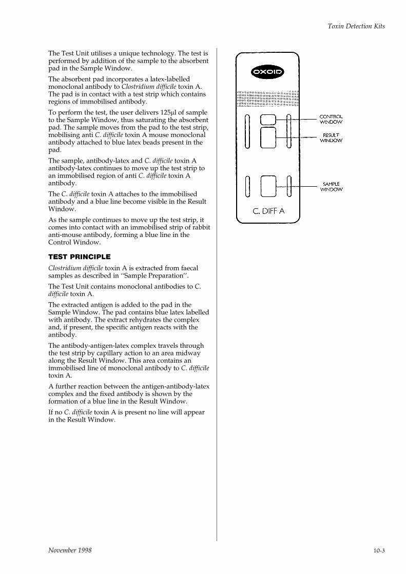
The Test Unit utilises a unique technology. The test isperformed by addition of the sample to the absorbentpad in the Sample Window.
The absorbent pad incorporates a latex-labelledmonoclonal antibody to Clostridium difficile toxin A.The pad is in contact with a test strip which containsregions of immobilised antibody.
To perform the test, the user delivers 125ml of sampleto the Sample Window, thus saturating the absorbentpad. The sample moves from the pad to the test strip,mobilising anti C. difficile toxin A mouse monoclonalantibody attached to blue latex beads present in thepad.
The sample, antibody-latex and C. difficile toxin Aantibody-latex continues to move up the test strip toan immobilised region of anti C. difficile toxin Aantibody.
The C. difficile toxin A attaches to the immobilisedantibody and a blue line become visible in the ResultWindow.
As the sample continues to move up the test strip, itcomes into contact with an immobilised strip of rabbitanti-mouse antibody, forming a blue line in theControl Window.
TEST PRINCIPLE
Clostridium difficile toxin A is extracted from faecalsamples as described in ``Sample Preparation''.
The Test Unit contains monoclonal antibodies to C.difficile toxin A.
The extracted antigen is added to the pad in theSample Window. The pad contains blue latex labelledwith antibody. The extract rehydrates the complexand, if present, the specific antigen reacts with theantibody.
The antibody-antigen-latex complex travels throughthe test strip by capillary action to an area midwayalong the Result Window. This area contains animmobilised line of monoclonal antibody to C. difficiletoxin A.
A further reaction between the antigen-antibody-latexcomplex and the fixed antibody is shown by theformation of a blue line in the Result Window.
If no C. difficile toxin A is present no line will appearin the Result Window.
Toxin Detection Kits
November 1998 10-3

11INDEX
Toxin Detection Kits
11-4 November 1998
PRODUCT INDEX
AAcid Egg Medium. . . . . . . . . . . . . . . . . . . . . . . . . . . 4-20`Actidione' Agar . . . . . . . . . . . . . . . . . . . . . . . . . . . . 4-18Aeromonas Medium (Ryan) . . . . . . . . . . . . . . . . . . . . . 2-32Agars . . . . . . . . . . . . . . . . . . . . . . . . . . . . . . . . . . 3-14Agar Bacteriological. . . . . . . . . . . . . . . . . . . . . . . . . . 3-15Agar Purified. . . . . . . . . . . . . . . . . . . . . . . . . . . . . . 3-15Agar Tablets (Agar No.3) . . . . . . . . . . . . . . . . . . . . . . 3-15Agar Technical (Agar No.3) . . . . . . . . . . . . . . . . . . . . . 3-15Amies Transport Medium . . . . . . . . . . . . . . . . . . . . . . 2-33Ampicillin Selective Supplement . . . . . . . . . . . . . . . . 2-32, 4-3Anaerobe Basal Agar . . . . . . . . . . . . . . . . . . . . . . . . . 2-34Anaerobe Basal Broth. . . . . . . . . . . . . . . . . . . . . . . . . 2-35Anaerobic gas generating kits . . . . . . . . . . . . . . . . . . . . . 7-2Anaerobic Indicators . . . . . . . . . . . . . . . . . . . . . . . . . . 7-6Anaerobic Jar. . . . . . . . . . . . . . . . . . . . . . . . . . . . . . 7-2, 3Anaerobic Selective Medium . . . . . . . . . . . . . . . . . . . . 2-219Anaerobe Selective Supplements . . . . . . . . . . . . . . . 2-219, 4-3AnaeroGen . . . . . . . . . . . . . . . . . . . . . . . . . . . . . . . . 7-6AnaeroGen Compact . . . . . . . . . . . . . . . . . . . . . . . . . . 7-7AnaeroJar . . . . . . . . . . . . . . . . . . . . . . . . . . . . . . . . . 7-8An-Ident Discs . . . . . . . . . . . . . . . . . . . . . . . . . . . . . . 5-8Antibiotic Assay Media . . . . . . . . . . . . . . . . . . . . . . . 2-35Antibiotic Medium No.1 . . . . . . . . . . . . . . . . . . . . . . . 2-37Antibiotic Medium No.2 . . . . . . . . . . . . . . . . . . . . . . . 2-38Antibiotic Medium No.3 . . . . . . . . . . . . . . . . . . . . . . . 2-38Antimicrobial Susceptibility Testing . . . . . . . . . . . . . . . . . 6-1Antimicrobial Susceptibility Testing - aura . . . . . . . . . . . . . 6-3Arcobacter Broth . . . . . . . . . . . . . . . . . . . . . . . . . . . 2-39Assay Media . . . . . . . . . . . . . . . . . . . . . . . . . . . . . . 2-35Atmosphere Generation System . . . . . . . . . . . . . . . . . . . 7-6aura - Complete Susceptibility System . . . . . . . . . . . . . . . . 6-3Azide Blood Agar Base . . . . . . . . . . . . . . . . . . . . . . . . 2-39Azide Dextrose Broth (Rothe) . . . . . . . . . . . . . . . . . . . . 2-40
BBacillus cereus Selective Agar Base. . . . . . . . . . . . . . . . . 2-41Bacillus cereus Selective Supplement . . . . . . . . . . . . . 2-41, 4-3Bacitracin Discs . . . . . . . . . . . . . . . . . . . . . . . . . . . . 5-12Baird-Parker Agar Base. . . . . . . . . . . . . . . . . . . . . . . . 2-43Barbitone Complement Fixation Test Diluent . . . . . . . . . . . 5-1Basic Fuchsin . . . . . . . . . . . . . . . . . . . . . . . . . 2-97, 145, 5-2BCET-RPLA . . . . . . . . . . . . . . . . . . . . . . . . . . . . . . 10-1Beef Extract . . . . . . . . . . . . . . . . . . . . . . . . . . . . . . . 3-10Beta Lactamase Broad Spectrum Mixture . . . . . . . . . . . . . 4-14Beta Lactamase Detection Papers . . . . . . . . . . . . . . . . . . . 5-3Beta Lactamase Touch Sticks . . . . . . . . . . . . . . . . . . . . . 5-4Biggy Agar . . . . . . . . . . . . . . . . . . . . . . . . . . . . . . . 2-45Bile Aesculin Agar . . . . . . . . . . . . . . . . . . . . . . . . . . 2-46Bile Salts . . . . . . . . . . . . . . . . . . . . . . . . . . . . . . 3-16, 17Bile Salts No.3 . . . . . . . . . . . . . . . . . . . . . . . . . . . . . 3-17Biochemical Reagents . . . . . . . . . . . . . . . . . . . . . . . . . . 5-1Biological extracts . . . . . . . . . . . . . . . . . . . . . . . . . . . . 3-1Bismuth Sulphite Agar . . . . . . . . . . . . . . . . . . . . . . . . 2-47Blaser-Wang (Campylobacter) Selective Medium . . . . . . 2-64, 4-4Blood Agar Base . . . . . . . . . . . . . . . . . . . . . . . . . . . . 2-48Blood Agar Base No.2 . . . . . . . . . . . . . . . . . . . . . . . . 2-49Blood Agar Base (Sheep) . . . . . . . . . . . . . . . . . . . . . . . 2-50Blood Culture Systems . . . . . . . . . . . . . . . . . . . . . . . . . 8-1Blood Culture System `Isolator' . . . . . . . . . . . . . . . . . . 8-5, 10Blood Culture System `SIGNAL' . . . . . . . . . . . . . . . . . . . 8-3Blood Products. . . . . . . . . . . . . . . . . . . . . . . . . . . . . 5-13Bordetella pertussis Selective Media . . . . . . . . . . . . . . . . 2-50Bordetella Selective Supplement . . . . . . . . . . . . . . . . 2-51, 4-3Brain Heart Infusion . . . . . . . . . . . . . . . . . . . . . . . . . 2-51Brain Heart Infusion Agar . . . . . . . . . . . . . . . . . . . . . . 2-52Brewers Thioglycollate Medium . . . . . . . . . . . . . . . . . . 2-199Brilliant Green Agar. . . . . . . . . . . . . . . . . . . . . . . . . . 2-53Brilliant Green Agar (Modified). . . . . . . . . . . . . . . . . . . 2-54Brilliant Green Bile (2%) Broth . . . . . . . . . . . . . . . . . . . 2-56Brucella Selective Media . . . . . . . . . . . . . . . . . . . . . . . 2-57BMPA (Legionella) Selective Medium . . . . . . . . . . . . . . . 2-120Buffered charcoal yeast extract agar (BCYE) . . . . . . . . . . . 2-120Buffered Listeria Enrichment Broth . . . . . . . . . . . . . . . . 2-126Buffered Peptone Water . . . . . . . . . . . . . . . . . . . . . . . 2-59
C`Calgon' Ringers Solution . . . . . . . . . . . . . . . . . . . . . . . 5-6CampyGen . . . . . . . . . . . . . . . . . . . . . . . . . . . . . . . . 7-9CampyGen Compact . . . . . . . . . . . . . . . . . . . . . . . . . 7-10Campylobacter Agar Base . . . . . . . . . . . . . . . . . . . . 2-63, 66Campylobacter Agar Base (Karmali) . . . . . . . . . . . . . . . . 2-65Campylobacter Blood-Free Selective Agar Base . . . . . . . . . 2-67Campylobacter Growth Supplement. . . . . . . . . . . . . . 2-64, 4-4Campylobacter Selective Media. . . . . . . . . . . . . . . . . . . 2-61Campylobacter Selective Supplement (Blaser-Wang) . . . . 2-64, 4-4Campylobacter Selective Supplement (Butzler) . . . . . . . 2-64, 4-4Campylobacter Selective Supplement (Karmali) . . . . . . . 2-65, 4-5Campylobacter Selective Supplement (Skirrow) . . . . . . . 2-63, 4-4Campylobacter Selective Supplement (Preston) . . . . . . . 2-66, 4-4Campylobacter Test Kit . . . . . . . . . . . . . . . . . . . . . . . . 9-1Cary-Blair Medium . . . . . . . . . . . . . . . . . . . . . . . . . . 2-69Casein Hydrolysate . . . . . . . . . . . . . . . . . . . . . . . . . . . 3-9CAT Supplement . . . . . . . . . . . . . . . . . . . . . . . . . 2-68, 4-5Catalyst - Anaerobic Jar . . . . . . . . . . . . . . . . . . . . . . . . 7-5CCDA Selective Supplement . . . . . . . . . . . . . . . . . . 2-67, 4-5CDMN Selective Supplement . . . . . . . . . . . . . . . . . . 2-79, 4-6CFC Pseudomonas Supplement . . . . . . . . . . . . . . . 2-171, 4-5C. Difficile Toxin A Test . . . . . . . . . . . . . . . . . . . . . . . 10-2Charcoal Agar . . . . . . . . . . . . . . . . . . . . . . . . . . 2-50, 2-70China Blue Lactose Agar . . . . . . . . . . . . . . . . . . . . . . . 2-71Chloramphenicol Selective Supplement . . . . . . . 2-92, 94, 180, 4-6Cholera Medium TCBS . . . . . . . . . . . . . . . . . . . . . . . . 2-72Chromogenic E. Coli/Coliform Medium . . . . . . . . . . . . . 2-73Chromogenic Urinary Tract Infection (UTI) Medium . . . . . . 2-74Clausen Medium . . . . . . . . . . . . . . . . . . . . . . . . . . . 2-75CLED Medium. . . . . . . . . . . . . . . . . . . . . . . . . . . . . 2-77CLED Medium (with Andrade indicator) . . . . . . . . . . . . . 2-77Clostridium difficile Agar Base . . . . . . . . . . . . . . . . . . . 2-78Clostridium difficile Selective Supplement . . . . . . . . . . 2-78, 4-6Clostridium perfringens Selective Media . . . . . . . . . . . . . 2-166COBA Streptococcus Selective Medium . . . . . . . . . . . . . . 2-195CO2Gen . . . . . . . . . . . . . . . . . . . . . . . . . . . . . . . . . 7-10CO2Gen Compact . . . . . . . . . . . . . . . . . . . . . . . . . . . 7-11Columbia Blood Agar Base . . . . . . . . . . . . . . . . . . . . . 2-80Cooked Meat Medium . . . . . . . . . . . . . . . . . . . . . . . . 2-81Columbia CNA Agar . . . . . . . . . . . . . . . . . . . . . . . . . 2-194Complement Test Diluent . . . . . . . . . . . . . . . . . . . . . . . 5-2CN Pseudomonas Supplement . . . . . . . . . . . . . . . . 2-171, 4-5Corn Meal Agar . . . . . . . . . . . . . . . . . . . . . . . . . . . . 2-82Corynebacterium diphtheriae Selective Medium . . . . . . . . 2-201Crossley Milk Medium . . . . . . . . . . . . . . . . . . . . . . . . 2-83CT Supplement . . . . . . . . . . . . . . . . . . . . . . . . . 2-141, 4-6Culti-Loops . . . . . . . . . . . . . . . . . . . . . . . . . . . . . . . 2-229Czapek Dox Agar . . . . . . . . . . . . . . . . . . . . . . . . . . . 2-84Czapek Dox Liquid Medium (Modified) . . . . . . . . . . . . . 2-85
DDCA . . . . . . . . . . . . . . . . . . . . . . . . . . . . . . . . . . . 2-88DCA Hynes . . . . . . . . . . . . . . . . . . . . . . . . . . . . . . 2-88DCLS Agar . . . . . . . . . . . . . . . . . . . . . . . . . . . . . . . 2-85Dermasel Agar Base. . . . . . . . . . . . . . . . . . . . . . . . . . 2-86Dermatophyte Selective Medium . . . . . . . . . . . . . . . . . . 2-86Dermasel Selective Supplement. . . . . . . . . . . . . . . . . 2-86, 4-6Desoxycholate Agar. . . . . . . . . . . . . . . . . . . . . . . . . . 2-87Dextrose Bacteriological . . . . . . . . . . . . . . . . . . . . . . . 3-17Dextrose Tryptone Agar . . . . . . . . . . . . . . . . . . . . . . . 2-89Dextrose Tryptone Broth . . . . . . . . . . . . . . . . . . . . . . . 2-90Diagnostic Discs . . . . . . . . . . . . . . . . . . . . . . . . . . . . . 5-8Diagnostic Kits and Reagents . . . . . . . . . . . . . . . . . . . . . 9-1Diagnostic Sensitivity Test Agar (DSTA) . . . . . . . . . . . . . 2-91Dichloran-Glycerol (DG 18) Agar Base . . . . . . . . . . . . . . 2-92Dichloran Rose Bengal Chloramphenicol Agar. . . . . . . . . . 2-94Dip Slides . . . . . . . . . . . . . . . . . . . . . . . . . . . . . . . . 4-23Disc dispensers . . . . . . . . . . . . . . . . . . . . . . . . . . . . . 6-3DNase Agar . . . . . . . . . . . . . . . . . . . . . . . . . . . . . . 2-93DRBC Agar Base. . . . . . . . . . . . . . . . . . . . . . . . . . . . 2-94Dryspot E. Coli O157 Latex Test . . . . . . . . . . . . . . . . . . . 9-8Dryspot IM . . . . . . . . . . . . . . . . . . . . . . . . . . . . . . . . 9-8Dryspot Staphytect Plus . . . . . . . . . . . . . . . . . . . . . . . . 9-8Dryspot Streptococcal Grouping Kit . . . . . . . . . . . . . . . . . 9-9DST Agar . . . . . . . . . . . . . . . . . . . . . . . . . . . . . . . . 2-91Dulbecco A . . . . . . . . . . . . . . . . . . . . . . . . . . . . . . . . 5-2
Index
November 1998 11-1
EE. coli O157 Latex Test . . . . . . . . . . . . . . . . . . . . . . . . . 9-2E. coli ST EIA. . . . . . . . . . . . . . . . . . . . . . . . . . . . . . 10-2Edwards Medium (Modified) . . . . . . . . . . . . . . . . . . . . 2-95EE Broth. . . . . . . . . . . . . . . . . . . . . . . . . . . . . . . . . 2-95Egg Yolk Emulsion . . . . . . . . . . . . . . . . . . . . . . . . . . 4-13Endo Agar Base . . . . . . . . . . . . . . . . . . . . . . . . . . . . 2-97Eosin Methylene Blue Agar (Modified) Levine . . . . . . . . . . 2-97Escherichia coli O157 Latex Test . . . . . . . . . . . . . . . . . . . 9-1
FFildes Extract . . . . . . . . . . . . . . . . . . . . . . . . . . . . . . 4-14Fluid Sabouraud Medium . . . . . . . . . . . . . . . . . . . . . . 2-183Fluid Thioglycollate Medium USP . . . . . . . . . . . . . . . . . 2-200Fraser Broth . . . . . . . . . . . . . . . . . . . . . . . . . . 2-98, 2-131Fraser Supplement . . . . . . . . . . . . . . . . . . . . 2-98, 2-131, 4-6Fuchsin Basic . . . . . . . . . . . . . . . . . . . . . . . . 2-97, 145, 4-14
GGardnerella vaginalis Selective Medium . . . . . . . . . . . . . 2-99Gardnerella vaginalis Selective Supplement . . . . . . . . . 2-99, 4-7Gas Generating Kits. . . . . . . . . . . . . . . . . . . . . . . . . . 7-2, 4GBS Agar Base (ISLAM) . . . . . . . . . . . . . . . . . . . . . . . 2-101GC Agar Base . . . . . . . . . . . . . . . . . . . . . . . . . . . . . 2-102GC Selective Supplement . . . . . . . . . . . . . . . . . . . 2-103, 4-7Gelatin Bacteriological . . . . . . . . . . . . . . . . . . . . . . . . 3-17Giolitti-Cantoni Broth. . . . . . . . . . . . . . . . . . . . . . . . . 2-106Glucose Bacteriological . . . . . . . . . . . . . . . . . . . . . . . . 3-17G-N Anaerobe Supplements. . . . . . . . . . . . . . . . . . 2-219, 4-3
HHaemoglobin Powder Soluble . . . . . . . . . . . . . . . 2-102, 3-17Haemophilus Test Medium Base . . . . . . . . . . . . . . . . . . 2-107Haemophilus Test Medium Supplement . . . . . . . . . . . . . 2-107Half Fraser Supplement . . . . . . . . . . . . . . . . . . . . . 2-99, 4-6Hand Dispensers (Discs) . . . . . . . . . . . . . . . . . . . . . . . . 6-3Hektoen Enteric Agar. . . . . . . . . . . . . . . . . . . . . . . . . 2-108Helicobacter pylori Kit . . . . . . . . . . . . . . . . . . . . . . . . . 9-2Helicobacter pylori Selective Supplement . . . . . . . . . . 2-109, 4-7High Resolution (HR) Medium . . . . . . . . . . . . . . . . . . . 2-110Horse Serum . . . . . . . . . . . . . . . . . . . . . . . . . . . . . . 5-13Hoyle Medium. . . . . . . . . . . . . . . . . . . . . . . . . . . . . 2-109HS-T Broth . . . . . . . . . . . . . . . . . . . . . . . . . . . . . . . 2-75
IIdentification Touch Sticks. . . . . . . . . . . . . . . . . . . . . . . 5-4Indicator-Anaerobic. . . . . . . . . . . . . . . . . . . . . . . . . . . 7-6Infectious Mononucleosis Kit . . . . . . . . . . . . . . . . . . . . . 9-2Iron Sulphite Agar . . . . . . . . . . . . . . . . . . . . . . . . . . 2-111Islam GBS Agar base . . . . . . . . . . . . . . . . . . . . . . . . . 2-101`ISOLATOR' Blood Culture System . . . . . . . . . . . . . . . . 8-5, 7`Iso-Sensitest' Agar . . . . . . . . . . . . . . . . . . . . . . . . . . 2-112`Iso-Sensitest' Broth . . . . . . . . . . . . . . . . . . . . . . . . . . 2-114
KKanamycin Aesculin Azide Agar Base. . . . . . . . . . . . . . . 2-114Kanamycin Sulphate Selective Supplement . . . . . . . . . 2-114, 4-7KF Streptococcus Agar . . . . . . . . . . . . . . . . . . . . . . . . 2-115Kligler Iron Agar . . . . . . . . . . . . . . . . . . . . . . . . . . . 2-116
L`Lab-Lemco' Agar . . . . . . . . . . . . . . . . . . . . . . . . . . . 2-118`Lab-Lemco' Broth. . . . . . . . . . . . . . . . . . . . . . . . . . . 2-118`Lab-Lemco' Powder . . . . . . . . . . . . . . . . . . . . . . . . . 3-10Laboratory Preparations . . . . . . . . . . . . . . . . . . . . . . . . 3-1Lab Ready Media . . . . . . . . . . . . . . . . . . . . . . . . . . . 4-19Lactalbumin Hydrolysate . . . . . . . . . . . . . . . . . . . . . . . 3-9Lactic Acid 10% Solution. . . . . . . . . . . . . . . . . . . . . . . 4-14Lactose Bacteriological . . . . . . . . . . . . . . . . . . . . . . . . 3-17Lactose Broth. . . . . . . . . . . . . . . . . . . . . . . . . . . . . . 2-118Laked Horse Blood . . . . . . . . . . . . . . . . . . . . . . . . . . 5-13Lauryl Sulphate Broth . . . . . . . . . . . . . . . . . . . . . . . . 2-119Lauryl Tryptose Broth . . . . . . . . . . . . . . . . . . . . . . . . 2-119LCAT Selective Supplement . . . . . . . . . . . . . . . . . . 2-103, 4-7Legionella BCYE Growth Supplement. . . . . . . . . . . . 2-120, 4-8Legionella BMPA Selective Supplement. . . . . . . . . . . 2-120, 4-8Legionella CYE Agar Base . . . . . . . . . . . . . . . . . . . . . . 2-120Legionella (GVPC) Selective Supplement . . . . . . . . . . 2-123, 4-8Legionella MWY Selective Supplement . . . . . . . . . . . 2-121, 4-8
Legionella Latex Test . . . . . . . . . . . . . . . . . . . . . . . . . . 9-3Legionella Selective Media. . . . . . . . . . . . . . . . . . . . . . 2-120Levine EMB Agar . . . . . . . . . . . . . . . . . . . . . . . . . . . 2-97Listeria Enrichment Broth Base (UVM) . . . . . . . . . . . . . . 2-129Listeria Enrichment Broth Base . . . . . . . . . . . . . . . . . . . 2-129Listeria Primary Selective Enrichment Supplement
(UVMI) . . . . . . . . . . . . . . . . . . . . . . . . . . . . 2-130, 4-8Listeria Rapid Test . . . . . . . . . . . . . . . . . . . . . . . . . . 9-10Listeria Secondary Selective Enrichment Supplement
(UVMII) . . . . . . . . . . . . . . . . . . . . . . . . . . . . 2-130, 4-9Listeria Selective Agar Base (Oxford) . . . . . . . . . . . . . . . 2-127Listeria Selective Enrichment
Supplement. . . . . . . . . . . . . . . . . . . . . . 2-126, 2-129, 4-9Listeria Selective Supplement (Oxford) . . . . . . . . . . . 2-127, 4-9Liver Broth . . . . . . . . . . . . . . . . . . . . . . . . . . . . . . . 2-133Liver Desiccated . . . . . . . . . . . . . . . . . . . . . . . . . . . . 3-11Liver Digest Neutralised . . . . . . . . . . . . . . . . . . . . . . . . 3-5Low Temperature Catalyst . . . . . . . . . . . . . . . . . . . . . . 7-5Lowenstein-Jensen Media . . . . . . . . . . . . . . . . . . . . . . 4-20Lysine Decarboxylase Broth . . . . . . . . . . . . . . . . . . . . . 2-133Lysine Iron Agar. . . . . . . . . . . . . . . . . . . . . . . . . . . . 2-134Lysine Medium . . . . . . . . . . . . . . . . . . . . . . . . . . . . 2-135
MM17 Agar . . . . . . . . . . . . . . . . . . . . . . . . . . . . . . . . 2-136M17 Broth. . . . . . . . . . . . . . . . . . . . . . . . . . . . . . . . 2-137MacConkey Agar . . . . . . . . . . . . . . . . . . . . . . . . . . . 2-138MacConkey Agar (without salt) . . . . . . . . . . . . . . . . . . 2-139MacConkey Agar No.2 . . . . . . . . . . . . . . . . . . . . . . . . 2-139MacConkey Agar No.3 . . . . . . . . . . . . . . . . . . . . . . . . 2-139MacConkey Broth . . . . . . . . . . . . . . . . . . . . . . . . . . . 2-142MacConkey Broth Purple . . . . . . . . . . . . . . . . . . . . . . 2-142MacConkey Sorbitol Agar . . . . . . . . . . . . . . . . . . . . . . 2-140Malt Extract . . . . . . . . . . . . . . . . . . . . . . . . . . . . . . 3-10Malt Extract Agar . . . . . . . . . . . . . . . . . . . . . . . . . . . 2-143Malt Extract Broth. . . . . . . . . . . . . . . . . . . . . . . . . . . 2-143Mannitol Salt Agar . . . . . . . . . . . . . . . . . . . . . . . . . . 2-144Mannitol Selenite Broth Base . . . . . . . . . . . . . . . . . . . . 2-187Maximum Recovery Diluent . . . . . . . . . . . . . . . . . . . . 2-144Membrane Endo Agar LES . . . . . . . . . . . . . . . . . . . . . 2-145Membrane Lauryl Sulphate Broth . . . . . . . . . . . . . . . . . 2-146Metronidazole Diagnostic Discs . . . . . . . . . . . . . . . . . . 5-10Milk Agar. . . . . . . . . . . . . . . . . . . . . . . . . . . . . . . . 2-147Milk Plate Count Agar . . . . . . . . . . . . . . . . . . . . . . . . 2-170Minerals Modified Glutamate Medium . . . . . . . . . . . . . . 2-148MLCB Agar. . . . . . . . . . . . . . . . . . . . . . . . . . . . . . . 2-150Modified Acid Egg Medium. . . . . . . . . . . . . . . . . . . . . 4-21Modified Lauryl Tryptose Broth With MUG . . . . . . . . . . . 2-151Modified Lowenstein-Jensen Medium . . . . . . . . . . . . . . . 4-21Modified Pyruvic Acid Egg Medium . . . . . . . . . . . . . . . 4-22Modified Semi-Solid Rappaport Vassiliadis (MSRV)
Medium Base. . . . . . . . . . . . . . . . . . . . . . . . . . . . 2-152Modified Wadowsky Yee Legionella Medium . . . . . . . . . . 2-121MRS Agar. . . . . . . . . . . . . . . . . . . . . . . . . . . . . . . . 2-153MRS Broth . . . . . . . . . . . . . . . . . . . . . . . . . . . . . . . 2-154MRVP Medium . . . . . . . . . . . . . . . . . . . . . . . . . . . . 2-155MSRV Selective Supplement . . . . . . . . . . . . . . . . . 2-152, 4-9Mueller Hinton Agar . . . . . . . . . . . . . . . . . . . . . . . . . 2-156Mueller Hinton Broth . . . . . . . . . . . . . . . . . . . . . . . . . 2-157Muller Kauffman Tetrathionate Broth Base . . . . . . . . . . . . 2-157MUG Reagent . . . . . . . . . . . . . . . . . . . . . . . . . . . 2-56, 5-2Mycobacteria Media . . . . . . . . . . . . . . . . . . . . . . . . . 4-21Mycological Peptone . . . . . . . . . . . . . . . . . . . . . . . . . . 3-7Mycoplasma Agar Base . . . . . . . . . . . . . . . . . . . . . . . 2-158Mycoplasma Broth Base . . . . . . . . . . . . . . . . . . . . . . . 2-160Mycoplasma Selective Supplement G . . . . . . . . . . . . 2-158, 4-9Mycoplasma Selective Supplement P . . . . . . . . . . . . 2-159, 4-9
NNeisseria Gonorrhoeae (GC) Selective Media. . . . . . . . . . . 2-102Nitrocefin . . . . . . . . . . . . . . . . . . . . . . . . . . . . . . . . 4-15Nitrous Acid (Streptococcal Grouping Kit) . . . . . . . . . . . . . 9-5N-S Anaerobe Selective Supplement . . . . . . . . . . . . . 2-219, 4-3New York City (GC) Medium (Modified) . . . . . . . . . . 2-105, 160Novobiocin Supplement . . . . . . . . . . . . . . . . . . . . . . . 9-11Nutrient Agar . . . . . . . . . . . . . . . . . . . . . . . . . . . . . 2-161Nutrient Broth . . . . . . . . . . . . . . . . . . . . . . . . . . . . . 2-161Nutrient Broth No.2. . . . . . . . . . . . . . . . . . . . . . . . . . 2-161Nutrient Gelatin . . . . . . . . . . . . . . . . . . . . . . . . . . . . 2-162
Index
11-2 November 1998
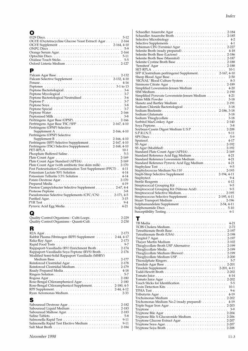
O0129 Discs. . . . . . . . . . . . . . . . . . . . . . . . . . . . . . . . 5-12OGYE (Oxytetracycline Glucose Yeast Extract) Agar . . . . . . 2-164OGYE Supplement . . . . . . . . . . . . . . . . . . . . . . 2-164, 4-10ONPG Discs . . . . . . . . . . . . . . . . . . . . . . . . . . . . . . . 5-9Orange Serum Agar. . . . . . . . . . . . . . . . . . . . . . . . . . 2-164Optochin Discs. . . . . . . . . . . . . . . . . . . . . . . . . . . . . 5-11Oxidase Touch Sticks . . . . . . . . . . . . . . . . . . . . . . . . . . 5-5Oxford Listeria Medium . . . . . . . . . . . . . . . . . . . . . . . 2-127
PPalcam Agar Base . . . . . . . . . . . . . . . . . . . . . . . . . . . 2-132Palcam Selective Supplement . . . . . . . . . . . . . . . . 2-132, 4-10Penase . . . . . . . . . . . . . . . . . . . . . . . . . . . . . . . . . . 4-16Peptones . . . . . . . . . . . . . . . . . . . . . . . . . . . . . . 3-1 to 13Peptone Bacteriological . . . . . . . . . . . . . . . . . . . . . . . . . 3-6Peptone Mycological . . . . . . . . . . . . . . . . . . . . . . . . . . 3-7Peptone Bacteriological Neutralised . . . . . . . . . . . . . . . . . 3-6Peptone P . . . . . . . . . . . . . . . . . . . . . . . . . . . . . . . . . 3-7Peptone Soya. . . . . . . . . . . . . . . . . . . . . . . . . . . . . . . 3-8Peptone Special . . . . . . . . . . . . . . . . . . . . . . . . . . . . . 3-7Peptone Water . . . . . . . . . . . . . . . . . . . . . . . . . . . . . 2-166Peptonised Milk . . . . . . . . . . . . . . . . . . . . . . . . . . . . . 3-8Perfringens Agar Base (OPSP). . . . . . . . . . . . . . . . . . . . 2-166Perfringens Agar Base TSC/SFP . . . . . . . . . . . . . . 2-167, 4-10Perfringens (OPSP) Selective
Supplement A . . . . . . . . . . . . . . . . . . . . . . . 2-166, 4-10Perfringens (OPSP) Selective
Supplement B . . . . . . . . . . . . . . . . . . . . . . . 2-166, 4-10Perfringens (SFP) Selective Suppplement . . . . . . . . . 2-167, 4-10Perfringens (TSC) Selective Suppplement . . . . . . . . . 2-168, 4-10PET-RPLA . . . . . . . . . . . . . . . . . . . . . . . . . . . . . . . 10-1Phosphate Buffered Saline . . . . . . . . . . . . . . . . . . . . . . . 5-3Plate Count Agar . . . . . . . . . . . . . . . . . . . . . . . . . . . 2-169Plate Count Agar Standard (APHA) . . . . . . . . . . . . . . . . 2-169Plate Count Agar (with antibiotic free skim milk) . . . . . . . . 2-170Post Pasteurisation Contamination Test Supplement (PPCT) . 4-11Potassium Lactate 50% Solution . . . . . . . . . . . . . . . . . . 4-14Potassium Tellurite 3.5% Solution . . . . . . . . . . . . . . . . . 4-14Potato Dextrose Agar . . . . . . . . . . . . . . . . . . . . . . . . . 2-170Prepared Media . . . . . . . . . . . . . . . . . . . . . . . . . . . . 4-19Preston Campylobacter Selective Supplement . . . . . . . . 2-67, 4-4Proteose Peptone . . . . . . . . . . . . . . . . . . . . . . . . . . . . 3-8Pseudomonas Selective Supplements (CFC/CN) . . . . . 2-171, 4-5Purified Agar. . . . . . . . . . . . . . . . . . . . . . . . . . . . . . 3-15PYR Test . . . . . . . . . . . . . . . . . . . . . . . . . . . . . . . . . 9-4Pyruvic Acid Egg Media . . . . . . . . . . . . . . . . . . . . . . . 4-21
QQuality Control Organisms - Culti-Loops . . . . . . . . . . . . . 2-229Quality Control Organisms - Quanti-Cult. . . . . . . . . . . . . 2-230
RR2A Agar . . . . . . . . . . . . . . . . . . . . . . . . . . . . . . . . 2-173Rabbit Plasma Fibrinogen (RPF) Supplement . . . . . . . 2-44, 4-17Raka-Ray Agar. . . . . . . . . . . . . . . . . . . . . . . . . . . . . 2-173Rapid Food Tests . . . . . . . . . . . . . . . . . . . . . . . . . . . . 9-7Rappaport-Vassiliadis (RV) Enrichment Broth . . . . . . . . . . 2-174Rappaport-Vassiliadis Soya Peptone (RVS) Broth . . . . . . . . 2-176Modified Semi-Solid Rappaport Vassiliadis (MSRV)
Medium Base. . . . . . . . . . . . . . . . . . . . . . . . . . . . 2-177Reinforced Clostridial Agar . . . . . . . . . . . . . . . . . . . . . 2-178Reinforced Clostridial Medium . . . . . . . . . . . . . . . . . . . 2-179Ready Prepared Media . . . . . . . . . . . . . . . . . . . . . . . . 4-18Ringers Solution . . . . . . . . . . . . . . . . . . . . . . . . . . . . . 5-7Rogosa Agar . . . . . . . . . . . . . . . . . . . . . . . . . . . . . . 2-180Rose-Bengal Chloramphenicol Agar . . . . . . . . . . . . . . . . 2-180Rose-Bengal Chloramphenicol Supplement. . . . . . . . . 2-180, 4-5RPF Supplement . . . . . . . . . . . . . . . . . . . . . . . . . 2-44, 4-12Ryan Aeromonas Medium. . . . . . . . . . . . . . . . . . . . . . 2-33
SSabouraud Dextrose Agar . . . . . . . . . . . . . . . . . . . . . . 2-182Sabouraud Liquid Medium . . . . . . . . . . . . . . . . . . . . . 2-183Sabouraud Maltose Agar. . . . . . . . . . . . . . . . . . . . . . . 2-183Saline Tablets. . . . . . . . . . . . . . . . . . . . . . . . . . . . . . . 5-8Salmonella Rapid Test . . . . . . . . . . . . . . . . . . . . . . . . 9-11Salmonella Rapid Test Elective Medium . . . . . . . . . . . . . 9-11Salt Meat Broth . . . . . . . . . . . . . . . . . . . . . . . . . . . . 2-184
Schaedler Anaerobe Agar . . . . . . . . . . . . . . . . . . . . . . 2-184Schaedler Anaerobe Broth . . . . . . . . . . . . . . . . . . . . . . 2-185Selective Microbiology . . . . . . . . . . . . . . . . . . . . . . . . . 4-2Selective Supplements . . . . . . . . . . . . . . . . . . . . . . . . . 4-1Schieman CIN (Yersinia) Agar . . . . . . . . . . . . . . . . . . . 2-227Selenite Broth (ready prepared) . . . . . . . . . . . . . . . . . . . 4-18Selenite Broth Base (Lactose) . . . . . . . . . . . . . . . . . . . . 2-186Selenite Broth Base (Mannitol) . . . . . . . . . . . . . . . . . . . 2-187Selenite Cystine Broth Base . . . . . . . . . . . . . . . . . . . . . 2-188`Sensitest' Agar . . . . . . . . . . . . . . . . . . . . . . . . . . . . 2-188SET-RPLA . . . . . . . . . . . . . . . . . . . . . . . . . . . . . . . 10-1SFP (Clostridium perfringens) Supplement . . . . . . . . 2-167, 4-10Sheep Blood Agar Base . . . . . . . . . . . . . . . . . . . . . . . . 2-50`SIGNAL' Blood Culture System . . . . . . . . . . . . . . . . . . . 8-3Simmons Citrate Agar . . . . . . . . . . . . . . . . . . . . . . . . 2-189Simplified Lowenstein-Jensen Medium . . . . . . . . . . . . . . 4-20SIM Medium . . . . . . . . . . . . . . . . . . . . . . . . . . . . . . 2-190Simplified Pyruvate Lowenstein-Jensen Medium . . . . . . . . 4-21Skim Milk Powder . . . . . . . . . . . . . . . . . . . . . . . . . . 3-18Slanetz and Bartley Medium . . . . . . . . . . . . . . . . . . . . 2-191Sodium Chloride Bacteriological . . . . . . . . . . . . . . . . . . 3-18Sodium Biselenite . . . . . . . . . . . . . . . . . . . . . . . 2-186, 3-18Sodium Glutamate . . . . . . . . . . . . . . . . . . . . . . . . . . 3-18Sodium Thioglycollate . . . . . . . . . . . . . . . . . . . . . . . . 3-18Sorbitol MacConkey Agar . . . . . . . . . . . . . . . . . . . . . . 2-140Soya Peptone . . . . . . . . . . . . . . . . . . . . . . . . . . . . . . . 3-8Soybean-Casein Digest Medium U.S.P. . . . . . . . . . . . . . . 2-208S.P.R.I.N.T. . . . . . . . . . . . . . . . . . . . . . . . . . . . . . . . 9-11SPS Discs . . . . . . . . . . . . . . . . . . . . . . . . . . . . . . . . . 5-9Sputasol . . . . . . . . . . . . . . . . . . . . . . . . . . . . . . . . . 4-17SS Agar . . . . . . . . . . . . . . . . . . . . . . . . . . . . . . . . . 2-192SS Agar (Modified) . . . . . . . . . . . . . . . . . . . . . . . . . . 2-192Standard Plate Count Agar (APHA) . . . . . . . . . . . . . . . . 2-169Standard Reference Acid Egg Medium . . . . . . . . . . . . . . 4-21Standard Reference Lowenstein Medium . . . . . . . . . . . . . 4-21Standard Reference Pyruvic Acid Egg Medium . . . . . . . . . 4-21Staphylase Test . . . . . . . . . . . . . . . . . . . . . . . . . . . . . 9-5Staphylococcus Medium No.110 . . . . . . . . . . . . . . . . . . 2-193Staph-Strep Selective Supplement . . . . . . . . . . . . . 2-194, 4-11Staphytect. . . . . . . . . . . . . . . . . . . . . . . . . . . . . . . . . 9-4Sterile Reagents . . . . . . . . . . . . . . . . . . . . . . . . . . . . 4-12Streptococcal Grouping Kit . . . . . . . . . . . . . . . . . . . . . . 9-5Streptococcal Grouping Kit (Nitrous Acid) . . . . . . . . . . . . . 9-5Streptococcal Selective Medium. . . . . . . . . . . . . . . . . . . 2-195Streptococcus Selective Supplement . . . . . . . . . . . . 2-195, 4-11Stuart Transport Medium . . . . . . . . . . . . . . . . . . . . . . 2-196Sulphamandelate Supplement . . . . . . . . . . . . . . . . 2-54, 4-11Sulphonamide Discs . . . . . . . . . . . . . . . . . . . . . . . . . 5-10Susceptibility Testing . . . . . . . . . . . . . . . . . . . . . . . . . . 6-1
TTB Media . . . . . . . . . . . . . . . . . . . . . . . . . . . . . . . . 4-21TCBS Cholera Medium . . . . . . . . . . . . . . . . . . . . . . . . 2-72Tetrathionate Broth Base . . . . . . . . . . . . . . . . . . . . . . . 2-197Tetrathionate Broth (USA) . . . . . . . . . . . . . . . . . . . . . . 2-198Tergitol 7 Agar. . . . . . . . . . . . . . . . . . . . . . . . . . . . . 2-197Thayer Martin Medium . . . . . . . . . . . . . . . . . . . . . . . 2-102Thioglycollate Broth USP Alternative . . . . . . . . . . . . . . . 2-199Thioglycollate Media . . . . . . . . . . . . . . . . . . . . . . . . . 2-199Thioglycollate Medium (Brewer) . . . . . . . . . . . . . . . . . . 2-199Thioglycollate Medium USP. . . . . . . . . . . . . . . . . . . . . 2-200Thiosulphate Ringers . . . . . . . . . . . . . . . . . . . . . . . . . . 5-7Tinsdale Agar Base . . . . . . . . . . . . . . . . . . . . . . . . . . 2-201Tinsdale Supplement . . . . . . . . . . . . . . . . . . . . . 2-201, 4-11Todd-Hewitt Broth . . . . . . . . . . . . . . . . . . . . . . . . . . 2-202Tomato Juice . . . . . . . . . . . . . . . . . . . . . . . . . . . . . . 4-14Tomato Juice Agar . . . . . . . . . . . . . . . . . . . . . . . . . . 2-202Touch Sticks for Identification. . . . . . . . . . . . . . . . . . . . . 5-5Toxin Detection Kits . . . . . . . . . . . . . . . . . . . . . . . . . 10-1TPHA Test . . . . . . . . . . . . . . . . . . . . . . . . . . . . . . . . 9-6Tributyrin Agar . . . . . . . . . . . . . . . . . . . . . . . . . . . . 4-19Trichomonas Medium . . . . . . . . . . . . . . . . . . . . . . . . 2-202Trichomonas Medium No.2 (ready prepared) . . . . . . . . . . 4-19Triple Sugar Iron Agar . . . . . . . . . . . . . . . . . . . . . . . . 2-203Tryptone . . . . . . . . . . . . . . . . . . . . . . . . . . . . . . . . . 3-9Tryptone Bile Agar . . . . . . . . . . . . . . . . . . . . . . . . . . 2-204Tryptone Bile X-Glucuronide Medium. . . . . . . . . . . . . . . 2-206Tryptone Glucose Extract Agar . . . . . . . . . . . . . . . . . . . 2-207Tryptone Soya Agar. . . . . . . . . . . . . . . . . . . . . . . . . . 2-207Tryptone Soya Broth . . . . . . . . . . . . . . . . . . . . . . . . . 2-208
Index
November 1998 11-3

Tryptone T . . . . . . . . . . . . . . . . . . . . . . . . . . . . . . . . 3-9Tryptone Water . . . . . . . . . . . . . . . . . . . . . . . . . . . . 2-209Tryptose. . . . . . . . . . . . . . . . . . . . . . . . . . . . . . . . . . 3-7Tryptose Blood Agar Base . . . . . . . . . . . . . . . . . . . . . . 2-209Tryptose Phosphate Broth . . . . . . . . . . . . . . . . . . . . . . 2-210TTC Solution (0.05%) . . . . . . . . . . . . . . . . . . . . . 2-197, 4-18TSC (Clostridium perfringens) Supplement. . . . . . . . 2-168, 4-10TST-RPLA . . . . . . . . . . . . . . . . . . . . . . . . . . . . . . . 10-1
UUniversal Beer Agar. . . . . . . . . . . . . . . . . . . . . . . . . . 2-212Urea Agar Base . . . . . . . . . . . . . . . . . . . . . . . . . . . . 2-212Urea Broth Base . . . . . . . . . . . . . . . . . . . . . . . . . . . . 2-213Urea 40% Solution. . . . . . . . . . . . . . . . . . . . . . . . . . . 4-14
VVCAT Selective Supplement. . . . . . . . . . . . . . . . . 2-104, 4-11VCN Selective Supplement . . . . . . . . . . . . . . . . . 2-103, 4-12VCNT Selective Supplement . . . . . . . . . . . . . . . . 2-103, 4-12VDRL Test Kit . . . . . . . . . . . . . . . . . . . . . . . . . . . . . . 9-7VET-RPLA . . . . . . . . . . . . . . . . . . . . . . . . . . . . . . . 10-1VTEC-RPLA . . . . . . . . . . . . . . . . . . . . . . . . . . . . . . 10-2V Factor Discs . . . . . . . . . . . . . . . . . . . . . . . . . . . . . 5-11Violet Red Bile Agars . . . . . . . . . . . . . . . . . . . . . . . . . 2-214Violet Red Bile Glucose Agar . . . . . . . . . . . . . . . . . . . . 2-216Violet Red Bile Lactose Agar . . . . . . . . . . . . . . . . . . . . 2-215Vitox . . . . . . . . . . . . . . . . . . . . . . . . . . . . . . . 2-103, 4-13
Vogel-Johnson Agar. . . . . . . . . . . . . . . . . . . . . . . . . . 2-217
WWadowsky Yee (Legionella) Medium . . . . . . . . . . . . . . . 2-121Wilkins Chalgren Anaerobe Agar . . . . . . . . . . . . . . . . . 2-219Wilkins Chalgren G-N Anaerobe Selective Supplement . 2-219, 4-3Wilkins Chalgren N-S Anaerobe Selective Supplement. . 2-219, 4-3Wilkins Chalgren Anaerobe Broth . . . . . . . . . . . . . . . . . 2-221WL Nutrient Agar. . . . . . . . . . . . . . . . . . . . . . . . . . . 2-222WL Nutrient Broth . . . . . . . . . . . . . . . . . . . . . . . . . . 2-222Wort Agar . . . . . . . . . . . . . . . . . . . . . . . . . . . . . . . 2-223
XX Factor Discs . . . . . . . . . . . . . . . . . . . . . . . . . . . . . 5-11X+V Factor Discs . . . . . . . . . . . . . . . . . . . . . . . . . . . 5-11XLD Medium . . . . . . . . . . . . . . . . . . . . . . . . . . . . . 2-224
YYeast Autolysate Growth Supplement. . . . . . . . . . . 2-102, 4-12Yeast Extract Agar . . . . . . . . . . . . . . . . . . . . . . . . . . 2-226Yeast and Mould Agar . . . . . . . . . . . . . . . . . . . . . . . . 2-226Yeast Extract Powder . . . . . . . . . . . . . . . . . . . . . . . . . 3-10Yersinia Selective Agar Base. . . . . . . . . . . . . . . . . . . . . 2-227Yersinia Selective Supplement . . . . . . . . . . . . . . . 2-227, 4-12
ZZaher & Marks (TB) Media . . . . . . . . . . . . . . . . . . . . . 4-21
CM1 Nutrient Broth . . . . . . . . . . . . . . . . . . . . . . . . . 2-1613 Nutrient Agar . . . . . . . . . . . . . . . . . . . . . . . . . 2-1615 MacConkey Broth . . . . . . . . . . . . . . . . . . . . . . . 2-1425a MacConkey Broth Purple (CM505) . . . . . . . . . . . . . 2-1426a MacConkey Broth Purple tablets (CM506) . . . . . . . . 2-1427 MacConkey Agar . . . . . . . . . . . . . . . . . . . . . . . 2-1387b MacConkey Agar (without Salt) (CM507). . . . . . . . . 2-1399 Peptone Water . . . . . . . . . . . . . . . . . . . . . . . . . 2-16615 Lab Lemco Broth . . . . . . . . . . . . . . . . . . . . . . . 2-11817 Lab Lemco Agar . . . . . . . . . . . . . . . . . . . . . . . . 2-11819 Yeast Extract Agar . . . . . . . . . . . . . . . . . . . . . . 2-22621 Milk Agar. . . . . . . . . . . . . . . . . . . . . . . . . . . . 2-14723 Thioglycollate Medium (Brewer) . . . . . . . . . . . . . . 2-19927 Edward's Medium . . . . . . . . . . . . . . . . . . . . . . 2-9529 Tetrathionate Broth Base . . . . . . . . . . . . . . . . . . . 2-19731 Brilliant Green Bile 2% Broth . . . . . . . . . . . . . . . . 2-5633 Kligler Iron Agar . . . . . . . . . . . . . . . . . . . . . . . 2-11635 Desoxycholate Citrate Agar . . . . . . . . . . . . . . . . . 2-8741 Sabouraud Dextrose Agar . . . . . . . . . . . . . . . . . . 2-18241a Sabouraud Maltose Agar. . . . . . . . . . . . . . . . . . . 2-18343 MRVP Medium . . . . . . . . . . . . . . . . . . . . . . . . 2-15549 Agar No.3 Tablets . . . . . . . . . . . . . . . . . . . . . . . 3-1553 Urea Agar Base . . . . . . . . . . . . . . . . . . . . . . . . 2-21255 Blood Agar Base . . . . . . . . . . . . . . . . . . . . . . . . 2-4857 Malt Extract Broth. . . . . . . . . . . . . . . . . . . . . . . 2-14359 Malt Extract Agar . . . . . . . . . . . . . . . . . . . . . . . 2-14367 Nutrient Broth No.2. . . . . . . . . . . . . . . . . . . . . . 2-16169 Eosin Methylene Blue Agar (Levine) . . . . . . . . . . . . 2-9771 Urea Broth Base . . . . . . . . . . . . . . . . . . . . . . . . 2-21373 Dextrose Tryptone Broth . . . . . . . . . . . . . . . . . . . 2-9075 Dextrose Tryptone Agar . . . . . . . . . . . . . . . . . . . 2-8977 Liver Broth . . . . . . . . . . . . . . . . . . . . . . . . . . . 2-13379 Iron Sulphite Agar . . . . . . . . . . . . . . . . . . . . . . 2-11181 Cooked Meat Medium . . . . . . . . . . . . . . . . . . . . 2-8183 Hoyle Medium. . . . . . . . . . . . . . . . . . . . . . . . . 2-10985 Mannitol Salt Agar . . . . . . . . . . . . . . . . . . . . . . 2-14487 Tryptone Water . . . . . . . . . . . . . . . . . . . . . . . . 2-20994 Salt Meat Broth . . . . . . . . . . . . . . . . . . . . . . . . 2-18495 Czapek Dox Liquid Medium (Modified) . . . . . . . . . 2-8597 Czapek Dox Agar . . . . . . . . . . . . . . . . . . . . . . . 2-8499 SS Agar . . . . . . . . . . . . . . . . . . . . . . . . . . . . . 2-192
103 Corn Meal Agar . . . . . . . . . . . . . . . . . . . . . . . . 2-82107 Violet Red Bile Agar . . . . . . . . . . . . . . . . . . . . . 2-214109 MacConkey Agar No.2 . . . . . . . . . . . . . . . . . . . . 2-139111 Stuart Transport Medium . . . . . . . . . . . . . . . . . . 2-196113 Tomato Juice Agar . . . . . . . . . . . . . . . . . . . . . . 2-202115 MacConkey Agar No.3 . . . . . . . . . . . . . . . . . . . . 2-139119 Charcoal Agar . . . . . . . . . . . . . . . . . . . . . . . . . 2-70127 Tryptone Glucose Extract Agar . . . . . . . . . . . . . . . 2-207129 Tryptone Soya Broth . . . . . . . . . . . . . . . . . . . . . 2-208131 Tryptone Soya Agar. . . . . . . . . . . . . . . . . . . . . . 2-207135a Nutrient Gelatin . . . . . . . . . . . . . . . . . . . . . . . . 2-162137 Lactose Broth. . . . . . . . . . . . . . . . . . . . . . . . . . 2-118139 Potato Dextrose Agar . . . . . . . . . . . . . . . . . . . . . 2-170145 Staphylococcus Medium No.110 . . . . . . . . . . . . . . 2-193147 Fluid Sabouraud Medium . . . . . . . . . . . . . . . . . . 2-189149 Reinforced Clostridial Medium . . . . . . . . . . . . . . . 2-179151 Reinforced Clostridial Agar . . . . . . . . . . . . . . . . . 2-178155 Simmons Citrate Agar . . . . . . . . . . . . . . . . . . . . 2-189161 Trichomonas Medium . . . . . . . . . . . . . . . . . . . . 2-202163 Desoxycholate Agar. . . . . . . . . . . . . . . . . . . . . . 2-87169 Brucella Medium Base . . . . . . . . . . . . . . . . . . . . 2-57173 Fluid Thioglycollate Medium USP . . . . . . . . . . . . . 2-207189 Todd Hewitt Broth . . . . . . . . . . . . . . . . . . . . . . 2-202191 Lysine Medium . . . . . . . . . . . . . . . . . . . . . . . . 2-135201 Bismuth Sulphite Agar . . . . . . . . . . . . . . . . . . . . 2-47209 China Blue Lactose Agar . . . . . . . . . . . . . . . . . . . 2-71213 Crossley Milk Medium . . . . . . . . . . . . . . . . . . . . 2-83225 Brain Heart Infusion . . . . . . . . . . . . . . . . . . . . . 2-51227 Desoxycholate Citrate Agar (Hynes) . . . . . . . . . . . . 2-87233 Tryptose Blood Agar Base . . . . . . . . . . . . . . . . . . 2-209247 Wort Agar . . . . . . . . . . . . . . . . . . . . . . . . . . . 2-223259 Azide Blood Agar Base . . . . . . . . . . . . . . . . . . . . 2-39261 DST Agar Base . . . . . . . . . . . . . . . . . . . . . . . . . 2-91263 Brilliant Green Agar. . . . . . . . . . . . . . . . . . . . . . 2-53271 Blood Agar Base No.2 . . . . . . . . . . . . . . . . . . . . 2-49275 Baird Parker Agar Base. . . . . . . . . . . . . . . . . . . . 2-43277 Triple Sugar Iron Agar . . . . . . . . . . . . . . . . . . . . 2-203283 Tryptose Phosphate Broth . . . . . . . . . . . . . . . . . . 2-210287 Antibiotic Medium No.3 . . . . . . . . . . . . . . . . . . . 2-38301 CLED Medium. . . . . . . . . . . . . . . . . . . . . . . . . 2-77308 Lysine Decarboxylase Broth . . . . . . . . . . . . . . . . . 2-138309 WL Nutrient Agar. . . . . . . . . . . . . . . . . . . . . . . 2-222317 EE Broth. . . . . . . . . . . . . . . . . . . . . . . . . . . . . 2-95
NUMERICAL PRODUCT LISTINGCULTURE MEDIA (CM), LABORATORY PREPARATIONS (L), STERILE REAGENTS (SR)
Index
11-4 November 1998
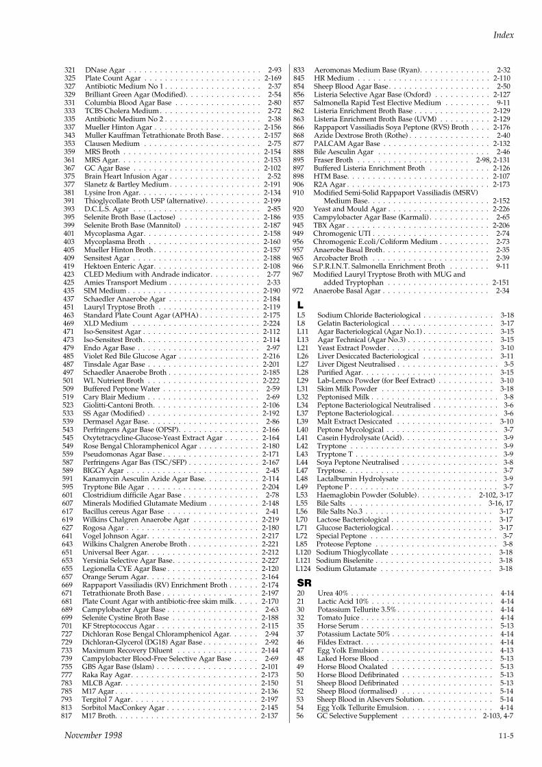
321 DNase Agar . . . . . . . . . . . . . . . . . . . . . . . . . . 2-93325 Plate Count Agar . . . . . . . . . . . . . . . . . . . . . . . 2-169327 Antibiotic Medium No 1 . . . . . . . . . . . . . . . . . . . 2-37329 Brilliant Green Agar (Modified). . . . . . . . . . . . . . . 2-54331 Columbia Blood Agar Base . . . . . . . . . . . . . . . . . 2-80333 TCBS Cholera Medium . . . . . . . . . . . . . . . . . . . . 2-72335 Antibiotic Medium No 2 . . . . . . . . . . . . . . . . . . . 2-38337 Mueller Hinton Agar . . . . . . . . . . . . . . . . . . . . . 2-156343 Muller Kauffman Tetrathionate Broth Base . . . . . . . . 2-157353 Clausen Medium . . . . . . . . . . . . . . . . . . . . . . . 2-75359 MRS Broth . . . . . . . . . . . . . . . . . . . . . . . . . . . 2-154361 MRS Agar. . . . . . . . . . . . . . . . . . . . . . . . . . . . 2-153367 GC Agar Base . . . . . . . . . . . . . . . . . . . . . . . . . 2-102375 Brain Heart Infusion Agar . . . . . . . . . . . . . . . . . . 2-52377 Slanetz & Bartley Medium . . . . . . . . . . . . . . . . . . 2-191381 Lysine Iron Agar. . . . . . . . . . . . . . . . . . . . . . . . 2-134391 Thioglycollate Broth USP (alternative) . . . . . . . . . . . 2-199393 D.C.L.S. Agar . . . . . . . . . . . . . . . . . . . . . . . . . 2-85395 Selenite Broth Base (Lactose) . . . . . . . . . . . . . . . . 2-186399 Selenite Broth Base (Mannitol) . . . . . . . . . . . . . . . 2-187401 Mycoplasma Agar. . . . . . . . . . . . . . . . . . . . . . . 2-158403 Mycoplasma Broth . . . . . . . . . . . . . . . . . . . . . . 2-160405 Mueller Hinton Broth . . . . . . . . . . . . . . . . . . . . . 2-157409 Sensitest Agar . . . . . . . . . . . . . . . . . . . . . . . . . 2-188419 Hektoen Enteric Agar. . . . . . . . . . . . . . . . . . . . . 2-108423 CLED Medium with Andrade indicator. . . . . . . . . . 2-77425 Amies Transport Medium . . . . . . . . . . . . . . . . . . 2-33435 SIM Medium . . . . . . . . . . . . . . . . . . . . . . . . . . 2-190437 Schaedler Anaerobe Agar . . . . . . . . . . . . . . . . . . 2-184451 Lauryl Tryptose Broth . . . . . . . . . . . . . . . . . . . . 2-119463 Standard Plate Count Agar (APHA) . . . . . . . . . . . . 2-175469 XLD Medium . . . . . . . . . . . . . . . . . . . . . . . . . 2-224471 Iso-Sensitest Agar . . . . . . . . . . . . . . . . . . . . . . . 2-112473 Iso-Sensitest Broth. . . . . . . . . . . . . . . . . . . . . . . 2-114479 Endo Agar Base . . . . . . . . . . . . . . . . . . . . . . . . 2-97485 Violet Red Bile Glucose Agar . . . . . . . . . . . . . . . . 2-216487 Tinsdale Agar Base . . . . . . . . . . . . . . . . . . . . . . 2-201497 Schaedler Anaerobe Broth . . . . . . . . . . . . . . . . . . 2-185501 WL Nutrient Broth . . . . . . . . . . . . . . . . . . . . . . 2-222509 Buffered Peptone Water . . . . . . . . . . . . . . . . . . . 2-59519 Cary Blair Medium . . . . . . . . . . . . . . . . . . . . . . 2-69523 Giolitti-Cantoni Broth. . . . . . . . . . . . . . . . . . . . . 2-106533 SS Agar (Modified) . . . . . . . . . . . . . . . . . . . . . . 2-192539 Dermasel Agar Base. . . . . . . . . . . . . . . . . . . . . . 2-86543 Perfringens Agar Base (OPSP). . . . . . . . . . . . . . . . 2-166545 Oxytetracycline-Glucose-Yeast Extract Agar . . . . . . . 2-164549 Rose Bengal Chloramphenicol Agar . . . . . . . . . . . . 2-180559 Pseudomonas Agar Base . . . . . . . . . . . . . . . . . . . 2-171587 Perfringens Agar Bas (TSC/SFP) . . . . . . . . . . . . . . 2-167589 BIGGY Agar . . . . . . . . . . . . . . . . . . . . . . . . . . 2-45591 Kanamycin Aesculin Azide Agar Base. . . . . . . . . . . 2-114595 Tryptone Bile Agar . . . . . . . . . . . . . . . . . . . . . . 2-204601 Clostridium difficile Agar Base . . . . . . . . . . . . . . . 2-78607 Minerals Modified Glutamate Medium . . . . . . . . . . 2-148617 Bacillus cereus Agar Base . . . . . . . . . . . . . . . . . . 2-41619 Wilkins Chalgren Anaerobe Agar . . . . . . . . . . . . . 2-219627 Rogosa Agar . . . . . . . . . . . . . . . . . . . . . . . . . . 2-180641 Vogel Johnson Agar. . . . . . . . . . . . . . . . . . . . . . 2-217643 Wilkins Chalgren Anerobe Broth . . . . . . . . . . . . . . 2-221651 Universal Beer Agar. . . . . . . . . . . . . . . . . . . . . . 2-212653 Yersinia Selective Agar Base. . . . . . . . . . . . . . . . . 2-227655 Legionella CYE Agar Base . . . . . . . . . . . . . . . . . . 2-120657 Orange Serum Agar. . . . . . . . . . . . . . . . . . . . . . 2-164669 Rappaport Vassiliadis (RV) Enrichment Broth . . . . . . 2-174671 Tetrathionate Broth Base . . . . . . . . . . . . . . . . . . . 2-197681 Plate Count Agar with antibiotic-free skim milk . . . . . 2-170689 Campylobacter Agar Base . . . . . . . . . . . . . . . . . . 2-63699 Selenite Cystine Broth Base . . . . . . . . . . . . . . . . . 2-188701 KF Streptococcus Agar . . . . . . . . . . . . . . . . . . . . 2-115727 Dichloran Rose Bengal Chloramphenicol Agar. . . . . . 2-94729 Dichloran-Glycerol (DG18) Agar Base . . . . . . . . . . . 2-92733 Maximum Recovery Diluent . . . . . . . . . . . . . . . . 2-144739 Campylobacter Blood-Free Selective Agar Base . . . . . 2-69755 GBS Agar Base (Islam) . . . . . . . . . . . . . . . . . . . . 2-101777 Raka Ray Agar. . . . . . . . . . . . . . . . . . . . . . . . . 2-173783 MLCB Agar. . . . . . . . . . . . . . . . . . . . . . . . . . . 2-150785 M17 Agar . . . . . . . . . . . . . . . . . . . . . . . . . . . . 2-136793 Tergitol 7 Agar. . . . . . . . . . . . . . . . . . . . . . . . . 2-197813 Sorbitol MacConkey Agar . . . . . . . . . . . . . . . . . . 2-145817 M17 Broth. . . . . . . . . . . . . . . . . . . . . . . . . . . . 2-137
833 Aeromonas Medium Base (Ryan). . . . . . . . . . . . . . 2-32845 HR Medium . . . . . . . . . . . . . . . . . . . . . . . . . . 2-110854 Sheep Blood Agar Base . . . . . . . . . . . . . . . . . . . . 2-50856 Listeria Selective Agar Base (Oxford) . . . . . . . . . . . 2-127857 Salmonella Rapid Test Elective Medium . . . . . . . . . 9-11862 Listeria Enrichment Broth Base . . . . . . . . . . . . . . . 2-129863 Listeria Enrichment Broth Base (UVM) . . . . . . . . . . 2-129866 Rappaport Vassiliadis Soya Peptone (RVS) Broth . . . . 2-176868 Azide Dextrose Broth (Rothe) . . . . . . . . . . . . . . . . 2-40877 PALCAM Agar Base . . . . . . . . . . . . . . . . . . . . . 2-132888 Bile Aesculin Agar . . . . . . . . . . . . . . . . . . . . . . 2-46895 Fraser Broth . . . . . . . . . . . . . . . . . . . . . . 2-98, 2-131897 Buffered Listeria Enrichment Broth . . . . . . . . . . . . 2-126898 HTM Base. . . . . . . . . . . . . . . . . . . . . . . . . . . . 2-107906 R2A Agar . . . . . . . . . . . . . . . . . . . . . . . . . . . . 2-173910 Modified Semi-Solid Rappaport Vassiliadis (MSRV)
Medium Base. . . . . . . . . . . . . . . . . . . . . . . . 2-152920 Yeast and Mould Agar . . . . . . . . . . . . . . . . . . . . 2-226935 Campylobacter Agar Base (Karmali) . . . . . . . . . . . . 2-65945 TBX Agar . . . . . . . . . . . . . . . . . . . . . . . . . . . . 2-206949 Chromogenic UTI . . . . . . . . . . . . . . . . . . . . . . . 2-74956 Chromogenic E.coli/Coliform Medium . . . . . . . . . . 2-73957 Anaerobe Basal Broth. . . . . . . . . . . . . . . . . . . . . 2-35965 Arcobacter Broth . . . . . . . . . . . . . . . . . . . . . . . 2-39966 S.P.R.I.N.T. Salmonella Enrichment Broth . . . . . . . . 9-11967 Modified Lauryl Tryptose Broth with MUG and
added Tryptophan . . . . . . . . . . . . . . . . . . . . 2-151972 Anaerobe Basal Agar . . . . . . . . . . . . . . . . . . . . . 2-34
LL5 Sodium Chloride Bacteriological . . . . . . . . . . . . . . 3-18L8 Gelatin Bacteriological . . . . . . . . . . . . . . . . . . . . 3-17L11 Agar Bacteriological (Agar No.1) . . . . . . . . . . . . . . 3-15L13 Agar Technical (Agar No.3) . . . . . . . . . . . . . . . . . 3-15L21 Yeast Extract Powder . . . . . . . . . . . . . . . . . . . . . 3-10L26 Liver Desiccated Bacteriological . . . . . . . . . . . . . . 3-11L27 Liver Digest Neutralised . . . . . . . . . . . . . . . . . . . . 3-5L28 Purified Agar. . . . . . . . . . . . . . . . . . . . . . . . . . 3-15L29 Lab-Lemco Powder (for Beef Extract) . . . . . . . . . . . 3-10L31 Skim Milk Powder . . . . . . . . . . . . . . . . . . . . . . 3-18L32 Peptonised Milk . . . . . . . . . . . . . . . . . . . . . . . . . 3-8L34 Peptone Bacteriological Neutralised . . . . . . . . . . . . . 3-6L37 Peptone Bacteriological . . . . . . . . . . . . . . . . . . . . . 3-6L39 Malt Extract Desiccated . . . . . . . . . . . . . . . . . . . 3-10L40 Peptone Mycological . . . . . . . . . . . . . . . . . . . . . . 3-7L41 Casein Hydrolysate (Acid). . . . . . . . . . . . . . . . . . . 3-9L42 Tryptone . . . . . . . . . . . . . . . . . . . . . . . . . . . . . 3-9L43 Tryptone T . . . . . . . . . . . . . . . . . . . . . . . . . . . . 3-9L44 Soya Peptone Neutralised . . . . . . . . . . . . . . . . . . . 3-8L47 Tryptose. . . . . . . . . . . . . . . . . . . . . . . . . . . . . . 3-7L48 Lactalbumin Hydrolysate . . . . . . . . . . . . . . . . . . . 3-9L49 Peptone P . . . . . . . . . . . . . . . . . . . . . . . . . . . . . 3-7L53 Haemaglobin Powder (Soluble) . . . . . . . . . . . 2-102, 3-17L55 Bile Salts . . . . . . . . . . . . . . . . . . . . . . . . . . 3-16, 17L56 Bile Salts No.3 . . . . . . . . . . . . . . . . . . . . . . . . . 3-17L70 Lactose Bacteriological . . . . . . . . . . . . . . . . . . . . 3-17L71 Glucose Bacteriological . . . . . . . . . . . . . . . . . . . . 3-17L72 Special Peptone . . . . . . . . . . . . . . . . . . . . . . . . . 3-7L85 Proteose Peptone . . . . . . . . . . . . . . . . . . . . . . . . 3-8L120 Sodium Thioglycollate . . . . . . . . . . . . . . . . . . . . 3-18L121 Sodium Biselenite . . . . . . . . . . . . . . . . . . . . . . . 3-18L124 Sodium Glutamate . . . . . . . . . . . . . . . . . . . . . . 3-18
SR20 Urea 40% . . . . . . . . . . . . . . . . . . . . . . . . . . . . 4-1421 Lactic Acid 10% . . . . . . . . . . . . . . . . . . . . . . . . 4-1430 Potassium Tellurite 3.5% . . . . . . . . . . . . . . . . . . . 4-1432 Tomato Juice . . . . . . . . . . . . . . . . . . . . . . . . . . 4-1435 Horse Serum . . . . . . . . . . . . . . . . . . . . . . . . . . 5-1337 Potassium Lactate 50% . . . . . . . . . . . . . . . . . . . . 4-1446 Fildes Extract . . . . . . . . . . . . . . . . . . . . . . . . . . 4-1447 Egg Yolk Emulsion . . . . . . . . . . . . . . . . . . . . . . 4-1348 Laked Horse Blood . . . . . . . . . . . . . . . . . . . . . . 5-1349 Horse Blood Oxalated . . . . . . . . . . . . . . . . . . . . 5-1350 Horse Blood Defibrinated . . . . . . . . . . . . . . . . . . 5-1351 Sheep Blood Defibrinated . . . . . . . . . . . . . . . . . . 5-1352 Sheep Blood (formalised) . . . . . . . . . . . . . . . . . . 5-1453 Sheep Blood in Alsevers Solution. . . . . . . . . . . . . . 5-1454 Egg Yolk Tellurite Emulsion. . . . . . . . . . . . . . . . . 4-1456 GC Selective Supplement . . . . . . . . . . . . . . . 2-103, 4-7
Index
November 1998 11-5
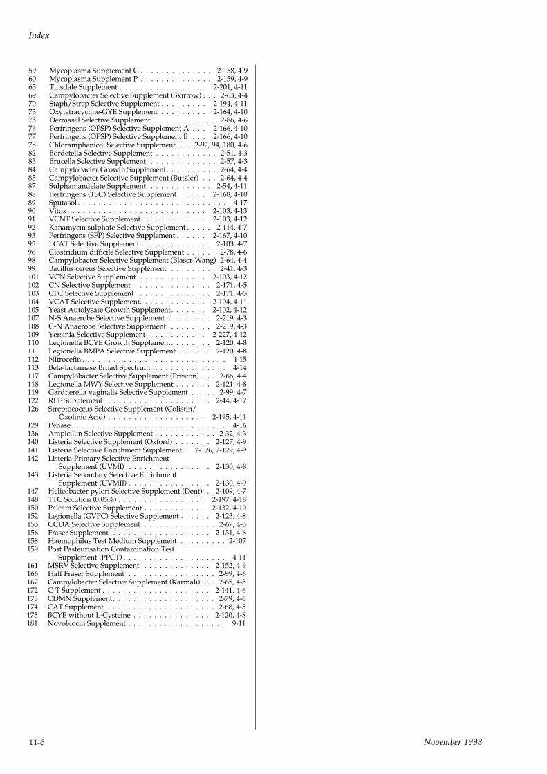
59 Mycoplasma Supplement G . . . . . . . . . . . . . . 2-158, 4-960 Mycoplasma Supplement P . . . . . . . . . . . . . . 2-159, 4-965 Tinsdale Supplement . . . . . . . . . . . . . . . . . 2-201, 4-1169 Campylobacter Selective Supplement (Skirrow) . . . 2-63, 4-470 Staph/Strep Selective Supplement . . . . . . . . . 2-194, 4-1173 Oxytetracycline-GYE Supplement . . . . . . . . . 2-164, 4-1075 Dermasel Selective Supplement. . . . . . . . . . . . . 2-86, 4-676 Perfringens (OPSP) Selective Supplement A . . . 2-166, 4-1077 Perfringens (OPSP) Selective Supplement B . . . 2-166, 4-1078 Chloramphenicol Selective Supplement . . . 2-92, 94, 180, 4-682 Bordetella Selective Supplement . . . . . . . . . . . . 2-51, 4-383 Brucella Selective Supplement . . . . . . . . . . . . . 2-57, 4-384 Campylobacter Growth Supplement. . . . . . . . . . 2-64, 4-485 Campylobacter Selective Supplement (Butzler) . . . 2-64, 4-487 Sulphamandelate Supplement . . . . . . . . . . . . 2-54, 4-1188 Perfringens (TSC) Selective Supplement. . . . . . 2-168, 4-1089 Sputasol . . . . . . . . . . . . . . . . . . . . . . . . . . . . . 4-1790 Vitox . . . . . . . . . . . . . . . . . . . . . . . . . . . 2-103, 4-1391 VCNT Selective Supplement . . . . . . . . . . . . 2-103, 4-1292 Kanamycin sulphate Selective Supplement . . . . . 2-114, 4-793 Perfringens (SFP) Selective Supplement . . . . . . 2-167, 4-1095 LCAT Selective Supplement . . . . . . . . . . . . . . 2-103, 4-796 Clostridium difficile Selective Supplement . . . . . . 2-78, 4-698 Campylobacter Selective Supplement (Blaser-Wang) 2-64, 4-499 Bacillus cereus Selective Supplement . . . . . . . . . 2-41, 4-3101 VCN Selective Supplement . . . . . . . . . . . . . 2-103, 4-12102 CN Selective Supplement . . . . . . . . . . . . . . . 2-171, 4-5103 CFC Selective Supplement . . . . . . . . . . . . . . . 2-171, 4-5104 VCAT Selective Supplement. . . . . . . . . . . . . 2-104, 4-11105 Yeast Autolysate Growth Supplement. . . . . . . 2-102, 4-12107 N-S Anaerobe Selective Supplement . . . . . . . . . 2-219, 4-3108 C-N Anaerobe Selective Supplement. . . . . . . . . 2-219, 4-3109 Yersinia Selective Supplement . . . . . . . . . . . 2-227, 4-12110 Legionella BCYE Growth Supplement. . . . . . . . 2-120, 4-8111 Legionella BMPA Selective Supplement. . . . . . . 2-120, 4-8112 Nitrocefin . . . . . . . . . . . . . . . . . . . . . . . . . . . . 4-15113 Beta-lactamase Broad Spectrum. . . . . . . . . . . . . . . 4-14117 Campylobacter Selective Supplement (Preston) . . . 2-66, 4-4118 Legionella MWY Selective Supplement . . . . . . . 2-121, 4-8119 Gardnerella vaginalis Selective Supplement . . . . . 2-99, 4-7122 RPF Supplement . . . . . . . . . . . . . . . . . . . . . 2-44, 4-17126 Streptococcus Selective Supplement (Colistin/
Oxolinic Acid) . . . . . . . . . . . . . . . . . . . 2-195, 4-11129 Penase . . . . . . . . . . . . . . . . . . . . . . . . . . . . . . 4-16136 Ampicillin Selective Supplement . . . . . . . . . . . . 2-32, 4-3140 Listeria Selective Supplement (Oxford) . . . . . . . 2-127, 4-9141 Listeria Selective Enrichment Supplement . 2-126, 2-129, 4-9142 Listeria Primary Selective Enrichment
Supplement (UVMI) . . . . . . . . . . . . . . . . 2-130, 4-8143 Listeria Secondary Selective Enrichment
Supplement (UVMII) . . . . . . . . . . . . . . . . 2-130, 4-9147 Helicobacter pylori Selective Supplement (Dent) . 2-109, 4-7148 TTC Solution (0.05%) . . . . . . . . . . . . . . . . . 2-197, 4-18150 Palcam Selective Supplement . . . . . . . . . . . . 2-132, 4-10152 Legionella (GVPC) Selective Supplement . . . . . . 2-123, 4-8155 CCDA Selective Supplement . . . . . . . . . . . . . . 2-67, 4-5156 Fraser Supplement . . . . . . . . . . . . . . . . . . . 2-131, 4-6158 Haemophilus Test Medium Supplement . . . . . . . . . 2-107159 Post Pasteurisation Contamination Test
Supplement (PPCT) . . . . . . . . . . . . . . . . . . . . 4-11161 MSRV Selective Supplement . . . . . . . . . . . . . 2-152, 4-9166 Half Fraser Supplement . . . . . . . . . . . . . . . . . 2-99, 4-6167 Campylobacter Selective Supplement (Karmali) . . . 2-65, 4-5172 C-T Supplement . . . . . . . . . . . . . . . . . . . . . 2-141, 4-6173 CDMN Supplement . . . . . . . . . . . . . . . . . . . . 2-79, 4-6174 CAT Supplement . . . . . . . . . . . . . . . . . . . . . 2-68, 4-5175 BCYE without L-Cysteine . . . . . . . . . . . . . . . 2-120, 4-8181 Novobiocin Supplement . . . . . . . . . . . . . . . . . . . 9-11
Index
11-6 November 1998


