Anaesthetic Care Russell Chilton and Ross Thompson
The word ‘anaesthesia’ comes from the Greek and translates to mean ‘without feeling’ (Williams and Smith 2008 ) and this is the objective of the anaesthetic process; to render the patient insensitive to pain, so that a procedure may take place, be that awake, sedated or under a general (unresponsive) anaesthetic.
The anaesthetic does not start when the patient comes through the anaesthetic room door, it begins when the patient comes for the preoperative assessment, which establishes his or her fitness for anaesthesia and surgery (Hughes and Mardell 2009 ). The National Institute for Health and Clinical Excellence (NICE) has devised a set of criteria that grade the type of surgery, in line with the ASA (American Society of Anesthesiologists) score, which is the assessment of a patient’s fitness before surgery (Carlisle 2006 ). This information, coupled with a pre-anaesthetic assessment and the nature of the procedure, will guide the anaesthetist to the most appropriate plan for the patient. This plan is communicated to the supporting practitioner prior to or on the day of the procedure.
Principles
The rationale for using anaesthesia is to provide the best possible set of conditions for surgery to take place (Oakley and Van Limburgh 2005 ). There are a number of techniques used to produce the ideal circumstances for the surgical team: general, regional and local anaesthesia or sedation (Wicker and O’Neil 2006 , p. 243). A combi-nation of these techniques can also be deployed. The state of the patient is accomplished by using a range of specialised drugs, commonly referred to as the ‘triad of anaesthesia’, a term introduced by Rees and Gray in 1950. It may prove difficult to maintain a balance during anaesthesia, so the ‘triad’ can be manipulated by the administration of further medicines, depending upon which aspect requires attention. This results in the triad transforming into the ‘star of anaesthesia’ (Figure 9.1 ).
102 Patient Safety and Managing Risks in Perioperative Care
Section 3
Traditionally, anaesthesia is viewed as being in four stages and the object is to obtain the third stage as quickly and smoothly as possible:
� Stage 1 – Disordered consciousness. This is now known as ‘induction’, and is the stage at which the patient passes from analgesia without amnesia to analgesia with amnesia, leading to unconsciousness.
� Stage 2 – Excitement. This is characterised by uncontrolled muscular movement, altered vital signs, pupil dilation, vomiting and possibly laryngeal spasm.
� Stage 3 – Surgical anaesthesia. This is achieved when the patient loses the ability to consciously respond to stimuli and reflexes serving muscle control become relaxed. It is at this stage a patient under a general anaesthetic will require support to maintain their airway via an appropriate airway adjunct.
� Stage 4 – Overdose. This stage is when a potential increased risk is present. If the anaesthetist does not titrate the level of anaesthetic to the surgical stimulation, this will cause an adverse impact upon the patient. Originally this was described as the ‘law of diminishing resistance’.
Staffing
Wherever anaesthesia is commenced, the anaesthetist must have exclusive and qualified assistance (AAGBI 2010 , p. 10). This assistance typically takes the form of an anaesthetic practitioner, who can be either a registered nurse (RN) or an operating department practitioner (ODP). These assistants have two fundamental functions: to provide support
to the anaesthetist and to support the patient, acting as advocate (Hughes and Mardell 2009 ). A third person should also be available to help support the patient and assist with any unforeseen occurrences.
Training
Any anaesthetic practitioner must comply with the National Occupational Standards (NOS) (AAGBI 2010 , p. 11). The RN is required to achieve additional professionally recog-nised and approved training before taking on the duties of an anaesthetic practitioner.
All relevant professional bodies acknowledge and positively encourage a programme of continuing professional development (CPD) for all anaesthesia practitioners. This will enable the practitioner to remain both vigilant and effective, thus maintaining a safe environment and delivery of the best quality care to the patient (Wicker and O’Neil 2006 , p. 243). CPD forms the basis of employer reviews and professional registration compliance.
A clear function of the anaesthetic practitioner is to promote the wellbeing of the patient, to act as advocate and provide a professional approach to their duties. This is achieved by using sound, patient-centred, evidence-based practice and critical thinking, supported by a positive, reflective attitude. The application of a range of professional and personal skills is necessary to function as part of the anaesthetic multi-disciplinary team (MDT). Particular proficiencies must be evident in the form of preparation of the environment, maintaining health and safety, preparation of medical devices, undertaking vital signs monitoring, supporting the patient’s cardiovascular requirements and airway management (College of Operating Department Practice 2009 ).
The role of the anaesthesia assistant
The guiding principle for any practitioner working in anaesthetics is to ensure that the patient’s safety is maintained at all times (Younger 2000, p. 148). In order to achieve the best possible outcome for the patient, the knowledge base of the practitioner is continually advancing with core skills (Figure 9.2 ).
Many practitioners will evolve how they practice and will develop habits based upon their experiences; however this must be underpinned by best practice. Wicker and O’Neil ( 2006 ) argue that the basis of the anaesthetic practitioner’s role is to:
� prepare the environment � prepare appropriate equipment
Excellent inter-personal skills Anaesthetic
proceduralknowledge
Infectionpreventionawareness
Technologymanagement
Anatomy &physiology
Anaestheticpharmacology
Professional
Responsive &adaptable
Problem solver
Anaestheticpractitioner
Figure 9.2 Qualities of an anaesthetic practitioner.
104 Patient Safety and Managing Risks in Perioperative Care
Section 3
� ensure the safety checklist is completed � support the patient as their needs dictate � support the chosen method of anaesthesia � work as part of a multi-disciplinary team (MDT) � attach appropriate monitoring � communicate any potential problems to relevant members of the MDT � ensure documentation is accurate and complete.
When consultant anaesthetists are asked ‘What makes a good anaesthetic practi-tioner?’, a common response is ‘Someone who has what I need before I know I need it!’ While this is true, it only comes with exposure to a wide range of differing situations.
A critically important component in the operating department is communication. We must recognise our potential barriers, including gender, ethnicity, prejudices, professional rivalry, own limitations or organisational confusion to be able to overcome them. Gopee and Galloway ( 2009 ) suggest that clear and effective communication/information given to the patient preoperatively:
� improves the patient’s outcomes � improves patient’s expectations � requires less postoperative analgesia and � reduces the patient’s length of stay in hospital.
The act of communication is a two-way meaningful process, not a rudimentary non- rhetorical remark or gesture (Russell 2005 ). To support the patient or member of the MDT an effective practitioner will adjust how, when and at what level there is a need to communicate (Figure 9.3 ). The NHS Institute for Innovation and Improvement ( 2009 ) advocates a framework referred to as ‘SBAR’ (situation, background, assessment, recommendation).
Registeredstaff:
scrub /circulating /
recoverypractitioners
Professionsallied to
medicine:Radiologists
Radiographer
Support staff:Theatre assistant
Administrationstaff
HSDU staff
Other services:Pharmacy /
CCUward or ED
staff
Medical staff:Anaesthetists
Surgeons
Patient
Figure 9.3 Immediate interdisciplinary team for surgical patients (from Hughes and Mardell 2009 ).
The size and layout of an anaesthetic room is guided by many factors, usually by recom-mended good practice through to statute intervention, such as the Association for Perioperative Practice (AfPP) (2011), Health and Safety Executive (HSE), Health and Safety at Work Act 1974 and NHS Estates ( 2005 ). The layout of the anaesthetic room should follow a logical sequence of when and where the practitioner needs to access items; for example, suction close to the anaesthetic machine, syringes close to needles, and so on. It should also be in close proximity to appropriately stocked stores.
The anaesthetic practitioner will develop his or her own way of working. Ideally, a routine should be established. Some may use the patient journey process – preparing monitoring first, then cannulae, airway equip, etc. Some may check their room in a clockwise manner. It does not matter which approach is used so long as it leads to nothing being forgotten or overlooked. Students and newly qualified practitioners should consider writing their ‘routine’ down so that they can refer to and adapt it.
Equipment
An essential aspect for any anaesthetic practitioner is the sound knowledge base regarding the equipment they will be expected to use. All medical devices are governed by the Medicines and Healthcare Regulatory Agency (MHRA), by the Medical Devices Regulations (MHRA 2002 ) plus subsequent amendments. Further regulation and guid-ance is taken from professional bodies, especially the AfPP (2011) and Association of Anaesthetists of Great Britain and Ireland (AAGBI 2004 ). The main tenets revolve around:
� not using unfamiliar equipment � not using out of serviceable date � following manufacturers instructions � not using faulty equipment � reporting faulty equipment as per the local protocol.
The primary guidance for checking anaesthetic equipment is issued by the AAGBI, which states that it is the responsibility of anaesthetists to be satisfied that checks have been carried out correctly. It is common practice for the anaesthetic practitioner to undertake initial checks. The AAGBI state that it is a mandatory activity to check anaesthetic equipment at the beginning of each session of use, and in the event of any changes to the current set-up (AAGBI 2004 ). These checks cover all aspects associated with the administration of anaesthesia, including the gas supply pipelines, the anaesthetic machine, filters, circuits, airway adjuncts, ventilators, suction, monitoring and ancillary equipment. A record of these checks should be kept with the anaesthetic machine (AAGBI 2004 ). A copy of the checklist produced by the AAGBI should be attached permanently to the anaesthetic machine.
The following advice is predominantly taken from the guidance issued by the AAGBI document entitled Checking Anaesthetic Equipment 3 (AAGBI 2004 ).
Anaesthetic machine
Most anaesthetic machines may be described simply as a box which draws in a medical gas (i.e. O 2 ), the option to mix it with an anaesthetic agent and deliver a specific quantity of this gas/mixture to the patient via a circuit. Usually anaesthetic machines are connected directly to the mains electrical supply. Although other equipment can be
106 Patient Safety and Managing Risks in Perioperative Care
Section 3
connected into it, multi-socket extension leads must not be used with these devices. In essence, the anaesthetic machine is a collection of medical devices assembled in a convenient, transportable trolley.
In the event of machine failure an alternative method of effective oxygen supply is required. This can be via self-inflating bag or a circuit coupled with an oxygen cylinder. These items must be present in the anaesthetic room and checked appropriately.
Medical gas supplies
The anaesthetic practitioner should identify and note which gases are being supplied by pipeline and note that each pipeline is correctly inserted into the appropriate gas supply terminal. The use of size-specific connections should prevent cross-connection.
All machines have an oxygen failure alarm; testing this may involve disconnecting the oxygen pipeline on some machines. Repeated disconnection of gas hoses may lead to premature failure of the Schroeder socket and probe. As a result of this, a ‘tug’ test to confirm correct insertion of each pipeline into the appropriate socket is sufficient. If a gas master switch is present that should be used instead. In addition to these checks, the oxygen failure alarm must be checked on a weekly basis by disconnecting the oxygen hose while the oxygen rotameter/flowmeter is turned on. A written record must be kept. In addition to sounding an alarm which must sound for at least 7 seconds, oxygen failure warning devices are also linked to a gas shut-off device.
All gas pressure gauges for pipelines connected to the anaesthetic machine should indicate between 400 and 500 kPa.
Adequate supplies of any other gases intended for use must be available and connected as appropriate. All reserve cylinders should be securely seated and turned off after checking their contents.
Where fitted to an anaesthetic machine, the operation of rotameters/flowmeters must be checked, ensuring that each control valve operates smoothly and that the bobbin spins freely. If nitrous oxide is to be used, the anti-hypoxia device should be tested by first turning on the nitrous oxide flow and ensuring that at least 25% oxygen also flows. The oxygen flow should then be turned off and a check made that the nitrous oxide flow also stops.
Vaporisers
These units hold specific anaesthetic agents, such as Sevoflurane™ and Isoflurane™. They are used to either induce or maintain anaesthesia (Oakley and Van Limburgh 2005 ). The vaporiser(s) must be fitted correctly to the anaesthetic machine and secured to the back bar and the control knobs rotated. They should be kept upright at all times and not overfilled. Reference should be made to local policy with regard to the appropriate filling method, although COSHH regulations require that it should take place in a fume cupboard, unless fixed to the delivery unit, when filling should be carried out in isolation.
Humidifiers
Humidifiers are used to protect the patient’s respiratory tract and to counter the ‘drying out’ effect of anaesthetic gases. They also act as a barrier to potential microbiological bacteria. They are placed between the catheter mount and the breathing circuit.
Anaesthetic gas scavenging system
This system assists in the removal of anaesthetic gases. The anaesthetic practitioner must ensure that the system is switched on, connected to the machine and working correctly.
Many modern anaesthetic machines have an absorption facility using modified soda lime crystals. This should be checked to ensure it is airtight and not exhausted. This process can produce caustic fluid, so glove-wearing is essential when changing the crystals and cleaning.
Breathings systems
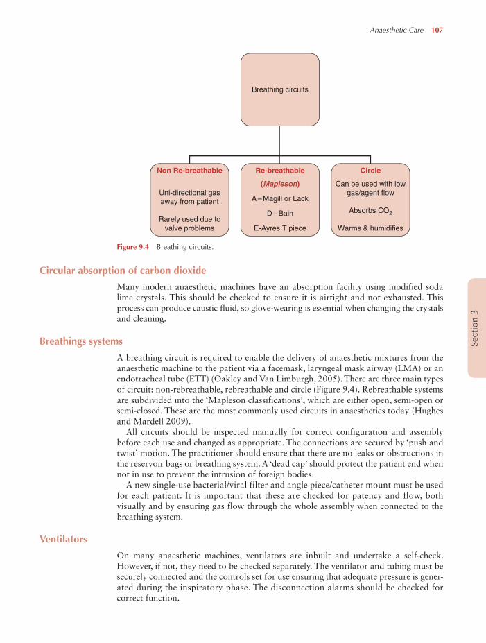
A breathing circuit is required to enable the delivery of anaesthetic mixtures from the anaesthetic machine to the patient via a facemask, laryngeal mask airway (LMA) or an endotracheal tube (ETT) (Oakley and Van Limburgh, 2005 ). There are three main types of circuit: non-rebreathable, rebreathable and circle (Figure 9.4 ). Rebreathable systems are subdivided into the ‘Mapleson classifications’, which are either open, semi-open or semi-closed. These are the most commonly used circuits in anaesthetics today (Hughes and Mardell 2009 ).
All circuits should be inspected manually for correct configuration and assembly before each use and changed as appropriate. The connections are secured by ‘push and twist’ motion. The practitioner should ensure that there are no leaks or obstructions in the reservoir bags or breathing system. A ‘dead cap’ should protect the patient end when not in use to prevent the intrusion of foreign bodies.
A new single-use bacterial/viral filter and angle piece/catheter mount must be used for each patient. It is important that these are checked for patency and flow, both visually and by ensuring gas flow through the whole assembly when connected to the breathing system.
Ventilators
On many anaesthetic machines, ventilators are inbuilt and undertake a self-check. However, if not, they need to be checked separately. The ventilator and tubing must be securely connected and the controls set for use ensuring that adequate pressure is gener-ated during the inspiratory phase. The disconnection alarms should be checked for correct function.
108 Patient Safety and Managing Risks in Perioperative Care
Section 3
Suction
Suction is required for rapid response to aspiration, regurgitation or vomiting by the patient during anaesthesia. The check should comprise confirmation function and connection security and also a test for the rapid development of an adequate negative pressure, done by occluding the tubing.
Cannulation
Intravenous access is a prerequisite for anaesthesia to be undertaken and is achieved by insertion of a cannula. Its placement in a blood vessel is for a variety of uses: administration of medicines, fluids or nutrition, invasive monitoring or haemo-dialysis (Wolverson 2008 ). In exceptional circumstances (e.g. babies or a needle-phobic patient), the patient can be induced without this access but it is established at the earliest opportunity.
In anaesthetics, the most common use is for the administration of anaesthetic medicines, usually via a peripheral vein.
Cannulae come in a range of sizes (Figure 9.5 ), the choice of which is dependent upon the size of vein and its use. The siting of peripheral cannulae can be into any viable vessel, though the most common are the back of the hand, forearm or antecubital fossa. A convention exists in the UK relating to the gauge size and specific colours. Once inserted, they must be secured with an appropriate transparent dressing. This is to enable the site to be observed for any potential signs of infection, reaction or haemor-rhage. The size, location, time and date are recorded on patient documentation.
The use of invasive monitoring via an arterial cannula is only justified in certain circumstances when a severe change in blood pressure is expected. The uses of arterial lines include continuous/precise blood pressure monitoring or repeated arterial blood gases (ABG). An arterial line is usually sited in the radial artery, on the non-dominant hand side, though where peripheral access is difficult, the brachial or femoral arteries may be used on a short-term basis (Wolverson 2008 ). There are increased risks of bleeding, haematoma, ischaemia and infection when arterial lines are used, so great care is required when they are being inserted, secured, connected to and removed. The cannula is connected to a transducer, which converts the signal into a blood pressure reading. The transducer must be placed approximately level with the heart and ‘zeroed’ to atmospheric pressure (Hughes and Mardell 2009 ).
Central venous lines via the internal jugular, subclavian or femoral veins are used for monitoring central venous pressure (CVP), administration of medicines, infusions,
Figure 9.5 Cannulae comparison. Image of BD Venflon TM Pro Safety IV cannula reproduced with kind permission of BD UK Ltd.
nutrition, and long-term access or if there is poor peripheral access. They are typically inserted using the Seldinger technique (Wolverson 2008 ).
Monitoring equipment
Pre, peri and post anaesthesia the patient needs to be monitored effectively (AAGBI 2007 ). Direct clinical observations are a vital and effective form of monitoring and should be utilised as such (Thompson 2009 ) although advances in technological monitoring devices have increasingly become more sophisticated to help with this task. All apparatus must be functionality checked before use.
Monitoring devices must be attached before induction of anaesthesia and their use continued until the patient has recovered from the effects of anaesthesia (AAGBI 2007 ). Particular attention must be paid to ensuring that all devices used are clean, fit for purpose and free from obstruction.
Induction and maintenance of anaesthesia The following are required for the induction and maintenance of anaesthesia:
� pulse oximetry – for measuring the circulating level of oxygen saturation, as well as heart rate (Oakley and Van Limburgh 2005 )
� non-invasive blood pressure via a cuff attached externally to the patient. � electrocardiograph � fresh inspired and end-tidal gases and anaesthetic agents � airway pressure – to avoid barotraumas and ensure adequate ventilation is taking place.
A nerve stimulator (whenever a neuromuscular blocking agent is used) and a way of measuring the patient’s temperature should also be available.
Patients who undergo an ‘operative procedure’ under either a regional technique or sedation must have a minimum of the following monitoring devices:
Thermoregulation NICE defines hypothermia as a core temperature of below 35.0 °C and recommends that patients should be maintained as ‘comfortably warm’ (between 36.5 °C and 37.5 °C), pre and post operatively.
From an anaesthetic viewpoint, effective management of thermoregulation revolves around monitoring before and during the procedures. The patient’s temperature should be measured and documented before induction of anaesthesia and then every 30 minutes until the end of surgery.
It is noteworthy that hypothermia can be used to manage a patient during their opera-tion (especially in neurological and cardiac surgery) after resuscitation or in the intensive care unit. This can be achieved via cooling catheters, cooling blankets, vests and leg wraps.
Airway adjuncts There are five main categories associated with these devices (Figure 9.6 ):
110 Patient Safety and Managing Risks in Perioperative Care
Section 3
For each device there are a range of versions, some reusable, most disposable, along with some developmental advances (e.g. Proseal™ and iLMA™). Combined use of devices is not uncommon (e.g. OP airway with an ET tube upon extubation). All practitioners should ensure that all appropriate sizes of relevant adjuncts and connectors are available. The devices should only be prepared for use and checked for patency at the point of use as this will avoid contamination, unnecessary wastage and possible confusion of size. Each device must be correctly sized and practitioners must know how it is inserted, in case of an emergency.
Ancillary equipment
A check should be made that all ancillary equipment, including intubating forceps (Magill’s), bougies, nerve stimulators etc. are present and working. The patient trolley/bed/operating table should be able to be rapidly tilted head-down in the event of an emergency. All syringes must have a colour-coded label that conforms to the national standard (AAGBI 2003 ). A stethoscope must always be available (AAGBI 2007 ).
A laryngoscope should be present with a variety of blade sizes. This enables the anaesthetist to examine the larynx and introduce an ET tube and/or nasogastric tube. Its functionality needs to be tested; spare batteries and lamps (if not fibreoptic) must also be readily available. Specialist laryngoscope (e.g. McCoy’s, fibreoptic laryngoscope or paediatric) attachments should be readily available within the department.
Techniques
The following section is a broad overview of the common techniques related to supporting the anaesthetist. The patient’s journey will be used as a guide to give the techniques logic, each progressing from a basic start point and ranging through to
increased complexity, covering a range of anticipated situations. It is in the interests of the patient that the practitioner considers the potential consequences of any treatment regime, anatomical anomaly or reactive condition, and is prepared.
It is essential that the anaesthetic practitioner attends the team briefing, to absorb the knowledge held by the surgeon and anaesthetist. Enquiries at this stage may include significant allergies, body dimension, including obesity, communication and learning difficulties and any expected complications that may impact on care provision.
Once the patient arrives and has been checked into the theatre by the registered practitioner, monitoring is applied and the next stage is venous cannulation. All equip-ment must be available a cannula tray ready, containing a range of cannulae, approved fixative dressings, swabs, and disposable disinfection wipes for skin.
Peripheral cannulation
Cannulation of the peripheral veins is a skilled activity, usually carried out by the anaesthetist assisted by the theatre assistant, who compresses the forearm tissues sufficiently to obstruct venous return distal to the placement of his or her hands (in circumference around the arm) but not to obstruct arterial supply. This will facilitate filling of the veins and enable safe and quick cannulation and reduce patient anxiety. The patient should be told what is going to happen before it occurs as cannulation can be uncomfortable. As the patient’s advocate, the theatre assistant could offer to get more experienced assistance if the inserter is struggling. In most cases, cannulation is achieved quickly and a dressing applied and reinforced, if necessary.
It is imperative to know the drugs that are in common use in the anaesthetic/post-anaesthetic care of the patient, plus those needed for any emergency situations.
Central venous catheterisation
Central venous catheters are used to measure returning blood flow pressure to the right side of the heart, provide access to central veins for fluid augmentation and allow access for the delivery of drugs. The catheter is usually inserted aseptically into either the internal or external jugular vein. The patient is tilted head-down, with the head turned to the left. The skin is prepared and disposable drapes applied. A sterile field is created on a trolley and the equipment placed on it. The catheter is introduced using the Seldinger method, using a wire through the needle, which is then removed and a larger dilator is threaded over the wire to increase the diameter of the lumen. Once the aperture is created, the catheter is introduced over the wire. The lumen(s) of the catheter are flushed and attached to a transducer. When the device is in situ it is secured by suturing and then covered with a transparent dressing.
Arterial cannulation
Arterial cannulation is carried out to provide accurate monitoring of blood pressure. The cannula is introduced in a similar method to the central venous catheter. Securing is of paramount importance, as the patient can lose a large volume of blood if the cannula comes out under the drapes. A small volume of fluid is given continually to keep the distal tip of the cannula clear. Once removed, a pressure dressing must be applied to prevent haemorrhage.
Basic airway management
The anaesthetist must ensure that all equipment is checked, so will usually check the anaesthetic delivery device/machine in accordance with guidelines prior to commencing the list. Where the anaesthetic device is checked by the anaesthetic practitioner, the
112 Patient Safety and Managing Risks in Perioperative Care
Section 3
permanent record is signed. The practitioner should inform the anaesthetist of the equipment that they have checked and whether there are any issues such as shortages or unavailability of items and discuss specific plans for each patient on the operating list.
The practitioner should prepare the airway trolley with the following items available:
� a range of appropriately sized, single-patient use silicone facemasks � appropriate sizes of disposable oropharyngeal and nasopharyngeal airways � patient-appropriate LMA – re-usable or disposable � syringes for cuff inflation and water-based lubricant jelly � Magill’s forceps, securing tapes, pads and tape for the eyes, if that is the preferred method.
The patient should be pre-oxygenated. As the patient is given the hypnotic agent, they will lose consciousness. Their airway can become compromised, as the tongue fills the oropharynx. The practitioner is required to concentrate on the anaesthetist’s actions, both in induction and airway management. Remember that they are a knowledgeable observer so should be noticing drug doses and the effect of the drug. Once the airway has been controlled, the practitioner should be prepared to hand out an oro- or naso-pharyngeal airway, having been taught how to size these, to ensure correct fitting and reduce airway obstruction. Before the anaesthetist indicates that they wish to insert the LMA, all of the necessary items must be checked to ensure that they are close to hand. The practitioner must be ready to open the airway to assist the insertion, by performing a jaw thrust and extending the airway entry. Once it is confirmed that the LMA is in place, the cuff is inflated, only with the recommended volume of air, and then secured with a cotton tie or adhesive tape (as long as the patient is not allergic). Some anaesthetists prefer to leave the LMA unsecured.
Once the airway is in place, an appropriate method is used to cover the eyes. This may be gauze pads, preformed dressings or simple tape.
Once the eyes are taped, the anaesthetist will be ready to move the patient to the theatre suite. Monitoring is disconnected; the anaesthetic circuit being the last to be separated from the airway. The anaesthetist should ensure that medical gases and vaporisers are turned off, as well as ventilators, if used.
Advances to basic airway management
The procedure highlighted above is for a routine, elective patient undergoing a minor or intermediate procedure. The patient may on the other hand, present with:
� an underlying medical condition, such as COPD � an anatomical defect (e.g. hiatus hernia) � having shared airway surgery or dual-life � being an emergency � another reason.
Definitive control of the airway requires insertion of an endo/nasotracheal tube. One of the key issues with preparing the tube is the length, which is usually indicated on the device, but can be guided by the anaesthetist, who will have met the patient. It is important to ensure, once the tube has been cut, that the circuit connector is inserted fully into the tube so that there is no gap. The tube tie may catch in the gap and pull the tube out of the trachea, or push it in and cause an obstruction. The tube must be fully tested prior to use for patency, cuff inflation, pilot line and cuff inflation valve. The remainder of the
equipment required for intubation should be laid out within easy reach and be checked for function and cleanliness. The following items may be required:
� two fully working laryngoscopes with a long blade available � appropriate sized gum-elastic bougies � Magill’s forceps, size dependent on patient type (i.e. small for a child or baby) � range of syringes to inflate cuff � full set of oropharyngeal and/or nasopharyngeal airways – dependent on patient/surgery type
� tube ties, tape or preformed devices and products to secure airway � anaesthetic swabs, not white, but may have X-ray detectable line through them � lubricating gel � a range of tube sizes and/or different styles – dependent on surgery/patient.
The tube is passed through the upper airway passages, under direct vision, using the laryngoscope. The distal tip of the ET tube is passed through the vocal cords and sits in the space externally identified as the suprasternal notch (Pattnaik and Bodra 2000 ). The cuff is then inflated. An overpressurised ET tube cuff can cause disruption to the mucociliary escalator and may lead to necrosis of the tracheal tissues (Al-Shaikh and Stacey 2007 ). It is worth noting here that the cuff pressure can rise during the procedure due to increase in air temperature (21–37 °C) and the diffusion of N 2 O, if used. The cuff should be monitored and assessed during long cases or if the patient is repositioned.
Additional airway management
So that individual lungs can be deflated (unilateral ventilation) to allow surgical procedures to be carried out, it is important that any devices that are inserted into the lungs have features to ensure an airtight seal. Endobronchial (EB) tubes are tubes which have extended length, they do not require cutting, and they have two cuffs, one normal-sized proximal cuff and a smaller distal cuff, which sits in the smaller main bronchus. The endobronchial tubes come in a kit, complete with all of the connections. The tubes need to be put together in the correct manner, tested and prepared for intubation. The techniques for insertion are the same as for the ET tube, but the individual passageways within the tube must be tested to ensure they are patent and will allow one lung to deflate while ventilating the other.
Longer suction catheters must also be available for EB tubes. A tube clamp is employed when using EB tubes. This is used to clamp off the fresh gas supply line prior to opening the exhaust/suction port.
Spinal anaesthesia
Used as an alternative to, and sometimes in conjunction with, a general anaesthetic, spinal anaesthesia involves the insertion of a needle into the spinal space distal to the termination of the spinal cord at L2. Through the needle a combination of drugs can be given to induce a total block of all sensations, usually from the umbilicus and below. The patient usually sits upright, in an arched-back position, supported on a pillow with a member of support staff in front of them to provide communication and positional support. The procedure is carried out under strict aseptic technique. The anaesthetist will scrub and don gown and gloves. The skin is prepared with a non-irritant solution (e.g. chlorhexidine with tint 2% isopropyl alcohol). The anaesthetist selects a location to insert the needle, usually around L3/L4. The patient’s position should encourage the vertebra to part, making insertion of the needle easier. The skin is anaesthetised with local anaesthetic, followed by deeper infiltration.
114 Patient Safety and Managing Risks in Perioperative Care
Section 3
The spinal needle is extremely thin and flexible to prevent it being manoeuvred within the tissue plains of the back, so it needs to be almost fully withdrawn before reinserting in a different angle, if required. Proper insertion is achieved when the anaes-thetist feels the needle ‘pop’ through the ligamentum flavum and into the spinal space. The indication that the needle is in the correct space is a flow of cerebral spinal fluid (CSF) back along the needle, until it drips out of the proximal hub. The practitioner should observe the CSF and also note its turbidity as it mixes with the local anaesthetic prior to injection. The fluid should be clear. The CSF fluid can be tested with a blood glucose monitor to confirm that it is CSF; it should show as positive for glucose (approximately equal to plasma concentration). Once the needle is positioned, the anaesthetist ‘fixes’ the position by holding the needle firmly and then injecting either a high concentration local anaesthetic (e.g. Heavy Marcain™) or a combination of normal local anaesthetic with diamorphine.
Because of the size of the needle bore, the hole self-seals when the needle is with-drawn. The skin can be sprayed with a dressing spray or an adhesive dressing applied to capture any leakage. Gradually, paralysis takes effect and the patient will lose sensation below the waist. The patient is usually then laid down, possibly requiring some manual handling that should be coordinated by the anaesthetist to ensure no patient or staff risk. The patient should be communicated with throughout the procedure, ensuring comfort and response to any questions. Elderly patients or those with hearing deficiencies may require the anaesthetist’s instructions to be repeated by the member of staff supporting the patient, so concentration and patient-centred care is essential.
Epidural anaesthesia
Similar to the spinal procedure above, epidurals are inserted in the back, but into the epidural space. The epidural space contains only the spinal roots and is a much larger physical space, so a larger volume of local anaesthetic can be given and more control achieved. Epidurals can be used for post-procedure analgesia for bowel surgery, caesarean section and a range of pelvic/abdominal procedures.
The positioning of the patient is similar to that used in spinal anaesthesia, with the same staff numbers involved. The main difference is the equipment used, in particular the needle. The Tuohy needle (Figure 9.7 ) has its distal opening on the side of the needle
rather than on the end. This facilitates the positioning of the epidural catheter, which is left in situ when the needle is withdrawn. The needle is also graduated in 1 cm sections so that the needle depth can be monitored and recorded.
The technique proceeds as for spinal anaesthesia initially. The in-dwelling catheter and 0.2 μm filter are flushed and left prepared. With the patient positioned to open the vertebrae, the anaesthetist, in gown and gloves, pushes the needle through the skin of the back. A loss of resistance (LOR) syringe, filled with saline, is attached to the back of the needle. The needle is slowly advanced a few millimetres at a time, with the LOR plunger being pressed gently at the end of each movement. If the distal end of the needle is up against tissue bulk, the saline cannot escape so resistance is felt in the syringe. As the needle tip breaks into the epidural space, the saline in the syringe, when pressed, will flow easily and into the space. This indicates a successful procedure.
The syringe is then removed and a thin catheter fed down the lumen of the needle. The patient may experience some discomfort as the catheter brushes against the nerve roots on the way to its final location. This is usually transient. The distal tip of the catheter can be directed by rotating the needle, with its side aperture, to locate the catheter in the desired position. Local anaesthetic is injected down the catheter once a filter has been connected. The volume of local anaesthetic is greater than that used for spinal anaesthesia and the effect is not so profound. The degree of block is dose-dependent and a complete block can be achieved if desired, for example to allow surgical delivery of a baby, with the mother awake and pain-free.
Once the catheter is in place, it is imperative that it is not moved and to ensure this a locking dressing is applied, followed by secure taping of the catheter to the patient’s back. The site of injection must be monitored to ensure it remains infection free and to check that there is no haemorrhage. Care must be taken while moving and positioning the patient.
Peripheral nerve blocks
As well as the techniques described above, the anaesthetist has a wide range of options available for alternative or augmented analgesia, including the blocking of regional nerves (e.g. sciatic, femoral or brachial plexus). This technique requires the nerve proximal to the site of surgery to be located, or an anatomically appropriate location selected, away from important structures and organs. The needle used is covered by a thin insulating layer, with just the tip exposed. This allows a low-voltage current to activate the tip so that when it comes into contact with the nerve it causes stimulation and the corresponding muscle contracts, indicating which nerve has been found.
The nerve is approximated following careful positioning and then stimulated by the electrical impulse produced by a peripheral nerve stimulator (PNS) device.
The technique commences with checking the equipment, battery power and function, collecting the appropriate drugs and checking expiry dates. The patient has the proce-dure explained to them and they are assisted to position themselves appropriately. The negative electrode wire is connected to the needle and the positive attached to an ECG electrode and affixed to the patient, near to the injection site. The anaesthetist flushes the needle with local anaesthetic and then withdraws the syringe. This allows for a free flow of blood in case a vessel is inadvertently punctured en route to the nerve. The needle is used to puncture the skin and advanced towards the nerve. Once close to the nerve, the PNS is turned on and set to 1 mA at 2 Hz. When the identified nerve responds by contracting strongly, the current is reduced to as low as possible, while still maintaining visual stimulation. Then 1 mL of local anaesthetic is injected through the needle, after aspiration to ensure that it is not in a blood vessel. Muscle twitch should be eliminated.
116 Patient Safety and Managing Risks in Perioperative Care
Section 3
The rest of the prescribed dose is then administered in short volumes with aspiration between, and the needle is removed. The entry site is checked to ensure that it is not bleeding. The local anaesthetic will need to develop for approximately 20 minutes.
The skin pain receptors are stimulated prior to the commencement of surgery to ensure that an adequate block has been achieved. Topping up may be required during the procedure and is usually administered by the surgeon directly into the tissues.
Ultrasound-guided transabdominal plane blocks
Transabdominal plane (TAP) blocks involve the placement of epidural catheters using a Tuohy needle, guided by an ultrasound scanner, into the neurovascular plane between the internal oblique muscles and transverse abdominal muscles. The block is usually carried out after the administration of a general anaesthetic. The purpose of the technique is to block the sensory and motor nerves of the abdominal wall, which is especially use-ful for bowel, gynaecological and pelvic floor surgery, including those carried out with endoscopes.
The equipment and techniques must be learnt prior to involvement and will form part of the CPD for new practitioners.
References
AAGBI (Association of Anaesthetists of Great Britain and Ireland) ( 2003 ) Syringe Labelling in Critical Care Areas . London : AAGBI. AAGBI ( 2004 ) Checking Anaesthetic Equipment 3 . London : AAGBI , pp. 1 – 2 . AAGBI ( 2007 ) Recommendations for Standards of Monitoring During Anaesthesia and Recovery , 4th edn. London : AAGBI . AAGBI ( 2010 ) The Anaesthesia Team 3 . London : AAGBI . http://www.aagbi.org/sites/default/files/anaesthesia_team_2010_0.pdf (accessed March 2012). Al-Shaikh B and Stacey S ( 2007 ) Essentials of Anaesthetic Equipment , 3rd edn. China : Churchill Livingstone . Association for Perioperative Practice ( 2011 ) Standards and Recommendations for Safe Perioperative Practice , 3rd edn. Harrogate : AfPP . Carlisle J ( 2006 ) In Allman KG and Wilson I (eds) Oxford Handbook of Anaesthesia . Oxford : Oxford University Press , p. 4 . College of Operating Department Practice ( 2009 ) Scope of Practice . London : CODP, London , p. 4 . Gopee N and Galloway J ( 2009 ) Leadership and Management in Healthcare . London : Sage , p. 20 . Hughes S and Mardell A ( 2009 ) Oxford Handbook of Perioperative Practice . Oxford : Oxford University Press . MHRA (Medicines and Healthcare Regulatory Agency) ( 2002 ) Medical Devices Regulations SI 2002/618 . www.mhra.gov.uk/Howweregulate/Devices/index.htm available from www.legislation.gov.uk/uksi/2002/618/contents/made (accessed 15 December 2011). National Patient Safety Agency ( 2009 ) WHO Surgical Safety Checklist, NHS . http://www.nrls.npsa.nhs.uk/resources/clinical-specialty/surgery/ (accessed 15 December 2011). NHS Estates ( 2005 ) Health Building Note 26: Facilities for Surgical Procedures , Vol. 1 . London : Stationery Office . NHS Institute for Innovation and Improvement ( 2009 ) The Productive Operating Theatre – Team Working . London : NHS Institute for Innovation and Improvement , p. 103 . Oakley M and Van Limburgh M ( 2005 ) Care of the patient undergoing anaesthesia . In Woodhead K and Wicker P (eds) A Textbook of Perioperative Care . London : Elsevier Churchill Livingstone , pp. 147 – 160 . Pattnaik SK and Bodra R ( 2000 ) Ability of cuff to confirm the correct intratracheal position of the endotracheal tube in the intensive care unit . European Journal of Anaesthesiology 17 : 587 – 590 . Radford M , County B and Oakley M ( 2004 ) Advancing Perioperative Practice . Cheltenham : Nelson Thornes . Rees GJ and Gray TC ( 1950 ) Methyl- n -propyl ether . British Journal of Anaesthesia 22 : 83 . Russell J ( 2005 ) Introduction to Psychology for Health Carers . Cheltenham : Nelson Thornes , p. 3 . Thompson R ( 2009 ) Introduction to Anaesthetics , Technic Journal , CODP, London. Wicker P and O’Neil J , ( 2006 ) Caring for the Perioperative Patient . Oxford : Blackwell Publishing . Williams T and Smith B , ( 2008 ) Operating Department Practice A–Z . Cambridge : Cambridge University Press , p. 12 . Wolverson A ( 2008 ) Chapter 4 . In Brooks A , Mahoney PF and Rowlands B (eds) ABC of Tubes, Drains, Lines and Frames . Chichester : BMJ Books, Wiley-Blackwell , p. 19 . Younger J ( 2000 ) Changing roles, changing titles in the perioperative environment . In Hind M and Wicker P (eds) Principles of Perioperative Practice . London : Churchill Livingstone , p. 148 .