Summary Introduction. Percutaneous endoscopic gastrostomy (PEG) performed using the Pull technique is associa- ted with a high rate of surgical infections. When PEG is performed using the Introducer technique, a lower rate of infection is seen. However, this technique can pose technical difficulties during gastropexy. Gastro- pexy using two straight needles, our initial method, causes the snare to be in contact with the sterile suture. We have recently used an original gastropexy technique performed with a long curved needle in which there is no contamination of the sterile suture. The aim of this study is to compare the rates of infection observed with these two methods of gastropexy. Methods. The Intro- ducer technique was performed in all patients with two different gastropexy techniques used during two se- parate, consecutive periods. Antibiotic prophylaxis was not used during either procedure. Any surgical infec- tions were treated with local wound care and/or anti- biotic therapy with treatment based on the severity of the infection. The surgical infection rates in each group were compared. Results. Group I consisted of 142 pa- tients who underwent gastropexy with two straight needles, and group II consisted of 435 patients on whom gastropexy was performed with a curved needle. The infection rates found in groups I and II were 2.8% and 0.2%, respectively (P = 0.03). Conclusions. Gas- tropexy performed with a curved needle was associated with a lower rate of infection when compared to gastro- pexy performed with two straight needles. Key words. Percutaneous endoscopic gastrostomy, gas- trostomy, surgical procedures, minimally invasive, en- doscopic surgical procedure, suture techniques. Infección periestomal en gastrostomía endoscópica percutánea (GEP): Estudio comparativo de dos técnicas de gastropexia en diseño antes y después Resumen Introducción. La gastrostomía endoscópica percutánea (GEP) realizada usando la técnica "Pull" (por tracción) está asociada a una alta tasa de infecciones quirúrgicas. Cuando la GEP se realiza usando la técnica "Introdu- cer" (con introductor), se observa una tasa de infección más baja. Sin embargo, esta técnica puede presentar di- ficultades técnicas durante la gastropexia. La gastrope- xia realizada usando dos agujas rectas, nuestro método inicial, hace que el lazo esté en contacto con la sutura es- téril. Recientemente hemos usado una técnica de gastro- pexia original realizada con una aguja larga curva en la cual no hay contaminación de la sutura estéril. El ob- jetivo de este estudio es comparar las tasas de infección observadas en estos dos métodos de gastropexia. Méto- dos. La técnica "Introducer" fue realizada en todos los pacientes usando dos tipos distintos de gastropexia en dos períodos separados, consecutivos. No se realizó profilaxis antibiótica en ninguno de los dos procedimientos. Todas las infecciones quirúrgicas fueron tratadas por medio de cuidado local de la herida y/o terapia antibiótica con un Correspondence: Paulo Moacir Oliveira Campoli Avenida 85, número 300, Condomínio Itatiaia. Setor Marista. Goiânia, Goiás, Brazil. Tel. 74160-010, E-mail: [email protected]◆ MANUSCRITO ORIGINAL Peristomal infection in percutaneous endoscopic gastrostomy (PEG): A comparative study of two gastropexy techniques in a before-and-after design Paulo Moacir Oliveira Campoli 1,2,3 Daniela Medeiros Milhomem Cardoso, 1,2 Marília Dalva Turchi, 3 Flávio Hayato Ejima, 1 Orlando Milhomem Mota 2 1 Department of Digestive Endoscopy, Araújo Jorge Hospital. 2 Department of Gastrointestinal Oncology, Araújo Jorge Hospital. 3 Institute of Tropical Pathology and Public Health, Federal University of Goiás. Goiânia, Goiás, Brazil. Acta Gastroenterol Latinoam 2011;41:296-301 296Acta Gastroenterológica Latinoamericana – Vol 41 / N° 4 / Diciembre 2011 11-008 Campoli 12/21/11 04:22 PM Página 296
Transcript
Summary
Introduction. Percutaneous endoscopic gastrostomy(PEG) performed using the Pull technique is associa-ted with a high rate of surgical infections. When PEGis performed using the Introducer technique, a lowerrate of infection is seen. However, this technique canpose technical difficulties during gastropexy. Gastro-pexy using two straight needles, our initial method,causes the snare to be in contact with the sterile suture.We have recently used an original gastropexy techniqueperformed with a long curved needle in which there isno contamination of the sterile suture. The aim of thisstudy is to compare the rates of infection observed withthese two methods of gastropexy. Methods. The Intro-ducer technique was performed in all patients withtwo different gastropexy techniques used during two se-parate, consecutive periods. Antibiotic prophylaxis wasnot used during either procedure. Any surgical infec-tions were treated with local wound care and/or anti-biotic therapy with treatment based on the severity ofthe infection. The surgical infection rates in each groupwere compared. Results. Group I consisted of 142 pa-tients who underwent gastropexy with two straightneedles, and group II consisted of 435 patients onwhom gastropexy was performed with a curved needle.The infection rates found in groups I and II were 2.8%and 0.2%, respectively (P = 0.03). Conclusions. Gas-tropexy performed with a curved needle was associated
with a lower rate of infection when compared to gastro-pexy performed with two straight needles.
Infección periestomal en gastrostomíaendoscópica percutánea (GEP): Estudiocomparativo de dos técnicas de gastropexia en diseño antes y después
Resumen
Introducción. La gastrostomía endoscópica percutánea(GEP) realizada usando la técnica "Pull" (por tracción)está asociada a una alta tasa de infecciones quirúrgicas.Cuando la GEP se realiza usando la técnica "Introdu-cer" (con introductor), se observa una tasa de infecciónmás baja. Sin embargo, esta técnica puede presentar di-ficultades técnicas durante la gastropexia. La gastrope-xia realizada usando dos agujas rectas, nuestro métodoinicial, hace que el lazo esté en contacto con la sutura es-téril. Recientemente hemos usado una técnica de gastro-pexia original realizada con una aguja larga curva enla cual no hay contaminación de la sutura estéril. El ob-jetivo de este estudio es comparar las tasas de infecciónobservadas en estos dos métodos de gastropexia. Méto-dos. La técnica "Introducer" fue realizada en todos lospacientes usando dos tipos distintos de gastropexia en dosperíodos separados, consecutivos. No se realizó profilaxisantibiótica en ninguno de los dos procedimientos. Todaslas infecciones quirúrgicas fueron tratadas por medio decuidado local de la herida y/o terapia antibiótica con un
Correspondence: Paulo Moacir Oliveira CampoliAvenida 85, número 300, Condomínio Itatiaia. Setor Marista.Goiânia, Goiás, Brazil.Tel. 74160-010, E-mail: [email protected]
� MANUSCRITO ORIGINAL
Peristomal infection in percutaneousendoscopic gastrostomy (PEG): A comparativestudy of two gastropexy techniques in abefore-and-after design
Paulo Moacir Oliveira Campoli 1,2,3 Daniela Medeiros Milhomem Cardoso,1,2
Marília Dalva Turchi,3 Flávio Hayato Ejima,1 Orlando Milhomem Mota 2
1 Department of Digestive Endoscopy, Araújo Jorge Hospital.2 Department of Gastrointestinal Oncology, Araújo Jorge Hospital.3 Institute of Tropical Pathology and Public Health, Federal University of Goiás. Goiânia, Goiás, Brazil.
tratamiento basado en la gravedad de la infección. Secompararon las tasas de infección quirúrgica de cadagrupo. Resultados. El grupo I estaba compuesto por142 pacientes, los cuales se sometieron a gastropexia condos agujas rectas, y el grupo II estaba compuesto por435 pacientes, a los cuales se les realizó la gastropexiacon una aguja curva. Las tasas de infección encontradasen los grupos I y II fueron de 2,8% y 0,2%, respectiva-mente (P = 0,03). Conclusión. La gastropexia reali-zada con aguja curva se asoció a una tasa de infecciónmás baja en comparación con la gastropexia realizadacon dos agujas rectas.
AbbreviationsPEG, percutaneous endoscopic gastrostomy.BMI, body mass index.SD, standard deviation.OR, odds ratio.CI, confidence interval.
Percutaneous endoscopic gastrostomy (PEG),first described in 1980 by two independent cen-ters,1,2 is currently the safest and most common wayof performing a gastrostomy. According to the tech-nique proposed by Hashiba 1 (known as the Intro-ducer technique), the gastrostomy tube is inserteddirectly into the stomach and involves a stage inwhich the stomach is fixated to the abdominal wall(gastropexy). This technique failed to gain popula-rity, however, likely due to its technical complexity.
In contrast, the procedure proposed by Gaudereret al.2 gained wide acceptance because it is easy toperform and results in good outcomes.3 Despite itssuitability, Gauderer’s technique (known as the Pulltechnique), is less than optimal in that the gastros-tomy tube is pulled through the mouth, resulting ina high risk of peristomal infections. If antibiotics areadministered, the rate of surgical infection associa-ted with this technique is 8.7%; however, the ratecan be as high as 26% if antibiotic prophylaxis isnot used.4,5 In addition, Gauderer’s technique is as-sociated with a non-negligible risk of tumor im-plantation at the surgical site in patients with esop-hageal or head and neck cancers.6
Hashiba’s Introducer technique,1 has undergone
several important changes that have made the pro-cedure easier to perform.7-12 The Introducer techni-que has several advantages, including its low risk ofinfection and the fact that it does not put patientswith malignant neoplasia at risk of tumor implanta-tion.6 In our institution, a large cancer center, theIntroducer technique for gastrostomy has been usedsince 2003, because the majority of our patientsneeding a gastrostomy have malignant neoplasia.More than 500 PEG using two straight needles orlong curved needle for fixation of the stomach tothe abdominal wall have been performed at our ins-titution, in a five year period. This present studycompares the rates of surgical infection betweenthese two methods of gastropexy in an oncologycenter in Brazil.
Patients and methods
Consecutive patients who underwent successfulPEG placement between 2003 and 2007 were enro-lled. Informed consent was obtained from all pa-tients, and the study was approved by the EthicalInstitutional Review Board. The Introducer techni-que was performed on all patients, and two differentmethods of gastropexy were used during two sepa-rate, consecutive periods.
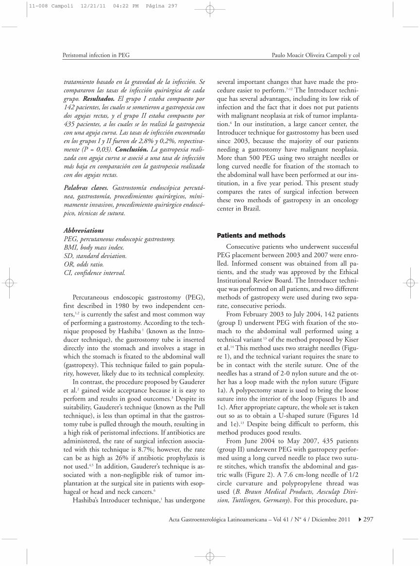
From February 2003 to July 2004, 142 patients(group I) underwent PEG with fixation of the sto-mach to the abdominal wall performed using atechnical variant 13 of the method proposed by Kiseret al.14 This method uses two straight needles (Figu-re 1), and the technical variant requires the snare tobe in contact with the sterile suture. One of theneedles has a strand of 2-0 nylon suture and the ot-her has a loop made with the nylon suture (Figure1a). A polypectomy snare is used to bring the loosesuture into the interior of the loop (Figures 1b and1c). After appropriate capture, the whole set is takenout so as to obtain a U-shaped suture (Figures 1dand 1e).13 Despite being difficult to perform, thismethod produces good results.
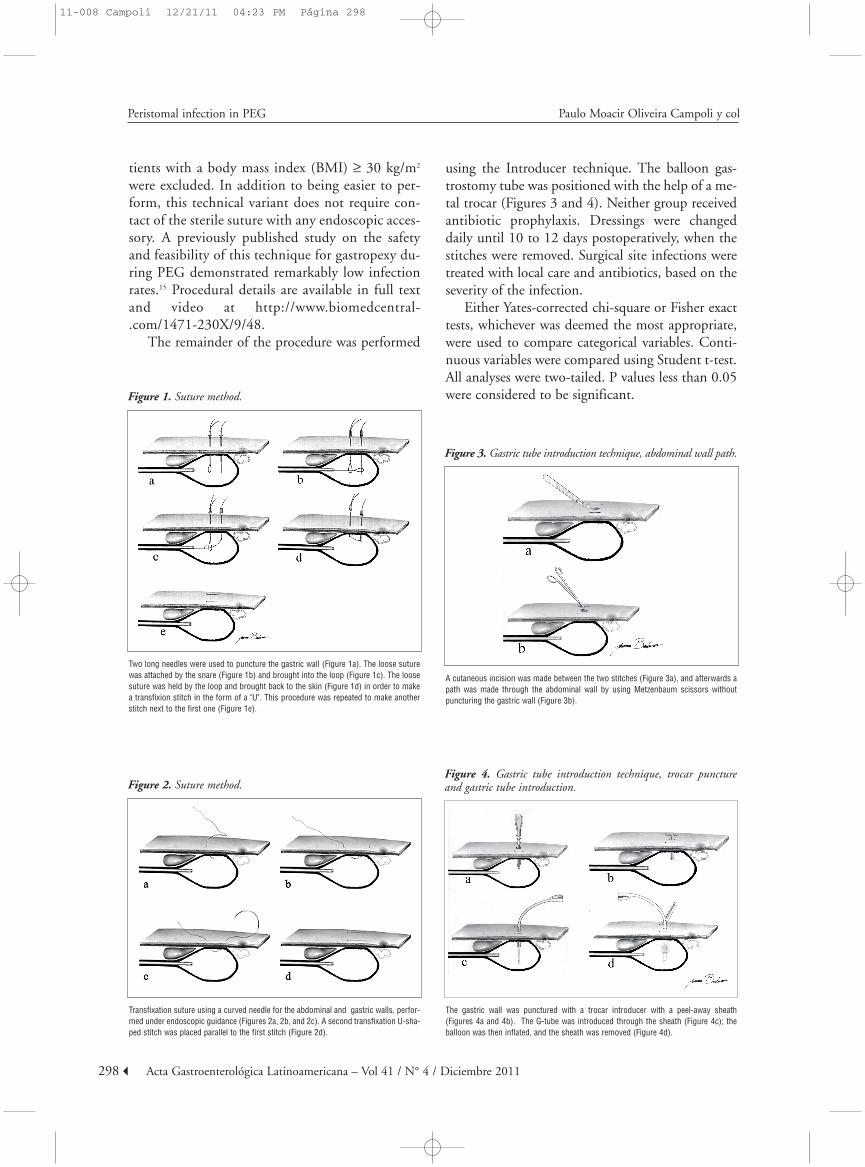
From June 2004 to May 2007, 435 patients(group II) underwent PEG with gastropexy perfor-med using a long curved needle to place two sutu-re stitches, which transfix the abdominal and gas-tric walls (Figure 2). A 7.6 cm-long needle of 1/2circle curvature and polypropylene thread wasused (B. Braun Medical Products, Aesculap Divi-sion, Tuttlingen, Germany). For this procedure, pa-
Peristomal infection in PEG Paulo Moacir Oliveira Campoli y col
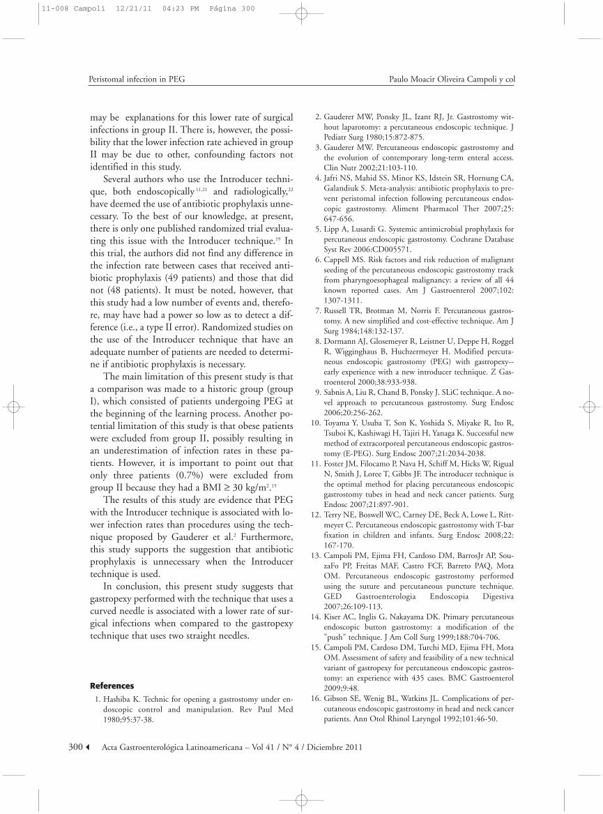
using the Introducer technique. The balloon gas-trostomy tube was positioned with the help of a me-tal trocar (Figures 3 and 4). Neither group receivedantibiotic prophylaxis. Dressings were changeddaily until 10 to 12 days postoperatively, when thestitches were removed. Surgical site infections weretreated with local care and antibiotics, based on theseverity of the infection.
Either Yates-corrected chi-square or Fisher exacttests, whichever was deemed the most appropriate,were used to compare categorical variables. Conti-nuous variables were compared using Student t-test.All analyses were two-tailed. P values less than 0.05were considered to be significant.
tients with a body mass index (BMI) ≥ 30 kg/m2
were excluded. In addition to being easier to per-form, this technical variant does not require con-tact of the sterile suture with any endoscopic acces-sory. A previously published study on the safetyand feasibility of this technique for gastropexy du-ring PEG demonstrated remarkably low infectionrates.15 Procedural details are available in full textand video at http://www.biomedcentral-.com/1471-230X/9/48.
Peristomal infection in PEG Paulo Moacir Oliveira Campoli y col
Figure 1. Suture method.
Two long needles were used to puncture the gastric wall (Figure 1a). The loose suturewas attached by the snare (Figure 1b) and brought into the loop (Figure 1c). The loosesuture was held by the loop and brought back to the skin (Figure 1d) in order to makea transfixion stitch in the form of a "U". This procedure was repeated to make anotherstitch next to the first one (Figure 1e).
Figure 2. Suture method.
Transfixation suture using a curved needle for the abdominal and gastric walls, perfor-med under endoscopic guidance (Figures 2a, 2b, and 2c). A second transfixation U-sha-ped stitch was placed parallel to the first stitch (Figure 2d).
A cutaneous incision was made between the two stitches (Figure 3a), and afterwards apath was made through the abdominal wall by using Metzenbaum scissors withoutpuncturing the gastric wall (Figure 3b).
The gastric wall was punctured with a trocar introducer with a peel-away sheath(Figures 4a and 4b). The G-tube was introduced through the sheath (Figure 4c); theballoon was then inflated, and the sheath was removed (Figure 4d).
11-008 Campoli 12/21/11 04:23 PM Página 298
Discussion
This present study suggests that gastropexy per-formed with a curved needle (group II) was associa-ted with lower morbidity when compared with gas-tropexy performed with two straight needles (groupI). Respiratory depression was the major complica-tion and occurred in a significantly higher percenta-ge of patients in group I. In 3 of the 6 cases of res-piratory depression in group I, a tracheotomy wasperformed while the patient was still in the endos-copy room. These patients had a large tumor volu-me affecting the larynx and had a borderline needfor tracheotomy prior to the procedure. Gibson etal.16 described this complication and expressed theimportance of identifying those patients already fa-cing respiratory difficulties and referring them fortracheotomy before performing PEG. In this pre-sent study, the lower rate of respiratory depressionamong patients that underwent gastropexy with theuse of curved needles (group II) may be due to ex-perience gained by the endoscopy team in identif-ying patients at risk and referring them for tracheo-tomy before performing PEG.
The overall surgical infection rate was low inboth groups. These results were achieved withoutthe use of antibiotic prophylaxis and are consistentwith rates reported elsewhere when PEG is perfor-med using the Introducer technique.10,11,17-19 In addi-tion, the rates reported in our study were lower thanthose reported when PEG is performed using thePull technique, even when antibiotic prophylaxis isused.4,5,20
When the rates of infection were compared bet-ween groups I and II, the latter group had a signifi-cantly lower risk of surgical infection. The lower ra-te of surgical infection in group II may be attribu-ted to the gastropexy technique used. The absenceof contact of endoscopic accessories with the sterilesuture and the ease of the procedure performance
Results
Of the 577 patients, 568 (98%) had a malig-nancy, mainly esophageal or head and neck cancers.The ratio of male to female patients was 4.2:1. Themajority of procedures were performed in the diges-tive endoscopy room, under conscious sedation andmonitored with pulse oximetry. Of the 577 pa-tients, 166 (28.8%) underwent the procedure on anoutpatient basis.
Table 1 shows the baseline characteristics of thetwo groups. There were no significant differencesbetween the two groups regarding the sex or age ofthe patients or in the indications for the procedure.However, the percentage of patients with head andneck tumors was significantly higher in group IIcompared with group I. The main findings on en-doscopy were stenosis, active peptic ulcer, and tra-cheoesophageal fistula; the differences in the fre-quency of these findings between the two groupsdid not reach statistical significance.
Procedure-related morbidity was significantly lo-wer in group II than in group I, primarily due tosignificantly lower risks of surgical site infection[odds ratio (OR) = 0.08, 95% confidence interval(CI) 0.01-0.72, P = 0.03) and respiratory depres-sion (OR = 0.16, 95%CI 0.04-0.64, P = 0.02) (Ta-ble 2). There was one procedure-related death ineach group; however, the observed percent differen-ce did not reach statistical significance.
4. Jafri NS, Mahid SS, Minor KS, Idstein SR, Hornung CA,Galandiuk S. Meta-analysis: antibiotic prophylaxis to pre-vent peristomal infection following percutaneous endos-copic gastrostomy. Aliment Pharmacol Ther 2007;25:647-656.
5. Lipp A, Lusardi G. Systemic antimicrobial prophylaxis forpercutaneous endoscopic gastrostomy. Cochrane DatabaseSyst Rev 2006:CD005571.
6. Cappell MS. Risk factors and risk reduction of malignantseeding of the percutaneous endoscopic gastrostomy trackfrom pharyngoesophageal malignancy: a review of all 44known reported cases. Am J Gastroenterol 2007;102:1307-1311.
7. Russell TR, Brotman M, Norris F. Percutaneous gastros-tomy. A new simplified and cost-effective technique. Am JSurg 1984;148:132-137.
8. Dormann AJ, Glosemeyer R, Leistner U, Deppe H, RoggelR, Wigginghaus B, Huchzermeyer H. Modified percuta-neous endoscopic gastrostomy (PEG) with gastropexy--early experience with a new introducer technique. Z Gas-troenterol 2000;38:933-938.
9. Sabnis A, Liu R, Chand B, Ponsky J. SLiC technique. A no-vel approach to percutaneous gastrostomy. Surg Endosc2006;20:256-262.
10. Toyama Y, Usuba T, Son K, Yoshida S, Miyake R, Ito R,Tsuboi K, Kashiwagi H, Tajiri H, Yanaga K. Successful newmethod of extracorporeal percutaneous endoscopic gastros-tomy (E-PEG). Surg Endosc 2007;21:2034-2038.
11. Foster JM, Filocamo P, Nava H, Schiff M, Hicks W, RigualN, Smith J, Loree T, Gibbs JF. The introducer technique isthe optimal method for placing percutaneous endoscopicgastrostomy tubes in head and neck cancer patients. SurgEndosc 2007;21:897-901.
12. Terry NE, Boswell WC, Carney DE, Beck A, Lowe L, Ritt-meyer C. Percutaneous endoscopic gastrostomy with T-barfixation in children and infants. Surg Endosc 2008;22:167-170.
14. Kiser AC, Inglis G, Nakayama DK. Primary percutaneousendoscopic button gastrostomy: a modification of the"push" technique. J Am Coll Surg 1999;188:704-706.
15. Campoli PM, Cardoso DM, Turchi MD, Ejima FH, MotaOM. Assessment of safety and feasibility of a new technicalvariant of gastropexy for percutaneous endoscopic gastros-tomy: an experience with 435 cases. BMC Gastroenterol2009;9:48.
16. Gibson SE, Wenig BL, Watkins JL. Complications of per-cutaneous endoscopic gastrostomy in head and neck cancerpatients. Ann Otol Rhinol Laryngol 1992;101:46-50.
Peristomal infection in PEG Paulo Moacir Oliveira Campoli y col
may be explanations for this lower rate of surgicalinfections in group II. There is, however, the possi-bility that the lower infection rate achieved in groupII may be due to other, confounding factors notidentified in this study.
Several authors who use the Introducer techni-que, both endoscopically 11,21 and radiologically,22
have deemed the use of antibiotic prophylaxis unne-cessary. To the best of our knowledge, at present,there is only one published randomized trial evalua-ting this issue with the Introducer technique.19 Inthis trial, the authors did not find any difference inthe infection rate between cases that received anti-biotic prophylaxis (49 patients) and those that didnot (48 patients). It must be noted, however, thatthis study had a low number of events and, therefo-re, may have had a power so low as to detect a dif-ference (i.e., a type II error). Randomized studies onthe use of the Introducer technique that have anadequate number of patients are needed to determi-ne if antibiotic prophylaxis is necessary.
The main limitation of this present study is thata comparison was made to a historic group (groupI), which consisted of patients undergoing PEG atthe beginning of the learning process. Another po-tential limitation of this study is that obese patientswere excluded from group II, possibly resulting inan underestimation of infection rates in these pa-tients. However, it is important to point out thatonly three patients (0.7%) were excluded fromgroup II because they had a BMI ≥ 30 kg/m2.15
The results of this study are evidence that PEGwith the Introducer technique is associated with lo-wer infection rates than procedures using the tech-nique proposed by Gauderer et al.2 Furthermore,this study supports the suggestion that antibioticprophylaxis is unnecessary when the Introducertechnique is used.
In conclusion, this present study suggests thatgastropexy performed with the technique that uses acurved needle is associated with a lower rate of sur-gical infections when compared to the gastropexytechnique that uses two straight needles.
References
1. Hashiba K. Technic for opening a gastrostomy under en-doscopic control and manipulation. Rev Paul Med1980;95:37-38.
17. Dormann AJ, Wejda B, Kahl S, Huchzermeyer H, EbertMP, Malfertheiner P. Long-term results with a new introdu-cer method with gastropexy for percutaneous endoscopicgastrostomy. Am J Gastroenterol 2006;101:1229-1234.
18. Horiuchi A, Nakayama Y, Tanaka N, Fujii H, Kajiyama M.Prospective randomized trial comparing the direct methodusing a 24 Fr bumper-button-type device with the pullmethod for percutaneous endoscopic gastrostomy. Endos-copy 2008;40:722-726.
19. Shastri YM, Hoepffner N, Tessmer A, Ackermann H, Sch-roeder O, Stein J. New introducer PEG gastropexy doesnot require prophylactic antibiotics: multicenter prospecti-ve randomized double-blind placebo-controlled study. Gas-trointest Endosc 2008;67:620-628.
20. Blomberg J, Lagergren P, Martin L, Mattsson F, LagergrenJ. Novel approach to antibiotic prophylaxis in percutaneousendoscopic gastrostomy (PEG): randomised controlledtrial. Br Med J 2010;341:c3115.
21. Saunders JR, Jr., Brown MS, Hirata RM, Jaques DA. Per-cutaneous endoscopic gastrostomy in patients with headand neck malignancies. Am J Surg 1991;162:381-383.
22. Dinkel HP, Beer KT, Zbaren P, Triller J. Establishing radio-logical percutaneous gastrostomy with balloon-retained tu-bes as an alternative to endoscopic and surgical gastrostomyin patients with tumours of the head and neck or oesopha-gus. Br J Radiol 2002;75:371-377.
Peristomal infection in PEG Paulo Moacir Oliveira Campoli y col