Page 1
Mar. Drugs 2015, 13, 2327-2346; doi:10.3390/md13042327
marine drugs ISSN 1660-3397
www.mdpi.com/journal/marinedrugs
Review
Fucoidan and Cancer: A Multifunctional Molecule with
Anti-Tumor Potential
Farzaneh Atashrazm 1, Ray M. Lowenthal 1, Gregory M. Woods 1, Adele F. Holloway 2
and Joanne L. Dickinson 1,*
1 Menzies Institute for Medical Research, University of Tasmania, Hobart, Tasmania 7000, Australia;
E-Mails: [email protected] (F.A.); [email protected] (R.M.L.);
[email protected] (G.M.W.) 2 School of Medicine, University of Tasmania, Hobart, Tasmania 7000, Australia;
E-Mail: [email protected]
* Author to whom correspondence should be addressed; E-Mail: [email protected] ;
Tel.: +61-3-6226-7622; Fax: +61-3-6226-7704.
Academic Editor: Keith B. Glaser
Received: 24 February 2015 / Accepted: 3 April 2015 / Published: 14 April 2015
Abstract: There is a wide variety of cancer types yet, all share some common cellular and
molecular behaviors. Most of the chemotherapeutic agents used in cancer treatment are
designed to target common deregulated mechanisms within cancer cells. Many healthy
tissues are also affected by the cytotoxic effects of these chemical agents. Fucoidan, a natural
component of brown seaweed, has anti-cancer activity against various cancer types by
targeting key apoptotic molecules. It also has beneficial effects as it can protect against
toxicity associated with chemotherapeutic agents and radiation. Thus the synergistic effect
of fucoidan with current anti-cancer agents is of considerable interest. This review discusses
the mechanisms by which fucoidan retards tumor development, eradicates tumor cells and
synergizes with anti-cancer chemotherapeutic agents. Challenges to the development of
fucoidan as an anti-cancer agent will also be discussed.
Keywords: fucoidan; cancer; apoptosis; synergy
OPEN ACCESS
Page 2
Mar. Drugs 2015, 13 2328
1. Introduction to Cancer
Cancers are multifactorial diseases of various etiologies. They arise largely as a result of acquired
genetic changes that alter cell function leading neoplastic cells to gain survival or growth
advantages [1]. For cancer cells to survive, the generation of new blood vessels (angiogenesis) is
required. Cancer leads to death mostly through tumor cell spread to distal organs (metastasis). Various
pathways are disrupted in tumor development, which result from unbalanced programmed cell death,
disordered signaling pathways, angiogenesis and poor immune response against cancer. Most of the
chemotherapeutic agents used in cancer treatment target these major deregulated pathways.
Unfortunately, as many of these therapies cause severe side effects, the toxicities limit the dose and
thus the efficacy of treatment. Therefore, there is strong interest in developing better-tolerated
anti-cancer agents.
2. A Role for Natural Products for Cancer Treatment
Chemotherapy has been a cornerstone of the standard cancer treatment regimens since the 1960s.
A variety of chemicals ranging from traditional agents such as methotrexate and folic acid analogues to
novel chemicals such as anthracyclines have been used in cancer treatment [2]. Despite promising
tumor growth-inhibitory effects in pre-clinical tests, many fail in clinical trials when adverse unexpected
side effects are revealed. Traditionally anti-cancer chemotherapy targets rapidly dividing and proliferating
cells. Therefore, normal cells which have high-proliferating potential are also affected.
Novel therapeutic agents are designed to target specific molecules (targeted therapy). However, these
targeted therapies are not always completely free of side effects either. For instance, vemurafenib, a
B-Raf enzyme inhibitor, is specific for oncogenic mutant V600E B-Raf positive melanoma cells. This
drug was the first targeted molecular therapy, which was approved for use in advanced stages of
melanoma. Although vemurafenib has shown significant beneficial anti-cancer effects, several studies
have reported the rapid emergence of acquired resistance and adverse dermatological effects. It also
stimulates B-Raf expression in V600E B-Raf negative patients promoting melanoma growth [3,4].
Monoclonal antibodies are another example of targeted therapy and are designed to specifically target
the cancer antigens located on tumor cells. Monoclonal antibodies are generally safer than chemotherapy
and the side effects caused by them include mild allergic reactions such as urticaria. But they can also
cause severe reactions such as infusion reactions and serum sickness. As an example, rituximab
(anti-CD20), which is widely used in treating B-cell lymphoma, generally causes only mild toxicities,
however, reports have described occasional cases with severe complications such as anaphylactic reactions
and myocardial infarction as well as high risk of tumor lysis syndrome in patients who have a high
burden of tumor cells in their circulation [5].
Concerns over toxicity, tumor cell resistance and development of secondary cancers from
chemotherapeutic chemicals have generated interest in exploiting natural products for cancer treatment.
Flavopiridol is a flavonoid derived from the indigenous Indian plant Dysoxylum binectariferum, which
inhibits cell cycle progression. It is the first cyclin-dependent kinase (CDK) inhibitor to be approved
for use in clinical trials [6]. Natural products are also being tested as adjuvants for use in synergy with
chemotherapeutic agents. For example those with immunomodulatory effects can reduce immune
Page 3
Mar. Drugs 2015, 13 2329
suppression and the associated increased risk of infection. In George et al. [7] study, Indukantha
Ghritha (IG), a polyherbal preparation consisting of 17 plant components, was used as an adjuvant to
cyclophosphamide cancer chemotherapy and shown to stimulate the hematopoietic system and induce
leukopoiesis in tumor-bearing mice. When administrated in combination with cyclophosphamide, it
reversed myelosuppression induced by cyclophosphamide suggesting its potential to minimize or
reverse chemotherapy-induced leukopenia.
Polysaccharides include a large family of diverse biopolymers. They are constituted by
monosaccharide residues linked together by O-glycosidic bonds that are found in natural and
semi-synthetic structures [8]. Due to structural diversity, polysaccharides display the highest biological
properties among macromolecules. Many natural polysaccharides obtained from natural sources such as
plants and algae have anti-cancer properties. The multifunctional structure of natural polysaccharides
also allows them to be used in conjugation with anti-cancer agents that lack physiochemical and
biopharmaceutical properties [8,9].
3. Fucoidan
Fucoidan is a natural sulfated polysaccharide that exists mainly in the cell wall matrix of various
species of brown seaweed such as mozuku, kombu, limumoui, bladderwrack and wakame [10]. Various
forms of fucoidan have also been recognized in some marine invertebrates such as sea urchins [11] and
sea cucumbers [12]. The brown seaweeds containing fucoidan are widely consumed as part of the
normal diet in East Asia, particularly Japan, China and Korea.
3.1. Fucoidan’s Anti-Cancer Potential
The anti-cancer property of fucoidan has been demonstrated in vivo and in vitro in different types of
cancers. Nevertheless, it has been rarely investigated for its anti-cancer properties in clinical trials.
Fucoidan mediates its activity through various mechanisms such as induction of cell cycle arrest, apoptosis
and immune system activation. Additional activities of fucoidan have been reported that may be linked to
the observed anti-cancer properties and these include induction of inflammation through immune system,
oxidative stress and stem cell mobilization. These activities have been reviewed by Kwak [13].
3.1.1. Fucoidan and Cell Cycle
Fucoidan treatment results in sub G0/G1 cell accumulation (suggestive of dead cells/apoptotic cells)
in a variety of cell types [14,15]. It can also induce cell cycle arrest in other phases; Riou et al. [16]
and Mourea et al. [17] reported arrest in G1 phase in a chemo-resistant non-small-cell bronchopulmonary
carcinoma line by fucoidan from Ascophyllum nodosum and Bifurcaria bifurcate, respectively.
In an investigation of the mechanism of the action, fucoidan demonstrated significant down
regulation of cyclin D1, cyclin D2 and CDK4 in cancer cells [18–20]. The crude fucoidan from Fucus
vesiculosus increased the level of p21/WAF1/CIP1 in PC3 cells and down-regulated E2F; a
transcription factor that controls progression of cells from G1 to S phase [18].
Page 4
Mar. Drugs 2015, 13 2330
Table 1. Effects of fucoidan on cell cycle and apoptosis molecules.
Ref Cell Type Fucoidan Source Dose (µg/mL) Effects on Cell Cycle Effects on Apoptosis Pathways Extrinsic Intrinsic Common
[15] Human lymphoma
HS-sultan cells F. vesiculosus 100
↑ sub G0/G1 - ↓ MMP Caspase 3 activation
No G0/G1 or G2/M arrest
[20] HTLV-1 infected
T-cell HUT-102- cells C. okamurans 3000
G1 arrest Apoptosis was reversed by
caspase 8 inhibitor
Caspase 9 activation Apoptosis was reversed by
caspase 3 inhibitor ↓ cyclin D2, c-myc No changes in Bcl-2 and Bcl-XL
No changes in p21,p53 ↓ survivin, cIAP-2
[21] Human hepatocellular
carcinoma cells Okinawa mozuku 22.5
↑ G2/M phase in HAK-1A,
KYN-2, KYN-3 cell lines - No clear caspase 9 activation in HAK-1B cell line
No clear caspase 3 activation in
HAK-1B cells
[22] Human breast cancer
MCF7 cells Not mentioned 1000 ↑ sub-G1 fraction
Caspase 8 activation Caspase 9 activation Caspase 7 activation
Caspase inhibitors blocked
apoptosis completely
↓ Bid, cytosolic Bax PARP cleavage
↑ whole lysate Bax, cytosolic cytochrome C
[23] Human acute leukemia
NB4 and HL-60 cells F. vesiculosus 150 ↑ sub-G1 fraction Caspase 8 activation
caspase 9 activation PARP cleavage
No changes in Bcl-2 or Bax Caspase 3 activation
↓ Mcl-1, ↑ cytochrome C
[24]
Human colon
cancer HT-29 and
HCT116 cells
F. vesiculosus -
Caspase 8 activation Caspase 9 activation PARP cleavage
↑ Fas, DR5, TRAIL ↑ cytochrome C, Smac/Diablo, Bak, t-Bid
Caspase 3 and 7 activation No significant effects on
FasL and DR4
No changes in Bcl-2, Bcl-xL, Bax, Bad, Bim, Bik
↓ XIAP, survivin
[25] Human lung cancer
A549 cells U. pinnatifida 50, 100, 200 ↑ Sub-G1frction -
Caspase-9 activation ↓ procaspase-3
↓ Bcl-2, ↑ Bax PARP cleavage
[14] Human breast cancer
MCF-7 cells
Cladosiphon
novae-caledoniae 82, 410, 820
↑ Sub-G1
No changes in caspase-
8
Mitochondrial dysfunction No activation of PARP and
caspase-7 No significant changes in cell
cycle distribution
AIF and cytochrome C release
No cleavage of caspase-9 and Bid. All caspase inhibitors failed to
attenuate FE-induced apoptosis ↓ Bcl-2, Bcl-xl ,↑ Bax, Bad
[26] Hela cells Sargassum
filipendula 1500 - -
No effect on caspase 9 activation No effect on caspase 3
(Caspase independent) ↑ cytosol AIF
[19] Human breast cancer
MCF-7 cells F. vesiculosus 400, 800, 1000
G1 phase arrest
Caspase-8 activation
↓ Bcl-2
Caspase-dependent pathway ↑ Sub G0/G1 ↓ cyclin D1 and
CDK-4 gene expression
↑ Bax
Release of cytochrome C and APAf-1
Page 5
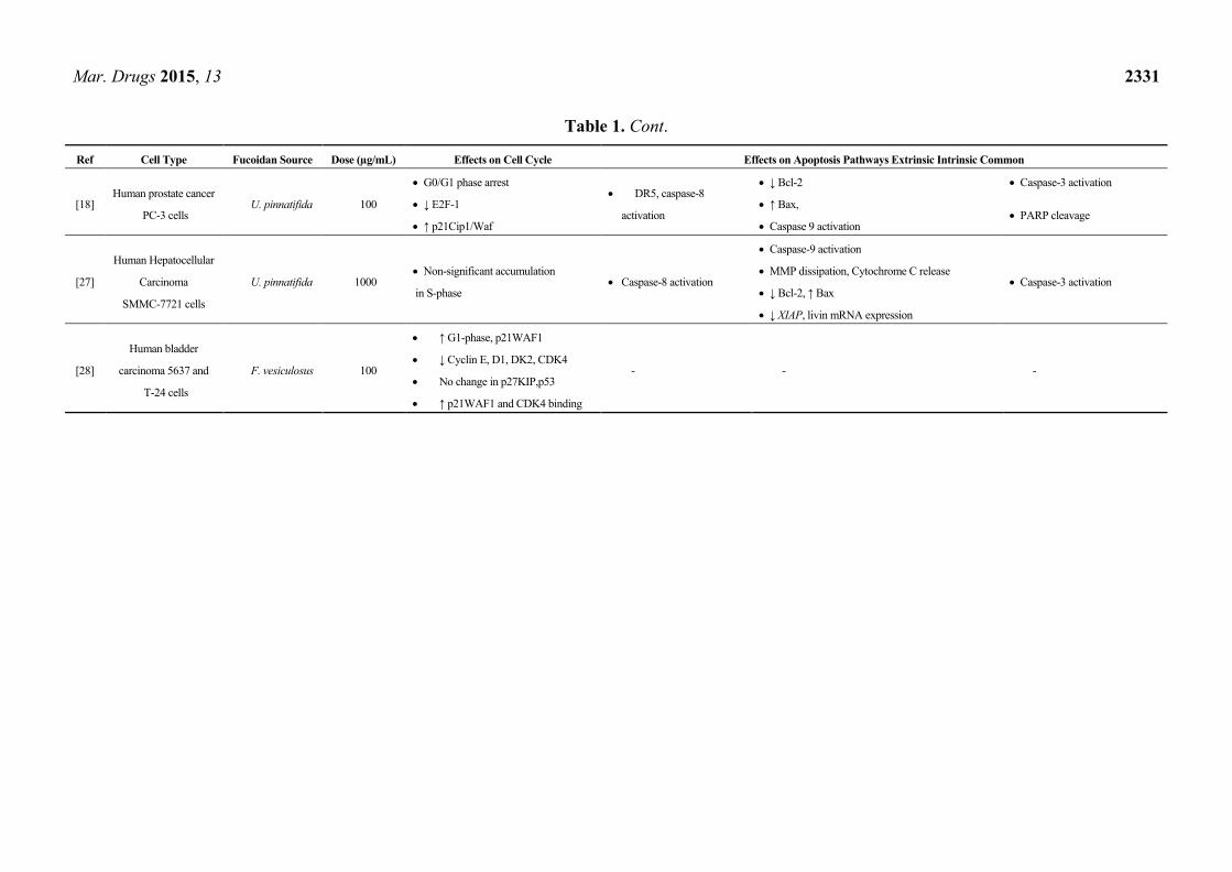
Mar. Drugs 2015, 13 2331
Table 1. Cont.
Ref Cell Type Fucoidan Source Dose (µg/mL) Effects on Cell Cycle Effects on Apoptosis Pathways Extrinsic Intrinsic Common
[18] Human prostate cancer
PC-3 cells U. pinnatifida 100
G0/G1 phase arrest DR5, caspase-8
activation
↓ Bcl-2 Caspase-3 activation
↓ E2F-1 ↑ Bax, PARP cleavage
↑ p21Cip1/Waf Caspase 9 activation
[27]
Human Hepatocellular
Carcinoma
SMMC-7721 cells
U. pinnatifida 1000 Non-significant accumulation
in S-phase Caspase-8 activation
Caspase-9 activation
Caspase-3 activation MMP dissipation, Cytochrome C release
↓ Bcl-2, ↑ Bax
↓ XIAP, livin mRNA expression
[28]
Human bladder
carcinoma 5637 and
T-24 cells
F. vesiculosus 100
↑ G1-phase, p21WAF1
- - - ↓ Cyclin E, D1, DK2, CDK4
No change in p27KIP,p53
↑ p21WAF1 and CDK4 binding
Page 6
Mar. Drugs 2015, 13 2332
In a recent study, fucoidan down-regulated cyclin E, CDK2, CDK4 resulting in G0/G1 arrest in
human bladder cancer 5637 cells. Furthermore, immunoprecipitation assays revealed a significant
increase in the binding of p21/WAF1/CIP1 to CDK2 and CDK4 in cells treated with fucoidan,
suggesting that the induced G0/G1 arrest is due to suppression of CDK activity following direct
binding of this CDK inhibitor to CDKs 2 and 4 [28]. Table 1 summarizes findings of studies
examining the effects of fucoidan on cell cycle.
3.1.2. Fucoidan and the Apoptosis Pathway
Apoptosis characterized by cytoplasmic shrinkage and chromatin condensation facilitates the
removal of cells without inducing inflammation [29]. Apoptosis occurs through either the extrinsic
(cytoplasmic) pathway whereby death receptors trigger the apoptosis, or the intrinsic (mitochondrial)
pathway in which changes in mitochondrial membrane potential (MMP) lead to cytochrome C release and
death signal activation. Both pathways activate executive caspases that cleave regulatory and structural
molecules [30]. Several studies examining a variety of cancers such as hematopoietic, lung, breast and
colon cancers have shown that fucoidan-mediated cell death occurs through triggering apoptosis
(Table 1) [14,22,24]. A very low dose of fucoidan from F. vesiculosus (20 µg/mL) activated common
caspases 3 and 7 in human colon cancer cells [24], whereas it induced the same activity in T-cell
leukemia at a much higher concentration (3 mg/mL) [20]. Caspase 8 and 9, two of the best characterized
molecules of the extrinsic and intrinsic pathways respectively are activated by fucoidan [24].
Yamasaki-Miyamoto et al. showed that pre-treatment with caspase 8 inhibitor completely blocked
fucoidan mediated apoptosis in MCF-7 breast cancer cell line [22]. In contrast, in Zhang et al. [14] study,
the mediated apoptosis by fucoidan from Cladosiphon okamuranus in MCF-7 human breast cancer cell
line was shown to be caspase independent. As cytochrome C and apoptosis inducing factor (AIF)
increased in the cytosol, it was concluded that fucoidan performed its activity through mechanisms
altering mitochondrial function.
Fucoidan also affects other components of extrinsic and intrinsic pathways. Analyzing the extrinsic
pathway, 20 µg/mL crude fucoidan from F. vesiculosus increased the levels of the death receptors Fas,
DR5 and TRAIL but not FasL and DR4 in human colon cancer cell lines [24]. Bcl-2 family members
include anti-apoptotic, pro-apoptotic and regulatory proteins, which are mainly involved in the
apoptosis intrinsic pathway. Contradictory results have been described in the expression of these regulatory
molecules in response to fucoidan (Table 1). Treatment of MDA-MB231 breast cancer cells with
820 µg/mL of low molecular weight (LMW) fucoidan resulted in a significant decrease in
anti-apoptotic proteins Bcl-2, Bcl-xl and Mcl-1 [31]. In contrast, no changes in expression of Bcl-2,
Bcl-xl, Bad, Bim and Bik were observed in colon cancer cells when they were treated with 20 µg/mL
fucoidan from Fucus vesiculosus [24]. Taken together, the results suggest that fucoidan may interact
with several components of the apoptosis pathway.
3.1.3. Fucoidan and Angiogenesis
Fucoidan inhibits the formation of new vessels by which tumor cells receive their oxygen and
required nutrients. Fucoidan has been found to inhibit the binding of VEGF, a key angiogenesis promoting
molecule, to its cell membrane receptor [32]. Xue et al. examined the anti-angiogenic properties of
Page 7
Mar. Drugs 2015, 13 2333
fucoidan in 4T1 mouse breast cancer cells both in vitro and in vivo and observed a significant
dose-dependent decrease in VEGF expression in cells treated with fucoidan. Further, in a mouse breast
cancer model using 4T1 cells, intraperitoneal injections of 10 mg/kg body weight fucoidan from
F. vesiculosus for 20 days markedly reduced the number of microvessels. Using immunohistochemistry,
fucoidan was shown to reduce VEGF expression compared to the control group [33]. In contrast, Zhu
et al. reported that fucoidan did not suppress angiogenesis and VEGF expression in human
hepatocarcinoma cell lines treated with 10 to 200 µg/mL of a commercial fucoidan purified from
Sargassum spp. Similarly no changes in VEGF expression were observed in xenograft tumors developed
in nude mice following 20 to 200 mg/kg/body weight fucoidan injected intraperitoneally once a day over
25 days [34]. It is postulated that different effects are observed with fucoidans of various MWs and
molecular structures and this is reviewed by Kwak [13].
3.1.4. Fucoidan and Metastasis
In 1987, Coombe et al. demonstrated that fucoidan significantly decreased tumor cells metastasis to
the lungs in animals that were intravenously injected with rat mammary adenocarcinoma 13762 MAT
cells [35]. It was first reported that fucoidan inhibits cell invasion through competing with tumor cell
binding with laminin in the basement membrane [36]. Subsequent studies then revealed that fucoidan
binds to fibronectin with high affinity and prevent attachment of tumor cells. In agreement with this
study, fucoidan reduced the spread of human breast adenocarcinoma cells plated on a surface containing
fibronectin [37].
Selectin inhibition by fucoidan interferes with tumor cell–platelet interaction. In Cumashi et al.
study [38], highly metastatic MDA-MB-231 breast cancer cells were plated in platelet-coated plates in
the presence or absence of 100 µg/mL fucoidan. The number of cells attached to the platelets
decreased by 80% in the presence of fucoidan. Interaction of tumor cells with platelets is one of the
key factors in facilitating the early steps of tumor cell migration. During tumor cell migration, most
circulating tumor cells do not survive attack from immune cells or the shear forces of the blood stream.
However, they can attach to platelets to induce platelet aggregation allowing the tumor cell cluster to
survive in the micro-vascular system. It was concluded that fucoidan inhibited P-selectin residing on
the platelet surface and led to reduced number of attached tumor cells. Fucoidan can also inhibit other
adhesion molecules such as integrins residing on the tumor cell surface and can modify distribution of
their subunits.
Tumor invasion requires the secretion of proteolytic enzymes by tumor cells to break down
the extracellular matrix (ECM) proteins (e.g., collagen, fibronectin and laminin), with the matrix
metalloproteinases (MMPs) MMP-2 and MMP-9 playing a major role. Fucoidan attenuates both
expression and activity of these enzymes [39].
3.1.5. Fucoidan and Signaling Pathways
The extracellular signal-regulated kinase (ERK) pathway (or Ras/Raf/MAPK pathway) is often
hyperphosphorylated and upregulated in a variety of human cancers. The potential for developing
anticancer agents that cause ERK’s dephosphorylation and pathway blockade have been explored.
Various studies have shown that fucoidan inhibits tumor cell proliferation by decreasing ERKs activity
Page 8
Mar. Drugs 2015, 13 2334
through reduction of its phosphorylation [15,40] while several studies have proposed that fucoidan
causes ERK activation rather than inactivation [41,42]. To explain these contradictions, it should be
noted that the ERK signaling pathway is highly complex. It induces a range of different responses
including cell proliferation, differentiation, migration and apoptosis depending on cell type, the type of
stimulus and duration of activation [43]. Therefore, some of the contradictory results of the
aforementioned studies can be explained by different fucoidan extracts with different molecular
structures being used on different tumor cell types. Another complication is that different studies have
examined ERK phosphorylation over different time periods ranging from 10 min to 48 h. Jin et al.
reported increased ERK1/2 phosphorylation in HL-60 leukemic cell line 10–15 min after fucoidan
treatment. The phosphorylation returned to the basal level after 1 h [23]. In Lee et al. study, crude
fucoidan progressively diminished phosphorylation of ERK1/2 from 1 h to 9 h after treatment [39].
JNK and p38 are other MAPK superfamily members whose activity is altered by fucoidan.
Fucoidan induced cell death in breast cancer cells through phosphorylation and activation of JNK and
p38 after 30 min. The fucoidan-induced apoptosis significantly annulled in the presence of JNK
inhibitor, indicating critical role of JNK in fucoidan-mediated apoptosis [14].
Figure 1. Overview of main signal transduction pathways involved in cell proliferation and apoptosis.
Similarly, the PI3K/AKT, GSK and Wnt pathways have been shown to be triggered by fucoidan.
PI3K/AKT pathway generally inhibits apoptosis. AKT over-activation is also associated with drug
resistance and tumor cell survival. As a result, deactivating this pathway could be another potential
target for anti-cancer drug development. Most of the studies have reported inactivation of AKT by
Page 9
Mar. Drugs 2015, 13 2335
fucoidan. PI3k, an upstream molecule of AKT, is also inhibited by fucoidan [39]. Upregulation of the
Wnt signaling pathway is believed to have a critical role in prostate cancer development, survival and
progression. Fucoidan from F. vesiculosus activated GSK-3β in PC3 human prostate cancer cells
resulting in hypo-phosphorylation and inactivation of β-catenin, a critical component of the Wnt pathway
(Figure 1) [18]. Figure 1 represents an overview of the mentioned signaling pathways.
3.1.6. Fucoidan and the Immune System
The effects of fucoidan on molecules of the immune system have been studied both in vitro and
in vivo and effects on both cellular and humoral elements have been described. Fucoidan increases
both activity and number of natural killer (NK) cells in vivo [44,45]. Increase in the number of
cytotoxic T-cells (CTLs) has also been reported. A high-molecular-weight (HMW) fucoidan from
Cladosiphon okamuranus (200–300 kDa) induced a large increase in the proportion of murine
cytotoxic T cells [46]. Investigation of the role of fucoidan on dendritic cell (DC)-mediated T-cell
cytotoxicity has revealed that the stimulation of CTLs was more effective in fucoidan-treated DCs as CTLs
co-cultured with fucoidan-treated DCs exerted a high level of specific lysis of breast cancer cells [47].
In a recent study, the role of fucoidan in DCs function and its adjuvant effect have been examined
in vivo. Fucoidan was systemically administrated to mice by intraperitoneal injection. Examination of
the spleen DCs revealed up-regulation of maturation markers as well as production of IL-6, IL-12 and
TNF-α. Fucoidan was then used as an adjuvant in vivo with ovalbumin antigen and induced Th1
mediated immune response and CTL activation [48].
3.1.7. Fucoidan and Malignant Transformation in Vitro and in Vivo
Few studies have reported the potential of fucoidan to inhibit neoplastic transformation. Teas et al. fed
rats with dietary seaweed (Laminira) for 55 days and administrated the carcinogen 7,12-dimethylbenz(a)
anthracene intragastrically. Following 26 weeks monitoring, experimental rats showed a significant
delay in the median time for tumor appearance (19 vs. 11 weeks in the control group) [49].
Transforming growth factor β1 (TGFβ1) is believed to promote tumor development and metastasis
through epithelial to mesenchymal transition (EMT), a process that enables epithelial cells migrate to
distant areas during late stages of breast cancer development [50]. To trigger tumor progression,
TGFβ1 recruits TGF receptors (TGFR) residing on the cell surface. The investigations of effects of
fucoidan on TGFβ1-promoted carcinogenesis in MDA-MB-231 breast cancer cells have indicated that
fucoidan decreased the expression of TGFRs and affected the downstream signaling molecules, which
are involved in TGFβ1-mediated EMT [41].
Epidermal growth factor (EGF) is another carcinogenesis promoter, which induces tumor
transformation through overexpression and activation of EGF receptor (EGFR). EGFR has a key role
in cell proliferation and differentiation and many carcinomas arise from its mutations [51].
Lee et al. examined the role of fucoidan on the activation of EGFR and EGF-mediated neoplastic
transformation [52]. They utilized murine JB6 Cl41 epidermal cells and induced cell transformation by
EGF in the presence of fucoidan from L. guryanovae. Fucoidan markedly reduced the EGFR activation
through hypo-phosphorylation. It also inhibited EGF-tumorigenic activity through inhibition of AP-1, a
transcription factor responsible for cell proliferation regulation.
Page 10
Mar. Drugs 2015, 13 2336
3.2. Fucoidan Metabolism
Fucoidanase, the enzyme responsible for fucoidan hydrolysis, has only been found in brown
seaweed and marine microorganisms such as some marine bacteria and fungi [53] and not in humans.
It is possible that the acidic conditions in the stomach could degrade fucoidan, but it has been reported
that the low gastric pH does have restricted effects on fucoidan [54].
Small amounts of dietary fucoidan can be endocytosed and cross the intestinal wall directly without
breaking down [54]. In Tokita et al. study, 10 volunteers were given oral fucoidan and the
concentrations of fucoidan in the serum and urine were analyzed. Fucoidan was detectable 3 h after
administration and increased to 100 ng/mL in serum and 1000 ng/mL in urine. However the rate of
absorption in the small intestine was highly variable among the participants. The MW of fucoidan in
serum was similar to administered fucoidan indicating that fucoidan was not hydrolyzed by digestive
enzymes [55]. However, the MW of the fucoidan detected in urine was significantly smaller than the
ingested fucoidan suggesting that fucoidan is degraded in the excretory system and possibly the kidney
and not by intestinal enzymes or normal flora.
To evaluate the fucoidan uptake process by cells, the internalization of LMW fucoidan into rabbit
smooth muscle cells (SMCs) was analyzed. Fucoidan was shown to be internalized by endocytosis at 6 h.
The number of vesicles containing fucoidan increased in the peri-nuclear region at 24 h, but nuclear
internalization was not observed at any time during the study [56]. However, examining the transport
of a native fucoidan from Cladosiphon okamuranus with MW of 80 kDa revealed a poor permeation of
fucoidan across the human colon adenocarcinoma Caco-2 cell monolayer [57].
Regarding the specific ligands by which fucoidan binds to the cells surface, several molecules have been
implicated including class A macrophage scavenger receptors for fucoidan attachment to macrophages [58]
as well as adhesion molecules such as L-selectin and P-selectin [59] and integrins [60]. However, some
reports have shown fucoidan mediates apoptosis through selectin-independent mechanisms [15].
3.3. Fucoidan as a Synergistic Anti-Cancer Agent
The ability of fucoidan to synergize with standard anti-cancer agents and/or reduce toxicity has
recently been investigated. Ikeguchi et al. examined the synergistic effect of a HMW fucoidan with
colorectal cancer chemotherapy agents; oxaliplatin plus 5-fluorouracil/leucovorin (FOLFOX) or
irinotecan plus 5-fluorouracil/leucovorin (FOLFIRI). The test patients received 150 mL/day for
6 months of liquid that contained 4.05 g fucoidan. From the commencement of chemotherapy, toxicities
and chemotherapy efficiency were compared. Fucoidan showed no side effects such as allergic
dermatitis. Diarrhea, neurotoxicity and myelosuppression were not suppressed by fucoidan, whereas
general fatigue was significantly decreased from 60% to 10%. The patients were followed for
approximately 15 months and the survival rate of the patients who received fucoidan was longer than
that of the control participants; however the difference was not significant, probably due to the small
numbers [61].
Fucoidan affects the migration and invasion of multiple myeloma (MM) cells treated with
chemotherapy drug cytarabine. The human myeloma cell lines RPMI8226 and U266 were treated with
crude fucoidan from F. vesiculosus for 72 h and then cytarabine for 6 h. Fucoidan reduced cell migration
Page 11
Mar. Drugs 2015, 13 2337
through a Boyden chamber and down-regulated expression of CXCR4 and MMP-9 [62]. Fucoidan
from Saccharina cichorioides has been reported to synergize with the anti-tumor activity of low dose
resveratrol (a natural polyphenol extracted from foods and beverages) on invasive and highly motile
HCT 116 colon cancer cell line [63]. In the colony formation assay, fucoidan plus resveratrol reduced the
colony number by 60% compared to 34% and 27% in resveratrol alone or fucoidan alone, respectively.
Zhang et al. studied the combinatory effect of fucoidan and three commonly used anti-cancer
agents; cis-platin (CDDP), tamoxifen (TAM) and paclitaxel (Taxol) on signal transduction pathways.
Fucoidan from Cladosiphon navae-caledoniae plus anti-cancer agents reduced the ERK phosphorylation in
MDA-MB-231 breast cancer cells compared to untreated control or fucoidan alone [64]. Dietary
fucoidan synergistically reduced cell growth in the OE33 cell line when it was combined with
lapatinib, a targeted therapy that acts as a tyrosine kinase inhibitor in advanced HER2-positive breast
cancer cells [65].
In a xenograft transplantation study, the effect of fucoidan alone or in combination with
cyclophosphamide was examined on tumor growth. Nine days after the injection of Lewis lung
carcinoma cells into mice, fucoidan from Fucus evanescens was administered to animals alone or
combined with cyclophosphamide. The fucoidan group showed marked antitumor (33% tumor growth
inhibition) and anti-metastatic (29% reduction of the number of metastases) activities. However,
fucoidan did not exhibit a synergistic effect with cyclophosphamide on tumor growth, but significantly
decreased the lung cancer cells metastasis [66].
3.4. Why Fucoidan Usage is Complicated?
Despite the promising results about the anti-cancer effect of fucoidan, there are still challenges
impeding utilization of fucoidan in the clinic. Variable and contradictory results being influenced by
endogenous and exogenous factors in fucoidan usage are of the main concerns. In this section we will
summarize important conditions, which have been undertaken in different experiments and have led to
such variable results in reported studies.
3.4.1. Structure and Molecular Weight Variation
Fucoidan is composed of α-(1-2) or α-(1-3)-linked L-fucose with a fucose content of 34-44%. It
also contains various amounts of other monosaccharaides such as galactose, mannose, xylose and
uronic acid all of which make up less than 10% of the total fucoidan structure [67,68]. The sulfate
groups in fucoidan structure are mainly at position 4 but they can also occupy position C2 and
occasionally C3 [53]. The fucoidan structure and monosaccharide composition vary depending on
different factors such as the source of fucoidan, the time and location of harvesting and the extraction
method, which can affect the fucoidan’s bioactivities. Most anti-cancer studies of fucoidan have used a
commercially available crude fucoidan extracted from Fucus vesiculosus (Sigma Co. St. Louis, MO,
USA). Some groups have extracted and purified fucoidan in their own laboratories. Okinawa mozuku,
C. Okamuranus tokida, Sargassum sp. and Undaria pinnatifida are the most common fucoidans
examined in cancer studies.
Cumashi et al. studied different biological aspects of fucoidan from nine different species of brown
seaweed in rats [38]. Analysis of P-selectin-mediated neutrophil adhesion to platelets revealed that
Page 12
Mar. Drugs 2015, 13 2338
extracted fucoidans from only some sources like F. evanescens and A. nodosum could serve as more
efficient P-selectin inhibitors. Furthermore, in contrast to other sources, fucoidan from C. okamuranus did
not exert anti-coagulant activity, which was suggested to be due to high content of 2-O-a-D-glucuronyl
substituent in the polysaccharide chain of fucoidan from C. okamuranus.
Sulfation is another key factor in fucoidan bioactivity. More sulfation is linked with greater
bioactivity and thus researchers have produced over-sulfated fucoidans to enhance its biological
properties [36]. It has been suggested that over-sulfation causes higher negative charge in the molecule
which can facilitate formation of fucoidan-protein complexes involved in cell proliferation [69].
Molecular weight is another crucial factor in fucoidan activity. Cho et al. produced three fucoidan
fractions with molecular weights of <5, 5–30 and >30 kDa and reported that the F5-30K showed the
most tumor growth inhibitory effect despite the sulfate amount in F<5K being greater than in the two
other fractions [70].
The extraction method can also affect fucoidan’s bio-properties. Fucoidan from Undaria pinnatifida
was hydrolyzed using different hydrolysis conditions and their anti-cancer activity was compared
in vitro. The native fucoidan showed 37% anti-cancer activity; hydrolyzed fucoidan generated under mild
conditions (in boiling water with HCl for 5 min) exhibited 75.9% anti-tumor activity; whereas
hydrolyzed fucoidan generated under harsh conditions (microwave for more than 90 s) slightly
enhanced the anti-cancer effect [71].
3.4.2. Fucoidan Dose and Route of Administration
As fucoidan is a large highly branched molecule, the dosage for in vitro studies mostly resides in
the range of µg/mL and not ng/mL. However, there is a large variation in the doses. Vischchuk et al.
treated HCT-116 colon cancer cells with 100–800 μg/mL fucoidan from the brown alga Saccharina
cichorioides Miyabe and observed that fucoidan exerted a low cytotoxicity and there was less than
15% reduction in cell number with the high dose of 800 μg/ml after 24 h [63]. In contrast, Kim et al.
demonstrated that 20 μg/mL fucoidan from F. vesiculosus caused 37% growth inhibition in the same cell
line after 72 h [24]. Though the difference between incubation times (24 h vs. 72 h) should be
considered, the dose difference (800 μg/mL vs. 20 μg/mL) was substantial. The source of fucoidan
appears to be the main factor leading to variation in results. Though most researchers have utilized
dosages of less than 1 mg/mL, there are reports of use of up to 3 mg/mL fucoidan.
Regarding the in vivo studies, both dose and the route of administration can affect outcome. To
select the most effective dose, mice were treated with various doses of fucoidan (10–400 mg/kg body
weight) followed by total-body irradiation. The mice injected with 100 mg/kg body weight fucoidan
showed the best survival rate at 30 days post-irradiation [72]. Other studies have used various doses
ranging from 5 mg/kg to 100 mg/kg and occasionally doses up to 500 mg/kg/body weight of different
fucoidan extracts. The amount and number of doses of fucoidan administration has also been shown to
be important for in vivo studies. Alekseyenko et al. studied mice with lung carcinoma that were treated
with fucoidan from Fucus evanescence. They found that a single injection of 25 mg/kg/body weight of
fucoidan did not inhibit tumor cell proliferation, while three-time injections of 10 mg/kg/body weight
significantly reduced tumor growth and metastasis [66]. Most in vivo studies of anti-tumor activity have
selected intraperitoneal (IP) injections, but subcutaneous (SC) or intravenous (IV) routes of
Page 13
Mar. Drugs 2015, 13 2339
administration have also been used. Oral fucoidan is another route for in vivo delivery either for its
anti-tumor properties following tumor induction or as a neoplastic transformation inhibitor
administered prior to cancer induction. Taken together, these studies indicate that different delivery
routes can affect the fucoidan metabolism in vivo and lead to variable outcomes.
3.5. Fucoidan Toxicity
Whilst fucoidan consumed in food in the form of 4% of the total dry weight of brown seaweeds is
generally regarded as safe, the fucoidan used for research is a highly purified extract. For in vitro
studies, researchers have utilized normal cells such as normal fibroblasts alongside tumor cell lines and
reported that fucoidan did not induce apoptosis within normal cells at the doses which were toxic for
cancer cell lines. A very high dose of 3 mg/mL fucoidan suppressed the viability of peripheral blood
mononuclear cells from healthy donors by 25% compared to 60%–90% in five different leukemic
T-cells [20]. In vivo, oral administration of up to 1 g/mL/body weight Undaria pinnatifida fucoidan was
non-toxic in mice but higher doses (2 g/mL/body weight) induced changes in thyroid weight and altered
levels of triglyceride and alanine transaminase activity [73]. In another study, daily administration of
300 mg/kg/body weight fucoidan from Laminaria japonica in Wister rats over 6 months did not induce
any adverse side effects, but higher doses (900–2500 mg/mL) resulted in coagulopathy and markedly
elevated clotting time [74].
Toxicity has also been examined in the context of fucoidan use as adjuvant. Oh et al. examined the
combinatory effect of fucoidan with the standard anti-Her2 inhibitor lapatinib in different breast cancer
cell lines in vitro [65] and found that fucoidan decreased the efficiency of lapitinib and exerted
antagonistic effects on cell proliferation in a few cell lines. Examining the effect of combination of
fucoidan from Fucus evanescence with cyclophosphamide, 7 out of 10 mice that were injected with
25 mg/kg/body weight fucoidan plus cyclophosphamide died whereas of the mice that were treated
with fucoidan alone, 3 out of 10 died [66].
Fucoidan has been examined in several clinical trials mainly for its anti-coagulant and anti-viral
properties. Administration of capsules containing 560 mg fucoidan from Undaria pinnatifida for up to
24 months did not induce any side effect when the participants took 4 capsules a day [75]. In
Mori et al. [76] and Irhimeh et al. [77] studies, daily consumption of 5 capsules contained 166 mg
fucoidan from C okamuranus Tokida for over one year and 3 g HMW fucoidan from Undaria
pinnatifida for up to 12 days, respectively, were revealed to be safe. However, Irhimeh et al.
demonstrated that orally administered fucoidan affected coagulation tests and prolonged the aPTT,
thrombin time and AT-III. Other studies have also shown the potential of bleeding complication
development due to fucoidan’s anti-thrombotic property [78]. Diarrhea is another reported side effect,
which was seen in 4 out of 17 participants within 1 month of daily administration of 6 g fucoidan [79].
When a blend of three different extracts (from Fucus vesiculosis (85% w/w), Macrocystis pyrifera
(10% w/w), and Laminaria japonica (5% w/w)) in capsules containing up to 187.5 mg were daily
given to volunteers, a statistically significant change in the potassium level was seen after 28 days.
Although, the change was minor and within the clinical reference range [80].
Page 14
Mar. Drugs 2015, 13 2340
4. Conclusions
The goal of cancer treatment is eradication of tumor cells ideally with minimal damage to healthy
tissues. Because of the side-effects of many current treatments, the use of natural substances of low toxicity
is of interest. A number of in vitro and in vivo studies have indicated that fucoidan contains strong
anti-cancer bioactivity. Since fucoidan also possesses immunomodulatory effects, it is postulated that it
may have protective effects against development of side effects when it is co-administered with
chemotherapeutic agents and radiation.
In this report, we reviewed the underlying cellular mechanisms by which fucoidan induces cell
death within tumor cells and increases the survival rate of tumor-bearing animal models by
suppression of metastasis and angiogenesis. However despite numerous promising pre-clinical reports,
there are few reported clinical studies so far [61]. In this review we also discussed the challenges
impeding utilization of fucoidan in the clinic which include the complex heterogeneous structure of
fucoidan, highly variable doses, different administration routes and possible negative interactions with
chemotherapy. Due to the wide variation of fucoidan structure and to make future experiments
reproducible, it is recommended that the critical bioactivity factors such as fucoidan content, sulfate
content, monosaccharide constituents and molecular weight be reported. Attention to these factors will
be likely to lead to more consistent reports and ultimately produce the required evidence to underpin
clinical studies in near future.
Acknowledgments
This work was supported by the Australian National Health and Medical Research Council
(NHMRC), the Australian Cancer Research Foundation and by Menzies Institute for Medical
Research, University of Tasmania philanthropic support. JLD is supported by an Australian Research
Council Future Fellowship.
Authors Contribution
FA conducted the literature research and drafted the manuscript. RML carried out the supervision
and edited the manuscript. GMW carried out the supervision and edited the manuscript. AFH carried
out the supervision and edited the manuscript. JLD carried out the supervision and edited the manuscript.
Conflicts of Interest
The authors declare no conflict of interest.
References
1. Cooper, G.M. The development and causes of cancer. In The Cell: A Molecular Approach, 2nd ed.;
Sinauer Associates: Sunderland, MA, USA, 2000.
2. Joo, W.D.; Visintin, I.; Mor, G. Targeted cancer therapy—Are the days of systemic chemotherapy
numbered? Maturitas 2013, 76, 308–314.
Page 15
Mar. Drugs 2015, 13 2341
3. Cohen, P.R.; Bedikian, A.Y.; Kim, K.B. Appearance of new vemurafenib-associated melanocytic
nevi on normal-appearing skin: Case series and a review of changing or new pigmented lesions in
patients with metastatic malignant melanoma after initiating treatment with vemurafenib. J. Clin.
Aesthet. Dermatol. 2013, 6, 27–37.
4. Huang, V.; Hepper, D.; Anadkat, M.; Cornelius, L. Cutaneous toxic effects associated with
vemurafenib and inhibition of the braf pathway. Arch. Dermatol. 2012, 148, 628–633.
5. Dotan, E.; Aggarwal, C.; Smith, M.R. Impact of rituximab (rituxan) on the treatment of b-cell
non-hodgkin’s lymphoma. P T 2010, 35, 148–157.
6. Senderowicz, A.M. Flavopiridol: The first cyclin-dependent kinase inhibitor in human clinical
trials. Investig. New Drugs 1999, 17, 313–320.
7. George, S.K.; Rajesh, R.; Kumar, S.S.; Sulekha, B.; Balaram, P. A polyherbal ayurvedic
drug—Indukantha ghritha as an adjuvant to cancer chemotherapy via immunomodulation.
Immunobiology 2008, 213, 641–649.
8. Caliceti, P.; Salmaso, S.; Bersani, S. Polysaccharide-based anticancer prodrugs. In
Macromolecular Anticancer Therapeutics; Reddy, L.H., Couvreur, P., Eds.; Springer: New York,
NY, USA, 2010; pp. 163–166.
9. Aravind, S.R.; Joseph, M.M.; Varghese, S.; Balaram, P.; Sreelekha, T.T. Antitumor and
immunopotentiating activity of polysaccharide pst001 isolated from the seed kernel of tamarindus
indica: An in vivo study in mice. Sci. World J. 2012, 2012, 361382.
10. Kalimuthu, S.; Kim, S. Fucoidan, a sulfated polysaccharides from brown algae as therapeutic
target for cancer. In Handbook of Anticancer Drugs from Marine Origin; Kim, S., Ed.; Springer
International Publishing: Cham, Switzerland, 2015; p. 147.
11. Mulloy, B.; Ribeiro, A.; Alves, A.; Vieira, R.; Mourao, P. Sulfated fucans from echinoderms have
a regular tetrasaccharide repeating unit defined by specific patterns of sulfation at the o-2 and o-4
positions. J. Biol. Chem. 1994, 22113–22123.
12. Ribeiro, A.; Vieira, R.; Mourao, P.; Mulloy, B. A sulfated a-l-fucan from sea cucumber.
Carbohydr. Res. 1994, 255, 225–240.
13. Kwak, J.Y. Fucoidan as a marine anticancer agent in preclinical development. Mar. Drugs 2014,
12, 851–870.
14. Zhang, Z.; Teruya, K.; Eto, H.; Shirahata, S. Fucoidan extract induces apoptosis in mcf-7 cells via
a mechanism involving the ros-dependent jnk activation and mitochondria-mediated pathways.
PLoS ONE 2011, 6, e27441.
15. Aisa, Y.; Miyakawa, Y.; Nakazato, T.; Shibata, H.; Saito, K.; Ikeda, Y.; Kizaki, M. Fucoidan
induces apoptosis of human hs-sultan cells accompanied by activation of caspase-3 and
down-regulation of erk pathways. Am. J. Hematol. 2005, 78, 7–14.
16. Riou, D.; Colliec-Jouault, S.; Pinczon du Sel, D.; Bosch, S.; Siavoshian, S.; Le Bert, V.;
Tomasoni, C.; Sinquin, C.; Durand, P.; Roussakis, C. Antitumor and antiproliferative effects of a
fucan extracted from ascophyllum nodosum against a non-small-cell bronchopulmonary
carcinoma line. Anticancer Res. 1996, 16, 1213–1218.
Page 16
Mar. Drugs 2015, 13 2342
17. Moreau, D.; Thomas-Guyon, H.; Jacquot, C.; Jugé, M.; Culioli, G.; Ortalo-Magné, A.; Piovetti, L.;
Roussakis, C. An extract from the brown alga bifurcaria bifurcata induces irreversible arrest of
cell proliferation in a non-small-cell bronchopulmonary carcinoma line. J. Appl. Phycol. 2006, 18,
87–93.
18. Boo, H.J.; Hong, J.Y.; Kim, S.C.; Kang, J.I.; Kim, M.K.; Kim, E.J.; Hyun, J.W.; Koh, Y.S.; Yoo, E.S.;
Kwon, J.M.; et al. The anticancer effect of fucoidan in pc-3 prostate cancer cells. Mar. Drugs
2013, 11, 2982–2999.
19. Banafa, A.M.; Roshan, S.; Liu, Y.Y.; Chen, H.J.; Chen, M.J.; Yang, G.X.; He, G.Y. Fucoidan
induces g1 phase arrest and apoptosis through caspases-dependent pathway and ros induction in
human breast cancer mcf-7 cells. J. Huazhong Univ. Sci. Technol. Med. Sci. 2013, 33, 717–724.
20. Haneji, K.; Matsuda, T.; Tomita, M.; Kawakami, H.; Ohshiro, K.; Uchihara, J.; Masuda, M.;
Takasu, N.; Tanaka, Y.; Ohta, T.; et al. Fucoidan extracted from cladosiphon okamuranus tokida
induces apoptosis of human t-cell leukemia virus type 1-infected t-cell lines and primary adult
t-cell leukemia cells. Nutr. Cancer 2005, 52, 189–201.
21. Fukahori, S.; Yano, H.; Akiba, J.; Ogasawara, S.; Momosaki, S.; Sanada, S.; Kuratomi, K.;
Ishizaki, Y.; Moriya, F.; Yagi, M.; et al. Fucoidan, a major component of brown seaweed,
prohibits the growth of human cancer cell lines in vitro. Mol. Med. Rep. 2008, 1, 537–542.
22. Yamasaki-Miyamoto, Y.; Yamasaki, M.; Tachibana, H.; Yamada, K. Fucoidan induces apoptosis
through activation of caspase-8 on human breast cancer mcf-7 cells. J. Agric. Food Chem. 2009,
57, 8677–8682.
23. Jin, J.O.; Song, M.G.; Kim, Y.N.; Park, J.I.; Kwak, J.Y. The mechanism of fucoidan-induced
apoptosis in leukemic cells: Involvement of erk1/2, jnk, glutathione, and nitric oxide. Mol.
Carcinog. 2010, 49, 771–782.
24. Kim, E.J.; Park, S.Y.; Lee, J.Y.; Park, J.H. Fucoidan present in brown algae induces apoptosis of
human colon cancer cells. BMC Gastroenterol. 2010, 10, 96, doi:10.1186/1471-230X-10-96.
25. Boo, H.J.; Hyun, J.H.; Kim, S.C.; Kang, J.I.; Kim, M.K.; Kim, S.Y.; Cho, H.; Yoo, E.S.; Kang, H.K.
Fucoidan from undaria pinnatifida induces apoptosis in a549 human lung carcinoma cells.
Phytother. Res. 2011, 25, 1082–1086.
26. Costa, L.S.; Telles, C.B.; Oliveira, R.M.; Nobre, L.T.; Dantas-Santos, N.; Camara, R.B.; Costa, M.S.;
Almeida-Lima, J.; Melo-Silveira, R.F.; Albuquerque, I.R.; et al. Heterofucan from sargassum
filipendula induces apoptosis in hela cells. Mar. Drugs 2011, 9, 603–614.
27. Yang, L.; Wang, P.; Wang, H.; Li, Q.; Teng, H.; Liu, Z.; Yang, W.; Hou, L.; Zou, X. Fucoidan
derived from undaria pinnatifida induces apoptosis in human hepatocellular carcinoma
smmc-7721 cells via the ros-mediated mitochondrial pathway. Mar. Drugs 2013, 11, 1961–1976.
28. Cho, T.M.; Kim, W.J.; Moon, S.K. Akt signaling is involved in fucoidan-induced inhibition of
growth and migration of human bladder cancer cells. Food Chem. Toxicol. 2014, 64, 344–352.
29. Elmore, S. Apoptosis: A review of programmed cell death. Toxicol. Pathol. 2007, 35, 495–516.
30. Ghobrial, I.M.; Witzig, T.E.; Adjei, A.A. Targeting apoptosis pathways in cancer therapy.
CA Cancer J. Clin. 2005, 55, 178–194.
31. Zhang, Z.; Teruya, K.; Eto, H.; Shirahata, S. Induction of apoptosis by low-molecular-weight
fucoidan through calcium- and caspase-dependent mitochondrial pathways in mda-mb-231 breast
cancer cells. Biosci. Biotechnol. Biochem. 2013, 77, 235–242.
Page 17
Mar. Drugs 2015, 13 2343
32. Koyanagi, S.; Tanigawa, N.; Nakagawa, H.; Soeda, S.; Shimeno, H. Oversulfation of fucoidan
enhances its anti-angiogenic and antitumor activities. Biochem. Pharmacol. 2003, 65, 173–179.
33. Xue, M.; Ge, Y.; Zhang, J.; Wang, Q.; Hou, L.; Liu, Y.; Sun, L.; Li, Q. Anticancer properties and
mechanisms of fucoidan on mouse breast cancer in vitro and in vivo. PLoS ONE 2012, 7, e43483.
34. Zhu, C.; Cao, R.; Zhang, S.X.; Man, Y.N.; Wu, X.Z. Fucoidan inhibits the growth of
hepatocellular carcinoma independent of angiogenesis. Evid Based Complement. Alternat. Med.
2013, 2013, 692549.
35. Coombe, D.R.; Parish, C.R.; Ramshaw, I.A.; Snowden, J.M. Analysis of the inhibition of tumour
metastasis by sulphated polysaccharides. Int. J. Cancer 1987, 39, 82–88.
36. Soeda, S.; Ishida, S.; Shimeno, H.; Nagamatsu, A. Inhibitory effect of oversulfated fucoidan on
invasion through reconstituted basement membrane by murine lewis lung carcinoma. Jpn. J.
Cancer Res. 1994, 85, 1144–1150.
37. Liu, J.M.; Bignon, J.; Haroun-Bouhedja, F.; Bittoun, P.; Vassy, J.; Fermandjian, S.;
Wdzieczak-Bakala, J.; Boisson-Vidal, C. Inhibitory effect of fucoidan on the adhesion of
adenocarcinoma cells to fibronectin. Anticancer Res. 2005, 25, 2129–2133.
38. Cumashi, A.; Ushakova, N.A.; Preobrazhenskaya, M.E.; D’Incecco, A.; Piccoli, A.; Totani, L.;
Tinari, N.; Morozevich, G.E.; Berman, A.E.; Bilan, M.I.; et al. A comparative study of the
anti-inflammatory, anticoagulant, antiangiogenic, and antiadhesive activities of nine different
fucoidans from brown seaweeds. Glycobiology 2007, 17, 541–552.
39. Lee, H.; Kim, J.S.; Kim, E. Fucoidan from seaweed fucus vesiculosus inhibits migration and
invasion of human lung cancer cell via pi3k-akt-mtor pathways. PLoS ONE 2012, 7, e50624.
40. Patel, M.K.; Mulloy, B.; Gallagher, K.L.; O’Brien, L.; Hughes, A.D. The antimitogenic action of
the sulphated polysaccharide fucoidan differs from heparin in human vascular smooth muscle
cells. Thromb. Haemost. 2002, 87, 149–154.
41. Hsu, H.Y.; Lin, T.Y.; Hwang, P.A.; Tseng, L.M.; Chen, R.H.; Tsao, S.M.; Hsu, J. Fucoidan
induces changes in the epithelial to mesenchymal transition and decreases metastasis by
enhancing ubiquitin-dependent tgfbeta receptor degradation in breast cancer. Carcinogenesis 2013,
34, 874–884.
42. Hyun, J.H.; Kim, S.C.; Kang, J.I.; Kim, M.K.; Boo, H.J.; Kwon, J.M.; Koh, Y.S.; Hyun, J.W.;
Park, D.B.; Yoo, E.S.; et al. Apoptosis inducing activity of fucoidan in hct-15 colon carcinoma
cells. Biol. Pharm. Bull. 2009, 32, 1760–1764.
43. Zhuang, S.; Schnellmann, R.G. A death-promoting role for extracellular signal-regulated kinase.
J. Pharmacol. Exp. Ther. 2006, 319, 991–997.
44. Ale, M.T.; Maruyama, H.; Tamauchi, H.; Mikkelsen, J.D.; Meyer, A.S. Fucoidan from sargassum sp.
And fucus vesiculosus reduces cell viability of lung carcinoma and melanoma cells in vitro and
activates natural killer cells in mice in vivo. Int. J. Biol. Macromol. 2011, 49, 331–336.
45. Azuma, K.; Ishihara, T.; Nakamoto, H.; Amaha, T.; Osaki, T.; Tsuka, T.; Imagawa, T.; Minami,
S.; Takashima, O.; Ifuku, S.; et al. Effects of oral administration of fucoidan extracted from
cladosiphon okamuranus on tumor growth and survival time in a tumor-bearing mouse model.
Mar. Drugs 2012, 10, 2337–2348.
Page 18
Mar. Drugs 2015, 13 2344
46. Shimizu, J.; Wada-Funada, U.; Mano, H.; Matahira, Y.; Kawaguchi, M.; Wada, M. Proportion of
murine cytotoxic t cells is increased by high molecular-weight fucoidan extracted from okinawa
mozuku (cladosiphon okamuranus). J. Health Sci. 2005, 51, 394–397.
47. Hu, Y.; Cheng, S.C.; Chan, K.T.; Ke, Y.; Xue, B.; Sin, F.W.; Zeng, C.; Xie, Y. Fucoidin enhances
dendritic cell-mediated t-cell cytotoxicity against ny-eso-1 expressing human cancer cells.
Biochem. Biophys. Res. Commun. 2010, 392, 329–334.
48. Jin, J.O.; Zhang, W.; Du, J.Y.; Wong, K.W.; Oda, T.; Yu, Q. Fucoidan can function as an
adjuvant in vivo to enhance dendritic cell maturation and function and promote antigen-specific t
cell immune responses. PLoS ONE 2014, 9, e99396.
49. Teas, J.; Harbison, M.L.; Gelman, R.S. Dietary seaweed (laminaria) and mammary carcinogenesis
in rats. Cancer Res. 1984, 44, 2758–2761.
50. Wakefield, L.M.; Roberts, A.B. Tgf-beta signaling: Positive and negative effects on
tumorigenesis. Curr. Opin. Genet. Dev. 2002, 12, 22–29.
51. Humphrey, P.A.; Wong, A.J.; Vogelstein, B.; Zalutsky, M.R.; Fuller, G.N.; Archer, G.E.;
Friedman, H.S.; Kwatra, M.M.; Bigner, S.H.; Bigner, D.D. Anti-synthetic peptide antibody
reacting at the fusion junction of deletion-mutant epidermal growth factor receptors in human
glioblastoma. Proc. Natl. Acad. Sci. USA 1990, 87, 4207–4211.
52. Lee, N.Y.; Ermakova, S.P.; Zvyagintseva, T.N.; Kang, K.W.; Dong, Z.; Choi, H.S. Inhibitory
effects of fucoidan on activation of epidermal growth factor receptor and cell transformation in
jb6 cl41 cells. Food Chem. Toxicol. 2008, 46, 1793–1800.
53. Silchenko, A.S.; Kusaykin, M.I.; Kurilenko, V.V.; Zakharenko, A.M.; Isakov, V.V.; Zaporozhets,
T.S.; Gazha, A.K.; Zvyagintseva, T.N. Hydrolysis of fucoidan by fucoidanase isolated from the
marine bacterium, formosa algae. Mar. Drugs 2013, 11, 2413–2430.
54. Irhimeh, M.R.; Fitton, J.H.; Lowenthal, R.M.; Kongtawelert, P. A quantitative method to detect
fucoidan in human plasma using a novel antibody. Methods Find Exp. Clin. Pharmacol. 2005, 27,
705–710.
55. Tokita, Y.; Nakajima, K.; Mochida, H.; Iha, M.; Nagamine, T. Development of a fucoidan-specific
antibody and measurement of fucoidan in serum and urine by sandwich elisa. Biosci. Biotechnol.
Biochem. 2010, 74, 350–357.
56. Deux, J.F.; Meddahi-Pelle, A.; le Blanche, A.F.; Feldman, L.J.; Colliec-Jouault, S.; Bree, F.;
Boudghene, F.; Michel, J.B.; Letourneur, D. Low molecular weight fucoidan prevents neointimal
hyperplasia in rabbit iliac artery in-stent restenosis model. Arterioscler. Thromb. Vasc. Biol. 2002,
22, 1604–1609.
57. Kimura, R.; Rokkaku, T.; Takeda, S.; Senba, M.; Mori, N. Cytotoxic effects of fucoidan
nanoparticles against osteosarcoma. Mar. Drugs 2013, 11, 4267–4278.
58. Thelen, T.; Hao, Y.; Medeiros, A.I.; Curtis, J.L.; Serezani, C.H.; Kobzik, L.; Harris, L.H.;
Aronoff, D.M. The class a scavenger receptor, macrophage receptor with collagenous structure, is
the major phagocytic receptor for clostridium sordellii expressed by human decidual
macrophages. J. Immunol. (Baltimore, Md. 1950) 2010, 185, 4328–4335.
59. Ding, Z.; Issekutz, T.B.; Downey, G.P.; Waddell, T.K. L-selectin stimulation enhances functional
expression of surface cxcr4 in lymphocytes: Implications for cellular activation during adhesion
and migration. Blood 2003, 101, 4245–4252.
Page 19
Mar. Drugs 2015, 13 2345
60. Yamasaki, Y.; Yamasaki, M.; Tachibana, H.; Yamada, K. Important role of beta1-integrin
in fucoidan-induced apoptosis via caspase-8 activation. Biosci. Biotechnol. Biochem. 2012, 76,
1163–1168.
61. Ikeguchi, M.; Yamamoto, M.; Arai, Y.; Maeta, Y.; Ashida, K.; Katano, K.; Miki, Y.; Kimura, T.
Fucoidan reduces the toxicities of chemotherapy for patients with unresectable advanced or recurrent
colorectal cancer. Oncol. Lett. 2011, 2, 319–322.
62. Lv, Y.; Song, Q.; Shao, Q.; Gao, W.; Mao, H.; Lou, H.; Qu, X.; Li, X. Comparison of the effects
of marchantin c and fucoidan on sflt-1 and angiogenesis in glioma microenvironment. J. Pharm.
Pharmacol. 2012, 64, 604–609.
63. Vishchuk, O.S.; Ermakova, S.P.; Zvyagintseva, T.N. The effect of sulfated (1-->3)-alpha-l-fucan
from the brown alga saccharina cichorioides miyabe on resveratrol-induced apoptosis in colon
carcinoma cells. Mar. Drugs 2013, 11, 194–212.
64. Zhang, Z.; Teruya, K.; Yoshida, T.; Eto, H.; Shirahata, S. Fucoidan extract enhances the
anti-cancer activity of chemotherapeutic agents in mda-mb-231 and mcf-7 breast cancer cells. Mar.
Drugs 2013, 11, 81–98.
65. Oh, B.; Kim, J. Anticancer effect of fucoidan in combination with tyrosine kinase inhibitor
lapatinib. Evid. Based Complement. Alternat. Med. 2014, 2014, 865375.
66. Alekseyenko, T.V.; Zhanayeva, S.Y.; Venediktova, A.A.; Zvyagintseva, T.N.; Kuznetsova, T.A.;
Besednova, N.N.; Korolenko, T.A. Antitumor and antimetastatic activity of fucoidan, a sulfated
polysaccharide isolated from the okhotsk sea fucus evanescens brown alga. Bull. Exp. Biol. Med.
2007, 143, 730–732.
67. Mabeau, S.; Kloareg, B.; Joseleau, J. Fractionation and analysis of fucans from brown algae.
Phytochemistry 1990, 29, 2441–2445.
68. Black, W. The seasonal variation in the combined l-fucose content of the common british
laminariaceae and fucaceae. J. Sci. Food Agric. 1954, 5, 445–448.
69. Haroun-Bouhedja, F.; Ellouali, M.; Sinquin, C.; Boisson-Vidal, C. Relationship between sulfate
groups and biological activities of fucans. Thromb. Res. 2000, 100, 453–459.
70. Cho, M.L.; Lee, B.Y.; You, S.G. Relationship between oversulfation and conformation of low and
high molecular weight fucoidans and evaluation of their in vitro anticancer activity. Molecules
2010, 16, 291–297.
71. Yang, C.; Chung, D.; Shin, I.S.; Lee, H.; Kim, J.; Lee, Y.; You, S. Effects of molecular weight
and hydrolysis conditions on anticancer activity of fucoidans from sporophyll of undaria
pinnatifida. Int. J. Biol. Macromol. 2008, 43, 433–437.
72. Lee, J.; Kim, J.; Moon, C.; Kim, S.H.; Hyun, J.W.; Park, J.W.; Shin, T. Radioprotective effects of
fucoidan in mice treated with total body irradiation. Phytother. Res. 2008, 22, 1677–1681.
73. Chung, H.J.; Jeun, J.; Houng, S.J.; Jun, H.J.; Kweon, D.K.; Lee, S.J. Toxicological evaluation of
fucoidan from undaria pinnatifidain vitro and in vivo. Phytother. Res. 2010, 24, 1078–1083.
74. Li, N.; Zhang, Q.; Song, J. Toxicological evaluation of fucoidan extracted from laminaria japonica
in wistar rats. Food Chem. Toxicol. 2005, 43, 421–426.
75. Cooper, R.; Dragar, C.; Elliot, K.; Fitton, J.H.; Godwin, J.; Thompson, K. Gfs, a preparation of
tasmanian undaria pinnatifida is associated with healing and inhibition of reactivation of herpes.
BMC Complement. Altern. Med. 2002, 2, 11; doi:10.1186/1472-6882-2-11.
Page 20
Mar. Drugs 2015, 13 2346
76. Mori, N.; Nakasone, K.; Tomimori, K.; Ishikawa, C. Beneficial effects of fucoidan in patients
with chronic hepatitis c virus infection. World J. Gastroenterol. 2012, 18, 2225–2230.
77. Irhimeh, M.R.; Fitton, J.H.; Lowenthal, R.M. Fucoidan ingestion increases the expression of cxcr4
on human cd34+ cells. Exp. Hematol. 2007, 35, 989–994.
78. Millet, J.; Jouault, S.C.; Mauray, S.; Theveniaux, J.; Sternberg, C.; Boisson Vidal, C.; Fischer,
A.M. Antithrombotic and anticoagulant activities of a low molecular weight fucoidan by the
subcutaneous route. Thromb. Haemost. 1999, 81, 391–395.
79. Araya, N.; Takahashi, K.; Sato, T.; Nakamura, T.; Sawa, C.; Hasegawa, D.; Ando, H.; Aratani, S.;
Yagishita, N.; Fujii, R.; et al. Fucoidan therapy decreases the proviral load in patients with human
t-lymphotropic virus type-1-associated neurological disease. Antivir. Ther. 2011, 16, 89–98.
80. Myers, S.P.; O’Connor, J.; Fitton, J.H.; Brooks, L.; Rolfe, M.; Connellan, P.; Wohlmuth, H.;
Cheras, P.A.; Morris, C. A combined phase I and II open label study on the effects of a seaweed
extract nutrient complex on osteoarthritis. Biologics 2010, 4, 33–44.
© 2015 by the authors; licensee MDPI, Basel, Switzerland. This article is an open access article
distributed under the terms and conditions of the Creative Commons Attribution license
(http://creativecommons.org/licenses/by/4.0/).