72
1 Maria Immaculata iwo/SF ITB
1
Maria Immaculata iwo/SF ITB

2
3.1 Cellular Organization• three main part of a human cell• Describe the structure, function and the role of the
plasma membrane, nucleus, endoplasmic reticulum and the Golgi apparatus.
• Describe the structure and the function of lysosomes and the role of these organelles in the breakdown of mlecules
• Describe the structure and the function of mitochondria and their role in produicng ATP
3.2 Crossing the Plasma membrane• Describe how substances move across the plasma
membrane, and distinguish between passive and active transport.
3.3. The Cell Cycle• Describe the phases of the cell cycle
Maria Immaculata iwo/SF ITB

3
3.1 Cellular Organization
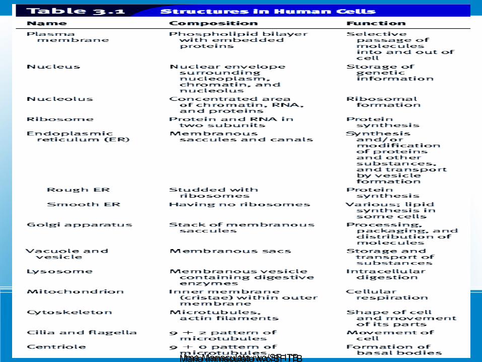
Three main part of every human cell: • a plasma membrane, • a nucleus,• cytoplasm
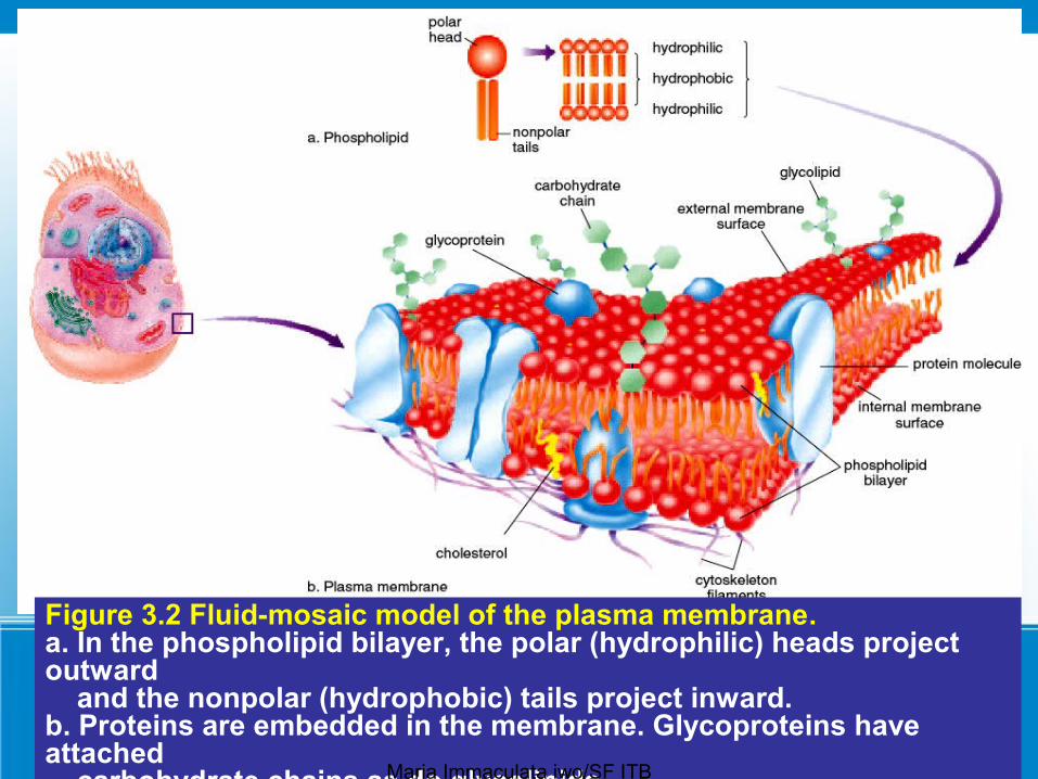
The plasma membrane, - surrounds the cell and keeps it intact, regulates what enters and exits a cell.- is a phospholipid bilayer that is said to be semipermeable
because it allows certain molecules but not others to enter the cell. - Proteins present in the plasma membrane play important
roles in allowing substances to enter the cell.Maria Immaculata iwo/SF ITB

4
The nucleus - is a large, centrally located structure that can often be seen
with a light microscope. - contains the chromosomes - is the control center of the cell. - It controls the metabolic functioning and structural characteristics of the cell. The nucleolus is a region inside the nucleus.- is of primary importance because it stores the genetic information that determines the characteristics of the body’s cells and their metabolic functioning. - Every cell contains a copy of genetic information, but each
cell type has certain genes turned on, and others turned off.
Maria Immaculata iwo/SF ITB

5
The cytoplasm - is the portion of the cell between the nucleus and
the plasma membrane. - The matrix of the cytoplasm is a semi-fluid medium that contains water and various types of molecules suspended or dissolved in the medium.
The presence of proteins accounts for the semi-fluid nature of the matrix.- contains various organelles
Maria Immaculata iwo/SF ITB

6
Organelles
- are small, usually membranous structures that are best seen with an electron microscope.
- Each type of organelle has a specific function. For example,
– one type of organelle transports substances, and another type produces ATP for the cell.
– Because organelles are composed of membrane, keeping the various cellular activities separated from one another. • Just as the rooms in your house have particular
pieces of furniture that serve a particular purpose, organelles have a structure that suits their function.
Maria Immaculata iwo/SF ITB

7
Cytoskeleton
● Cells also have a cytoskeleton, a network of interconnected filaments and microtubules in the cytoplasm.
● The name cytoskeleton is convenient in that it allows us to compare the cytoskeleton to our bones and muscles. – Bones and muscles give us structure and produce
movement. • Similarly,
– the elements of the cytoskeleton maintain cell shape and allow the cell and its contents to move.
● Some cells move by using cilia and flagella, which are made up of microtubules.
Maria Immaculata iwo/SF ITB

8
Maria Immaculata iwo/SF ITB

9
Maria Immaculata iwo/SF ITB

10
Maria Immaculata iwo/SF ITB
11
Ima Mariaculata iwo/SF ITBMaria Immaculata iwo/SF ITB
12
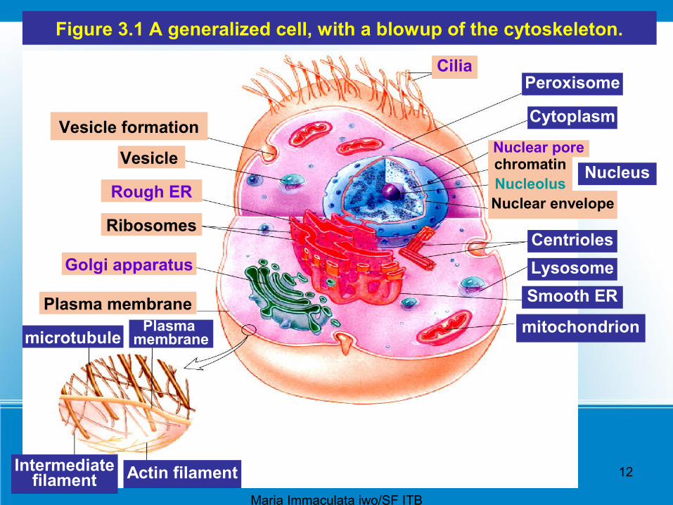
Figure 3.1 A generalized cell, with a blowup of the cytoskeleton.
Vesicle formation
Vesicle
Rough ER
Ribosomes
Golgi apparatus
Plasma membranePlasma
membranemicrotubule
Intermediatefilament Actin filament
Cilia
Nuclear pore
Cytoplasm
mitochondrion
CentriolesLysosomeSmooth ER
Nucleus
Peroxisome
Nucleoluschromatin
Nuclear envelope
Maria Immaculata iwo/SF ITB
13
Figure 3.2 Fluid-mosaic model of the plasma membrane. a. In the phospholipid bilayer, the polar (hydrophilic) heads project outward and the nonpolar (hydrophobic) tails project inward. b. Proteins are embedded in the membrane. Glycoproteins have attached carbohydrate chains as do glycolipids.Maria Immaculata iwo/SF ITB

14
Maria Immaculata iwo/SF ITB

15
Ribosomes
Ribosomes are composed of two subunits, one large and one small.
Each subunit has its own mix of proteins and rRNA.Protein synthesis occurs at the ribosomes.Ribosomes are found free within the cytoplasm
either singly or in groups called polyribosomes.Ribosomes are often attached to the endoplasmic
reticulum
Maria Immaculata iwo/SF ITB

16
Endomembrane System
The endomembrane system consists of - the nuclear envelope,
– the endoplasmic reticulum, – he Golgi apparatus, – lysosomes,– vesicles (tiny membranous sacs)
These components of the cell work together to produce and secrete a product.
Maria Immaculata iwo/SF ITB
17
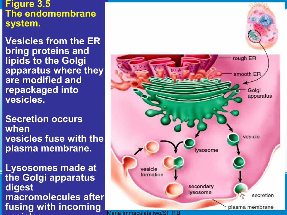
Figure 3.5 The endomembrane system.
Vesicles from the ERbring proteins and lipids to the Golgi apparatus where they are modified and repackaged into vesicles.
Secretion occurs whenvesicles fuse with the plasma membrane.
Lysosomes made at the Golgi apparatus digest macromolecules after fusing with incoming vesicles. Maria Immaculata iwo/SF ITB
18
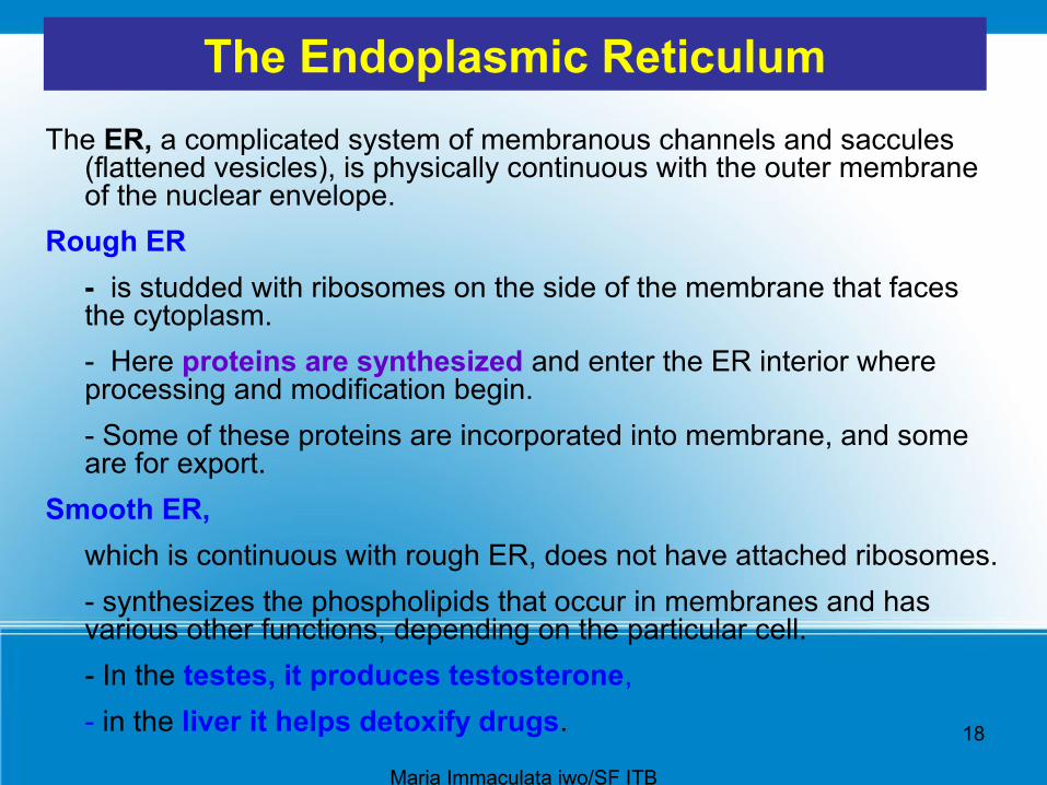
The Endoplasmic ReticulumThe ER, a complicated system of membranous channels and saccules
(flattened vesicles), is physically continuous with the outer membrane of the nuclear envelope.
Rough ER- is studded with ribosomes on the side of the membrane that faces the cytoplasm. - Here proteins are synthesized and enter the ER interior where processing and modification begin. - Some of these proteins are incorporated into membrane, and some are for export.
Smooth ER, which is continuous with rough ER, does not have attached ribosomes. - synthesizes the phospholipids that occur in membranes and has various other functions, depending on the particular cell. - In the testes, it produces testosterone, - in the liver it helps detoxify drugs.
Maria Immaculata iwo/SF ITB

19
Figure 3.4 Rough endoplasmic reticulum is studded with ribosomes where protein synthesis occurs.
Smooth endoplasmicreticulum, which has no attached ribosomes, produces lipids and often has other functions as well in particular cells.
ER also forms vesicles in which large molecules are transported to other parts of the cell. Smooth ER
Rough ER
RibosomeNuclearenvelope
Maria Immaculata iwo/SF ITB

20
The Golgi Apparatus
The Golgi apparatus is named for Camillo Golgi, who discovered its presence in cells in 1898.
● The Golgi apparatus receives protein and/or lipid-filled vesicles that bud from the ER.
● The Golgi apparatus contains enzymes that modify proteins and lipids.
For example, it can add a chain of sugars to proteins and lipids, thereby making them glycoproteins and glycolipids, which are molecules found in the plasma membrane.
Maria Immaculata iwo/SF ITB
21
Lysosomes● Lysosomes, membranous sacs produced by the Golgi
apparatus, contain hydrolytic digestive enzymes. ● Sometimes macromolecules are brought into a cell by
vesicle formation at the plasma membrane (Fig. 3.5). ● When a lysosome fuses with such a vesicle, its contents
are digested by lysosomal enzymes into simpler subunits that then enter the cytoplasm.
● Even parts of a cell are digested by its own lysosomes (called autodigestion).
● Normal cell rejuvenation most likely takes place in this manner, but autodigestion is also important during development.
Maria Immaculata iwo/SF ITB
22
Lysosomes
For example, The fingers of a human embryo are at first webbed, but they are freed from one another as a result of lysosomal action.
Occasionally, a child is born with Tay-Sachs disease, a metabolic disorder involving a missing or inactive lysosomal enzyme. In these cases, the lysosomes fill to capacity with macromolecules that cannot be broken down.
The cells become so full of these lysosomes that the child dies. Someday soon, it may be possible to provide the missing enzyme for these children.
Maria Immaculata iwo/SF ITB

23
Mitochondria• Although the size and shape of mitochondria
(sing., mitochondrion) can vary, all are bounded by a double membrane.
● The inner membrane is folded to form little shelves called cristae, which project into the matrix, an inner space filled with a gel-like fluid
● Mitochondria are the site of ATP (adenosine triphosphate) production involving complex metabolic pathways. ATP molecules are the common carrier of energy in cells.
Maria Immaculata iwo/SF ITB
24
● The chemical transformation that involves mitochondria is as follows:
• In the process, mitochondria use up oxygen and give off carbon dioxide and water.
● Because oxygen is involved, we say that mitochondria carry on cellular respiration.
Maria Immaculata iwo/SF ITB
25
● The matrix of a mitochondrion contains enzymes for breaking down glucose products.
• ATP production then occurs at the cristae. ● Every cell uses a certain amount of ATP energy to
synthesize molecules, but many cells use ATP to carry out their specialized functions,
For example,
- muscle cells use ATP for muscle contraction, which produces movement
- nerve cells use it for the conduction of nerve impulses, which make us aware of our environment.
Maria Immaculata iwo/SF ITB

26
cristae
Maria Immaculata iwo/SF ITB
27
The Cytoskeleton
Several types of filamentous protein structures form a cytoskeleton that helps maintain the cell’s shape and either anchors the organelles or assists their movement as appropriate.
The cytoskeleton includes – microtubules, – intermediate filaments,– actin filaments
Maria Immaculata iwo/SF ITB
28
Microtubules are hollow cylinders whose wall is made up
of 13 logitudinal rows of the globular protein tubulin. Microtubule assembly is regulated by the centrosome which lies near the nucleus.
Microtubules radiate from the centrosome, helping to maintain the shape of the cell and acting as tracks along which organelles move.
During cell division, microtubules form spindle fibers, which assist the movement of chromosomes.
Maria Immaculata iwo/SF ITB
29
• Intermediate filaments – differ in structure and function.
• Actin filaments – are long, extremely thin fibers that usually occur in
bundles or other groupings. – Actin filaments have been isolated from various
types of cells, especially those in which movement occurs.
– Microvilli, which project from certain cells and can shorten and extend, contain actin filaments.
– Actin filaments, like microtubules, can assemble and disassemble.
Maria Immaculata iwo/SF ITB
30
Centrioles are short cylinders with nine outer microtubule triplets and no center microtubules
● Each cell has a pair of centrioles in the centrosome near the nucleus.
● The members of each pair of centrioles are at right angles to one another.
● Before a cell divides, the centrioles duplicate, and the members of the new pair are also at right angles to one another.
● During cell division, the pairs of centrioles separate so that each daughter cell gets one centrosome.
● Centrioles may be involved in the formation of the spindle that functions during cell division. Centrioles also give rise to basal bodies that direct the formation of cilia and flagella.
31
• Cilia and Flagella
● Cilia and flagella (sing., cilium, flagellum) are projections of cells that can move either in an undulating fashion, like a whip, or stiffly, like an oar.
● Cilia are shorter than flagella● Cells that have these organelles are capable of self
movement or moving material along the surface of the cell.
For example, sperm cells, carrying genetic material to the egg, move by means of flagella. The cells that line our respiratory tract are ciliated. These cilia sweep debris trapped within mucus back up the throat, and this action helps keep the lungs clean.
Maria Immaculata iwo/SF ITB
32
● Each cilium and flagellum has a basal body at its base, which lies in the cytoplasm.
• Basal bodies, like centrioles, have (9+0 pattern): nine outer microtubule triplets and no center microtubules
They are believed to organize the structure of cilia and flagella even though cilia and flagella have a 9+ 2 pattern of microtubules.
In cilia and flagella, nine microtubule doublets surround two central microtubules.
● This arrangement is believed to be necessary to their ability to move.
Maria Immaculata iwo/SF ITB

33
Maria Immaculata iwo/SF ITB
34
3.2 Crossing the Plasma Membrane
● The plasma membrane keeps a cell intact.
It allows only certain molecules and ions to enter and exit the cytoplasm freely;
therefore, the plasma membrane is said to be selectively permeable.
● Both passive and active methods are used to cross the plasma membrane
Maria Immaculata iwo/SF ITB

35
Diffusion
● Diffusion is the random movement of molecules from the area of higher concentration to the area of lower concentration until they are equally distributed.
– To illustrate diffusion, imagine putting a tablet of dye into water. The water eventually takes on the color of the dye as the dye molecules diffuse.
● The chemical and physical properties of the plasma membrane allow only a few types of molecules to enter and exit a cell simply by diffusion.
● Lipid-soluble molecules such as alcohols can diffuse through the membrane because lipids are the membrane’s main structural components.
Maria Immaculata iwo/SF ITB

36
Diffusion● Gases can also diffuse through the lipid bilayer;
this is the mechanism by which oxygen enters cells and carbon dioxide exits cells.
After inhalation (breathing in), the concentration of oxygen in the alveoli is higher than that in
the blood; therefore, oxygen diffuses into the blood.
● When molecules simply diffuse from higher to lower concentration across plasma membranes, no cellular energy is involved.
Maria Immaculata iwo/SF ITB

37
OsmosisOsmosis is the diffusion of water across a plasma
membrane.– It occurs whenever an unequal concentration of water
exists on either side of a selectively permeable membrane.
Normally, – body fluids are isotonic to cells, that is, there is an
equal concentration of solutes (substances) and solvent (water) on both sides of the plasma membrane, and cells maintain their usual size and shape.
– Intravenous solutions medically administered usually have this tonicity.
Tonicity is the degree to which a solution’s concentration of solute versus water causes water to move into or out of cells.
Maria Immaculata iwo/SF ITB
38
Hypotonic & hypertonic solutionsSolutions (solute plus solvent) that cause cells to swell or
even to burst due to an intake of water are said to be hypotonic solutions.
If red blood cells are placed in a hypotonic solution, which has a higher concentration of water (lower concentration of solute) than do the cells, water enters the cells and they swell to bursting (Fig. 3.8b).
The term lysis refers to disrupted cells; hemolysis, then, is disrupted red blood cells.
Solutions that cause cells to shrink or to shrivel due to a loss of water are said to be hypertonic solutions. If red blood cells are placed in a hypertonic solution, whichhas a lower concentration of water (higher concentration of solute) than do the cells, water leaves the cells and they shrink
(Fig. 3.8c).Maria Immaculata iwo/SF ITB
39
Solutions that cause cells to shrink or to shrivel due to a loss of water are said to be hypertonic solutions. If red blood cells are placed in a hypertonic solution, which has a lower concentration of water (higher concentration of solute) than do the cells, water eaves the cells and they shrink (Fig. 3.8c).
● The term crenation refers to red blood cells in this condition. These changes have occurred due to osmotic
pressure.• Osmotic pressure is the force exerted on a selectively
permeable membrane because water has moved from the area of higher concentration of water to the area of lower concentration (higher concentration of solute).
Maria Immaculata iwo/SF ITB
40
Maria Immaculata iwo/SF ITB
41
Filtration
• Because capillary walls are only one cell thick, small molecules (e.g., water or small solutes) tend to passively diffuse across these walls, from areas of higher concentration to those of lower concentration.
However, blood pressure aids matters by pushing water and dissolved solutes out of the capillary.
This process is called filtration.– Filtration is easily observed in the laboratory when a solution is
poured past filter paper into a flask. Large substances stay behind, but small molecules and water pass through.
Filtrasi memerlukan tekanan!
Maria Immaculata iwo/SF ITB

42
• Filtration of water and substances in the region of capillaries is largely responsible for the formation of tissue fluid, the fluid that surrounds the cells.
● Filtration is also at work in the kidneys when water and small molecules move from the blood to the inside of the kidney tubules.
Maria Immaculata iwo/SF ITB

43
Transport by Carriers
Most solutes do not simply diffuse across a plasma membrane; rather, they are transported by means of protein carriers within the membrane.
During facilitated transport, a molecule (e.g., an amino acid or glucose) is transported across the plasma membrane from the side of higher concentration to the side of lower concentration. The cell does not need to expend energy for this type of transport because the molecules are moving down their concentration gradient.
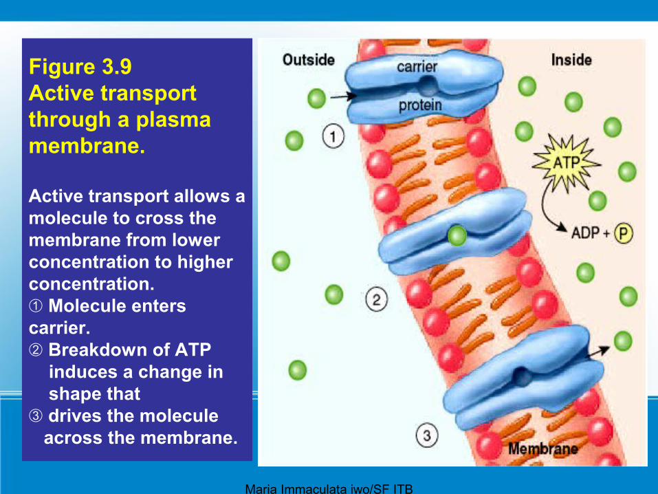
During active transport, a molecule is moving contrary to the normal direction—that is, from lower to higher concentration (Fig. 3.9).
Maria Immaculata iwo/SF ITB
44
For example,- iodine collects in the cells of the thyroid gland; - sugar is completely absorbed from the gut by cells that line
the digestive tract; - and sodium (Na) is sometimes almost completely withdrawn
from urine by cells lining kidney tubules. Active transport requires a protein carrier and the use of
cellular energy obtained from the breakdown of ATP.
When ATP is broken down, energy is released, and in this case the energy is used by a carrier to carry out active transport.
Therefore, it is not surprising that cells involved in active transport have a large number of mitochondria near the plasma membrane at which active transport is occurring.
Maria Immaculata iwo/SF ITB

45
● Proteins involved in active transport often are called pumps because just as a water pump uses energy to move water against the force of gravity, proteins use energy to move substances against their concentration gradients. – One type of pump that is active in all cells but is especially
associated with nerve and muscle cells moves sodium ions (Na) to the outside of the cell and potassium ions (K) to the inside of the cell.
● The passage of salt (NaCl) across a plasma membrane is of primary importance in cells. – First, sodium ions are pumped across a membrane;
then, chloride ions simply diffuse through channels that allow their passage.
– Chloride ion channels malfunction in persons with cystic fibrosis, and this leads to the symptoms of this inherited (genetic) disorder.
Ima Mariaculata iwo/SF ITBMaria Immaculata iwo/SF ITB

46
Endocytosis and Exocytosis
● During endocytosis, commonly called phagocytosis, a portion of the plasma membrane invaginates to envelop a substance, and then the membrane pinches off to form an intracellular vesicle (see Fig. 3.1, top).
● Digestion may be required before molecules can cross a vesicle membrane to enter the cytoplasm.
● During exocytosis, a vesicle fuses with the plasma membrane as secretion occurs (see Fig. 3.1, bottom).
● This is the way insulin leaves insulin-secreting cells, for instance.
● Table 3.2 summarizes the various ways molecules cross the plasma membrane.
Maria Immaculata iwo/SF ITB
47
Figure 3.9 Active transport through a plasma membrane.
Active transport allows a molecule to cross the membrane from lower concentration to higher concentration.
Molecule enters ➀carrier.
Breakdown of ATP ➁ induces a change in shape that
drives the molecule ➂ across the membrane.
Maria Immaculata iwo/SF ITB

48
Maria Immaculata iwo/SF ITB

49
Dehydration and Water Intoxication
Dehydration is due to a loss of water. ● The solute concentration in extracellular fluid increases—
that is, tissue fluid becomes hypertonic to cells, and water leaves the cells.
● Common causes of dehydration are excessive sweating, perhaps during exercise, without any replacement of the water lost.
● Dehydration can also be a side effect of any illness that causes prolonged vomiting or diarrhea.
● The signs of moderate dehydration are a dry mouth, sunken eyes, and skin that will not bounce back after light pinching.
Maria Immaculata iwo/SF ITB
50
Dehydration and Water Intoxication
If dehydration becomes severe, – the pulse and breathing rate are rapid, – the hands and feet are cold, – the lips are blue.
● Although dehydration leads to weight loss, it is never a good idea to dehydrate on purpose for this reason.
• To cure dehydration, intake of a low-sodium solution is needed because water intake alone could lead to water intoxication.
Maria Immaculata iwo/SF ITB

51
• Water intoxication is due to a gain in water. – The solute concentration in extracellular fluid decreases
—that is, tissue fluid becomes hypotonic to the cells, and water enters the cells.
● One cause can be the intake of too much water during a marathon race.
● Marathoners who collapse and have nausea and vomiting after a race are probably not suffering from a heart attack, but they may be suffering from water intoxication, which can lead to pulmonary edema and swelling in the brain.
• The cure, an intravenous solution containing high amounts of sodium, is the opposite of that for dehydration.
● Therefore, it is important that physicians be able to diagnose water intoxication in athletes who have had an opportunity to drink fluids for the past several hours.
Maria Immaculata iwo/SF ITB

52
Figure 3A Dehydration versus water intoxication.a. If extracellular fluid loses much water, cells lose water
by osmosis, and become dehydrated.Maria Immaculata iwo/SF ITB
53
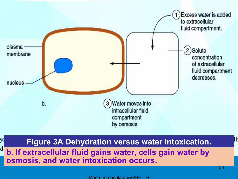
Figure 3A Dehydration versus water intoxication.b. If extracellular fluid gains water, cells gain water by osmosis, and water intoxication occurs.
Maria Immaculata iwo/SF ITB
54
3.3 The Cell Cycle
Maria Immaculata iwo/SF ITB

55
Figure 3.10 The cell cycle consists of interphase, during which cellular components duplicate,
a mitotic stage, during which the cell divides.
Interphase consists of two so-called “growth” phases (G1 and G2) and a DNA synthesis (S) phase. The mitotic stage consists of the phases noted plus cytokinesis.
Maria Immaculata iwo/SF ITB
56
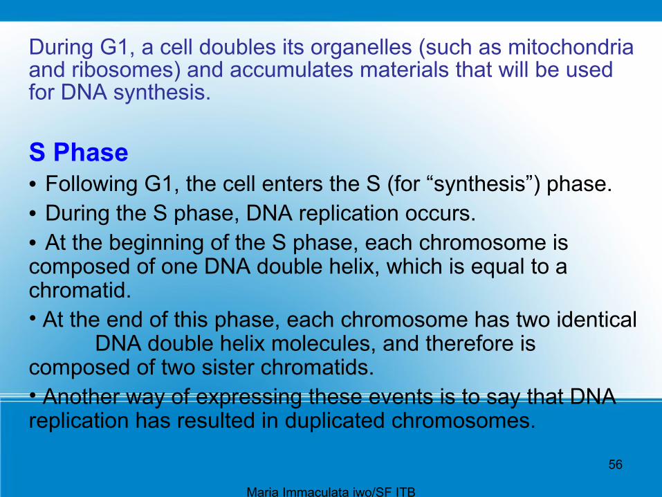
During G1, a cell doubles its organelles (such as mitochondria and ribosomes) and accumulates materials that will be used for DNA synthesis.
S Phase • Following G1, the cell enters the S (for “synthesis”) phase. • During the S phase, DNA replication occurs. • At the beginning of the S phase, each chromosome is composed of one DNA double helix, which is equal to a chromatid.• At the end of this phase, each chromosome has two identical
DNA double helix molecules, and therefore is composed of two sister chromatids.• Another way of expressing these events is to say that DNA replication has resulted in duplicated chromosomes.
Maria Immaculata iwo/SF ITB

57
G2 Phase During this phase, the cell synthesizes proteins that will assist
cell division, such as the protein found in microtubules.● The role of microtubules in cell division is described later
in this section. ● Also, chromatin condenses, and the chromosomes
become visible.
Mitotic StageFollowing interphase, the cell enters the M (for mitotic) stage.● This cell division stage includes mitosis (division of the
nucleus) and cytokinesis (division of the cytoplasm). ● During mitosis, daughter chromosomes are distributed to
two daughter nuclei. When cytokinesis is complete, two daughter cellsare present.
Maria Immaculata iwo/SF ITB

58
Events During Interphase
● Two significant events during interphase are – replication of DNA – protein synthesis.
Maria Immaculata iwo/SF ITB

59
Replication of DNA● During replication, an exact copy of a DNA helix is
produced.● The double-stranded structure of DNA aids replication
because each strand serves as a template for the formation of a complementary strand.
● A template is most often a mold used to produce a shape opposite to itself. – In this case, each old (parental) strand is a template
for each new (daughter) strand.
Maria Immaculata iwo/SF ITB
60
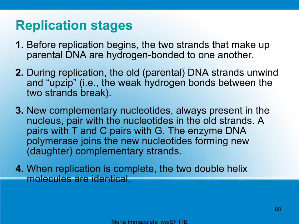
Replication stages 1. Before replication begins, the two strands that make up
parental DNA are hydrogen-bonded to one another.
2. During replication, the old (parental) DNA strands unwind and “upzip” (i.e., the weak hydrogen bonds between the two strands break).
3. New complementary nucleotides, always present in the nucleus, pair with the nucleotides in the old strands. A pairs with T and C pairs with G. The enzyme DNA polymerase joins the new nucleotides forming new (daughter) complementary strands.
4. When replication is complete, the two double helix molecules are identical.
Ima Mariaculata iwo/SF ITBMaria Immaculata iwo/SF ITB

61
• Each strand of a double helix is equal to a chromatid, which means that at the completion of replication each chromosome is composed of two sister chromatids.
They are called sister chromatids because they are identical. The chromosome is called a
duplicated chromosome.
Cancer, which is characterized by rapidly dividing cells, is treated with chemotherapeutic drugs that stop replication and therefore cell division.
Some chemotherapeutic drugs are analogs that have a similar, but not identical, structure to the four nucleotides in DNA. When these are mistakenly used by the cancer cells to synthesize DNA,
replication stops, and the cells die off.
Maria Immaculata iwo/SF ITB

62
Figure 3.11 Overview of DNA replication.
During replication, an old strand serves as a template for a new strand.The new double helix is composed of an old (parental) strand and a new (daughter) strand.
Maria Immaculata iwo/SF ITB
63
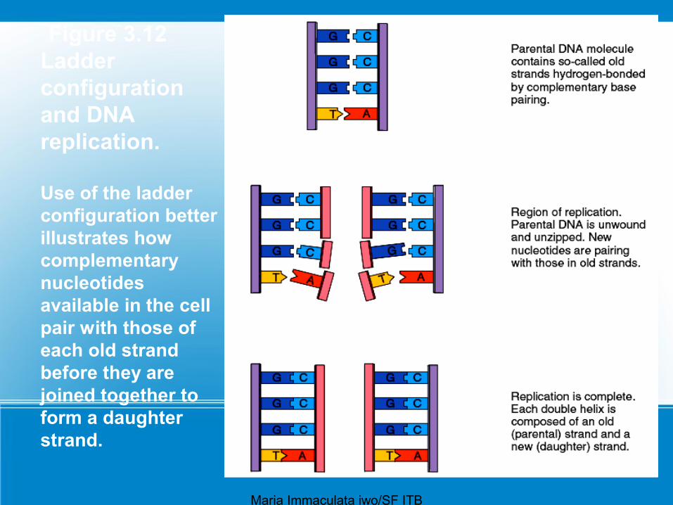
Figure 3.12 Ladder configuration and DNA replication. Use of the ladder configuration better illustrates how complementary nucleotides available in the cell pair with those of each old strand before they are joined together to form a daughter strand.
Maria Immaculata iwo/SF ITB

64
Protein SynthesisDNA not only serves as a template for its own replication, but
is also a template for RNA formation.
Protein synthesis requires two steps, called - transcription - translation.
During transcription, an mRNA molecule is produced, and during translation, this mRNA specifies the order of amino acids in a particular polypeptide (Fig. 3.13).
A gene (i.e., DNA) contains coded information for the sequence of amino acids in a particular polypeptide. The code is a triplet code: Every three bases in DNA (and therefore in mRNA) stands for a particular amino acid.
Maria Immaculata iwo/SF ITB

65
Figure 3.13 Protein synthesis. The two steps required for protein synthesis are transcription, which occurs in the nucleus, and translation, which occurs in the cytoplasm at the ribosomes.
Maria Immaculata iwo/SF ITB

66
Events During the Mitotic StageThe mitotic stage of the cell cycle consists of mitosis and
cytokinesis.● By the end of interphase (Fig. 3.14, upper left), the
centrioles have doubled and the chromosomes are becoming visible.
● Each chromosome is duplicated—it is composed of two chromatids held together at a centromere.
● The mitosis process is divided into four phases: prophase, metaphase, anaphase, and telophase (Fig.3.14).
● The parental cell is the cell that divides, and the daughter cells are the cells that result.
Maria Immaculata iwo/SF ITB

67
Maria Immaculata iwo/SF ITB

68
Prophase● The two pairs of centrioles outside the nucleus begin moving away
from each other toward opposite ends of the nucleus. ● Spindle fibers appear between the separating centriole pairs, the
nuclear envelope begins to fragment, and the nucleolus begins to disappear.
● The chromosomes are now fully visible. ● Spindle fibers attach to the centromeres as the chromosomes
continue to shorten and thicken. ● During prophase, chromosomes are randomly placed in the nucleus.
Structure of the Spindle ● At the end of prophase, a cell has a fully formed spindle. ● A spindle has poles, asters, and fibers.● The asters are arrays of short microtubules that radiate from the
poles, and the fibers are bundles of microtubules that stretch between the poles.
● Centrioles are located in centrosomes, which are believed to organize the spindle.
Maria Immaculata iwo/SF ITB

69
Metaphase
● During metaphase, the nuclear envelope is fragmented, and the spindle occupies the region formerly occupied by the nucleus.
● The chromosomes are now at the equator (center) of the spindle.
● Metaphase is characterized by a fully formed spindle, and the chromosomes, each with two sister chromatids, are aligned at the equator (Fig. 3.15).
Maria Immaculata iwo/SF ITB

70
AnaphaseAt the start of anaphase, the sister chromatids separate. Once
separated, the chromatids are called chromosomes. ● Separation of the sister chromatids ensures that each cell receives a
copy of each type of chromosome and thereby has a full complement of genes.
● During anaphase, the daughter chromosomes move to the poles of the spindle.
● Anaphase is characterized by the movement of chromosomes toward each pole.
Function of the Spindle The spindle brings about chromosome movement. Two types of spindle fibers are involved in the movement of
chromosomes during anaphase.● Spindle fibers, as stated earlier, are composed of microtubules.● Microtubules can assemble and disassemble by the addition or
subtraction of tubulin (protein) subunits. This is what enables spindle fibers to lengthen and shorten, and it ultimately causes the movement of the chromosomes.
Maria Immaculata iwo/SF ITB
71
Telophase and CytokinesisTelophase begins when the chromosomes arrive at the poles.● During telophase, the chromosomes become indistinct
chromatin again. ● The spindle disappears as nucleoli appear, and nuclear
envelope components reassemble in each cell. ● Telophase is characterized by the presence of two
daughter nuclei.
Cytokinesis is division of the cytoplasm and organelles. ● In human cells, a slight indentation called a cleavage
furrow passes around the circumference of the cell. ● Actin filaments form a contractile ring, and as the ring gets
smaller and smaller, the cleavage furrow pinches the cell in half. As a result, each cell becomes enclosed by its own plasma membrane.
Maria Immaculata iwo/SF ITB

72
Importance of Mitosis● Because of mitosis, each cell in our body is
genetically identical, meaning that it has the same number and kinds of chromosomes.
● Mitosis is important to the growth and repair of multicellular organisms. When a baby develops in the mother’s womb, mitosis occurs as a component of growth.
● As a wound heals, mitosis occurs, and the damage is repaired.
Maria Immaculata iwo/SF ITB


