NOAA Technical Report NMFS Circular 440 Marine Flora and Fauna of the Northeastern United States. Turbellaria: Acoela and Nemertodermatida Louise F. Bush July 1981 u.s. DEPARTMENT OF COMMERCE Malcolm Baldrige, Secretary National Oceanic and Atmospheric Administration National Marine Fisheries Service Terry L. Leitzell, Assistant Administrator for FisherIes
Transcript
NOAA Technical Report NMFS Circular 440
Marine Flora and Fauna ofthe Northeastern United States.Turbellaria: Acoela andNemertodermatida
Louise F. Bush
July 1981
u.s. DEPARTMENT OF COMMERCEMalcolm Baldrige, Secretary
National Oceanic and Atmospheric Administration
National Marine Fisheries ServiceTerry L. Leitzell, Assistant Administrator for FisherIes
FOREWORD
This NMFS Circular is part of the subseries "Marine Flora and Fauna of the NortheasternUnited States;' which consists of original, illustrated, modern manuals on the identification,classification, and general biology of the estuarine and coastal marine plants and, animals of thenortheastern United States. The manuals are published at irregular intervals on as many taxa ofthe region as there are specialists available to collaborate in their preparation.
Geographic coverage of the "Marine Flora and Fauna of the Northeastern United States" isplanned to include organisms from the headwaters of estuaries seaward to approximately the 200m depth on the continental shelf from Maine to Virginia, but may vary somewhat with each majortaxon and the interests of collaborators. Whenever possible representative specimens dealt with inthe manuals are deposited in the reference collections of major museums of the region.
The "Marine Flora and Fauna of the Northeastern United States" is being prepared in collaboration with systematic specialists in the United States and abroad. Each manual is basedprimarily on recent and ongoing revisionary systematic research and a fresh examination of theplants and animals, Each major taxon, treated in a separate manual, includes an introduction,illustrated glossary, uniform originally illustrated keys, annotated checklist with information\vhen available on distribution, habitat, life history, and related biology, references to the majorliterature of the group, and a systematic jnde:\.
These manuals are intended for use by biology students, biologists, biologicaloceanographers, informed laymen, and others wishing to identify coastal organisms for thisregion. Often they can serve as guides to additional information about species or groups.
The manuals arc an outgrowth of the widely used "Keys to Marine Invertebrates of theWoods Hole Region;' edited by R. I. Smith in 1964, and produced under the auspices of theSystematics Ecology Program, Marine Biological Laboratory, Woods Hole, Mass. After a sufficient number of manuals of related taxonomic groups have been published, the manuals will berevised, grouped, and issued as special volumes, which will consist of compilations for phyla orgroups of phyla.
The National 1Iarine Fisheries Service (Nl\fFS) does not approve, rec~
ommend or endorse any proprietary product or proprietary materialmentioned in this publication. No reference shall be made to NMFS, orto this publication furnished by N!'.IFS, in any advertising or sales promotion which would indicate or imply that N:\IFS approves, recommendsor endorses any proprietary product or proprietary material mentionedherein, or which has as its purpose an intent to cause directly or indirectlythe advertised product to be used or purchased because of this NMFSpublication,
iii
Marine Flora and Fauna of the Northeastern United States.Turbellaria: Acoela and Nemertodermatida
LOUISE F. BUSH'
ABSTRACT
This manual contains an introduction to the general biolo~y, an illustrated key to the genera, and an annotated s~'stematic list of the orders AcacIa and Nemertodermatida. The key facililates identification of 16 familiesand 75 ~cncra of Acocla and includes the 3 genera of the order Nemerlodcrmalida. The s)'slemalic list includes113 s\ledel> whkh have been described from the North Atlantic, including some more southern species that mightbe encountered here, and gives the habitat and known distribution for each species.
INTRODUCTION
Acoel turbellarians are almost exclusively marine, and onlytwo species, Oligochoerus /imnophilous Ax and Dorjes, 1966and Limnoposthia polonica Kolasa and Faubel, 1974, havebeen described from freshwater and none from any terrestrialhabitat. They are common on various substrata, both intertidally and subtidaUy, on the continental shelf. A few arepelagic, but none have been reported to date in deep seas.Most of the work on this order, aside from the series of papersby Marcus (1950,1952,1954) in Brazil, has been carried out byscientists working on collections made in Europe, and newspecies and genera from these areas are being added even today. Relatively few species have been described from thenortheast coast of the United States, but experience andmaterials collected thus far indicate that many, ifnot most, ofthe genera present in the eastern North Atlantic are alsorepresented in the western North Atlantic. Therefore, in orderfor this publication to be most useful in the northeasternUnited States, I have included all known genera from theworld in the key and all genera and species from the NorthAtlantic are included in the systematic list. The known speciesof the order Nemertodermatida, all of which are marine, arealso from the North Atlantic.
The position of Acoela in the class Turbellaria has undergonevarious changes and, while almost always being recognized as anatural division, they have been ranked variously as a subclass(Graff 1904a, b), a suborder (Kading 1940, under orderArchoophora), or an order (Ax 1956; Dorjes 1968a; Karling1974). The ranking of Acoela as a separate order has the advantage of conforming to the common usage of the term "acoe1"and is so used here. The separation of the order Nemertodcrmatida from Acoela is a more recent development as isexplained below.
The subdivisions of AcoeIa have not been generally agreedupon. Graff (1882) recognized two families, Proporidae andAphanastomidae (~ Convolutidae). Luther (1912) retainedProporidae and Convolutidae as major subdivisions of thegroup based on the presence or absence of a seminal bursa, andthis arrangement into two principal families was more or lessfollowed by other workers until the series of papers by Westblad
lGeorgc M. Gray Museum. Marine Biological Laboratory, Woods Hole, MA02543.
appeared (1940, 1942, 1945, 1946, 1948). Westblad in his finalpaper proposed the division of Acoela into three tribes:Opisthandropora-Abursalia, Proandropora-Abursalia, andProandropora-Bursalia, with a series of families included ineach tribe. As the names imply, these were based on the positionof the male genital pore and on the presence or absence of theseminal bursa. Recently, Dorjes (1968a), in his comprehensivesurvey of Acoela, criticized Westblad's arrangement in detailand simply divided the order into 15 families, based primarily onthe structure of the male organs. This avoids at least some of theproblems and inconsistencies of Westblad's system and is thearrangement, with the following exceptions, which 1 have usedin this paper. An additional family, Antroposthiidae, has beendefined recently by Faubel (1976) and is included here as the16th family. Also, it must be noted that two of Dorjes' families,Hofsteniidae and Nemertodermatidae, have been considered bysome authors to deserve ranking as orders separate fromAcaela; recent discussions by Ax (1961), Kading (1967, 1974),Tyler and Rieger (1977), and Faubel and Dorjes (1978) lead meto agree that certainly Nemertodermatidae do not belong inAeoela since they have a gut cavity during at least part of theirlife cycle and also have unitlageIlate sperm which are distinctlydifferent from those of Acoela. Specimens of Nemenodermasp. taken by me in Vineyard Sound near Woods Hole, Mass.,clearly show a gut space in some of the specimens. Observationson Meara (Westblad 1949), and on Flage/lophora (Faubel andDorjes 1978), which have open guts and characters dose toNemertodenna, also indicate that the family should be in aseparate order. I concur with the proposals for an order Nemertodermatida (Westblad 1947; Karling 1940), and am placingNemertoderma, Meara, and Flagellophora in the order Nemertodermatida. I am indicating the possibility of Hofstcniidaedeserving placement in a scparate order by a footnote in thc key.
Although future work may be expected to add to the families,genera, and species of AcocIa, publication of this key to familiesand known genera should be useful to workers in OUf area andencourage students on this side of the Atlantic to further workon the Turbellaria in general and Acoela in particular.
DIAGNOSTIC CHARACTERS OFTHE ORDER ACOELA
The order Acoela, as the name implies, comprises thosemembers of the class Turbellaria which do not have clearly
defined, large open spaces in the body. They not only lack abody cavity, as is the case in all Platyhelminthes, but they alsodo not possess a digestive cavity with a lining of epithelial cells.The act of digestion is carried out in the central part of the bodyin a tissue which appears under the microscope as a meshworkof vacuolated cytoplasm with scattered nuclei. This central(digestive) parenchyma is surrounded by a denser tissue, theperipheral parenchyma, in which are imbedded the organsmaking up the hermaphroditic reproductive system of theseanimals. Surrounding the peripheral parenchyma, and more orless penetrating it, are several layers of muscle fibers and variousepithelial gland cells. The exterior of the body is clothed by alayer of ciliated epithelial cells. The muscle fibers, epithelialgland cells, and outer epithelium form a complex that gives stiffness and preserves the shape of the animal. The German term"hautmuskelschlauch" (outer muscle sheath), often used in theliterature, applies only to the layers of muscles, but the epithelium and its often in sunken nuclei and derived gland cellstogether with the muscle layers make a more or less interwovenunit which can be thought of as an outer body wall. The brain(cerebral ganglion) and large nerve chords lie in the peripheralparenchyma, or, in a few species, in the base of the outerepithelial layer. There are no skeletal or excretory systems. As inmany marine flatworms, protonephridia are not present.
The acoels are small animals from ruO.5 to rvlO.5 mm in length,mostly oval to elongate oval in shape and without striking surface features such as appendages or special markings. They tendto be more or less transparent or white or pale in color, althougha few species are conspicuously colored by the presence of symbionts or special pigments. The lack of a gut cavity as well as ofdefined body spaces, usually the lack of conspicuous internalorgans aside from those of the reproductive system, and thepresence of a uniform parenchyma around the organs give thema homogenous appearance that enables the animals to be rather
easily distinguished from other Turbellaria. Under lowmagnification, other characters are visible which aid indistinguishing acoe1s, namely the presence of a statocyst, thesimple mouth opening without a pharynx or with a very simpletubelike one, and the arrangement of large eggs full of storedfood material (Fig. 1). Location of the mouth and location andarrangement of sperm and the male copulatory complex (and ofstored sperm in the female parts of some species) are useful forrecognizing families and genera. Diagnostic characters whichrequire observation with the higher powers of the microscope orrecourse to the making of histological sections include details ofthe reproductive system, presence or absence of frontal or otherglands, arrangement of muscle fibers in the outer body wall, andtype of nervous system.
The presence of a statocyst is not unique to the acoels butthis, together with the lack of a conspicuous pharynx anddigestive tract and the presence of large egg cells. is a certain dueto their recognition. The circular clear vesicle of the statocystwith an enclosed statolith is easy to see in the anterior end of theliving animal, but it is harder to find in fixed materiaL In sections it is found lying close to or imbedded in the anterior(cerebral) ganglion.
The mouth in acoels is difficult to see, since in most species itis simply a break in the outer epithelial layer closed by a fewsphincter muscle fibers. It opens directly into the centraldigestive parenchyma (Fig. 2). In relatively few genera, there is asimple tube-shaped pharynx (pharynx simplex) (see Figs. 7, 8, 9,23a, 24a)2 which is essentially an invagination of the outer bodywall layers. This lack of a specialized pharynx is characteristic ofthe Acoela as compared with the other orders of Turbellaria,
ZReferenccs in the remainder of this section, unless otherwise indicated, are to the examplesin the key wllere the structures are listed or are indicated by arrows on the Figures.
frontal gland ~I~~@ ,
statocyst
, 0'male germinal o 0
00
center '?~ o()\ ,,0.001-0, • 0'90, , o 'eo ell female°0' (§fb"00' D at> <t J
germinal centermouth (..(,0'\ ®0 €'€) 6~o,
IO~'®@ @ '"".' @® {,("u( ®@ ®®~'"
~ @ <,mature egg ,
® @)f~
seminal bursasperm entering
seminal vesiclecopulatory organ
Figure I.-Generalized diagram of acoel strudure, as seen in surface view. when the turbeUarian is swimmin~orquiet under a covcr-o;lip.
2
each of which is defined by a particular and usually more complex pharynx structure. The highly vacuolated central parenchyma can be seen in sections to be more or less sharply distinctfrom the denser peripheral parenchyma. The terms "endocytium" and "ectocytium" have been used for these twotissues since cell membranes are hard to distinguish in them.However, since some recent studies with electron microscopyhave shown that many turbellarian tissues, once thought to besyncytial, are truly cellular, the use of these as general terms maybe appropriate no longer. See Ax (961) for a long discussion ofthis point and also Boguta and Mamkaev (972) and Ivanov andMamkaev (1977).
The parts of the hermaphroditic reproductive system areimportant in identification of genera and species. In the key, anattempt has been made to use only parts which can bedetermined without sections, but this is not always possible.
The female parts of an acoel may consist of only the developing and mature eggs. Thus female accessory organs may betotaUy lacking or there may be present an organ for storing andreleasing sperm received from a partner (a seminal bursa or bursa seminalis). In a few species, a vagina is present. There are nooviducts in any of the acoels. The large size of the mature egg isdue to its being entolecithal, i.e., each egg develops and storesnutritive material (yolk) in its cytoplasm, Entolecithal eggs arefound in some other turbellarian orders (Archoophora), but themore usual condition in so-called higher Turbellaria(Neoophora) is for smaller ectolecithal eggs with the nutritivematerials developed and stored in separate and conspicuousyolk glands, Thus the acoels lack yolk glands although in a veryfew species, Hallangia proporoides" Po!ychoerus caudatus,Nadina pu!chella, and some species of Oligochoerus, a group ofyolk-carrying cells near the eggs have been described. Thus, inmost acoels, the eggs increase in size gradually as they matureand yolk accumulates so that a series of eggs of increasing sizescan be seen arranged conspicuously along the length of thebody. The youngest stages are found in a definite area of thebody (a female germinal center) that is characteristic for eachspecies, This germinal center may be considered to be the ovaryalthough there is no ovary wall or delimiting capsule and thecenter may be more or less diffuse. The term "ovary" is
variously used in the literature to indicate either the germinalcenter or the entire mass of developing eggs. In some species,there may be a common germinal center where both eggs andsperm start their development in close proximity and then moveapart as they mature (Fig. 16). The seminal bursa may be only aloosely defined vacuolated space in the parenchyma (Fig. 138) orit may be a more or less elaborate structure with epithelial ormuscular walls (Figs. 29, 30). Nozzles (Figs. 42, 43) or spermaticducts (Figs. 62, 63) or bursal appendages (Figs. 99, 100, 101)may be present. The bursa may open directly from the femalepore (Figs. 69, 99), from a common genital pore (Fig. 30), orfrom an associated female antrum or vagina (Fig. 29). Groupsof sperm associated with bursal nozzles may also be found in theparenchyma without any discernible outside connection (Figs.46, 72a, 72b) in which case it may be postulated that sperm arestored in this way after they have been deposited either on theoutside of the body or through the epidermis by hypodermicinjection. However, it has been suggested also that spermdeposited in a vagina or antrum may move through the wall toform such a disconnected bursa or to lie in vacuolated spaces inthe parenchyma.
The sperm develop in a male germinal center (testis) which,like the ovary, is not delimited by a definite wall. The developingstages may be arranged in a rather compact mass, may appear tobe organized in follicles as in some other Turbellaria, or may bemore or less scattered ("diffm,e testis"). Distinctions betweenthese types of testes and their exact definitions are not clear (seeSteinbtick 1966:84-85). As already noted, the male and femalegerminal centers may be closely associated. In most cases,however, the sperm develop dorsal to the eggs and moveposteriorly in the dorsal parenchyma, since there are no spermducts in the male system of the acoels.
Some sort of male copulatory complex is always presentalthough it may consist of nothing more than a space in theparenchyma where sperm accumulate close to a male genitalpore (Fig. 74). Such an unwalled space in the parenchyma wheresperm accumulate before ejaculation is termed a "false seminalvesicle" and such may occur not only with a simple genital pore,but also with many other types of copulatory apparatus (Figs.80, 169, 171). A walled structure where sperm are accumulated
male antrum
sperm enteri ngseminal
vesicle
antrumcell
female
egg
digestive parenchyma
seminal bursa
center
'\mouth
mature
female germi nal
male germinal center
frontal gland
statocystbody parenchyma
Figure 2.-Gcneralized diagram of acoel structures, longitudinal reconstruction.
3
is considered a true seminal vesicle (Figs. 18, 26a, 91); inside ofthis type there is usually present, besides the sperm, a granularsecretion supplied by the walls of the vesicle or by associatedgland cells.
Arrangements for the ejaculation of sperm in the acoels rangefrom a simple outside opening at the end of a true or falseseminal vesicle to more complex situations that include apassage to the outside (male antrum) and a well~developed
penis. The penis, when present, may be composed of variousarrangements of fine or coarse needles (Figs. 32b, 54,106), mayconsist of a cone-shaped group of muscles (Fig. 107), or may bea glandUlar or muscular tube (Figs. 91, 94, 115). In some cases,it is inverted into the seminal vesicle itself when at rest andeverted to the exterior when sperm are to be discharged. It maybe enclosed by a penis sheath or sac which may also enclose theseminal vesicle. A special feature of some families is cuticularneedles (adenodactyls) which do not make up a penis, but whichare associated with the reproductive passages and whichapparently have a stimulatory function (Figs. 37, 38).
The sperm of acoels are biflagellate (Hendelberg 1977) andinternal fertilizatjon is the rule, as in other Turbellaria. Thearrangement of the reproductive organs suggests cross fertiliza~
tion. Behavior that suggests mutual insemination has beenobserved a number of times and it has been shown conclusivelyto be the case by histological sections of animals in coitus(Hyman J937, in Amphiscolops langerhansi; Westblad 1946, inConaperta flavibadllum).
Asexual reproduction has been reported several times, but hasbeen shown clearly to be a normal method only for the speciesParatomella unichaeta (Dorjes 1966) and Pseudohaplogonariamacnaei (du BoJs-Reymond Marcus 1957). Acoels have ratherlimited powers of regeneration, a characteristic which is un~
doubtedly associated with the lack of asexual reproduction. Asummary of studies on regeneration in this order is given bySteinbOck (1967) and should be referred to for its bibliographyas well as for his view of the evolutionary significance of exper~
iments in this area.Gland cells and their secretions play an important role in the
physiology of acoels judging by their large numbers andfrequent occurrences. Some types of gland cells are so conspicuous as to be useful in the identification of species. Most 0bvious are those whose secretions are released onto the surface ofthe body. Rhabdites are common, and under the microscopethey can be seen as small elongate bodies lying in or close to theouter epithelial layer. They are produced in the epithelium or inspecial epithelial cells which are sunken below the surface butwhich retain their connections to the surface so that the rhabdites may be extruded to the outside. Rhabdites may bescattered irregularly over the body, be present only in limitedareas, or be arranged in beautiful rows running the length of theanimal. Often they are colorless, but they may carry color thatgives a characteristic hue to the entire animal. Rhammites aremore elongated or irregular bodies lying deeper in the interior ofthe body.
A group of gland cells makes up the frontal gland which ischaracteristic of and occurs in most acoels as well as in someother turbellarians. This gland may not be conspicuous in livinganimals, but in stained preparations it appears as a varyingnumber of differentially staining cells with ducts opening closetogether at the anterior tip of the body. In some cases, the ductsunite to form a small ampule before opening to the outside.Studies of the detaih~d structure of this gland suggest that it is in-
4
valved in chemoreception as well as in secretion (Antonius1970).
Other gland cells are present in association with the reproductive organs. There do not appear to be any special digestiveglands, although some gland cells may be developed around themouth or pharynx (Fig. 14). Adhesive glands in conspicuouspapillae, as in some other turbellarians, are not usually present,but groups of special adhesive cilia, "haptocilia:' in Hesioficiuminops, have been described recently (Tyler 1973; Crezee andTyler 1976). Some cells in the parenchyma that produce pigmentwould be classed as gland cells.
In most species a layer of circular muscle fibers lies to the out~
side next to the epithelium with a layer of longitudinal fibers justinside this (Fig. J30). In the few genera where this arrangementis reversed, it is considered to be of taxonomic significance andhas been used to define at least one genus (Fig. 128). There mayalso be a third layer of fibers arranged diagonally between, orclose to the others, and sometimes there is an additional layer ofcircular fibers (Bush 1975). Other fibers, probably derived fromthese, form the sphincter around the mouth, and, in thereproductive system, muscle fibers are arranged as sphincters,retractors, protractors, and as sheets in the walls of someorgans. In many acoels, muscle fibers that extend through theparenchyma from side to side are only weakly developed so thatmovement is restricted mostly to elongations or shortenings ofthe body with only a limited amount of twisting or coiling.Locomotion in these species is a characteristic gliding movementaccomplished by means of the cilia on the surface. However, insome species, intraparenchymal muscles are better developedand act as retractors of the anterior or posterior ends, may beused in rolling the sides of the body ventrally as when capturingprey, or, in some interstitial species, provide more varied coilingand twisting of the body as the animals move about in spacesbetween the sand grains.
The nervous system of acoels has been described in relativelyfew species since the nerve tissue is usually not clearly differentiated in routine sections. In most acoels, however, it can be seenthat there is a more or less complex cerebral ganglion lying inthe parenchyma at the anterior end of the body with three to sixnerve chords extending posteriorly, Some smaller nerves extendto the surface at the anterior end, to the statocyst, and to theneighborhood of the frontal gland. In a few species of acoelsand in the order Nemertodermatida, all or part of the nervetissue, instead of lying in the parenchyma inside the outer wallmuscle layers, is found instead lying next to or even somewhatintermeshed with the bases of the outer epithelial cells. Such anepithelial nervous system is considered by some authors to represent a primitive situation and is, therefore, held to be of importance in plotting the evolutionary changes and relationshjps ofthe Turbellaria. For differing opinions on this point, see discussions by Ax (1961) and Karling (1974).
Sensory structures appear to be rather few and simple, but,since most of them must be represented by single cells, they arenot usually identified and are probably more numerous than isrealized. Observations of the reactions of living animals indicatethat among the cilia there may be sensory hairs, but acoels donot have the large conspicuous sensory hairs, or the ciliated pitsor grooves found in some other turbellarians. Only a few specieshave pigmented eye spots or pigment-cup ocelli. Electronmicroscopy will undoubtedly give needed infonnation on sensory structures (e.g., the recent work by Crezee and Tyler (1976)on the caudal organ in Hesiolicium inops).
The life cycles of acoels have been determined mostly by inference from collection data. For more complete information,laboratory culture of the animals is required, and this has rarelybeen done. A recent report by Ax (1977) gave an account basedon collection data for 14 species of acoels, as well as otherTurbellaria, from the North Sea Island of Syl1. He concludedthat in 12 of the 14 acoels there is a polyvoltine life cycle (i.e.,with marc than one generation per year). Also, he noted that•'the polyvoltine life cycle is an important prerequisite for theimmense population development of several interstitial acoels.In addition the brood size for acoels commonly consists of manyeggs:' Apelt (1969) cultured successfully several species of acoelsthrough at least one generation. He followed the embryologicaldevelopment, and his observations agree with those of earlierworkers showing that the acoels have a spiral-duet type ofcleavage. He also shows the speed of development to be directlyrelated to temperature. Almost all acoels arc oviparous butApelt mentions a few viviparous species. All have a direct typeof development without larval stages. See the review by Henley(1974) for a good summary of earlier work on acoelreproduction.
DIAGNOSTIC CHARACTERS OFTHE ORDER NEMERTODERMATIDA
The order Nemertodermatida includes two genera, Nemertoderma and Meara, which were originally induded in theAcoela, plus a recently defined genus, Flagellophora, whichappears to be related to the first two and which is therefore alsoincluded in this order. All these species are small forms whosegeneral appearance and habits made it natural when they werefirst observed to assume they were acoels. However, theuniflagellate sperm in Nemertoderma and Meara (Tyler andRieger 1975; Hendelberg 1977) and the presence of a gut cavityat least some time during their life history differentiate thesegenera from acoels. The presence of two statoliths instead ofone in the statocyst makes the distinction between nemertoder·matids and acoels relatively easy, the only reported cases of twostatoliths in acoels being in fusion and regenerated specimens(Steinbock 1966). The known species of Nemertoderma andFlagellophora are free-living sublittoral forms; Meara isrepresented by one species from the gut or body cavity ofholothurians.
The size range for nemertodermatids is from f\.JO.5 to ru3.0 mm.They are generally without conspicuous external features exceptthat in Nemertoderma bathycola the thick outer epithelium hasvery large vacuoles so that under low magnification the livinganimal appears to have a shining outer layer. While the otherknown nemertodermatids are generally oval in form, Nemertoderma rubra is a typical interstitial form with an elongatedbody and a coiling, twisting type of movement.
The digestive tract is a somewhat variable cavity lined by anepithelium that includes two types of cells, amoeboid digestivecells and club-shaped gland ("kornerkolben") cells, with coarsegranules in their cytoplasm. These gland cells are considered tobe an important characteristic of the order. The mouth, locatedon the ventral side, may have slightly thickened lips as in Mearaor an inturned short section of the epithelium as inNemertoderma, but in Flage/{ophora it appears to be lackingaltogether. This last genus is characterized by a flagellar organ(Fig. 181a, b) which reaches from the anterior end to theneighborhood of the gut and which may take the place of a
5
mouth in securing food, but there is little known about thehabits of this species.
As already noted, the nervous system in the Nemertodermatida is epithelial or subepithelial. A mass of nerve tissue at theanterior end just under or closely associated with the epitheliumrepresents a cerebral center. Associated with the epithelium allover the body there is a generally diffuse nerve plexus whichthins out toward the posterior end and which also showslongitudinal thickenings that probably function as longitudinalnerves. Aside from the statocyst, sense organs have not beendescribed.
The reproductive systems in the Nemertodermatida arerelatively simple and similar to those in the acoels. Testes andovaries may be represented by a common germinal center ormay be clearly separated as compact or follicular units. Femaleaccessory organs are lacking in Nemertoderma and Meara, butin Flagellophora there is a large seminal bursa which opens dorsally at the center of the body. The male pore is located in theposterior end of the body, ventrally in Flagellophora and terminally in Nemertoderma and Meara. In Nemertoderma there isa long male antrum with a seminal vesicle and a mass ofassociated glands. In Meara the antrum is shorter and theseminal vesicle is smaller and without conspicuous glands. InFlagellophora there is a short male antrum with only a falseseminal vesicle. The eggs, as in Acoe1a, are entolecithal but, asalready noted, the sperm are uniflagellate in contrast to thebiflagellate sperm of the acoels.
ECOLOGY
Little is known concerning the ecology of the acoels and evenless about the nemertodermatids for whom we have only theevidence from collection records. Certainly the acoels are morecommon and occur in a wider variety of habitats than thenemertodermatids. As noted above, our records for the nemertodermatids are all from sublittoral habitats or, in the case ofMeara, as entocommensals from holothurians. The acoels arecommon subtidally on the surface of sand or sandy mud orintertidally in rock pools and among detritus or algae in shallowwater. Neither group can withstand drying and their soft bodiesseem to prevent their burrowing within soft mud deposits;relatively few species have been found associated with the interstitial fauna of sandy beaches although more are being describedfrom this habitat as it is being explored further. A few species ofacoels are pelagic and some acoels have been classed as parasitic(probably commensals) since they are found on, or in the bodyspaces of, larger animals.
Laboratory studies which determine accurately the kind andrange of factors to which these animals react await their culturein vitro. The recent work by Apelt (1969), which has been men~
tioned above, gives the best example of quantitative data.Observations on behavior are found scattered in the literature.
The role played by acoels in animal communities may beinferred from their food relationships as well as from thenumbers present in anyone area. Food items may be determinedin some specimens from remains found in the digestive parenchyma. Many species of acoels seem to be predators orscavengers, much the same as other turbellarians. In spite ofsmall size and fragile bodies acoels may be fast swimmers; I haveobserved Neochildia fusca moving very quickly to overwhelmprey such as small worms, crustaceans, and other small invertebrates. When young, members of this species are found to
contain diatoms ahnost exclusively, but they shift to the role ofactive predators as they grow larger and mature. In somehabitats acoels are only a smaIl part of the biological communitystructure, but often they are present in enormous numbers (Ax1977). In such cases they must playa significant role in the foodweb of the community.
COLLECTING AND EXAMINATION METHODS
The small size and rather fragile body structure of both theacoels and the nemertodermatids present difficulties in their col~
lection and study. Both their fragility and small size result inmost species not being recovered by general sampling techniquessuch as benthic nets or sledges. Relatively undisturbed samplesof bottom materials, algae, or other plant materials which arebrought into the laboratory and processed there give the bestresults. The most effective methods for processing are: 1) allowing natural concentration of the specimens at the surface ofbottom samples or at the surface of the overlying water afterstanding, 2) washing out or sieving of specimens with or withoutthe use of narcotic solutions, and 3) seawater ice treatment. Afew of the larger or more conspicuous species (e.g., Po!ychoeruscaudatus, a bright orange species) may be collected by handfrom stones or shells in the intertidal zone. The seawater icemethod has been shown to be superior for quantitative analysisof interstitial species (Uhlig et aI. 1973).
Some species which normally live on or near the surface of thesubstratum tend to go downwards when disturbed and manybecome mixed: with the substratum in spite of care in collecting.If the substratum samples or masses of algae or other plants areallowed to stand in the collecting water in the laboratory,however, many of the ~ima1s concentrate at the surface of thesubstratum or at the surface of the water. The length of timenecessary for this to occur may vary from a few hours to a dayor so. Apparently, the animals are reacting to lack of oxygen inlower layers, although change of temperature may also beinvolved. They may be picked up from the surface with amedicine dropper, either with or without the aid of a dissectingscope.
Washing out of specimens from the substratum may be donein several ways. Vigorous shaking of algae or other plantmaterial in seawater will dislodge some specimens. Vigorous stirring of sand samples in seawater followed by quick decanting ofthe water as sand particles settle will recover many specimenssince neither the acoels nor the nemertodennatids have thestrongly developed adhesive organs found in some otherturbeJJarians. For most interstitial organisms a narcotizing solution (usually 7% MgClz in tap water) is used so that the animalsmay be passively washed out by stirring and decanting, by sieving, or by eIutriation of the material in a separation funnel (seeHulings and Gray 1971, for an account of various methods). Onreplacement of the narcotizing solution the animals recoverreadily if they have not been left in the narcotic too long, butacoels tend to die very soon and also to break up when handledin the relaxed state. 1f the material is fIxed and stained with ageneral stain, such as Rose Bengal, before washing out,this difficulty is avoided, but the process of sorting afterwards is muchmore difficult since most turbellarians, and especially acoels andnematodennatids, look like small oval lumps after fixation andcan be easily mistaken for broken bits of tissue or for ciliateswhich may be about the same size.
The seawater ice method is an effective method (Uhlig et aI.1973) for recovering Turbellaria brought into the laboratorywith sand or sandy mud and is being adopted by many workerswho desire a quantitative analysis of such material. This methodinvolves the use of a temperature and water salinity gradient toinduce the animals to leave the substratum. A simple apparatus(Fig. 3) is set up consisting of a tube to hold the substratumsamples plus seawater ice which provides a slight temperaturegradient but, more importantly l results in a change from low tohigh salinity of the water that runs through the sample asmelting occurs. The live animals collect in the dish at the bottomof the apparatus and can then be sorted and prepared for further study. The chief drawback of this method is that it issomewhat time consuming and may not be worthwhile if only aquick determination of the most prominent members of a faunais desired.
)'jgure 3.-Apparatus for seawater ice treatment of sediment (redrawn from Uhlig et a!. 1973). Use of the insulation layer is optional.
6
Acoels may be kept in small dishes in the laboratory forhours, days, or even months. In plain seawater many species willlive for a day or so. However, adding a bit of substratum mayprovide enough food for them to live much longer, and whenfood preferences are known they may be maintained through atleast part of a life cycle. Eggs are often laid by mature adultsduring the first few days in dishes in the laboratory and younghatched from such a "culture" may survive for a time evenwithout further feeding. Apelt (1969) maintained several speciesby supplying cultured diatoms to the animals and was thus ableto study the complete life cycles.
For the easiest identification by use of the following key, it isbest to begin with living animals. In some cases fixation andperhaps sectioning will be necessary after the living animals havebeen observed. Where only fixed material is available, wholemounts are of some help but in most cases sectioning isnecessary.
Observation of living animals is done in a drop of water undera coverslip with the amount of water adjusted so that thecoverslip just slightly flattens the acoel, thus holding the animalquiet and also flattening the body to achieve greatertransparency. A narcotic such as MgCl2 may be added to thedrop of water, but the difference between achieving just strongenough a solution of the narcotic to quiet the animal and toostrong a solution which kills it and causes it to disintegrate isvery narrow. Such treatment is difficult to use with acoels. Flattening of the animal may also be achieved by the use of aspecially built "rotocompressor" such as Heunert and Uhlig(1966) described but, so far as I am aware, these are notavailable commercially. The larger structures referred to in thekey and many of the finer details of organs such as the seminalbursa may be observed by study of living animals with ordinarylow power (100 x) or high dry (430 x) magnification. The use ofphase contrast or interference contrast is best for studying thesetransparent animals, but is not essential.
Fixing of materials for further examination may be done withany of the standard histological fixatives. The one most commonly used, especially in field work, is hot or cold Bouin's sincespecimens may be left in this for an indefinite period of time.Fixation with this solution, although not considered as good forcytological details, is quite satisfactory for routine identification. Animals can be picked up in a finely drawn-out medicinedropper with as little water as possible and dropped directly intothe fixative. For animals as small as most species of these twoorders, the fixation is almost instantaneous, and there isrelatively little contraction or shrinking of the specimens.Specimens fixed in Formalin with other animals, plant material,or both may be used, but the fixation is poor and oftenspecimens are distorted or broken.
Whole mounts may be stained in Borax Carmine which is probably best for museum specimens since it does not fade as muchas many of the hematoxylins. However, I have had better differentiation with a trichrome stain (stock solution:Chromotrope 2R, 0.6 g; Fastgreen FCF, 0.3 g; phosphotungsticacid, 0.7 g; acetic acid, 1.0 m1; distilled water 100.0 m1. SeeHoren 1957), if it is used in a very dilute solution. For this,specimens which have been fixed in Bouin's (10-20 min) shouldbe washed (several changes for about 1 h) in 70% alcohol. Afterwashing they are left in the dilute stain overnight, thendehydrated and mounted. For the best whole mounts, animalsshould be fixed by allowing the Bouin's to flow over the animalheld slightly flattened under a coverslip.
7
For sections, any of the standard histological methods may beused. Imbedding in paraffin and sections 6-10 IA m thick hasproved satisfactory. Various stains may be used, but since thestructure of acoels depends almost entirely on the structure ofindividual cells, the most satisfactory stains will be those whichbring out as clearly as possible the nuclei and shape of cells andmuscle fibers. For this the old standby, a combination of ironhematoxylin with a counterstain such as eosin or orange G, isstill the most reliable. Mallory's triple stain is fairly good, butmany of the stains for special tissues used in histology are noteffective since acoels do not have such specialized tissues. Theuse of electron microscopy is producing much needed information about these animals but cannot be used primarily for identification of species.
GLOSSARY
Terms are defined here as used in this key; included are someequivalent terms common in the literature. Terminology usedfor reproductive organs has varied considerably in the past anddefinitions given here are an attempt to render in English themost recent usage of Dorjes (1968a), Antonius (1968), and otherEuropean workers.
Accessoryorgans Organs which aid in reproduction as accessories to the ovary and testis.
Adenodactyl A bundle of fine cuticular needles lying withinner ends in the wall of the male antrum and with pointsprojecting into the antrum; the inner ends are associated withgland cells. Functions as a stimulatory ("reizorgan") oradhesive organ; compare "prostatoid organ;'
Antrum See "female antrum," "male antrum" (in someauthors "atrium" is used instead of "antrum."
Archipharynx A type of pharynx unique to the family Nadinidae; consists of a short ciliated outer section and long,funnel-shaped inner part; the wall of the outer section hasmuscle fibers which are extensions of those of the outer bodywall; the wall of the inner section has an added inner layer ofvery heavy circular muscles; a sphincter muscle separates thetwo parts.
Atrium See "common genital atrium;'Bursal appendage An appendage on the seminal bursa; see
"spermatic duct."Bursal nozzle A cuticularized structure associated with the
seminal bursa or with groups of stored sperm.Bursal sphincter See "spermatic duct."Bursa seminalis Same as seminal bursa.Central parenchyma See "parenchyma."Common genital atrium Passage leading from the exterior to
both male and female reproductive organs.Common genital pore Opening from the exterior to both
female and male reproductive organs; may be associated witha common genital atrium or the male and female parts mayopen directly from it.
Common germinal center The site in some species where theearly stages of both eggs and sperm are intermingled or lieclose together.
Ductus spermaticus Same as spermatic duct.False seminal vesicle See "seminal vesicle."Female antrum Passage leading to the female organs which is
considered to be an inpocketing of the outer body wall as
indicated by an epithelial lining that is continuous with andsimilar to the outer epithelium.
Female accessory organs Organs in the female beside the ovaryand eggs; in the acoels these consist of the seminal bursa andassociated parts, but may be lacking altogether.
Female germinal center Area where the earliest stages of development of eggs are found; in the acoels it is not delimited bya capsule and is continuous with the string of eggs whichmove along the body as they develop; "ovary" is often usedto include both the germinal center and the string of eggs.
Flagellar organ Organ consisting of several flagella in anelongated channel at the anterior end of the body in somemembers of the order Nemertodermatida. The flageIIa arise inan oval enlargement at the proximal end of the channel andmay be protruded through the open end of the channel atthe anterior tip of the body.
Frontal gland Group of gland cells whose ducts open at theanterior end of the body either singly or variously combined,with or without a small ampule at the outer end of the ducts.
Germinal center See "female germinal center," "male germi~
nal center."Haptocilia Specialized cilia with adhesive tips.K'Ornerkolben cells Large club-shaped gland cells with coarse
secretory granules; characteristic of the intestine of theNemertodermatida.
Male antrum Passage leading to the male organs which isconsidered to be an inpocketing of the outer body wall asindicated by an epithelial lining which is continuous withand similar to the outer epithelium.
Male copulatory complex All organs involved in the transferof sperm to a partner. This includes the penis and associatedglands and ducts.
Male germinal center Area where the earliest stages of spermdevelopment take place; not set off by a wall or capsule andmay be scattered groups of cells; "testes" often used forthis general area.
Nozzle See "bursal nozzle,"Outer body wall Includes the outer epithelial layer and the
underlying circular, longitudinal, and, if present, obliquemuscle layers plus intermingled parenchyma and glandcells.
Ovary Includes the female germinal center and the string ofdeveloping eggs; see "female germinal center,"
Parenchyma The mass of generalized cells filling most of thebody in acoels; peripheral parenchyma is the outer part of thismass which surrounds the body organs; the central parenchyma is the more vacuolated central part in which digestionoccurrs.
Penis Glandular, muscular, or cuticularized structure which
8
serves directly to convey sperm to the partner; a wide varietyof structures are included under this term.
Penis sac Sac) not part of the wall of the penis, that surroundsthe penis and may also surround the seminal vesicle and, in afew cases, the male antrum also.
Peripheral parenchyma See "parenchyma."Pharynx simplex A simple tube that structurally is an inturned
part of the outer body wall.Prostatoid organ A thick-walled pocket containing a secretion
and, sometimes, with a cuticularized point or needle at itsopen end; may open either into the male antrum or onto theventral surface of the body; believed to function as astimulatory ("reizorgan") or adhesive organ; compare"adenodactyl. "
Rhabdites Oval to elongate-oval rods, secreted by, and lyingin, outer epithelium or in epithelial cells sunken into theparenchyma; may be clear, semitransparent, or colored andmay protrude on the surface of the body.
Rhammites Similar to rhabdites but long, sinous, or irregularin shape and always found in cells sunken into the parenchyma.
Seminal bursa Walled sac that receives and stores sperm froma partner.
Seminal vesicle Area where sperm accumulate before ejaculation; a true seminal veskle js a walled organ associated withthe male copulatory organ; a false seminal vesicle is simply aspace in the parenchyma associated with the copulatoryorgan or the male genital pore.
Spermatic duct Duct leading from the seminal bursa; maybe outside of the bursa (bursal appendage) or may be inverted inside the bursa; may be cuticularized (bursal nozzle)or may consist only of cellular and/ or muscular elements;may act as a sphincter. Its evolutionary precursor appears tobe only a cap of cells (see Dorjes 1968a:73-74).
Statocyst Sense organ in anterior end of body. In Acoela it iscomposed of a cellular vesicle which contains a lithocyteenclosing a statolith; in Nemertodermatida it is similar butcontains two lithocytes, each with a statolith.
Syncytial bursa Area in the parenchyma where bundles ofsperm received from a partner accumulate and which thusserves as a bursa in addition to or in the place of a seminalbursa.
Testis See "male germinal center."True seminal vesicle See "seminal vesicle."Vagina .Passage leading to the seminal bursa whose lining
epithelium and wall structure does not correspond to that of afemale antrum, or where a passage is present in addition tothe female antrum; this term is loosely used and not welldefined.
KEY TO THE FAMILIES AND GENERA OF ACOELA AND NEMERTODERMATIDAm'THEWORLD
This key includes the known families and genera of the worldsince the acoels in the western North Atlantic are poorly known,and anyone working with material from the northeastern UnitedStates will need to start by comparing specimens with thefamilies and genera already described from other areas. Particularly pertinent are the genera from the eastern North Atlantic since we already know that many of the same genera andsome of the same species also OCCur here. New genera are beingdefined from time to time, and Some appearing very recently,since the key was completed, are added by means of footnotes atappropriate places,
The arrangement of the families and parts of the key arebased on Db'rjes (l968a) and the debt owing to him is gratefullyacknowledged, but I am responsible, of course, for the key andsystematic list as here presented.
In identifying material it is most desirable to start with a study
of living specimens slightly flattened under a coverslip; the identification to genus often can be made in this way. When thisdoes not suffice, the only recourse is to fix the animals and makesections as suggested on page 7. Unfortunately, in my experience, whole mounts satisfactory for identification purposes arevery difficult to attain. The inclusion of a complete diagram forat least one species in each genus will facilitate identification andthe annotated systematic list of species for each genus will provide further clues. Page references in the key after each familyrefer to its location in the systematic list. All figures in the keyhave been redrawn from the original descriptions so far as possible and the type species for each genus is included in all but afew cases. Except where otherwise noted, all figures are sagittalreconstructions of the entire animal or of male and femaleaccessory organs. Arrows on the figures indicate characterslisted in the key.
Statocyst absent-three instances are reported where statocyst is lacking in species otherwise having the characters of AcoeIa: Amphiscolops sargassi Hyman frorn Bermuda; Amphiscolops evelinae Marcus from Brazil;Haplodiscuspiger Weldon from the North Atlantic (Fig. 4).
3 (2) Mouth opening with or without a pharynx but not terminal at the posterior end of the body. . . . . . . . . . . . . . . . . . . . . . . . . .. 4
4 (3) Mouth with pharynx at the anterior end or very dose to front end and with male pore at posterior end ofbody (Fig. 8). Archiproporus minimus An der Lan has been placed in Proporus (Westblad 1945) but is nowconsidered to deserve ranking in a separate genus (Karling 1974:8); description of this species is unclear onsome points and position of the genus seems to me uncertain (Fig. 9) , .o000 000 0 0 0 0 0 000 000 000 00 0 0 0 0 0 000 000 0 0 000 000 000 0 0 0 0 0 0 0 0 Famiiy PROPORIDAE (po 58) 0 0 0 0 0 0 000 0 0
Genus Proporus.Genus Archiproporus.
Hgure 8.-Proporus venenosus.
F'igure 9.-Archiproporus minimus.
4 (3) Mouth opening with or without a pharynx but not terminal at anterior end of body, or, if terminal or subter-minal at anterior end, then male pore not terminal at posterior end " 5
10
5 (4) String of developing eggs extends behind the male copulatory organ into the posterior end of the body (Figs.10,11) , ,. '" , . 6
\(~
Figure 1O.-Dillgramatic longitudinal section.
Figure ll.-Diagramatic longitudinal section.
5 (4) String of developing eggs ends anteriorto or at thelevel of the male copnlatory organ (Fig. 12) 13
8----------->C- ~~d
Figure 12.-Dillgramatic longitudinal section.
6 (5) Mouth with a pharynx simplex (Fig. 13) Family SOLENOFlLOMORPHlDAE (p. 63) 7
Fi~urc 13.-0ligojilofflorpha illterstitiophiluffI: a-sagillal reconstruction; b-sa~ittal section of phar)'nx.
9 (8) Seminal bursa absent (Fig. 16) .. . . . , Genus Solenojilomorpha.
Figure 16.-So1enofilomorpka longissima.
12
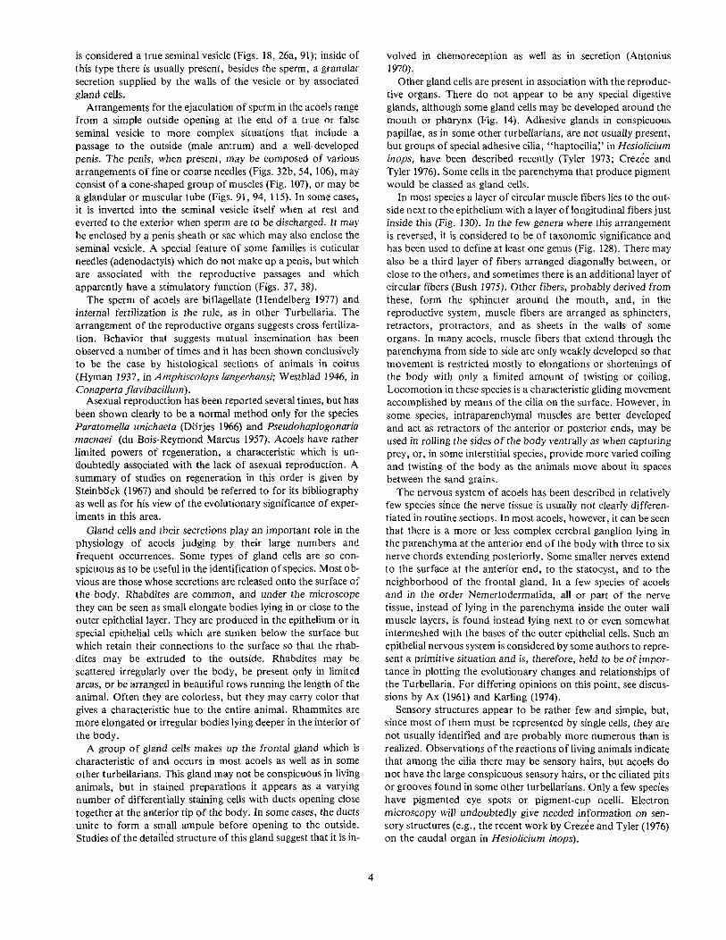
10 (7) Rhammites scattered near the entire ventral surface but concentrated around the mouth; seminal vesicle verylarge so as to almost fill the body in cross section; male antrum with a network of longitudinal musclesrunning through it (Fig. 17) Genus Fusantrum.
Figure 17.-/·usantrum rhammiphorum.
10 (7) Rhammites in rhammite tracts anterior to the mouth; seminal vesicle and male antrum well defined withmuscle fibers in the walls (Fig. 18) , Genus Endocincta.
Fi2ure 18.-Endocincta punctara.
11 (6)
11(6)
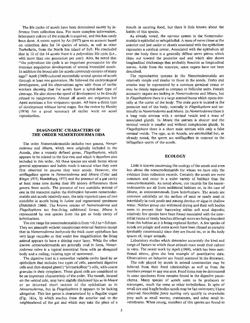
Male pore in anterior of body close to mouth; tubelike ciliated male antrum (Fig. 19) ..................................................... " Family ANTlGONARllDAE (p. 62)
Figure 19.-Antigonaria arenaria.
Male pore not in anterior end; male antrum absent; haptocilia present (Fig. 20) .. . . . . . . . . . . . . . . . . . . . . . . . . . . . ... Family PARATOMELLlDAE (p. 61) ..
13
., 12
12 (11) A caudal sensory organ present (Fig. 20); no asexual reproduction .· ; Genus Hesioliciurn.
12 (11)
Figure ZO.-Hesiolicium inops.
Caudal sensory organ absent (Figs. 21,22); both sexual and asexual reproduction .· Genus Paratofnella.
Figure 21.-ParatomelJa unichaeta.
Figure 22.-Paratomella rubr«, sagittal section through body at level of male accessory organs.
13 (5) Mouth opening at or close to the anterior end of body . ......................... 14
13 (5) Mouth opening not close to anterior end 18
14 (13) Male pore close to mouth at anterior end; bursa seminalis absent .· < .Family HOFSTENIIDAE' (p. 61) 15
14 (13) Male pore not dose to mouth; bursa seminalis present , . " '" " .· Family HALLANGIIDAE (p. 60) 17
3The Hofsteniidae arc sometimes placed in an order or suborder separate from the Acocla (e.g.• Karling 1940). However, until this point has been resolved by furlher studies, the tendency at present appears to be to retain them in the Aeocla (sec Steinbock 1966, for a complete review of Ihis question).
15 (14) Pharynx with only weakly muscular walls, either tube-shaped or short and globular; mouth opening ventralnear anterior end 16
16 (15) Pharynx long (about one-fourth body length), thin and tube~shaped (Fig. 24a); seminal vesicle enclosed in athick muscular wall which encloses also the cuticular elements of the penis (Fig. 24b). One species from theMediterranean Genus Hofstenio[a.
Figure 24.-Hojsleniola pardii.
16 (15) Pharynx short (about one·sixth body length), thin-walled and globular (Fig. 25a); seminal vesicle clearlyseparated from the bulb of the penis by a constriction (Fig. 25b). One species from .Brazil .. . . . . . . . . . . . . . . . . . . . . . . . Genus Marcusiola.
18 (13) Mouth with archipharynx (Fig. 28)... Family NADINIDAE (p. 61)
1 Genus Nadina.
Figure 28.-Nadina pulchella.
18 (13) Mouth with pharynx simplex or without a pharynx 19
19 (18) Seminal bursa present, opening posterior to the malecopulatory organ either through a common genital poreor through a separate female pore (Figs. 29, 30) .
Family OTOCELlDIDAE (p. 61). . . . . . . . . .20
Figure 29.-0tocelis westbladi.
16
Figure 30.-Archocelis macrorhabditis.
19 (18) Seminal bursa present or absent but when present openinganterior to the male copulatory organ , 25
21 (20) Testes paired and the two ovaries fused (Fig. 31a); penis muscular (Fig. 31 b) .. . . . . . . . . . . . . . . . , '" " , Genus Notocelis.
ab
Figure 3t.-Notocelis gullmarensis: a-dorsal view; b-sagitlal reconstruction of male and female accessory organs.
21 (20) Testes and ovaries paired (Fig. 32a); penis muscular (Fig. 33b) or with cuticular needles (Fig. 32b) 22
aFigure 32.-Philoceli!; cellala: a-dorslll view; b.sagitlal reconstruction of male and female accessory organs.
4Genus Exocelis. recently described from the Galapagos Islands, has many small nozzles, a feature which makes it easily distinguishable from the other genera of the Otocilididae. Also, theseminal bursa in this ~enus lies caudal to the male m~an, a sit\\atim\ found ill \\0 other acod.
Figure 31c.-f.:xocelis exopenis.
17
22 (2/) Penis with cuticular needles (Fig. 32) .· Genus Phi/aceUs.
22 (21)
23 (20)
Penis a muscular tube (Fig. 33) .· Genus Gtace/is.
Figure 33.-0tocelis rubropunctata: a-dorsal view; 'b-sagi((al reconstruction of male and female accessory organs.
Ovary paired (Fig. 34a); two coiled spermatic ducts present (Fig. 34b). One species from Brazil· Genus Hap/ocelis.
a
penis
b
23 (20)
24 (23)
Figure 34.-Haplocelis dichone: a-sagittal reconstruction; b-sagittal reconstruction of seminal bursa.
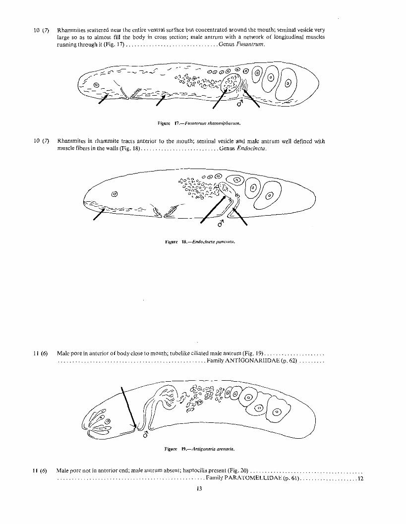
Ovary unpaired; spermatic duct lacking or very short 24
Spermatic duct lacking; common genital pore terminal at posterior end (Fig. 35a, b)....................................................... Genus Archace/is.
Genus Parotacelis.5
Figure 35a.-Archocelis macrorhabditis.
.~.......,\-seminal bursa
W+.I4/--Penis
5Genus ParOlOce!is, recently described from the Galapagos Islands, is rcported as being close to Archocelis and would fall here in the key. It differs from Archoccfis in having a short ciliated malean!rum and II muscular penis papilla surrounded by a crown of gland cells. III ArdlOcefis a male antrum is lacking and the muscular wall of the seminal vesicle appears to serve as a penis.
25 (19) Male antrum with adenodactyls bearing large needles and conspicuous rosettes of gland cells (Figs. 37, 38);one exception, see Genus Paranaperus , Family ANAPERIDAE (p. 61) , , 26
Figure 37.-Anaperus tvaerminnensis. dorsal and sagittal view~ ofposterior end. Figure 38.-Achoerus pachycaudatus, dorsal and sa?,i.ttat views of posterior end.
25 (19) Male antrum without adenodactyls or with adenodactyls very small and gland cells very inconspicuous(Figs. 39,40) .. . .. .. . . .. . . . , , 30
"'igurc 39.-Kuma monogonopllOra,dorsal view of poslerior end.
Hgure 4O.-Hapioposthia ruhra.
19
26 (25) Either colored rhabdites or epithelial pigment present 27
26 (25) Colored rhabdites and epithelial pigment lacking; adenodactyls very small. This genus is taxonomically connected to both Anaperidae and Haploposthiidae but is placed here in Anaperidae by Dorjes (1968a) (Fig.41) " Genus Paranaperus.
a
Figure 41.-Paranaperus pellucidus: a-sagittal reconstruction; b-dorsal view of posterior end,
27 (26) Seminal bursa and bursal nozzles lacking 28
27 (26) Either seminal bursa with a nozzle (Fig. 42) or nozzles without walled bursae (Fig. 43) present 29
Figure 42.-Phi/achoerus johwmi.
28 (27) Color provided by rhabdites (Fig. 44)
Figure 43.-Anaperus tvaerminnensis.
....................................................... Genus Achoerus.
Figure 44.-Achoerus pachycaudatus.
28 (27) Color provided by epithelial pigment granules (Fig. 45). Single species from Brazil " .. . . . . . .. . . . . . , , " , Genus Pseudanaperus.
20
a
.'igure 45.-Pseudanaperus tinctus: a-sagittal section; b-dorsal view of male accessory Organs.
30 (25) Male organ simple; penis either lacking (Figs. 48, 49) or very weakly developed (Fig. 50) or its place taken by aciliated male antrum invaginated into a sheath or sac (Fig. 51) 31
30 (25) Male organ well developed; either a glandular cellular penial area associated with the seminal vesicle (Fig. 52)or a tube-shaped penis (Fig. 53) or a cuticularized penis stylet (Fig. 54) 46
31 (30) Male organ simple; penis lacking or very weakly developed 32
22
31 (30) Male organ consists of a ciliated male antrum which isinserted at its proximal end into a sheath Or sac in sucha way that it opens directly from an unciliated granularvesicle with either a true or false seminal vesicle present(Fig. 55) .. . . . . . . . . . .Family ANTROPOSTHIIDAE (p. 64) .43
Figure 55.-Convoluella bru.nea.
32 (31) Male organ with a very small penis. Two species, Hap/oposthia rubra (Fig. 50) and Haploposthia (= Kuma?)a/biventer (Fig. 86) with other characters which have placed them in the Family Haploposthiidae have verysmall penes. Paranaperus pel!ucfdus (Fig. 41) with a very small penis and small adenodactyls appears to betaxonomically intermediate between the Family Anaperidae and the Family Haploposthiidae.
32 (31) Penis lacking (Figs. 56, 57, 58); often with a tube-shaped male antrum which is not inserted into a sheath orsac (Figs. 57, 58) Family HAPLOPOSTHIIDAE (p. 60) 33
33 (32) Seminal bursa present either with a distinct wall (Fig. 59) Or as a more or less well-defined vacuolated or syn-cytial space in the parenchyma (Fig. 60) " 34
Figure 59.-1Iap{ogonariu eleguns.
33 (32) Seminal bursa lacking .
23
FiRUrc 6O.-Pseudohaplogonaria vacua.
.. 38
34 (33) Either no cuticular nozzle on the seminal bursa (Fig. 61), or an attached (Fig. 62), or an invaginated (Fig. 63)muscular spermatic duct may be present instead 35
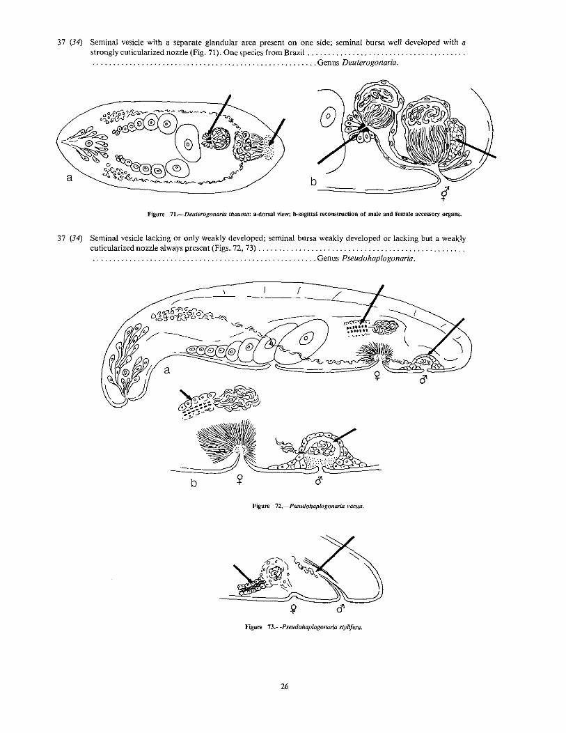
37 (34) Seminal vesicle with a separate glandular area present on one side; seminal bursa well developed with astrongly cuticularized nozzle (Fig. 71). One species from Brazil " .. . . . . .. . '" " " Genus Deuterogonaria.
b-==--==:=::::=::;;;y
Figure 71.-Deuterogonaria thauma: a·dorsal view; b-sagittai re<:onstroction of male and female accessory organs.
38 (33) Male antrum, penis, and seminal vesicle all lacking; a false seminal vesicle present at the posterior end of thebody (Fig. 74) Genus Simplicomorpha.
figure 74.-Simplicomorpha gigantorhabditis.
38 (33) Male antrum present(Figs. 75, 76) 39
Figure 75.-Kuma monogonophora, dorsal view of posterior end. F~ure 76.-Parahap{oposthia avesicutn, sagittal section of posterior end.
39 (38) Semiglobular caudal organ present, consisting of a ciliated inpocketing of the terminal end of the body whichis surrounded by and into which open gland cells containing pear-shaped droplets of secretion (Fig. 77) .. . . . . . . . . . . . . . . . . . . . . . . . . . . . . . . . . . . . . . . . . . . , Genus Adenocauda.
Figure 77.-Adenocauda helgolandica.
39 (38) Caudal organ iacking .
27
40 (39) Frontal organ lacking (Figs. 78, 79) .· Genus AJronta.
Figure 78.-Afronta aurantiaca, dorsal view.
Figure 79.-Afrrmta rubra: a-sagittal
reconstruction; b-dorsal
"iew of posterior end.
-~
40 (39) Frontal gland present .41
41 (40) A pair of false seminal vesicles present (Figs. 80, 81) .· , Genus Parahaploposthia.
'/~,J}@~/~~
Fij::ure SO.-Parahaploposthia cerebroepiiheliatQ.
41 (40) Either a true seminal vesicle or a single false seminal vesicle present .
Figure 81.-Parahaploposthla avesicula.
. .42
42 (41) Colored by rhabdites or stored body pigment; common germinal center for eggs and sperm (Figs. 82, 83) ....· . . . . . . . . . . . . . . . . . . . .. . . . . . . . . . Genus Hap/oposthia.
45 (44) her than a nozzle (Fig, 90) . , . . . . , .Seminal bursa with a small cap of cells, rat .... , . Genus Convoluella............ , .. , .... "
Figure 9O.-ConvofueUa bmnea,
b
30
46 (30) Copulatory organ a globular structure inside of which is a proximal seminal vesicle and a distal glandularregion which serves as a penis (Figs. 91, 92) Family MECYNOSTOMIDAE (p. 63) 47
Figure 92.-Pseudmecynostomum pelophilum: a-dorsal view of posterior end; b.sagi.ttat
section of copulatory o~an.
46 (30) Copulatory organ either a conical penis made up of cellular, muscular, or cuticular parts (Fig. 93), or a tube-shaped penis formed by an invagination of the body wall (Fig. 94) 52
Figure 93.-Actinoposthib haploraln: a-dorsal view of posterior end; b-sagillal
section of copulator)' organ.
a
Figure 94.-Archaphallosloma macrospinferum: a-dorsal view of posterior end; b-sagittal
47 (46) Seminal bursa present ........................................... a
48 (47) Seminal bursa lacking an appendage (Fig. 97); in Pseudmecynostomum maritimum the bursal wall develops aprotrusion over the ripe eggs (Fig. 98) Genus Pseudmecynostomum.
a
b/
Figure 97.-Pseudmecynostomum pe/ophilum.
32
Figure 98.-Pseudmecynostomum maritimum.
48 (47) Seminal bursa with a cellular (Fig. 99), muscular (Fig. 100), or cuticular (Fig. 101) appendage .49
50 (49) Seminal bursa weakly developed without a well~defined wall but with spermatic duct strongly cuticularized toform a conspicuous nozzle (Fig. 102) , Genus Paedomecynostomum.
FiJl.ure l02.-Paedomecynostomum bruneum.
50 (49) Seminal bursa well developed with an appendage formed of matrix cells whose inner ends are weaklycuticularized to form a weak nozzle (Fig. 103) Genus Philomecynoslomurn.
Fi~ure I03.-PlJilomec)'f/OS10mUm lapillu!Il.
~)
33
51 (49) Appendage of bursa muscular; rhabdites present; variegated coloring from dissolved pigment and yellow tobrown granules (Fig. 104) Genus Paramecynostomum.
Figure l04.-Paramecynostomum diversicolor.
51 (49) Appendage of bursa cellular; rhabdites absent; no dissolved or granular pigment (Fig. 105) ,...................... ' Genus Postmecynostomum.
Genus Limnoposthia,6
Figure ]()Sa.-Postmecynostomum pictum.
F~ure ]()sb.-Limnoposthiil polonica. (See next page and text footnote 6.)
@
b
6The genus LII/II/Oposthill (Kolasa and Faubel 1974) was placed tentacively by che authors dose 10 Com'aluta, but it seems to me illc illustratioDS anQ Q('5criplions of the penial struclures wouldlead a user of this key to a position close to PoslmecynoSlromum, The structure of the penis in the illustration of Litlll/opos/hiu polonicu suggests the glandUlar penis of POSI!/Iecynostomum and thepart labeled "vas deferens" may cOrrespond 10 the enclosed seminal vesicle of the Mecynostomidae. Study of actual specimens is needed to clear up this point. From freshwater lake in POland.
34
o\,,-- /
C) I
~)~(
o
Fl~ure UJ5c.,d.-Limnoposthia polonica.
52 (46) Copulatory organ with a cone-shaped penis built up of cuticular (Fig. 106), or muscular (Fig. 107 elementsand never invaginated into a seminal vesicle Family CHILDIIDAE (p. 62) '" 53
52 (46) Copulatory organ with a tube-shaped, muscular and often glandular penis. This may be simply a direct invagination of the body wall without a male antrum or true seminal vesicle (Fig. 108), or it may be combined witheither one or both of a male antrum or a seminal vesicle. It is often invaginated into or surrounded by theseminal vesicle (Fig. 109) or a penis sac (Fig. 110) Family CONVOLUTlDAE (p. 58) 63
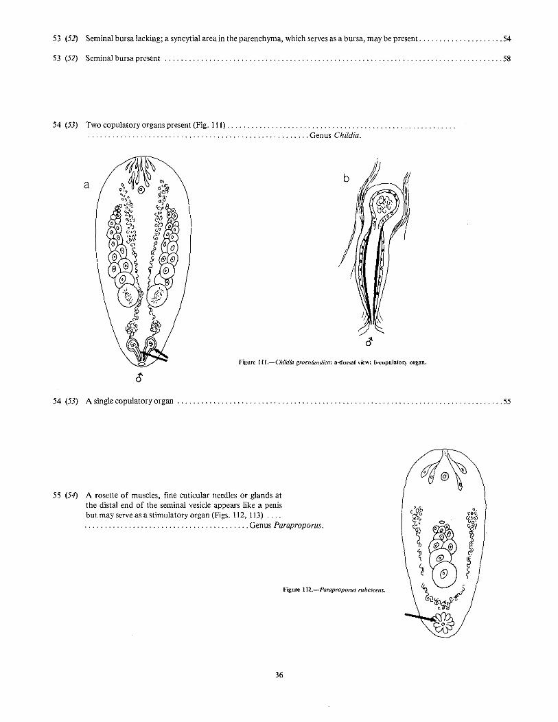
55 (54) A rosette of muscles, fine cuticular needles or glands atthe distal end of the seminal vesicle appears like a penisbut may serve as a stimulatory organ (Figs. 112, 113) . .... . . . . . . . " Genus Paraproporus.
Figure 112.-Parapropoms rubescens.
36
Figure 113.--Paraproporus dUwalUs.
55 (54) Copulatory organ not a rosettelike arrangement; penis with strongly cuticularized needles (Fig. 114). musclelayers in its wall (Fig. 115), or a cuticularized papilla surrounded by matrix cells (Fig. 116) 56
56 (55) Male antrum long, tube-shaped and ciliated; penis a thin cuticularized tube clothed in matrix cells (Fig. 117)....................................................... Genus Atriojronta.
Figure 117.-Atriofronta polyvacuola.
56 (55) Male antrum short; penis with cuticularized stylet needles or muscle layers in its wall 57
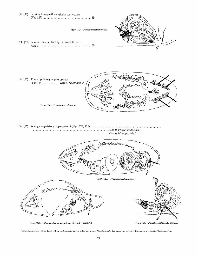
Figure 12Sb.-Monoposthia pselldovesicula. (See lext footnote 7.) Figure lU.-Phiklctinoposthia adenogonaria.
7Gcnus MOl1opowhia, recently described from Ihc Galapagos Islands, is dose to the gCllllS Phi{aclinoposrhiu but lacks a true seminal vesicle, such as is present in Phila,"linopoiilhill.
39
60 (58) Outer body wall muscle layers consist of outer longitudinal and inner circular layers (Fig. 128); frontal glandvery small or lacking (Figs. 127, 129) '" , .Genus Paraphanostoma
Figure 127.-Paraphanos(oma macroposthium.
longitud ina I muscleo 0 0 0 • 0 "circular muscle
'I\ma((I(Ill UII(MIlli0" \ \ 0 l\ , C'::l \
Fi~ure 128.-Longitudinal section of body wall.
Figure 129.-Paraphanostoma gracilis.
60 (58) Outer body wall muscle layers with outer circular and inner longitudinal fibers (Fig. 130); frontal gland welldeveloped 61
\J%\\)~WMII \I IIIIII( II (((,\\\I\liu/iJ.., .) 0 I, ,0,J.o (~circular muscleo a a 0 000
64 (63) Seminal bursa an extensive syncytial area in the posterior end of the body with bundles of sperm scatteredtherein (Figs. 139, 140, 141) '" .Genus Archaphanostoma.
64 (63) Seminal bursa a definite walled structure 65
42
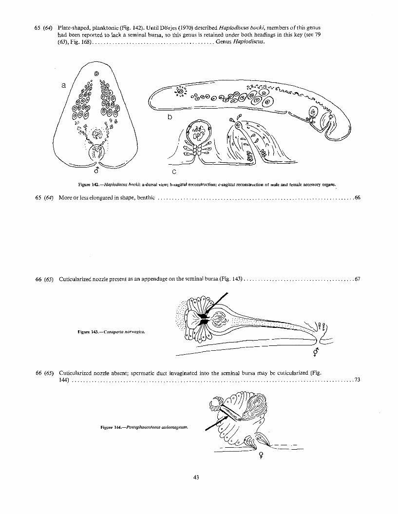
65 (64) Plate-shaped. planktonic (Fig. 142). Until Dorjes (1970) described Haplodiscus bockl, members of this genushad been reported to lack a seminal bursa, so this genus is retained under both headings in this key (see 79(63), Fig. 168) Genus Haplodiscus.
a
c
)
Figure 142.-Haplodiscus bocki: a-dorsal view; b.sagittal reconstruction; c-sagiual reconstruction of male and female accessory organs.
65 (64) More or less elongated in shape. benthic " , 66
66 (65) Cuticularized nozzle present as an appendage on the <;eminal bursa (Fig. 143) , , 67
70 (67) Special yolk-carrying cells absent; yolk deposited in the eggs 72
70 (67) Developing eggs in one layer toward the ventralside of the body with a layer of special yolkcarrying cells (yolk-glandlike layer) dorsal or dor-solateral to the eggs (Fig. 149) 71
.;igure 149.-Po/ychoerus, cross section.
45
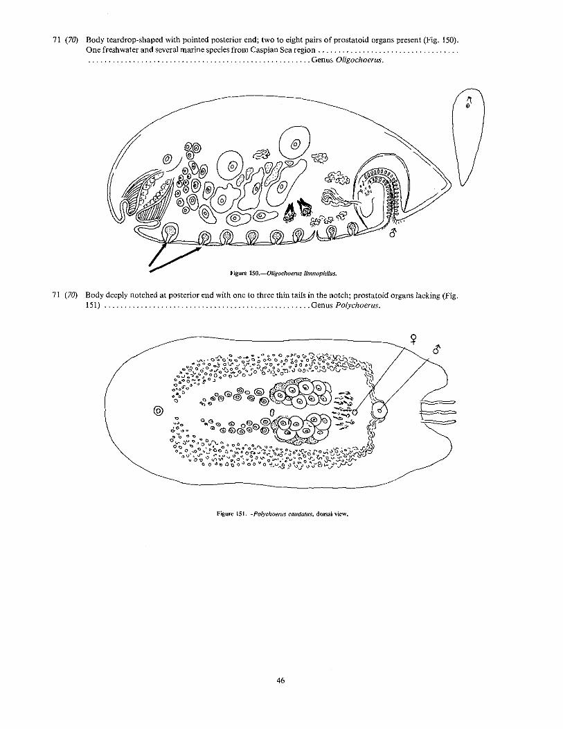
71 (70) Body teardrop-shaped with pointed posterior end; two to eight pairs of prostataid organs present (Fig. 150).One freshwater and several marine species from Caspian Sea region .. . . . . . . . . . . . . . . . . . . . . . . . . . . . . , Genus Oligochoerus.
Hgure lSO.-Oligochoerns limnophilus.
71 (70) Body deeply notched at posterior end with one to three thin tails in the notch; prostatoid organs lacking (Fig.151) Genus Polychoerus.
@
Figure lSI.-Po{ychoerus caudafus, dorsal view.
46
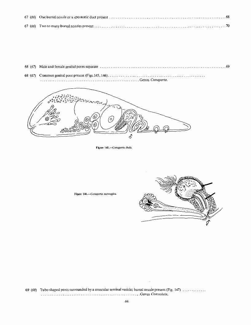
72 (70) Separate genital pores; colored by the presence of symbionts or pigment; seminal bursa with two, more or lesscurved or spiraled nozzles (Figs. 152, 153, 154) Genus Amphiscolops.
Figure 154.-Amphisco{ops bermudensis: a--dorsal view; b-sagittal rei:onstruction of male and female acces;ory organs.
72 (70) Common genital pore; white, or colored only by the presence of food products; seminal bursa with two ormore short cone-shaped nozzles. From the Pacific coasts of the United States and U.S.S.R. (Fig. 155)....................................................... Genus Diatomovora.
Figure 155.-Diatomovora umoena.
47
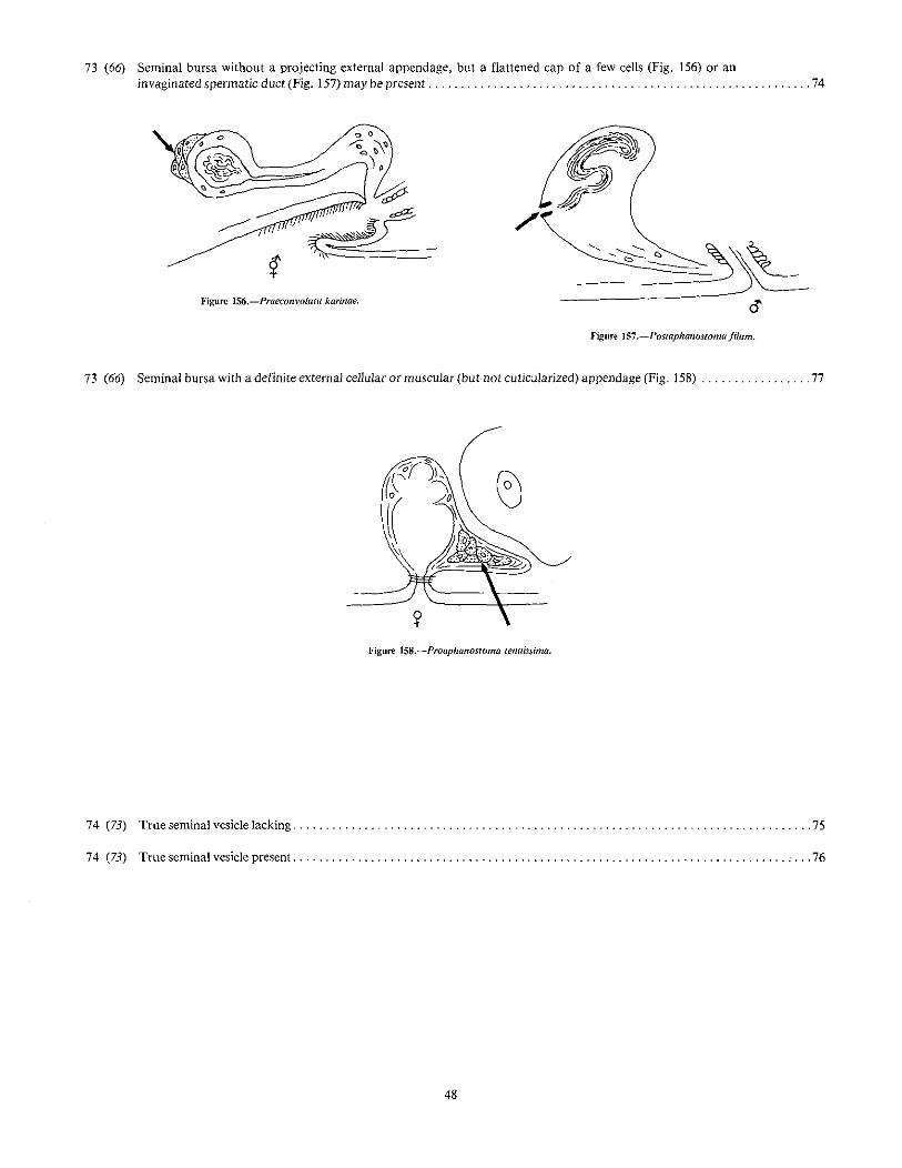
73 (66) Seminal bursa without a projecting external appendage, but a flattened cap of a few cells (Fig. 156) or aninvaginated spermatic duct (Fig. 157) may be present , , , " , , , , ' ' . , 74
Figure 1S6.-Praeconvoluta karinae.
"'~~~~
oFigllJ1! lS7.-l'ostaphanostoma filum.
73 (66) Seminal bursa with a definite external cellular or muscular (but not cuticularized) appendage (Fig. 158) " 77
75 (74) Small rhabditcs present, arranged in rows; female germinal center and string of eggs unpaired; penis a simple,tube-shaped invagination of the outer body waH (Figs. 159, 160) .. . . . . . . . . . . . . . . . . . . . . . . . . . . . . . . . . . . . . . . . . . . , ,Genus Postaphanostoma.
•'igllre 159.-Postaphanostoma atriomagnum.
Figure 160.-Postaphanostoma filum.
75 (74) Rabditcs lacking; female germinal center and string of eggs paired; penis very glandular and invaginated intoa muscular penis sac (Fig. 161) . . . . , " , .
. . . . . Genus Praeconvoluta.
Figure 161.-Praeconmluta karinae.
49
76 (74) Seminal bursa opens from a long, tubelike ciliated female antrum and is surrounded by a mass of netliketissue (a bursal tissue), but with no appendage (Fig. 162) .. . . . . . . . . . . . . . . . . . . . . . . . . . . . . . . . . . . , Genus Bursosaphia.
}<'igure 162.-Bursosaphia baltalimaniafonnis.
76 (74) Seminal bursa opens almost directly from the exterior without any or with an extremely short female antrum;no appendage and no bursal tissue present but there may be an invaginated spermatic duct (Figs. 163, 164) .... . . . . . . . . . . . . . . . . . . . . . . . . . . . . . . . . . . . . . . . . . . . . . . . . . . . . . . Genus Praeaphanostoma.
Figure 163.-Praephanostoma chaetocauCfatum.
Figure 164.-I'raeaphanostoma lutheri.
77 (73) Rhabdites present, arranged in irregular rows; seminal vesicle and ciliated male antrum present (Fig. 165) .. . . . . . , , Genus Aphanostoma.
50
Figure 165.-Aphanostoma album.
77 (73) Rhabdites absent; male antrum lacking; seminal vesicle present or absent 78
78 (77) Seminal vesicle absent; penis a simple invagination of outer wall layers with a ball of sperm at the proximalend that is surrounded by a parenchymal net with a few muscle fibers and gland cells (Fig. 166) .
78 (77) Seminal vesicle present; penis a tubelike invagination which is further invaginated into the seminal vesicle(Fig. 167) Genus Proaphanostoma.
)
b
Figure 167.-Proaphanostoma telluissima.
51
79 (63) Plate-shaped; planktonic (Fig. 168); until Dorjes (1970) described Haplodiscus bocki, the members of thisgenus had none of them been reported as having a seminal bursa, so the genus is retained under both headingsin this key (see 65 (64), Fig. 142) Genus Haplodiscus.
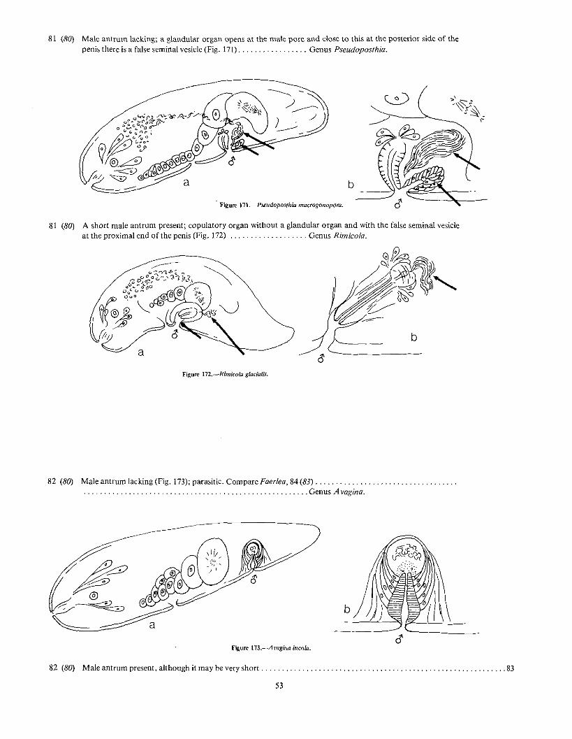
81 (80) Male antrum lacking; a glandular organ opens at the male pore and dose to this at the posterior side of thepenis there is a false seminal vesicle (Fig. 171) Genus Pseudoposthia.
Figure 111.-Pseudopos/hia macrogonopora.
b
81 (80) A short male antrum present; copulatory organ without a glandular organ and with the false seminal vesicleat the proximal end of the penis (Fig. 172) Genus Rimicola.
Figure 174.-Neochi/dia fusca: a-dorsal view; b-sagitlal reconstruction of posterior end.
83 (82) One copulatory apparatus present 84
84 (83) Male antrum very short (Fig. 175). Compare with A vagina 82 (80), which was originally (Westblad 1945)separated from Faerlea on the basis of the latter being free-living. Description of a parasitic species in Faerleaby DiJrjes in 1972, leaves onJy the slight differences in the maJe antra to distinguish these genera. They mayneed to be combined Genus Faer/ea.
a
Fi~ure 175.-Faerlea fragiiis.
84 (83) Male antrum long, tUbe-shaped and ciliated
54
............ 85
85 (84) Penis and male antrum without a clear line demarking one from the other (Fig. 176); from the Caspian Sea....................................................... Genus Baltalimania.
85 (84) Penis and male antrum with a clear line distinguishing one from the other. 86
86 (85) Colored rhabdites present as well as other pigment; a tube-shaped organ present at the anterior end as well asa frontal gland (Fig. 177). From Sakhalin Island, Pacific coast of U.S.S.R .. . . . . . . . . . . . . . . . . . . . . . . . . . . . . . . . . . . . . . . . . . . . . . . . . . . . . . .Genus Oxyposthia.
a
Figure lii.-Ox}po~thia praedator.
55
86 (85) Colored rhabdites lacking, but small uncolored rhabdites in rows; tube-shaped organ lacking; frontal glandpresent (Figs. 178, 179, 180) , Genus Pseudaphanostoma.
87 (2) Flagellar organ present; mouth opening absent (Fig. 181) ." , , , , Genus F/agellophora.
a
c
Figure 181.-Flagellophora ape/Ii: a-sagittal reconstruction; b-basc of flagellar organ next to intestine; c-male accessory organs.
56
87 (2) Flagellar organ absent; mouth opening present 88
88 (87) Frontal gland absent; ovary and testes folicular; male antrum with a side pocket but no gland cells aroundbase of the seminal vesicle (Fig. 182); commensal in holothurians , > , , • , ••••• > •••• > > • > ., •
... " , .. ,.' , , .. ,.' Genus Meara.
Figure 182.-Meara stichopi.
88 (87) Frontal gland well developed; compact testes and compact ovary; male antrum with many large gland cellsaround base of the seminal vesicle (Figs. 183, 184); free-living , ' , , , , ... . . . . . . . , , Genus Nemertoderma.
Figure l83.-Nemertodemw bathycola.
Figure 184.-Nemertodenna rubra: a-sagittal reconstruction; b-sagittal section through phal)'Rx; c-sagittlll section of male accessory organs.
57
ANNOTATED SYSTEMATIC LIST OFNORTH ATLANTIC GENERA AND SPECIES OF
ACOELA AND NEMERTODERMATIDA
Species listed here include: 1) species reported in the literatureor collected by the author in the area, and 2) genera and speciesthat may be expected in this area because of their occurrence inthe North Sea, the North Atlantic Ocean, or adjacent areas.Since the fauna here is as yet poorly described, it is felt to bemost useful to include as many of the species and genera likelyto be encountered as possible. Synonyms likely to be encountered in the literature are also included. Most of the doubtfulspecies I have omitted altogether.
The list is arranged systematically in families as in Dorjes(1968a) with the new family Antroposthiidae Faubel added atthe end. Genera are arranged alphabetically within the familiesand species alphabetically within the genera. Geographicdistribution and habitat are-indicated for each species. Asterisksindicate species for which I have records or which have beenreported in the literature from the northeast coast of NorthAmerica.
Order ACOELA
Family PROPORIDAE Graff, 1852
Genus A rchiproporus An der Lan, 1936
Archiproporus minimus :=. Proporus minimus according toWestblad (1945), but see comment under 4 (3) in key.
Genus ProPQrus O. Schmidt, 1882
Proporus brochii Westblad, 1945. Sweden, Norway. 100150 m.
200-300 m.Proporus rubropunctatus O. Schmidt, 1852 = Gtace/is rubro~
punctata.Proporus venenosus (0. Schmidt, 1852). British Isles,
Mediterranean Sea, Black Sea. Subtidal. Includes subspecies: P. v. venenosus Graff, 1904, P. v. violaceus Graff,1882, and P. v. viridijlavus Riedl, 1959.
Genus Schizoporus O. Schmidt, 1852
Only one species S. venenosus == Proporus venenosus.
Amphiscolops cinereus (Graff, 1874). North Atlantic,Mediterranean Sea. On algae.
58
*Amphiscolops gardineri Graff, 1910. Graff listed this in hisfirst report on American Turbellaria, but later (1911) erectedthe new genus Anaperus for it.
Amphiscolops /angerhansi (Graff, 1882). North Atlantic,Mediterranean. Tide pools, rocky shores. A species identified as this (?) was collected on algae at Virginia Key, Fla.
Many species assigned to this genus have been shuffled backand forth several times. I have followed Dorjes (l968a) as totheir disposition in the following list.
Aphanostoma album Dorjes, 1968. North Sea. Frommuddy sand.
*?Aphanostoma aurantiacum Verrill, 1892. Newport,R.I. Listed as a doubtful species by various authors andnot definitely identified since.
Aphanostoma auritum (M. S. Schultze, 1851) = Mecynostomum aurilum.
Aphanostoma caudatum (An der Lan, 1936) := Actinoposthiacaudata.
Aphanostoma divaceum Verrill, 1892. Listed by Dorjes(1968a) as a doubtful species. Probably a typographical erroron A. olivaceum.
The number of species in this genus is very large and manyplaced here in the past have since been removed to other genera.I have followed Dorjes (1968a) as to disposition of these.
rhammi/era.Convoluta rascoffens!s Graff, 1904. Dorjes (1968a) says this is
59
a species whose definition }s uncertain; there are many references in the literature and many reports on experiments withanimals identified as this species.
*Neochildia jusca Bush, 1975. North Carolina to Maine.In or on sand or muddy sand along marsh streams or inestuaries.
Genus PolYchoerus Mark, 1892
*Polychoerus caudatus Mark, 1892. Formerly common fromNew Jersey to Maine (Verrill 1892). Now rare but has beencollected on empty scallop shells (1-2 m) in Great Bay nearWauwinet, Mass., and once in bottom sand sample (5 m) atMenemsha, Mass. This beautiful orange species has been usedat Woods Hole in experimental studies (Child 1907; Keil1929).
Genus Posfaphanostoma Dorjes, 1968
Postaphanostoma atriomagnum Db'rjes, 1968. NorthSea. In sand without mud or detritus, 3-18 m.
Postaphanostomajilum Dorjes, 1968. North Sea. Commoninterstitial form in various sands, 7-18 m.
Postaphano.stoma glandulosum Dorjes, 1968. North Sea.From gravel deposits.
Genus Praeaphanostoma Dlirjes, 1968
Praeaphanostoma brevifrons Dorjes, 1968. North Sea.From very fine sand, 5 m.
Praeaphanostoma chaetocaudatum Dodes, 1968. North Sea.From coarse sand in shallow water.
Praeaphanostoma longum Dt1rjes, 1968. North Sea. In finesand, 6 m.
Praeaphanostoma lutheri (Westblad, 1946) ~ Peiophila lutheri
Praeaphanostoma rubtum Dorjes, 1968. North Sea. In finesand, 5 m.
Genus Praeconvoluta Dorjes, 1968
Praeconvoluta karinae Dorjes, 1968. North Sea. In graveland rock pools, 1-2 m.
Praeconvoluta minor Faubel, 1974. North sea. From beachsand.
Praeconvoluta schmidti Faubel, 1977. Norway. From beachsand.
Genus Proaphanostoma Dorjes, 1972
Proaphanostoma tenuissima (Westblad, 1946). North Sea.30 ffi.
Proaphanostoma viridis An der Lan, 1936 = Haploposthiaviridis.
Genus Proconvoluta Dorjes, 1968
Proconvoluta primitiva Dorjes, 1968. North Sea. Fromdetritus zone in shallow water.
Genus Pseudaphanostoma Westblad, 1946
Pseudaphonostoma brevicaudatum Dorjes, 1968. North SeaIn muddy sand, shallow water.
Pseudaphanostoma pelophilum Dorjes, 1968. North Sea.In muddy sand, often with Zostera or Mytilus.
Pseudaphanostoma psammophilum Dorjes, 1968. North SeaIn sand of the swash zone and sublittorally in unstablesand areas.
Pseudaphanostoma variabilis Westblad, 1946. North Sea.From clay with sand, 10-20 m.
Genus Pseudoposthia Westblad, 1946
Pseudoposthia macrogonopora Westblad, 1946. North Sea.From clay with sand.
Family NEMERTODERMATIDAE Steinbock, 1930
See under Order NEMERTODERMATIDA at end.
Family DIOPISTHOPORlDAE WestbJad, 1940
Genus Diopisthoporus WestbJad, 1940
Diopisthoporus brachypharyngeus Dorjes, ]968. North SeaFrom fine sand, 5-8 m.
Diopisthoporus longitubus Westblad, 1940. North Sea,Adriatic. From soft bottom to 360 m.
Diopisthoporus psammophilus Dorjes, 1968. North Sea.From fine sand, 5 m.
FamBy HALLANGIIDAE Westblad, 1948
Genus Halll1ngia WestbJad, 1946
Ha/langia proporoides Westblad, 1946. European Atlantic.From soft bottom to 100 m.
60
FamBy HAPLOPOSTHIIDAE Westblad, 1948
Genus Adenocauda Dorjes, 1968
Adenocauda helgolandica Dorjes 1968. North Sea. In finesand, 5 ffi.
Genus Afronta Hyman, 1944
*Afronta aurantiaca Hyman, ]944. Maine coast. Dredged insurface mud, 30 ft.
Afronta rubra Faubel, 1976. North Sea. From surf zone.
Genus Haplogonaria Dorjes, 1968
Haplogonaria elegans Faubel, ]976. North Sea. From sandyshallows.
Haplogonaria glandulijera Dorjes, 1968. North Sea. In finesand, 5 m.
Haplogonaria macrobursalia Dorjes, 1968. North Sea.Surface layers of coarse sediments, 2-6 m.
Haplogonaria minima (Westblad, 1946). North Sea, Mediterranean. From soft bottom, 16~150 m.
Haplogonaria psammalia Faubel, 1974. North Sea. In beachsand.
Haplogonaria simplex Dorjes, 1968. North Sea. Fromcoarse sand, 32 m.
Haplogonaria sinubursalia Dorjes, 1968. North Sea.From fine sand, 5-8 m.
Haplogonaria syltensis Dorjes, 1968. North Sea. Fromvarious sands to 15 m.
Haplogonaria viridis Dorjes, 1968. North Sea. From sandymud, 5-8 m.
Genus Haploposthia An der Lan, 1936
Haploposthia brunea An der Lan, 1936. Greenland. Frommuddy bottoms, to 180 ffi.
Archocelis macrorhabditis Dorjes, 1968. North Sea. Onshells and mud, 5 m.
Genus Haplotestis Dorjes, 1968
Haplotestis curvitubus Dorjes, 1968. North Sea. Fromcoarse sand, 32 m.
Genus Notocelis Dorjes, 1968
Notocelis gullmarensis (Westblad, 1946). North Sea, Mediterranean. From coarse gravel with shells, shallow water.
Genus Otocelis Diesing, 1862
Otocelis ruhropunctata (0. Schmidt, 1852). Mediterranean,Black Sea. Northern records of this are a separate species,Otocelis westbladi, according to Ax (1959). In detritus-richfine sand.
Otocelis westbladi Ax, 1959. North Sea. From mud, 5-20 m.
Genus Phl/ocelis Dijrjes, 1968
Philocelis cellala DUrjes, 1968. North Sea. From detritusrich sand, 2-6 m.
Philocelis karlingi (Westblad, 1946). North Sea, Baltic. Incourse sand, 2--6 m.
Family HOFSTENlIDAE Bock, 1923
Genus HOfstenia Bock, 1923
Hofstenia giselae Steinbock, 1966. Bimini. On Thalassia.Hojstenia miami Correa, 1960. Florida and Curacao, On
algae.
61
Family PARATOMELLlDAE Dijrjes, 1%8
Genus Hesiolicium Crezee and Tyler, 1976
Hesiolicium inops Crezee and Tyler, 1976. New River, N.C.In seawater.
Genus Paratomella Dorjes, 1966
*Paratomella rubra Rieger and Ott, 1971. North Carolina,Mediterranean. In clean sand in Shallow water.
Paratomella unichaeta Dorjes, 1966. North Sea. Clean sandand gravel, 3-5 m.
Family NADlNlDAE Dorjes, 1%8
One genus and one species only. Genus Nadinff U}janin,1870. Mediterranean.
Family ANAPERlDAE Dorjes, 1968
Genus Achoetus Beklemischev, 1914
Achoerus pachycaudatus Dorjes, 1968. North Sea. Shallowwater, soft bottom.
Microposthia listensis Faubel, 1974. North Sea. In sandybeach.
Genus Paraphanostoma SteinbOck, 1931
Paraphanostoma brachyposthium Westblad, 1942. NorthSea. Common in dredge hauls. From muddy bottom, to360m.
Phi/actinoposthia adenogonaria Dorjes, 1968. North Sea.From mud, 8 m.
Philactinoposthia diploposthia Dorjes, 1968. North Sea.Fine sand with some detritus, 5 m.
Philactinoposthia helgo/andica Dorjes, 1968. North Sea.From coarse sand, 33 m.
Philactinoposthia pusiJla (Westblad, 1946). North Sea.From sand and clay, 10-20 m.
Philactinoposthia rhammifera (Westblad, 1946). North Sea toIceland. Occurs sporadically, 15-20 m.
PhiJaclinoposlhia saliens (Graff, 1882). North Sea,Mediterranean. From sand and mud, mostly shallow water.
Philactinoposthia slyli!era (Westblad, 1946). North Sea.From clay with sand, 10-20 m.
Philactinoposthia viridis Dorjes, 1968. North Sea. Fromgrowth on an old shipwreck.
Philactinoposthia viridorhabditis Dorjes, 1968. North Sea.From fine sand.
9In his key and definition of genera, Dorjes (I968a) lists a genus ProaelinopoSlhiQ and aspecies ProaC/illopos/hia pelophila, but description of a species Archm:!inopos!hia pelophi/ais placed in position for this genus (p. 335) and it is so listed in Dorjes' (l968b) article on theecology of the acoels.
Mediterranean, Black Sea, salt spring in Germany. Fromsand and mud in shallow water. Past records of this speciesmay actually involve the following: 1) M. auritum without abursa and 2) these species with a bursa: M. aurilum formatypica changed to Pseudmecynostomum westbladi Dorjes1968; M. aurilum forma flavescens changed toPseudrnecynostomum flavescens (Westblad 1946); and M.auritum forma glandulosum changed to Postmecynostomumglandulosum (Westb1ad 1946) (see Dorjes 1968a:116; Dorjesand Karling 1975:181).
ANTONIUS. A.1968. Faunistische Studien am Roten Meer im Winter 1961/62, TeillV.
Neue Convolutidae und eine Bearbeitung des VerwandtsehaftskreisesCon yo/uta (Turbellaria Acocla). Zool. Jahrb. Abt. Syst. Oekol.Geogr. Tiere 95:297-394.
1970. Sense organs in marine Aeoela. Am, Zool. 10:550.APELT, G,
AX, P.1956. Les Turbellaries des etangs co tiers du Jittoralmediteranneen de la
France meridinale. Vic Milieu Supp!. 5:1-215.1959. Zur Systematik. o.:ologie und Tiergeographie der Turbellariell
fauna in den ponto-kaspischen Brackwassermeeren. Zool. Jahrb. Abt.Syst. OekoL Geogr. Tiere 87:43-184.
1961. Verwandtschaftsbeziehungen und Phylogenie der Turbellarien.Ergeb. BioI. 24:1-68.
1977. Life cycles of interstitial Turbellaria from the eulittora1 of the NorthSea. Acta Zool. Fenn. 154:11-20.
AX, P.• and J. DORJES.1966. Oligochoerus Iimnophilus nov. spec., ein kaspisches Faunen
clement ais erster Siisswasservertreter cler Turbellaria Acoeia in FIiissenMittdeuropas. Int. Rev. gesamten Hydrobiol. 51:15-44.
AX. P., and E. SCHULZ.1959. Ungeschleehtliche Fortpllanzung durch Paratomie bei acoelen
Turbellaricn. BioI. Zentralbl. 78:613-622.BEKLEM1SCHEW. W.
1914. Uber einige aci.:ile Turbellarien des Kaspischen Meeres.Zool. Anz. 45:1-7.
1915. Sur les turbcllaries parasites de la cote Mourmanne, I Acoela.Tr. Imp. S. Pcterb. O-va. Estestvoispyt. 43:103-172. (Trav. Soc. Imp.Nat. petrograd 43:103-172.)
1963. Die Turbellarien des Kaspischen Meere. III die Gattung Oligochoerus nov. gen. (Aeoela, Convolutidae). Byull. Mosk. O-va. Ispyt.Prir. Otd. Bioi. 68:29-40.
BOCK, S.1923. Eine neue marine Turbellarien-ga!!ung aus Japan. Vniv. Arsskr.
Mat. Nat. 1:1-55.BOGUTA, K. K., and.Y, V. MAMKAEY.
1972. [On the structure of parenchyma in acoelus Turbellaria.} {InRuss.} Vestn. Leningr. Univ. Biolg. 1972(2):15-27.
BOOMIG, L.1895. Die Turbellaria acoela der Plankton-Expedition. Ergebnisse der
Plankton-Expedition der Humboldt-Stiftung II, 48 p.1908. Reports Sc:ientifiques. Turbellarien. In Resultats du voyage du S.
Y. Belgica en 1897-1898-1899 sous Ie commandemcnt de A. de Gerlachede Gomery. Zoologie. 32 p.
BUSH, L.1975. Biology of Neochildia fusca n. gen., n. sp. from the nOrtheastern
coost of the United States (Platyhelminthes: Turbellaria). BiD!. Bull.(Woods Hole) 148:35-48.
CHILD, C. M.1907. The localization of different methods of form-regulation in
CORREA, D. D.1960. Two new marine Turbellaria from Florida. Bull. Mar. Sci. Gulf
Caribb. 10:208-216.1%3. The turbellarian Hojs(enla miamla in the Caribbean Sea. Stud.
fauna Curacao Other Caribb. lsI. 17:38-40.CREZEE. M.
1975. Monograph of the Solenofilomorphidae (Turbellaria: Acoela).Int. Rev. gesamten Hydrobiol. 60;769-845.
1978. partomella rubra Rieger and Ott, an amphiatlantic: acoel turbelJarian. Cah. Bio!. Mar. 19: 1-9.
CREZEE, M., and S. TYLER,1976. Hesiolicium gen.n. (Turbcllaria Acocla) and observations on its
ultrastructure. Zoo!. Scr. 5:207-216.DIESING. K. M.
1862. Revision der Turbellarien. Abtei!ung: Rhabdocoelen. Sitzungsber.Math-Naturwiss. Kl. Kais. Akad. Wlss. Wien 45:191-318.
In coarse sand.Sea. In beach
Antroposthia axi Faubel, 1974. North Sea.Antroposthia unipara Faubel, 1974. North
sand.
ABILDGAARD. P. CH.1806. Zoologica danica seu animalium Danieae et Norwegiae rariorum
ae mines notorum descriptorum et historia aus O. F. Muller. Zool.Dan. 4, 26 p.
Meara stichopi Westblad1 1949. North Sea. In intestineand body cavity of holothurians.
Flagellophora apel!i Faubel and Dorjes, 1978. North Sea.From coarse sand and shell gravel, sublittoraL
Order NEMERTODERMATIDA Karling, 1940
Unantra polyvacuola Faubel, 1976. North Sea. In sublittoralcoarse sand.
Genus Solenojilomorpha Dorjes, 1968
Convoluella brunea Faubel, 1974. North Sea. In shallowwater to 10 em in sand.
Family NEMERTODERMATlDAE Steinbock, 1930
Genus Flagellophora Faubel and Dorjes, 1978
Genus Antroposthia Faubel, 1974
Adenopea illardalus (Lohner and Micoletzky, 1911). Pelagicin Mediterranean.
Genus Adenopea Antonius, 1968
Famay ANTROPOSTHIIDAE Faubel, 1976
Solenojilomorpha longissima Dorjes, 1968. North Sea.From muddy sand,S m.
Genus Unantra Faubel, 1976
*Nemertoderma bathyco!a, Steinbock, 1930. North Atlanticto Greenland, North Sea, Mediterranean; Buzzards Bayand Vineyard Sound, Mass. Subtidal. Four geographicvarities of this species have been designated (Dorjes 1968;Riedl 1960). Variety in our area may be different.
Nemertoderma rubra Faubel, 1976. North Sea. From coarsesand.
Nemertoderma sp. A, B. D. Reported by Tyler and Rieger(1977) from North Carolina.
HULINGS, N. c., and J. S. GRAY.1971. A manual for the study of meiofauna. Smithson. Contrib. Zool.
78, 84 p.HYMAN, L. H.
\931. Reproductive system and copulation in Amphiscolops iangerhansi(Turbellaria Acoela). BioI. BulL (Woods Hole) 72:319-326.
1939. Acoe! and polyclad Turbellaria from Bermuda and the Sargassum.Bull. Bingham Oceanogr. Collect. Yale Univ. 7:1-35.
1944. Marine Turbellaria from the Atlantic Coast of North America.Am. Mus. Novit. 1266, 15 p.
IVANOV, A. V.1952. Beskishechnye turbellyarii (Acoela) yuzhnogo poberezh "ya Sak
halina." [In Russ.) Tr. Zool. Inst. Akad. Nauk SSSR 12:40-132.IVANOV, A: V., and Y. V. MAMKAEV.
1977. Uber die Struktur dcs Digestionsparenchyms bei Turbellaria Acoela.(In Ger., Engl. abstr.J Acta Zool. Fenn. 154:59-61.
JENSEN, O. S.1878. Turbellaria
Norges Veslkyst.p.
KARLlNG, T. G.1940. Zur Morpho10gie und Systematik der Alloeocoela Cumulata und
Rhabdocoela Lccithophora (Turbellaria). Fauna Fenn. 26:1-260.1967. Zur Frage von dem systematischen Wert der Katgegorien Arcna
aphora und Neoophora (Turbellaria). Commentat. BioI. Soc. Sci.Fenn.30:1-11.
1974. On the anatomy and affinities of the tvrbellarian orders. InN. W. Riser and M. P. Morse (editors), Biology of the Turbellaria,p.1-\6. McGraw-Hili, N.Y.
KEIL, E. M.1929. Regeneration in Polychoerus caudatlis Mark. BioI. Bull. (Woods
Hole) 57:225-244.KOLASA, J., and A. FAUBEL.
1974. A preliminary description of a freshwater Acacia (Turbellaria):Oligochoerus po!onicus nov. spec. Boll. Zool. 41:81-85.
KOZLOFF, E. N.1965. New species of awel turbellarians from the Pacific Coast. BioI.
Bull. (Woods Hole) 129:151-166.LEIPER, R. T.
1902. On an acoelous Turbellarian inhabiting the common heart urchin.Nature (Lond.) 66:641.
LEVINSEN, G. R. M.1879. Bidrag til kundskab om GrO"nlands Tmbellariefauna. Vidensk.
Mcdd. Dan. Naturhist. Foren. 1879: 165-204.LOHNER, L., and H. MICOLETZKY.
1911. Uber zwei neue pclagische Acolen des Golfes von Triest (Convo!ulapelagica und Monochoerus illardatus). Z. wiss. Zoot. 98:381-429.
LUTHER, A.1912. Studien tiber Acole Turbellarien aus dem Finnischen Mcerbusen.
MARK, E. L.1892. Polychoerus caudatus nov. gen., et nov. spec. Festschrift zum 70
Gebirtstage R. Leuckart. p. 298-309.MEIXNER, J.
1938. Turbellaria (Strudelwurmer).Part IVb.,l46 p.
0RSTED, A. S.1845. Fortegnelse over Oyr, samlede i Christiamifjord ved Dobrak fra
21-24 Julie 1844. Naturh. Tidessk., udgivel of H. Kroyer 2:400-427.PAPt, F.
1957. Sopra un nuovo Turbellario arcooforo di particolare significatofilelico e sulla posizione della fam. Hofsteniidae nel sistema dei TurbeJlari. Pubbl. Stn. Zool. Napoli 30:132-148.
RIEDL, R.1954. Neue Turbellarien aus dem mcditerranen Fe1s1itoral. Zool. Jahrb.
Abt. Syst. Oekol. Geogr. Tiere 82:157-244.1959. Turbellarian aus submarinen Hohten, I. Archoopnora. Ergebnisse
der o,terreichischen Tyrrhenia-Expedition 1952, Part VII. PubbL Stil.Zool. Napoli Suppl. 30: 178-208.
DORJES, J.1966. Partomella unichaeta nov. gen. nov. spec., Vertreter einer neuen
Familie der Turbellaria Acoc1amit asexueller Fortpflanzung durchParatomie. Veroeff. Inst. Meeresforsch. Bremerhaven Suppl. 2:187-200.
\968a. Die Acoela (Turbellaria) dcr Deutschen Nordscekuste undein neues System der Ordnung. Z. Zool. Syst. Evolutionsforsh. 6:56-452.
1968b. Oeologie der Acocla (Turbellaria) in der Deutschen Bucht. [InGer., Eng!. abstr.) Helga!. wiss. Meeresuntcrs. 18:78-115.
1970. Haplodiscus bocki spec. nov., eine neue pe1agische Turbellarie derOrdnung Acoela von der Molukken-See mit einer Diskussion cler Gattung. lIn Ger., Engl. summ.) Ark. Zool., Ser. 2, 23:255-266.
\971. Monographie der proporidae und Solenofilomorphidae (TurbellariaAcoela). Senckenb. BioI. 52:113-137.
1972. Faer!ea echinocardii sp. n. und Diskussion der Gattungen A vaginaLeiper und Faerlea Westblad (Turbellaria, Acoela). Zool. Scr.1:185-189.
D()RJES, J., and T. G. KARLING.1975. Species of Turbellaria Acoela in the Swedish Museum of Natural
History, with remarks on their anatomy, taxonomy anddistribution. Zool. SCc. 4: 175-189.
Ou BOIS-REYMOND MARCUS, E.1957. On Yurbellaria. An. Acad. Bras. Cienc. 29:153-191.
EHLERS, U., and J. DORJES.1979. lnterstitielle fauna von Galapagos, XXIII Acoela
FORSIUS, R.1925. Palmenio!a n. n. statt Palmenia Luthcr 1912 nec Arc 1910. Soc.
Fauna Flora Fcnn. Medd. 49:61-62.GRAFF, L. v.
1874. Zur Kenntniss der Turbellarien. Z. Wiss. Zool. 24:123-\60.1882. Monographic der Turbellarien, I Rhabdocoelida. 2 voL, Leipzig.1904a. Marine Turbellarien Orotavas und der Kiisten Europas. Arb.
aus dem Zool. Inst. zu Oraz NO.7. Or see Z. Wiss. Zoo!' 78:190-244.1904b. Acoela und Rhabdocoelida. In H. G. Bronn's Klassen und Ord
nungen des Thier-Reichs, Wissenschaftlich dargestellt in Wort und Bild.IV(Part IC):1733-2599.
1910. Vergleichung der Nordamerikanischen und EuropaischenTurbellarienfauna. !n Proc. Seventh Int. Zool. Congress, Boston, 19-24August 1907, p. 944-948. The University Press, Cambridge, Mass.
1911. AcacIa Rhabdocoela und Alloeocoela des Ostcns der VereinigtenStaaten von Amcrika. Z. Wiss. Zool. 99:1-108.
HANSON, E. D.1961. Convoluta sutc!iffei, a new species of acoelous Turbellaria. Trans.
Am. Microse_ Soc. 80:423-433.HENDELBERG, J.
1977. Comparative morphology of turbellarian spermatozoa studied byelectron microscopy. Acta Zool. Fenn. 154:149-162.
HENLEY, C.\974. Platyhelminthes (Turbellaria). Tn A. C. Giese and J. S. Pearse
(editors), Reproduction of marine invertebrates. Vol. I, p.267-343. Academic Press. N.Y.
HEUNERT, H. H., and G. UHLIG.1966. Erfahrungcn mit einer neuen Kammcr zur Lcbendbeobachtung
beweglicher Mikroorganismen. [In Ger., Eng]. summ.] Film 5:642-649.
HOREN, W. P.1957.The trichome stain: a useful technique for staining helminths. J.
ParasitoL 43:669.
65
1960, Uber einige nordatlantische und mediterrane Nemerloderma-Funde.Zool. Anz. 165:232-248.
RfEGER, R., and J. OTT.1971. Gezeitenbedingte Wanderungen von Turbellarien und Nematoden
eines nordadrjatischen Sandstranden. [In Ger., Engl. and Fr. summ.JIn Troisi~me Symposium European de Biologie Marine. Vie Milieu(Suppl. 22)1:425-447.
SCHMIDT, O.1852. Neue Rhabdococlcn aus dem nordischen und dcm adriatischen
SCHULTZE, M. S.1851. Beitrage zur Naturgeschichte der Turbellarien. Griefswald.
STEINBOCK, O.1930. Nemertoderma balhyco/a nov. gen. nov. spec., eine eigenartige
Turbellarie aus der Ticfe der Diskobay; nebst einem Beitrag zur Kenntnisdes Nemertinenepithels. In Ergebnisse eioer von E. Reisinger & o.Steinbock mit Hilfe des Rask-0rsted Fonds durchgcfiihrten Reise inGronland 1926. Vidensk. Medd. Dan. Naturhist. Foreo. 90:47-84.
1931. Marine Turbellaria. Zool. Faroes 8:1-26.1966. Die Hofsteniiden (Turbellaria Acoe1a). Grundsa·tzliches zur Evolu
tion der Turbellarien. Z. Zool. Syst. Evolutionforsch. 4:58-195.[967. Regenerationsversuche mit Ho/stenia giselae Steinb. (TurbeUaria
acoela). Arch. Entwicklungsmech. Org. (Wilhelm Rou.x) 158:394-458.TYLER, S.
1973. An adhesive function for modified cilia in an interstitialturbellarian. Acta Zoo!. (Stockholm) 54:139-151.
TYLER, S., and R. M. RIEGER.1975. Unillagellate spermatozoa in lVemerfoderma (TurbelJaria) and their
phylogenetic significance. Science (Wash., D.C.) 188:730-732.1977. Ultrastructural evidence for the systematic position of the Nemer
todermatida (Turbellaria). Acta Zool. Fenn. 154:193-207.
66
UHLIG, G., H. THIEL, and J. S. GRAY.1973. The quantitative separation of meiofauna, a comparison of
methods. Helgo/. wiss. Meeresumers. 25:173-195.ULJANlN, W.
1870. Die Turbellarien der Bucht von Sebastopol. BeL VercinsFreunde Naturwiss. Moskau, 76 p.
VAN BENEDEN, E.1870. Etude zoologique et anatomique de genre Macrostomum, etc.
Bull. Acad. R. Be1g. 30:114-133.VERRILL, A. E.
1892. Marine planarians of New England. Trans. Conn. Acad. Arts Sci.8:459-520.
WELDON, W. F. R.1889. Haplodiscus piger, a new pelagic organism from the Bahamas.
Q. J. Micrqsc. Sci., New Set., 29:1-8.WESTBLAD, E.
1937. Die Turbelhuien-Gattung Nemerfoderma Steinbock. Acta Soc.Fauna Flora Fenn. 60:45-89.
1940. Studiens iiber skandinavische TurbeUaria: Acacia. I. Ark. Zool.32A(20): 1-28.
1942. Studien iiber skandinavische Turbellaria: Acoela. II. Ark. Zoo!'33A(l4):1-48.
1945. Studien iiber skandinavische Turbcllaria: Acoela. Ill. Ark. Zool.36A(5): 1-56.
1946. Studien iiber skandinavische Turbellaria: Acoela. IV. Ark. Zool.38A(I):1-56.
1948. Studien iiber skandinavische Turbellaria: Acaela. V. Ark. Zool.41A(7):1-82.
1949. On Meara s!ichopi (Bock) Wes!blad, a new representative ofTurbellaria archoophora. Ark. Zool., Set. 2, 1:43-56.
1953. New Turbellaria parasites in Echinoderms. Ark. Zool., Ser. 2,5:269-288.
Preparation of the "Marine Flora and Fauna of the Northeastern United States" is being coordinated by the followingBoard:
Robert T. WHce, Department of Botany, University of Massachusetts, Amherst, MA 02116.
Coordinating Editor:Melbourne R. Carriker, College of Marine Studies, Univer
sily of Delaware, Lewes, DE 19958.
Editorial Advisers:Marie B. Abbott, 259 High Street, Coventry, CT 06238.Arthur G. Humes, Boston University Marine Program,
Marine Biological Laboratory, Woods Hole, MA 02543.Wesley N. Tiffney, Professor Emeritus, Boston University,
226 Edge Hill Road, Sharon, MA 02067.Ruth D, Turner, Museum of Comparative Zoology, Harvard
University, Cambridge, MA 02138.Roland L. Wigley, National Marine Fisheries Service, North
easl Fisheries Center, NOAA, Woods Hole, MA 02543.
The Board, which established the format for the "MarineFlora and Fauna of the Northeastern United States;' invitessystematists to collaborate in the preparation of manuals,reviews manuscripts, and advises the Scientific Editor of theNational Marine Fisheries Service.
Gratitude is expressed to Melbourne R. Carriker and tomembers of the Systematics-Ecology Program and the MarineBiological Laboratory at Woods Hole, who provided assistance,laboratory space, and library facilities for the study ofTurbellaria. Drew University also provided funds, supplies, andthe use of a small laboratory on the campus. Suggestions andcritical reviews by a number of persons have been very helpfuland are acknowledged here with gratitude. Grace Beach, EvaMontiero, and Susan Wier were most patient in typing andproofreading the manuscript.
70
COORDINATING EDITOR'S COMMENTS
Publication of the "Marine Flora and Fauna of the Northeastern United States" is most timely in view of the growinguniversal emphasis on environmental work and the urgent needfor more precise and complete identification of coastalorganisms than has been available. It is mandatory, where possible, that organisms be identified accurately to species. Accuratescientific names unlock the great quantities of biological information stored in libraries, obviate duplication of researchalready done, and often make possible prediction of attributesof organisms that have been inadequately studied.
Louise Bush has degrees in Zoology and Entomology and wasteaching Invertebrate Zoology at Drew University when an NSFScience Faculty Fellowship and a sabbatical leave from DrewUniversity provided her a year of study in marine biology inFlorida and at Woods Hole, Mass. Her interest in Turbellariabegan at this time and has continued with summer work at theMarine Biological Laboratory and at the Cornell University ofNew Hampshire Marine Laboratory on Appledore Island,Maine. Professor Emeritus of Zoology at Drew, she continuesher work with Turbellaria and serves as assistant curator in theGeorge M. Gray Museum at the Marine Biological Laboratoryin Woods Hole.
Preparation of this manual was supported in part by a grantfrom the Environmental Protection Agency to the Editorial
Board of the "Marine Flora and Fauna of the NortheasternUnited States;' Work on the "Marine Flora and Fauna of theNortheastern United States" by the Coordinating Editor is supported by the College of Marine Studies, University ofDelaware.
Manuals are available from the following:
Superintendent of Documents, U.S. Government PrintingOffice, Washington, D.C. 20402.
User Service Branch, Library and Information Services,Division D822, Washington Science Center, Building #4,Rockville, MD 20852, at no charge as long as the supplylasts.
National Technical Information Services, U.S. Departmentof Commerece, 5285 Port Royal Road, Springfield, VA22161, either as paper copy or microfiche, for a charge.
Manuals are not copyrighted and so may be photocopiedfrom the NOAA Technical Report NMFS Circulars available inmost major libraries.
The manuals so far published in the NOAA Technical ReportNMFS Circular Series are listed below by author, title, Circularnumber, and NTIS accession number.
MARINE FLORA AND FAUNA OF THE NORTHEASTERN UNITED STATES:
COOK, DAVID G., and RALPH O. BRINKHURST. Annelida: Oligochaeta.BORROR, ARTHUR C. Protozoa: CiliophoraMOUL, EDWIN T. Higher Plants of the Marine Fringe.McCLOSKEY, LAWRENCE R. Pycnogonida.MANNING, RAYMOND B. Crustacea: Stomatopoda.WILLIAMS, AUSTIN B. Crustacea: Decapoda.POLLOCK, LELAND W. Tardigrada.LARSON, RONALD J. Cnidaria: Scyphozoa.CAVALIERE, A. R. Higher Fungi: Ascomycetes, Deuteromycetes, and Basidiomycetes.COULL, BRUCE C. Copepoda: Harpacticoida.CUTLER, BDWARD B. Sipuncula.PAWSON, DAVID L. Echinodermata: Holothuroidea.HO, JU~SHEY. Copepoda: Lernaeopodidae and Sphyriidae.HO. JU~SHEY. Copepoda: Cyclopoids Parasitic on Fishes.CRESSEY, ROGER F. Crustacea: Branchiura.BOVEE, EUGENE c., and THOMAS K. SAWYER. Protozoa: Sarcodina: Amoebae.WATLING, LES. Crustacea: Cumacea.ZULLO, VICTOR A. Arthropoda: Cirripedia.