98:2022-2037, 2007. First published Jun 20, 2007; doi:10.1152/jn.00258.2007 J NeurophysiolMark M. G. Walton, Bernard Bechara and Neeraj J. Gandhi
You might find this additional information useful...
63 articles, 31 of which you can access free at: This article cites http://jn.physiology.org/cgi/content/full/98/4/2022#BIBL
1 other HighWire hosted article: This article has been cited by
[PDF] [Full Text]
, October 1, 2007; 98 (4): 1847-1848. J NeurophysiolV. Stuphorn
Focus on "The Role of Primate Superior Colliculus in the Control of Head Movements"New Functions for an Old Structure: Superior Colliculus and Head-Only Movements.
including high-resolution figures, can be found at: Updated information and services http://jn.physiology.org/cgi/content/full/98/4/2022
can be found at: Journal of Neurophysiologyabout Additional material and information http://www.the-aps.org/publications/jn
This information is current as of January 31, 2008 .
Role of the Primate Superior Colliculus in the Control of Head Movements
Mark M. G. Walton,1 Bernard Bechara,3 and Neeraj J. Gandhi1–4
1Departments of Otolaryngology, 2Neuroscience, and 3Bioengineering and 4the Center for the Neural Basis of Cognition, University ofPittsburgh, Eye and Ear Institute, Pittsburgh, Pennsylvania
Submitted 7 March 2007; accepted in final form 18 June 2007
Walton MM, Bechara B, Gandhi NJ. Role of the primate superiorcolliculus in the control of head movements. J Neurophysiol 98: 2022–2037,2007. First published June 20, 2007; doi:10.1152/jn.00258.2007. Oneimportant behavioral role for head movements is to assist in theredirection of gaze. However, primates also frequently make headmovements that do not involve changes in the line of sight. Virtuallynothing is known about the neural basis of these head-only move-ments. In the present study, single-unit extracellular activity wasrecorded from the superior colliculus while monkeys performedbehavioral tasks that permit the temporal dissociation of gaze shiftsand head movements. We sought to determine whether superiorcolliculus contains neurons that modulate their activity in associationwith head movements in the absence of gaze shifts and whetherclassic gaze-related burst neurons also discharge for head-only move-ments. For 26% of the neurons in our sample, significant changes inaverage firing rate could be attributed to head-only movements. Mostof these increased their firing rate immediately prior to the onset of ahead movement and continued to discharge at elevated frequency untilthe offset of the movement. Others discharged at a tonic rate when thehead was stable and decreased their activity, or paused, during headmovements. For many putative head cells, average firing rate wasfound to be predictive of head displacement. Some neurons exhibitedsignificant changes in activity associated with gaze, eye-only, andhead-only movements, although none of the gaze-related burst neu-rons significantly modulated its activity in association with head-onlymovements. These results suggest the possibility that the superiorcolliculus plays a role in the control of head movements independentof gaze shifts.
I N T R O D U C T I O N
Over the last several decades, much neurophysiologicalwork has been done to clarify the role of the superior colliculus(SC) in the generation of saccadic eye movements. This struc-ture is also known to be involved in coordinated eye-head gazeshifts. For example, numerous studies have shown that micro-stimulation of SC evokes movements of both the eyes and headin both cats (Guillaume and Pelisson 2001, 2006; Munoz et al.1991; Pare et al. 1994; Roucoux et al. 1980) and monkeys(Freedman et al. 1996; Klier et al. 2001). In addition, theactivity of single SC neurons, particularly of the classic gaze-related burst cells (sometimes referred to as “burst neurons” inthis report), is well correlated with gaze amplitude (Freedmanand Sparks 1997a; Munoz et al. 1991). Data from a variety ofspecies have led to the idea that the role of this structure in thecontrol of gaze is part of a more general role in the control oforienting movements. Stein and Clamann (1981) stimulated theSC in cat and found a topographic map of pinna movements inregister with the map of saccadic eye movements. Stimulation
of the optic tectum in goldfish evokes not only eye movementsbut also movements of the tail (Herrero et al. 1998). In rodents,stimulation of SC evokes a whole-body circling response(Tehovnik 1989; Tehovnik and Yeomans 1986). In the bat,stimulation of SC not only evokes movements of the pinnaeand head but also sonar vocalizations (Valentine et al. 2002). Inthe primate SC, a number of studies have reported neuralactivity that is correlated with arm movements (Kutz et al.1997; Stuphorn et al. 1999, 2000; Werner 1993; Werner et al.1997). Stimulation of the cat SC during ongoing reach move-ments induces perturbations in mid-flight (Courjon et al. 2004).
Data also exist to support the notion that the SC has accessto head movement control circuitry. Anatomical studies havedemonstrated tecto- and tectoreticulospinal projections (Cowieet al. 1994; Huerta and Harting 1982; May and Porter 1992;Robinson et al. 1994). In terms of electrophysiological evi-dence, Corneil et al. (2002) reported that when SC was stim-ulated at current levels below the threshold to evoke gazeshifts, head movements were sometimes evoked in the absenceof a gaze shift [i.e., vestibuloocular reflex (VOR) gain of one].These authors also reported that subthreshold stimulationevokes neck muscle electromyographic activity (Corneil et al.2002, 2007). The onset of this activity is time-locked to thevisual response of SC during the performance of a gap task(Corneil et al. 2004). Petit and Beauchamp (2003) used fMRIto study head movements in humans. These authors reportedthat head-only movements were associated with activation of anumber of areas, including frontal eye fields, supplementaryeye fields, intraparietal sulcus, precuneus, MT, basal ganglia,thalamus, and SC, suggesting that head-only movements maybe subserved by many of the same areas that control saccades.Although these studies do not convincingly establish a role forSC in the control of head movements independent of gazeshifts, they do at least suggest the possibility.
Humans and monkeys do sometimes make head movementswithout gaze shifts. Humans do this frequently, such as whenshaking the head to indicate “no” or nodding the head toindicate “yes” while speaking to another person. Monkeys maymake head movements without gaze shifts as part of displaysof aggression and/or when biting off a chunk of food whilefixating on a distant target. When the eyes are at somewhateccentric orbital positions, such head movements could be usedto more favorably orient the pinnae toward the current target ofvisual fixation and restore the eyes to a more optimal, centralorbital position. It is possible that SC might play a role in thegeneration of such movements.
Address for reprint requests and other correspondence: M. Walton, Dept. ofOtolaryngology, University of Pittsburgh, Eye and Ear Institute, 203 LothropSt., Rm. 153, Pittsburgh, PA 15213 (E-mail: [email protected]).
The costs of publication of this article were defrayed in part by the paymentof page charges. The article must therefore be hereby marked “advertisement”in accordance with 18 U.S.C. Section 1734 solely to indicate this fact.
J Neurophysiol 98: 2022–2037, 2007.First published June 20, 2001; doi:10.1152/jn.00258.2007.
Although none of the available data demonstrates unambig-uously that SC is involved in the generation of head move-ments independent of gaze shifts, this possibility is worthinvestigating. The issue, unfortunately, is difficult to addressusing standard behavioral tasks that elicit coordinated eye-headgaze shifts. The problem is that the eyes and head are tempo-rally coupled, which makes it difficult to demonstrate thatsingle-unit activity is truly related to head movements and noteye or gaze. This problem is particularly difficult to addressbecause the relationship between eye and head movementsduring gaze shifts is highly lawful and consistent (Freedmanand Sparks 1997b).
With these considerations in mind, we have devised a seriesof behavioral tasks that dissociate movements of the eyes andhead. Monkeys were trained to make eye movements withouthead movements, head movements without gaze shifts, andcoordinated eye-head gaze shifts. Using these tasks, we soughtto determine whether the SC contains neurons that discharge inassociation with head movements in the absence of gaze andwhether saccade-related burst neurons also fire for head move-ments.
Portions of these results have been published previously inpreliminary form (Gandhi and Walton 2006).
M E T H O D S
Two male macaque monkeys (Macaca mulatta) were used. Ap-proval for this study was granted by the Institutional Animal Care andUse Committee for the University of Pittsburgh, and all procedureswere in compliance with guidelines of the Public Health ServicePolicy on Humane Care and Use of Laboratory Animals. Eachmonkey initially underwent sterile surgery to implant a Teflon-coatedcoil of wire underneath the conjunctiva of one eye to monitor gazeposition using the magnetic search coil technique (Fuchs and Robin-son 1966; Judge et al. 1980) and to affix a stainless steel post to theskull to permit head restraint. This head post was also used as a baseto which a miniature laser module (Edmund Optics, Model No.P57-100) was mounted during experiments (Gandhi and Sparks
2001). This laser was used to provide the animal with visual feedbackregarding his head position during some of the behavioral tasks (seefollowing text). A second coil of wire was then implanted in the bonecement to monitor head position. After these procedures, the animalswere trained for 4–6 mo on the tasks described in the following text.After the animals had attained proficiency on these tasks, a secondsurgery was performed to prepare for neurophysiological recording. Asingle stainless steel chamber was positioned over a 15-mm holetrephined in the skull. In each animal, the chamber was roughlycentered on the midline and tilted 38° with respect to the coronalplane, such that the electrode penetrations were approximately orthog-onal to the SC surface.
Single-unit recording
Tungsten microelectrodes (Microprobe) were used to record extra-cellular activity from the intermediate and deep layers of SC. On-line,the SC was identified by the presence of distinctive bursting ofbackground activity associated with flashes of room lights and gazeshifts, stimulation-evoked gaze shifts, and recordings of typical gaze-related burst neurons and buildup cells.
Behavioral tasks and visual display
Data acquisition was accomplished through the use of customsoftware written in LabView running the Real Time module (Bryantand Gandhi 2005). Targets were presented on a cylindrical, tri-statelight-emitting diode (LED) board spanning 96° horizontally and 80°vertically. LEDs were spaced 2° apart.
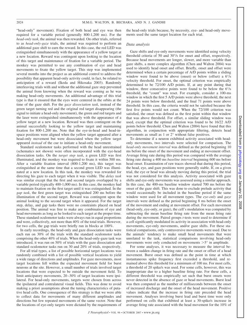
Head-movement-related activity was primarily assessed using aseries of eye-head dissociation tasks (Fig. 1). These began with theappearance of a red LED to which the monkey was asked to align thelaser spot within 3 s. Next, a green LED was illuminated at a secondlocation, and the animal was required to look to the new target withoutallowing the laser spot to move �5–7° away from the red LED(hereafter referred to as an “eye-only movement”). For some trial-types (Fig. 1A), the red target was extinguished and the green targetchanged to red. Because the animal was required to continue lookingat this target after the color change, completion of this stage requireda head movement without a change in gaze (hereafter referred to as a
Pos
ition
(deg
)
Time (msec)
Gaze
Head
Eye
100 200 300 400 0 100 200 300 4000
0
100 200 300 4000
A
B
Pos
ition
(deg
)
Gaze
HeadEye
Time (msec)100 200 300 400 0 100 200 300 400 5000
0
n = 9
n = 15
FIG. 1. Schematic representation of the“head-only” and “head-only-gaze” tasks (A)and the gaze task (B). In A, the arrows depict2 different possibilities, a reward (depictedby the drop of water) or the appearance of ayellow target at a new location, resulting inthe “head only” and “head-only-gaze” tasks,respectively. A miniature laser (yellowoval), mounted on the head, produces a redspot on a light-emitting diode (LED) board.Green lines indicate the animal’s line ofsight (gaze). The dashed red line indicatesthat the laser may be either on or off at thisstage of the task. Below the schematic rep-resentations of the tasks, eye (blue), head(red), and gaze (green) position traces areshown as a function of time. In each trace,100 ms are shown before the onset of themovement.
2023SUPERIOR COLLICULUS AND HEAD MOVEMENTS
J Neurophysiol • VOL 98 • OCTOBER 2007 • www.jn.org
“head-only” movement). Fixation of both head and eye was thenrequired for a variable period (generally 800-1,200 ms). For thehead-only task, the animal was then rewarded. On other trials, referredto as head-only-gaze trials, the animal was required to perform anadditional gaze shift to earn the reward. In this case, the red LED wasextinguished simultaneously with the appearance of a yellow target ata new location. Reward was contingent upon looking to the locationof this target and maintenance of fixation for a variable period. Themonkey was permitted to use any combination of eye and headmovements to fixate the yellow target. This step was implementedseveral months into the project as an additional control to address thepossibility that apparent head-only activity could, in fact, be related toanticipation of a reward (Ikeda and Hikosaka 2003). Randomlyinterleaving trials with and without the additional gaze step preventedthe animal from knowing when the reward was coming as he wasmaking head-only movements. An additional advantage of this trialtype is that it ensured that the eyes were centered in the orbits at thetime of the gaze shift. For the gaze dissociation task, instead of thegreen target turning red and the original red target disappearing (thesignal to initiate a head-only movement), the green and red targets andthe laser were extinguished simultaneously with the appearance of ayellow target at a new location. Reward was then contingent on theanimal successfully looking to the yellow target and maintainingfixation for 800-1,200 ms. Note that the eye-in-head and head-in-space positions were aligned when the yellow target appeared after ahead-only movement but were dissociated when the yellow targetappeared instead of the cue to initiate a head-only movement.
Standard oculomotor tasks performed with the head unrestrained(schematics not shown) were often incorporated with the eye-headdissociation tasks. On the target step task, a green LED was firstilluminated, and the monkey was required to fixate it within 500 ms.After a variable fixation interval (800-1,200 ms), this target wasextinguished at the same time that a second green LED was illumi-nated at a new location. In this task, the monkey was rewarded fordirecting his gaze to each target when it was visible. The delay taskwas similar except that the first and second targets overlapped by avariable period (typically 400-1,000 ms). In this case, the monkey hadto maintain fixation on the first target until it was extinguished. In thegap task, the first green target was extinguished 200 ms before theillumination of the second target. Reward was contingent upon theanimal looking to the second target when it appeared. For the targetstep, delay, and gap tasks there were no constraints placed on headposition. The animal was free to make any combination of eye andhead movements as long as he looked to each target at the proper time.These standard oculomotor tasks were always run in equal proportionsand were, collectively, not more than 40% of the total trials. However,for two cells, the gap trials were briefly run in blocks at 100%.
In early recordings, the head-only and gaze dissociation tasks wereeach run on 30% of the trials with the standard oculomotor taskscomprising the other 40% of trials. When the head-only-gaze task wasintroduced, it was run on 50% of trials with the gaze dissociation andstandard oculomotor tasks run on 30 and 20% of trials, respectively.
For all trial types, a list of possible horizontal target locations wasrandomly combined with a list of possible vertical locations to yielda wide range of directions and amplitudes. For gaze movements, mosttarget locations fell within the expected movement field for burstneurons at the site. However, a small number of trials involved targetlocations that were expected to be outside the movement field. Tolimit anticipatory movements, 20–30% of target locations were ipsi-lateral. For head-only movements, targets were chosen from acrossthe ipsilateral and contralateral visual fields. This was done to avoidmaking a priori assumptions about the tuning characteristics of puta-tive head cells. One consequence of this strategy is that we were ableto collect data for movements of many different amplitudes anddirections but few repeated movements of the same vector. Note thatthe locations of eye-only targets were dictated by the requirements of
the head-only trials because, by necessity, eye- and head-only move-ments used the same target location for each trial.
Data analysis
Gaze shifts and eye-only movements were identified using velocitythreshold criteria of 50 and 30°/s for onset and offset, respectively.Because head movements are longer, slower, and more variable thangaze shifts, a more complex algorithm (Chen and Walton 2004) wasused to identify head onset and offset. Briefly, onset and offset weredetermined when a certain percentage of A/D points within a slidingwindow were found to be above (onset) or below (offset) a 6°/svelocity threshold. For onset, the optimal criterion was empiricallydetermined to be 72/100 A/D points. If, at any point during thatwindow, three consecutive points were found to be below the 6°/sthreshold, the “count” was reset. For example, consider a 100-mswindow in which the first 5 A/D points were above threshold, the next24 points were below threshold, and the final 71 points were abovethreshold. In this case, the criteria would not be satisfied because thefirst five points would not count. When the 72/100 criterion wassatisfied, the onset was defined as the first point within that windowthat was above threshold. For offset, a similar sliding window wasused, except that the optimal criterion was found to be 16/22 A/Dpoints below threshold. Chen and Walton (2004) have found that thisalgorithm, in conjunction with appropriate filtering, detects headmovements as small as 1 or 2° without false positives.
To determine if a neuron exhibited activity associated with head-only movements, two intervals were selected for comparison. Thehead-only movement interval was defined as the period beginning 10ms before the onset of the movement and ending at movement offset.Average firing rate during this period was compared with the averagefiring rate during a 400 ms baseline interval beginning 600 ms beforehead onset. Examination of raw traces showed that during this period,the head, eye, and gaze positions were generally stable. If, on a giventrial, the eye or head was already moving during this period, the trialwas not considered for this analysis. Activity associated with gazeshifts and eye-only movements was assessed using a similar approach.In this case, the 400-ms baseline window started 700 ms before theonset of the gaze shift. This was done to exclude prelude activity thatwould occasionally be included in the baseline period if the samewindow were used for all movement types. The eye-only and gazeintervals were defined as the period beginning 8 ms before the onsetof the movement and ending at movement offset. For each movement(head-only, eye-only, or gaze), the cell’s modulation was computed bysubtracting the mean baseline firing rate from the mean firing rateduring the movement. Paired groups t-tests were used to determine ifa cell significantly modulated its activity in association with head-onlymovements, eye-only movements, and/or gaze shifts. For these sta-tistical comparisons, only contraversive movements were used. Due tothe animals’ tendency to make small head movements that wereunrelated to the task, statistical comparisons involving head-onlymovements were only conducted on movements �5° in amplitude.
For some analyses, it was necessary to measure the interval be-tween a sudden change in firing rate and the onset or offset of a headmovement. Burst onset was defined as the point in time at whichinstantaneous spike frequency first exceeded a threshold, and re-mained above this threshold for a minimum of 100 ms. For most cells,a threshold of 20 spike/s was used. For some cells, however, this wasinappropriate due to a higher baseline firing rate. For these cells, adifferent threshold was empirically set such that burst onsets werenever detected in the absence of gaze or head movements. Burst leadwas then computed as the number of milliseconds between the onsetof increased discharge and the onset of the head movement. Positivevalues indicate that the increase in firing rate preceded the headmovement. Analyses involving burst lead and burst time were onlyperformed on cells that exhibited at least a 30-spike/s increase inaverage firing rate associated with the head movement for the 10% of
2024 M.M.G. WALTON, B. BECHARA, AND N. J. GANDHI
J Neurophysiol • VOL 98 • OCTOBER 2007 • www.jn.org
trials with the largest modulation. Pause onset was defined as the pointin time at which instantaneous spike frequency dropped below 10spike/s and remained below this threshold for a minimum of 100 ms.For a trial to be considered, the instantaneous firing rate over theprevious 200 ms had to be consistently in excess of 30 spike/s. Pauselead was defined as the number of milliseconds between pause onsetand head onset. Pause offset was defined as the point in time at whichinstantaneous spike frequency rose to a threshold of 20 spike/s for�50 ms.
An inverse distance interpolation algorithm (Sigmaplot 2004, Sys-tat Software) was used to construct filled contour plots with neuronalactivity (number of spikes or average firing rate) plotted as a functionof horizontal and vertical amplitude (head or gaze). These plots wereused to gain insight into the general tuning characteristics of each cell.
R E S U L T S
Sufficient data were obtained from 210 neurons to charac-terize their discharge properties with respect to head move-ments. Neurons were identified as possible head cells if theysignificantly modulated their activity in association with head-only movements, and this modulation could not be moreparsimoniously explained by variables other than head move-ments (n � 56; see “additional controls” in the following text).In the subsections that follow, the basic discharge characteris-tics of head cells are first described followed by an evaluationof their tuning characteristics during head-only movements,eye-only movements, gaze shifts, and the head component ofgaze shifts. The next section examines the discharge charac-teristics of high-frequency gaze-related burst neurons in asso-ciation with head-only movements. Next, the modulation of allcells during gaze shifts, eye-only movements, and head-onlymovements are summarized and considered as a function oftheir locations within the SC. Finally, potential alternativeexplanations for the present results are considered.
Modulation of collicular activity during head-onlymovements: basic characteristics
The majority (n � 39; 70%) of these 56 neurons werecharacterized by increased discharge rate (typically 50–200
spike/s) during head movements and a lower firing rate (any-where from 0 to 100 spike/s) when the head was stable. Afterthe end of the head movement, the activity returned towardbaseline. Many were completely quiescent when the head wasnot moving. Raster plots, averaged spike density functions, andcorresponding movement waveforms are shown for one exam-ple neuron during head-only movements (Fig. 2A), coordinatedeye-head movements (Fig. 2B), and eye-only movements (Fig.2C). Head, eye, and gaze positions are plotted as a function oftime by red, blue, and green traces, respectively. Note that theincreased discharge precedes head onset in A and B. Also inthese panels, the cell can be seen to discharge at a very low rateduring the low-velocity head drifts near the beginning of theplot, then activity ceases when the head is completely stable.Finally, the firing rate dramatically increases in associationwith the higher velocity, visually guided head movement. Afterthe offset of these head movements, firing rate declines butdoes not always return to baseline because the head oftendrifted at subthreshold velocities after the end of the move-ment. For head-only movements (A), these often occurred asthe monkey made small adjustments to head position after theend of the primary head movement to more precisely align thelaser spot with the red LED.
For some of these cells, upward activity modulation wassometimes observed in association with the gaze shift itself, inaddition to the activity accompanying the head movement,although the gaze-related modulation was much weaker thanthe high-frequency bursts that characterize gaze-related burstneurons. This can be seen from the spike density function inFig. 2B. Activity was also observed in association with eye-only movements (C), but this result is difficult to interpretbecause it is likely that some head cells discharge in associa-tion with such movements.
Aligning the head-only data on head movement offset(Fig. 3A) or eye-only offset (Fig. 3C) had little effect other thana shift of the averaged spike density function because similarvector movements generally had highly similar durations. InFig. 2B, data are synchronized on gaze onset so that the readercan see the weak bursts that often accompanied gaze shifts. In
-800 -600 -400 -200 0 200 400 600 800 1000
Time (ms)
Hor
izon
tal
Am
plitu
de (
deg)
-800 -600 -400 -200 0 200 400 600 800 1000
Time (ms)
0
20
40
60
Spi
ke D
ensi
ty (
sps)
-20
0
20
-20
0
20
Ver
tical
Am
plitu
de (
deg)
A B
n = 10n = 14
-800 -600 -400 -200 0 200 400 600 800 1000
Time (ms)
C
Gaze, Head, Eye
n = 13Cell WL10140503
FIG. 2. Example data for 1 cell that increased its firing rate in association with head movements. Eye, head, and gaze position traces, raster plots, and averagedspike density functions are shown for head-only movements (A), gaze shifts (B), and eye-only movements (C). Data are aligned on movement onset, indicatedby the vertical dashed lines. Note that the cell begins to discharge before the onset of the head movement in both A and B. Same color conventions used for Fig. 1.
2025SUPERIOR COLLICULUS AND HEAD MOVEMENTS
J Neurophysiol • VOL 98 • OCTOBER 2007 • www.jn.org
Fig. 3B, data are synchronized on the offset of the headmovement associated with the gaze shift so that the reader cansee what this cell does after the end of the head movement.Surprisingly, the spike density function shows no obviousdecline after the offset of the movement. This is due to twofactors. First, it should be remembered that there were noconstraints on head position when the animals were makingeye-head gaze shifts or after these movements. Thus low-velocity head drifts inevitably occurred on some trials after thecoordinated eye-head gaze shifts were over. These drifts werenot necessarily in the same direction as the measured headmovement and usually occurred after a brief (50–200 ms)period of no movement. Second, the animals were rewarded800-1,200 ms after the animal fixated the target. Thus theposition traces in B include large gaze shifts the animalsperformed after the end of the trial. A close inspection of therasters for individual trials in B reveals some trials in whichdischarge continued beyond the measured head offset and thendecreased and others in which the firing rate declined forseveral hundred milliseconds and then increased again. Theformer correspond to trials in which head drifts occurred afterthe offset of the measured head movement and the latterrepresent increased discharge associated with the large gazeshifts visible on the right side of the plot.
We also isolated neurons that were characterized by thefollowing properties: tonic discharge when the head was notmoving, an abrupt decrease in firing rate—often a completecessation of discharge—that was temporally associated withhead-only movements, and resumption (or increase) in firingrate around the time of head-only movement offset. Low-velocity head drifts were generally associated with decreasedfiring rate or even cessation of discharge. Data are shown forone example neuron in Fig. 4. In A, the firing rate can be seento increase as the low-velocity head drifts slow down and endprior to the visually guided head-only movement. Then thefiring rate abruptly decreases shortly before the visually guidedhead-only movement begins. The averaged spike density func-tion gives the impression that resumption was very gradual buta close inspection of the rasters for individual trials shows that
the cell often returned to baseline firing rate over a period of50–100 ms (looked at over the longer time scales of headmovements, this seems “abrupt,” and this is what is meant bythis term in this manuscript). These “head pauser” cells (n �17; 30% of 56 neurons) also paused in association with gazeshifts (Fig. 4B) and sometimes eye-only movements (C). How-ever, only one such cell was recorded in the rostral portion ofSC and satisfied all of the criteria introduced by Munoz andWurtz (1993) for classification as a fixation cell.
As with Fig. 3, aligning the data on movement offset hadlittle effect on the averaged spike density functions for head-only (Fig. 5A) and eye-only data (C). It is important to note,however, that there was only a loose temporal associationbetween head movement offset and pause end (A). This may bedue, in part, to subthreshold drifts that occurred shortly afterthe end of the measured head movement. In A, these drifts areparticularly apparent in the vertical head position traces. In B,data are aligned on head movement offset. The firing rateremained at a low rate after the end of the measured headmovement, even when the head was stable.
Spatial tuning of putative head cells
Most putative head cells modulated their discharge for awide range of movements across the contralateral hemifield;few had circumscribed movement fields within the limit ofhead movements we were able to measure. Figure 6A shows amovement field plot for one typical cell. Although the trial-to-trial variability is high, the average firing rate for this cell isclearly higher for down-right head-only movements than formovements in other directions. To reveal structure that may beobscured by this variability, raw movement field plots wereconverted into filled contour plots using an inverse-distancebased smoothing algorithm in Sigmaplot. Panel B shows anexample, computed from the raw data points shown in A.
Cells that modulated their activity in association with head-only movements displayed a variety of tuning profiles. Mostincreased their firing rate monotonically with amplitude, al-though this relation often saturated for larger head movements.
-750 -500 -250 0 250 500 750 1000
Time (ms)
A
-20
0
20
Hor
izon
tal
Am
plitu
de (
deg)
-20
0
20
Ver
tical
Am
plitu
de (
deg)
0
20
40
60
-750 -500 -250 0 250 500 750 1000Time (ms)
B
-750 -500 -250 0 250 500 750 1000Time (ms)
C
Spi
ke D
ensi
ty (
sps)
FIG. 3. Example data for the same cell shown in Fig. 2. Data are aligned on head movement offset (A and B) or eye-only offset (C). Same format as Fig. 2.
2026 M.M.G. WALTON, B. BECHARA, AND N. J. GANDHI
J Neurophysiol • VOL 98 • OCTOBER 2007 • www.jn.org
However, varying degrees of directional biases were alsoobserved, ranging from weak to quite strong. This can beappreciated from Fig. 7, which shows filled contour plots forthree cells that displayed robust increases in discharge associ-ated with head-only movements. This heterogeneity meant thatthere was no single curve fitting approach that was appropriatefor every cell. Some cells (for example, see A) were bestdescribed by fitting a quadratic equation relating firing ratemodulation to vectorial amplitude, regardless of direction. Thequadratic fits were used because this relation tended to saturatefor large head movements in many cells. Other neurons (B)were better described by fitting a plane to the horizontalamplitude, vertical amplitude, and firing rate modulation data.
All 56 putative head cells were analyzed using both of theseapproaches. The results were not strongly affected by whetherthe modulation was positive or negative for head movements.The quadratic fits produced significant correlations for 25/56cells (44.6%). Next, planar fits were performed with andwithout an interaction term, as follows: FRmod � y0 � aX �bY and FRmod � y0 � aX � bY � cXY. where FRmod is thepredicted modulation of the cells firing rate, X is the horizontalhead displacement, and Y is the vertical head displacement.The planar model significantly accounted for the firing ratemodulation for 26/56 cells (46.4%). Eight cells showed signif-icant correlations for the quadratic fits but not for the planarfits. Average R2 values were 0.08 � 0.07 (SD) and 0.10 � 0.10
-40
-20
0
20
40
Hor
izon
tal
Am
plitu
de (
deg)
-20
-10
0
10
20
Ver
tical
Am
plitu
de (
deg)
n = 8
A B C
0
25
50
75
100
n = 6 n = 7
Gaze, Head, Eye
-750 -500 -250 0 250 500 750 1000
Time (ms)
-750 -500 -250 0 250 500 750 1000Time (ms)
-750 -500 -250 0 250 500 750 1000
Time (ms)
Spi
ke D
ensi
ty (
sps)
Cell BN10130603
FIG. 4. Example data for 1 cell that decreased its firing rate in association with head movements. Data are aligned on movement onset. Same format as Fig.2. Note that, prior to the head movement, the firing rate abruptly decreased to near 0.
-750 -500 -250 0 250 500 750 1000
Time (ms)
A
-750 -500 -250 0 250 500 750 1000Time (ms)
B C
-40
-20
20
40
Hor
izon
tal
Am
plitu
de (
deg)
-20
-10
10
20
Ver
tical
Am
plitu
de (
deg)
0
25
50
75
100
0
0
-750 -500 -250 0 250 500 750 1000Time (ms)
Spi
ke D
ensi
ty (
sps)
FIG. 5. Example data, aligned on movement offset. Same format as Fig. 3. At approximately the time of head offset, the firing rate abruptly returned tobaseline. On gaze trials, the neuron did not always return to baseline after the end of the head movement associated with the gaze shift.
2027SUPERIOR COLLICULUS AND HEAD MOVEMENTS
J Neurophysiol • VOL 98 • OCTOBER 2007 • www.jn.org
for the quadratic and planar fits, respectively. For the plane fits,average coefficients for horizontal and vertical amplitudeswere 0.15 � 0.50 and 0.05 � 0.06, respectively. Including aninteraction term improved the fit somewhat, yielding an aver-age R2 of 0.12 � 0.10 but only 25/56 were significant. Averagecoefficients for horizontal amplitude, vertical amplitude, andthe interaction term were 0.06 � 0.35, �0.16 � 0.78, and0.001 � 0.06, respectively.
These analyses were also conducted using the mean firingrate during the head movement instead of the average modu-lation. This approach resulted in slightly worse fits for thequadratic equation (R2 mean � 0.07 � 0.06) but improved theplanar fits. Average R2 values were 0.13 � 0.11 and 0.15 �0.12 with and without the interaction term, respectively. Av-erage coefficients for horizontal and vertical amplitudes were0.05 � 0.34 and �0.06 � 0.65, respectively, without theinteraction term and 0.06 � 0.35, �0.08 � 0.84, and 0.001 �0.06, respectively, when the interaction term was used. Usingthis approach, the distribution of average firing rate was sta-tistically accounted for in 34/56 neurons by the planar modelwithout the interaction term, in 33/56 by the planar model withthe interaction term, and in 25/56 by the quadratic fit.
For some cells, the timing of discharge was predictive ofhead displacement. To assess this relationship, burst lead wascomputed as the interval (in ms) between the onset of activityand the onset of the head movement. This analysis could onlybe performed on cells that strongly increased their activity inassociation with head movements and that displayed a fairlyabrupt onset of activity on a majority of trials (see METHODS formore detail). Ten cells met these criteria and, of these, burstlead was significantly related to head displacement for 8/10(80%). Two of these cells showed more complex effects.Figure 8 shows the relationship between burst lead and headdisplacement for these two cells. The data were fit with afour-parameter Gaussian
y � y0 � ae�� 0.5� x � x0
b� 2�
For the cell shown in the top row (which is the same cell shownin Figs. 2 and 3), burst lead was strongly predictive of theamplitude of the vertical component (R2 � 0.44) when theinitial vertical head position was 10° up (A). Burst onset ledhead onset for horizontal movements and those that wereslightly down. However, for upward movements, burst onset
0
5
10
15
20
25
30
35
40
45
50
-30 -20 -10 0 10 20 30
Horizontal Head Amplitude (deg)
-30
-20
-10
0
10
20
30
Ver
tical
Hea
d A
mpl
itude
(de
g)
-20 -10 0 10 20 30-30
-20
-10
0
10
20
0 5 10 15 20 25 30
Ave Firing Rate (spikes/sec)
A B
Cell WL10140503
FIG. 6. Movement field plots for an examplehead cell. Each data point represents a singlemovement. A: average firing rate during themovement is plotted as a function of horizontaland vertical head-only amplitude. B: filled con-tour plot constructed from the raw data pointsshown in A. For this example cell, vectorial headdisplacement seems to be predictive of firingrate.
Horizontal Head Amplitude
-20 -15 -10 -5 10 15 20
Horizontal Head Amplitude
-20 -10 00 5 10 20
Horizontal Head Amplitude
-20 -10 0 10 20
Ver
tical
Hea
d A
mpl
itude
-20
-10
0
10
20
0 5 10 15 2025304045
C
WL02200604
Ver
tical
Hea
d A
mpl
itude
-20
-15
-10
-5
0
5
10
15
20
15 20 25 30 35 40
A
BN05250604
Ver
tical
Hea
d A
mpl
itude
-20
-10
0
10
20
18 20 22 24 26 28 30 32
B WL02010603
FIG. 7. Filled contour plot movement fields for 3 example head cells. Putative head cells had highly heterogeneous tuning profiles with some more sensitiveto amplitude than direction (A), some more sensitive to direction (B), and some showing movement fields similar to those for gaze cells in superior colliculus(C).
2028 M.M.G. WALTON, B. BECHARA, AND N. J. GANDHI
J Neurophysiol • VOL 98 • OCTOBER 2007 • www.jn.org
generally lagged head onset. This relationship, however, dis-appeared when the same analysis was repeated for trials inwhich the vertical initial head position was 20° up (R2 � 0.05;B). Similarly, for the cell shown in the bottom row, burst leadwas predictive of horizontal head displacement (R2 � 0.24)when the vertical initial head position was 20° up (D), yet norelationship was found when the head started at 10° up (R2 �0.04; C).
For putative head cells that decreased their firing rate forhead-only movements (n � 17), analyses involving pauseduration or pause lead were only performed on cells with �15trials with clearly defined pauses (see METHODS). Seven putativehead cells met these criteria. Pause duration was significantlycorrelated with head movement duration for 2/7 cells. Norelationship was found between the timing of the resumption ofactivity and head displacement or the time of head offset. Thisnegative result may have been due to slow, subthreshold driftsafter the measured end of the movement. These cells oftendischarged at a substantially reduced frequency or were quies-cent during these slow drifts. For 4/7 cells, pause lead wassignificantly correlated with vertical and/or vectorial headdisplacement. Figure 9 shows this relationship for these cells.Note that, for three of the four cells, the correlation wasnegative with the cell pausing earlier for small head move-ments than for large ones.
Comparison of movement fields
Next, we compared each putative head cell’s movement fieldresponse during head-only movements, gaze shifts, and the
Ver
t Bur
st L
ead
(mse
c)
-20 -10 0 10 20-400
-200
0
200
400
600
Hor
Bur
st L
ead
(mse
c)
Hor Head Amplitude (deg)
Ver head Amplitude (deg)
A
C D
-20 -10 0 10 20-400
-200
0
200
400
600
-25 -20 -15 -10 -5 0 5 10 15
-400
-300
-200
-100
0
100
200
300
400
500
B
-25 -20 -15 -10 -5 0 5 10 15
-400
-300
-200
-100
0
100
200
300
400
500
BN05250604
WL10140503
FIG. 8. Relationship between burst timing and head dis-placement. Data in all panels are fitted with 4 parameter Gauss-ian functions. Top and bottom: data from 2 different cells. Leftand right: data from trials in which the vertical initial headposition was 10° up or 20° up, respectively. A and B: verticalburst lead (see text) was highly predictive of vertical headdisplacement for this example neuron when the head started at10° up (A) but not when it started at 20° up (B). C and D: for thiscell, horizontal burst lead was highly predictive of horizontalhead displacement when the initial head position was 20° up (D)but not when it was 10° up (C).
WL10170502
-100
-50
0
50
100
150
200
250
300WL10200505
-300
-200
-100
0
100
200
0 5 10 15 20 25 -20 -10 0 10 20 30
Head Amplitude
Pau
se L
ead
Vertical Head Amplitude
Ver
tical
Pau
se L
ead
= 0.30R2R2 = 0.39
BN10130602
Head Amplitude
Pau
se L
ead
-200
-100
0
100
200
300
4 6 8 10 12 14 16 18 20
R2 = 0.24
BN10130603
Vertical Head Amplitude
Ver
tical
Pau
se L
ead
-200
-100
0
100
200
300
400
-20 -15 -10 -5 0 5 10
R2= 0.13
A B
C D
FIG. 9. The relationship between pause lead (see text) and head displace-ment is shown for 4 cells that paused in association with head-only move-ments. Left: pause lead is correlated with vectorial head displacement. Right:vertical pause lead is correlated with vertical head displacement.
2029SUPERIOR COLLICULUS AND HEAD MOVEMENTS
J Neurophysiol • VOL 98 • OCTOBER 2007 • www.jn.org
head component of gaze shifts. Figure 10 illustrates this com-parison for one neuron, and it also demonstrates the difficultiesencountered in performing a robust quantitative analysis. Thehead-only movement field (A) shows, despite the considerabletrial-to-trial variability, clearly higher activity for downwardhead-only movements than for movements in other directions.B shows a movement field for head movements associated withgaze shifts. Activity may appear weaker than that shown in A,but this is difficult to interpret because these movements wereprimarily horizontal. C shows modulation associated with gazeshifts (i.e., the same movements used in B) and eye-onlymovements (not shown in B). Most of the gaze data recordedfor this cell involved large-amplitude movements because burstneurons recorded on this track had movement fields centerednear (40, �40). Eye-only data were therefore included in C toshow how this cell responded for small-amplitude movements(that would normally be accomplished with eye-only move-ments anyway). This cell showed little modulation for gazeshifts, even when they were directed downward, and no evi-dence of a gaze movement field.
We sought to perform a comparison between head-onlymovements and head movements associated with gaze shifts.Our monkeys were trained to make head-only movements in alldirections, up to an amplitude of �30°. Head movementsassociated with gaze shifts, however, tend to be mostly hori-zontal, even if the gaze shift itself has a strong verticalcomponent (Fig. 10B) (Freedman and Sparks 1997b). Weconsidered restricting the comparison to horizontal head move-ments but, given our current lack of understanding of theneurophysiological basis of eye-head coordination, wecouldn’t assume that the head movement that accompanies agaze shift is the same as the one requested by SC, a reflectionof the neural uncertainty problem (Sparks 1999). For thesereasons, we did not pursue any statistical comparisons of firingrate between the two different types of head movements.
We also attempted to perform direct statistical comparisonof the movement fields associated with head-only movementsand gaze shifts. If a topographic map existed for head-onlymovements, and, furthermore, if it was in spatial register withthe topographic map known for gaze shifts, we could havecompared the responses for movements of the same “hot-spot”vector. This approach, however, was quickly deemed futile aswe found no topography for head movements and no hot-spotvector for head-only movements. In fact, the heterogenoushead-only movement fields rarely resembled the circumscribedmovement fields associated with gaze shifts. In the end, weopted for a generalized comparison of modulation in activityfor head-only movements and gaze shifts across contralateralmovements. This analysis is presented later, after the charac-terization of classic gaze-related burst neurons for head-onlymovements.
Do classic gaze-related burst neurons modulate inassociation with head movements?
The data presented so far indicate that the SC contains cellsthat modulate their response in association with head-onlymovements as well as during gaze shifts but that the dischargeduring gaze shifts is not the classic high-frequency burst thatcharacterizes many SC neurons (quantitative details to beprovided in the following text). Thus the next question to beaddressed was whether neurons displaying high-frequencygaze-related bursts also discharged in association with headmovements. In general, cells displaying high-frequency burstsassociated with gaze shifts fired weakly—usually not at all—for head-only movements. Rasters and averaged spike densityfunctions are shown in Fig. 11 for one example neuron, alongwith eye, head, and gaze position traces. On head unrestrainedgap trials (B), this cell displayed clear prelude activity and ahigh-frequency burst of spikes associated with the gaze shift.The bottom inset shows the mean spike density for the samegap trials aligned on target onset and emphasizes the buildup
0.0
5.8
11.6
17.4
23.2
29.0
34.8
40.6
46.4
52.2
58.0
Ave Firing Rate (spikes/sec)
-40 -20 0 20 40
Hor Head Amplitude (deg)
-40 -20 0 20 40
Hor Head Amplitude (deg)
Head movements associated with Gaze
-50
-40
-30
-20
-10
0
10
20
30
40
50
Ver
t Hea
d A
mpl
itude
(de
g)
B
-40 -20 0 20 40
Hor Gaze Amplitude (deg)
C
Gaze/eye-only
-50
-40
-30
-20
-10
0
10
20
30
40
50
Ver
t Gaz
e A
mpl
itude
(de
g)
-50
-40
-30
-20
-10
0
10
20
30
40
50
Ver
t Hea
d A
mpl
itude
(de
g)A
Head only
WL02010603
FIG. 10. Movement fields for 1 putative head cell. A: average firing rate plotted as a function of horizontal and vertical head-only amplitude. Although muchtrial to trial variability in firing rate is apparent, the cell clearly discharges at a higher rate for downward head movements. B: average firing rate plotted as afunction of horizontal and vertical amplitude of head movements associated with gaze shifts. When contributing to a gaze shift, head movements were mostlyhorizontal, even if the gaze shift had a substantial vertical component. Nonetheless, the cell was clearly active for head movements of all directions thataccompany gaze shifts (i.e., there are very few blue points in this panel). C: average firing rate plotted as a function of horizontal and vertical gaze amplitude.Although the cell does discharge at a low rate during gaze shifts, there was no indication of organization or tuning. Data points shown in this panel include trialsfrom small amplitude eye-only movements and larger amplitude gaze shifts.
2030 M.M.G. WALTON, B. BECHARA, AND N. J. GANDHI
J Neurophysiol • VOL 98 • OCTOBER 2007 • www.jn.org
response (arrow). On head-only trials (A), this cell dischargedat low frequency, but this activity was nowhere near as robustas either the prelude or burst associated with gaze shifts. Manycells, particularly classic gaze-related burst neurons, did notdischarge at all during head-only movements. This can be seenin Fig. 12, which shows movement field plots for one cell forgaze shifts (A) and head-only movements (B). This cell wasrecorded at a relatively rostral site in the right SC and preferredsmall gaze shifts (�8, �5). There was little or no dischargeassociated with head only movements. To emphasize thispoint, average firing rates for head-only movements are plotted
on a different scale (Fig. 12B). Indeed, classic gaze-relatedburst neurons (i.e., no prelude activity) never significantlymodulated their activity in association with head-only move-ments (see following text for more details).
Comparison of modulation for gaze andhead-only movements
Two analyses were conducted across all neurons to comparethe level of modulation for head-only movements and gazeshifts and between head- and eye-only movements. The objec-
-250 0 250 500 750 1000
Time (ms)-500
Cell WL09260501
A B
Time (ms)
0
20
40
0
200
300
100
-40
-20
0
20
-40
-20
Hor
izon
tal
Pos
ition
(de
g)S
pike
Den
sity
(sp
s)V
ertic
alP
ositi
on (
deg)
-250 0 250 500 750 1000
Time (ms)-500
Gaze, Head, Eye
00S
pike
Den
sity
(sp
s)
n = 15 n = 18
FIG. 11. Example data for a high-frequency gaze related burst cell. Same format as Fig. 2. On gap trials (B), the cell displays a classic buildup of activityprior to the gaze shift. Bottom inset: averaged spike density function for the same trials, aligned on target onset (gap duration � 200 ms). The x and y scalesfor the inset are the same as for the outer panel. The offset of the fixation target and the onset of the gaze target are denoted by vertical solid and dashed lines,respectively. The arrow shows buildup activity. On head-only trials (A), the cell discharged at a very low rate, but did not show the robust burst of activity thatwas characteristic of gaze shifts into its response field.
0
20
41
61
82
103
123
144
164
185
206
-30 -20 -10 0 10 20 30-30
-20
-10
0
10
20
30
-30 -20 -10 0 10 20 30
-30
-20
-10
0
10
20
30
Hor Gaze Amplitude (deg)
Ver
t Gaz
e A
mpl
itude
(de
g)
Ave Firing Rate (spikes/sec)
Hor Head Amplitude (deg)
Ver
t Hea
d A
mpl
itude
(de
g)
A B
0
1
2
3
4
5
6
7
8
9
10
Ave Firing Rate (spikes/sec)
WL10200502
FIG. 12. Movement field plots for a gazerelated, high-frequency burst neuron. A: av-erage firing rate during gaze shifts, plotted asa function of horizontal and vertical gazeamplitude. B: average firing rate during head-only movements, plotted as a function ofhorizontal and vertical head displacement. Ingeneral, this cell was completely quiescentduring head-only trials. To emphasize thispoint, the average firing rate is plotted on adifferent scale in B.
2031SUPERIOR COLLICULUS AND HEAD MOVEMENTS
J Neurophysiol • VOL 98 • OCTOBER 2007 • www.jn.org
tives were to determine if the strength of modulation is differ-ent for these movement types and whether different or over-lapping populations of SC neurons showed significant changein activity for eye-only/gaze movement and head-only move-ments.
In the first case, a modulation parameter was computed asthe difference in activity during the appropriate movementinterval and its baseline periods, yielding “gaze modulation” or“eye-only modulation” and/or “head-only modulation” valuesfor each trial. A paired t-test was applied to the distribution ofvalues in the two intervals for each type of movement directedinto the contralateral field (see following text). This proceduredetermined whether a given neuron showed statistically signif-icant modulation for just head-only movements, eye move-ments (eye-only or gaze) only, both types of movements, orneither. Figure 13A plots each neuron’s mean modulationduring gaze shifts against its mean modulation during head-only movements. The outcome of the statistical test was usedto classify each cell as exhibiting significant sensitivity to gaze(red circles), head (blue squares), both (green diamonds), orneither (black stars). As mentioned previously, classic gaze-related burst neurons never significantly modulated their dis-charge in association with head-only movements and thereforeconsistently fell close to the horizontal dashed lines in thisfigure. In contrast, it was common to find cells exhibiting gazeprelude activity and/or weak gaze-related bursts in the “both”category. Overall, the modulation associated with gaze shifts
was greater than the modulation associated with head-onlymovements for neurons in the “gaze” (naturally) and “both”categories (2-tailed t-test; Table 1). Similar results were ob-tained when comparing head-only to eye-only movements (C).
In the second analysis, a modulation index was computed as(mean firing rate during movement – baseline firing rate)/(mean firing rate during movement � baseline firing rate) foreye-only movements, head-only movements, and gaze shifts.Again, a paired t-test was performed as explained in thepreceding text. This resulted in slightly different percentages ofneurons in the four categories. Figure 13B compares themodulation indices of the neurons during gaze shifts andhead-only movements, and D compares the modulation indicesof neurons during eye-only and head-only movements.
In all panels of Fig. 13, the numbers of cells in the fourcategories do not add up to 210 because cells that significantlymodulated their activity in association with head-only move-ments are shown only if they were considered to be good headcell candidates (see Additional controls). Furthermore, it mustbe emphasized that these analyses were not restricted to trialsthat fell within the movement field of the cell (if any). Foreye-only and gaze, all contraversive trials were included; forhead-only movements, all contraversive movements over 5° inamplitude were used. This was done because cells displayinghead-movement-related activity usually were active for allcontraversive head movements. For head-movement-relatedactivity, averaging across the entire contralateral hemifield
32 Both42 Neither56 Gaze23 Head
Hea
d-O
nly
Mod
ulat
ion
(spi
kes/
sec)
-100 0 100 200-100
-50
0
50
Gaze Modulation (spikes/sec)
Hea
d-O
nly
Mod
ulat
ion
Inde
x
-1 -0.5 0 0.5 1
-1
-0.5
0
0.5
1
Gaze Modulation Index
A B
42 Both35 Neither62 Gaze14 Head
-50 0 50 100 150 200 250-60
-40
-20
0
20
40 24 Both50 Neither49 Eye31 Head
Hea
d-O
nly
Mod
ulat
ion
(spi
kes/
sec)
Eye Modulation (spikes/sec)-1 -0.5 0 0.5 1
-1
-0.5
0
0.5
1
30 Both55 Neither45 Eye24 Head
Hea
d-O
nly
Mod
ulat
ion
Inde
x
Eye Modulation Index
C D
FIG. 13. Comparison of modulation associated with gaze andeye-only movements to modulation associated with head-onlymovements. �, E, and � denote cells that significantly modulatedtheir activity in association with movements of the head, gaze, orboth, respectively. �, cells that did not significantly modulate foreither. A and B compare data for gaze and head-only movements.C and D compare data for eye-only and head-only movements. Aand C: change in firing rate during the movement in spike/s. B andD: modulation index (see text). Values of 0 indicate no modula-tion at all; values of 1 or �1 indicate that the cell fired exclusivelyduring or before the movement, respectively.
TABLE 1. Summary of modulation analyses
Category n Gaze Modulation Head Modulation N Gaze Modulation Index Head Modulation Index
Gaze modulation (red circles) is significantly different from head modulation (blue squares), P � 0.01 (2 tailed t-test). Gaze modulation (green diamonds) issignificantly different from head modulation (green diamonds), P � 0.05 (2-tailed t-test). Gaze modulation (blue squares) is not significantly different from headmodulation (blue squares), P � 0.1 (2-tailed t-test). Gaze modulation index (red circles) is NOT significantly different from head modulation index (bluesquares), P � 0.7 (2-tailed t-test). Gaze modulation index (green diamonds) is NOT significantly different from head modulation index (green diamonds), P �0.2 (2-tailed t-test). Gaze modulation index (blue squares) is significantly different from head modulation index (blue squares), P � 0.01 (2-tailed t-test). Valuesare means � SD.
2032 M.M.G. WALTON, B. BECHARA, AND N. J. GANDHI
J Neurophysiol • VOL 98 • OCTOBER 2007 • www.jn.org
appeared to more accurately represent the apparent head move-ment activity of these cells. An undesirable consequence wasthat the modulation associated with eye-only and gaze isunderestimated in this figure due to the inclusion of trialsoutside of the movement field. However, because saccade/gazerelated activity in SC has been well described in many studies,we felt that it was more important to accurately representneural activity potentially related to head movements. Restrict-ing these analyses to the 10% of trials with the highest firingrate produced more accurate estimates of eye-only and gaze-related activity, but the estimates for some head cells weremisleading because the mean was affected too strongly byoutliers. Overall, however, the figures were highly similarusing these two approaches (data not shown). This “top 10%”approach was also used to assess the gaze-related activity forthe 56 cells that showed significant head-movement-relatedactivity. When this was done, only 7/56 had gaze-related firingrates in excess of 100 spike/s for the 10% of trials with thehighest gaze-related firing rates. All seven of these cells dis-played prelude activity that began �100 ms before gaze onset,and all seven continued to discharge for �100 ms after gazeoffset. Spike density functions computed for the peri-gazeperiod never exceeded 500 spike/s for any of these seven cells.Thus classic gaze-related burst neurons and head-movementrelated cells represented two distinct, nonoverlapping popula-tions of cells.
We next examined whether the modulation was a function ofthe location of the neurons, in terms of both the SC map andthe depth from the SC surface. Cells displaying head-move-ment related activity were found throughout the mediolateraland rostrocaudal extent of SC (Fig. 14A). Typically, they werefound between 1 and 4 mm below the collicular surface withthe greatest concentration between 1 and 2 mm (Fig. 14B).Thus these cells were found intermingled between gaze-relatedneurons. Indeed, we often isolated gaze-related burst neuronsand head cells in the same penetration, �1 mm from eachother.
Additional controls
In our sample of neurons, we found that 87/210 (41%)significantly modulated their activity in association with head-only movements. However, 19/87 cells were rejected as headcell candidates despite significant modulation because themodulation associated with head-only movements was �10spike/s for the 10% of trials with the largest modulation. Forexample, some cells that were usually silent during head onlymovements occasionally discharged three or four spikes duringthese movements. Such cells often exhibited a significantmodulation only because they never discharged during thebaseline period. It would be difficult to argue that such neuronsare head cells. In addition, 12/87 cells were rejected as headcell candidates because the modulation was more parsimoni-ously explained as microsaccade or eye position effects (seefollowing text for details). Thus 56 neurons were regarded asputative head cells.
It is possible that microsaccades may be more likely to occurduring head-only movements than when the head is stationary.If this is the case, a cell that bursts in association withmicrosaccades would show a significant modulation for head-only movements in our analysis. To test for this possibility,
saccade onset and offset velocity thresholds were modified(15°/s for onset and offset) to detect saccades of 0.5–2°. For allbut five cells, modulation of firing rate was confirmed in theabsence of microsaccades. These five cells clearly dischargedin association with microsaccades and were therefore elimi-nated as potential head cells.
Before interpreting the present results as evidence that SCcontains head-movement cells, it is necessary to verify that themodulation of activity in the present data are not due to cellscarrying eye-in-head signals. During head-only movements, ofcourse, the eyes counter-rotate in the orbits due to the VOR. Acell that encodes the positions of the eyes in the orbits wouldtherefore significantly modulate its activity in association withhead-only movements. To test this possibility, we performedregression analyses on the eye-in-head position during thebaseline period when the eyes and head were stable. The slopeof a linear regression line was found to be significantly differ-ent from zero for seven cells. These cells also were eliminatedas potential head-movement cells.
One must consider the possibility that apparent head move-ment activity might, in fact, be a vestibular signal. During ourhead-only movements, the VOR gain was approximately one.Thus changes in firing rate would be expected for any vestib-ular cell. To test this possibility, for four cells that showed
0 2 4 6
-2
0
2
2
510 20 50 100
Rostral-Caudal SC Location (mm)
Med
ial-L
ater
al S
C L
ocat
ion
(mm
)
0 2 4 6
4
3
2
1
0
Dep
th fr
om S
C s
urfa
ce (
mm
)
Rostral-Caudal SC Location (mm)
A
B
FIG. 14. Cells with head-movement related activity were found intermin-gled with gaze-related neurons across the rostrocaudal and mediolateral extentof SC, and were usually found 0.5–2.5 mm below the surface of SC. A:locations of recordings on the SC map. Shape conventions are the same usedfor Fig. 13. Colors indicate the mediolateral location of each cell on thecollicular map. B: estimated depth of each recording below the collicularsurface. Color conventions are retained from A.
2033SUPERIOR COLLICULUS AND HEAD MOVEMENTS
J Neurophysiol • VOL 98 • OCTOBER 2007 • www.jn.org
particularly robust modulation associated with head move-ments, the head was restrained and passive, whole-body rota-tions performed while the monkey fixated a target LED.Rotations were performed manually in the horizontal dimen-sion at speeds approximating the speed of typical head-onlymovements. None of the four cells showed any modulationassociated with whole-body rotations.
One must also consider the possibility that apparent head-movement related activity is a visual response. Potentially,visual responses might be observed in response to the laser spotsweeping across the visual field during head-only movements.To test this possibility, the laser spot was turned off during thehead only movement in some or all head-only trials once themonkeys had extensive experience with the tasks (generallyafter 3 or 4 mo). The laser was turned back on when the headpassed within 5° of the target. For all cells in which theselaser-blank trials were used, the presence or absence of thelaser spot was found to have no effect on firing rate.
D I S C U S S I O N
We have found 56 cells in SC that can be regarded as likelyhead cells. Some (n � 39) increased their firing rate inassociation with head movements, others (n � 17) decreased oreven paused. Cells exhibiting high-frequency bursts for gazeshifts generally showed little or no modulation associated withhead-only movements. However, many cells with significanthead-movement-related activity also exhibited lower-fre-quency bursts for gaze shifts. For many cells with significanthead-movement activity, average firing rate was predictive ofhead displacement, and many showed biases for movements ina particular direction. Furthermore, burst lead was predictive ofhead displacement for 8/10 cells for which a detailed analysiswas possible. For two of these cells, the presence of thisrelationship depended on the initial head position. The fact thatthe timing and level of activity of these cells is predictive ofhead-movement parameters supports the contention that thesecells carry information related to head movements.
At least two previous studies have reported cells in the catSC that may have been head cells. Straschill and Schick (1977)reported 15 neurons in cat SC that discharged before andduring head movements. The firing rate of these neurons wascorrelated with head velocity. Peck (1990) described fiveneurons in cat SC that were characterized by bursts of spikesthat were temporally associated with head movements. Neitherof these studies, however, employed a head-only task; thus it isdifficult to unequivocally state that the activity was related tohead movements and not gaze shifts. To our knowledge, thepresent work represents the first direct evidence for the exis-tence of head-only signals in the primate SC.
Potential alternative explanations
As mentioned in the preceding text, cells carrying vestibularsignals would be expected to alter their firing rate in associa-tion with head-only movements. However, this is unlikely to bethe case for the neurons in our sample for two reasons. First,increased discharge usually preceded the onset of the headmovement. Second, passive whole body rotation performed onseveral putative head cells did not produce changes in firingrate.
One might suggest the possibility that increased activityduring head movements might be a visual response to themoving laser spot. However, turning off the laser spot duringhead movements did not affect the activity of these cells duringthe movement. It should also be pointed out that the laser spotalways swept through the visual hemifield opposite to thedirection of the movement. During contraversive head-onlymovements, for example, the laser spot swept through theipsilateral hemifield. Therefore if the present results wereattributable to movement of the laser spot, the firing rate shouldhave been higher for ipsiversive movements but the oppositepattern was found.
It is also conceivable that the activity could be associatedwith movement of the visual target across the retina during thehead-only movement. This could occur in either of two ways.First, microsaccades might be more common during the headmovement. Such microsaccades might tend to provoke in-creased activity from visual cells. Increased activity associatedwith microsaccades has, indeed, been reported in primaryvisual cortex and lateral geniculate nucleus (Martinez-Conde etal. 2002). In the present data, however, there were many trialsin which head-only movements occurred in the absence ofdetectable microsaccades and robust discharge was often seenon these trials. Retinal slip would also occur during headmovements if the VOR gain is not one. This drift could,potentially, increase the activity of visual cells. However, ifthis was the explanation for our results, increased activityshould have been observed in response to passive, whole bodyrotation, but this was not the case. It should also be pointed outthat many of these cells were recorded from SC sites encodinglarge gaze shifts. Visual receptive fields at such sites would notbe expected to involve the parafoveal region of the visual field.Finally, retinal slip hypotheses would not explain why changesin discharge rate tended to precede the head movement.
One must also consider the possibility that apparent head-movement related activity is actually associated with covertsaccade planning (Sparks 1999). During the head-only move-ment, however, the only target that is consistently visible is onethat the animal is currently fixating. Nonetheless, it is notimpossible that the animals might be planning a saccade backto the remembered location of the now-extinguished centraltarget. However, this is unlikely to be the explanation for ourresults. The head cells we recorded generally displayed stron-ger activity associated with contraversive head movementsthan ipsiversive movements. If apparent head-movement re-lated activity were the result of covert planning of saccades tothe remembered location of the central target, the oppositepattern of activity should have been observed. In fact, thecovert planning hypothesis predicts that “head movementfields” should have been found that closely resemble gazemovement fields, only reversed (i.e., in the “ipsilateral” hemi-field). However, this was not the case. Furthermore, robustmodulation of discharge was often observed during spontane-ous head movements (including head-only movements) duringthe intertrial interval.
Cancellation of planned saccades is associated with sharpdecreases in activity in many SC neurons (Pare and Hanes2003). In our tasks, prior to the cue to initiate a head-onlymovement, the animal does not know whether he is going to beasked to make a head-only movement or a gaze shift. Conceiv-ably, during this period, the monkey might be anticipating, and
2034 M.M.G. WALTON, B. BECHARA, AND N. J. GANDHI
J Neurophysiol • VOL 98 • OCTOBER 2007 • www.jn.org
planning, a gaze shift that he then cancels when it becomesclear that it is a head-only trial. If this is the case, one might seeneural activity during the dissociation period followed by apause. Thus one must consider this as a possible alternativeexplanation for decreases in activity that temporally coincidewith head movements. However, it must be pointed out thattarget direction and amplitude were randomized. Gaze targetswere presented across a wide range of ipsi- and contralaterallocations. In addition, the dissociation period was always morelikely to be followed by a head-only movement than a gazeshift. For these reasons, we think it unlikely that the animalswere planning gaze shifts during the dissociation period. It isalso not clear why, if pauses in discharge were due to cancel-lation of planned saccades, the cells often abruptly startedfiring again around the time that the head movement ended.Finally, the fact that pause lead was often significantly corre-lated with head displacement suggests that decreases in activitywere related to the head movement itself.
Another possibility that must be considered is that apparenthead-movement-related activity is associated with microsac-cades. It is conceivable that microsaccades might be morecommon during head-only movements and that SC neuronsmight participate in the generation of these movements. (Notethat this is not the same as the above-mentioned possibility thatmicrosaccades may lead to visual responses by inducing retinalslip. The question here is whether or not SC activity specifi-cally related to microsaccades might give the false impressionof head-movement activity.) Indeed, five neurons were rejectedas head cell candidates for this reason. These cells were easilyidentified because robust correlations (r � 0.9) were foundbetween burst onset and microsaccade onset time. For most ofthe cells in our sample, robust changes in discharge were oftenobserved during head movements in the complete absence ofdetectable microsaccades. Microsaccades were also commonduring the dissociation period before head-only movements,and no activity was observed for such movements for mosthead cells. The microsaccade hypothesis also would not ex-plain the relationships between neural activity and head-move-ment parameters.
It is also possible that SC cells with reward-related activity(Ikeda and Hikosaka 2003) might increase their firing rateduring head-only movements if the animals come to expect areward immediately after these movements. However, for mostof our cells, one of our tasks required the animal to make acoordinated eye-head gaze shift after the head-only movement.Thus the animals should not have been expecting a reward atthis stage of the task. Even when the head-only movement wasnot followed by a required gaze shift, the animal still had tomaintain fixation for 800-1,200 ms to obtain the reward. Inaddition, one would not expect reward-related activity to returnto baseline after the head-only movement as it consistently didfor our head cells.
Finally, one must consider the possibility that apparenthead-movement-related activity might be the result of a task-specific cognitive parameter rather than head movements perse. For example, it might be supposed that the head-only tasksare, in a sense, more demanding than standard eye-movementtasks. If this is the case, then changes in discharge might beassociated with increased attention. This seems unlikely be-cause changes in firing rate were also observed in associationwith coordinated eye-head gaze shifts, which, presumably, are
not particularly difficult for the animal. Moreover, many ofthese cells were recorded in animals that had many months ofexperience with these tasks. Finally, for many of our putativehead cells, robust changes in activity were observed in asso-ciation with head movements during the intertrial intervalwhen the monkeys were, of course, completely off task (datanot shown).
Possible functional roles of head cells in SC
What role might SC cells play in head-movement control? Inview of the broad tuning of neurons in our sample, one must becautious about assigning an important functional role to thesecells. Nonetheless, it is well known that populations of broadlytuned neurons can carry far more information than the activityof any single neuron. It should also be emphasized that,although the correlation coefficients reported in the presentstudy would be quite low by the standards of most researchinvolving the oculomotor system, they are comparable to thosecommonly found in the skeletal motor literature (e.g., Church-land et al. 2006; Werner et al. 1997).
There are many possible roles that head cells in SC mightplay. At least two studies, (Corneil et al. 2002; Cowie andRobinson 1994) have reported that low-frequency, long-dura-tion stimulation evoked head-only movements from some SCsites. The authors of both of these studies suggested that SCmay carry a parallel “head-only” drive. It is possible that thehead-only movements that these authors reported might bemediated, at least in part, by cells of the same type reported inthe present study. Thus it is possible that SC plays a role in thecontrol of head movements independent of gaze shifts.
It is also possible that these cells might carry a corollarydischarge signal from SC to other structures (Sommer andWurtz 2004a,b). For example, accurate eye-head coordinationmay require information regarding the initial positions of theeyes in the orbits. Head-only movements clearly result in achange in eye-in-head position. It is not unreasonable tosuppose that circuits controlling “head-only” movementsmight also relay information to the saccadic/gaze system toupdate a representation of eye position in head and that thesecircuits might involve SC.
With regard to potential functions, the head-pause cellswarrant some discussion. It should be pointed out that therewas often a negative correlation between pause lead and headdisplacement, a result that seems to argue against a simple“inhibitory gate” role analogous to the role that pontine omni-pause neurons play in the saccadic system. Given the morecomplex musculature of the head, however, this should not besurprising. It is also worth discussing the observation that thecells resumed discharge after head-only movements but oftendid not after the end of head movements associated with gazeshifts. This was true even when the head came to a completestop. This also suggests that these cells should not be thoughtof as “head omnipause neurons.” It is possible that this differ-ence between head-only and head-with-gaze movements isrelated to the fact that the animal was required to maintain afairly stable head position after the former type of movementbut not the latter.
The present data also may shed some light on the neuralbasis of head-movement control in general. Head-only move-ments and head movements associated with gaze shifts serve
2035SUPERIOR COLLICULUS AND HEAD MOVEMENTS
J Neurophysiol • VOL 98 • OCTOBER 2007 • www.jn.org
entirely different behavioral roles. With this in mind, one mightbe led to hypothesize that these different types of head move-ments may be subserved by separate circuits, analogous to thedifferent subsystems involved in the control of eye movements.It is worth pointing out that many of the cells in the presentsample clearly modulated their activity in association with bothhead-only movements and gaze shifts. If these neurons are,indeed, head cells, then the present data would seem to suggestthat head-only movements and head movements associatedwith gaze shifts share some elements at the level of the SC.
Gaze coding versus separate eye and head control
In recent years, many studies have explored eye-head coor-dination in various brain regions with much disagreementabout whether particular structures encode the eye and headcomponents separately or as a gaze command (frontal eyefields: Bizzi and Schiller 1970; Chen 2006; Guitton and Mandl1978a, b; Knight and Fuchs 2007; Tu and Keating 2000; caudalfastigial nucleus: Brettler and Fuchs 2002; Quinet and Goffart2005; nucleus reticularis gigantocellularis: Freedman andQuessy 2004; Quessy and Freedman 2004; supplementary eyefields: Martinez-Trujillo et al. 2003b; nucleus raphe interposi-tus: Bergeron and Guitton 2002; Pare and Guitton 1990;Phillips et al. 1999; paramedian pontine reticular formation:Gandhi and Sparks 2007; Sparks et al. 2002; central mesence-phalic reticular formation: Pathmanathan et al. 2006a,b).
This issue has also been discussed in the SC literature.Historically, the separate channel hypothesis was motivatedlargely by anatomical studies that demonstrated that projec-tions to downstream oculomotor and neck muscle structuresoriginate in different sublayers of the stratum griseum inter-mediale (Cowie and Robinson 1994; May and Porter 1992).However, electrophysiological studies have mainly supportedthe gaze hypothesis (Freedman and Sparks 1997a; Freedman etal. 1996; Klier et al. 2001; Martinez-Trujillo et al. 2003a;Munoz et al. 1991; Pare et al. 1994). In the monkey, Freedmanand Sparks (1997a) found that the discharge of individualneurons was more strongly correlated with gaze than witheither eye or head. Freedman et al. (1996) also reported thatgaze shifts evoked by microstimulation of SC exhibited pat-terns of eye-head coordination that were highly similar tovisually guided movements, even when initial eye position wasvaried. Because the head contribution to gaze normally variesdepending on the initial position of the eyes in the orbits, theirresults are not easy to explain in the context of a separate eyeand head cell hypothesis. If separate eye and head maps existin SC, then one might expect stimulation to evoke the samecombination of eye and head movements, regardless of initialeye position.
Superficially, the present results might be interpreted as achallenge to the gaze hypothesis. Some of the neurons in thepresent sample were active during coordinated eye-head gazeshifts and head-only movements, a result that may be inter-preted as evidence that some neurons that are active for gazeshifts really encode head movements. The interpretation of thisresult, however, is not straightforward. For one thing, asmentioned in the preceding text, the function of the cellsreported in this manuscript is unknown. Thus one should notjump to the conclusion that these neurons actively drive move-ment of the head as opposed to, for example, relaying a
corollary discharge signal to cortex. Second, gaze-related burstneurons never significantly modulated their activity in associ-ation with head-only movements; cells that significantly mod-ulated their activity for both gaze shifts and head movementsgenerally were characterized by much weaker gaze-relatedbursts (i.e., Fig. 2B).
The interpretation of this latter result is also not straightfor-ward. Given the different behavioral functions of head-onlymovements and head movements associated with gaze shifts, itis possible that some cells could participate in one type of headmovement and not the other. Thus a neuron that dischargesduring gaze shifts but not head-only movements could, theo-retically, be a “head movement associated with a gaze shift”cell.
With these considerations in mind, the present results arebest interpreted as supporting the more general conclusion thatSC is involved in the control of head movements in addition toits already-documented role in the control of gaze shifts.Identification of the precise nature of this role will requirefurther study.
A C K N O W L E D G M E N T S
The authors are grateful to C. Bryant and K. Pearson for computer program-ming, B. Hughes and T. Wheeler for machining services, J. Buhrman and Dr.Charles Scudder for electronics assistance, and G. Duffy for general assistance.
G R A N T S
This study was supported by National Eye Institute Grants EY-015485 toN. J. Gandhi and EY-015060 to M.M.G. Walton and the University ofPittsburgh Eye and Ear Foundation.
R E F E R E N C E S
Bergeron A, Guitton D. In multiple-step gaze shifts: omnipause (OPNs) andcollicular fixation neurons encode gaze position error; OPNs gate saccades.J Neurophysiol 88: 1726–1742, 2002.
Bizzi E, Schiller PH. Single unit activity in the frontal eye fields of unanes-thetized monkeys during eye and head movement. Exp Brain Res 10:150–158, 1970.
Brettler SC, Fuchs AF. Role of caudal fastigial neurons during head-free gazeshifts in the monkey. Ann NY Acad Sci 978: 505–506, 2002.
Bryant CL, Gandhi NJ. Real-time data acquisition and control system for themeasurement of motor and neural data. J Neurosci Methods 142: 193–200,2005.
Chen LL. Head movements evoked by electrical stimulation in the frontal eyefield of the monkey: evidence for independent eye and head control.J Neurophysiol 95: 3528–3542, 2006.
Chen LL, Walton MM. Use of a central difference differential algorithm incombination with a flexible, sliding window for defining head movementonset and offset thresholds. Neural Control Move Abstr 9, 2004.
Churchland MM, Afshar A, Shenoy KV. A central source of movementvariability. Neuron 52: 1085–1096, 2006.
Corneil BD, Munoz DP, Olivier E. Priming of head premotor circuits duringoculomotor preparation. J Neurophysiol 97: 701–714, 2007.
Corneil BD, Olivier E, Munoz DP. Neck muscle responses to stimulation ofmonkey superior colliculus. II. Gaze shift initiation and volitional headmovements. J Neurophysiol 88: 2000–2018, 2002.
Corneil BD, Olivier E, Munoz DP. Visual responses on neck muscles revealselective gating that prevents express saccades. Neuron 42: 831–841, 2004.
Courjon JH, Olivier E, Pelisson D. Direct evidence for the contribution of thesuperior colliculus in the control of visually guided reaching movements inthe cat. J Physiol 556: 675–681, 2004.
Cowie RJ, Robinson DL. Subcortical contributions to head movements inmacaques. I. Contrasting effects of electrical stimulation of a medial pon-tomedullary region and the superior colliculus. J Neurophysiol 72: 2648–2664, 1994.
Cowie RJ, Smith MK, Robinson DL. Subcortical contributions to headmovements in macaques. II. Connections of a medial pontomedullaryhead-movement region. J Neurophysiol 72: 2665–2682, 1994.
2036 M.M.G. WALTON, B. BECHARA, AND N. J. GANDHI
J Neurophysiol • VOL 98 • OCTOBER 2007 • www.jn.org
Freedman EG, Quessy S. Electrical stimulation of rhesus monkey nucleusreticularis gigantocellularis. II. Effects on metrics and kinematics of ongoinggaze shifts to visual targets. Exp Brain Res 156: 357–376, 2004.
Freedman EG, Sparks DL. Activity of cells in the deeper layers of thesuperior colliculus of the rhesus monkey: evidence for a gaze displacementcommand. J Neurophysiol 78: 1669–1690, 1997a.
Freedman EG, Sparks DL. Eye-head coordination during head-unrestrainedgaze shifts in rhesus monkeys. J Neurophysiol 77: 2328–2348, 1997b.
Freedman EG, Stanford TR, Sparks DL. Combined eye-head gaze shiftsproduced by electrical stimulation of the superior colliculus in rhesusmonkeys. J Neurophysiol 76: 927–952, 1996.
Fuchs AF, Robinson DA. A method for measuring horizontal and vertical eyemovement chronically in the monkey. J Appl Physiol 21: 1068–1070, 1966.
Gandhi NJ, Sparks DL. Experimental control of eye and head positions priorto head-unrestrained gaze shifts in monkey. Vision Res 41: 3243–3254,2001.
Gandhi NJ, Sparks DL. Dissociation of eye and head components of gazeshifts by stimulation of the omnipause neuron region. J Neurophysiol 98:360–373, 2007.
Gandhi NJ, Walton MM. Superior colliculus activity associated with headmovements. Soc Neurosci Abstr 211.212, 2006.
Guillaume A, Pelisson D. Gaze shifts evoked by electrical stimulation of thesuperior colliculus in the head-unrestrained cat. I. Effect of the locus and ofthe parameters of stimulation. Eur J Neurosci 14: 1331–1344, 2001.
Guillaume A, Pelisson D. Kinematics and eye-head coordination of gazeshifts evoked from different sites in the superior colliculus of the cat.J Physiol 577: 779–794, 2006.
Guitton D, Mandl G. Frontal “oculomotor ” area in alert cat. II. Unitdischarges associated with eye movements and neck muscle activity. BrainRes 149: 313–327, 1978a.
Guitton D, Mandl G. Frontal “oculomotor ” area in alert cat. I. Eyemovements and neck activity evoked by stimulation. Brain Res 149: 295–312, 1978b.
Herrero L, Rodriguez F, Salas C, Torres B. Tail and eye movements evokedby electrical microstimulation of the optic tectum in goldfish. Exp Brain Res120: 291–305, 1998.
Huerta MF, Harting JK. Tectal control of spinal cord activity: neuroana-tomical demonstration of pathways connecting the superior colliculus withthe cervical spinal cord grey. Prog Brain Res 57: 293–328, 1982.
Ikeda T, Hikosaka O. Reward-dependent gain and bias of visual responses inprimate superior colliculus. Neuron 39: 693–700, 2003.
Judge SJ, Richmond BJ, Chu FC. Implantation of magnetic search coils formeasurement of eye position: an improved method. Vision Res 20: 535–538,1980.
Klier EM, Wang H, Crawford JD. The superior colliculus encodes gazecommands in retinal coordinates. Nat Neurosci 4: 627–632, 2001.
Knight TA, Fuchs AF. Contribution of the frontal eye field to gaze shifts inthe head-unrestrained monkey: effects of microstimulation. J Neurophysiol97: 618–634, 2007.
Kutz DF, Dannenberg S, Werner W, Hoffmann KP. Population coding ofarm-movement-related neurons in and below the superior colliculus ofMacaca mulatta. Biol Cybern 76: 331–337, 1997.
Martinez-Conde S, Macknik SL, Hubel DH. The function of bursts of spikesduring visual fixation in the awake primate lateral geniculate nucleus andprimary visual cortex. Proc Natl Acad Sci USA 99: 13920–13925, 2002.
Martinez-Trujillo JC, Klier EM, Wang H, Crawford JD. Contribution ofhead movement to gaze command coding in monkey frontal cortex andsuperior colliculus. J Neurophysiol 90: 2770–2776, 2003a.
Martinez-Trujillo JC, Wang H, Crawford JD. Electrical stimulation of thesupplementary eye fields in the head-free macaque evokes kinematicallynormal gaze shifts. J Neurophysiol 89: 2961–2974, 2003b.
May PJ, Porter JD. The laminar distribution of macaque tectobulbar andtectospinal neurons. Visual Neurosci 8: 257–276, 1992.
Munoz DP, Guitton D, Pelisson D. Control of orienting gaze shifts by thetectoreticulospinal system in the head-free cat. III. Spatiotemporal charac-teristics of phasic motor discharges. J Neurophysiol 66: 1642–1666, 1991.
Munoz DP, Wurtz RH. Fixation cells in monkey superior colliculus. I.Characteristics of cell discharge. J Neurophysiol 70: 559–575, 1993.
Pare M, Crommelinck M, Guitton D. Gaze shifts evoked by stimulation ofthe superior colliculus in the head-free cat conform to the motor map butalso depend on stimulus strength and fixation activity. Exp Brain Res 101:123–139, 1994.
Pare M, Guitton D. Gaze-related activity of brainstem omnipause neuronsduring combined eye-head gaze shifts in the alert cat. Exp Brain Res 83:210–214, 1990.
Pare M, Hanes DP. Controlled movement processing: superior colliculusactivity associated with countermanded saccades. J Neurosci 23: 6480–6489, 2003.
Pathmanathan JS, Cromer JA, Cullen KE, Waitzman DM. Temporalcharacteristics of neurons in the central mesencephalic reticular formation ofhead unrestrained monkeys. Exp Brain Res 168: 471–492, 2006a.
Pathmanathan JS, Presnell R, Cromer JA, Cullen KE, Waitzman DM.Spatial characteristics of neurons in the central mesencephalic reticularformation (cMRF) of head-unrestrained monkeys. Exp Brain Res 168:455–470, 2006b.
Peck CK. Neuronal activity related to head and eye movements in cat superiorcolliculus. J Physiol 421: 79–104, 1990.
Petit L, Beauchamp MS. Neural basis of visually guided head movementsstudied with fMRI. J Neurophysiol 89: 2516–2527, 2003.
Phillips JO, Ling L, Fuchs AF. Action of the brain stem saccade generatorduring horizontal gaze shifts. I. Discharge patterns of omnidirectional pauseneurons. J Neurophysiol 81: 1284–1295, 1999.
Quessy S, Freedman EG. Electrical stimulation of rhesus monkey nucleusreticularis gigantocellularis. I. Characteristics of evoked head movements.Exp Brain Res 156: 342–356, 2004.
Quinet J, Goffart L. Saccade dysmetria in head-unrestrained gaze shifts aftermuscimol inactivation of the caudal fastigial nucleus in the monkey. J Neu-rophysiol 93: 2343–2349, 2005.
Robinson FR, Phillips JO, Fuchs AF. Coordination of gaze shifts in pri-mates: brain stem inputs to neck and extraocular motoneuron pools. J CompNeurol 346: 43–62, 1994.
Roucoux A, Guitton D, Crommelinck M. Stimulation of the superiorcolliculus in the alert cat. II. Eye and head movements evoked when thehead is unrestrained. Exp Brain Res 39: 75–85, 1980.
Sommer MA, Wurtz RH. What the brain stem tells the frontal cortex. I.Oculomotor signals sent from superior colliculus to frontal eye field viamediodorsal thalamus. J Neurophysiol 91: 1381–1402, 2004a.
Sommer MA, Wurtz RH. What the brain stem tells the frontal cortex. II. Roleof the SC-MD-FEF pathway in corollary discharge. J Neurophysiol 91:1403–1423, 2004b.
Sparks DL. Conceptual issues related to the role of the superior colliculus inthe control of gaze. Curr Opin Neurobiol 9: 698–707, 1999.
Sparks DL, Barton EJ, Gandhi NJ, Nelson J. Studies of the role of theparamedian pontine reticular formation in the control of head-restrained andhead-unrestrained gaze shifts. Ann NY Acad Sci 956: 85–98, 2002.
Stein BE, Clamann HP. Control of pinna movements and sensorimotorregister in cat superior colliculus. Brain Behav Evol 19: 180–192, 1981.
Straschill M, Schick F. Discharges of superior colliculus neurons during headand eye movements of the alert cat. Exp Brain Res 27: 131–141, 1977.
Stuphorn V, Bauswein E, Hoffmann KP. Neurons in the primate superiorcolliculus coding for arm movements in gaze-related coordinates. J Neuro-physiol 83: 1283–1299, 2000.
Stuphorn V, Hoffmann KP, Miller LE. Correlation of primate superiorcolliculus and reticular formation discharge with proximal limb muscleactivity. J Neurophysiol 81: 1978–1982, 1999.
Tehovnik EJ. Head and body movements evoked electrically from the caudalsuperior colliculus of rats: pulse frequency effects. Behav Brain Res 34:71–78, 1989.
Tehovnik EJ, Yeomans JS. Two converging brainstem pathways mediatingcircling behavior. Brain Res 385: 329–342, 1986.
Tu TA, Keating EG. Electrical stimulation of the frontal eye field in a monkeyproduces combined eye and head movements. J Neurophysiol 84: 1103–1106, 2000.
Valentine DE, Sinha SR, Moss CF. Orienting responses and vocalizationsproduced by microstimulation in the superior colliculus of the echolocatingbat, Eptesicus fuscus. J Comp Physiol [A] 188: 89–108, 2002.
Werner W. Neurons in the primate superior colliculus are active before andduring arm movements to visual targets. Eur J Neurosci 5: 335–340, 1993.
Werner W, Dannenberg S, Hoffmann KP. Arm-movement-related neuronsin the primate superior colliculus and underlying reticular formation: com-parison of neuronal activity with EMGs of muscles of the shoulder, arm andtrunk during reaching. Exp Brain Res 115: 191–205, 1997.
2037SUPERIOR COLLICULUS AND HEAD MOVEMENTS
J Neurophysiol • VOL 98 • OCTOBER 2007 • www.jn.org