49
Giorgina Specchia Ematologia con Trapianto 4 Ottobre 2018 Markers dei Tumori Ematologici

Giorgina SpecchiaEmatologia con Trapianto
4 Ottobre 2018
Markers dei Tumori Ematologici

✓ Indagini Fondamentali per la Diagnosi !!!
✓ Indagini Necessarie per lo score Prognostico
In Ematologia…….

Appropriatezza Dx in Ematologia
• WHO 2008-2016
• Linee Guida/ELN
• PDTA/LEA

Multiple approach to the diagnosis of
Haematological Neoplasms
Haferlach T, EHA 2015.

LAM
MDS
NMPCit/Mol
Cit/Mol
Criteri WHO nella DX delle Patologie Ematologiche
ImmunofenotipoIstologia
Istologia
Mol/Cit
Immunofenotipo
Neoplasie Linfoproliferative

…….……………………… “
…………………..
MORPHOLOGYCYTOGENETICS
MOLECULARGENETICS
IMMUNOPHENOTYPE
WHO 2016-2017
“


Diagnostica nelle LAM
ELN 2010/2013

M0
M1
M2
M4
M5
M7
M6

CD15 MPO CD7
CD
117
cyC
D79a
CD
33
AML M0
CD34+
CD117+
CD33+
MPO -


Criticità Diagnostiche per le Leucemie Acute
➢ Agoaspirato midollo ipocellulare : SMD, Mielofibrosi….
➢ Morfologia atipica : Carcinosi Midollare (?)…..
BOM (Conta Blasti, Immunoistochimica, Fibrosi …….)

LLC

SINDROMI MIELODISPLASTICHE

Myelodysplastic syndromes / Neoplasms
Clonal haematopoietic stem cell diseases characterized bycytopenia(s), dysplasia, ineffective haematopoiesis, increasedrisk of AML development
Progression to AML(25-30%)

Peripheral Cytopenia
Dysplasia
SideroblastsBlasts
Clonal cytogenetic abnormalities Gene Mutations : SF3B1, ASXL1, TP53……..
Current diagnosis of MDS2018
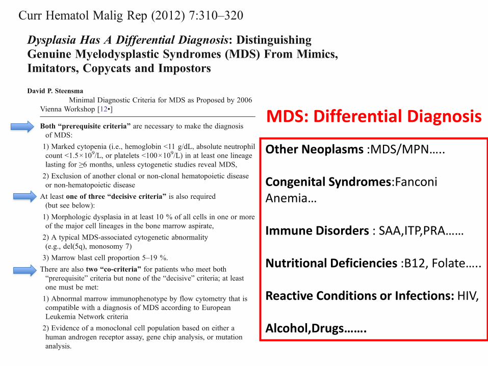
MDS: Differential Diagnosis
Other Neoplasms :MDS/MPN…..
Congenital Syndromes:Fanconi Anemia…
Immune Disorders : SAA,ITP,PRA……
Nutritional Deficiencies :B12, Folate…..
Reactive Conditions or Infections: HIV,
Alcohol,Drugs…….

Diagnosi Differenziale✓Anemia Aplastica-EPN
✓Disordini Nutrizionali (Anemia Megaloblastica)
✓Alterazioni citomorfologiche da Alcolismo
✓Infezioni virali (HBV, HCV, CMV,Parvovirus B19,HIV, etc)
✓Sostanze tossiche (antibiotici,chemioterapici, piombo,benzene)
✓Anemia dell’anziano non altrimenti definita
✓ Epatopatie
✓Patologie Autoimmuni
✓ ………..

PERCORSO DIAGNOSTICO…………..
• Anamnesi !!
• Emocromo-Reticolociti
• Es. morfologico s. periferico
• Es. morfologico s. midollare con Perls
• Citogenetica/FISH
• Biopsia Osteomidollare*
• s-EPO
• Assetto marziale
• HLA*
• ……………

Per la Diagnosi di SMD
➢ E’ indispensabile rilevare una displasia in > 10% delle cellule della linea mieloide /eritroide/megacariocitaria
➢ E’ necessario contare i blasti nel SP e MI
➢ E’ fondamentale effettuare l’analisi cariotipica (normaleperò nel 40-50% circa dei casi !!)
➢ E’ importante ,soprattutto nei casi senza evidenza di anomalie cromosomiche e con displasia modesta monitorare il paziente (SMDI ? o altro ?)


Sindrome 5q-• 5q- unica anomalia
• M/F 1:4
• Anemia Macrocitica
• Megacariociti mononucleati
• Ipoplasia eritroide……
• Decorso clinico favorevole
• Rara trasformazione in Leucemia Acuta
• Risposta alla terapia con lenalidomide

Peripheral blood and BM findings of MDS

SMD con eccesso di blasti
SP
✓Anisopoichilocitosi con macrocitosi
✓ Disgranulopoiesi
✓Blasts <5% (AREB-1)5-19% (AREB-2)

MI✓ Disgranulopoiesi, Diseritropoiesi, Dismegacariocitopoiesi
✓BOM: Ipercellulare o ipocellulare in 10-15%; localizzazione abnorme
di precursori immaturi (ALIP)
✓Blasti 5-9% (EB-1)
10-19% (EB-2)
✓o Blasti con corpi di Auer (EB-2)
SMD con eccesso di Blasti

Cytopenias defined as haemoglobin <10 g/dL, platelet count <100 x 109/L, and absolute neutrophil count <1.8 x 10⁹/L
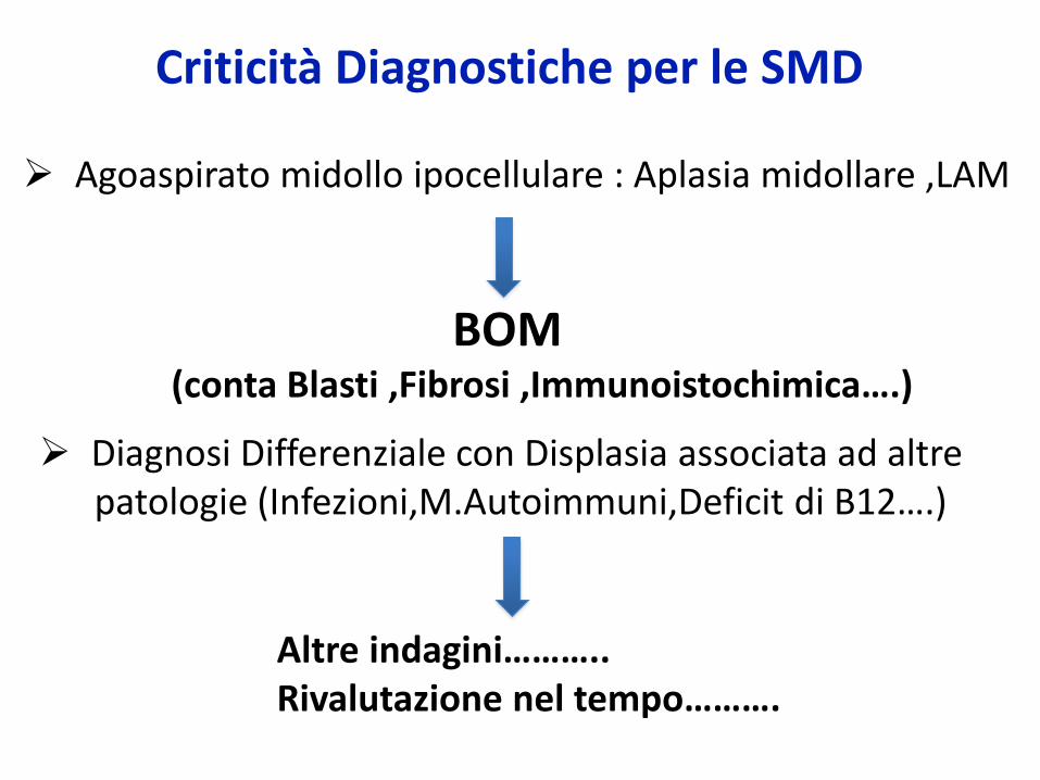
Criticità Diagnostiche per le SMD
➢ Agoaspirato midollo ipocellulare : Aplasia midollare ,LAM
BOM (conta Blasti ,Fibrosi ,Immunoistochimica….)
➢ Diagnosi Differenziale con Displasia associata ad altrepatologie (Infezioni,M.Autoimmuni,Deficit di B12….)
Altre indagini………..Rivalutazione nel tempo……….

Per la diagnosi e Classificazione delle
NMPcr
✓ Caratteristiche cliniche (Splenomegalia,Trombosi..)
✓Caratteristiche Istologiche /Citologiche (BOM*)
✓Anomalie molecolari (JAK2,MPL..) (Mut.JAK2..)
✓Altri Tests (Hb, Hct,WBC, Plts , MorfologiaPe
EPO,Citogenetica/ FISH,Rx….)

24
23
21
1312
12
13
21
22
313233
34
9
131211.2
11.2
12
13
22
34
131211.2
11.2
24
23
21
1312
12
13
21
22
313233
12
13
der(9)
der(22) o Ph
5’ABL-3’BCR
5’BCR-3’ABL
La diagnosi della LMC
RT-PCR
BCR-ABL
Citogenetica Convenzionalet(9;22)(q34;q11.2)
Morfologia Pe +Morfologia MI (Asp)
FISHe/o

Novel Marker in MPN: Mutations of CALR
Klampfl et al., N Engl J Med 2013
Screening for JAK2mut + CALRmut + MPLmut→ Clonality in ~95% of MF

Megakaryocytes
•Anisocytosis(small-large)
•Abnormal nuclear-cytoplasmic ratio
•Abnormal chromatin clumping
•Hyperchromatic nuclei,plump lobulation“bulbous,cloud-like,balloon-shaped,bare”

Criticità Diagnostiche per le NMP Ph-
➢ Casi con Eritrocitosi ,Trombocitosi “reattive”
➢ Casi con Fibrosi Midollare sec. ad altre Patologie
➢ Casi senza evidenza di alterazioni molecolari !!
Altre indagini………..Rivalutazione nel tempo……….

Neoplasie Linfoproliferative /
Linfomi
Criteri morfologici
Criteri immunofenotipici
Criteri genetici e molecolari
PRESENTAZIONE CLINICA
DIAGNOSI ISTOLOGICA
Linfomi indolenti
Linfomi aggressivi


Diagnosis of CLLNCI, iwCLL and ESMO guidelines
Variable NCI1 iwCLL2 ESMO3
Diagnosis
Lymphocytes (x 109/L)
> 5
Markers: CD19, CD20, CD23, CD5
≥ 5
Markers: CD5, CD19, CD20, CD23 surface immunoglobulin, CD79b
≥ 5
Markers: CD5, CD19, CD20, CD23 surface immunoglobulin, CD79b
Prolymphocytes (%) ≤ 55 ≤ 55 Not stated
Duration of lymphocytosis
None defined ≥ 3 months ≥ 3 months
Bone marrow lymphocytes (%)
≥ 30 > 30 Bone marrow biopsy not needed for diagnosis
Staging Modified Rai,correlate with Binet
Modified Rai or Binet
Modified Rai or Binet
Graphical Elaboration from text data

CLL
PLL
SML
HCL
MW

WHO Classification of Lymphoid Neoplasms
✓ Precursor B –cell Neoplasms
Precursor B-lymphoblastic leukemia/lymphoma
(precursor B-ALL)
Precursor T-cell and NK-cell Neoplasms
Precursor T-lymphoblastic lymphoma/leukemia
(precursor T-ALL)

Review Series
THE UPDATED WHO CLASSIFICATION OF HEMATOLOGICAL MALIGNANCIES
The2016 revision of theWorld Health Organization classi cation oflymphoid neoplasms
Steven H. Swerdlow,1 Elias Campo,2 Stefano A. Pileri,3 Nancy Lee Harris,4 Harald Stein,5 Reiner Siebert,6 Ranjana Advani,7
Michele Ghielmini,8 Gilles A. Salles,9 Andrew D. Zelenetz,10 and Elaine S. Jaffe11
1Division of Hematopathology, Department of Pathology, University of Pittsburgh School of Medicine, Pittsburgh, PA; 2Department of Pathology, Hospital
Clinic, University of Barcelona, August Pi i Sunyer Biomedical Research Institute, Barcelona, Spain; 3Haematopathology Unit, European Institute of
Oncology, Milan, and Department of Experimental, Diagnostic and Specialty Medicine, Bologna University Medical School, Bologna, Italy; 4Department of
Pathology, Harvard Medical School and Massachusetts General Hospital, Boston, MA; 5Pathodiagnostik, Berlin, Germany; 6Institute of Human Genetics,
Christian Albrechts University Kiel, Kiel, Germany; 7Division of Oncology, Department of Medicine, Stanford University, Stanford, CA; 8Department of
Medical Oncology, Oncology Institute of Southern Switzerland, Bellinzona, Switzerland; 9Department of Hematology, Hospices Civils de Lyon, and
Universite Claude Bernard Lyon-1, Lyon, France; 10Department of Medicine, Memorial Sloan Kettering Cancer Center and Weill Cornell Medical College,
New York, NY; and 11Hematopathology Section, Laboratory of Pathology, National Cancer Institute, Bethesda, MD
A revision of the nearly 8-year-old World
Health Organization classification of the
lymphoid neoplasms and the accompa-
nying monograph is being published. It
reflects a consensus among hematopa-
thologists, geneticists, and clinicians re-
garding both updates to current entities
as well as theaddition of alimited number
of new provisional entities. The revision
clarifies the diagnosis and management
of lesions at the very early stages of
lymphomagenesis, refines the diagnostic
criteria for some entities, details the
expanding genetic/molecular landscape
of numerous lymphoid neoplasms and
their clinical correlates, and refers to
investigations leading to more targeted
therapeuticstrategies.Themajorchanges
are reviewed with an emphasis on the
most important advances in our under-
standing that impact our diagnostic
approach, clinical expectations, and thera-
peutic strategies for the lymphoid neo-
plasms. (Blood. 2016;127(20):2375-2390)
Introduction
The 2008 World Health Organization (WHO) classi cation of
hematopoieticandlymphoidtumorsandtheassociatedmonograph
represent theestablished guidelinesfor thediagnosisof malignant
lymphomas;however,subsequentlytherehavebeenmajoradvances
withsigni cantclinical andbiologicimplications.1Amajorrevisionis
thereforebeingpublishedthat will beanupdateof thecurrent fourth
editionandnotatrulynew ftheditionastherearestill othervolumes
pending inthefourtheditionof theWHOtumor monographseries.
Because it is considered a part of the fourth edition, while some
provisional entitieswill bepromoted tode niteentitiesandasmall
numberofnewprovisionalentitiesadded,therewill benonewde nite
entities.
Aswiththe2001and2008classi cations,anall-importantClinical
Advisory Committeemeetingwasheldin2014toobtaintheadvice
andconsentofclinical hematologists/oncologistsandotherphysicians
critical totherevision(supplementalAppendix,availableontheBlood
Website). Additional editorial meetingsandconsultationsfollowed
leadingtotheupdatedclassi cation(Table1).2 Although thereare
only limitedalterationsin theclassi cationcomparedwith2008,
therevisedmonographwill incorporatealargebodyof information
published over the last 8 years relating to existing entities with
some important diagnostic, prognostic, and therapeutic implica-
tions. Theclassi cationmaintainsthegoalsof helpingtoidentify
homogeneous groupsof well-de ned entitiesand facilitating the
recognitionof uncommondiseasesthat requirefurtherclari cation.3
Thismanuscriptwill reviewthemajorareasinlymphoid,histiocytic,
and dendritic neoplasmswherechanges fromtheprior edition are
foreseenaswell asemphasizeconceptual themes(Table2).
Mature B-cell lymphoid neoplasms
Animportantelement thatpervadesmanypartsof thenewmonograph
derivesfromanexplosionof newclinical, pathological, andgenetic/
moleculardataconcerningthe“small B-cell” lymphomas.Theconcept
that therearelymphoidproliferationsthatweusedtodiagnoseasovert
lymphoidneoplasmsbutwhicharenotconsideredassuchin2016will
befurtheremphasized.AmongtheaggressiveB-cell lymphomas,there
aremajorchangesthat impacthowthesecasesshouldbeevaluatedand
diagnosedthathaveimportanttherapeuticimplicationsaswell asbeing
of biologicinterest.
Chronic lymphocytic leukemia/small lymphocytic lymphoma
and monoclonal B-cell lymphocytosis
The2008monographrecognizedmonoclonal B-cell lymphocytosis
(MBL) as thepresenceof monoclonal B-cell populations in the
peripheral blood(PB)ofupto53 109/Leitherwiththephenotypeof
chronic lymphocytic leukemia (CLL), atypical CLL, or non-CLL
(CD52)Bcellsintheabsenceof otherlymphomatousfeatures.Found
inupto12%of healthy individuals, insomeit may beanextremely
Submitted December 31, 2015; accepted February 9, 2016. Prepublished
onlineas Blood First Edition paper, March 15, 2016; DOI 10.1182/blood-2016-
01-643569.
The online version of this article contains a data supplement.
BLOOD, 19 MAY 2016 xVOLUME 127, NUMBER 20 2375
For personal use only.on May 27, 2016. by guest www.bloodjournal.orgFrom


Criticità Diagnostiche in Ematologia
• Citopenie ………….SMD
• Eosinofilie…………..LECr
• Monocitosi………….SMD, LMMcr
• Piastrinosi/Eritrocitosi……………TE,LMC,MF
• Linfocitosi…………….. LLC , Linfomi
• MGUS………………..Mieloma Multiplo


Criticità Diagnostiche in Ematologia
• Citopenie ………….SMD
• Eosinofilie…………..LECr
• Monocitosi………….SMD, LMMcr
• Piastrinosi/Eritrocitosi……………TE,LMC,MF
• Linfocitosi…………….. LLC , Linfomi
• MGUS………………..Mieloma Multiplo

Bejar, Curr Hematol Malig Rep 2015
CYTOPENIAS
….the diagnosis of Cytopenias remains a core task of hematology clinical practice……..

C .A. 41 y
Anemia Megaloblastica

Caso Clinico• Paz. 55aa
• Febbre
• Disfagia,Disfonia ,Tosse
• Tumefazioni corde vocali
• Rx Torace ndp
• E+P Monocitosi
• Episodio Influenzale : Tp Ab/Prd
• Apiressia 7 gg
• Febbricola /Disfagia
• E+P Monocitosi*
• ……………….

Biologi/Medici
di Laboratorio
MMG
EMATOLOGI
Per la Dx in Ematologia……….
ANATOMOPATOLOGI


