+ All Categories
Home > Documents > MASTERARBEIT / MASTER S THESIS - univie.ac.atothes.univie.ac.at/46990/1/49734.pdf · appear to have...

MASTERARBEIT / MASTER S THESIS - univie.ac.atothes.univie.ac.at/46990/1/49734.pdf · appear to have...

Date post: 15-Jun-2020
Category:
Upload: others
View: 3 times
Download: 0 times
Share this document with a friend
46
MASTERARBEIT / MASTERS THESIS Titel der Masterarbeit / Title of the Master‘s Thesis Morphological convergence in distantly related myxozoans: Spores as ecotypes “ verfasst von / submitted by Anna Sophia Feix BSc angestrebter akademischer Grad / in partial fulfilment of the requirements for the degree of Master of Science (MSc) Wien 2017/ Vienna 2017 Studienkennzahl lt. Studienblatt / degree programme code as it appears on the student record sheet: A >066 831< Studienrichtung lt. Studienblatt / degree programme as it appears on the student record sheet: Masterstudium Zoologie Betreut von / Supervisor: Mitbetreut von / Co-Supervisor: Dr. Astrid Sybille Holzer PhD Ao. Univ.-Prof. i. R. Dr. Waltraud Klepal
Transcript

MASTERARBEIT / MASTER’S THESIS

Titel der Masterarbeit / Title of the Master‘s Thesis

„ Morphological convergence in distantly related myxozoans: Spores as ecotypes “

verfasst von / submitted by

Anna Sophia Feix BSc

angestrebter akademischer Grad / in partial fulfilment of the requirements for the degree of

Master of Science (MSc)

Wien 2017/ Vienna 2017

Studienkennzahl lt. Studienblatt / degree programme code as it appears on the student record sheet:

A >066 831<

Studienrichtung lt. Studienblatt / degree programme as it appears on the student record sheet:

Masterstudium Zoologie

Betreut von / Supervisor:

Mitbetreut von / Co-Supervisor:

Dr. Astrid Sybille Holzer PhD

Ao. Univ.-Prof. i. R. Dr. Waltraud Klepal

2

3

Acknowledgements

At first I would like to thank my two supervisors Astrid Sybille Holzer, for discussing every detail and

her availability at any time possible and Waltraud Klepal for giving a lot of amazing advice. Further

thanks to the Laboratory of Electron Microscopy of the Biology Centre ASCR - Institute of Parasitology

in Ceske Budejovice for helping me with the preparation of the electron microscopy samples and

letting me use their facilities. I also want to thank the whole Laboratory of Fish Protistology of the

Institute of Parasitology of the Biology Centre ASCR, Hana Pecková for teaching me the molecular

techniques, Ivan Fiala for helping me with the Phylogenetic analysis, and Ana Isabel Born-Torrijos for

helping me with the statistics and everyone else in this Department for making my stay enjoyable.

Danksagungen

Als erstes möchte ich mich bei meinen beiden Betreuerinnen Astrid Sybille Holzer, für detailreiche

Diskussionen und ihre Erreichbarkeit zu jeder Zeit und Waltraud Klepal für gute Ratschläge und

Korrekturen bedanken. Weiteren Dank an das Labor für Elektronenmikroskopie des Biologie Zentrums

in Budweis (Tschechische Republik) für die Mithilfe bei der Vorbereitung der

Elektronenmikroskopischen Proben und für die Zurverfügungstellung der Geräte. Außerdem möchte

ich mich herzlich bei der ganzen Abteilung für Fisch Protistologie bedanken. Besonders Hana Pecková,

welche mir die molekularen Methoden beigebracht hat, Ivan Fiala für die Hilfe mit den

Phylogenetischen Analysen und Ana Isabel Born-Torrijos für die Hilfe mit den statistischen Analysen

und allen anderen KollegInnen die meine Zeit in Budweis so wundervoll gemacht haben.

4

Table of Contents

Acknowledgements………………………………………………………………………………………………………………………. 3

Abstract………………………………………………………………………………………………………………………………………… 5

Zusammenfassung………………………………………………………………………………………………………………………… 6

1.Introduction………………………………………………………………………………………………………………………………. 7

1.1. General introduction to the Myxozoa…………………………………………………………………………..…………. 7

1.2. Myxozoan taxonomy contradicts phylogeny……………………………………………………………….…………… 7

1.3. Biological characteristics reflecting myxozoans phylogeny……………………………………………………….. 8

1.4. Spore morphotypes……………………………………………………………………………………………………….…………9

2. Aims…………………………………………………………………………………………………………………………………………11

3. Material and Methods……………………………………………………………………………………………………….…….12

3.1. Collection of spores from fish…………………………………………………………………………………………………12

3.2. Spore morphology………………………………………………………………………………………………………………….13

3.3. Molecular analyses…………………………………………………………………………………………………………………14

3.4. Database of morphological and habitat features……………………………………………………………………..16

3.5. Statistical analyses…………………………………………………………………………………………………………….……17

3.6. Phylogeny………………………………………………………………………………………………………………………….…..17

4. Results……………………………………………………………………………………………………………………………..….…..18

4.1. Ultrastructure of myxozoan spores……………………………………………………………………………….………..18

4.2. Phylogenetic relationship between myxozoan taxa based on 18S rDNA sequences……….….…….. 20

4.3. Spores as ecotypes……………………………………………………………………………………………………….………..21

4.3.1. Relation between spore sizes/shapes and their habitats……………………………………………………….21

4.3.2. Relation between spore shape, appendages and ornamentation………………………..……………..…23

4.3.3. Relation between water current and spore surface/appendages……………………………………….… 24

4.3.4. Relation between spore ornamentation and ancestral habitat………………………………..…….……..25

4.3.5. Relation between spore appendages, ornamentation, shape and external habitat………………..25

4.3.6. Relation between spore shape and host target organ system ……………………………..……………….27

5. Discussion …..…..…..…..…..…..…..…..…..…..…..…..…..…..…..…..…..…..…..…..…..…..…..…..…..…..………29

5.1. Phylogenetic relationship between species …..…..…..…..…..…..…..…..…..…..…..…..…..…..…..………29

5.2. Spore measurements and ratios …..…..…..…..…..…..…..…..…..…..…..…..…..…..…..…..…..…..…………30

5.3. Relationships between spore morphology and intrapiscine/external habitats…..…..…..…..………..31

6. Conclusion …..…..…....…..…..…..…..…..…..…..…..…..…..…..…..…..…..…..…..…..…..…..…..…..…..…..…….34

7. References…..…..…..…..…..…..…..…..…..…..…..…..…..…..…..…..…..…..…..…..…..…..…..…..…..…..……… 35

Appendix…..…..…..…..…..…..…..…..…..…..…..…..…..…..…..…..…..…..…..…..…..…..…..…..…..…..…..………. 39

5

Abstract

Myxozoans are miniaturized endoparasites belonging to the Cnidaria, with roughly 2200 species

currently described. They are characterized by a two-host live cycle, including an invertebrate and a

vertebrate host (mainly fish). Spores are formed as durable transmission stages between hosts and

they represent pluricellular stages in a wide variety of shapes, sometimes with ornamentation and

appendages. Myxozoan taxonomy has been based predominantly on these morphological features,

however the incongruence of spore morphology and phylogenetic clustering of myxozoans is obvious.

Different spore morphotypes, i.e. genera, can be extremely closely related while other morphotypes

appear to have emerged more than once during the myxozoan evolution. The aim of the present study

was to determine whether a relationship exists between the spore features and their habitats, both

within the host and the environment. Spores of 10 species were collected, their SSU rDNA was

sequenced and their surface structure characteristics studied by SEM. Furthermore, they were

included in a large database of 258 taxa to perform statistical analyses in R. The most prominent

correlation was found between spore characteristics and external habitat. Spores in freshwater

habitats are usually characterized by some form of surface structure enlarging their surface area and

increasing their buoyancy, whereas marine spores are predominantly smooth. Most exceptions can be

explained by the habitat of the ancestor (as determined by SSU rDNA phylogeny). Despite significant

differences between projections and ornamentation in slow flowing water, the water current is more

likely to influence the shape of the spore than its ornamentation. Characteristic spore shapes exist also

for each host organ system, while ornamentation is more variable. In summary, the present study

shows that intrapiscine and external environment account for most of the variation in morphological

spore features, demonstrating that spores are in fact ecotypes.

Keywords: Myxozoa, spore morphology, vertebrate host, ecology, infection habitat

6

Zusammenfassung

Myxozoa sind wenige Mikrometer große Parasiten, welche zu den Cnidaria gehören. Zurzeit sind

ungefähr 2200 Arten innerhalb der Myxozoa beschrieben. Sie leben meistens als Endoparasiten

innerhalb aquatischer Habitate und besitzen einen komplizierten Wirtswechsel zwischen Evertebraten

und Wirbeltieren (am häufigsten Fischen). In den letzten Jahren zeichnete sich immer deutlicher ab,

dass es einen Zusammenhang zwischen Form, Oberflächenstruktur (Rillen, Höhlen…) und Anhängen

(Schwänze, Flügel, Lappen, Haare…) der Sporstadien und dem ökologischen Habitat, dem Wirtsgewebe

und andern Faktoren geben muss. Somit müsste es auch möglich sein, Sporen anhand der

beeinflussenden Faktoren als Ökotypen zu beschreiben. Diese Studie zeigt, dass das Infektionshabitat

eine bedeutende Rolle in der Sporentwicklung einnimmt. Sporstadien aus dem Süßwasser zeigen

immer eine Form von Oberflächenstrukturen (meistens Rillen), da sie ihre Oberfläche und so auch ihr

Volumen vergrößern, um die im Wasser vorhandenen Strömungs- und Auftriebskräfte zu nutzen.

Marine Arten sind in den meisten Fällen glatt, da im Salzwasser der Auftrieb aufgrund der höheren

Dichte des Wassers größer ist. Ausnahmen lassen sich häufig durch die Verwandtschaftsbeziehungen

erklären, da einige Arten ihr Habitat während ihrer Evolution wechselten. Weitere Unterschiede bei

der Oberflächenstruktur und den Anhängen konnten nur in langsam fließenden Gewässern festgestellt

werden, obwohl die Fließgeschwindigkeit am ehesten die Form der Sporen verändert. Die Form

verändert sich jedoch wesentlicher in Abhängigkeit vom Organsystem innerhalb des Fisches, während

die Oberflächenstruktur geringere Veränderungen aufweist. Zusammenfassend, konnte diese Studie

jedoch bestätigen, dass sowohl Organsysteme als auch Faktoren des externen Habitats für die meisten

morphologischen Veränderungen verantwortlich sind. Somit konnte bestätigt werden, dass die Sporen

von Myxozoen tatsächlich Ökotypen darstellen.

Stichwörter: Myxozoa, Sporen, Wirbeltiere als Wirt, Parasiten, Infektionshabitat

7

1. Introduction

1.1. General introduction to the Myxozoa

Myxozoans are microscopic parasites belonging to the Cnidaria, with roughly 2200 species (Lom and

Dyková 2006), currently described in 64 genera (Okamura, Gruhl and Bartholomew 2015). Myxozoans

are extremely diverse and currently compose one fifth of all known cnidarians, however in hypothetical

calculations as many as 16 000 myxozoan species are suggested for the Neotropics alone (Naldoni et

al. 2011) and eDNA analyses predict the existence of many more taxa and phylogenetic lineages

(Hartikainen et al. 2016), making the prospected number higher. Certain myxozoans cause severe

diseases in their hosts, both in wild populations and aquaculture facilities, contributing to large

ecological and economical losses. Especially in wild populations disease control is difficult. Young fish

are often more susceptible to diseases and parasite infections causing extreme losses in wild stocks

and in aquaculture.

Myxozoa were first described in 1880, however their classification changed from protozoans to the

animal kingdom. Now it is clear that they are Cnidaria (Jimenez-Guri 2007; Jimenez-Guri, Okamura and

Holland 2007, Holland et al. 2011), which diverged from a single ancestor. During this process

myxozoans simplified their morphology and evolved an intricate parasitic life cycle. The two hosts live

cycle is generally completed in aquatic habitats and usually includes an invertebrate and a vertebrate

host. Fish serve as intermediate hosts (Okamura et al. 2015), whereas annelids or bryozoans suit as

the final host. Though approximately 50 life cycles have been elucidated (Okamura et al. 2015), details

of the development are only scarcely known in either host. However, a sophisticated host-parasite

relationship and host manipulation has enabled them to spread from their teleost hosts to amphibians

and reptiles (Jirků et al. 2011; Lom and Dyková 1993; Hartigan, Phalen and Slapeta 2013), waterfowl

(Bartholomew et al. 2008) and even small mammals (Székely et al. 2015; Prunescu et al. 2007) in their

host range.

1.2. Myxozoan taxonomy contradicts phylogeny

Myxozoan taxonomy has been and continues to be based predominantly on morphological features,

mainly of the spore formed in the vertebrate host. Since their first discovery taxonomy was changed

several times. Several changes were executed in the 1980s with the last revision by Lom and Noble

(1984), however the important characters for classification stayed the same. Although spores vary in

their appearance, they are all composed of a number of shell valves that are joined by a suture line, at

least one polar capsule and one or more sporoplasms and their nuclei (Lom and Noble 1984). To

characterize orders and suborders of myxosporeans two of those characteristics are important, either

the number and configuration of the shell valves or the position of the polar capsules with regard to

8

the suture. Other characteristics for identification include details of the polar filaments, dimensions of

the polar capsules, the presence of ridges or striations on the valve surface, the presence of

appendages and the presence or absence of a mucous envelope. To determine a spore on species level

Lom and Arthur (1989) proposed that characters, like host identification, host habitat, information

about the vegetative stages and whether the sporogonic stages are mono-, di- or polysporic are

important for a complete description.

Systematics is still mainly based on spore morphology, but since the 1990s, when first DNA sequences

became available this system has become much debated, due to the incongruences between

morphology and DNA based phylogenetic approaches. As myxozoan diversification started hundreds

of millions of years ago, conservative markers are required to reconstruct their evolution. The 18S

rDNA is found in all eukaryotic taxa and it has become the most commonly used phylogenetic marker

of Myxozoans (Okamura et al. 2015). After phylogenetic analysis of molecular data for approx. 700

myxozoan taxa (National Centre of Biotechnology Information, NCBI, status April 2017) it is clear that

most myxozoan genera are either para- or polyphyletic (Diamant et al. 2005; Diamant, Whipps and

Kent 2004; Fiala 2006). While some spore morphotypes (genera) have emerged several times,

occupying 5 or more different phylogenetic position (Heiniger, Gunter and Adlard 2011), molecularly

monophyletic groups are artificially separated into different genera by minor differences such as

presence/absence of caudal appendages, used to distinguish Henneguya from Myxobolus (Fiala 2006)

or the number of sporoplasms which differentiated Polysporoplasma and Sphaerospora (Bartošová et

al. 2013).

Although the SSU marker is very suitable for myxozoan phylogeny, there are certain problems with

sticking to only one marker. The sequence length of myxosporeans varies a lot between marine and

freshwater species. Marine species show the shortest SSU sequences (1500-1700 basepairs), whereas

freshwater clades can be up to 2100 basepairs long. The Sphaerospora sensu stricto clade breaks ranks,

as with up to 3700 basepairs they have one of the longest SSU rRNA sequences in all eukaryotes

(Eszterbauer et al. 2013).

To produce the best outcome for myxozoan phylogeny Heiniger and Adlard (2013) propose a

combination of morphological biological and molecular characters. Their data on ceratomyxids from

cardinal fishes in Australian waters show significant differences between genetics and biological

characters, but insignificant variances amongst spore characters.

1.3. Biological characteristics reflecting myxozoans phylogeny

Based on the controversy between spore morphology and molecular phylogeny, naturally one

important question arises: Which characters if not morphological ones explain the phylogenetic

clustering of the Myxozoa and play a key role in their evolution? Tissue localisation in the intermediate

9

host and host habitat (freshwater vs marine) was first identified as a factors explaining phylogenetic

clustering of the morphologically paraphyletic genera (Holzer, Sommerville and Wootten 2004; Fiala

2006). Following further life cycle discoveries, it was suggested that the invertebrate host group

mirrored large-scale myxozoan phylogeny and SSU rRNA secondary structure (Holzer, Wootten and

Sommerville 2007). Most recently, Kodádková et al. (2015) determined that vertebrate host groups

also mirror myxozoan phylogeny with cartilaginous fish representing an ancestral state for most

myxozoan lineages.

An effect of geography on phylogenetic clustering was confirmed by Whipps and Kent (2006).

Differences between four oceanic regions suggest significant barriers on a global scale, whereas the

genetical exchange on a smaller geographic scale is not as meaningful, as morphological differences

between different host species and locations could not be found. Therefore, this genetic study on

Kudoa thyrsites suggests that the examined regions compromise endemic populations. The influence

of geography on the 18S rDNA diversity of single species was also observed by other others as well

(e.g. for Zschokkella nova (Fiala 2006) and for Myxidium truttae (Holzer et al. 2004)).

1.4. Spore morphotypes

If biological characteristics such as host tissue location, invertebrate host group or geography mirror

phylogenetic clustering, another question arises: What influences spore shape, size and

ornamentation? Apart from enduring unfavourable conditions outside the host, spore morphology

should favour rapid/easy exit from the host, dispersion in the environment and should guarantee

reaching the adequate invertebrate host. As such, spores likely represent ecotypes.

The common myxosporean ancestor had a smooth spore surface and was marine (Kodádková et al.

2014; Kodádková et al. 2015) Shell valves with ridges and striations appeared on entry into freshwater

environments (Kodádková et al. 2014). Surface structures (ridges, pits…) and appendages of the spore

appear to have the same main purpose, which is increasing the surface area of the spore. Based on the

Archimedes’ principle an upward buoyant force that is exerted on a body immersed in water. The

density of the surrounding water is lower than the density of the spore, hence helping the spore to

float. As any appendage or surface structure increases the surface area of the spore the surrounding

buoyancy forces have more area to operate on and as such slow down the spore when sinking. Thus,

spores have more time to float and can disperse further. As saltwater has a higher salinity as freshwater

it is more viscous and spores sink slower than in freshwater, forcing freshwater spores to use

mechanisms that counteract dispersal fast settlement rate. Hence one could speculate that spores in

marine habitats can disperse even with a smooth surface, whereas freshwater spores should more

likely have surface structures and appendages. In reality, this is not always the case as species

descriptions show that most marine species have surface structures, whereas freshwater do not.

10

Bipteria vetusta spores enlarge their surface area with wing-like structures (Shul’man 1964). The deep-

water genus Palliatus evolved a membranous veil (Kodádková et al. 2014). Both might enable better

flotation in the high-pressure water column (Fiala and Bartošová 2010). Generally, appendages in

myxozoans are thought to be adaptations to the out-of-host environment, and evolved many times

independently e.g. in Myxodavisia, Pseudalatospora and Henneguya (Kodádková et al. 2014).

However, parasites can also adapt to the hosts’ organ structure. Myxozoans live in restrained areas

making morphological simplifications vital (Lom, Rohde and Dyková 1992). Therefore, absence or

minimization of structures might represent an adaptation to the intrapiscine habitat. Most fish organs

can be infested, myxozoans can be either coelozoic, inhabiting body cavities, or histozoic (inhabiting

tissues). Myxospores with long appendages could be expected to be found in coelozoic environments,

as release from the fish is otherwise difficult (Feist and Longshaw 2006). However, Henneguya, is

known to have the longest appendages in myxozoans, lives in both marine and freshwater

environments, and is found mainly in histozoic habitats, like gills (Barassa et al. 2003) or kidney

(Wagner 2016). This hinders the spore from a rapid exit from the host organ. Most coelozoic spores do

not have long appendages (Arndt et al. 2006, Landsberg and Lom 1991), but are covered with surfaces

structures (Eiras et al. 2011; Eiras et al. 2012; Bartholomew et al. 2008; Hartigan et al. 2012). These

surface structures might be useful to float in the e.g. bile of the host.

The water flow rate has direct and indirect effects on the spores. The temperature of the water body

is highly dependent on the pace of the water flow. Low water flow promotes higher temperatures, as

the water gets heated by the sun. Experimental studies also show that slow flowing water has a higher

amount of temperature fluctuations, which may interfere with myxozoan development (Hallett and

Bartholomew 2008). High flows might damage the spores and cause a higher dilution of the spores,

suggested on actinospores (Kerans and Zale 2002). In a one-year experiment Hallett and Bartholomew

(2008) demonstrated that habitats with lower water flow promote higher infection prevalence and

greater dispersal in the invertebrate host. They also propose that the attachment of the actinospore

to the fish is affected by the flow velocity, due to two observations, first more actinospores lacking

sporoplasms could be found in slow flowing waters, second more fish became infected in the slow

flowing experimental tanks. These are strong indications that myxozoan spores adapted their

morphology to optimize host exit and transmission in the environment.

11

2. Aims

The aim of this thesis was to determine if a relationship exists between shape, cellular organisation

and fine structure of myxozoan spores and the characteristics of their habitat, both within and outwith

the fish host. Therefore, a comprehensive database of 258 taxa from all phylogenetic clades and known

morphotypes (genera) was produced and a number of statistical models were tested to understand

which morphological features can be related to which habitat. For example: Do the surface ridges and

appendages of myxozoan spores vary in relation to a specific host habitat? Which spore structure

represents which ecotype? Which myxozoan spore type is found in which host habitat? Additionally,

morphological and ultrastructural data as well as 18S rDNA sequences were produced for

new/previously undescribed taxa which were also added to the database.

12

3. Material and Methods

3.1. Collection of spores from fish

In each fish, the gallbladder, kidney (head and hind kidney), liver, gills, and skin (smears of lateral body

surface of fish and from under pectoral fin) were checked for myxozoans by light microscopy (400x

magnification). To take samples from the inner organs a ventral cut from the anus to the pelvic fins of

the fish was performed. Then the body cavity was opened. During a 5-month-period, 125 fish belonging

to 22 species and 13 families were checked for myxozoans (table 1). While myxozoan species were

found in 16 fish species (table 1), only 10 were used for this study as for these species a sufficient

number of spores for electron microscopy were found. If spores were detected in an organ they were

isolated and observed at x400 magnification under an Olympus BX51 light microscope (Olympus

Optical Co. (Europa) GmbH, Hamburg, Germany). Spores were immediately stored in TNES for further

molecular examination as well as in water or saltwater (depending on origin of fish) for Scanning

Electron Microscopy (SEM).

Fish species dissected(infected) fish Fish family Found myxozoan species

Albramis brama 17 (5) Cyprinidae Zschokella nova/Myxobolus sp.

Anguilla anguilla 1 (1) Anguillidae Myxidium giardi

Centropyge acanthops 5 Pomacanthidae -

Carassius auratus 1(1) Cyprinidae Sphaerospora sp.

Crassius gibelio 14(3) Cyprinidae Myxobulus sp.

Ctenopharyngodon idella 3 (2) Cyprinidae Zschokella nova, Chloromyxum sp.

Cyprius carpio 4(2) Cyprinidae Thelohanellus wuhanensis/Myxobolus sp.

Datnioides microlepsus 3 Datnioididae Plasmodium

Esox lucius 8 Esocidae -

Gobiodor okinawae 3(1) Gobiidae Ceratomyxa sp.

Merluccius productus 1(1) Merlucciidae Kudoa sp.

Microsynodontis batesii 5 Mochokidae -

Oligoancistrus sp. 1 Loricariidae -

Otocincius affinis 5 Loricariidae -

Paracanthurus hepatus 5(1) Acanthuridae -

Pseudanthias squamipinnis 5(5) Serranidae Ceratomyxa cardinalis

Rhodeus amarus 1(1) Cyprinidae Myxidium sp.

Rutilus rutilus 16(14) Cyprinidae Zschokella nova, Myxobolus sp.

Scathophagus argus 5(2) Scatophagidae Myxobolus sp.

Squalius cephalus 1(1) Cyprinidae Myxobolus muelleri

Tetradon nigrovirigis 18(11) Tetraodontidae Ortholinea aurata, Ceratomyxa arcuata

Tinca tinca 3(1) Cyprinidae Chloromyxum cyprini

Table 1: Number of dissected and infected fish species and found myxozoan species. In the column of dissected fish, the

number in parenthesis shows how many fish were infected with the myxozoan species in the corresponding column.

13

3.2. Spore morphology

Digital photos of isolated spores were taken at x1000 magnification with an Olympus DP70 camera

(Olympus Optical Co. (Europa) GmbH, Hamburg, Germany) mounted on the Nomarski interference

microscope Olympus BX51. Measurements of 10 spores of each myxozoan species were taken on

digital images with the program ImageJ, Version: 64-bit Java 1.6.0_20 for Windows (National Institutes

of Health, Maryland, US). Measurements of the spores follow the guidelines of Lom & Arthur (1989)

and include spore length, spore width, spore thickness as well as polar capsule length and polar capsule

width. The definitions and how to measure was incurred from Lom and Dyková (1992). Spore length is

defined by the distance between the apex and the posterior end. The width is measured perpendicular

to the length from one end of the suture to the other. The thickness is measured perpendicular to the

suture plane, from the most distant point of one valve to the other. Polar capsule length and width are

measured like the rest of the spore (figure 1).

Ultrastructure of myxozoan spores

For SEM sample preparation, a protocol of Alama-Bermejo (2009) was followed. Round cover slips

were washed in 96% ethanol (EtOH). The dried slides were coated with 0,1% poly-D-lysine on the

surface of which spores were left to settle for 30 min in their preferred medium (water or seawater,

depending on origin of fish). Afterwards the spores were fixed for 30 min on the coverslip using 2,5%

glutaraldehyde in 0,1M Cacodylate buffer. Then the spores were rinsed twice in the same buffer for

15 minutes each. Post-fixation was performed with 1% osmium tetroxide in 0,1M sodium cacodylate

buffer, for 30 minutes. The coverslips where washed with distilled water for 15 minutes and

dehydrated in an ascending alcohol series from 30% to 100% EtOH, for 5 minutes each. Thereafter, the

samples were critical point dried (PELCO CPD2, Ted Pella Inc, Redding, USA), mounted on stubs and

gold-sputtered with a BAL-TEC, SCD 050 sputter coater (BAL-TEC, Leica Biosystems Nussloch GmbH,

Figure 1: How to measure length (L), width (W) and thickness (T) of a myxozoaon spore. (Lom and Dyková, 1992)

14

Nussloch, Germany). The spores were examined with a JEOL JSM-7401F scanning electron microscope

(JEOL, Akishima Tokyo, Japan).

3.3. Molecular analyses

DNA analysis

To extract DNA from the spores they were stored in 400µl TNES urea buffer (10mM Tris-HCl, 125mM

NaCl, 10mM ethylenediaminetetraacetic acid, 0,5% sodium dodecyl sulphate, 4M urea) directly after

fish dissection. The spores were digested with Proteinase K (20 µg/ml) at 55°C overnight. For DNA

extraction, 400µl phenol (pH=8 buffered) and 400µl chloroform were added. After mixing, the different

phases were separated by centrifugation (15 000g for 5 min). Thereafter, 330µl of the aqueous top

layer including the DNA was removed into a fresh tube. The DNA was then precipitated mixing the

sample with triple amount of ice-cold 92% EtOH. After pelleting the DNA by centrifugation (13 000 g,

20 min) and washing the sample with 70% EtOH, the sample was dried on a heating plate at 55°C until

all alcohol had completely evaporated. The dried DNA was then resuspended in 50µl of RNAsa/DNAsa-

free water and left in the fridge to dissolve overnight.

Polymerase Chain Reaction was performed with a

programmable thermal cycler (Thermal Cycler-Life

Pro, BIOER Technology, Hangzhou, China or

TPersonal, Biometra GmbH, Göttingen, Germany) in

a final volume of 10µl. Each tube contained 1µl

dissolved DNA (50-100 ng), 7.35 µl nanopure water,

1 µl titanium buffer (containing 1,5Mm MgCl2,

ABgene, Epsom, UK), 0.2 mM dNTPs each, 0.5 µM forward and 0.5 µM reverse Primer, as well as 0,25

U Titanium Taq Polymerase. In the first PCR a universal set of primers targeting the 18S rDNA was used

(18E, 18R). For the nested PCR different primer pairs were used depending on the screened myxozoan

species. The volume and composition of the nested PCR were the same, with 1µl of the first PCR

product used in the nested assays. The PCR protocol consisted of initial DNA denaturation at 95°C for

3min, and 30 cycles of DNA separation at 94° for 50 seconds, annealing for 50 sec at different

temperatures depending on the Primers (table 2; table 3), 2 minutes elongation at 68°C, as well as a

final elongation step at 68°C for one minute and a final hold at 10°C. After checking if DNA was present

in a 1% agarose gel in TRIS-borate-EDTA buffer, the eluted DNA was purified for sequencing with a Kit

(PCR Cleanup Kit Gel/PCR DNA Fragments Kit (DF300), Geneaid Biotech Ltd., Taiwan) and sent off for

commercial sequencing by SEQme (SEQme s.r.o., Czech Republic).

Table 2: Annealing temperatures of all primer pairs

15

Cloning

When the obtained sequences had double peaks (contamination or mixed infection), the nested PCR

product was cloned. Therefore 2.5 µl of ligation master mix, 2.0 µl of the eluted PCR product and 0.5

µl cloning vector were mixed and incubated for 2 hours at 14°C in a thermal cycling block (ligation).

After placing the ligation reaction mix on ice, 50µl of competent bacterial cells (Escherichia coli- strain

DH5α) were added. The mixture was then incubated for 5-8 minutes on ice. The tubes were heated in

a 42°C water bath for 30 seconds (vector inclusion) and then incubated on ice again for 2 minutes.

Then 200µl of room temperature SOC medium was added and the tubes were shaken horizontally at

37°C for an hour. Meanwhile 40µl of X-gal was spread on each LB agar plate and the plates were

incubated until ready for use at 37°C. After spreading the transformation mixture on the prepared agar

plates, they were incubated overnight at 37°C.

The next day white and blue colonies were visible on the agar plates. White colonies, containing the

PCR product, were taken up with a pipette tip and placed into a tube containing 30µl of nanopure

water. The tubes where then shaken at 37°C for 10 minutes. After that a new PCR was performed with

a total amount of 10µl per tube. Each tube contained 2µl of cell suspension, 1µl of 10x Taq buffer, 0.2

mM of dNTP, 10µM of M13 forward primer and 10µM M13 reverse Primer, 0.24 U of polymerase and

6µl of PCR water. The PCR protocol consisted of an initial denaturation step at 95°C for 10min, and 20

cycles of DNA denaturation at 95° for 30 seconds, annealing at 54°C for 1 minute, 1 minute elongation

at 72°C and a final extension at 72°C, for 10 minutes. Finally, the plasmid PCR products were visualized

by agarose gel electrophoresis. If the electrophoresis showed a band of the expected size, the

corresponding bacterial colonies were put into tubes with 12µl ampicillin and 3ml culture solution and

shaken overnight at 37 °C. To isolate the plasmids from the competent cells, the High Pure Plasmid

Isolation Kit (Roche Diagnostics GmbH, Mannheim, Germany) was used. DNA was then sent for

commercial sequencing to SEQme.

The obtained sequences were submitted to the Basic Local Alignment Search Tool (BLAST) of the

National Centre for Biotechnology Information (NCBI) to screen for matches or close relatives. For the

alignment of 18S rDNA sequences Geneious (Biomatters Ltd., Auckland, New Zealand) was used.

Primers Sequence paired with Primers Sequence paired with

18e TGGTTGATCCTGCCAGT 18R, 18g Myx1F GTGAGACTGCGGACGGCTCAG Mx3

18g GGTAGTAGCGACGGGCGGTGT 18e, MyxGEN4F Mx3CCAGGACATCTTAGGGCATCACAG

Myx1F

18R CTACGGAAACCTTGTTACG 18e SSU CER-ForCTWGTTGGTADGGTAGTG

SSU CER-Rew

MyxGP2F GGATAACCGTGGGAAATCTA Act1R SSU CER-RewGTACAAGAGGCAGAGACGTAT

SSU CER-For

Act1R AATTTCACCTCTCGCTGCCA MyxGP2F MyxGEN4F GTGCCTTGAATAAATCAGAG 18g

Table 3: Primer sequences. The table shows all used primer sequences and the corresponding basepairs

16

3.4. Database of morphological and habitat features

To determine whether a relationship exists between the morphological features of myxozoan spores

(size, shape, surface structure, appendages etc.) and their habitats, both within the host and the

environment a database of 258 species was complied, including all currently recognised myxozoan

genera. For statistical analyses the following characteristics were picked: spore length, spore width,

spore thickness (and ratios of the latter three), the occurrence, size and structure of appendages, the

occurrence of surface structures, the type of surface structure and general information about the

habitat of the spore/host (marine, freshwater, brackish), as well as the organ location of the spore

inside the vertebrate host (table 4).

Table 4: For statistical analysis, all characteristics were changed into categories (represented by numbers). For all spore

measurements the mean was calculated. The infection habitat of the spore was grouped into freshwater, marine and

all habitats (FW, M, FWMB). Ancestral habitat is represented by four categories (FW, M, FW to M, M to FW). Categories

were ascribed for each organ system and for the general location within the host (coelozoic, histozoic, ectoparasitic).

Water current was categorized from standing water (1) to very fast flowing water (5). Surface structures and appendages

were put into subjective categories from none at all (as the lowest number) to a lot of them (the highest number). The

category “others” in appendages includes appendages that only occur in one myxozoan species. The shape of the spore

was also put into categories based on descriptions in the literature. The shape category “others” includes shapes that

occur only in one myxozoan species.

17

3.5. Statistical analyses

The ratio between length and thickness, thickness and width, and width and length was calculated by

dividing one by the other. Normality of the measurements and ratios was tested with the Shapiro-

Wilkinson-Test in the R-package “nortest” version 2.0-4 (Gross and Ligger 2015) and Q-Q plots version

7.3-47 (Wickham 2009). Thereafter a Kruskal-Wallis-Test (R-package rcompanion, version 1.5.6,

Mangiafica 2017) was performed. If a significant relationship between a measurement and habitat

could be detected a Dunn-Test, R-package “FSA” version 0.8.13 (Ogle 2017) was performed to show

significance between the different habitat categories. To test significance of the association between

two kinds of nominal categories a Fisher's exact test of contingency tables was performed, including

multiple comparisons for each combination with the R-package rcompanion, e.g. differences between

ornamentation types in the marine habitat or differences between appendages in the biliary system.

All statistical analysis was conducted in R (R Core Team, version 3.3.3, 2017, Vienna, Austria), plots

were done with ggplot2 version 2.2.1 (Wickham 2009).

3.6. Phylogeny

To draw an exemplary phylogenetic tree including all SSU rDNA sequences obatained in this study as

well as selected sequences from GenBank, sequences were assembled and aligned with Geneious

(Biomatters Ltd., Auckland, New Zealand) using MAFFT v7.017 (Katoh et al. 2002) with default

parameters. Phylogeny was reconstructed using the maximum likelihood method in RAxML

(Stamatakis 2006) using the GTR+G model. The malacosporean Buddenbrockia plumatellae was set as

an outgroup. The tree was visualized in Fig-Tree v 1.4.3. (Rambaut 2007).

18

4. Results

4.1. Ultrastructure of myxozoan spores

Examples presented in figure 2 show that most marine myxozoan spores have a smooth surface and

no ornamental structures of any kind. Kudoa sp. ex Merluccius productus (Figure 2C) has four valve

cells, whereas the three-other species have two valve cells. Both, Ceratomyxa sp. ex Paracanthurus

hepatus (Figure 2A) and Ceratomyxa cardinalis (Figure 2B) have elongated, banana shaped spores with

a straight suture. Myxobolus portucalensis from Anguilla anguilla (Figure 2D) is disc shaped with a

straight suture line.

Figure 2: Examples of myxospores with a smooth spore surface. A: Ceratomyxa sp. from the gallbladder of

Paracanthurus hepatus, marine B: Ceratomyxa cardinalis from the gallbladder of Pseudanthias squamipinnis, marine

C: Kudoa sp. from the muscle tissue of Merluccius productus, marine D: Myxobolus portucalensis from the gills of

Anguilla anguilla, freshwater scale bar= 5µm

A

D

C

B

19

Figure 3: Examples myxospores with an ornamented spore surface and from freshwater habitats. A: Myxobolus dispar

from the muscle of Cyprinis carpio, B: Thelohanellus sp. from the fin rays of Cyprius carpio, C: Myxidium rhodei from the

kidney of Rhodeus amarus, D: Zschokkella sp. from the gallbladder of Ctenopharyngodon idella, E: Ortholinea sp. from

the kidney tubules of Tetradon nigrovirigis, F: Chloromyxum cristatum from the gall bladder of Tinca tinca, scale bar=

2µm

A A

F A

E A

D A

C A

B A

20

As seen in figure 3 most freshwater species have surface structures. However, the species-rich

histozoic genera Myxobolus spp. (figure 3A and 2B) and Thelohanellus spp. (figure 3B) have disc-like

spores that have smooth valves and straight sutures. Myxidium rhodei from Rhodeus amarus has a

straight suture and thin longitudinal ridges (figure 3C). The pictured Zschokkella spp. from gallbladder

of Ctenopharyngodon idella shows only three longitudinal ridges on either side of the straight suture

line, which appears to run diagonal due to the position of the polar capsules (figure 3D). Otherwise the

spores are smooth. Parallel to the suture of the spores of Ortholinea spp. three circular ridges are

visible (figure 3E). In the middle of the circle thin longitudinal parallel ridges are formed. Chloromyxum

cristatum from the gallbladder of Tinca tinca shows a sinuous suture and 8-10 tall ridges on either

spore valve (figure 3F).

4.2. Phylogenetic relationship between myxozoan taxa based on 18S rDNA sequences

Ten new 18S rDNA sequences were obtained in the present study which clustered in 8 different

subclades (figure 4). Henneguya, Thellohanellus and Myxobolus cluster together in several subclades

of the oligochaete-infecting freshwater clade of myxozoans. As previously stated, most myxozoan

genera are poly- or paraphyletic. Myxidium spp. serve as an extreme example for polyphyly whereas

other genera like Ceratomyxa and Kudoa are almost monophyletic.

While all belonging to a single genus, Myxidium species with smooth valves cluster together in one

subclade. All other species have ridges somewhat parallel to the suture. Differences in spore length,

thickness and width as well as variations of the polar capsules have no visible effect on the

phylogenetic position. Yet, one clade including Myxidium amazonense, Myxidium rhodei and Myxidium

cuneiforme seems to have slightly elongated spores. This subclade also is an exception to all other

Myxidium species as those three species have a suture perpendicular to the polar capsule plane and

striations, whereas a perpendicular suture is normally found in smooth spores while polar capsules

and suture are in the same plane in spores with striations. As fine, longitudinal ridges are present in all

but one subclade, and the shape of all subclades with striations is very similar it is difficult to

differentiate these subclades, even by morphology supported by ultrastructure.

21

4.3. Spores as ecotypes

4.3.1 Relation between spore sizes/shapes and their habitats

Most myxozoans are between 10-29µm long, 15-20µm thick and around 10µm wide. In marine

habitats, they appear to be slightly longer and wider than in freshwater habitats (figure 5A). In the

biliary system spore length, thickness and width show a much higher variation than in all other organ

systems. However only thickness is significantly different from all other categories in the biliary system

and the external epithelia. Spores in the muscle (support, mostly marine species belonging to the

genera Kudoa or Unicauda) are the smallest.

Oligo

chae

te- In

fectin

g P

olych

aete

- Infe

cting

Figure 4: 18S rDNA-based phylogenetic tree indicating the polyphyletic nature of myxozoan genera as demonstrated for

Myxidium (in orange boxes), a genus (morphotype) which evolved multiple times in both, the oligochaete- (freshwater)

and polychaete-infecting (marine) clades of myxozoans. Sequences obtained in the present study are highlighted in blue

ǁ indicates branch shortening by 90% in basal branches (left part of image) and by 50% for S. elegans and C. anko; B

plumatellae was used as an outgroup.

22

When comparing the ratios between different external habitats, the variety in the marine habitat is

still the highest. However, the ratio length:thickness shows that the highest values are obtained in the

freshwater habitat, meaning spores in freshwater are as long as in the marine habitat (figure 6A) but

are significantly thinner and hence disc shaped rather than spherical. This is mostly the case for

myxozoans producing spores in external epithelia (genera Myxobolus, Henneguya, Thelohanellus). The

ratio thickness:width only shows differences in the biliary system, whereas the ratio width: length is

very variable, but shows significance in four organ systems.

Figure 5: Relation between absolute spore measurements (length, thickness and width) and their external as well as

host organ habitat. A: spore length - external habitat, B: spore length - infected organ system, C: thickness - external

habitat, D: thickness - infected organ system, E: width - infection habitat, F: spore thickness - infected organ. FW-

freshwater, M-marine, FWMB-all aquatic habitats (freshwater, marine and brackish), * shows categories significantly

different from all other categories

23

4.3.2 Relation between spore shape, appendages and ornamentation

When testing the correlation between appendages and their spore-shape, tails are significantly more

common in club-shaped spores than in any other spore shape. Wings were mainly found in the shape

category “others”. As expected appendages were absent in all categories except the club shaped and

“others” spore category. All other appendages (mucous envelope, hairs and other special structures)

were not significantly related to any specific spore shape. Similarly, ornamentations (thin ridges, thick

ridges, circular structures as well as pockets and pits) showed no statistically significant relation to a

specific spore shape, however, a smooth spore surface can be found in all shape categories (Figure 7).

Figure 6: Relation between spore measurement ratios (length: thickness, thickness: width, width: length) and their

external as well as host organ habitat. A: ratio length:thickness - external habitat, B: ratio length:thickness - infected

organ system, C: ratio thickness:width - external habitat, D: ratio thickness:width - infected organ system, E: ratio

width:length - external habitat, F: ratio width:length - infected organ system. FW-freshwater, M-marine, FWMB-all

aquatic habitats (freshwater, marine and brackish), * shows categories significantly different from all other categories

24

4.3.3 Relation between water current and spore surface/appendages

In slow-flowing water ecosystems significantly more myxozoans have tails than any other appendage

structure. All other water currents did not show a significant correlation with a specific appendage or

ornamentation. However, any form of ridges (thin and thick ridges as well as pockets and pits) on the

spore surface is significantly correlated with slow flowing water (figure 8).

Figure 8: Relation between water flow and A. appendages or B. ornamentation of the spores. Significance was tested

with a Fisher's exact test of contingency tables.

25

4.3.4 Relation between spore ornamentation and ancestral habitat

Marine spores, whose ancestors also occurred in marine habitats as of their phylogenetic position, are

smooth rather than ridged. Spores with pockets and pits show no significant difference to smooth

spores, when comparing spore ornamentation in different ancestral habitats (figure 9B). In marine

species with an ancestor occurring in freshwater, ridges are most prominent. In freshwater spores with

an ancestor in freshwater, ridges are more likely to occur than a smooth spore surface. However,

smooth spores also occur in freshwater. A significant difference between circular and fine ridges was

also observed (figure 9A). In marine species with an ancestor occurring in freshwater, ridges are most

prominent. Thick ridges occur in 100% of the spores that switched from freshwater habitats to marine

habitats. However, the proportion between thin and thick ridges is not significant, whereas circular

ridges show a significant difference in comparison with thick and thin ridges (figure 9C).

4.3.5. Relation between spore appendages, ornamentation, shape and external habitat

Significant differences between appendages, ornamentation and shape of spores could only be

observed in freshwater and marine habitats. The most common appendage in freshwater habitats are

tails. Wings could not be found in any myxozoan species from freshwater. A significant difference

between all appendages and freshwater habitat can be observed, except for the categories

“appendages missing” and “others”. Most species with wings can be found in marine habitats.

Significant differences were found between all appendages, except for the categories “hairs” and

“others”, and the external habitat they occur in (figure 10 A and B).

Figure 9: Relation between different ancestral habitats and ornamentation of the spore. A: ancestor and successor occur

in freshwater habitats, B: ancestor and successor occur in marine habitats, C: ancestor occurred in freshwater and

successor is living in marine habitats

26

When testing the relation between the external habitats and ornamentation, a clear difference

between the freshwater habitat and the marine habitat can be seen. Any form of ridges occur mainly

in freshwater habitats, with circular ridges only occurring in freshwater habitats. A smooth surface and

pockets and pits are more popular in marine habitats. However, all different ornamentation categories,

except circular ridges, can be found in both habitats (figure 10 C and D).

The shape of spores varies a lot between those of freshwater habitats and those of marine habitats.

All different shapes can be found in marine habitats, and the “shamrock” shape does not occur in

freshwater habitats. In freshwater, no significant difference between club, spherical and other shapes

could be found. Disc-shaped myxozoans are most common in freshwater habitats, however there is

only a 9% difference to the next popular shape (spherical). In marine habitats “shamrock” shape and

“banana” shape are the most common ones, disc-shaped spores are hardly found amongst marine

species (figure 10 E and F).

Figure 10: Relation between external habitat and appendages, spore ornamentation and shape. A: appendages -

freshwater habitats, B: appendages - marine habitats, C: ornamentation - freshwater habitats, D: ornamentation -

marine habitats, E: shape- freshwater habitats, F: shape - marine habitat

27

4.3.6. Relation between spore shape and host target organ system

In the external epithelia of the host, disc-shaped myxozoas are most common, club-shaped and others

are also significantly more prevalent in external epithelia though less common than disc-shaped ones.

In the urinary system, spherical, subspherical spores as well as “others” shapes are significantly more

common, with little difference between each other. In the biliary system, banana-shaped myxozoans

are most common. Despite spherical and subspherical spores showing a similar high prevalence in the

biliary system their morphology differs significantly from each other. In the muscle “shamrock” shaped

myxozoans are most common with 76.9% of the tested myxozoans in the muscle having this shape. In

the nervous system, the only shape present are disk shaped myxozoans belonging to the genus

Myxobolus (figure 11).

Figure 11: Relation between spore shape and host target organ systems. A: external epithelia, B: urinary system,

C: biliary system, D: muscle, E: nervous system

28

In the hosts’ urinary system spores with pockets and pits are most common, however, all different

forms of ridges are present in the urinary system. In the external epithelia, spores are most likely

smooth (figure 12 C and D). A significant difference between appendage types was found in the biliary

system and the external epithelia. Within the biliary system no significant differences were found in

the prevalence of “appendages missing”, “others” and “wings”, the most common appendages. In the

external epithelia tails are the most common feature, a shape that is significantly more common than

all other appendages in the epithelia (figure 12 A and B).

When comparing histozoic with coelozoic myxozoans it is imminent that species with a membranous

sheet are only histozoic, whereas species with wings are mainly coelozoic. However, all other

appendages occur in both, histozoic and coelozoic myxozoans (figure 13 A and B). Differences between

the different ornamentation categories are not significant. When comparing the different shapes, disc-

like spores are strictly histozoic and shamrock-shaped myxozoans are mainly histozoic. Coelozoic

spores are mainly banana shaped, but also frequently spherical and subsperical (figure 13C and D).

Figure 12: Relation between spore ornamentation/appendages and different host organ systems. A: appendages - biliary

system, B: appendages – external epithelia, C: ornamentation-urinary system, D: ornamentation – external epithelia

29

5. Discussion

Possible correlations between spore morphology and spore environment were speculated long time

ago (Shulman 1966), and later in relation to contradicting morphology- and SSU- based phylogenetic

trees (Fiala and Bartošová, 2010), however, past studies never focused on evaluating this correlation

based on statistical models applied to a large database, making this study the first of its kind. The

statistical evaluation of spore morphology and ultrastructure in relation to the intrapiscine and aquatic

spore environment has proven a valuable tool for understanding that the large variety of different

spore morphologies that evolved in myxozoans represent a response to environmental and functional

pressures, and explain much of their evolution into different, highly specialized morphotypes in

different habitats.

5.1. Phylogenetic relationship between species

Myxozoans were collected from a number of different organs and habitats, hence resulting in a variety

of phylogenetic origins in the oligochaete- (freshwater) and polychaete-infecting (marine) clades of

myxozoans. In this tree, Myxidium was chosen as a representative to demonstrate the general

polyphyletic distribution of most myxozoan genera, with 4 different origins within all urinary and biliary

tract parasite clades. The different clustering of Myxidium species can be partially explained by

different spore morphotypes. One Myxidium clade tends to be smooth, whereas all others are with

Figure 13: Relation between spore appendages/shape and coelozoic or histozoic location in the host. A: distribution of

appendages in coelozoic myxozoans, B: distribution of appendages in histozoic myxozoans, C: different shapes of

coelozoic myxozoans, D: different shapes of coelozoic myxozoans

30

ridges, however their shape differs between all subclades. Summarizing, there are

morphological/ultrastructural features that allow to differentiate some Myxidium spp. subclades that

cluster separately based on their molecular phylogeny. As Myxidium is not an isolated case, I would

suggest combining DNA sequences with specific morphological characters for designing a new

systematic scheme of the Myxozoa that will be able to correctly accommodate newly described

species. While this combination was also recommended in previous studies (Fiala and Bartošová, 2010)

the present study points out which morphological features are statistically significantly ascribed to a

certain phylogenetic genotype. It would make sense to merge subclades together when morphological

differences are minor, and hence reduce the number of genera. The position of the suture to the polar

capsules as well as the ornamentation of the spore would be suitable to differentiate Myxidum species

of different phylogenetic origin.

5.2. Spore measurements and ratios

In general, myxozoan spores are microscopic, showing very little size variation between species, likely

due to the enormous size reduction as an adaptation to parasitism. Most myxozoans are between 10-

29µm long, 15-20 µm thick and around 10µm wide. In marine habitats, they appear to be slightly longer

and wider than in freshwater habitats, which was already recognized by Shulman (1966), who stated

that the genera Chloromyxum, Unicapsula and Sphaerospora are the smallest, whereas Thellohanellus,

Henneguya and partly Myxobolus and Ceratomyxa are the largest. In the biliary system, length,

thickness and width have a considerably higher variability than in all other organ systems.

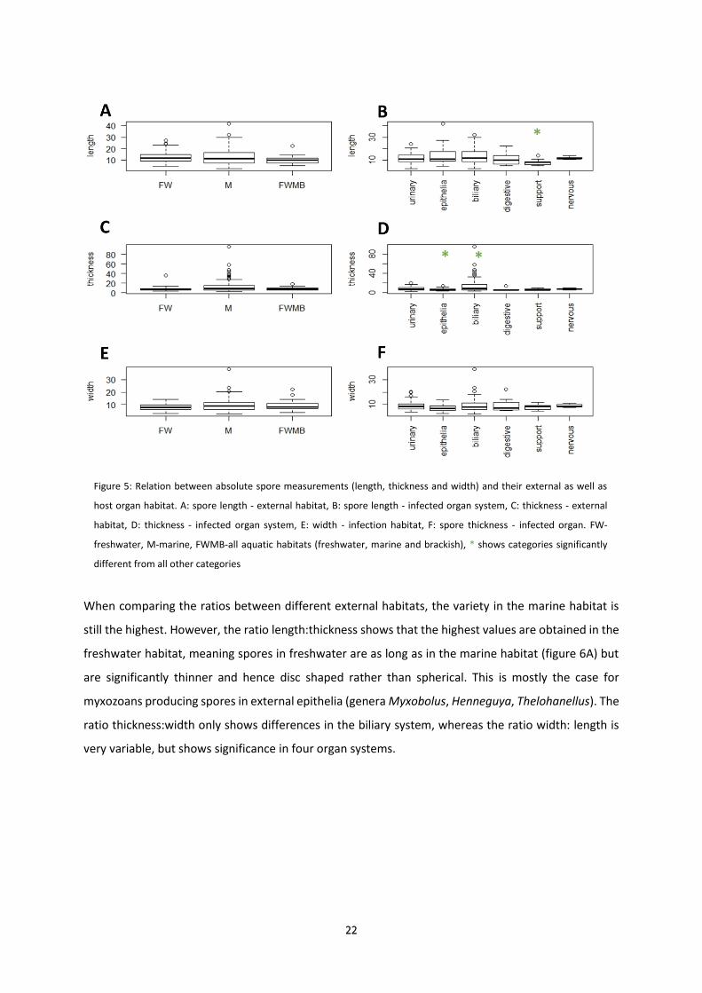
When comparing the ratios with the infection habitat, the variation in the marine habitat is still the

highest. However, the ratio length:thickness shows that the highest ratio can be found in the

freshwater habitat. This high ratio might be an intent to increase surface in relation to weight. Previous

studies already showed that spore dimension is a shared character within phylogenetic groups and

suggested that their ancestor had a spore thicker or of the same thickness as width, i.e. a spherical

shape (Fiala and Bartošová, 2010). The definitions for spore measurements (length, width, thickness)

are based on the orientation of the polar capsules relative to the spore and the orientation of the

suture relative to the polar capsules, and while important for taxonomy, as individual measurements

they fail to represent the spore shape as a whole. While ratios are more likely to address shape and

proved to improve statistical significance for the analyses relating spore shapes and their intrapiscine

or external habitat characteristics in the present analyses, they still do not fully represent the three

dimensions of different spore shapes. A mathematical model is required to define this relationship and

simple models have been developed e.g. for plant pollen (Reponen et al., 2010), however, this model

was not complex enough to reflect all different shapes found in myxozoan spores. In the future, it

would be useful to develop a mathematic model for all myxozoan spore shapes as empirical evaluation

31

as well as the superior significance of relationships from ratios rather than raw measurements indicate

that spore shape is essential for myxozoans in their specific habitats.

Most spore measurements are performed following clear definitions and guidelines by Lom and Arthur

(1989). However, one cannot be completely sure that thickness, length and width are always measured

correctly in original descriptions. Especially if descriptions predate the guidelines for species

descriptions by Lom & Arthur (1989) one of the three measurements is frequently missing, or if all

spores always lie in a certain way in the microscope, so that one measurement cannot be taken (e.g.

thickness in Ceratomyxa spp.). Furthermore, some species do not comply to the standard form of

myxozoans. The thickness of Ceratomyxa is a lot larger than in all other genera, whereas in other

species the suture is sinuous, and measurements become somewhat subjective, depending on the

angle at which they were taken. Moreover, some species do not have the standard spore features of

two valves, two polar capsules, one suture. Kudoa has four valves and four polar capsules, causing the

suture-lines to “cross”. Other species of this genus can have up to thirteen valves and polar capsules

(Kudoa permulticapsula). Unicapsula has three valves, two smaller and one larger one. Others i.e.

Auerbachia only has one polar capsule but two valves. In summary, measurements are difficult to

compare between different myxozoan genera and a shape-based mathematical model would

eliminate most of these problems.

5.3. Relationships between spore morphology and intrapiscine/external habitats

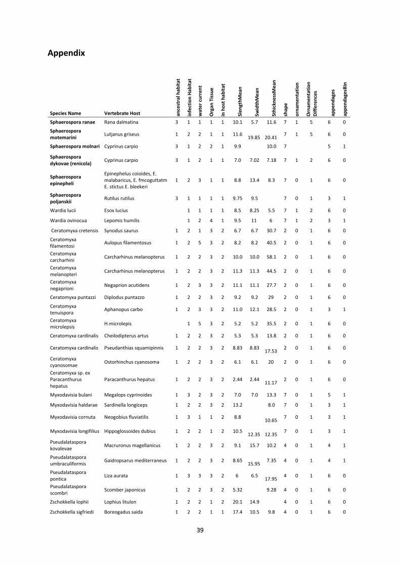
Using a database of 258 species and 64 taxa it was determined that most marine myxozoans have a

smooth spore surface, whereas a large number of freshwater species have some form of surface ridges.

As saline waters are more viscous spores sink slower to the ground anyway and therefore a surface

enlargement as in freshwater spores is not needed. Ridges of any form are more present in freshwater

habitats than in marine habitats. Due to the Archimedes principle this surface enlargement results in

a higher surface area that the surrounding buoyancy forces can interact with and hence the spores

have more time in the water column and therefore get dispersed further from their infection site. Fine

ridges create the highest surface area, which may be why circular ridges are very prominent as well.

Circular ridges are only found in freshwater habitats. However, thick ridges were as popular in marine

habitats as in freshwater habitats. As thick ridges produce a smaller surface area than thin ridges on

the valve, thick ridges can be an evolutionary adaptation to a new habitat, derived from thin ridges,

which is also shown by the fact that all spores with thick ridges in the database have a freshwater

ancestor and are now marine.

The main function of appendages is the same as for surface ornamentation, however the increase in

surface area might not be the only reason to prefer appendages. The most popular projection in

freshwater habitats, as well as slow flowing water current, are tails. This might be to hook the spore

32

on the ground/vegetation and stay there over a longer period of time after dispersal. Correlations

between spore shape and type of appendage showed a clear significance regarding club and drop-like

shaped spores developing a tail as appendage. This tail is always located on the narrower end of the

spore, which might result in a more streamlined appearance, presenting little resistance to water flow

in their environment. Most species with wings were found in marine habitats. Most wings have a

similar form and function as in seeds of a maple tree or in veils on sailing boats. However, wings are

rare and were hence designated to the category “others”, which included spore shapes that did not fit

in any other category. Difficulties when describing the spores within the “others” category may occur,

as not all spore descriptions are strictly after Lom and Arthur, 1989.

The theoretical ancestor of all myxozoans had a spherical spore (Kodadkova et al 2014). New shapes

occurred in both freshwater and marine species (Fiala and Bartošová, 2010). The present study shows

that, shape variations between freshwater and marine infection habitat complement each other. The

shamrock shape is only present in marine habitat, within the genera Kudoa and Unicapsula, with more

than two valves. Banana-shaped spores were mainly found in marine environments as well. Disc-

shaped spores are more popular in freshwater than in marine habitats. The disc-like spore shape might

have developed to impede the opening of the spore in stress situations or to get a higher surface to

volume ratio to prevent a rapid descent in the water. In stress situations, a spore will break along the

suture and the disc-like spore shape will work as a lever breaking the valves from each other. Spherical

spores have a lever-arm as large as the radius of the spore. Disc shaped spores with a suture lining the

edge of the disc have a reduced lever length and are therefore least likely to open in a stress situation

(Shulman, 1966). Cigar-shaped, banana- or horseshoe-shaped spores are more likely to break due to

the increased leverage (Noble, 1950). With this regard, surface ridges may not only increase surface

area but may also serve as an enforcement to the valve (Shulman, 1966). Another way of staying longer

in the water habitat, regardless of marine or freshwater habitat, is to decrease the volume to surface

ratio. Spheres have a small surface area, which makes them sink to the bottom quicker, which also

explains the size reduction of spherical species. Discs have an intermediate ratio, whereas cigar-shaped

spores have a large surface to volume ratio. However, Kudoa spp. slow down their descent by their

shamrock shape, which functions as a parachute (Shulman, 1966) and allows them to stay in the water

column as long as necessary. Although a significant correlation between shape and water current was

not found in our data. Spherical spores sink slowly, disc-shaped with an intermediate speed and

fusiform spores very quickly (Wittenburg et al 1989). However, banana-shaped spores and elongated

subspherical spores tend to rotate when sinking, which slows the sinking speed down (Leger, 1931).

This might also be a possible explanation for the formation of pockets and pits in spherical spores, as

this specific ornamentation even intensifies the tumbling process.

33

When testing the correlation between organ systems of the host and different morphological spore

characters it becomes clear that most myxozoans adapt a specific shape for each organ system. In

nervous tissue only disc-shaped spores can be found and external epithelia is preferred by disc-shaped

spores as well. However, the disc-shaped category is the only one present in all organ systems.

Epithelia-inhabiting myxozoans are mainly represented by the Myxobolus clades, which includes all

disc-shaped freshwater myxozoans which lack ornamentation. The fact that disc-shaped spores are

prevalent in every organ system suggests that this is a highly practical shape and likely one of the best

shape solutions for transmission in the environment. Observations by light microscopy show that disc

shaped spores are located very close to each other and they can be packed better than spherical spores

into a plasmodium. This enlarges the number of spores leaving the intermediate host and, more

importantly, infecting the final host even greater. The urinary system has spherical and subspherical

genera which is also mirrored in the biliary system, likely because of the similar pressure coming from

each all sides in liquid-filled spaces such as the gallbladder (Akhmerov et al., 1958). Banana-shaped

spores are most common in the biliary system. This shape might be used for flotation in the bile.

However, not all shapes are organ-dependent (Leger, 1931), but likely represent an adaptation to the

external habitat, since the time spores spend in the fish is limited (Shulman, 1966). Significant organ-

specific ornamentation was found in the urinary system. Most popular are spores with pockets and

pits, followed by any kind of ridges. Therefore, the pockets and pits might be a spore feature for better

grasping in the host tissue, since most of these spores are in close contact with the microvilli of the

excretory canals. Tails are the most common appendages in epithelia. These tails can entangle with

the host tissue for a better hold, but could also be used for a better release from the host tissue. The

high variability of appendages in different organs indicates that they are not so much an adaptation to

the in-host environment, which is supported by our statistical analyses and by different descriptions

over the years (Feist and Longshaw, 2006; Barassa et al, 2003).

Regardless of explanatory incongruences for the function of different ornamentations, appendages

and shapes of myxozoans are strongly correlated with their habitat. This study shows the vast

differences between myxozoan species and genera and tries to explain ecological adjustments of their

spore features, for the first time based on statistical analyses, making it a fundamental starting point

for in-depth future studies into the topic of functional myxozoan spore morphology.

34

6. Conclusion

This is the first time that correlations between myxozoan spore morphology/ultrastructure and habitat

within the fish host and the environment were analysed statistically. The results clearly demonstrate

that myxozoan spores represent ecotypes rather than morphotypes mirroring the phylogenetic tree

based on SSU rDNA sequence data. Some genera, that are strongly polyphyletic (e.g. Myxidium) in

reality represent multiple groups of similar spore morphotypes with specific and differentiable spore

dimensions and surface structures. The results of this study identify characteristics of myxozoan spore

morphology and ultrastructure that are useful for myxozoan taxonomy as they mirror phylogenetic

clustering. At the same time, it unveils some presently used taxonomic features as redundant as they

clearly represent ecological adaptations. While it has been repeatedly stated that a better taxonomic

scheme for the Myxozoa is absolutely required it can be concluded from the present study that such a

system would clearly involve not only spore features which are strongly biased by ecological

requirements but more details on earlier stages of parasite development in the host as well as

phylogenetic information on the origin and ancestry of a taxon to be described. Follow-on studies

concentrating on ecological adaptations of myxozoan spores should focus on developing a mathematic

model to better describe spore shape than simple measurements and optimizing/narrowing down the

artificially designed morphological categories for statistical analysis to join functionally identical

features and better reflect their correlation with habitat features. However, this study only gives a

first insight into spore ecology and changes in the spore because of influencing factors.

35

7. References

Alama-Bermejo G., M. Cuadrado, J.A. Raga & A. S. Holzer(2009) Morphological and molecular redescription of the myxozoan Unicapsula pflugfelderi Schubert, Sprague & Reinboth 1975 from two teleost hosts in the Mediterranean. A review of the genus Unicapsula Davis 1924. Journal of Fish Diseases, 32, 335–350

Akhmerov A.Kh. & I.P. Mart’yanova (1958) K. metodike opredeleiya slizistykh sporoviko v roda

Chloromyxum Mingazzini, 1890 (Method of identifying myxosporids of the Genus Chloromyxum Mingazzini, 1890. Zoolo.Zhurn., 37, 4, 619

Arndt R.E., E. J. Wagner, C. Bobo & T.St. John (2006) Laboratory and hatchery-scale evaluation of sand

filters and their efficacy at controlling whirling disease infection. Journal of Aquatic Animal Health, 18, 215-222.

Barassa B., E. A. Adriano, S. Arana & N. S. Cordeiro (2003) Henneguya curvata sp n. (Myxosporea :

Myxobolidae) parasitizing the gills of Serrasalmus spilopleura (Characidae : Serrasalminae), a South American freshwater fish. Folia Parasitologica, 50, 151-153.

Bartholomew J. L., S.D. Atkinson, S.L. Hallett, L.J. Lowenstine, M.M. Garner, C.H. Gardiner, B.A. Rideout,

M.K. Keel & J.D. Brown (2008) Myxozoan parasitism in waterfowl. International Journal for Parasitology, 38, 1199-1207.

Bartošová P., I. Fiala, M. Jirků, M. Cinkova, M. Caffara, M. Fioravanti, S. Atkinson, J. Bartholomew & A.

Holzer (2013) Sphaerospora sensu stricto: Taxonomy, diversity and evolution of a unique lineage of myxosporeans (Myxozoa). Molecular Phylogenetics and Evolution, 68, 93-105.

Diamant A., M. Ucko, I. Paperna, A. Colorni & A. Lipshitz (2005) Kudoa iwatai (Myxosporea :

Multivalvulida) in wild and cultured fish in the Red Sea: Redescription and molecular phylogeny. Journal of Parasitology, 91, 1175-1189.

Diamant A., C.M. Whipps & M.L. Kent (2004) A new species of Sphaeromyxa (Myxosporea :

Sphaeromyxina : Sphaeromyxidae) in devil firefish, Pterois miles (Scorpaenidae), from the northern Red Sea: Morphology, ultrastructure, and phylogeny. Journal of Parasitology, 90, 1434-1442.

Eiras J.C., Y.S. Lu, D.I. Gibson, I. Fiala, A. Saraiva, C. Cruz & M. J. Santos (2012) Synopsis of the species

of Chloromyxum Mingazinni, 1890 (Myxozoa: Myxosporea: Chloromyxidae). Systematic Parasitology, 83, 203-225.

Eiras J.C., A. Saraiva, C. F. Cruz, M. J. Santos & I. Fiala (2011) Synopsis of the species of Myxidium

Butschli, 1882 (Myxozoa: Myxosporea: Bivalvulida). Systematic Parasitology, 80, 81-116. Eszterbauer E., D. Sipos, B. Forro, P. Bartošová & A. Holzer (2013) Molecular characterization of

Sphaerospora molnari (Myxozoa), the agent of gill sphaerosporosis in common carp Cyprinus carpio carpio. Diseases of Aquatic Organisms, 104, 59-67.

Feist S.W., Longshaw M. (2006). Phylum myxozoa. In PTK Woo (ed.), Fish diseases and disorders.

Protozoan and metazoan infections. Vol. 1, 2nd ed., CAB International, Oxfordshire, 230-296. Fiala I. (2006) The phylogeny of Myxosporea (Myxozoa) based on small subunit ribosomal RNA gene

analysis. International Journal for Parasitology, 36, 1521-1534.

36

Fiala I. & P. Bartošová (2010) History of myxozoan character evolution on the basis of rDNA and EF-2

data. Bmc Evolutionary Biology, 10, 13. Gross J. and Ligges, U. (2015) nortest: Test for Normality. R package version 1.0-4. https://CRAN.R-

project.org/package=nortest Hartigan A., N.K. Dhand, K. Rose, J. Slapeta & D. N. Phalen (2012) Comparative Pathology and Ecological

Implications of Two Myxosporean Parasites in Native Australian Frogs and the Invasive Cane Toad. Plos One, 7.

Hartigan A., D.N. Phalen & J. Slapeta (2013) Myxosporean parasites in Australian frogs: Importance,

implications and future directions. International journal for parasitology. Parasites and wildlife, 2, 62-8.

Hartikainen H., D. Bass, A.G. Briscoe, H. Knipe, A. J. Green & B. Okamura (2016) Assessing myxozoan

presence and diversity using environmental DNA. International Journal for Parasitology, 46, 781-792.

Heiniger H., N.L. Gunter & R.D. Adlard (2011) Re-establishment of the family Coccomyxidae and

description of five novel species of Auerbachia and Coccomyxa (Myxosporea: Bivalvulida) parasites from Australian fishes. Parasitology, 138, 501-515.

Holland J.W., B. Okamura, H. Hartikainen & C. J. Secombes (2011) A novel minicollagen gene links

cnidarians and myxozoans. Proceedings of the Royal Society B-Biological Sciences, 278, 546-553.

Holzer A.S., C. Sommerville & R. Wootten (2004) Molecular relationships and phylogeny in a

community of myxosporeans and actinosporeans based on their 18S rDNA sequences. International Journal for Parasitology, 34, 1099-1111.

Holzer A.S., R. Wootten & C. Sommerville (2007) The secondary structure of the unusually long 18S

ribosomal RNA of the myxozoan Sphaerospora truttae and structural evolutionary trends in the Myxozoa. International Journal for Parasitology, 37, 1281-1295.

Jimenez-Guri E., B. Okamura & P. W. H. Holland (2007) Origin and evolution of a myxozoan worm.

Integrative and Comparative Biology, 47, 752-758. Jirků M., P. Bartošová, A. Kodádková & F. Mutschmann (2011) Another Chloromyxid Lineage:

Molecular Phylogeny and Redescription of Chloromyxum careni from the Asian Horned frog Megophrys nasuta. Journal of Eukaryotic Microbiology, 58, 50-59.

Jirků M., M. G. Bolek, C. M. Whipps, J. Janovy, Jr., M. L. Kent & D. Modry (2006) A new species of

Myxidium (Myxosporea : Myxidiidae), from the western chorus frog, Pseudacris triseriata triseriata, and Blanchard's cricket frog, Acris crepitans blanchardi (Hylidae), from Eastern Nebraska: Morphology, phylogeny, and critical comments on amphibian Myxidium taxonomy. Journal of Parasitology, 92, 611-619.

Katoh K., K. Misawa, K. Kuma, T. Miyata (2002) MAFFT: a novel method for rapid multiple sequence

alignment based on fast Fourier transform. Nucleic Acids Res, 30(14), 3059-66.

37

Kodádková A., P. Bartošová-Sojkova, A.S. Holzer & I. Fiala (2015a) Bipteria vetusta n. sp - an old parasite in an old host: tracing the origin of myxosporean parasitism in vertebrates. International Journal for Parasitology, 45, 269-276.

Kodádková A., I. Dyková, T. Tyml, O. Ditrich & I. Fiala (2014) Myxozoa in high Arctic: Survey on the

central part of Svalbard archipelago. International journal for parasitology. Parasites and wildlife, 3, 41-56.

Landsberg J.H. & J. Lom (1991) Taxonomy of the Genera of the Myxobolus-Myxosoma group

(Myxobolidae, Myxosporea), current listing of the species and revision of synonyms. Systematic Parasitology, 18, 165-186.

Leger L. (1930) Myxosporidies nouvelles ou peu connues du Genre <Myxidium> Chez les poissons d'eau

douce. Laboratoire D'Hydrobiologie et de Pisciculture, 21, 232-243. Lom J. & I. Dyková (2006) Myxozoan genera: definition and notes on taxonomy, life-cycle terminology

and pathogenic species. Folia Parasitologica, 53, 1-36. Lom, J. & E. R. Noble (1984) Revised classification of the class myxosporea Butschli, 1881. Folia

Parasitologica, 31, 193. Lom J., K. Rohde & I. Dyková (1992) Studies on the Protozoan Parasite of Australian fishes.1. New

species of the genera Coccomyxa Leger et Hsse, 1907, Ortholinea, Shul'man, 1962 and Kudoa Meglitsch, 1947 (Myxozoa, Myxosporea). Folia Parasitologica, 39, 289-306.

Salvatore Mangiafico (2017). rcompanion: Functions to Support Extension Education Program Evaluation. R package version 1.5.6. https://CRAN.R-project.org/package=rcompanion

Noble E. R. (1950) On a myxosporidian (Protozoan) Parasite of California trout. Journal of Parasitology,

36, 457-460. Ogle D.H. (2017) FSA: Fisheries Stock Analysis. R package version 0.8.12. https://cran.r-

project.org/web/packages/FSA/index.html

Okamura B., A. Gruhl & J. Bartholomew. 2015. Myxozoan Evolution, Ecology and Development. Vol1,

Springer-Verlag, Switzerland Prunescu C.C., P. Prunescu, Z. Pucek & J. Lom (2007) The first finding of myxosporean development

from plasmodia to spores in terrestrial mammals: Soricimyxum fegati gen. et sp n. (Myxozoa) from Sorex araneus (Soricomorpha). Folia Parasitologica, 54, 159-164.

Rambaut A. (2007) Molecular evolution, phylogenetics and epidemiology.

http://tree.bio.ed.ac.uk/software/figtree/ R Core Team (2017) R: A Language and Environment for Statistical Computing. version 3.3.3 Shul’man S. (1966) Myxosporidia of the USSR. Nauka, Moscow-Leningrad, 288-306. Shul’man S. (1988) Myxosporidia of the USSR. New Delhi: Amerinf publishing Co. Pvt. Ltd.

38

Shul’man S. S. (1964) Evolution and phylogeny of Myxosporidia. Academy of sciences of the USSR, Zoological institute, 1-9.

Stamatakis A. (2006) RAxML-VI-HPC: Maximum likelihood-based phylogenetic analyses with thousands

of taxa and mixed models. Bioinformatics, 22, 2688–2690. Székely C., G. Cech, S. D. Atkinson, K. Molnár, L. Egyed & A. Gubányi (2015) A novel myxozoan parasite

of terrestrial mammals: description of Soricimyxum minuti sp. n.(Myxosporea) in pygmy shrew Sorex minutus from Hungary. Folia parasitologica, 62, 045.

Wickham H. (2009) ggplot2: Elegant Graphics for Data Analysis. Springer-Verlag New York

Wittenburg J., J.Zierep, K., Bühler (1989) Technische Mechanik. In Czichos, H. Hütte- Die Grundlagen der Ingenieurwissenschaften. Vol.29, Springer-Verlag, Heidelberg, E1-E188

Whipps C. M. & M. L. Kent (2006) Phylogeography of the cosmopolitan marine parasite Kudoa thyrsites (Myxozoa : Myxosporea). Journal of Eukaryotic Microbiology, 53, 364-373.

39

Appendix

Species Name Vertebrate Host ance

stra

l hab

itat

infe

ctio

n H

abit

at

wat

er

curr

ent

Org

an T

issu

e

in h

ost

hab

itat

Sle

ngt

hM

ean

Swid

thM

ean

Sth

ickn

ess

Me

an

shap

e

orn

ame

nta

tio

n

Orn

ame

nta

tio

n

Dif

fere

nce

s

app

en

dag

es

app

en

dag

esB

in

Sphaerospora ranae Rana dalmatina 3 1 1 1 1 10.1 5.7 11.6 7 1 5 6 0

Sphaerospora motemarini

Lutjanus griseus 1 2 2 1 1 11.6

19.85

20.41 7 1 5 6 0

Sphaerospora molnari Cyprinus carpio 3 1 2 2 1 9.9 10.0 7 5 1

Sphaerospora dykovae (renicola)

Cyprinus carpio 3 1 2 1 1 7.0 7.02 7.18 7 1 2 6 0

Sphaerospora epinepheli

Epinephelus coioides, E. malabaricus, E. fmcoguttatm E. stictus E. bleekeri

1 2 3 1 1 8.8 13.4 8.3 7 0 1 6 0

Sphaerospora poljanskii

Rutilus rutilus 3 1 1 1 1 9.75 9.5 7 0 1 3 1

Wardia lucii Esox lucius 1 1 1 1 8.5 8.25 5.5 7 1 2 6 0

Wardia ovinocua Lepomis humilis 1 2 4 1 9.5 11 6 7 1 2 3 1

Ceratomyxa cretensis Synodus saurus 1 2 1 3 2 6.7 6.7 30.7 2 0 1 6 0

Ceratomyxa filamentosi

Aulopus filamentosus 1 2 5 3 2 8.2 8.2 40.5 2 0 1 6 0

Ceratomyxa carcharhini

Carcharhinus melanopterus 1 2 2 3 2 10.0 10.0 58.1 2 0 1 6 0

Ceratomyxa melanopteri

Carcharhinus melanopterus 1 2 2 3 2 11.3 11.3 44.5 2 0 1 6 0

Ceratomyxa negaprioni

Negaprion acutidens 1 2 3 3 2 11.1 11.1 27.7 2 0 1 6 0

Ceratomyxa puntazzi Diplodus puntazzo 1 2 2 3 2 9.2 9.2 29 2 0 1 6 0

Ceratomyxa tenuispora

Aphanopus carbo 1 2 3 3 2 11.0 12.1 28.5 2 0 1 3 1

Ceratomyxa microlepsis

H.microlepis 1 5 3 2 5.2 5.2 35.5 2 0 1 6 0

Ceratomyxa cardinalis Cheilodipterus artus 1 2 2 3 2 5.3 5.3 13.8 2 0 1 6 0

Ceratomyxa cardinalis Pseudanthias squamipinnis 1 2 2 3 2 8.83 8.83

17.53 2 0 1 6 0

Ceratomyxa cyanosomae

Ostorhinchus cyanosoma 1 2 2 3 2 6.1 6.1 20 2 0 1 6 0

Ceratomyxa sp. ex Paracanthurus hepatus

Paracanthurus hepatus 1 2 2 3 2 2.44 2.44

11.17 2 0 1 6 0

Myxodavisia bulani Megalops cyprinoides 1 3 2 3 2 7.0 7.0 13.3 7 0 1 5 1

Myxodavisia haldarae Sardinella longiceps 1 2 2 3 2 13.2 8.0 7 0 1 3 1

Myxodavisia cornuta Neogobius fluviatilis 1 3 1 1 2 8.8 10.65

7 0 1 3 1

Myxodavisia longifilius Hippoglossoides dubius 1 2 2 1 2 10.5

12.35

12.35 7 0 1 3 1

Pseudalataspora kovalevae

Macruronus magellanicus 1 2 2 3 2 9.1 15.7 10.2 4 0 1 4 1

Pseudalataspora umbraculiformis

Gaidropsarus mediterraneus 1 2 2 3 2 8.65

15.95 7.35 4 0 1 4 1

Pseudalataspora pontica

Liza aurata 1 3 3 3 2 6 6.5

17.95 4 0 1 6 0

Pseudalataspora scombri

Scomber japonicus 1 2 2 3 2 5.32 9.28 4 0 1 6 0

Zschokkella lophii Lophius litulon 1 2 2 1 2 20.1 14.9 4 0 1 6 0

Zschokkella sigfriedi Boreogadus saida 1 2 2 1 1 17.4 10.5 9.8 4 0 1 6 0

40

Zschokkella hildae Gadus morhua 1 2 3 1 1 16.2 10.4 9.6 4 0 1 6 0

Sinuolinea dimorpha Cynoscion regalis, C. nebulosus

1 2 2 1 2 14.9 14.8 14.9 3 0 1 6 0

Sinuolinea phyllopteryxa, Sinuolinea sp. 2. Dykova 2012

Phyllopteryx taeniolatus 1 2 2 1 1 17.1 16.4 15.6 3 0 1 6 0

Sinuolinea tetraodontoni

Tetraodon palembangensis (Pao palembangensis)

3 1 3 1 2 9.56

10.11 3 0 1 6 0

Sinuolinea arctica Myoxocephalus scorpius 1 2 2 1 2 15.7 15.4 16.1 3 0 1 6 0

Parvicapsula minibicornis

Oncorhynchus spp. 1 3 2 1 2 11.0 7.5 6.8 7 0 1 5 1

Parvicapsula pseudobranchicola

Salmo salar, S. truttae 1 3 5 2 1 12.4 6.2 7 0 1 6 0

Parvicapsula irregularis

Hippoglossoides platessoides

1 2 3 1 2 11.0 8.9 8.7 7 0 1 6 0

Parvicapsula bicornis Pleuronectes platessa 1 2 3 1 1 6.8 5.3 3.44 7 0 1 3 1

Parvicapsula spinachiae

Spinachia spinachia 1 2 2 1 1 10 5 3.98 7 1 2 5 1

Gadimyxa atlantica Gadus morhua 1 2 3 1 1 5.3 7.5 5.3 4 0 1 6 0

Gadimyxa sphaerica Gadus morhua 1 2 3 1 2 4.8 10.0 4.1 4 0 1 6 0

Gadimyxa arctica Arctogadus glacialis 1 2 3 1 2 5.7 10.0 5.6 4 0 1 6 0

Gadimyxa ex Sprattus sprattus (Karlsbakk)

Sprattus sprattus 1 2 2 1 1 2.55 4.2 2.45 4 0 1 6 0

Gadimyxa ex Clupea harengus (Karlsbakk)

Clupea harengus 1 2 2 1 1 2.55 4.2 2.45 4 0 1 6 0

Auerbachia pulchra Coryphaenoides rupestris 1 2 2 3 2 30 11 11 5 0 1 6 0

Auerbachia maamouni Gnathanodon speciosus 1 2 2 3 2 6.2 7.9 7.9 5 0 1 6 0

Auerbachia scomeroidi

Scomberoides lysan 1 2 2 3 2 21.4 7.5 7.5 5 0 1 6 0

Auerbachia chaetodonti

Chaetodon unimaculatus 1 2 2 3 2 32.2 9.1 9.1 5 0 1 6 0

Auerbachia caranxi Caranx papuensis 1 2 2 3 2 16.8 6.7 6.7 5 0 1 6 0

Coccomyxa gobiodoni Gobiodon citrinus 1 2 2 3 2 10.6 6.5 6.5 4 0 1 6 0

Coccomyxa colurodontis

Colurodontis paxmani 1 2 2 3 2 12.1 6 6 4 0 1 6 0

Coccomyxa jirilomi Bathygobius cyclopterus 1 2 2 3 2 10.1 6 6.1 4 0 1 6 0

Coccomyxa baleswarensis

Hilsa ilisha 1 3 5 3 2 11.36 5.17 5.17 4 0 1 6 0

Coccomyxa morovi Sardina pilchardus 1 3 3 3 2 14 5.5 5.5 4 0 1 6 0

Myxidium gadi Merlangius merlangus, Pollachius virens

1 2 1 3 2 11.3 5.3 5.3 4 0 1 6 0

Myxidium bergense Pollachius virens 1 2 2 3 2 18.54 6.87 6.25 4 0 1 6 0

Myxidium finnmarchicum

Merlangius merlangus 1 2 1 3 2 15.3 9.2 9 4 0 1 6 0

Myxidium milleri Corythoichthys schultzi 1 2 2 3 2 9.2 5.2 5.97 4 0 1 6 0

Myxidium incurvatum Callionymus lyra 1 2 2 3 2 11.62 4.92 4.92 4 0 1 6 0

Myxidium amazonense

Corydoras melini 2 1 1 3 2 17.0 3.7 2 1 2 6 0

Myxidium sp. Ex Rhodeus amarus

Rhodeus amarus 2 1 1 1 2 4.9 14.3 4.9 4 1 2 6 0

Zschokkella sp1 IF2006

Eugerres plumieri 4 3 1 3 2 11.5 13.4 4 6 0

Sigmomyxa sphaerica Belone belone 1 2 3 3 2 18.0 11.7 8.2 7 6 0

Ellipsomyxa gobii Pomatoschistus microps 1 3 1 3 2 7.0 8.7 11.6 4 0 1 6 0

Ellipsomyxa syngnathi Syngnathus rostellatus 1 2 2 3 2 6.8 8.1 10 4 0 1 6 0

41

Ellipsomyxa mugilis Liza saliens 1 2 1 3 2 7.25

11.75 6.8 4 0 1 6 0

Ceratonova gasterostea

Gasterosteus aculeatus 1 3 2 4 1 5.2 22.4 5.2 2 0 1 6 0

Ceratonova shasta (before Ceratomyxa shasta)

Gasterosteus aculeatus 1 3 3 4 1 6 14 6 2 0 1 6 0

Enteromyxum leei Takifugu rubripes 1 3 4 4 1 14.1 7.75 7 0 1 6 0

Enteromyxum scophthalmi

Scophthalmus maximus 1 3 1 4 1 22.2 11.7 14 7 0 1 6 0

Gastromyxum bulani Elops machnata 1 2 2 4 1 10.29 5.64 4.15 7 0 1 6 0

Gastromyxum rafii Elops machnata 1 2 3 4 1 7.92 5.42 5.35 7 0 1 6 0

Unicapsula pflugfelderi

Spicara smaris 1 2 2 5 1 5.16 6.02 6.02 6 0 1 6 0

Unicapsula fatimae Siganus canaliculatus 1 2 2 4 1 6.23 6.8 6.8 6 0 1 6 0

Unicapsula pyramidata

Scolopsis monogramma 1 2 2 5 1 5.1 7 7 6 0 1 6 0

Unicapsula seriolae Seriola dumerili 1 2 3 5 1 6.1 6.1 6.1 6 0 1 6 0

Unicapsula andersenae

Acanthopagrus australis 1 2 2 5 1 5.8 5.8 5.8 6 0 1 6 0

Kudoa sp. ex Merluccius productus

Merluccius productus 1 2 2 5 1 6.23 5.59 5.59 6 0 1 6 0

Kudoa carcharini Carcharhinus cautus 1 2 2 5 1 8.16

10.14 9.79 6 0 1 6 0

Kudoa thyrsites Paralichthys olivaceus 1 2 2 7.31

15.26 7.57 6 0 1 6 0

Kudoa iwatai Acanthopagrus schlegelii 1 2 2 6.0 6.8 6.0 6 0 1 6 0

Kudoa septempunctata

Paralichthys olivaceus 1 2 2 5 1 8.5 11.8 9.4 6 0 1 1 1

Kudoa gunterae Neoglyphidodon melas 1 2 2 5.42 10 5.62 6 0 1 6

Kudoa azoni Pleurogrammus azonus, Hexagrammos octogrammus

1 2 2 5 1 5.5 6.7 5.6 6 0 1 6 0

Kudoa permulticapsula

Scomberomorus commerson,

1 2 2 5 1 6 9.15 7.7 6 0 1 6 0

Chloromyxum clavatum

Raja clavata 4 2 1 3 2 14.4 11.9 9.4 3 0 1 5 1

Chloromyxum menticirrhi

Squalus acanthias 4 2 3 1 2 10.5 9.8 10.1 3 1 3 6 0

Chloromyxum riorajum

Rioraja agassizii 4 2 1 11.41 8.48 5.92 3 1 3 2 1

Sphaeromyxa artedielli

Triglops murrayi 4 2 1 3 2 17.5 5.6 5.6 2 1 2 6 0

Sphaeromyxa balbianii Gaidropsarus vulgaris 4 2 1 3 2 15 5 5 2 1 2 6 0

Sphaeromyxa longa Trisopterus minutus 4 2 1 3 2 20 5 5 2 0 1 6 0

Sphaeromyxa cannolii Hippocampus erectus 4 2 2 3 2 17.6 5.5 5.5 2 0 1 6 0

Sphaeromyxa schulmani

Salilota australis 4 2 3 3 2 19.3 5 5 2 0 1 6 0

Sphaeromyxa kenti Gobiosoma bosc 4 2 3 3 2 18.5 4.4 4.4 2 1 2 6 0

Sphaeromyxa zaharoni

Pterois miles 4 2 2 13.7 4.8 4.2 2 1 2 6 0

Sphaeromyxa lycodi Lycodes sp. 4 2 2 3 1 22.4 5.7 4.5 2 0 1 6 0

Soricimyxum fegati Sorex minutus 2 3 1 7.0 5.4 3.5 4 1 2 6 0

Soricimyxum minuti Sorex minutus 2 3 1 12.6 9.2 8.0 4 1 2 6 0

Myxidium baueri Macruronus magellanicus 4 2 2 3 2 17.55 4.65 4.65 4 1 2 6 0

Myxidium coryphaenoideum

Coryphaenoides rupestris 4 2 2 3 2 23.3 5 5.0 4 1 2 6 0

Cystodiscus melleni Pseudacris triseriata triseriata

1 3 2 12.3 7.6 4.82 4 1 2 6 0

Cystodiscus axonis Typhlonectes compressicauda

2 1 1 3 2 13.66 7.83 4.84 4 1 2 3 1

42

Cystodiscus australis Limnodynastes peronii 2 1 1 3 2 16.0 8.7

10.34 4 1 2 6 0

Cystodiscus immersus Rhinella marina 2 1 1 3 2 12.55 8.05 4 1 2 6 0

Myxidium anatidum Anas platyrhynchos 2 1 1 3 1 23.1 10.8 11.2 4 1 2 6 0

Myxidium scripta Trachemys scripta elegans (turtle)

2 1 1 1 1 18.5 5.1 5.1 4 1 2 6 0

Myxidium chelonarum Kachuga smithii (turtle) 2 1 1 3 2 14.5 4.5 5.5 4 1 2 6 0

Myxidium truttae Oncorhynchus kisutch 2 3 3 3 1 11.5 7.2 7.15 4 1 2 6 0

Myxidium ceccarellii Leporinus elongatus 2 1 1 3 2 17.7 10.4 10.1 4 1 2 6 0

Zschokkella jaimeae Tylosurus gavialoides 4 2 2 3 2 11.4 7.1 4 1 2 6 0

Zschokkella balistoidi Balistoides viridescens 4 2 2 3 2 11.1 6.3 5.86 4 1 2 6 0

Zschokkella auratis Sparus aurata 4 2 2 3 2 9.5 7.1 6.55 4 6 0

Zschokkella icterica Siganus luridus 4 2 2 3 2 12.4 7.1 4.4 4 1 2 6 0

Zschokkella nova Carassius carassius, Carassius auratus gibelio

2 1 1 3 2 10 6 6 4 1 2 6 0

Zschokkella sp. ex Ctenopharyngodon idella

Ctenopharyngodon idella 2 1 1 3 2 4.88

10.46 4.88 4 1 2 6 0

Chloromyxum auratum

Carassius auratus 2 1 2 3 2 18.8 9.8 9.8 3 3 1

Chloromyxum cristatum

Hypophthalmichthys molitrix

2 1 2 3 2 13.6 12.6 13.1 3 1 2 6 0

Chloromyxum cyprini Tinca tinca 2 1 1 3 2 13.24 12

13.34 3 1 2 6 0

Chloromyxum fluviatile

Hypophthalmichthys molitrix, Rutilus rutilus, leuciscus cephalus

2 1 2 3 2 7.8 8.0 7.45 3 1 4 6 0

Chloromyxum truttae Salmo salar 4 1 2 3 2 9.4 7.4 9.15 3 1 4 6 0

Chloromyxum thymalli Thymallus nigrescens 2 1 2 3 2 9.3 6.65 8.9 3 1 2 5 1

Chloromyxum careni Megophrys nasuta 1 1 1 7.25 5.75 5.76 3 1 2 6 0

Myxidium lieberkuehni

Esox lucius 2 1 3 1 1 20 6 6 4 1 2 6 0

Chloromyxum legeri Cyprinus carpio and other cyprinids

2 1 2 3 2 7.5 7.5 7.5 3 1 2 6 0

Myxobolus arcticus Oncorhynchus masou masou 2 3 1 6 1 14.1 8.7 9.76 1 0 1 6 0

Myxobolus neurobius Salmo trutta 2 3 2 6 1 11 8.0 6 1 0 1 6 0

Myxobolus fryeri Oncorhynchus clarki 2 3 3 6 1 12.9 8.6 7.2 1 0 1 6 0

Myxobolus neurotropus

Oncorhynchus mykiss 2 3 3 6 1 11.8 10.8 8.8 1 0 1 6 0

Myxobolus murakamii Oncorhynchus masou ishikawae

2 3 3 6 1 11.15

10.05 7.4 1 0 1 6 0

Ortholinea auratae Sparus aurata 4 2 2 1 2 9.0 8.3 7.2 4 1 2 6 0

Ortholinea orientalis Clupea harengus, Sprattus sprattus

4 2 3 1 2 9.0 7.9 5.6 4 1 2 6 0

Ortholinea labracis Dicentrarchus labrax 4 3 2 7.6 7.2 6.5 4 1 2 6 0

Ortholinea aurata Tetradon nigrovirigis 2 1 5 1 1 6.97 8.97 4 1 2 6 0

Hoferellus alosae Alosa alosa 2 3 2 1 1 9.7 8.4 7.7 7 1 2 2 1

Hoferellus cyprini Cyprinus carpio 2 1 3 1 1 8.5 6.7 5.15 7 1 2 2 1

Hoferellus gnathonemi

Gnathonemus petersii 2 1 2 1 1 11.9 11 7 1 2 3 1

Hoferellus anurae Hyperolius kivuensis 2 1 1 1 1 8 7 3.9 7 1 2 3 1

Hoferellus wuchangensis

Crassius auratus auratus 2 1 2 1 2 9.5 7.5 6.6 7 1 2 5 1

Hoferellus liocasis Tachysurus brashnikowi = Liocassis brashnikowi

2 1 2 1 8.5 7.2 7.3 7 1 2 5 1

Hoferellus gilsoni Anguilla anguilla 2 3 3 7.8 7.6 8.19 7 1 2 2 1

43

Hoferellus carassii Carassius gibelio 2 1 2 1 2 13.1 9.6 7.25 5 1 2 2 1

Myxobolus muelleri Leuciscus cephalus 2 1 2 2 1 8.3 7 4.7 1 0 1 6 0

Myxobolus buckei Leuciscus cephalus 2 1 3 5 1 14.0 11.5 1 0 1 6 0

Myxobolus muellericus

Leuciscus cephalus 2 1 3 2 1 9.7 8.1 5.05 1 0 1 6 0

Myxobolus cerebralis Oncorhynchus mykiss 2 1 3 5 1 8.55 8.5 6.8 1 0 1 6 0

Myxobolus tambroides

Tor tambroides 2 1 2 2 1 9.9 7.4 7.2 1 0 1 6 0

Myxobolus sp ex C. carpio

Cyprinus carpio 2 1 2 2 1 10.39 5.75 8.38 1 0 1 6 0

Myxobolus sp. ex Anguilla anguilla

Anguilla anguilla 2 3 3 2 1 9.66 5.21 1 0 1 6 0

Henneguya zschokkei Prosopium williamsoni 2 1 4 5 1 11 9 5 0 1 3 1

Henneguya chydadea Astyanax altiparanae 2 1 1 2 1 18.8 4.4 3.8 5 0 1 3 1

Henneguya tunisiensis Symphodus tinca 4 2 2 2 1 41.8 9.1 8 5 0 1 3 1

Henneguya curimata Curimata inormata 2 1 2 1 1 16.6 6.2 5 0 1 3 1

Thelohanellus kitauei Cyprinus carpio 2 1 2 1 2 23.9 10.0 8.6 4 0 1 6 0

Thelohanellus hovorkai

Cyprinus carpio 2 1 2 21.25 12.5 4 0 1 1 1

Thelohanellus nikolskii Cyprinus carpio 2 1 2 2 3 18.9 9.6 8 4 0 1 6 0

Thelohanellus wuhanensis

Carassius auratus gibelio 2 1 2 2 3 22.9 13.3 10.6 4 0 1 3 1

Thelohanellus wuhanensis

Cyprius carpio 2 1 2 2 3 20.22 8.68 4 0 1 3 1

Cardimyxobolus japonensis

Odontobutis obscura 2 1 2 9.4 11.9 5.8 4 0 1 6 0

Acauda hoffmani Lepomis macrochirus 2 1 2 1 1 19.85 8.95 9.12 4 1 2 6 0

Acauda elongata Lepomis cyanellus 2 1 2 1 1 16 5.5 5 4 1 2 6 0

Agarella gracilis Lepidosiren paradoxa, Anura 2 1 1 4 1 17.7 6.6 5.9 7 0 1 3 1

Alataspora samaroidea

Chlorophthalmus atlanticus 1 2 3 3 2 10.15 39.75

7 0 1 4 1

Alataspora budegassai Lophius budegassa 1 2 3 3 2 13.2 35.8 7 0 1 6 0

Alataspora africana Callanthias ruber 1 2 1 3 2 12.8 33.1 7 0 1 4 1

Alataspora longialata Hippoglossoides dubius 1 2 1 3 2 13.0 96 7 0 1 4 1

Alataspora adelia Glyptocephalus stelleri 1 2 1 3 2 7.5 48 7 0 1 4 1

Alataspora merluccii Merluccius australis 1 2 2 3 2 13.51 9.89 7 0 1 4 1

Biptera formosa Merlangius merlangus 1 2 2 1 1 6.6 6.6 11 7 1 5 4 1

Bipteria vetusta Chimaera monstrosa 1 2 3 3 2 9.2 12 9.1 7 0 1 4 1

Bipteria admiranda Pagellus acarne 1 2 2 1 2 12.3

10.82

11.31 7 0 1 4 1

Bipteria nototheniae Patagonotothen ramsayi 1 2 2 1 1 11.35

12.35 12.7 7 0 1 4 1

Bipteria magna Coryphaenoides pectoralis 1 2 2 1 2 17.29 13.3

14.63 7 0 1 4 1

Bipteria minima Coryphaenoides pectoralis 1 2 2 1 2 11.68

11.82

16.95 7 0 1 4 1

Caudomyxum nanum Lota lota 1 1 1 1 5.8 5.25 5.25 7 0 1 3 1

Dicauda atherinodi Notropis atherinoides 1 2 5 3 10.3 9.3 7.3 1 0 1 3 1

Fabespora nana Scorpaena porcus, Proterorhinus marmoratus

2 2 3 2 3.25 7.15 4 0 1 3 1

Fabespora vermicola Archosargus probatocephalus, Crassicutis archosargi,

2 2 5 1 8.4 4.7 4.7 4 1 2 6 0

Hennegoides longitudinalis

Osphronemus gourami 2 1 2 4 1 11.5 5.4 7 0 1 3 1

Hennegoides obpyriformis

Noemacheilus yingjangensis, Plectorhinchus polytaenia

4 2 2 2 1 10.2 6.5 5.0 7 0 1 3 1

44

Hennegoides berlandi Pangasius hypophthalmus 2 1 2 2 1 8.5 2.8 3.0 7 0 1 3 1

Hennegoides malayensis

Pangasius hypophthalmus 2 1 2 2 1 13.7 6.8 5.1 7 0 1 3 1

Hennegoides pangasii Pangasius hypophthalmus 2 1 2 2 1 27.3 12.6 13.6 7 0 1 3 1

Kentmoseria alata Chaetodon rainfordi 2 2 1 1 12.6 9.6 9.9 4 0 1 4 1

Kentmoseria fluviatilis Dichotomyctere fluviatilis 1 2 1 1 8.3 7.8 6.8 4 1 4 6 0

Kentmoseria indica Macrospinosa cuja 3 2 1 1 7.38 6.17 4 0 1 6 0

Laterocaudata mastacembala

Mastacembelus aculeatus 1 2 2 3 8.4 5.0 4.1 1 0 1 3 1

Laterocaudata armati Mastacembelus armatus 1 2 2 1 8.4 5.2 1 0 1 3 1

Latyspora scomberomori

Scomberomorus guttatus 1 2 2 1 1 9.2 9.6 16.1 1 0 1 6 0

Meglitschia mylei Myleus rubripinnis 1 1 3 2 24.6 8.7 5.1 2 1 3 3 1

Myxobilatus anguillaris

Taenioides anguillaris 3 1 2 1 6.2 3.7 7 0 1 3 1

Myxobilatus cheni Schizothorax davidi 1 1 1 2 11.9 5.1 6.3 7 1 2 3 1

Myxobilatus gasterostei

Gasterosteus aculeatus 2 1 1 1 1 12.9 5.8 7 1 2 3 1

Myxobilatus gobii Gobio gobio 2 1 2 1 1 11.85 7.25 6.1 7 1 2 3 1

Myxobilatus hemiculteri

Hemiculter leucisculus 2 1 5 1 1 12.2 6.7 6.25 7 1 2 3 1

Myxobilatus mictosporus

Micropterus salmoides 2 1 1 1 2 14.25 8.5 6.75 7 1 2 3 1

Myxobilatus noturi Noturus gyrinus 2 1 3 1 2 8.9 6.6 6.3 7 1 2 3 1

Myxobilatus sichuanensis

Carassius auratus auratus 2 1 1 1 2 12.9 4.8 4.6 7 0 1 3 1

Myxoproteus cheni Thamnaconus septentrionalis

2 1 1 2 14.0 12.8 12.5 4 0 1 6 0

Myxoproteus elongatus

Anarhichas lupus, Licichthys deuyiculatus

2 2 1 2 11.75 6.5 7.75 4 0 1 6 0

Myxoproteus meridionalis

Merluccius hubbsi 2 2 1 2 10.5 8.65 4 0 1 6 0

Myxoproteus cujaeus Macrospinosa cuja 3 2 1 2 10.5 9.2 4 0 1 6 0

Myxoproteus hubbsi Coelorinchus coelorhincus carminatus

2 2 1 2 5.8 5.2 3.9 4 0 1 6 0

Myxoproteus abyssus Bathygadus antrodes, Coryphaenoides ariommus, C. armatus

2 2 1 2 11.8 10.2 7.2 4 0 1 6 0

Myxoproteus rosenblatti

Coryphaenoides acrolepis 2 2 1 2 17.0 10.5 9.5 4 0 1 6 0

Neobipteria macrouri Coryphaenoides acrolepis 2 2 1 2 13.8 16 7 0 1 4 1

Neohenneguya tetraradiata

Odontamblyopus rubicundus

2 2 2 3 18.9 5.4 5.4 7 0 1 3 1

Neomyxobolus ophiocephalus

Channa argus 1 2 1 1 9.05 10 1 1 3 6 0

Noblea admiranda Urophycis chuss 2 2 1 2 17.3 13.3 3 0 1 4 1

Neothelohanellus catlae

Catla catla 1 2 1 1 8.5 7.7 4 0 1 6 0

Neothelohanellus krishnagarensis

Labeo calbasu 1 2 6 1 11.2 7.7 6.5 4 0 1 6 0

Neoparvicapsula ovalis

Gobionotothen gibberifrons 2 2 1 2 14.6 8.2 8.2 5 1 2 3 1

Neoparvicapsula monoalata

Microspinosa cuja 3 2 1 2 14.25 7.0 8.4 5 0 1 4 1

Octospina tongrensis Ctenopharyngodon idella 1 1 3 1 12.9 9.0 9.0 5 0 1 3 1

Palliatus mirabilis Xenodermichthys copei,(old name Xenodermichthys socialis)

1 2 2 3 2 19.45 23.5 4 0 1 5 1

Palliatus grandis Alepocephalus australis 1 2 3 3 2 26 17 25 4 0 1 5 1

45

Palliatus indecorus Alepocephalus rostratus 1 2 3 3 2 23.5 21 4 0 1 5 1

Palliatus indicus Liza macrolepis 1 3 3 3 2 9.8 7.35 4 0 1 5 1

Palliatus binus Hippoglossoides dubius 1 2 1 3 2 18.5

18.25 4 0 1 5 1

Palliatus magellanicus Macruronus magellanicus 1 2 3 3 2 14.4 16.3 17 4 0 1 5 1

Paramyxoproteus reinhardti

Reinhardtius hippoglossoides

2 3 1 2 20.4

20.12 11.5 4 0 1 3 1

Phlogospora mysti Mystus bleekeri 1 1 2 1 16.0 4.25 5 0 1 3 1

Phlogospora oculatus Mystus oculatus 1 3 2 1 18.47 3.07 5 0 1 3 1

Phlogospora gulio Mystus gulio 1 3 2 1 19.8 5.9 5 0 1 3 1

Pseudalataspora lophii

Lophius piscatorius 2 2 3 2 9.8 18.2 17.5 7 0 1 4 1

Pseudalataspora atlantica

Chlorophthalmus agassizi 2 2 3 2 8.35

38.55

38.55 7 0 1 4 1

Pseudalataspora indecora

Dentex angolensis 2 2 3 2 4 16.7 16.7 7 0 1 4 1

Pseudalataspora pontica

Liza aurata 3 2 3 2 6

17.95

17.95 7 0 1 4 1

Pseudalataspora scombri

Scomber japonicus 2 2 3 2 5.32 9.26 9.28 7 0 1 4 1

Pseudalataspora squamiformis

Lepidonotothen squamifrons

2 3 3 2 6 20.6 20.6 7 0 1 4 1

Pseudalataspora umbraculiformis

Gaidropsarus mediterraneus 2 2 3 2 8.65

15.95

15.95 7 0 1 4 1

Renispora simae Patagonotothen sima 2 2 3 2 24.4 10.8 2 0 1 4 1

Spirosuturia hypophthalmichthydis

Hypophthalmichthys molitrix

1 2 5 1 8.4 8.4 5.5 4 0 1 6 0

Spirosuturia carassi Carassius auratus 1 2 1 1 13.8 8.6 8.6 4 0 1 6 0

Schulmania aenigmatosa

Hippoglossoides platessoides

1 2 2 1 2 20.3 16.9 16.4 7 0 1 4 1

Schulmania pacifica Coryphaenoides longifilis 1 2 3 3 2 19

11.35

11.35 7 0 1 4 1

Schulmania ovale Lycodes esmarkii 1 2 2 3 2 19

13.95

13.95 7 0 1 4 1

Schulmania albinae Sebastolobus macrochir, Sebastes helvomaculatus

1 2 3 3 2 14.65 8.95 8.95 7 0 1 4 1

Schulmania japonica Dasycottus setiger 1 2 2 3 2 17.5 11.3 11.3 7 0 1 4 1

Trigonosporus acanthogobii

Acanthogobius flavimanus 3 3 2 1 8.7 13.9 5.5 7 0 1 3 1

Tetrauronema macropodus

Macropodus opercularis 1 1 2 1 11.9 6.04 5.45 7 0 1 3 1

Tetrauronema desaequalis

Hoplias malabaricus 1 1 2 3 13.6 6.5 3.7 7 0 1 3 1

Trilospora californica Gibbonsia elegans, Typhlogobius californiensis

2 1 3 2 7.2 16 16 6 0 1 6 0

Trilospora muscularis Molva dypterygia 2 2 5 1 5.9 8.25 8.25 6 0 1 6 0

Trilosporoides platessae

Pleuronectes platessa L., 2 2 3 2 24.4 9.4 9.4 6 0 1 6 0

Trigonosporus acanthogobii

Acanthogobius flavimanus 3 2 2 1 8.7 13.9 5.5 7 0 1 5 1

Triangula egyptica Oreochromis niloticus 1 2 1 1 12.85

11.22 7.32 1 0 2 6 0

Triangula illinoisensis Ictiobus bubalus 1 2 2 1 10.2 12.8 1 0 1 6 0

Triangula yangkiangensis

Rhinogobius giurinus, Rhodeus lightii

3 2 2 3 9.6 9.1 1 0 1 6 0

Triangula percae Perca fluviatilis 1 2 6 1 10.6 10.4 1 0 1 6 0

Unicauda wuhanensis Arabibarbus grypus 2 1 3 2 3 4.85 3.05 6.1 7 0 1 3 1

Unicauda pelteobragus

Hybognathus nuchalis 2 1 1 2 1 13.58 7.38 5.4 7 0 1 3 1

46

Unicauda magna Pimephales promelas 2 1 1 2 3 15.5 12.1 7 0 1 3 1

Unicauda lumae Carassius auratus gibelio 2 1 1 2 3 11.0 8.5

11.75 7 0 1 3 1

Unicauda crassicauda Campostoma anomalum 2 1 2 2 1 9.5 6.5 6.5 7 0 1 3 1

Unicauda clavicauda Notropis blennius, N. hudsonius

2 1 2 2 1 12.3 10.1 6 7 0 1 3 1

Unicauda brachyura Notropis anogenus 2 1 1 5 1 10.75 8.4 4.5 7 0 1 3 1

Unicauda aristichthyalis

Hypophthalmichthys nobilis 2 1 2 1 2 9.53 5.08 4.6 7 0 1 3 1


Recommended