19
Presented by ABID HUSSAIN Abdul Wasey
| Date post: | 18-Jul-2015 |
| Category: |
Education |
| Upload: | muhammad-abid |
| View: | 72 times |
| Download: | 0 times |
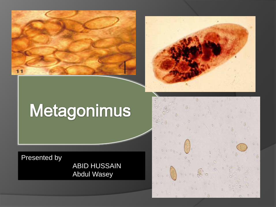
Presented by
ABID HUSSAIN
Abdul Wasey

Metagonimus is a genus of family Heterophyidae.
Cause disease Metagonimiasis.
Metagonimiasis was discovered by Katsurasa in 1911-1913 when he first observed eggs of M. yokagawai in feces .
M. takahashii was described later first by Suzuki in 1930
M. Miyatai was describe in 1984 by Saito
All three species are hermaphrodite.
Smallest intestinal fluke
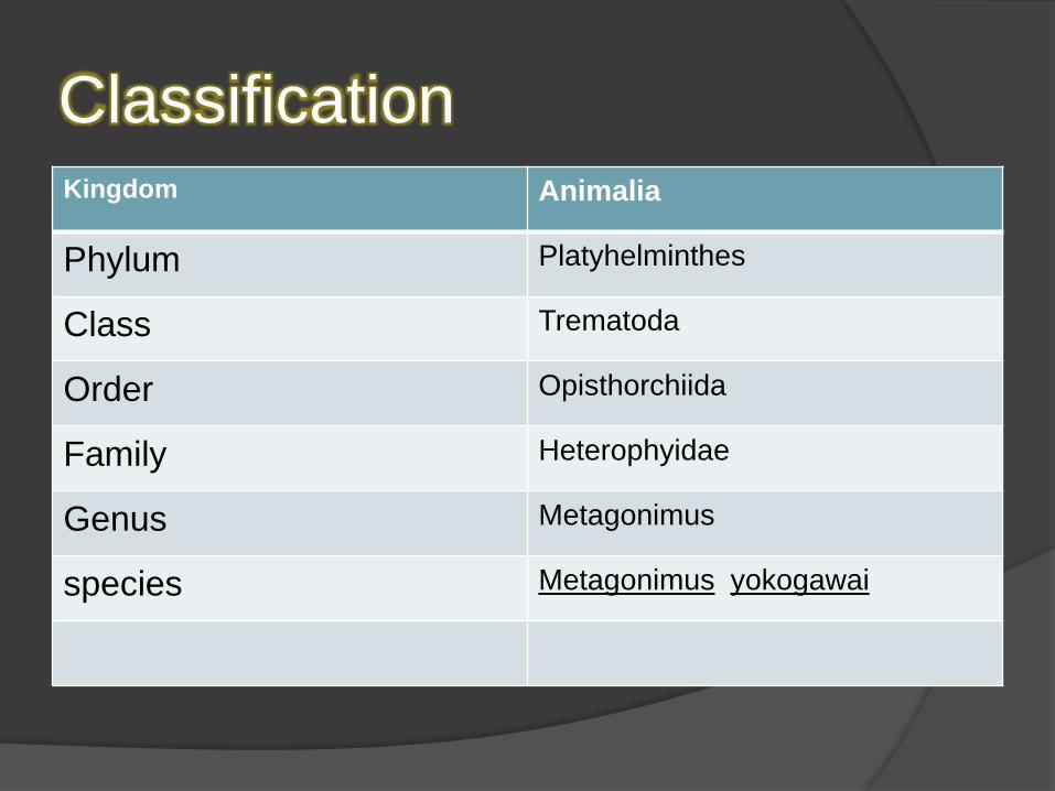
ClassificationKingdom Animalia
Phylum Platyhelminthes
Class Trematoda
Order Opisthorchiida
Family Heterophyidae
Genus Metagonimus
species Metagonimus yokogawai

Metagonimiasis infections are endemic or
potentially endemic in following countries
including
Japan, Korea, China, Taiwan, the Balkans,
Spain, Indonesia, the Philippines , Russia,
India and Pakistan

Metagonimus yokagawai also called
the Japanese fluke
Metagonimus Takahashii
Metagonimus miyatai

Transmission requires two intermediate hosts,
The first of which is snails, most commonly of
species Thiara granifera.
Infection is acquired through the secondary
intermediate host, fish, that haven’t been
thoroughly cooked. Metacercariae encyst under
the scales or in the flesh of fish
Definitive hosts include humans and various fish-
eating mammals, primarily dogs, cats, and pigs.
Fish-eating birds may also be infected with
metagonimiasis

Eggs are very small. Eggs have a
smooth, hard shell that is transparent and
yellow-brown in color, ovoid egg shape.
They are about 26-28 μm length and 15-
17μm width.
The egg also has a very slight opercular
shoulder

leaf-shaped.
It is one of the smallest intestinal flukes, its ventral sucker is deflected to the right of its midline and is closely associated with the opening of the genital pore.
The testes are large and diagonal to each other
Ovary is anterior to the testes and the uterus is filled with eggs. The uterus is the largest organ in the body.
The size of the adult fluke is 2.5 mm length by .75 mm width.


oral sucker (OS), pharynx (PH), intestine
(IN), genitoacetabulum (GA), ovary
(OV), the large, paired testes (TE), and
eggs within the uterus (EG).

Life cycleAdults release fully embryonated eggs each with a fully-developed miracidium, and eggs are passed in the host’s feces
After ingestion by a suitable snail (first intermediate host), the eggs hatch and release miracidia which penetrate the snail’s intestine
The miracidia undergo several developmental stages in the snail, i.e. sporocysts , rediae , and cercariae. Many cercariae are produced from each rediae. The cercariae are released from the snail
It is encyst as Metacercariae in the tissues of fish (second intermediate host)
The definitive host becomes infected by ingesting undercooked or salted fish containing Metacercariae
After ingestion, the Metacercariae excyst, attach to the mucosa of the small intestine and mature into adults


Pathogenesis
It cause Metagonimiasis
Site of infection is small intestine
The incubation period is around 14 days
Infestation may persist for more than one
year

Sign and Symptoms
Diarrhea
Abdominal pain
Nausea
lethargy
anorexia
Fatigue
malaise
seizures, neurologic deficits

Diagnosis
Fecal test
Serological test(Elisa)

Avoid eating uncooked food in endemic area
Prevent fecal contamination of fish ponds.
Education regarding method of transmission
Snail control
Avoid feeding raw fish to cats and dogs in endemic area.

Praziquantel(drug of choice)
Tetrachloroethylene
bithionol
Niclosamide
Nicoflan