+ All Categories
Home > Documents > Material-specific processes in tactile short-term memory

Material-specific processes in tactile short-term memory

Date post: 02-Apr-2022
Category:
Upload: others
View: 3 times
Download: 0 times
Share this document with a friend
37
Portland State University Portland State University PDXScholar PDXScholar Dissertations and Theses Dissertations and Theses 1978 Material-specific processes in tactile short-term Material-specific processes in tactile short-term memory memory Christina Anne Meyers Portland State University Follow this and additional works at: https://pdxscholar.library.pdx.edu/open_access_etds Part of the Cognitive Neuroscience Commons, and the Cognitive Psychology Commons Let us know how access to this document benefits you. Recommended Citation Recommended Citation Meyers, Christina Anne, "Material-specific processes in tactile short-term memory" (1978). Dissertations and Theses. Paper 2736. https://doi.org/10.15760/etd.2732 This Thesis is brought to you for free and open access. It has been accepted for inclusion in Dissertations and Theses by an authorized administrator of PDXScholar. Please contact us if we can make this document more accessible: [email protected].
Transcript

Portland State University Portland State University

PDXScholar PDXScholar

Dissertations and Theses Dissertations and Theses

1978

Material-specific processes in tactile short-term Material-specific processes in tactile short-term

memory memory

Christina Anne Meyers Portland State University

Follow this and additional works at: https://pdxscholar.library.pdx.edu/open_access_etds

Part of the Cognitive Neuroscience Commons, and the Cognitive Psychology Commons

Let us know how access to this document benefits you.

Recommended Citation Recommended Citation Meyers, Christina Anne, "Material-specific processes in tactile short-term memory" (1978). Dissertations and Theses. Paper 2736. https://doi.org/10.15760/etd.2732

This Thesis is brought to you for free and open access. It has been accepted for inclusion in Dissertations and Theses by an authorized administrator of PDXScholar. Please contact us if we can make this document more accessible: [email protected].

AN ABSTRACT OF THE THESIS OF Christina Anne Meyers for the Mslster of

Science in Psychology presented May 30, 1978.

Title: Material-specific Processes in Tactile Short-term Memory

APPROVED BY MEMBERS OF THE THESIS COMMITTEE:

Barry D/'Anderson, Chairman

1 Shane Haydon

Studies· concerning tactile short-term memory (short-term memory

~or the sense of touch) have often been contr9dictory. Some of these

s tu.dies support the existence of rµodali ty-specific tactile memor:'{, a

separote, independent storage system for tactile information. Other

studies do not support such a system. Further, coniusion has arisen

regarding the tactile test materials, since many of them use common

shapes which are easily labeled verbally. It is hypotheslzed that

information which can be labeled is stored in material-specific verbal

memory in the left hemisphere,.while patterned or spatial information

is·stored in mnt~rial-specific nonverbal memory in the right hemisphere.

This paper reports two studies conducted to demonstrate both

verbal and nonverbal material-speci~ic memory using tactile test

materials. The first experiment utilized the Seguin Formboard,

which has wooden shapes that are easily labeled verbally. The test

2

was administered to brain damaged patients and to normal controls.

Results showed that the performance of the.people with left hemisphere

brain damage was significantly impaired relative to the normal controls.

This was expected since verbBl material is p~ocessed in the ·1eft

hemisphere. People with damage in this area have dif'ficulty nai¢-ng

objects and storing the names.

The second experiment utilized. wooden shapes that were presumed

difficult to label. This test was again administered to brain damaged

subjects as well us to normal controls. The results were not

signifieant. 1Fni s may have been because the test was too difficult

or because H did not. tsp nonverbal spatial information. Thus, people

with right hemisphere damage were expected to have difficulty processing

this type of material. Tne right damaged group did tend to do more

poorly than the other groups. It is not known if this difference

would be significan~ were the test shnplified, or if there were

actually no group differences.

The first study suggests that the Seguin Formboard, thought to

be a nonverbal tactile memory test, is actually verbally mediated. The

second study did not yield significant results, but suggests a line of

further research into the area of nonverbal material-specific memory·

tested in the ta_atile modality.. These experiments suggest the import-

ance of carefully evaluating test materials to determine what abilities

they actually.measure in order to obtain a fine analysis of memory function ..

MATERIAL-SffiCIFIC PROCESSES IliI TACTILE SHORT-'I$Rt~ MEMORY

by

CHRIS 'J?INA ANNE ~YERS

A thesis submitted in partiol fulfillment of the requirements for the der;ree of

MASTER OF 8C IENCE in

PSYCHOLOGY

FOrtland State U~iv~rsity

1978

PORTlAND STATE UNIVERSITY UBRABY

:------·------ -----------

TO THE OFFICE OF GRADUATE STUDIES AND RESEARCH:

The members of the Committee approve the thesis of

Christina Anne Meyers presented May 30, 1978.

APPROVED:

Barry D. AnderOil, Chairman '

shaneo. "H',.,

of Psychology

Studies and Research

~/

ACKNOWLEDGMENTS

I wish to extend my appreciation to Dr. Ylllriel Le·zak for the

use of her research material, and to the Portland M=tropolitan

Stroke Club for their generoiJ.s cooperation.

ACKNOWLEDGMENTS •

LIST OF TABLES • • • •

LIST OF FIGURES

Ilfl'RODUCTION . . .

TABLE OF CONTENTS

. . . . . . .

Components of STM • •. • • • • • • • • • •

Physiological Basis of ~terial-Specific STM

Physiological Basis of Modality-Specific STM

EXIERIMENT 1

EXPERil-1ENT 2

GENERAL DISCUSS ION

REFERENCES

. . . .

PAGE

iii

v

vi

l.

2

6

7

16

21

25

27

LIST OF TABLES

TABLE PAGE

I Statistical Results for Experiment 1:

Timed Trials • • • . • • • • J • • • • • • • • • 17

II Statistical Results for E:>..""Periment 1:

Memory and Location. Scores • 17

III Statistical Results for Experiment 2 . 23

:.- .... 11

~

I I

LIST OF FIGURES

FIGURE

1. Short-term V~mory Systems (l~ssaro, 1973) . . -

2. Thalamus (Netter, i972, p .. 48) .•.•.• ; .•

3. Tactile M:!mory Task Used by Ghent et al. (1955)

4. Shapes of the Seguin Formboard

5. Means (± SEM) for Site o:f Lesion, Experiment 1:

Timed Trials • •

6. Means {~ SEM) for Hand Used, Experiment 1:

Timed· Trials • • • • • • # , • • • • • • • • • • • •

··:

7.

8.

Mean~---(!' SEM) for Site of' Iiesion, Experiment 1:

Memory and Location Scores • • • • •

Means (~ SEM) of Ail Trials, Experinient 2

PAGE

3

7

11·

14

18

19

19

23

__ ,, _____ ,, -----------·----------·-·--------- ------------------·----

INTRODUCTION

Short-term memory (STM) is a transient, unstable trace of very

recent events (Horton & Turnage, 1976, p. 152). Information in this

system decays within approximately 30 seconds unless a control process,

such as rehearsal, maintains it for a longer period of time (Atkinson

& Shiffrin, 1968). S'I'M is thought to consist of two systems; material-

. specific· and modality-specific STM. Modality-specific STM is the·

relatively unprocessed information comin.g in directly from the senses.

A separate, independent storage system is hypothesized for each· sensory

modality, each of which ~s processed bilaterally in tbe brain, that is,

equa·lly in both hemispheres. Visual and auditory ST.M have been studied

most extensively. :V~terial-specific STM refers to tee storage systems

that depend upon the way in· which information is coded, verbally or

nonverbally. The left hemisphere o~ the brain stores the verbal

material, and the right hemisphere stores nonverbal material.

A number of studies have concerned tactile memory (memory for the

sense of touch), but findings are contradictory. While some studies

suggest there is a tactile memory, as there are visual and auditory

memories, others fail'to show the existence of a separate tactile

memory. Further, some confusion has arise·n regarding the tactile test

materials, since many of them utilize connnon shapes (e.g .. , squares and

circles), which are easily labeled verbally. This confounds the

modality-specific tactile memory results with a material-specific

·component.

. I

-:

2

This paper will begin by discussing material- and modality-

specific STM, including the possible.neuroanatomical correlates of

these sys.tems, and then focus on tactile memory. Two studies will be

presented on tactile STM; one using connnon, easily labeled shapes, and

one using shapes that cannot be easily labeled. These studies will

try to demonstrate both verbal and nonverbal material-specific

tactile memory.

COMRJNENTS OF STM

Memory can be discus·sed in terms of the physical phenomena, such

as sound and light, that carry the information to the senses.

Massaro (1973) makes the·assumption that since memory is closely tied

. to perception, the dimensions of ..memory are ana1agous to the dimensions

6f' ·sound and light after·they· are processed in the brain.

Massaro postulates an information-processing m~del of' the.

processing operations between reception of' the initial stimulus and

the meaning that is derived from it. First, the stimulus is held in

preperceptual storage for approximately 250 msec. There is a one-

to-one relationship between the stimulus and the in:formation in this

system. At this point feature detection occurs, which is the analysis

of simple· physical attributes such as size, shape, and color.

Secondly, higher pattern recognition occurs. This is a transformation

of the features held in preperceptual storage into a percept. This

11gestalt" is stored in synthesized memory.. Finally, conceptual

processing takes place to derive meaning. This information is then

stored in generated abstract memory. This memory store contains abstract

I I I

l.

rather than modality-specific information (:Massaro, 1975, p. 7-12).

Massaro hypothesizes that both synthesized and abstract memory are

parts of STM, and that information in either form can be maintained

independently (see Figure 1). Synthesized memory is commonly termed

modality-specific memory. Generated abstraet memory is equivalent

to material-specific memory •

sound wave p:i:tte rn

• ~ preperceptual 't synthesized ~, ~auditory .;, auditory

storoge memory .J, generated

3

abstract--· ;..meaning

ligh wave patt

memory

preperceptual synthesized T ;,visual ~visual rn storage . memory

detection perception conception

Figure 1. Short-term memory systems (¥assaro, 1973).

To test this model, YDssaro (1973) used same-different reaction

time tasks.. The subjects were presented with two spoken letters, and

were to decide if they had the same or different names. The indepe_nd-

ent variable was whether the two letters were presented by the same or

different speakers. Massaro found a faster. reaction time on both the

same and different name trials when the same speaker articulated the

·letters. This faster reaction time was independent of a delay between

the presentation of the two letters. Massaro suggests the subjects

retained the experimenter's voice in synthesized auditory memory and

used that inf'ormatj_on to facilitate recognition of the second letter.

4

Massaro found a similar resu1t for visually presented letters.

Reaction times were approximately 80 msec faster with physical matches

(i.e., letters printed in the same way) than with name matches under

a no-delay condition when the letters were printed differently.

When an interval was interpolated between the two stin1ulus letters, the

reaction times were the same. Apparently, when a delay is introduced

subjects compare the letters on a name basis-; utilizing material-

specific memory. Beller (1967) also found that the response ~ime to

physically identical letters was faster than to physically different

letters with the same name· (i.e.,~ and~)· Posner & Mitchell (1967)

found similar results. Tnus, the faster reaction time to physically·

match letters, as gpposed to naming them, points to serial processing

.of' the informatio~ in this memory. sys.tem, from modality-s:peci:fic to •·

~aterial-specific memory. If the subjects are instructed to say the

names of the letters aloud, then the reaction times between physically

matching and naming are identical. That is, it takes just as long to

recognize three same and three different letters (Ingalls, 1974).

Thus, if the instructions are to internally decide sa~e or different,

modality-specific memory is tapped, and the physical attributes are

compared. If the instructions are to speak the letter names and decide

if they are the same or different, material-specific memory is tapped,

and the semantic attributes are compared. V.i.assaro (1975, p. 13) also

notes that there is probably some overlap of these systems. For

instance, modality-specific memory may eliminate alternatives to what

is heard (such as shoes or choose). Mlterial-specific memory may note

that only shoes is. correct· semantically (as in "Take off your shoes"}

5

and feeds back that information into modality-specific store so the

word is heard as shoes. Although the information usually goes from

percept to meaning, conceptual information may modify perceptual

experience.

Massaro (1973) also presented evidence for separate visual and

auditory memory stores. Subjects were given a list·of first auditory

digits, and then visual letters for one presentation. Since STMhas a

limited capacity, the auditory list should have caused a decrease in

the recollection of the visual list. H9wever, the correct recall for

each list was identical. This then suggests the existence of modality-

specific STM; a separate, independent memory store for each sensory

modality • .' M9.ssaro also noted that in a shadowing task, where subjects

first remember a letter ~resented visually or orally, and then repeat

back the auditory list, auditory shadowing interfered with the recall

of the auditory list much more than the visual shadowing did.

Short-term memory can then be discussed in terms of two systems:

Modality-speci~ic, such as visual and auditory memory, and material-

specific, such as verbal and nonverbal memory. There are several

behavioral differences that distinguish these systems. Forgetting is

very rapid in modality-specific memory (about 5-15 seconds), and is as

rapid for filled as fur unfilled retention intervals if the interpolated

task is in a modality'different from.the one under observation.

Events occuring in other sensory modalities do not affect the modality-

specific memory of a particular modality. Events in the same modality

as a given sensory storage system will interfere with it and cause it

to be lost (Sch~n, et al., 1973). Verbal STM, due to the higher

r---

6

level processing of the inforrr~tion, may last up to 30 seconds

unrehearsed. In verbal STM, interference comes from several modalities

simultaneously. Also, loss from verbal STM is greater when the retention

interval is filled with verbal material than with nonverbal material.

Investigators are now determining the physical correlates in the.brain

of these two types of STI~.

PHYSIOLOGICAL BASIS OF MATERIAL-SPECIFIC STM

Fedio & Van Buren (1972) have implicated the ventrolateral

thalamus in verbal and nonverbal short-term memory. The subject pop-

ulation consisted of people undergoing unilateral left or right

thalamotonzy- for relief of Parkinsonism. · Each patient had a therapeutic

electr.ode ~nserted through a medi~l parietal burr bole into the pulvinar

~ucleus and the remainder thalamus (anterior and inferior to the pulvinar)

{see Figure 2). Each patient was given verbal and nonverbal memory

tests. Fedio & Van Buren found that naming errors were the result of

left p\llvinar stimulation, that is, the patient was unable to recall

the names of co:m:mon objects correctly identified before the stimulation,

even though there were no speech disturbances. These patients had no

difficulty with the nonverbal task. Right pulvinar stimulation

produced perceptual discrimination errors with objects correctly

perceived before sti..."llulation, but no difficulty with naming or speech.

In this case, the pat'ient was unable to recognize complex patterns he

identified before the stimulation. Excitation of regions ·outside the

pulvinar nucleus produced no disturbances of memory.

Fedie & Van Buren also found that at lower levels of stimuJ.ation

·

"

7

verbal and nonverbal memory remained basically intact, but visual STM

was disrupted. This results from the reciprocal connections the

pulvinar nucleus has with the visual association areas of the cortex

{Clark, 1975). Thus, although the thalamus is not a "language center"

as such, there may be a system involving the cortex and the thalamus

the regulates language in the left hemisphere and nonverbal abilities

in the right hemisphere. There has been increasing clinical evidence

of patients with thalamic lesions that manifest language disorders

(Brown, 1974 and Riklan & Cooper, 1975).

MEO.GEN .. BODY

,_,.T. Gf;i11. 6oOY

Figure 2.. Thalamus (Netter·, 1972, p. 48).,

PHYSIOLOGICAL BASIS OF MODALITY-SIECIFIC STM

The foregoing indicates that subcortical structures are involved

with material-specific (i.e., verbal and nonverbal) STM, and that the

left hemisphere processes verbal information and the right hemisphere

processes nonverbal information (Fedio & Van Buren, 1972). Tnere are

'

. j

f

I I

!

I

j

I . I . I

I 1 ·

8

now a large nwnber of studies investigating cortical damage and impliM

eating these lesions in modality-specific memory. l·:Odality-specific

memory involves separate, independent storage systems for each sensory

modality. These sensory systems contain information in a relatively

unprocessed form as compared to verbal memory (Schurman et al., 1973).

Visual STM

.Butters et al. (1970) investigated patients with left and right

parietal lobe damage. The hallmark symptoms of these people include

.impaired spatial orientation. They a~e unable to visualize how an

objec~ looks from different perspectives. This inability to rotate

objects mentally may be just a manifestation of a more basic· deficit, .·

the ina.bili ty to retain visual in1ages. . A visual STM disruption would

·~~lp explain the various· constructional-spatial problems of pa~ietal ~

patients, such as the inability to recognize faces or to reproduce a

design with blocks. It is kno~n1 that the parietal lobes are involved

with the processing and storage of visual information.

Butters et aL .. (1970) tested the hypothesis of visual memory

disruption in parietal lobe damage. The subjects were presented with

.. a visual or auditory stimulus, and required to identify it. after no-

delay or a delay of 3, 9, or 18 seconds. If the patient correctly

identified the object.under.the no-delay condition, it suggested that

the information was getting into the brain ·correctly, ruling out a

registration difficulty. If both the delay and no-delay-condition

per~ormances were impaired, perceptual processes were assumed to be

disrupted. If the patient was able to perfbr rn at no-delay, but was

unable to perform adequately after a delay, a deficit of memory was

•I I

l

9

indicated.

This study compared people with lef't parietal (LP), left frontal

(LF), right parietal (RP), and right frontal (RF) lobe damage, and

normal controls (NC). Butters and his coworkers found no group di:ff-.

erences in the ability to identify geometric patterns under the no-

delay conditions. The RP and LP groups made significantly more errors

und.e1• the visual delay conditions. The performance of the RP group

decayed most rapidly as the delay interval increased from 0-18 seconds,

although both the RP and LP groups were significantly impaired. The

same results were obtained using visually presented single consonants

and consonant trigrams. Butters and his coworkers also found alexic

symptoms in the LP group when consonants were presented visually,

J~:hich introduced a regi"stration deficit for this group. The LP group

was also impaired for the consonants presented in the auditory modality

because of' this language disturbance.- Butters hypothesized that the

lef't hemisphere may be involved in both the processing of' verbal

information in any modality and in the storage of patterned visual

material. The major conclusion of this study was that both parietal

lobes are involved in the storage of' modality-specific visual

material.

Auditory STM

Samuels et al. (1972) did a similar study with patients who

had unilateral excisions of' the temporal lobes for the relief of

epilepsy. Again, the patients were presented with visual and auditory

information and asked to identify it immediately or after a delay. The

stimuli were visually presented geometric patterns, visually presented

•,

10

consonant trigrams, and orally presented consonant trigrams. Samuels

and her coworkers found no group effects :for visual patterns or visual

consonants. With auditory consonants, they found no differences

between the groups under the no-delay condition, but both left temporal

{LT) and right temporal {RT) damaged groups made more errors under

the delay condition.. There were no differences between the LT and RT

groups. Both left temporal and rigbt temporal lesions appear to

disturb identification of auditory letters, while only left hemisphere

lesions disrupt the processing of words. · The left hemisphere processes

the syntactic-semantic aspects of language, while the more basic

acoustic analyses are processed bilaterally. The consonant trigrams

used had low association or linguistic value, and were thus similar to

nonsense words. These ~ere proce~sed equally in both ?emispheres,

which indicates modality-specific memory is bilateral.. Material- .

·specific memory is :pro·cessed only in the h~mis:phere that handles verbal

or nonverbal material.

Tactile STM

Thus far the discussion of modality-specific memory has been

limited to auditory and visual memory.. There is also a large body of

research on tactile memory, but much of it contains contradictory

results.

Ghent et al .. (1955) used tactile patterns made of raised metal

strips on a wooden block (see Figure 3). The blindfolded patient feels

a sample pattern for 5 seconds and then selects that pattern from the

array on the board. Ghent gave each patient six trials:

l. Using hand ipsilateral to the lesion ..

-" ....

'

.l l

11

2. · Using hand contralateral to the lesion. 3. Contralateral hand. 4. Ipsilateral hand. 5. Ipsilateral hand. 6. Contralateral hand.

The patient's score was the number of correct choices made. Among

the normal controls, there were fewer errors on Trial 3 than ~or Trial

1, which indicates learning. Among the brain damaged patients, the

ipsilateral hand showed improvement over trials 1, 4, and 5, but the

contralateral hand did not show improvement. Among the controls, the

improvement between their own ipsilateral and contralateral hands was

not significant, but there was a difference between the performance of

the ipsilateral and contralateral hands in the brain damaged group •

" ......... , .... .._..,...._ ........... -. ........ -

'.( '•

I J

L _J

Figure·3. Tactile _mem~ry task used by Ghent et al. (1955).

Ghent and her coworkers at first thought the lack of improvement in the

brain damaged contralateral hand was due to sensorimotor disturbance •

...........

12

They then divided the patient. group into those with somesthetic or .

motor defects of the hand, and.those with no such defects. The

contralateral hand still showed no improvement in either group,

while the ipsilateral hand. improved in both groups. Ghent then

divided the, patient group into various other categories, such as

locus of lesion, presence of aphasia, or presence of epilepsy. None

of these subgroups showed improvement in the hand contralateral to

the brain damage, while all .improved with the ipsilatera1 hand.

Ghent et al. (1955) describe th.is tactile memory impairment ·in the

contralateral hand as a difficulty in learning, but it could also be

thought of as a deficit of tactile modality-specific STM. Ghent found

that a lesion anywhere in one hemisphere caused this impairment ..

. This would ·seem to rule out a specifi.c location within the brain that ......

handles tactile material •

. Schurman et al. (1973) investigated memory for two successive

touches on the arm to determine if the interval between the touches

and the presence or absence of an interpolated task in this interval

affected tactile memory as it·does visual memory. They found· a gradual

decrease in correct recall for both filled and unfilled intervals

over time. Events occuring in other modalities, such as. auditory

counting, did not affect performance. This study supports modality-

specific memory for touch.· However, Helgoe (1972), also working

with touches· to the forearm, found that recall was negatively affected

by counting backward during the retention interval.·

The interpolated task also interfered with tactile memory in a

study by J. Clark (1974). When subjects were giyen a tactile pattern

' -.,

.!

.1

13

to retain, they made more errors when a visual search task was inter-

polated in the retention interval. When a tactile search task was

introduced in the interval, performance also deteriorated, but not as

much as with the visual search. Clark_ presented two possible explana-

tions for his results. First, the tactile pattern was somehow coded·

and stored in visual STM. The alternative explanation was that both

visual and tact:!.le information were coded in some combination. Clark

may have instead tapped material-specific memory for nonverbal tactile

patterns. This would account for the interference from the visual

task."

A connnon tactile memory test used by clinicians is the Seguin.

Formboard. This test is commonly thought to test nonverbal tactile

memory •. The test consis:ts of' ten~ wooden shapes placed in appropriate

holes in a wooden board (see Figure 4). Each patient is blindfolded,

and the s-hapes are place.d in front .of the formboard within easy reach.

The patient, using first his preferred hand (Pl), places the shapes

into their appropriate holes. The score is the time to place all ten

shapes, in seconds. The second trial is with the subject's nonpreferred

hand- .(NP). Both hands (B) are used for the third trial, and finally

the preferred hand (P2) for the last trial. ·The test materials are

then removed and the patient unblindfolded. The patient is then asked

to draw on a piece of.paper the shapes (memory score) and their

approximate locations on the board (location score).

Some investigators have found that left hemisphere damaged patients

do better than right damaged patients on the blindfolded task, but

right damaged patients do better on the recall task. The better recall

'-...

14

of the right damaged patients may be due to the ease with which the

shapes may be labeled, utilizing verbal memory (Lezak, 1976, p. 381).

Lezak also notes (p. 383) that if trial Pl takes about 420-480 seconds

and trial NP takes about 180-300 seconds, a left hemisphere lesion is

indicated. If trial NP takes longer than trial Pl, but trial B is

·shorter and the memory score is adequate, a right hemisphere lesion

is indicated.

D D [ J •.

'j

O<=>.D Figure 4. Shapes of the Seguin Formboard.

There is some controversy coqcerning the type of brain damage

to which the Seguin Formboard is most sensitive. Reitan (1964, p.

308) reported his frontal lobe damaged groups performed worse than the

non-frontal groups. He· round differences between right frontal and

le:ft nonfrontal groups, and between left frontal and right nonfrontal

groups, which is not an appropriate comparison (Lezak, 1976, pl 382). ·

'-..,

15

Reitan also found differenc~s between left frontal and right frontal

groups on Trials NP, B, and total time score. He did not find

differences between the frontal and nonfrontal groups within the same

hemisphere. Reitan also found that .the left damaged groups did better

with their ipsilateral hand, which is consistent with the findings

of Ghent et al. (1955)r

· Teuber (1964, p. 421) fou~d that the nonfrontal groups did worse

than the frontal groups on both the formboard task and memory scores.

Other researchers have alco found the frontal groups to perform better

(Lezak, 1976, p. 382).

Because of the contradictory nature of the research on tactile

memory, and the lack of distinction bet.ween modality-specific and

material-specific tactil~ memory, the following two studies seek to .. ~ determine how brain damaged groups process tactile material that can

be labeled verbally and tactile material that ·cannot be labeled

verbal~y. These studies will investigate the possibility of the

existence ·of" material-specific tactile memory for both verbal and

nonverbal material.

'-..,

EXPERIMENT 1

The purpose of this eXJ;>eriment was to demonstrate material­

specific tactile memory for verbal material.

Method

fJubjects. Twenty-seven subjects were selected from a population

of brain damaged people being tested :tn an eight-year longitudinal

study conducted .by Dr. Muriel Lezak at the Portland VA HospitaL Nine

of the patients had left hemisphere damage, nine patients had right

hemisphere damage, and nine.patients had bilateral-diffuse damage. The

experimenta.l subjects (all males) ranged in age from 20 to 47, with a

mean age of 28. Twenty-one had brain damage as a result of traumatic

injury, 3 from .cerebral-vascular acci~ents, and one each from infection,

tumor, and anoxia. Neurological reports and the side of hemiparesis,

if any, 'Vere used to group the subjects into lef't, right, or bilateral

diffuse categories. Nine subjects were also tested as normal controls.

These subjects (all males) ranged j_n age frorn 19 to 39, with a mean

age of 26.

Procedure. Each patient was given the Seguin Formboard test in

accordance with the standard administration as des~ribed previously.

Scores were obtained for trials Pl, NP, B, and P2, memory, and location

for each subject. Differences between the left and right hemisphere

damaged groups were expected since the Seguin forms are easily labeled

17 verbally, which the right damaged people might utilize to facilitate

recall.

Results and Discussion

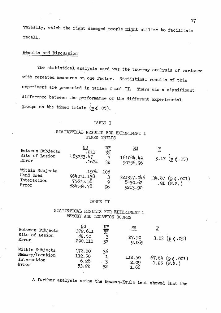
The statistical analysis used was the two-way analysis of variance

with repeated mea.sures on one factor. Statistical results of this

experiment are presented in Tables I and II. There was a significant

difference between the performance of the different experimental

groups on the timed trials (]2 ( . 05).

TABLE I

STATISTICAL RESULTS FOR EXPERil!'.lENT 1 TIMED TRIAI.S

SS DJ:i, MS Between Subjects -:-211 35 Site of Lesion 483253,.47 3 161084.49 Error .1624 32 50756.,96 Within Subjects .1924 108 Hand Used 964071.138 3 321357.046 Interaction 75875.58 9 8430.62 Error 884534.78 9? 9213.90

TABLE II

STATISTICAL RESULTS FOR EXPERIMENT 1 MEMORY AND LOCATION SCORES

SS DF M3 Between Subjects 372.""611 35 Site of Lesion 82. 50 3 . 27. 50 Error 290.111 32 9.o65 Within Subjects 172.00 36 Memory/Location 112.50 l 112.50 Interaction 6.28 . 3 2.09 Error 53.22 32 1.66

F

3 -17 (g < . 05 )

34. 87 ( p < . 001 ) . 91 (N. s.)

F

3 . 03 (I! ( . 05 )

67.64 (12 ( .001) 1.25 (N.S.)

A further analysj.s using the Newman-Keuls test showed that the

I

I

I

-- - --- --- - ------ -------- ---

18

left damaged group was significantly impaired relative to the normal

·control group (J2 < . 05). No other comparisons between groups proved

to be significant (see Figure 5). On the memory and location scores,

the left damaged group was significantly impaired relative to the

right damaged and control groups <.~<·05). The bilateral-diffuse

group was also significantly impaired relative to the controls

(E, (. 05) (see Figure 7) •.

The overall time taken for· each trial was significantly different

(J!{.001), except for trials P2 and B, which did not differ (see Figure

6). The interaction between the site of damage and each trial was not

signi~icant. The number correct for the memory and location scores

differed significantly (:2, <. 001), favoring the memory scores. There

was no intera.ction between the site of damage and. the memory or

location scores.

700

600 {ll

.ro 500 s:l

0 ()

GJ 400 m

300

200

100

.Left (322.8)

Right (273.75)

Experimental Group

Bilateral (261.44)

Control (163.16)

Figure 5.. Means (± SEM) for site of lesion, Experiment 1, timed trials ..

600

500

400 tll rd ~ 0 300 0 CIJ ti)

200

100

Pl (376. 25)

. NP (283.6 )'

Trial

B (174.4)

P2 (186.58)

Figure 6. Means (~ SEM) for hand used, Experiment 1, timed trials.

7t .p 6 C) CIJ

51 H H 0

0

H 4 Cl)

~ z 3

2

1

I

.Left (4.44)

r!

I

Right (6. 72)

I

Experimental Group

r-I

Bilateral (5.22)

.-l

Control (7.05)

Fig~r·e· ·7,, Means (:.t SEM) for site of lesiop, Experiment 1, memory and location scores.

19

I I j'

20

These results suggest the Seguin Formboard taps verbal short-

term memory. The people with right hemisphere lesions and the control

group were able to label the Seguin shapes verbally to facilitate

recall, while the lef't hemisphere damaged people were unable to do

so. The Seguin Formboard has been traditionally considered a test of'

nonverbal skills, which this study seriously questions. The following

study investigates an aJ_terna.tive test specifically designed to assess

nonverbal abilities.

EXPERIMENT 2

Method

Subjects. Twelve subjects volunteered from the Portland

Metropolitan Stroke Club, Portland, Oregon. Half of these people had

left hemisphere strokes, and half had right hemisphere strokes. The

experimental subjects (9 males and 3 females) ranged in age from 44 to

67, with a mean age of 57. The site of da:m.&.ge was determined by the

side of hemiparesis, if any, presence of aphasia, and verbal reports

from the subject or his family. Another six normal control subjects

were also tested. The control group ranged in age from 46 to 75, with

a mean age of 62.

Procedure. Each subject was first given a tactile acuity test

to determine if his sense of touch was adequate for the tactile memory

test. This also determined if the tactile information was being

received in the brain correctly. The test consisted of having the

subject feel two wooden shapes conceaJ_ed behind a curtain. The tactile

materials consisted of 6 three-dimensional shapes made by gluing five

wooden cubes (3/4 in. sq.) into various configurations. The subject

judged whether the shapes felt alike or different. There were three

trials under this no-delay condition.

The subject was then presented with a sarnple shape, concealed

:~behind a cur~ain. He felt. the shape with the hand ipsilateral to the

stroke for· as long as desired (control subjects used their preferred

••

22

hand). After a 5-second delay, the subject was then asked to pick out

that same shape from an array of six different shapes behind the curtain.

The subject was then given another sample shape to feel behind the

curtain, and asked to identif'y it visually from the array of six

shapes after a 5-second retention interval. Three trials were given

under both the tactile-tactile and the tactile-visual conditions. The

score for each Ct)ndition was the number of correct choices made,

ranging from 0-3 for each condition.. A tape of' hospital pages was

played throughout the test to help confound any attempts at

verbalization.

It was expected that the right hemisphere damaged group would

be impaired relative to the left damaged and control groups. These

shapes are primarily spatial, and any attempts to verbalize them ·wouJ.d

be inefficient. The· left damaged and control groups would use their

nonverbal memory store to retain the information.

Resul'ts and Discussion

A two-way analysis of' variance with repeated measures on one

factor was used to analyze the data. Statisti~l results for

Experi..~ent 2 are presented in Table III. The site of damage did not

significantly affect performance, although the differences were in

the predicted direction (see Figure 8). The right damaged subjects

tended to do more poorly than either the left damaged group or normal

controls. 'I'he right damaged group also tended to go more slowly

during the test and to use cues such as the number of grooves in the

shape to facilitate recall. Several subjects in this group tried to

23

scratch the surface of the design to lea.ve an identifying mark.

TABLE III

STATISTICAL RESULTS FOR EXPERIMENT 2

SS DF M3 F Between Subjects 22."54 17 Site of Lesion 3.37 2 i.685 L 32 (N.S.) Error 19.17 15 1.278

Within Subjects 24.oo 36 '

Trials I 9.15 2 4.575 10. 05 (p1

(. 01) Interaction l.19 . 4 .2975 .654 "{°N.S.) Error 13.66 30 .455

The performance under the two memory trials differed significantly

(£( .01) from the no-delay condition. The tactile-tactile and tactile-

visual conditions did not differ ·s.ignificantly. This was expected

if material-specific memory was being tapped, since this system stores

info~mation from all sensory modalities.

.µ ()

<l> H S--1 0 0

S--1 <l>

~ z

7

6

5

4

3

2

1

Left (5.67)

.Right (~. 83)

Experimental Group

~

Control (6.67)

Figure 8'. Means (± SEM) of' all trials, EX]?eriment 2.

There are several possible factors that might account for the

lack of significant differences between experimental groups. One

is that the procedure and test designs were too difficult for a

24

large distribution of performance to be seen. In fact, the average

per cent correct across all conditions and groups was 63. Only one

subject in the control group perf'ormed at lOCP/o correct, a.nd one scored

35% correct. If the test figures and procedure were redesigned to

yield a wider distribution of performance, significant differences

may appear between the groups. The significant difference between

the no-delay and delay conditions would probably remain stable, since

it reflects that the minimum ability necessary to take the test (ie.,

tactile acuity) is not dependent upon memory function.

Another reason for the lack of significant ~ifferences in this

study may have been the age of the Eubjects. The ages ranged from 44

to 75 years, and the older subjects tended to perform more poorly

regardless of whicq experimental group they were in.

I

GENERAL DISCUSSION

Material-specific tactile memory for verbal material was

demonstrated in Experiment 1. Since verbal material is processed

and stored in the left hemisphere, people with damage in this area

have difficulty with labeling and storing these verbal labels. This

experiment also suggests that the Seguin Formboard, thought to be a

nonverbal tactile memory test, is verbally mediated. It is of great

importance that clinicians are aware of what a given test actually

measures, otherwise the results obtained may be very misleading and

cause problems in the diagnosis of organic or functional disorders.

Ma.terial-specific tactile memory for nonverbal material wa.s

not demonstrated in Experiment 2. As previously discussed, the diff­

iculty of the test and the age of the subjects may have obscured any

real differences between the experimental groups. ·A similar test

with simpler figures may indicate whether this test is indeed sensitive

to right hemisphere damage, suggesting a nonverbal memory component,

or if material-specific· nonverbal memory is not being examined.

Another possibility is that material-specific nonverbal memory is not

located in the right hemisphere.

The results in Experiment 1, using the Seguin Formboard, were

obtained from timed trials, while the results· :from Experiment 2 were

obtained from the numbe~ of correct decisions made by the subjects.

The two studies may be made more comparable if Experiment 2 was

modified to be a timed task. In this case, the test itself could be

performed at 1000~ a.ccuracy by all subjects, but the time taken to

complete the task may vary by experimental group.

26

The most important conclusion of these studies is the questioning

of the adequacy of memory tests, or tests in general. In the clinical

evaluation of memory fu.nction, discriminative testing will yield

valuable clues as to the locus of the brain damage, the amount of

lntellectu.al and behavioral compromj_se, and the types of remedie.l

treatments that would be most effective. Thus, it is of utmost

importance to have a clear understanding of wha.t the memory tests

actually measure in order to obtain an accurate and fine analysis of

memory functioning.

REFERENCES

Atkinson, R. C. & Shiffrin, R. M. Human memory: A proposed system and its control processes. The Psychology of Learning and Motivation~ New York: Academic Press, 1968.

Beller, H. K. Farallel and serial stages in matching. Journal of Expe:~iment.al Ps~~~~' 1970, 84, 213-19.

Brown, J. W. Language, cognition, and the thalamus. Confinia Neurologica, 1974, 36,. 33-60.

Butters, N., Samuels, I., Goodglass, H., and Brody, B. Short-term . vtsual and auditory memory disorders after parietal and frontal lobe damage. Cortex, 1970, VI, 440-459.

Clark, J. L. Short-term memory for visual and tactual patterns: The effects of information processing load and interpolated task modality. Thesis submitted to the University of Cincinnati, 1974. Dissertation Abstracts International, 1974, 35, 3053-4.

Cra.ik, F. I. M. Short-term storage in a 'levels of processing' framework. Paper presented at the meeting of' the Midwestern Psychological Association, Chicago, 1973.

Fedio P. & Van Buren, J. Electrical stimulation of thalamic mechanisms for immediate memory in man. Paper presented at the annual meeting of the American Psychological Association, Honolulu, Hawaii, 1972. ·

Ghent, L., Weinstein, S., Semmes, J., and Teuber, H.-L. Effect of unilateral brain injury in man on learning of a tactual discrimination. Journal of Comparative and H-1ysiological Psychology, 1955, E:Q, 478-481. -

Helgoe, R. Effects of repetition in tactile memory. Thesis submitted to the University of D.linois at Urbana-Champaign, 1971. Dissertation Abstracts International, 1972, Jg_, 6077.

Ingalls, R. P. Effects of same-different patterns on tachistoscopic recognition of letters. Journal of Experimental Psychology, 1974, 102, 209-214.

Horton D. & Turnage, T. Human learning. Englewood Cliffs, New Jersey: Prentice-Hall Inc., 1976.

Lezak, M. D. Neuropsychological assessment. New York: Oxford University Press, 1976.

28

Massaro, D. W. The dimensions of short~term memory. ~per presented at the meeting of the Midwestern Psychological Association, Chicago, 1973.

Massaro, D. W. (ed.) Understanding language. New York: Academic Press, 1975.

Netter, F. H. CIBA collection of medical illustrations, VoL I: The nervous system. CIBA Pharmaceutical Co., 1972.

Posner, M. I. & Mitchell, R. F. Chronometric analysis of classifi­cation. Psychology Review, 1967, 74, 392-409.

Reitan, R. M. · Psychological deficits· resu1ting from. cerebral lesions b man. In J .. M. Warren & K. P..kert (eds.) The f:r.ontal granular cortex a.nd behav~or. New York: McGraw Hill, 1§61~ ...

Riklan M. & Cooper, I. S. Psychometric studies of verbal functions following thalamic lesions in humans. Brain & Language, 1975, g_, 45-64 ..

Samuels, I., Butters, N., and Fedio, P. Short-term memory disorders following temporal lobe removals in hmnans. Cortex, 1972, VIII, 283-298.

Schurman, D. L., Bernstein, I. H .. , and Proctor, R. W. :Modality­specific short-term storage for pressure. Bulletin of the Psy~honomic Society, 1973, 1:_, 71-74.

Teube~, H .. -L. The riddle of f~ontal lobe function in man. In J. M. Warren & K. Akert (eds.) The frontal granular cortex and behavior. New York: McGraw Hill, 1964 ..


Recommended