JAVIER ARSUAGA∗, YUANAN DIAO† , AND MARIEL VAZQUEZ‡
Abstract. In recent years, knot theory and low-dimensional topology have beeneffectively used to study the topology and geometry of DNA under different spatialconstraints, and to solve the topological mechanisms of enzymes such as site-specificrecombinases and topoisomerases. Through continuous collaboration and close inter-action with experimental biologists, many problems approached and the solutions pro-posed remain relevant to the biological community, while being mathematically andcomputationally interesting. In this paper, we illustrate the use of mathematical andcomputational methods in a variety of DNA topology problems. This is by no meansan exhaustive description of techniques and applications, but is rather intended to in-troduce the reader to the exciting applications of topology to the study of DNA. Manymore examples will be found throughout this book.
Key words. DNA knots, bacteriophage P4, DNA packing, random knots, site-specific recombination, Xer, tangles.
Motivation. DNA presents high levels of condensation in all organ-isms. Volume reduction, defined as the ratio between the volume occupiedby a given genome and the volume occupied by a random walk of thesame length as the genome, ranges from 102 in Escherichia coli to 104 inhumans[50].
These large condensation values lead to questions such as how theDNA is packed inside the eukaryotic cell nucleus, the prokaryotic cell, aswell as inside other organisms such as DNA viruses. The complexity of thepacking problem is magnified when one considers that the DNA moleculeneeds to be readily available to multiple biological processes essential to theproper functioning of the organism, such as DNA replication, transcription,recombination and repair. The cell has evolved tools to remove unwantedDNA entanglement and solve other topological problems, such as DNA un-(or over-)winding, knotting or linking, and formation of multimers, thatmay interfere with its functions. DNA topology, the study of geometrical
∗Department of Mathematics, San Francisco State University, San Francisco, CA94132, USA ([email protected]). This research was supported in part by the Institutefor Mathematics and its Applications (IMA). J. Arsuaga is partially supported by NIHgrant NIH-2S06GM52588-12.
†Department of Mathematics and Statistics, University of North Carolina at Char-lotte, Charlotte, NC 28223, USA ([email protected]). Y. Diao is partially supported byNSF grant DMS-0712958.
‡Department of Mathematics, San Francisco State University, San Francisco, CA94132, USA ([email protected]). M. Vazquez is partially supported by the Institutefor Mathematics and its Applications (IMA), and by NIH grants 2S06GM052588 and20123550 U56 Mentoring core.
(supercoiling) and topological (knotting) properties of circular DNA, pro-vides the necessary experimental and computational techniques to describeand quantify these problems and their solutions.
The paper is divided into two parts. In each part we present an impor-tant problem in DNA topology, and the mathematical and computationaltools used to address it.
In Part I we discuss the formation of knots in bacteriophages and itsimplications for phage packing geometry. Bacteriophages are viruses thatpropagate in bacteria. Most dsDNA bacteriophages pack their genomein a similar way inside the capsid, a proteinic enclosure with icosahedralsymmetry. In the 1980s Liu and colleagues found that DNA extractedfrom bacteriophages P4 and P2 capsids was mostly knotted [63, 64]. Theorigin of these knots and whether they contained any information about theorganization of the DNA inside the capsid remained unexplored. Here wewill describe our current knowledge on how these DNA knots are formed,in particular we will focus on different mathematical models that havebeen proposed to explain their formation. We will also emphasize how thisproblem has been amenable to interdisciplinary studies and has generatednew mathematics [2, 7, 66].
Part II deals with the resolution of topological obstructions arisingduring replication of the E. coli chromosome. The bacterial chromosome,a 4.6Mbp double-stranded DNA circle, is condensed 103 times inside thenucleoid. The two DNA strands are wrapped around each other an averageof 420, 000 times in the supercoiled bacterial chromosome and therefore theDNA double-helix must be unwound in order to be copied. Interwinding ofnewly replicated sister chromosomes in a partially replicated chromosomeforms precatenanes, which become catenanes (links) upon completion ofreplication. Without careful management by cellular machinery, replica-tion of the bacterial chromosome would lead to two sister molecules highlylinked together. The cell must solve the topological problem of separatingthe two linked sister chromosomes to ensure proper segregation at cell di-vision. Unlinking of replication catenanes is mainly achieved by the typeII topoisomerase Topo IV (reviewed in [41, 81]).
Furthermore, stalled or broken replication forks are repaired by homol-ogous recombination. Occasionally crossing-over by homologous recombi-nation generates DNA dimers, which may be knotted [84]. The dimersare resolved by Xer recombination. The Xer system consists of enzymesXerC and XerD, which act cooperatively and co-localize at the septum withthe protein FtsK. FtsK plays an essential role in dimer resolution, coordi-nates chromosome segregation and cell division (reviewed in [9]). Recentexperimental evidence shows that XerCD-FtsK recombination can unlinkcatenanes formed by site-specific recombination in vitro [52], as well ascatenanes formed by replication in vivo [47]. Here we will review the tan-gle method for site-specific recombination. We will illustrate the method
METHODS IN DNA TOPOLOGY 9
with applications to Xer recombination. The analysis will lead to severalpossible topological pathways followed by the enzymes. The question isposed as to whether the different pathways are simple planar projectionsof the same 3-dimensional topological mechanism.
Part I. DNA Knotting in Bacteriophages.
In this part of the paper we present the problem of DNA knottingin bacteriophage P4 as well as the various tools from the theory of ran-dom knotting used to approach this problem. Bacteriophage P4 knots areformed by random cyclization. In section 1 we introduce the problem ofrandom cyclization of DNA in free solution. We discuss several compu-tational methods currently used to simulate this process, as well as thecorresponding analytical results to estimate the knotting probability of arandom polygonal curve in ℜ3. This work is used as a framework to studythe problem of DNA knotting in bacteriophages. Section 2 deals withcyclization of DNA in confined volumes. First, we review some of the ex-perimental results on DNA knots found in the bacteriophage P4 system.This is followed by the description of three computational models and howthese models have been used to address the biological problem. In section3 we discuss the limitations of these approaches and future directions.
1. Cyclization of DNA molecules in free solution.
1.1. Experimental studies on random cyclization of DNA
molecules. Random cyclization of long linear DNA molecules with sticky(i.e. complementary) ends produces knots with non-trivial probability.This knotting probability was independently estimated by Rybenkov etal. [78] and by Shaw and Wang [83]. Both groups showed that the for-mation of these knots depends on the length of the DNA molecule andon the ionic conditions of the solution (i.e. the effective diameter of theDNA molecule). In [78] it was found that the knotting probability for P4DNA molecules circularized in solution was 3% and that the trefoil was theprevalent knot population followed by smaller amounts of the four crossingknot and even smaller amounts of the five crossing knots. Monte-Carlosimulations of idealized polymer chains (e.g [58, 66]) and analytical results[34] support these experimental results as explained below.
1.2. Simulations of Gaussian and equilateral random poly-
gons without confinement. The wormlike chain is the most accuratepolymer model for simulating DNA in solution. However other models suchas the equilateral random polygon (ERP) or the Gaussian random poly-gon are good for estimating properties of long DNA molecules in the bulkand at the same time are more amenable to the development of rigorousanalytical results. Several algorithms have been proposed for generatingsamples of equilateral random polygons. These include the crankshaft al-gorithm [58, 68], the hedgehog algorithm [58] and the pairwise rotation
10 JAVIER ARSUAGA, YUANAN DIAO, AND MARIEL VAZQUEZ
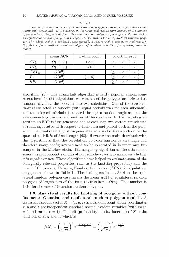
Table 1
Summary results concerning various random polygons. Results in parenthesis arenumerical results and – is the case when the numerical results vary because of the choicesof parameters. GPn stands for a Gaussian random polygon of n edges, EPn stands foran equilateral random polygon of n edges, CEPn stands for an equilateral random poly-gon of n edges within a confined space (usually a sphere with a predetermined radius),Rn stands for a uniform random polygon of n edges and SPn for spooling randommodel.
mean ACN leading coeff. knotting prob
GPn O(n ln n) 1/2π ≥ 1 − e−nǫ → 1
EPn O(n ln n) 3/16 ≥ 1 − e−nǫ → 1
CEPn O(n2) −− (≥ 1 − e−nǫ → 1)
Rn O(n2) (.115) (≥ 1 − e−nǫ → 1)
SPn O(n2) −− (≥ 1 − e−nǫ → 1)
algorithm [73]. The crankshaft algorithm is fairly popular among someresearchers. In this algorithm two vertices of the polygon are selected atrandom, dividing the polygon into two subchains. One of the two sub-chains is selected at random (with equal probabilities for each subchain),and the selected subchain is rotated through a random angle around theaxis connecting the two end vertices of the subchain. In the hedgehog al-gorithm an ERP is first generated and at each step two vectors are selectedat random, rotated with respect to their sum and placed back in the poly-gon. The crankshaft algorithm generates an ergodic Markov chain in thespace of all ERPs of fixed length [68]. However the main drawback withthis algorithm is that the correlation between samples is very high andtherefore many configurations need to be generated in between any twosamples in the Markov chain. The hedgehog algorithm on the other handgenerates independent samples of polygons however it is unknown whetherit is ergodic or not. These algorithms have helped to estimate some of thebiologically relevant properties, such as the knotting probability and themean of the Average Crossing Number distribution (ACN), for equilateralpolygons as shown in Table 1. The leading coefficient 3/16 in the equi-lateral random polygon case means the mean ACN of equilateral randompolygons of length n is of the form (3/16)n lnn + O(n). This number is1/2π for the case of Gaussian random polygons.
1.3. Analytical results for knotting of polygons without con-
finement: Gaussian and equilateral random polygon models. AGaussian random vector X = (x, y, z) is a random point whose coordinatesx, y and z are independent standard normal random variables (with mean= 0 and variance = 1). The pdf (probability density function) of X is thejoint pdf of x, y and z, which is
f(X) =
(
1√2π
)3
e−x2+y2+z2
2 =
(
1√2π
)3
e−|X|2
2 .
METHODS IN DNA TOPOLOGY 11
A Gaussian random walk of n steps (denoted by GWn) consists ofn + 1 consecutive points X0 = (0, 0, 0) = O, X1, X2, ... , Xn such thatYk+1 = Xk+1 − Xk (k = 0, 1, ..., n − 1) are independent Gaussian randomvectors. It follows that the joint pdf for all the vertices is
f(X1, X2, ..., Xn) =
(
1√2π
)3n
e−12(|Y1|
2+|Y2|
2+···+|Yn|2)
=
(
1√2π
)3n
e−12(|X1|
2+|X2−X1|
2+···+|Xn−Xn−1|
2).
A Gaussian random polygon GPn is a conditioned GWn of n stepssuch that the last vertex Xn coincides with the starting point X0 = O.Thus, if we let gn(Xn) be the pdf of Xn for a GWn,
then the joint pdf of X1, X2, ..., Xn−1 of a GPn is
h(X1, X2, ..., Xn) = f(X1, X2, ..., Xn)/gn(O).
The one advantage of the Gaussian random polygons (over other ran-dom polygon models) is that the joint probability density function of itsvertices is of an explicitly nice form. This enabled the derivation of thefollowing result concerning the knotting probability of a GPn [34].
Theorem 1.1. [34] Let K be any knot type, then there exists a positiveconstant ǫ such that GPn contains K as a connected sum component witha probability at least 1 − exp(−nǫ) provided that n is large enough.
One can obtain a similar result for equilateral random polygons.
Suppose Y1, Y2, ... , Yn are n independent random vectors uniformlydistributed on S2. An equilateral random walk of n steps, denoted byEWn, is defined as the sequence of points in the three dimensional spaceR
3: X0 = O, Xk = Y1 + Y2 + · · · + Yk, k = 1, 2, ..., n. Each Xk is calleda vertex of the EWn and the line segment joining Xk and Xk+1 is calledan edge of EWn (which is of unit length). Notice that the coordinates ofeach point are not independent from each other due to the fact that thedistance between consecutive points in the polymer needs to be one. If thelast vertex Xn of EWn is fixed, then we have a conditioned random walkEWn|Xn. In particular, EWn becomes a polygon if Xn = O. In this case,it is called an equilateral random polygon and is denoted by EPn. The jointprobability density function f(X1, X2, ..., Xn) of the vertices of an EWn isf(X1, X2, ..., Xn) = ϕ(U1)ϕ(U2) · · ·ϕ(Un) = ϕ(X1)ϕ(X2 − X1) · · ·ϕ(Xn −Xn−1). Where ϕ(Ui) is the density function of selecting a random pointover the surface of the sphere.
Let Xk be the k-th vertex of an EWn (n ≥ k > 1), its density functionis defined by
fk(Xk) =
∫ ∫
· · ·∫
ϕ(X1)ϕ(X2 − X1) · · ·
ϕ(Xk − Xk−1)dX1dX2 · · · dXk−1 (1.1)
12 JAVIER ARSUAGA, YUANAN DIAO, AND MARIEL VAZQUEZ
and it has the closed form fk(Xk) = 1
2π2r
∫∞
0x sin rx
(
sin xx
)kdx [74]. In the
case of EPn, the density function of the vertex Xk can be approximatedby a Gaussian distribution, as given in the following lemma [30, 33, 34].
Lemma 1.1. Let Xk be the k-th vertex of an EPn and let hk be itsdensity function, then
hk(Xk) =
(√
3
2πσ2
nk
)3
exp
(
−3|Xk|22σ2
nk
)
+O
(
1
k5/2+
1
(n − k)5/2
)
, (1.2)
where σ2
nk = k(n−k)
n .
In other words the density of the k step of an EPn can be approximatedby a Gaussian distribution with mean 0 and a standard deviation thatdepends on The vertex number k and on the distance from the vertex tothe origin (or first vertex in the polygon).
This lemma provided the key link to apply the methods used in [34] forthe Gaussian random polygons to the equilateral random polygons, whichleads to the following theorem.
Theorem 1.2. [30] Let K be any knot type, then there exists a positiveconstant ǫ such that EPn contains K as a connected sum component witha probability at least 1 − exp(−nǫ), provided that n is large enough.
Numerical studies on EPn suggest a scaling law of 1−exp(−n/a) witha = 244 ± 5 (see [66] and references therein).
The above two theorems imply that a long GPn or EPn contains manyconnected sum components (with a high probability), which makes it highlyunlikely for the polygon to be achiral. This is stated in the followingcorollary. However, this only provides reason for the long GPn and EPn
to favor chiral knots than achiral ones. For relatively short polygons, thisis not clear.
Corollary 1.1. [30, 34] There exists some constant θ > 0 such thatthe probability that a GPn or an EPn is a chiral knot is at least 1 − 1
nθ .
The determination of the knot type of a circular molecule can tell usits topological (minimum) crossing number, i.e., the minimum number ofcrossings one will see no matter how this molecule is artificially stretched,twisted, or bent. However, the average crossing number (ACN), definedas the average of crossing numbers over all orthogonal projections of themolecule, is a more natural geometric measure of the molecule entangle-ment as it refers to the actual number of crossings that can be perceivedwhile observing a non-perturbed trajectory of a given molecule [55]. Fur-thermore, it is believed that DNA knots migrate in gel eletrophoresis ac-cordingly with their ACN [99].
The following theorems are presented in [31, 32] and establish theO(n ln n) behavior of the mean ACN for the Gaussian and equilateral ran-dom polygons as illustrated in Table 1.
METHODS IN DNA TOPOLOGY 13
Theorem 1.3. Let χn be the ACN of an equilateral random walk ofn steps; then
E(χn) =3
16n ln n + O(n).
On the other hand, if χ′n is the ACN of a Gaussian random polygon of n
steps, then
E(χ′n) =
1
2πn lnn + O(n).
2. Cyclization of DNA molecules in confined volumes: DNA
knotting in bacteriophage P4 capsids.
2.1. Experimental studies on DNA knots in bacteriophages.
In dsDNA bacteriophages the volume of the bacteriophage genome is re-duced 100 times inside the capsid [53]. This volume reduction imposessevere physical constraints on the DNA molecule. For instance the DNAmolecule is under (at least) 50 atmospheres of pressure [42, 93] and ata concentration of 800mg/ml [56]. Despite these conditions the dsDNAmolecule is believed to preserve its double helical structure [8] and not tohave sequence-specific associations with the protein capsid. A number ofmodels have been proposed to describe the organization of the viral chro-mosome under such extreme conditions of condensation. These includecoaxial and concentric spooling models [4, 20, 35, 76, 82], coaxial models[10], toroidal models [51, 72], and liquid-crystaline models [61].
Bacteriophage P4 is an icosahedral phage of radius r = 180A and alinear dsDNA genome of 11.5 kb (l = 120 × 103A). The genome is flankedby two 16bp long single stranded complementary sequences of DNA calledcos sites. During phage morphogenesis a protein enclosure called capsidis assembled first. This is followed by the packing of a single linear DNAmolecule into the capsid through the portal vertex. Infective viruses keepat least one of their cos sites attached near the portal [21]. This attachmentprevents the two cos ends from meeting within the capsid and circulariz-ing the chromosome. However in the experiments performed by Liu andcolleagues it was found that most of the DNA molecules phenol extractedfrom bacteriophage P4 are circular and non-trivially knotted [63, 64].
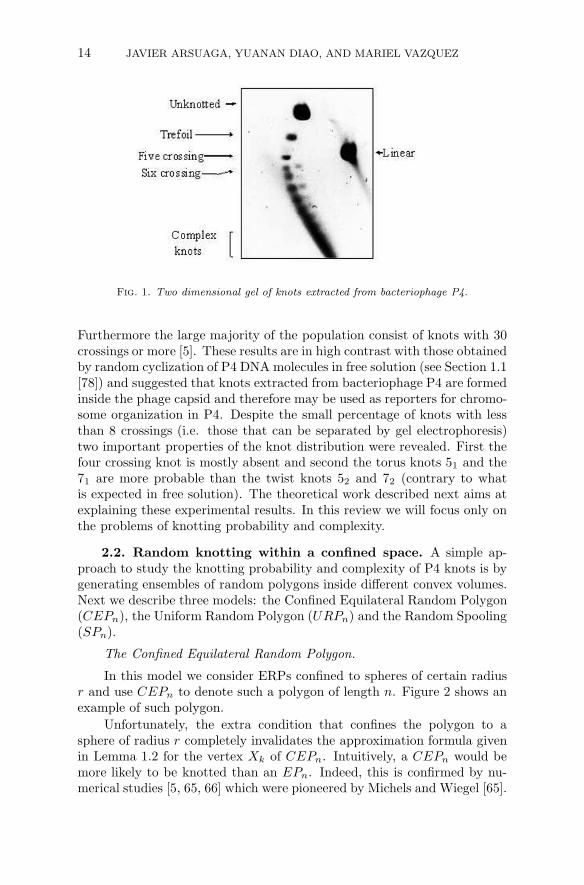
Recent work [5, 6, 91, 92] reproduced and extended the results of Liu etal. Figure 1 shows a two dimensional gel of DNA knots from bacteriophageP4 in which different conditions are used in each dimension [91]. In thisfigure the top spot corresponds to the unknotted molecule followed, along abell-shaped curve, by the trefoil knot, the figure eight (four-crossing) knot,and so on. The spot ahead of the bell is the linear chain. The most remark-able fact about this distribution is that about 95% of the DNA moleculesare knotted and only about 2% are knots between 3 and 10 crossing knots.
14 JAVIER ARSUAGA, YUANAN DIAO, AND MARIEL VAZQUEZ
Fig. 1. Two dimensional gel of knots extracted from bacteriophage P4.
Furthermore the large majority of the population consist of knots with 30crossings or more [5]. These results are in high contrast with those obtainedby random cyclization of P4 DNA molecules in free solution (see Section 1.1[78]) and suggested that knots extracted from bacteriophage P4 are formedinside the phage capsid and therefore may be used as reporters for chromo-some organization in P4. Despite the small percentage of knots with lessthan 8 crossings (i.e. those that can be separated by gel electrophoresis)two important properties of the knot distribution were revealed. First thefour crossing knot is mostly absent and second the torus knots 51 and the71 are more probable than the twist knots 52 and 72 (contrary to whatis expected in free solution). The theoretical work described next aims atexplaining these experimental results. In this review we will focus only onthe problems of knotting probability and complexity.
2.2. Random knotting within a confined space. A simple ap-proach to study the knotting probability and complexity of P4 knots is bygenerating ensembles of random polygons inside different convex volumes.Next we describe three models: the Confined Equilateral Random Polygon(CEPn), the Uniform Random Polygon (URPn) and the Random Spooling(SPn).
The Confined Equilateral Random Polygon.
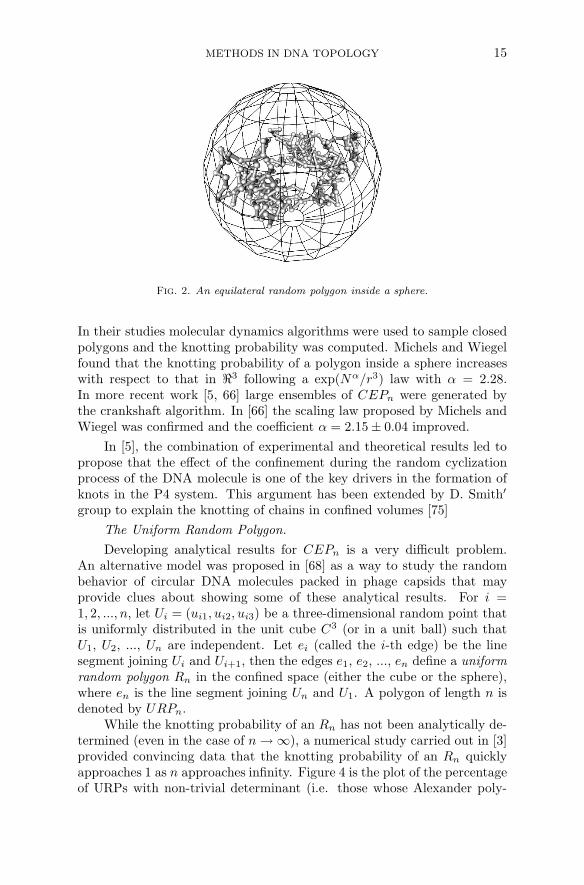
In this model we consider ERPs confined to spheres of certain radiusr and use CEPn to denote such a polygon of length n. Figure 2 shows anexample of such polygon.
Unfortunately, the extra condition that confines the polygon to asphere of radius r completely invalidates the approximation formula givenin Lemma 1.2 for the vertex Xk of CEPn. Intuitively, a CEPn would bemore likely to be knotted than an EPn. Indeed, this is confirmed by nu-merical studies [5, 65, 66] which were pioneered by Michels and Wiegel [65].
METHODS IN DNA TOPOLOGY 15
Fig. 2. An equilateral random polygon inside a sphere.
In their studies molecular dynamics algorithms were used to sample closedpolygons and the knotting probability was computed. Michels and Wiegelfound that the knotting probability of a polygon inside a sphere increaseswith respect to that in ℜ3 following a exp(Nα/r3) law with α = 2.28.In more recent work [5, 66] large ensembles of CEPn were generated bythe crankshaft algorithm. In [66] the scaling law proposed by Michels andWiegel was confirmed and the coefficient α = 2.15 ± 0.04 improved.
In [5], the combination of experimental and theoretical results led topropose that the effect of the confinement during the random cyclizationprocess of the DNA molecule is one of the key drivers in the formation ofknots in the P4 system. This argument has been extended by D. Smith′
group to explain the knotting of chains in confined volumes [75]
The Uniform Random Polygon.
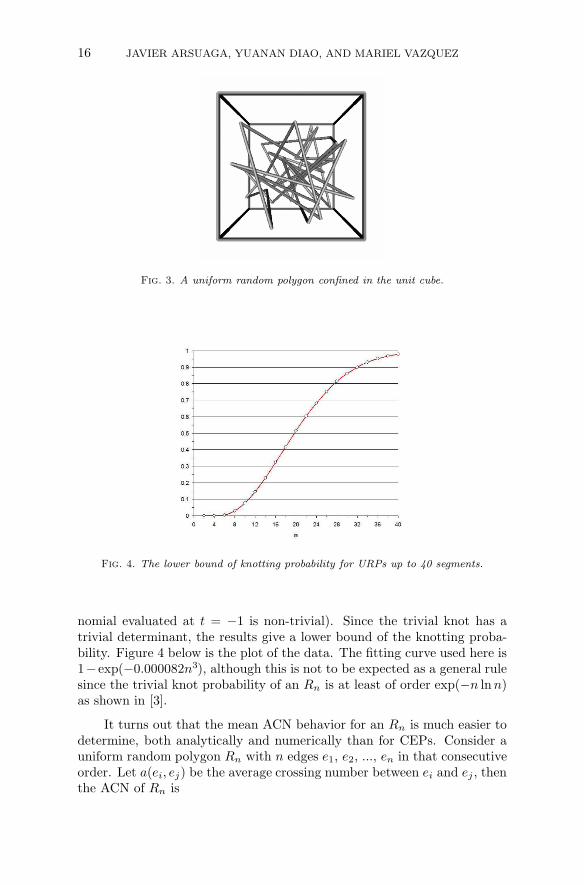
Developing analytical results for CEPn is a very difficult problem.An alternative model was proposed in [68] as a way to study the randombehavior of circular DNA molecules packed in phage capsids that mayprovide clues about showing some of these analytical results. For i =1, 2, ..., n, let Ui = (ui1, ui2, ui3) be a three-dimensional random point thatis uniformly distributed in the unit cube C3 (or in a unit ball) such thatU1, U2, ..., Un are independent. Let ei (called the i-th edge) be the linesegment joining Ui and Ui+1, then the edges e1, e2, ..., en define a uniformrandom polygon Rn in the confined space (either the cube or the sphere),where en is the line segment joining Un and U1. A polygon of length n isdenoted by URPn.
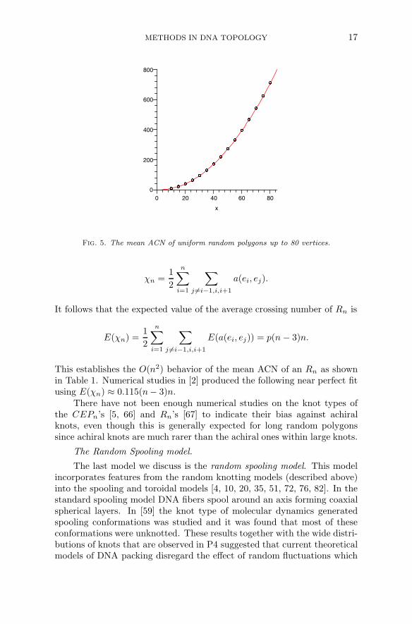
While the knotting probability of an Rn has not been analytically de-termined (even in the case of n → ∞), a numerical study carried out in [3]provided convincing data that the knotting probability of an Rn quicklyapproaches 1 as n approaches infinity. Figure 4 is the plot of the percentageof URPs with non-trivial determinant (i.e. those whose Alexander poly-
16 JAVIER ARSUAGA, YUANAN DIAO, AND MARIEL VAZQUEZ
Fig. 3. A uniform random polygon confined in the unit cube.
Fig. 4. The lower bound of knotting probability for URPs up to 40 segments.
nomial evaluated at t = −1 is non-trivial). Since the trivial knot has atrivial determinant, the results give a lower bound of the knotting proba-bility. Figure 4 below is the plot of the data. The fitting curve used here is1− exp(−0.000082n3), although this is not to be expected as a general rulesince the trivial knot probability of an Rn is at least of order exp(−n ln n)as shown in [3].
It turns out that the mean ACN behavior for an Rn is much easier todetermine, both analytically and numerically than for CEPs. Consider auniform random polygon Rn with n edges e1, e2, ..., en in that consecutiveorder. Let a(ei, ej) be the average crossing number between ei and ej , thenthe ACN of Rn is
METHODS IN DNA TOPOLOGY 17
80
x
600
0
200
06020
400
800
40
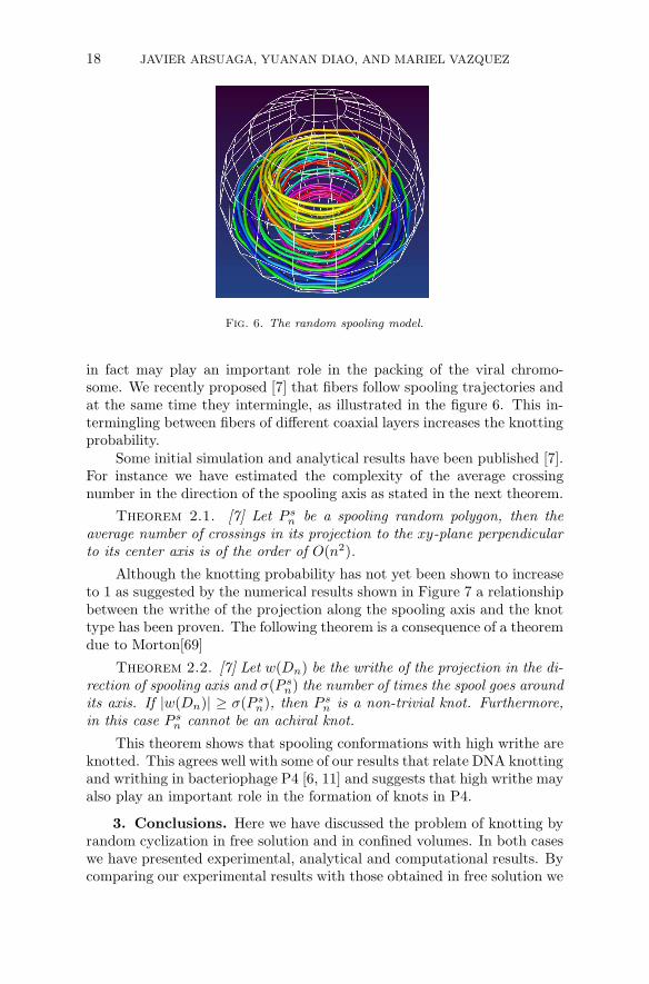
Fig. 5. The mean ACN of uniform random polygons up to 80 vertices.
χn =1
2
n∑
i=1
∑
j 6=i−1,i,i+1
a(ei, ej).
It follows that the expected value of the average crossing number of Rn is
E(χn) =1
2
n∑
i=1
∑
j 6=i−1,i,i+1
E(a(ei, ej)) = p(n − 3)n.
This establishes the O(n2) behavior of the mean ACN of an Rn as shownin Table 1. Numerical studies in [2] produced the following near perfect fitusing E(χn) ≈ 0.115(n− 3)n.
There have not been enough numerical studies on the knot types ofthe CEPn’s [5, 66] and Rn’s [67] to indicate their bias against achiralknots, even though this is generally expected for long random polygonssince achiral knots are much rarer than the achiral ones within large knots.
The Random Spooling model.
The last model we discuss is the random spooling model. This modelincorporates features from the random knotting models (described above)into the spooling and toroidal models [4, 10, 20, 35, 51, 72, 76, 82]. In thestandard spooling model DNA fibers spool around an axis forming coaxialspherical layers. In [59] the knot type of molecular dynamics generatedspooling conformations was studied and it was found that most of theseconformations were unknotted. These results together with the wide distri-butions of knots that are observed in P4 suggested that current theoreticalmodels of DNA packing disregard the effect of random fluctuations which
18 JAVIER ARSUAGA, YUANAN DIAO, AND MARIEL VAZQUEZ
Fig. 6. The random spooling model.
in fact may play an important role in the packing of the viral chromo-some. We recently proposed [7] that fibers follow spooling trajectories andat the same time they intermingle, as illustrated in the figure 6. This in-termingling between fibers of different coaxial layers increases the knottingprobability.
Some initial simulation and analytical results have been published [7].For instance we have estimated the complexity of the average crossingnumber in the direction of the spooling axis as stated in the next theorem.
Theorem 2.1. [7] Let P sn be a spooling random polygon, then the
average number of crossings in its projection to the xy-plane perpendicularto its center axis is of the order of O(n2).
Although the knotting probability has not yet been shown to increaseto 1 as suggested by the numerical results shown in Figure 7 a relationshipbetween the writhe of the projection along the spooling axis and the knottype has been proven. The following theorem is a consequence of a theoremdue to Morton[69]
Theorem 2.2. [7] Let w(Dn) be the writhe of the projection in the di-rection of spooling axis and σ(P s
n) the number of times the spool goes aroundits axis. If |w(Dn)| ≥ σ(P s
n), then P sn is a non-trivial knot. Furthermore,
in this case P sn cannot be an achiral knot.
This theorem shows that spooling conformations with high writhe areknotted. This agrees well with some of our results that relate DNA knottingand writhing in bacteriophage P4 [6, 11] and suggests that high writhe mayalso play an important role in the formation of knots in P4.
3. Conclusions. Here we have discussed the problem of knotting byrandom cyclization in free solution and in confined volumes. In both caseswe have presented experimental, analytical and computational results. Bycomparing our experimental results with those obtained in free solution we
METHODS IN DNA TOPOLOGY 19
50 100 150 200
0.0
0.2
0.4
0.6
0.8
x
y
Fig. 7. Knotting probability as a function of the length of the chain for the randomspooling model.
concluded that knotting in bacteriophage P4 occurs before, or very soonafter, the disruption of the capsid and therefore P4 knots can be used asreporters of DNA packing. The large amount of knotting is still a featurethat is not fully explained by current mathematical models. In this reviewwe have presented three random knotting models: the confined equilateralpolygon, the uniform random polygon and the spooling random polygon.All these models present consistent results however they do not reach thehigh levels of complexity found in bacteriophages. This is specially trueif more accurate representations of the DNA molecule are taken into ac-count. Nevertheless some information about the biological system has beenextracted from these theoretical models. For instance our current simula-tions results suggest that DNA knotting in P4 is mainly driven by theconfinement imposed by the capsid during the cyclization reaction, andperhaps also by possible biases introduced by the arrangement of the vi-ral chromosome [6, 11]. Importantly none of the current idealized modelsproposed in the literature account for the formation of knots inside thecapsid and previous simulations results failed to do so [59] thus suggestingthat they may not reflect some important properties of the DNA packing.The random spooling model is our first attempt to address this issue. Itremains to be seen if such models can reproduce the knot distributionsobserved experimentally. New experimental results have recently obtainedfor P4 deletion mutants whose genomes range between 5 and 8 kb [92].These experiments hold a great promise for unveiling the properties thatdrive knotting in phage capsids as well as some of the essential features ofthe viral packing.
20 JAVIER ARSUAGA, YUANAN DIAO, AND MARIEL VAZQUEZ
Part II. Enzymes that change the topology of DNA.
The cell has evolved a set of tools to remove unwanted DNA entan-glement and solve other topological problems. Noteworthy are enzymesthat change the topology of DNA such as site-specific recombinases andtopoisomerases. For example, replication of the circular E. coli genomeproduces two catenated (linked) circles that cannot be separated withoutbreaking the DNA chain; the type II topoisomerase topoIV plays an impor-tant role in unlinking the newly replicated genomes, thus ensuring propersegregation at cell division. Similarly, circular chromosome dimers ariseoccasionally due to crossing over by homologous recombination of newlyreplicated chromosomes. The site-specific recombination system XerCDcoupled with the multifunctional protein FtsK resolves the chromosomedimers, thus allowing for stable inheritance. In Section 4 we introduce thebiology of site-specific recombination. In Section 5 we briefly review themathematics used in the tangle method, and the method itself, includingthe main assumptions. We finish the review by summarizing the tangleanalysis of Xer recombination at psi and of XerCD-FtsK recombination atdif. We emphasize the 3-dimensional interpretation of the data.
4. Site-specific recombination. Site-specific recombinases are en-zymes able to change the genetic code of their DNA substrate. They me-diate important biological processes such as inversion of DNA segments,transposition of a DNA segment from one location to another along thegenome, integration and excision of viral DNA into and out of its hostgenome, and resolution of multimeric DNA molecules [48, 79].
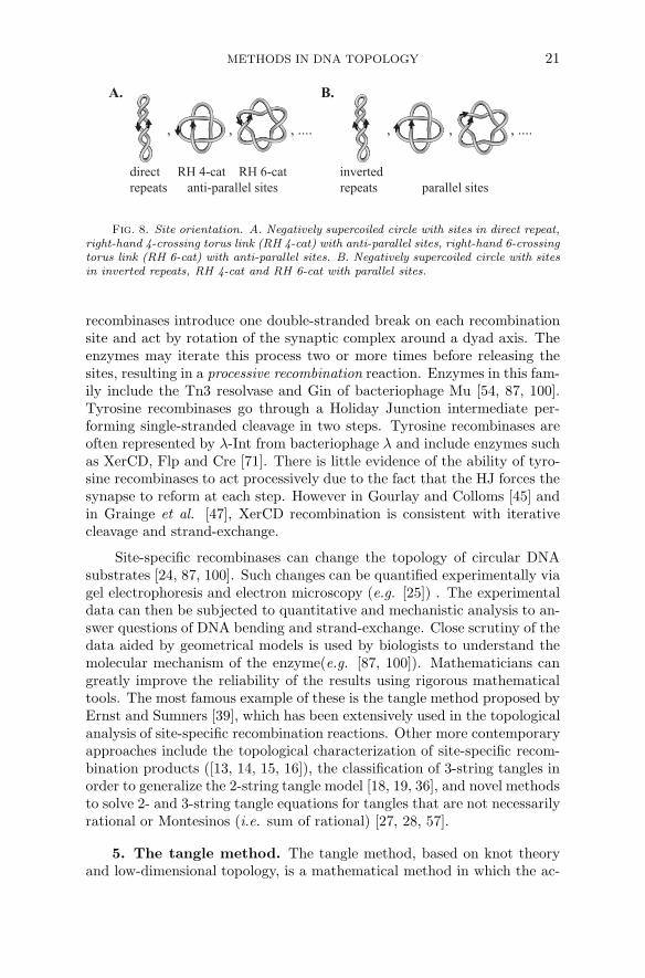
Site-specific recombination occurs in two steps. First, during synapsetwo short DNA segments of specific sequence are brought together by thesite-specific recombinase(s) and any necessary accessory proteins. Thesesegments are called recombination sites. Second, during strand-exchange,each recombination site is cleaved, the lose ends recombined and the re-combined pieces are rejoined. The DNA sequence in a recombination site isusually non-palindromic, thus allowing to define a site orientation. Whenthe DNA substrate consists of circular DNA molecules, the two recombi-nation sites may occur in a single DNA circle or in two separate circles.In the first case, for intramolecular recombination, if the sites induce thesame orientation on the circle they are said to be in direct repeat, otherwisethey are in inverted repeat (Figure 8). Relative orientation of the sites isharder to characterize in the case of intermolecular recombination (i.e. twosites on separate DNA circles). In the simple case of T (2, n) torus linkswith 4 or more crossings the sites are said to be in parallel or anti-parallelorientation with respect to each other (Figure 8).
Based on sequence homology and strand-passage mechanism, site-specific recombinases are divided into two families: serine recombinasesand tyrosine recombinases [60, 71]. After synapse formation, the serine
Fig. 8. Site orientation. A. Negatively supercoiled circle with sites in direct repeat,right-hand 4-crossing torus link (RH 4-cat) with anti-parallel sites, right-hand 6-crossingtorus link (RH 6-cat) with anti-parallel sites. B. Negatively supercoiled circle with sitesin inverted repeats, RH 4-cat and RH 6-cat with parallel sites.
recombinases introduce one double-stranded break on each recombinationsite and act by rotation of the synaptic complex around a dyad axis. Theenzymes may iterate this process two or more times before releasing thesites, resulting in a processive recombination reaction. Enzymes in this fam-ily include the Tn3 resolvase and Gin of bacteriophage Mu [54, 87, 100].Tyrosine recombinases go through a Holiday Junction intermediate per-forming single-stranded cleavage in two steps. Tyrosine recombinases areoften represented by λ-Int from bacteriophage λ and include enzymes suchas XerCD, Flp and Cre [71]. There is little evidence of the ability of tyro-sine recombinases to act processively due to the fact that the HJ forces thesynapse to reform at each step. However in Gourlay and Colloms [45] andin Grainge et al. [47], XerCD recombination is consistent with iterativecleavage and strand-exchange.
Site-specific recombinases can change the topology of circular DNAsubstrates [24, 87, 100]. Such changes can be quantified experimentally viagel electrophoresis and electron microscopy (e.g. [25]) . The experimentaldata can then be subjected to quantitative and mechanistic analysis to an-swer questions of DNA bending and strand-exchange. Close scrutiny of thedata aided by geometrical models is used by biologists to understand themolecular mechanism of the enzyme(e.g. [87, 100]). Mathematicians cangreatly improve the reliability of the results using rigorous mathematicaltools. The most famous example of these is the tangle method proposed byErnst and Sumners [39], which has been extensively used in the topologicalanalysis of site-specific recombination reactions. Other more contemporaryapproaches include the topological characterization of site-specific recom-bination products ([13, 14, 15, 16]), the classification of 3-string tangles inorder to generalize the 2-string tangle model [18, 19, 36], and novel methodsto solve 2- and 3-string tangle equations for tangles that are not necessarilyrational or Montesinos (i.e. sum of rational) [27, 28, 57].
5. The tangle method. The tangle method, based on knot theoryand low-dimensional topology, is a mathematical method in which the ac-
22 JAVIER ARSUAGA, YUANAN DIAO, AND MARIEL VAZQUEZ
(0) (+1) (-1) (0,0)
0 1 -1
(4) (-3,0) (-3,-1) (-5,-1,-1,0)
4 -1/3 -4/3 -6/11
8q/p =
q/p =
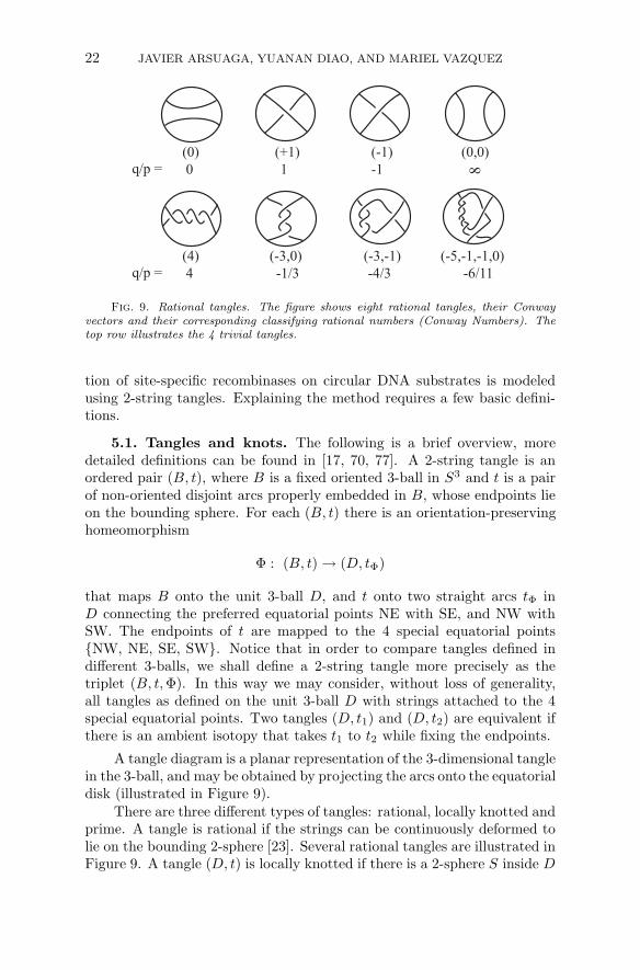
Fig. 9. Rational tangles. The figure shows eight rational tangles, their Conwayvectors and their corresponding classifying rational numbers (Conway Numbers). Thetop row illustrates the 4 trivial tangles.
tion of site-specific recombinases on circular DNA substrates is modeledusing 2-string tangles. Explaining the method requires a few basic defini-tions.
5.1. Tangles and knots. The following is a brief overview, moredetailed definitions can be found in [17, 70, 77]. A 2-string tangle is anordered pair (B, t), where B is a fixed oriented 3-ball in S3 and t is a pairof non-oriented disjoint arcs properly embedded in B, whose endpoints lieon the bounding sphere. For each (B, t) there is an orientation-preservinghomeomorphism
Φ : (B, t) → (D, tΦ)
that maps B onto the unit 3-ball D, and t onto two straight arcs tΦ inD connecting the preferred equatorial points NE with SE, and NW withSW. The endpoints of t are mapped to the 4 special equatorial points{NW, NE, SE, SW}. Notice that in order to compare tangles defined indifferent 3-balls, we shall define a 2-string tangle more precisely as thetriplet (B, t, Φ). In this way we may consider, without loss of generality,all tangles as defined on the unit 3-ball D with strings attached to the 4special equatorial points. Two tangles (D, t1) and (D, t2) are equivalent ifthere is an ambient isotopy that takes t1 to t2 while fixing the endpoints.
A tangle diagram is a planar representation of the 3-dimensional tanglein the 3-ball, and may be obtained by projecting the arcs onto the equatorialdisk (illustrated in Figure 9).
There are three different types of tangles: rational, locally knotted andprime. A tangle is rational if the strings can be continuously deformed tolie on the bounding 2-sphere [23]. Several rational tangles are illustrated inFigure 9. A tangle (D, t) is locally knotted if there is a 2-sphere S inside D
METHODS IN DNA TOPOLOGY 23
=
(-3,O) + (-1) = (-3,-1)
=
N(-3,-1) = N (-4/3) = <1,2,1> = b(4,3)
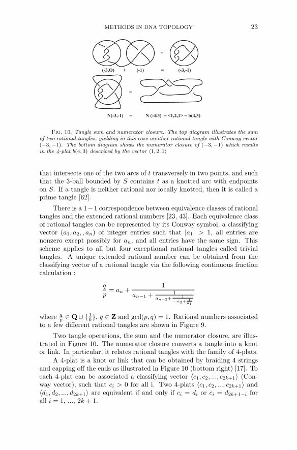
Fig. 10. Tangle sum and numerator closure. The top diagram illustrates the sumof two rational tangles, yielding in this case another rational tangle with Conway vector(−3,−1). The bottom diagram shows the numerator closure of (−3,−1) which resultsin the 4-plat b(4, 3) described by the vector 〈1, 2, 1〉
that intersects one of the two arcs of t transversely in two points, and suchthat the 3-ball bounded by S contains t as a knotted arc with endpointson S. If a tangle is neither rational nor locally knotted, then it is called aprime tangle [62].
There is a 1−1 correspondence between equivalence classes of rationaltangles and the extended rational numbers [23, 43]. Each equivalence classof rational tangles can be represented by its Conway symbol, a classifyingvector (a1, a2, , an) of integer entries such that |a1| > 1, all entries arenonzero except possibly for an, and all entries have the same sign. Thisscheme applies to all but four exceptional rational tangles called trivialtangles. A unique extended rational number can be obtained from theclassifying vector of a rational tangle via the following continuous fractioncalculation :
q
p= an +
1
an−1 + 1
an−2+1
···a2+ 1a1
where qp ∈ Q ∪ { 1
0}, q ∈ Z and gcd(p, q) = 1. Rational numbers associated
to a few different rational tangles are shown in Figure 9.
Two tangle operations, the sum and the numerator closure, are illus-trated in Figure 10. The numerator closure converts a tangle into a knotor link. In particular, it relates rational tangles with the family of 4-plats.
A 4-plat is a knot or link that can be obtained by braiding 4 stringsand capping off the ends as illustrated in Figure 10 (bottom right) [17]. Toeach 4-plat can be associated a classifying vector 〈c1, c2, ..., c2k+1〉 (Con-way vector), such that ci > 0 for all i. Two 4-plats 〈c1, c2, ..., c2k+1〉 and〈d1, d2, ..., d2k+1〉 are equivalent if and only if ci = di or ci = d2k+1−i forall i = 1, ..., 2k + 1.
24 JAVIER ARSUAGA, YUANAN DIAO, AND MARIEL VAZQUEZ
From the Conway vector, we can obtain a classifying rational numberfor each 4-plat through a continued fraction:
β
α=
1
c1 + 1
c2+···+ 1c2k+1
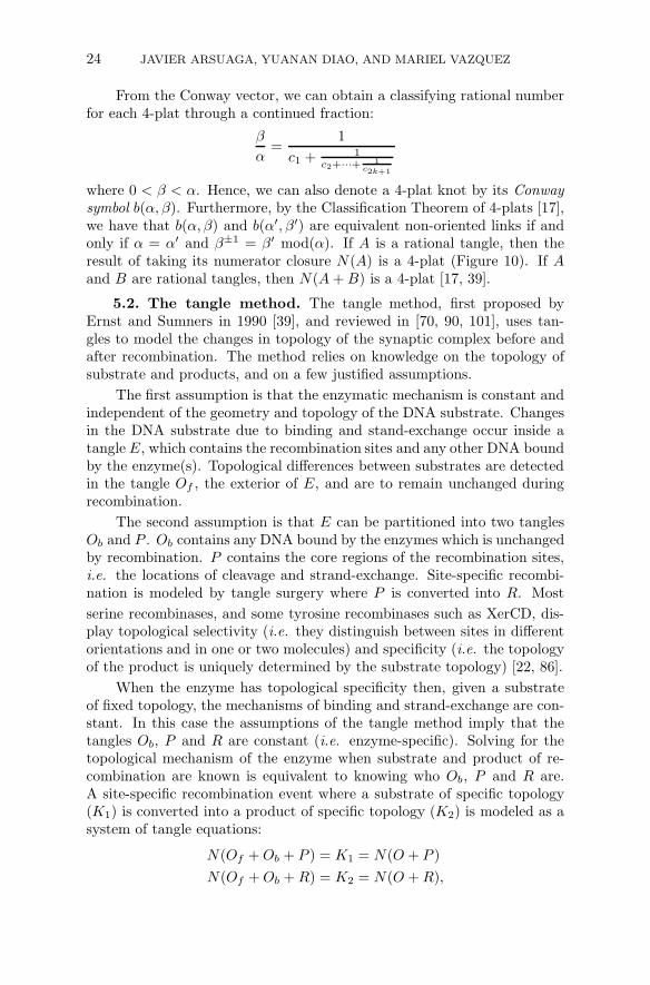
where 0 < β < α. Hence, we can also denote a 4-plat knot by its Conwaysymbol b(α, β). Furthermore, by the Classification Theorem of 4-plats [17],we have that b(α, β) and b(α′, β′) are equivalent non-oriented links if andonly if α = α′ and β±1 = β′ mod(α). If A is a rational tangle, then theresult of taking its numerator closure N(A) is a 4-plat (Figure 10). If Aand B are rational tangles, then N(A + B) is a 4-plat [17, 39].
5.2. The tangle method. The tangle method, first proposed byErnst and Sumners in 1990 [39], and reviewed in [70, 90, 101], uses tan-gles to model the changes in topology of the synaptic complex before andafter recombination. The method relies on knowledge on the topology ofsubstrate and products, and on a few justified assumptions.
The first assumption is that the enzymatic mechanism is constant andindependent of the geometry and topology of the DNA substrate. Changesin the DNA substrate due to binding and stand-exchange occur inside atangle E, which contains the recombination sites and any other DNA boundby the enzyme(s). Topological differences between substrates are detectedin the tangle Of , the exterior of E, and are to remain unchanged duringrecombination.
The second assumption is that E can be partitioned into two tanglesOb and P . Ob contains any DNA bound by the enzymes which is unchangedby recombination. P contains the core regions of the recombination sites,i.e. the locations of cleavage and strand-exchange. Site-specific recombi-nation is modeled by tangle surgery where P is converted into R. Most
serine recombinases, and some tyrosine recombinases such as XerCD, dis-play topological selectivity (i.e. they distinguish between sites in differentorientations and in one or two molecules) and specificity (i.e. the topologyof the product is uniquely determined by the substrate topology) [22, 86].
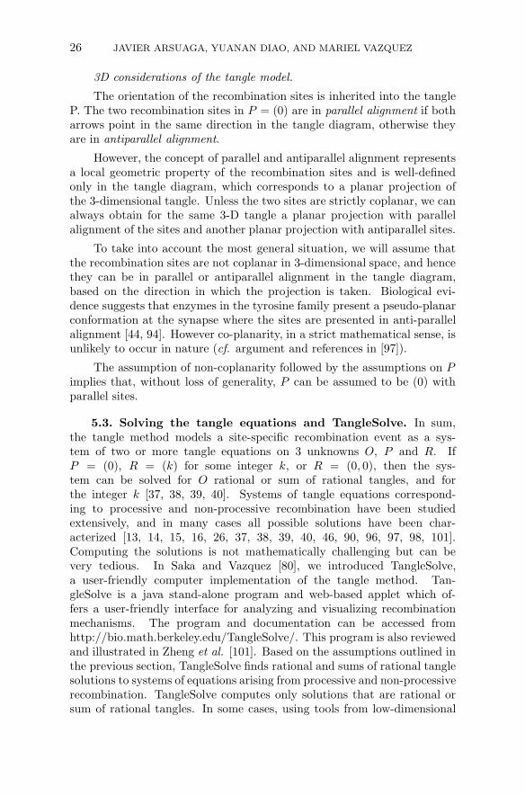
When the enzyme has topological specificity then, given a substrateof fixed topology, the mechanisms of binding and strand-exchange are con-stant. In this case the assumptions of the tangle method imply that thetangles Ob, P and R are constant (i.e. enzyme-specific). Solving for thetopological mechanism of the enzyme when substrate and product of re-combination are known is equivalent to knowing who Ob, P and R are.A site-specific recombination event where a substrate of specific topology(K1) is converted into a product of specific topology (K2) is modeled as asystem of tangle equations:
N(Of + Ob + P ) = K1 = N(O + P )
N(Of + Ob + R) = K2 = N(O + R),
METHODS IN DNA TOPOLOGY 25
where O = Of +Ob is the outside tangle consisting of all DNA (bound andunbound) which is not changed by strand-exchange.
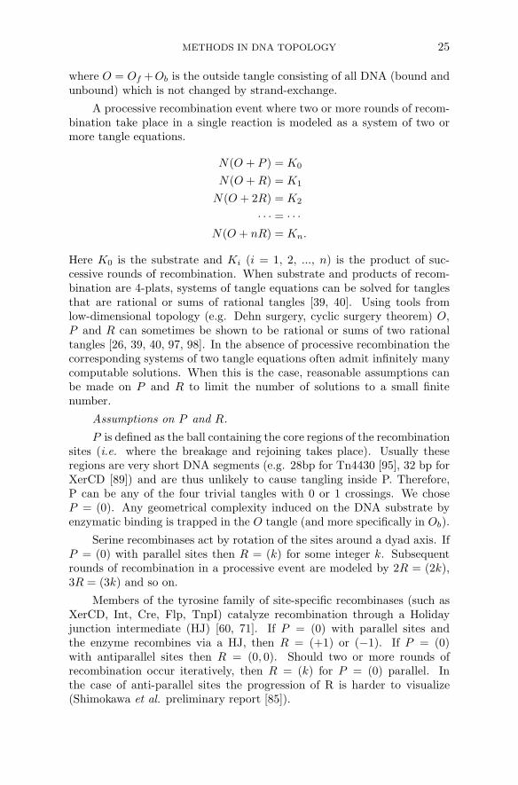
A processive recombination event where two or more rounds of recom-bination take place in a single reaction is modeled as a system of two ormore tangle equations.
N(O + P ) = K0
N(O + R) = K1
N(O + 2R) = K2
· · · = · · ·N(O + nR) = Kn.
Here K0 is the substrate and Ki (i = 1, 2, ..., n) is the product of suc-cessive rounds of recombination. When substrate and products of recom-bination are 4-plats, systems of tangle equations can be solved for tanglesthat are rational or sums of rational tangles [39, 40]. Using tools fromlow-dimensional topology (e.g. Dehn surgery, cyclic surgery theorem) O,P and R can sometimes be shown to be rational or sums of two rationaltangles [26, 39, 40, 97, 98]. In the absence of processive recombination thecorresponding systems of two tangle equations often admit infinitely manycomputable solutions. When this is the case, reasonable assumptions canbe made on P and R to limit the number of solutions to a small finitenumber.
Assumptions on P and R.
P is defined as the ball containing the core regions of the recombinationsites (i.e. where the breakage and rejoining takes place). Usually theseregions are very short DNA segments (e.g. 28bp for Tn4430 [95], 32 bp forXerCD [89]) and are thus unlikely to cause tangling inside P. Therefore,P can be any of the four trivial tangles with 0 or 1 crossings. We choseP = (0). Any geometrical complexity induced on the DNA substrate byenzymatic binding is trapped in the O tangle (and more specifically in Ob).
Serine recombinases act by rotation of the sites around a dyad axis. IfP = (0) with parallel sites then R = (k) for some integer k. Subsequentrounds of recombination in a processive event are modeled by 2R = (2k),3R = (3k) and so on.
Members of the tyrosine family of site-specific recombinases (such asXerCD, Int, Cre, Flp, TnpI) catalyze recombination through a Holidayjunction intermediate (HJ) [60, 71]. If P = (0) with parallel sites andthe enzyme recombines via a HJ, then R = (+1) or (−1). If P = (0)with antiparallel sites then R = (0, 0). Should two or more rounds ofrecombination occur iteratively, then R = (k) for P = (0) parallel. Inthe case of anti-parallel sites the progression of R is harder to visualize(Shimokawa et al. preliminary report [85]).
26 JAVIER ARSUAGA, YUANAN DIAO, AND MARIEL VAZQUEZ
3D considerations of the tangle model.
The orientation of the recombination sites is inherited into the tangleP. The two recombination sites in P = (0) are in parallel alignment if botharrows point in the same direction in the tangle diagram, otherwise theyare in antiparallel alignment.
However, the concept of parallel and antiparallel alignment representsa local geometric property of the recombination sites and is well-definedonly in the tangle diagram, which corresponds to a planar projection ofthe 3-dimensional tangle. Unless the two sites are strictly coplanar, we canalways obtain for the same 3-D tangle a planar projection with parallelalignment of the sites and another planar projection with antiparallel sites.
To take into account the most general situation, we will assume thatthe recombination sites are not coplanar in 3-dimensional space, and hencethey can be in parallel or antiparallel alignment in the tangle diagram,based on the direction in which the projection is taken. Biological evi-dence suggests that enzymes in the tyrosine family present a pseudo-planarconformation at the synapse where the sites are presented in anti-parallelalignment [44, 94]. However co-planarity, in a strict mathematical sense, isunlikely to occur in nature (cf. argument and references in [97]).
The assumption of non-coplanarity followed by the assumptions on Pimplies that, without loss of generality, P can be assumed to be (0) withparallel sites.
5.3. Solving the tangle equations and TangleSolve. In sum,the tangle method models a site-specific recombination event as a sys-tem of two or more tangle equations on 3 unknowns O, P and R. IfP = (0), R = (k) for some integer k, or R = (0, 0), then the sys-tem can be solved for O rational or sum of rational tangles, and forthe integer k [37, 38, 39, 40]. Systems of tangle equations correspond-ing to processive and non-processive recombination have been studiedextensively, and in many cases all possible solutions have been char-acterized [13, 14, 15, 16, 26, 37, 38, 39, 40, 46, 90, 96, 97, 98, 101].Computing the solutions is not mathematically challenging but can bevery tedious. In Saka and Vazquez [80], we introduced TangleSolve,a user-friendly computer implementation of the tangle method. Tan-gleSolve is a java stand-alone program and web-based applet which of-fers a user-friendly interface for analyzing and visualizing recombinationmechanisms. The program and documentation can be accessed fromhttp://bio.math.berkeley.edu/TangleSolve/. This program is also reviewedand illustrated in Zheng et al. [101]. Based on the assumptions outlined inthe previous section, TangleSolve finds rational and sums of rational tanglesolutions to systems of equations arising from processive and non-processiverecombination. TangleSolve computes only solutions that are rational orsum of rational tangles. In some cases, using tools from low-dimensional
METHODS IN DNA TOPOLOGY 27
topology, it can be proven that all possible solutions to a system of tangleequations are rational or sums of two rational tangles. Such instances arehighlighted in TangleSolve.
It is our goal to make this program available to the wider scientificcommunity of mathematicians, molecular biologists and computational bi-ologists. Therefore the calculations rest heavily on a graphical interface.Mathematical notation, or deep knowledge of the tangle method are notrequired to insert substrate and product topologies and compute enzymaticmechanisms.
TopoICE-R is another computer implementation of the tangle modelwhich is available through KnotPlot [29]. TangleSolve and TopoICE-Rhave complementary features, noteworthy TopoICE-R provides a 3D ren-dition of the tangle equations and benefits from all the capabilities withinKnotplot . TangleSolve has the ability to compute solutions for processiverecombination reactions. Also the two applications use different sets ofassumptions.
6. Tangle analysis of Xer recombination.
6.1. Dimer resolution at psi sites. In Colloms et al. [22] it wasshown that, when acting on unknotted DNA circles with two psi recombina-tion sites in direct repeats, XerC/XerD yield products of unique topologyb(4,3) (the right-hand 4-crossing torus catenane) and anti-parallel sites(Figure 8). The reaction requires two accessory proteins PepA and ArgR[12], and there is experimental evidence that the sites wrap around theaccessory proteins approximately three times prior to recombination [1].This reaction can be written as the following system of tangle equations:
N(O + P ) = b(1, 1), N(O + R) = b(4, 3)
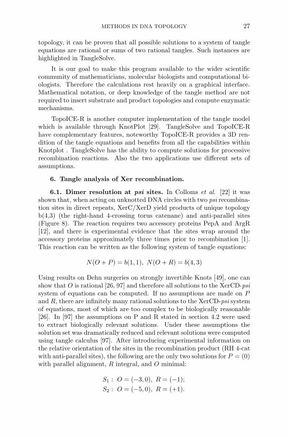
Using results on Dehn surgeries on strongly invertible Knots [49], one canshow that O is rational [26, 97] and therefore all solutions to the XerCD-psisystem of equations can be computed. If no assumptions are made on Pand R, there are infinitely many rational solutions to the XerCD-psi systemof equations, most of which are too complex to be biologically reasonable[26]. In [97] the assumptions on P and R stated in section 4.2 were usedto extract biologically relevant solutions. Under these assumptions thesolution set was dramatically reduced and relevant solutions were computedusing tangle calculus [97]. After introducing experimental information onthe relative orientation of the sites in the recombination product (RH 4-catwith anti-parallel sites), the following are the only two solutions for P = (0)with parallel alignment, R integral, and O minimal:
S1 : O = (−3, 0), R = (−1);
S2 : O = (−5, 0), R = (+1).
28 JAVIER ARSUAGA, YUANAN DIAO, AND MARIEL VAZQUEZ
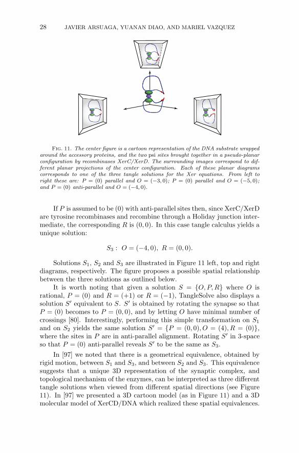
Fig. 11. The center figure is a cartoon representation of the DNA substrate wrappedaround the accessory proteins, and the two psi sites brought together in a pseudo-planarconfiguration by recombinases XerC/XerD. The surrounding images correspond to dif-ferent planar projections of the center configuration. Each of these planar diagramscorresponds to one of the three tangle solutions for the Xer equations. From left toright these are: P = (0) parallel and O = (−3, 0); P = (0) parallel and O = (−5, 0);and P = (0) anti-parallel and O = (−4, 0).
If P is assumed to be (0) with anti-parallel sites then, since XerC/XerDare tyrosine recombinases and recombine through a Holiday junction inter-mediate, the corresponding R is (0, 0). In this case tangle calculus yields aunique solution:
S3 : O = (−4, 0), R = (0, 0).
Solutions S1, S2 and S3 are illustrated in Figure 11 left, top and rightdiagrams, respectively. The figure proposes a possible spatial relationshipbetween the three solutions as outlined below.
It is worth noting that given a solution S = {O, P, R} where O isrational, P = (0) and R = (+1) or R = (−1), TangleSolve also displays asolution S′ equivalent to S. S′ is obtained by rotating the synapse so thatP = (0) becomes to P = (0, 0), and by letting O have minimal number ofcrossings [80]. Interestingly, performing this simple transformation on S1
and on S2 yields the same solution S′ = {P = (0, 0), O = (4), R = (0)},where the sites in P are in anti-parallel alignment. Rotating S′ in 3-spaceso that P = (0) anti-parallel reveals S′ to be the same as S3.
In [97] we noted that there is a geometrical equivalence, obtained byrigid motion, between S1 and S3, and between S2 and S3. This equivalencesuggests that a unique 3D representation of the synaptic complex, andtopological mechanism of the enzymes, can be interpreted as three differenttangle solutions when viewed from different spatial directions (see Figure11). In [97] we presented a 3D cartoon model (as in Figure 11) and a 3Dmolecular model of XerCD/DNA which realized these spatial equivalences.
METHODS IN DNA TOPOLOGY 29
The molecular model was based on x-ray crystallographical data of theaccessory proteins PepA and Arg R[88], and of the Cre/DNA complex[44, 94]. Cre is a tyrosine recombinase which shares high degree of homologywith Xer.
This study indicates a limitation of the tangle model and suggests theneed to consider equivalence classes of planar tangle diagrams related by3D rigid motion (rotations and translations).
6.2. Unlinking by XerCD-FtsK. In [52] it was shown that XerCD-FtsK recombination at dif sites can unlink catenanes produced by λ-Int re-combination with both parallel and anti-parallel sites (cf Figure 8). Theseresults led to the hypothesis that in vivo XerCD-FtsK recombination atdif may work with topoIV to unlink catenanes produced by DNA replica-tion. To test this hypothesis, supercoiled catenated plasmids with a dif siteproduced in vivo by replication in topoIV-deficient cells were incubated invitro with XerCD-FtsK50C . FtsK50C is a biochemically active form of FtsK[52]. In addition to catenanes with 2–14 crossings, a few dimeric knots werealso extracted from the cells. The reaction, which was ATP dependent, effi-ciently produced unlinked circles. Experimental data suggested a stepwisereaction where crossings would be removed one at a time, thus convertingcatenanes into knots, into catenanes, iteratively until the two free circleswould be obtained. A control experiment was done to demonstrate thatXerCD-Ftsk50C recombination could convert knotted dimers of the typepredicted above to free circles. These substrates were RH torus knots withtwo directly repeated dif sites produced by Cre-loxP recombination.
We used the tangle method to confirm that the recombination mecha-nism most consistent with the experimental data is one of stepwise unlink-ing where RH torus catenanes with parallel sites are converted to RH torusknots with directly repeated sites, and such knots are converted to RHtorus catenanes until the reaction stops at two open circles. These resultsare summarized in Figure 12 and in [47]. The details of the mathematicalanalysis will be reported elsewhere [85].
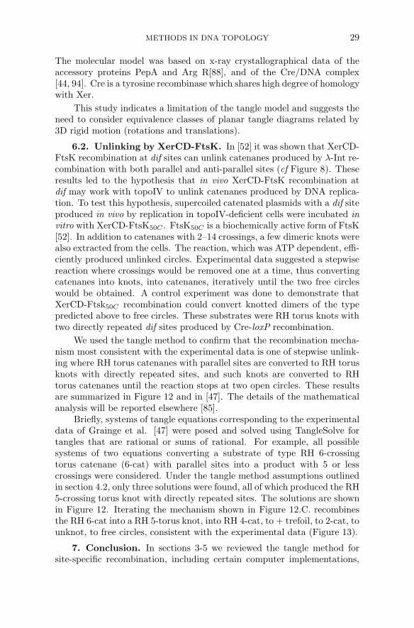
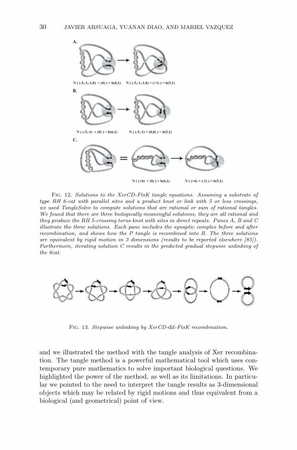
Briefly, systems of tangle equations corresponding to the experimentaldata of Grainge et al. [47] were posed and solved using TangleSolve fortangles that are rational or sums of rational. For example, all possiblesystems of two equations converting a substrate of type RH 6-crossingtorus catenane (6-cat) with parallel sites into a product with 5 or lesscrossings were considered. Under the tangle method assumptions outlinedin section 4.2, only three solutions were found, all of which produced the RH5-crossing torus knot with directly repeated sites. The solutions are shownin Figure 12. Iterating the mechanism shown in Figure 12.C. recombinesthe RH 6-cat into a RH 5-torus knot, into RH 4-cat, to + trefoil, to 2-cat, tounknot, to free circles, consistent with the experimental data (Figure 13).
7. Conclusion. In sections 3-5 we reviewed the tangle method forsite-specific recombination, including certain computer implementations,
30 JAVIER ARSUAGA, YUANAN DIAO, AND MARIEL VAZQUEZ
N ( (-5,-1) + (0) ) = b(6,1) N ( (-5,-1) + (0,0) ) = b(5,1)
B.
N ( (+6) + (0) ) = b(6,1) N ( (+6) + (-1) ) = b(5,1)
C.
N ( (-5,-1,-1,0) + (0) ) = b(6,1) N ( (-5,-1,-1,0) + (+1) ) = b(5,1)
A.
Fig. 12. Solutions to the XerCD-FtsK tangle equations. Assuming a substrate oftype RH 6-cat with parallel sites and a product knot or link with 5 or less crossings,we used TangleSolve to compute solutions that are rational or sum of rational tangles.We found that there are three biologically meaningful solutions, they are all rational andthey produce the RH 5-crossing torus knot with sites in direct repeats. Panes A, B and Cillustrate the three solutions. Each pane includes the synaptic complex before and afterrecombination, and shows how the P tangle is recombined into R. The three solutionsare equivalent by rigid motion in 3 dimensions (results to be reported elsewhere [85]).Furthermore, iterating solution C results in the predicted gradual stepwise unlinking ofthe 6cat.
Fig. 13. Stepwise unlinking by XerCD-dif-FtsK recombination.
and we illustrated the method with the tangle analysis of Xer recombina-tion. The tangle method is a powerful mathematical tool which uses con-temporary pure mathematics to solve important biological questions. Wehighlighted the power of the method, as well as its limitations. In particu-lar we pointed to the need to interpret the tangle results as 3-dimensionalobjects which may be related by rigid motions and thus equivalent from abiological (and geometrical) point of view.
METHODS IN DNA TOPOLOGY 31
REFERENCES
[1] C. Alen, D.J. Sherratt, and S.D. Colloms, Direct interaction of aminopep-tidase A with recombination site DNA in Xer site-specific recombination.,EMBO J. 16 (1997), pp. 5188–5197.
[2] J. Arsuaga, T. Blackstone, Y. Diao, E. Karadayi, and M. Saito, Linkingof Uniform Random Polygons in Confined Spaces, J. Physics A 40 (2007),pp. 1925–1936.
[3] J. Arsuaga, T. Blackstone, Y. Diao, E. Karadayi, and M. Saito, SamplingLarge Random Knots in a Confined Space, J. Physics A 40 (2007), pp. 11697–11711.
[4] J. Arsuaga, R. Tan, M. Vazquez, D.W. Sumners, and S.C. Harvey, Investi-gation of viral DNA packaging using molecular mechanics models, Biophys.Chem. 101 (2002), pp. 475–484.
[5] J. Arsuaga, M. Vazquez, S. Trigueros, D.W. Sumners, and J. Roca, Knot-ting probability of DNA molecules confined in restricted volumes: DNA knot-ting in phage capsids. Proc. Natl. Acad. Sci. USA 99 (2002), pp. 5373–5377.
[6] J. Arsuaga, M. Vazquez, P. McGuirk, S. Trigueros, D.W. Sumners, and
J. Roca, DNA knots reveal a chiral organization of DNA in phage capsids,Proc. Natl. Acad. Sci. USA 102 (2005), pp. 9165–9169.
[7] J. Arsuaga and Y. Diao, DNA Knotting in Spooling Like Conformations inBacteriophages, Computational and Mathematical Methods in Medicine 9(3)(2008), pp. 303–316.
[8] K. Aubrey, S. Casjens, and G. Thomas, Secondary structure and interactionsof the packaged dsDNA genome of bacteriophage P22 investigated by Ramandifference spectroscopy, Biochemistry 31 (1992), pp. 11835–11842.
[9] F.X. Barre and D.J. Sherratt, Chromosome dimer resolution. In The BacterialChromosome (Higgins, N.P., ed.), Washington, DC: ASM Press (2005), pp.513–524.
[10] L. Black,W. Newcomb, J. Boring, and J. Brown, Ion etching bacteriophageT4: support for a spiral-fold model of packaged DNA, Proc. Natl. Acad. Sci.USA 82 (1985), pp. 7960–7964.
Acknowledgements. The authors thank the following people: JungHun Koh for his help generating the spooling conformation, Rob Schareinfor his assistance in generating figures using Knotplot, David Sherratt, IanGrainge, Sean Colloms and Jonathan Bath for valuable discussions on thebiology of Xer recombination, Kai Ishihara and Koya Shimokawa for theirinsights on the mathematical analysis of Xer. Special thanks go to DeWitt L. Sumners for guiding the authors into this field. The three authors arevery grateful to Nick Cozzarelli. All of them were fellows of the Programin Mathematics and Molecular Biology (PMMB). This program fosteredthe authors′ interdisciplinary interest. Y. Diao was inspired by the jointwork of Nick Cozzarelli and De Witt L. Sumners on random knot theory. J.Arsuaga was a postdoctoral fellow with N. Cozzarelli and is grateful for theinvaluable training and for his career advice. M. Vazquez is most grateful toNick Cozzarelli for his vision of a world where topology could help biology(and vice versa), and for letting this vision materialize by promoting closeinteractions between mathematicians and molecular biologists.
32 JAVIER ARSUAGA, YUANAN DIAO, AND MARIEL VAZQUEZ
[11] T. Blackstone,P. McGuirck,C. Laing, M. Vazquez, J. Roca, and J. Ar-
suaga, The role of writhe in DNA condensation, Proceedings of InternationalWorkshop on Knot Theory for Scientific Objects. OCAMI Studies Volume 1
(2007). Osaka Municipal Universities Press; pp. 239–250.[12] M. Bregu, D.J. Sherratt, and S.D. Colloms, Accessory factors determine the
order of strand exchange in Xer recombination at psi., EMBO J. 21 (2002),pp. 3888–3897.
[13] D. Buck and E. Flapan, Predicting Knot or Catenane Type of Site-SpecificRecombination Products, J Molecular Biology 374(5) (2007), pp. 1186-1199.
[14] D. Buck and E. Flapan, A topological characterization of knots and links arisingfrom site-specific recombination., J. Phys. A: Math. Gen. 40 (2007), pp.12377–12395.
[15] D. Buck and C. Verjovsky-Marcotte, Tangle-solutions for a family of DNArearranging proteins., Math Proc Camb Phil Soc 139 (2005), pp. 59–80.
[16] D. Buck and C. Verjovsky-Marcotte, Classification of Tangle Solutions forIntegrases, A Protein Family that Changes DNA Topology., J. Knot. TheoryRamifications 16 (2007), pp. 969–995.
[17] G. Burde and H. Zieschang, Knots., vol. 5, In de Gruytier Studies in Mathe-matics (Gabriel, P., ed.) Walter de Gruyter, Berlin., 1985.
[18] H. Cabrera Ibarra, On the classification of rational 3-tangles, J. Knot TheoryRamifications 12(7) (2003), pp. 921-946.
[19] H. Cabrera Ibarra, Results on the classification of rational 3-tangles, J. KnotTheory Ramifications 13(2) (2004), pp. 175–192.
[20] K. Cerritelli, N. Cheng, A. Rosenberg, C. McPherson, F. Booy, and
A. Steven, Encapsidated conformation of bacteriophage T7 DNA, Cell 91
(1997), pp. 271–280.[21] D.K. Chattoraj and R.B. Inman, Location of DNA ends in P2, 186, P4 and
lambda bacteriophage heads, J. Mol. Biol. 87 (1974), pp. 11–22.[22] S.D. Colloms, J. Bath, and D.J. Sherratt, Topological selectivity in Xer site-
specific recombination, Cell 88 (1997), pp. 855–864.[23] J.H. Conway, An enumeration of knots and links, and some of their algebraic
properties., Computational Problems in Abstract Algebra, Pergamon, Ox-ford, UK (1967), pp. 329–358.
[24] N.R. Cozzarelli, M.A. Kraznow, S.P. Gerrard, and J.H. White, A topo-logical treatment of recombination and topoisomerases., Cold Spring HarborSymp. Quant. Biol. 49 (1984), pp. 383–400.
zarelli, The topological mechanism of phage lambda integrase., J. Mol. Biol.289 (1999), pp. 747–775.
[26] I. Darcy, Biological distances on DNA knots and links: applications to Xerrecombination., J. Knot Theory Ramification 10 (2001), pp. 269–294.
[27] I.K. Darcy, J. Chang, N. Druivenga, C. McKinney, R.K. Medikonduri, S.
Mills, J. Navarra-Madsen, A. Ponnusamy, J. Sweet, and T. Thompson,sl Coloring the Mu transpososome., BMC Bioinformatics 7 (2006), pp. 435.
[28] I.K. Darcy, J. Luecke, and M. Vazquez Tangle analysis of difference topol-ogy experiments: applications to a Mu-DNA protein complex, IMA preprintseries, (2008), https://www.ima.umn.edu/preprints/oct2007/2177.pdf.
[29] I.K. Darcy and R.G. Scharein, TopoICE-R: 3D visualization modeling thetopology of DNA recombination., Bioinformatics 22(14) (2006), pp. 1790–1791.
[30] Y. Diao, The Knotting of Equilateral Polygons in R3, Journal of Knot Theory
and its Ramifications, 4(2) (1995), pp. 189–196.[31] Y. Diao, A. Dobay, R.B. Kusner, K. Millet, and A. Stasiak, The Aver-
age Crossing Number of Equilateral Random Polygons J. Physics A 36(46)(2003), pp. 11561–11574.
METHODS IN DNA TOPOLOGY 33
[32] Y. Diao and C. Ernst, The Average Crossing Number of Gaussian RandomWalks and Polygons, Physical and numerical models in knot theory, J.A.Calvo, K.C. Millett, E.J. Rawdon, and A. Stasiak, editors, Series on Knotsand Everything 36 (2005), World Scientific, pp. 275–292.
[33] Y. Diao, J. Nardo, and Y. Sun, Global Knotting in Equilateral Random Poly-gons; Journal of Knot Theory and its Ramifications, 10(4) (2001), pp. 597–607.
[34] Y. Diao, N. Pippenger, and D.W. Sumners, On Random Knots, Journal ofKnot Theory and its Ramifications, 3(3) (1994), pp. 419–429.
[35] W.C. Earnshaw and S.R. Casjens, DNA packaging by the double-strandedDNA bacteriophages, Cell 21 (1980), pp. 319–331.
[36] J. Emert and C. Ernst, N-string tangles, J. Knot. Theory Ramifications9(8)(2000), pp. 987–1004.
[37] C. Ernst, Tangle equations, J. Knot. Theory Ramifications 5(1996), pp. 145–159.[38] C. Ernst, Tangle equations II, J. Knot. Theory Ramifications 6 (1997), pp. 1–11.[39] C. Ernst and D.W. Sumners, A calculus for rational tangles: applications to
DNA recombination, Math. Proc. Cambridge Phil. Soc. 108 (1990), pp. 489–515.
[40] C. Ernst and D.W. Sumners, Solving tangle equations arising in a DNA recom-bination model, Math. Proc. Cambridge Phil. Soc. 126 (1999), pp. 23–36.
[41] O. Espeli and K.J. Marians,, Untangling intracellular DNA topology, Mol Mi-crobiol 52 (2004), pp. 925–931.
[42] A. Evilevitch, L. Lavelle, C.M. Knobler, E. Raspaud, and W.M. Gelbart,Osmotic pressure inhibition of DNA ejection from phage, Proc. Natl. Acad.Sci. USA 100 (2003), pp. 9292–9295.
(1997), pp. 300–332.[44] D.N. Gopaul, F. Guo, and G.D. Van Duyne, Structure of the Holliday junction
intermediate in Cre–loxP site-specific recombination, EMBO J. 17 (1998),pp. 4175–4187.
[45] S.C. Gourlay and S.D. Colloms, Control of Cre recombination by regulatoryelements from Xer recombination systems, Mol. Microbiol. 52 (2004), pp.53–65.
[46] I. Grainge, D. Buck, and M. Jayaram, Geometry of site alignment during Intfamily recombination: antiparallel synapsis by the FLP recombinase, J. Mol.Biol. 298 (2000), pp. 749–764.
[47] I. Grainge, M. Bregu, M. Vazquez, V. Sivanathan, S.C. Ip, and D.J. Sher-
ratt, Unlinking chromosomes catenated in vivo by site-specific recombina-tion, EMBO J 26(19) (2007), pp. 4228–4238.
[48] B. Hallet and D.J. Sherratt, Transposition and site-specific recombinationadapting DNA cut-and paste mechanism to a variety of genetic rearrange-ments, FEMS Microbiol. Rev. (1997), p. 21.
[49] M. Hirasawa and K. Shimokawa, Dehn surgeries on strongly invertible knotswhich yield lens spaces, Proc. Am. Math. Soc. 128 (2000), pp. 3445–3451.
[50] V.F. Holmes and N.R. Cozzarelli, Closing the ring: links between SMC pro-teins and chromosome partitioning, condensation, and supercoiling, Proc.Natl. Acad. Sci. USA 97 (2000), pp. 1322–1324.
[51] N. Hud, Double-stranded DNA organization in bacteriophage heads: an alterna-tive toroid-based model, Biophys. J. 69 (1995), pp. 1355–1362.
[52] S.C. Ip, M. Bregu, F.X. Barre, and D.J. Sherratt, Decatenation of DNA cir-cles by FtsK-dependent Xer site-specific recombination., EMBO J 22 (2003),pp. 6399–6407.
[53] P.J. Jardine and D.L. Anderson, DNA packaging in double-stranded DNAphages The bacteriophages (2006), Ed. Richard Calendar, Oxford UniversityPress, pp. 49–65.
34 JAVIER ARSUAGA, YUANAN DIAO, AND MARIEL VAZQUEZ
[54] R. Kanaar, A. Klippel, E. Shekhtman, J. M. Dungan, R. Kahmann, and
N.R. Cozzarelli, Processive recombination by the phage Mu Gin system:implications for the mechanisms of DNA strand-exchange, DNA site align-ment, and enhancer action, Cell 62 (1990), pp. 353–366
[55] V. Katritch, Bednar, D. Michoud, R. G. Scharein, J. Dubochet, and A.
Stasiak, Geometry and physics of knots, Nature 384 (1996), pp. 142–145.[56] E. Kellenberger, E. Carlemalm, J. Sechaud, A. Ryter, and G. Haller,
Considerations on the condensation and the degree of compactness in non-eukaryotic DNA-containing plasmas, In Bacterial Chromatin: Proceedingsof the Symposium “Selected Topics on Chromatin Structure and Function”(eds. C. Gualerzi and C. L. Pon), Springer, Berlin (1986), pp. 11–25.
[57] S. Kim and I.K. Darcy, Topological analysis of DNA-protein complexes, In-cluded in this volume, Mathematics of DNA Structure, Function and Inter-actions (eds C.J. Benham, S. Harvey, W.K. Olson, D.W. Sumners and D.Swigon), Springer Science + Business Media, LLC, New York, (2009).
Frank-Kamenetskii, Effect of Excluded Volume on Topological Propertiesof Circular DNA, J. Biomolec. Str. and Dyn. 5 (1988), pp. 1173–1185.
[59] J.C. LaMarque, T.L. Le, and S.C. Harvey, Packaging double-helical DNA intoviral capsids, Biopolymers 73 (2004), pp. 348–355.
[60] A. Landy,Coming or going its another pretty picture for the lambda-Int familyalbum, Proc. Natl Acad. Sci. USA 96 (1999), pp. 7122–7124.
[61] J. Lepault, J. Dubochet, W. Baschong, and E. Kellenberger, Organiza-tion of double-stranded DNA in bacteriophages: a study by cryo-electronmicroscopy of vitrified samples EMBO J. 6 (1987), pp. 1507–1512.
[62] W.B.R. Lickorish, Prime knots and tangles., Trans. Am. Math. Soc. 267 (1981),pp. 321–332.
[63] L.F. Liu, J.L. Davis, and R. Calendar, Novel topologically knotted DNA frombacteriophage P4 capsids: studies with DNA topoisomerases, Nucleic AcidsRes. 9 (1981), pp. 3979–3989.
[64] L.F. Liu, L. Perkocha, R. Calendar, and J.C. Wang, Knotted DNA frombacteriophage capsids, Proc. Natl. Acad. Sci. USA 78 (1981), pp. 5498–5502.
[65] J.P.J. Michels and F.W. Wiegel, On the topology of a polymer ring, Proc. R.Soc. London Ser A 403 (1986), pp. 269–284.
[66] C. Micheletti, D. Marenduzzo, E. Orlandini, and D.W. Sumners, Knottingof random ring polymers in confined spaces, J. Chem. Phys. 124 (2006), pp.064903.1–10.
[67] K. Millett, Knotting of regular polygons in 3-space, Random knotting andlinking (Vancouver, BC, 1993), World Sci. Publishing, Singapore (1994), pp.31–46.
[68] K. Millett, Monte Carlo Explorations of Polygonal Knot Spaces, Knots in Hel-las’98 (Delphi), Ser. Knots Everything 24 (2000), World Scientific, pp. 306–334.
[69] H.R. Morton, Seifert circles and knot polynomials, Math. Proc. Cambridge Phil.Soc. 99 (1986), pp. 107–109.
[70] K. Murasugi, Knot Theory, Its Applications (Translated by B. Kurpita),Birkhauser, Boston, MA. 1996.
[71] S.E. Nunes-Duby, H.J. Kwon, R.S.T. Tirumalai, T. Ellenberger, and A.
Landy, Similarities and differences among 105 members of the Int family ofsite-specific recombinases, Nucl. Acids Res. 26 (1998), pp. 391–406.
[72] A.S. Petrov, M.B. Boz, and S.C. Harvey, The conformation of double-stranded DNA inside bacteriophages depends on capsid size and shape, JStruct Biol. 160 (2007) pp. 241–248.
[73] P. Plunkett, M. Piatek, A. Dobay, J.C. Kern, K. Millet, A. Stasiak, and
E. Rawdon, Total curvature and total torsion of knotted polymers, Macro-molecules 40 (2007), pp. 3860–3867.
METHODS IN DNA TOPOLOGY 35
[74] L. Rayleigh, On the problems of random vibrations, and of random flights inone, two, or three dimensions, Phil. Mag. S. 6. 37(220) (1919), pp. 321–347.
[75] D. Raymer and D. Smith, Spontaneous knotting of an agitated string Proc.Natl. Acad. Sci
[76] K. Richards, R. Williams, and R. Calendar, Mode of DNA packing withinbacteriophage heads, J. Mol. Biol. 78 (1973), pp. 255–259.
[77] D. Rolfsen, Knots Mathematics Lecture Series 7, Publish or Perish, Berkeley,CA., 1976.
[78] V.V. Rybenkov, N.R. Cozzarelli, and A.V. Vologodskii, Probability of DNAknotting and the effective diameter of the DNA double helix, Proc. Natl.Acad. Sci. USA 90 (1993), pp. 5307–5311.
[79] P.D. Sadowski, Site-specific genetic recombination: hops, flips, and flops,FASEB J. 7 (1993), pp. 760–767.
[80] Y. Saka and M. Vazquez, TangleSolve: topological analysis of site-specific re-combination, Bioinformatics 18 (2002), pp. 1011–1012.
[81] J.B. Schvartzman and A. Stasiak, A topological view of the replicon, EMBORep. 5(3) (2004), 256–261.
[82] P. Serwer, Arrangement of double-stranded DNA packaged in bacteriophagecapsids: An alternative model, J. Mol. Biol. 190 (1986), pp. 509–512.
[83] S.Y. Shaw and J.C. Wang, Knotting of a DNA chain during ring closure, Science260 (1993), pp. 533–536.
[84] Arciszewska, L.K. and D.J. Sherratt Site-specific recombination and circu-lar chromosome segregation, Philos. Trans. R. Soc. Lond. B. Biol. Sci. 347
(1995), pp. 37–42.[85] K. Shimokawa, K. Ishihara, I. Grainge, D.J. Sherratt, and M. Vazquez,
DNA unlinking by site-specific recombination: topological analysis of XerCD-FtsK action, Preliminary report.
[86] W.M. Stark and M.R. Boocock, Topological selectivity in site-specific recom-bination, In Mobile Genetic Elements (Sherratt, D. J., ed.), IRL Press atOxford University, Oxford (1995), pp. 101–129.
[87] W.M. Stark, D.J. Sherratt, and M.R. Boocock, Site-specific recombinationby Tn3 resolvase: topological changes in the forward and reverse reactions,Cell 58 (1989), pp. 779–790.
[88] N. Strater, D.J. Sherratt, and S.D. Colloms, X-ray structure of aminopep-tidase A from Escherichia coli and a model for the nucleoprotein complex inXer site-specific recombination., EMBO J. 18 (1999), pp. 4513–4522.
[89] D.K. Summers and D.J. Sherratt, Multimerization of high copy number plas-mids causes instability: ColE1 encodes a determinant essential for plasmidmonomerization and stability, Cell 36 (1984), pp. 1097–1103.
[90] D.W. Sumners, C. Ernst, N.R. Cozzarelli, and S.J. Spengler, Mathematicalanalysis of the mechanisms of DNA recombination using tangles, QuarterlyReviews of Biophysics 28 (1995), pp. 253–313.
[91] S. Trigueros, J. Arsuaga, M. Vazquez, D.W. Sumners, and J. Roca, Noveldisplay of knotted DNA molecules by two dimensional gel electrophoresis,Nucleic Acids Research 29 (2001), e67.
[92] S. Trigueros and J. Roca, Production of highly knotted DNA by means ofcosmid circularization inside phage capsids, BMC Biotechnol 7(1) (2007),pp. 94.
[93] S. Tzill, J.K. Kindt, W.M. Gelbart, and A. Ben-Shaul, Forces and Pressuresin DNA Packaging and Release from Viral Capsids, Biophys. J. 84 (2003),pp. 1616–1627.
[94] G.D. Van Duyne, A structural view of Cre-loxP site-specific recombination,Annu. Rev. Biophys. Biomol. Struct. 30 (2001), pp. 87–104.
[95] V. Vanhooff, C. Galloy, H. Agaisse, D. Lereclus, B. Revet, and B. Hal-
let, Self-Control in DNA site-specific recombination mediated by the tyro-sine recombinase TnpI, Molecular Microbiology 60(3) (2006), pp. 617–629.
36 JAVIER ARSUAGA, YUANAN DIAO, AND MARIEL VAZQUEZ
[96] M. Vazquez, Tangle analysis of site-specific recombination: Gin and Xer systems,PhD dissertation in mathematics, Florida State University, Tallahassee, FL,2000.
[97] M. Vazquez, S.D. Colloms, and D.W. Sumners, Tangle analysis of Xer re-combination reveals only three solutions, all consistent with a single 3-dimensional topological pathway, J. Mol. Biol. 346 (2005), pp. 493–504.
[98] M. Vazquez and D.W. Sumners, Tangle analysis of Gin site-specific recombina-tion, Math. Proc. Cambridge Phil. Soc. 136 (2004), pp. 565–582.
[99] A.V. Vologodskii, N.J. Crisona, B. Laurie, P. Pieranski, V. Katritch, J.
Dubochet, and A. Stasiak, Sedimentation and electrophoretic migrationof DNA knots and catenanes, J. Mol. Biol. 278 (1998), pp. 1–3.
[100] S.A. Wasserman, J.M. Dungan, and N.R. Cozzarelli, Discovery of a predictedDNA knot substantiates a model for site-specific recombination, Science 229
(1985), pp. 171–174.[101] W. Zheng, C. Galloy, B. Hallet, and M. Vazquez, The tangle model for
site-specific recombination: a computer interface and the TnpI-IRS recom-bination system, Knot Theory for Specific Objects, OCAMI studies 1(2)(2007), pp. 251–271.