UNIVERSITY OF CALIFORNIA, SAN DIEGO Mathematical Model for Leukocyte Migration in Prosthetic Materials BENG 221 – Mathematical Methods for Bioengineers – Fall 2012 Term Project Ana Moreno, Shawn R Mailo, Sheng-Hsiou Hsu, Jahir Gutierrez 11/9/2012 By means of the “convection-diffusion partial differential equation”, a model for the migration of leukocyte towards the implantation site of a vascular prosthetic material has been developed. Although this is a simplified version of more complex models used to describe cell populations’ dynamics, it provides meaningful data on leukocytes’ behavior in the presence of chemotactic factors released by the implanted prosthetic material. Both the analytical solution and the numerical approximation are provided along with three-dimensional graphs to accompany the results.
Transcript
UNIVERSITY OF CALIFORNIA, SAN DIEGO
Mathematical Model for Leukocyte Migration in
Prosthetic Materials BENG 221 – Mathematical Methods for
Bioengineers – Fall 2012 Term Project
Ana Moreno, Shawn R Mailo, Sheng-Hsiou Hsu, Jahir Gutierrez
11/9/2012
By means of the “convection-diffusion partial differential equation”, a model for the migration of leukocyte towards the implantation site of a vascular prosthetic material has been developed. Although this is a simplified version of more complex models used to describe cell populations’ dynamics, it provides meaningful data on leukocytes’ behavior in the presence of chemotactic factors released by the implanted prosthetic material. Both the analytical solution and the numerical approximation are provided along with three-dimensional graphs to accompany the results.
Mathematical Model for Leukocyte Migration in Prosthetic Materials
Ana Moreno, Shawn R Mailo, Sheng-Hsiou Hsu, Jahir Gutierrez
Page 2
INTRODUCTION
Cardiovascular disease is the second leading cause of death the world and the second leading cause
of death in the United States [1]
. Among all cardiovascular diseases, atherosclerosis is the most
important because almost all life-threatening cardiovascular disorders (e.g. coronary artery disease)
are derived from it.
Atherosclerosis, in brief, is the abnormal accumulation of plaque (which is mainly composed of fat
and calcium) inside the arteries [2]
. This plaque abates the artery’s normal blood flow and, at an
advanced stage of the disease, ruptures (becomes a thrombus) and eventually starts to flow in the
bloodstream until it completely occludes one small-diameter artery. This finally leads to ischemia
and infarction of the organ (e.g. heart or even the brain) that used to be nourished by the now
occluded artery [3]
.
Tissue engineered vascular grafts (TEVG) are promising alternatives for treating cardiovascular
diseases related to atherosclerosis (e.g. carotid artery disease). A TEVG is a prosthetic material that
behaves as an artificial blood vessel to replace atherosclerotic arteries in the patient’s circulatory
system. Although much effort and many years of research have been dedicated to the task of
improving these prosthetic materials, it has not been possible to build a reliable and durable TEVG
yet. It has been well established that the presence of a foreign body in the tissue, such as a
prosthetic material, increases the risk of infection [4]
and this is actually the main reason that lies
beneath complications with vascular prosthetic materials implantation [5]
.
Because leukocytes serve as the key acute inflammatory mediators, the control of their motility on
the surfaces of vascular prosthetic materials is a crucial factor in promoting implant infection
resistance [6]
. Whereas leukocyte behavior and motility on normal vascular tissue has been
extensively studied [7]
, very few details are known in terms of the regulation of their migration on
vascular prosthetic biomaterials. Right after a prosthetic material is implanted, an immune response
is triggered and, eventually, several circulating proteins begin to modulate the behavior of adherent
leukocytes in order to direct their migration to the implant site [8]
. Leukocytes will then flow in the
blood towards the prosthetic material to adhere to its surface in order to attack any infectious agent
that could be present along the prosthetic vascular graft and try to eliminate them.
PROBLEM STATEMENT
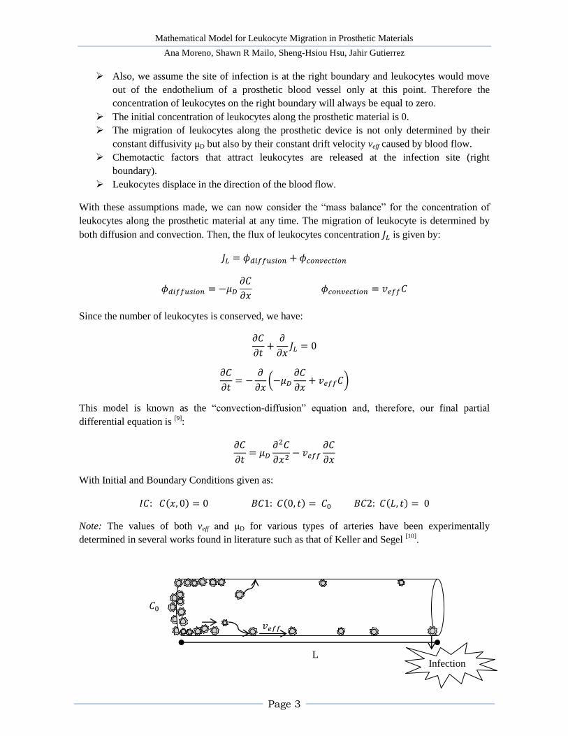
We consider the following problem: A linear prosthetic material (i.e. engineered vascular graft) of
length L and constant cross sectional area has just been implanted in one of the arteries of a patient
and immediately triggers an immune response with the subsequent migration of leukocytes towards
the implantation site. We would like to find a mathematical expression for the concentration of
leukocytes , along the vascular prosthetic material as a function of distance and time .
Assumptions
We assume the concentration of leukocytes is constant on the left boundary of the
prosthetic at all times (it can be seen as a source of leukocytes) and equal to C0.
Mathematical Model for Leukocyte Migration in Prosthetic Materials
Ana Moreno, Shawn R Mailo, Sheng-Hsiou Hsu, Jahir Gutierrez
Page 3
L
Also, we assume the site of infection is at the right boundary and leukocytes would move
out of the endothelium of a prosthetic blood vessel only at this point. Therefore the
concentration of leukocytes on the right boundary will always be equal to zero.
The initial concentration of leukocytes along the prosthetic material is 0.
The migration of leukocytes along the prosthetic device is not only determined by their
constant diffusivity μD but also by their constant drift velocity veff caused by blood flow.
Chemotactic factors that attract leukocytes are released at the infection site (right
boundary).
Leukocytes displace in the direction of the blood flow.
With these assumptions made, we can now consider the “mass balance” for the concentration of
leukocytes along the prosthetic material at any time. The migration of leukocyte is determined by
both diffusion and convection. Then, the flux of leukocytes concentration is given by:
Since the number of leukocytes is conserved, we have:
This model is known as the “convection-diffusion” equation and, therefore, our final partial
differential equation is [9]
:
With Initial and Boundary Conditions given as:
, , ,
Note: The values of both veff and μD for various types of arteries have been experimentally
determined in several works found in literature such as that of Keller and Segel [10]
.
Infection
Mathematical Model for Leukocyte Migration in Prosthetic Materials
Ana Moreno, Shawn R Mailo, Sheng-Hsiou Hsu, Jahir Gutierrez
Page 4
ANALYTICAL SOLUTION
Part 1: Simplified model
Assumption: Convection (drift) term is zero, i.e. v .
The diffusion-convection equation can be reduced to diffusion equation.
Reduced to Diffusion Equation
(The migration of Leukocyte in
the absence of blood flow.)
, t
t
, t
Boundary Conditions
Initial Condition
, t , , t , t
, ,
To homogenize the boundary conditions, let , t , t , we can solve steady-state
solution and make time-varying solution , t easy to solve.
Steady-state solution Time-varying solution , t
, t
t
, t
, , t , , t
,
(1) Steady-state solution: The second derivative of the steady-state solution is equal to zero. The solution has the general
form:
B
Implementing with the boundary conditions shown above we get the following equations:
B
B
Using these equations the variable A can be found:
***Plugging A back into the general form of the steady-state solution results,
,
Mathematical Model for Leukocyte Migration in Prosthetic Materials
Ana Moreno, Shawn R Mailo, Sheng-Hsiou Hsu, Jahir Gutierrez
Page 5
(2) Time-varying solution: , t Solve for diffusion equation with homogeneous boundary conditions:
Use separation of variables:
, t t
We can solve for time-dependence and space-dependence solutions separately.
t t t
(a) Time-dependence solution
t e
(b) Space-dependence solution:
(i) For , B. With boundary conditions, B
(ii) For , e Be , With boundary conditions,
(iii) For , cos Bsin , With boundary conditions,
Bsin , where
, n , ,
Combine (a) and (b),
, t sin n
e
Solve coefficient to satisfy the initial condition.
, sin n
sin
n
d
Combine steady-state and time-varying solution, we obtain the final solution:
, t
sin n
e
Mathematical Model for Leukocyte Migration in Prosthetic Materials
Ana Moreno, Shawn R Mailo, Sheng-Hsiou Hsu, Jahir Gutierrez
Page 6
Part 2: General Model
Now we consider the effect of blood flow and thus the convection (drift) term in our general model.
Diffusion-convection Equation , t
t
, t
, t
Boundary Conditions
Initial Condition
, t , , t , t
, ,
Similarly, we can homogenize the boundary condition. Let , t , t ,
Steady-state solution Time-varying solution , t
, t
t
, t
, t
, , t , , t
,
(1) Steady-state solution: Integrating both sides two times, we have the general form of the solution:
e
B
By boundary conditions,
B , e
B
, B
We have the steady-state solution.
Note that the steady-state solution is not linear as in reduced model. It’s the effect of blood flow
which carries leukocytes further. For more discussion, please see the numerical simulation.
(2) Time-varying solution: , t Solve for diffusion equation with homogeneous boundary conditions.
Mathematical Model for Leukocyte Migration in Prosthetic Materials
Ana Moreno, Shawn R Mailo, Sheng-Hsiou Hsu, Jahir Gutierrez
Page 7
Use separation of variables:
, t t
We can solve for time-dependence and space-dependence solutions separately.
t t t
(a) Time-dependence solution:
t e
(b) Space-dependence solution:
The general model has a convection term. It is a secondary derivative ordinary differential
equation.
Its auxiliary equation is,
r
The roots of the auxiliary equation are,
r
(i) For
, the equation has double root r
The solution has the form e B e .
With boundary conditions: , B e , B
(ii) For
, the equation has positive real roots and the solution is
e Be
With boundary conditions: B , e Be , B
(iii) For
,
The equation has complex roots and solution has general form,
e
sin
Bcos
where .
With boundary conditions: B , and
e
sin
Mathematical Model for Leukocyte Migration in Prosthetic Materials
Ana Moreno, Shawn R Mailo, Sheng-Hsiou Hsu, Jahir Gutierrez
Page 8
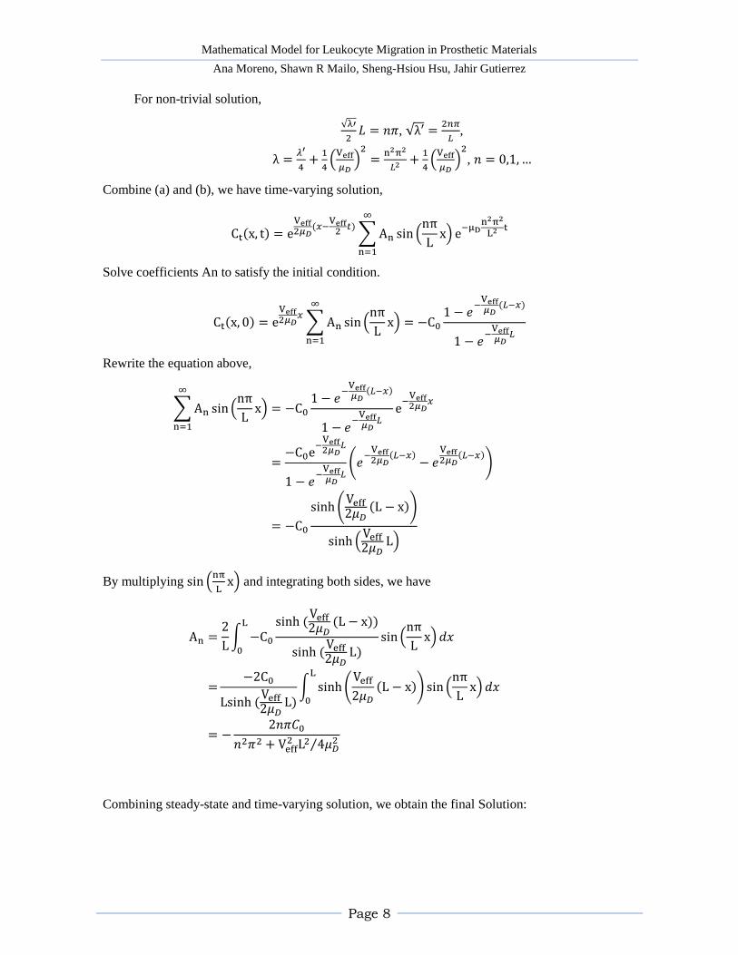
For non-trivial solution,
,
,
, , ,
Combine (a) and (b), we have time-varying solution,
, t e
sin
n
e
Solve coefficients An to satisfy the initial condition.
, e
sin
n
Rewrite the equation above,
sin n
e
e
sinh
sinh
By multiplying sin
and integrating both sides, we have
sinh
sinh
sin
n
sinh
sinh
sin n
Combining steady-state and time-varying solution, we obtain the final Solution:
Mathematical Model for Leukocyte Migration in Prosthetic Materials
Ana Moreno, Shawn R Mailo, Sheng-Hsiou Hsu, Jahir Gutierrez
Page 9
, t e
sin
n
e
At a quick inspection of the solution, except for the nonlinear steady-state term, there is a
e
moving wave term with velocity
and direction of +x. This is the result of
convection term and thus the blood flow.
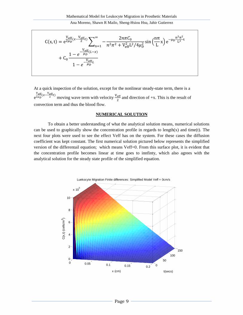
NUMERICAL SOLUTION
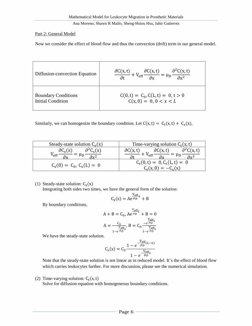
To obtain a better understanding of what the analytical solution means, numerical solutions
can be used to graphically show the concentration profile in regards to length(x) and time(t). The
next four plots were used to see the effect Veff has on the system. For these cases the diffusion
coefficient was kept constant. The first numerical solution pictured below represents the simplified
version of the differentail equation; which means Veff=0. From this surface plot, it is evident that
the concentration profile becomes linear at time goes to innfinty, which also agrees with the
analytical solution for the steady state profile of the simplified equation.
0
50
100
150
0 0.05 0.1 0.15 0.2
0
2
4
6
8
10
x 105
t(secs)
Luekocyte Migration Finite differences: Simplified Model Veff = 0cm/s
x (cm)
C(x
,t)
(cells
/m2)
Mathematical Model for Leukocyte Migration in Prosthetic Materials
Ana Moreno, Shawn R Mailo, Sheng-Hsiou Hsu, Jahir Gutierrez
Page 10
0
50
100
150
0 0.05 0.1 0.15 0.2
0
2
4
6
8
10
x 105
t(secs)
Luekocyte Migration Finite differences: General Model Veff = 1.5*10-3cm/s
x (cm)
C(x
,t)
(cells
/m2)
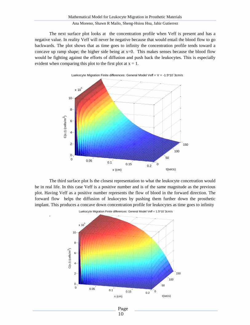
The next surface plot looks at the concentration profile when Veff is present and has a
negative value. In reality Veff will never be negative because that would entail the blood flow to go
backwards. The plot shows that as time goes to infinity the concentration profile tends toward a
concave up ramp shape; the higher side being at x=0. This makes senses because the blood flow
would be fighting against the efforts of diffusion and push back the leukocytes. This is especially
evident when comparing this plot to the first plot at x = 1.
The third surface plot Is the closest representation to what the leukocyte concetration would
be in real life. In this case Veff is a positive number and is of the same magnitude as the previous
plot. Having Veff as a positive number represents the flow of blood in the forward direction. The
forward flow helps the diffusion of leukocytes by pushing them further down the prosthetic
implant. This produces a concave down concentration profile for leukocytes as time goes to infinity
.
0
50
100
150
0 0.050.1
0.150.2
0
2
4
6
8
10
x 105
t(secs)
Luekocyte Migration Finite differences: General Model Veff = V = -1.5*10-3cm/s
x (cm)
C(x
,t)
(cells
/m2)
Mathematical Model for Leukocyte Migration in Prosthetic Materials
Ana Moreno, Shawn R Mailo, Sheng-Hsiou Hsu, Jahir Gutierrez
Page 11
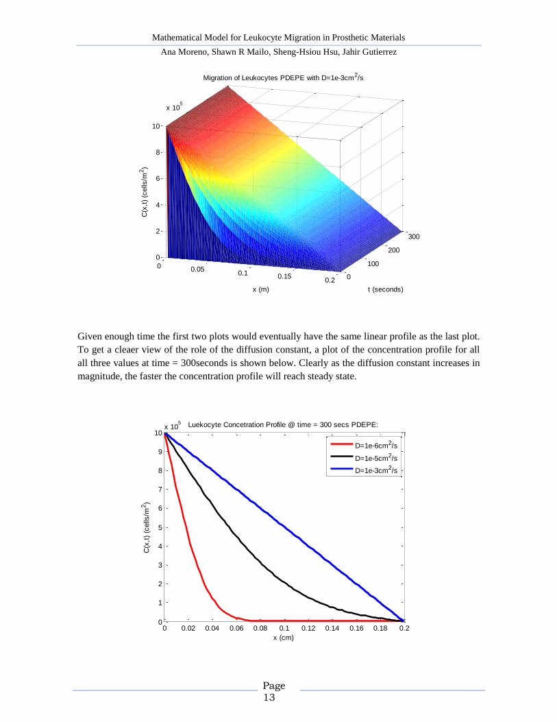
The next plot shows more cleary what the steady state concentration profile is for this
equation at the different values of Veff.
Along with seeing the effects of Veff has on the equation, it is also important to understand
how the diffusion constant plays a role in the leukocyte concetration. For all of the following plots,
Veff is 0 in order to make the effects of diffusion more clear. Also, take note that all diffusion
constants, although at different magnitudes, are all positive. This is because is the definition of
diffusion is the act of spreading out, the postive case, and not retracting, the negative case. By
comparing the next three suraface plots to each other it is evident that the main effect of the
diffusion constant is on the time it takes for the system to reach steady state.
[4] C. C. Chang, R. S. Rosenson-Schloss, T. D. Bhoj and P. V. Moghe, "Leukocyte chemosensory
migration on vascular prosthetic biomaterial is mediated by an integrin B2 receptor chain,"
Biomaterials, no. 21, pp. 2305-2313, 2000.
[5] J. Goldstone and W. Moore, "Infection in vascular prostheses.," American Journal of Surgery,
no. 128, pp. 225-233, 1974.
[6] R. S. Rosenson-Schloss, C. C. Chang, A. Constantinides and P. V. Moghe, "Alteration of
leukocyte motility on plasma-conditioned prosthetic biomaterial, ePTFE, via a flow-responsive
cell adhesion molecule, CD43," JBM - John Wiley & Sons, Inc., vol. 1278, pp. 1-18, 2001.
[7] L. McIntire, "Bioengineering and vascular biology," Annals of Biomedical Engineering, no. 22,
pp. 2-13, 1994.
[8] C. Chang, M. Lieberman and P. Moghe, "Quantitative analysis of the regulation of leukocyte
chemosensory migration by a vascular prosthetic biomaterial," Journal of Materials Science:
Materials in Medicine, vol. 11, pp. 337-344, 2000.
[9] J. R. Brannan and W. E. Boyce, Differential Equations With Boundary Value Problems. An
introduction to Modern Methods and Applications, New York: John Wiley & Sons, 2011.
[10] E. Keller and L. Segel, "Model for Chemotaxis," Journal of Theoretical Biology, no. 30, pp.
225-234, 1971.
Mathematical Model for Leukocyte Migration in Prosthetic Materials
Ana Moreno, Shawn R Mailo, Sheng-Hsiou Hsu, Jahir Gutierrez
Page 16
APPENDIX: MATLAB CODE
%% Simplified Model - Finite difference method % diffusion constant clear all global D D = 1*10^-4; % cm^2/s
global C0 C0 = 1*10^6;
global V V = 0; % cm/s
% domain dx = 0.005; % step size in x dimension cm dt = .1; % step size in t dimension weeks xmesh = 0:dx:.2; % domain in x cm tmesh = 0:dt:150; % domain in t weeks
% solution using finite differences (see Week 1 class notes) nx = length(xmesh); % number of points in x dimension nt = length(tmesh); % number of points in t dimension stepsize = D * dt / dx^2; % stepsize for numerical integration sol_fd = zeros(nt, nx); sol_fd(:, 1) = C0; % left boundary conditions; constant value sol_fd(:, nx) = 0; % left boundary conditions; zero value sol_fd(1, :) = 0; % initial conditions; zero
for t = 1:nt-1 for x = 2:nx-1 sol_fd(t+1, x) = sol_fd(t, x) + stepsize * ... (sol_fd(t, x-1) - 2 * sol_fd(t, x) + sol_fd(t, x+1)) -
(V*dt/dx) * ... (sol_fd(t, x+1) - sol_fd(t, x)); end end
figure(1) surf(tmesh,xmesh,sol_fd','EdgeColor','none') title('Luekocyte Migration Finite differences: Simplified Model Veff =
%%%%%%%%%%%%%%%%%% %% General Model - Finite difference method % diffusion constant global D D = 1*10^-4; % cm^2/s
Mathematical Model for Leukocyte Migration in Prosthetic Materials
Ana Moreno, Shawn R Mailo, Sheng-Hsiou Hsu, Jahir Gutierrez
Page 17
global C0 C0 = 1*10^6;
global V V = -1.5*10^-3; % cm/s
% domain dx = 0.005; % step size in x dimension cm dt = .1; % step size in t dimension weeks xmesh = 0:dx:.2; % domain in x cm tmesh = 0:dt:150; % domain in t weeks
% solution using finite differences (see Week 1 class notes) nx = length(xmesh); % number of points in x dimension nt = length(tmesh); % number of points in t dimension stepsize = D * dt / dx^2; % stepsize for numerical integration sol_fd = zeros(nt, nx); sol_fd(:, 1) = C0; % left boundary conditions; constant value sol_fd(:, nx) = 0; % left boundary conditions; zero value sol_fd(1, :) = 0; % initial conditions; zero
for t = 1:nt-1 for x = 2:nx-1 sol_fd(t+1, x) = sol_fd(t, x) + stepsize * ... (sol_fd(t, x-1) - 2 * sol_fd(t, x) + sol_fd(t, x+1)) -
(V*dt/dx) * ... (sol_fd(t, x+1) - sol_fd(t, x)); end end
figure(2) surf(tmesh,xmesh,sol_fd','EdgeColor','none') title('Luekocyte Migration Finite differences: General Model Veff = V = -
%%%%%%%%%%%%%%%%%% %% General Model - Finite difference method % diffusion constant global D D = 1*10^-4; % cm^2/s
global C0 C0 = 1*10^6;
global V V = 1.5*10^-3; % cm/s
% domain dx = 0.005; % step size in x dimension cm dt = .1; % step size in t dimension weeks
Mathematical Model for Leukocyte Migration in Prosthetic Materials
Ana Moreno, Shawn R Mailo, Sheng-Hsiou Hsu, Jahir Gutierrez
Page 18
xmesh = 0:dx:.2; % domain in x cm tmesh = 0:dt:150; % domain in t weeks
% solution using finite differences (see Week 1 class notes) nx = length(xmesh); % number of points in x dimension nt = length(tmesh); % number of points in t dimension stepsize = D * dt / dx^2; % stepsize for numerical integration sol_fd = zeros(nt, nx); sol_fd(:, 1) = C0; % left boundary conditions; constant value sol_fd(:, nx) = 0; % left boundary conditions; zero value sol_fd(1, :) = 0; % initial conditions; zero
for t = 1:nt-1 for x = 2:nx-1 sol_fd(t+1, x) = sol_fd(t, x) + stepsize * ... (sol_fd(t, x-1) - 2 * sol_fd(t, x) + sol_fd(t, x+1)) -
(V*dt/dx) * ... (sol_fd(t, x+1) - sol_fd(t, x)); end end
figure(3) surf(tmesh,xmesh,sol_fd','EdgeColor','none') title('Luekocyte Migration Finite differences: General Model Veff =
function leukocytemig global miuD global Co global veff global L
Co = 1e6; %cells/cm^2 miuD = 1e-6; %cm^2/s L = 0.2; %cm veff = 1e-5; %cm/s
Mathematical Model for Leukocyte Migration in Prosthetic Materials
Ana Moreno, Shawn R Mailo, Sheng-Hsiou Hsu, Jahir Gutierrez
Page 19
x = 0:0.01*L:L; t = 0:1:300;
sol_pdepe = pdepe(0,@pdefun,@ic,@bc,x,t);
A= sol_pdepe(300, :)'; figure(4) plot(x, A, 'r', 'linewidth', 2.5); hold on figure(1) surf(t,x,sol_pdepe','EdgeColor','none') title('Migration of Leukocytes PDEPE with D=1e-6cm^2/s') xlabel('t (seconds)') ylabel('x (cm)') zlabel('C(x,t) (cells/m^2') % function definitions for pdepe: % -------------------------------------------------------------- function [c, f, s] = pdefun(x, t, u, DuDx) % PDE coefficients functions global miuD global veff c = 1; f = miuD * DuDx; % diffusion s = -veff * DuDx; % convection % -------------------------------------------------------------- function u0 = ic(x) global Co
% Initial conditions function u0 = (x==0);
% -------------------------------------------------------------- function [pl, ql, pr, qr] = bc(xl, ul, xr, ur, t) % Boundary conditions function
global Co
pl = ul-Co; % value left boundary condition ql = 0; % flux left boundary condition pr = ur; % value right boundary condition qr = 0; % flux right boundary condition
%Migration of Leukocytes with "pdepe"
function leukocytemig1 global miuD global Co global veff global L
Co = 1e6; %cells/cm^2
Mathematical Model for Leukocyte Migration in Prosthetic Materials
Ana Moreno, Shawn R Mailo, Sheng-Hsiou Hsu, Jahir Gutierrez
Page 20
miuD = 1e-5; %cm^2/s L = 0.2; %cm veff = 1e-5; %cm/s x = 0:0.01*L:L; t = 0:1:300;
sol_pdepe1 = pdepe(0,@pdefun2,@ic2,@bc2,x,t); A1= sol_pdepe1(300, :)'; figure(4) plot(x, A1, 'k', 'linewidth', 2.5); hold on figure(2) surf(t,x,sol_pdepe1','EdgeColor','none') title('Migration of Leukocytes PDEPE with D=1e-5cm^2/s') xlabel('t (seconds)') ylabel('x (cm)') zlabel('C(x,t) (cells/m^2') % function definitions for pdepe: % -------------------------------------------------------------- function [c, f, s] = pdefun2(x, t, u, DuDx) % PDE coefficients functions global miuD global veff c = 1; f = miuD * DuDx; % diffusion s = -veff * DuDx; % convection % -------------------------------------------------------------- function u0 = ic2(x) global Co
% Initial conditions function u0 = (x==0);
% -------------------------------------------------------------- function [pl, ql, pr, qr] = bc2(xl, ul, xr, ur, t) % Boundary conditions function
global Co
pl = ul-Co; % value left boundary condition ql = 0; % flux left boundary condition pr = ur; % value right boundary condition qr = 0; % flux right boundary condition
%Migration of Leukocytes with "pdepe"
function leukocytemig2 global miuD global Co global veff global L
Mathematical Model for Leukocyte Migration in Prosthetic Materials
Ana Moreno, Shawn R Mailo, Sheng-Hsiou Hsu, Jahir Gutierrez
Page 21
Co = 1e6; %cells/cm^2 miuD = 1e-3; %cm^2/s L = 0.2; %cm veff = 1e-5; %cm/s x = 0:0.01*L:L; t = 0:1:300;
sol_pdepe2 = pdepe(0,@pdefun3,@ic3,@bc3,x,t); A2= sol_pdepe2(300, :)'; figure(3) surf(t,x,sol_pdepe2','EdgeColor','none') title('Migration of Leukocytes PDEPE with D=1e-3cm^2/s') xlabel('t (seconds)') ylabel('x (m)') zlabel('C(x,t) (cells/m^2)')
figure(4) plot(x, A2, 'b', 'linewidth', 2.5); hold on
title('Luekocyte Concetration Profile @ time = 300 secs PDEPE:') xlabel('x (cm)'); ylabel('C(x,t) (cells/m^2)') legend('D=1e-6cm^2/s','D=1e-5cm^2/s','D=1e-3cm^2/s') % function definitions for pdepe: % -------------------------------------------------------------- function [c, f, s] = pdefun3(x, t, u, DuDx) % PDE coefficients functions global miuD global veff c = 1; f = miuD * DuDx; % diffusion s = -veff * DuDx; % convection % -------------------------------------------------------------- function u0 = ic3(x) global Co
% Initial conditions function u0 = (x==0);
% -------------------------------------------------------------- function [pl, ql, pr, qr] = bc3(xl, ul, xr, ur, t) % Boundary conditions function
global Co
pl = ul-Co; % value left boundary condition ql = 0; % flux left boundary condition pr = ur; % value right boundary condition qr = 0; % flux right boundary condition