1 MCI & Dementia Catherine Price, Ph.D. 01-30-2006 Part 1: A Review of the Basics Higher Cortical Function Denotes Denotes complex complex brain functions brain functions such as “reading” or such as “reading” or “language” “language” Based on a hierarchical concept Based on a hierarchical concept - A.R. A.R. Luria Luria Retina Primary “Associated” with visual impressions Higher Cortical Functions and Association Cortices Attending Selecting Recognizing Imitating Remembering Association cortices = cognition The “Association Cortices” have a distinctive neocortex Cortical Maps: Brodmann Lateral Medial Cytoarchitecture = Cell packing density and type ~50 regions N e o c o r t e x White Matter Connections White Matter Connections Additional Behavioral Influences
Transcript
1
MCI & Dementia
Catherine Price, Ph.D.01-30-2006
Part 1:A Review of the Basics
Higher Cortical Function
Denotes Denotes complexcomplex brain functions brain functions such as “reading” orsuch as “reading” or
“language”“language”
Based on a hierarchical concept Based on a hierarchical concept -- A.R.A.R. LuriaLuria
Retina Primary “Associated” with visual impressions
Higher Cortical Functions and Association Cortices
Attending
Selecting
Recognizing
Imitating
Remembering
Association cortices = cognition
The “Association Cortices” have a distinctive neocortex
Small and Large Vessel Vascular SupplySmall and Large Vessel Vascular Supply
Blood vessels in human brain. A plastic emulsion was injected into brain vessels and brain tissue was dissolved. Zlokovic & Apuzzo: Neurosurgery 43(4):877-878, 1998.
Additional Behavioral Influences
Behavior = An Integration of all these ElementsBehavior = An Integration of all these Elements
Part 2: Normal Aging and Mild Cognitive Impairment
Mild Cognitive Impairment:
a) only represents the prodromal stage of Alzheimer’s Disease
b) represents the prodromal stage of ANY dementia syndrome
c) only represents the mild stage of Alzheimer’s Disease
d) represents the mild stage of ANY dementia syndrome
Question:
Mild Cognitive Impairment:
a) requires neuropsychological assessment for diagnosis
b) can be diagnosed based on informant and patient interview only
c) can be diagnosed based on memory tests only
d) can be diagnosed based on patient complaints only
Question:
3
Mild Cognitive Impairment:
a) has not been associated to changes in any brain structure
b) has been associated with reduced levels of cerebral spinal fluid
c) has been associated with smaller entorhinal cortices
d) has been associated with larger sylvian fissures
Question:
Mild Cognitive Impairment:
True or False:Neuropsychiatric symptoms are common in MCI
True or False:Depression and apathy are the most common symptoms in MCI
Heterogeneity of MCI from clinical and etiological perspectives.Open cells are most common.
PathoPatho--physiologyphysiology
Progression of Neurodegenerative Diseases
Normal
Prodromal
Clinical Dx
Disease ProgressionDisease Progression
CognitiveCognitiveFunctionFunction
Progression of Alzheimer’s Disease
Normal
Prodromal
Clinical Dx
Disease ProgressionDisease Progression
CognitiveCognitiveFunctionFunction
Therapeutic Implications of Disease Course
Normal
Prodromal
ClinicalDementia
Disease ProgressionDisease Progression
PreventOnset
SlowProgression Treat
Symptoms &
Slow Decline
6
Controversial Issue #2:
How do you diagnose MCI?
St. Louis Method
Clinical Dementia Rating
Developed by John C . Morris, M.D.– Morris, Ernesto, Schafer, et al. (1997)
Informant corroborated interview
What is the CDR?
5 point scale to characterize 6 domains of cognitive and functional performance to AD and related dementias:– Memory, Orientation, Judgment & Problem solving,
Community Affairs, Home & Hobbies, and Personal Care
CDR Scale
0 = Normal0.5 = Very Mild Dementia1 = Mild Dementia2 = Moderate Dementia3 = Severe Dementia
Normal MCI AD
0 CDR 0.5 >1
1 2 3 >4
Petersen (2000)
GDS
Memory Complaints, preferably corroborated
Objective memory impairment
Normal general cognitive function
Intact activities of daily living
Not demented
Mayo Criteria
7
Defined with Neuropsychology
Memory complaint/ corroborated by InformantNot demented– Preserved general cognitive function– Normal activities of daily living
Memory impaired for age and education (tends to be 1.5 S.D.
California Verbal Learning TestTotal Learning Score at Baseline
Marilyn S. Albert, Ph.D.
0
1020
3040
5060
7080
Controls Questionables Converters
Mean Scores
P<.01
19% 24%P<.001
n=32 n=21 n=24
Prevalence Rates
Cardiovascular Health Study (CHS)
General elderly population=653, Overall MCI prevalence = 19%Wash U, Johns Hopkins
01020304050607080
AmnesticMCI Other
28% 72%
Prevalence Rates
Unverzagt (2004)
-Nature of criteria affect prevalence-3 to 28% for MCI-9-27% for CIND and AACD
-Study design affects prevalence:-Adherence to criteria-Age range of sample
Reversion RatesLarrieu et al (2002), Neurology
Community based cohortFollowed for 5 yearsAt baseline, there were 58 prevalent cases of MCI (2.8% of the sample). At 5-year follow-up - 40 incident cases of MCI occurred in 1,265 subjects.
MCI was a good predictor of AD with an annual conversion rate of8.3%, but it was very unstable over time:
Within 2 to 3 years, only 6%6% of the subjects continued to have MCI, whereas >40% reverted to normal.whereas >40% reverted to normal.
8
Other Issues:
Non-Cognitive Features of MCI
Non-Cognitive Features of MCI
0102030405060708090
NC MCI Mild AD
% withsymptomson NPI
Hwang, et al., ADAD, 2004
Non-Cognitive Features
•UCLA Sample•Mood, apathy distinguished MCI from Normal Adults•Symptoms, when present, were of moderate severity
•Cardiovascular Health Study•Depression is common in MCI (40%)•Depression equally common in amnestic type and multiple cognitive deficit type MCI
•Depression may signal the presence of MCI
Other Issues:
Neuroanatomy
Hippocampal VolumeJack, C (2004)
Risk of Converting to AD
•9% - Hippocampal Volume > 50th percentile
•26% - Hippocampal Volume between 1st and 50th %ile
•50% - Hippocampal Volume < 1st percentile
Non-Cognitive Features of MCINestor et al., (2003)
Limbic structural changes in MCI on MRI
Limbic functional changes in MCI on PET-Hippocampus-Thalamus-Posterior cingulate
Neuropsychiatric features may reflect limbic dysfunction
9
Case RW
Case RW – 83 year-old right-handed Caucasian male
•Referred by neurologist who initially treated him for an episodewhere he observed a series of dark wavy lines moving across hisleft visual field. This was later found to be benign. MRI normal.
•Neurologist’s general examination revealed mild memory difficulties and she recommended that he begin a course of Aricept (inhibitor of the enzyme acetylcholinesterase). Death of recent wife that same year.
•Approximately 6 weeks ago RW requested that he be weaned off of the medication in order to facilitate his admission into a lifetime care community: Oak Hammock.
•One requirement for the community is a medical history that contains nodiagnosis of, or treatment for, any form of dementia.
•The current evaluation was directed toward determining whether RW shows signs of early-stage dementia of the Alzheimer’s type or if they are typical of normal age-related memory functioning.
Case RW – 83 year-old right-handed Caucasian male
•Other Points:•Wife died a few months before neurologist prescribed Aricept•RW reports that he has had problems all of his life with his memory.
•Education – Bachelor’s degree in electrical engineering•Occupation – engineer and supervisor for a large company.
•Current activities – no changes in activities of daily living, very socially active.
Review: Causes of DementiaToday - Our Progression Through the Syndromes:
Subcortical, Cortical, Mixed
Syndromes for Today’s Discussion
Parkinson’s DiseaseParkinson’s DiseaseParkinson’s Disease with DementiaParkinson’s Disease with DementiaParkinson’s Disease Plus SyndromesParkinson’s Disease Plus Syndromes
–– Progressive Supranuclear Palsy (PSP)Progressive Supranuclear Palsy (PSP)–– Multiple System AtrophyMultiple System Atrophy–– Diffuse Lewy Body DiseaseDiffuse Lewy Body Disease–– Cortical Basal DegenerationCortical Basal Degeneration
Small Vessel Vascular Dementia vs. MultiSmall Vessel Vascular Dementia vs. Multi--Infarct DementiaInfarct DementiaAlzheimer’s DiseaseAlzheimer’s DiseaseFrontotemporal DementiaFrontotemporal Dementia
••Which of these symptoms areWhich of these symptoms aremost likely subcortical in nature?most likely subcortical in nature?
••Which denote involvement of Which denote involvement of association cortices?association cortices?
12
Dementia Syndromes
Parkinson’s DiseaseParkinson’s DiseaseParkinson’s Disease with DementiaParkinson’s Disease with DementiaParkinson’s Disease Plus SyndromesParkinson’s Disease Plus Syndromes
–– Progressive Supranuclear Palsy (PSP)Progressive Supranuclear Palsy (PSP)–– Multiple System AtrophyMultiple System Atrophy–– Diffuse Lewy Body DiseaseDiffuse Lewy Body Disease–– Cortical Basal DegenerationCortical Basal Degeneration
Pronounced gait disturbances and moderate generalized disability; postural instability with tendency to fall; still independent. To stage 4 in ~42 months.
Stage Stage 33
Bilateral involvement with early postural changes; slow, shuffling gait with decreased excursion in legs; tremor on both sides; rigidity. To stage 3 in ~25 months.
Stage Stage 22
Unilateral involvement; blank faces; affected arm in semiflexed position with tremor; patient leans to unaffected side. Progression to stage 2 in ~18 months.
Stage Stage 11
Stage 4Stage 4
Stage 5Stage 5
Complete invalidism; patient confined to bed or chair; cannot stand or walk with assistance.
Stage 5
Significant disability; limited ambulation with assistance.Progression to stage 5 in ~17 months.
Stage 4
Parkinson’s Disease:
Typical Cognitive Profile:– Retention of Problem Solving Abilities with only fluctuations in
attention and processing speed– Intact learning and memory, although rapid retrieval is
compromised– Visuoperceptual abilities may be variable– Not demented
Typical Emotional Profile:– Depressive symptoms reported or may appear– May appear apathetic or report apathetic symptoms.
The picture for Parkinson's disease is very encouraging.
In 1997, the U.S. Food and Drug Administration (FDA) approved DBS for the treatment of tremor in Parkinson's disease using a single implanted electrode. In January 2002 the FDA approved DBS using two implanted electrodes (bilateral, meaning one on each side of the brain).
PD:Treatment
Clinical Spectrum of Lewy Body Disorders
Modified from Arch Neurol 2001; 58:186
DLB
Visual Hallucinations
Behavioral Abnormalities
PD PD WithDementia
LB VariantOf AD
AD
Extrapyramidal Disorder
Memory Disorder
Dementia Syndromes
Parkinson’s DiseaseParkinson’s DiseaseParkinson’s DiseaseParkinson’s Disease with DementiaParkinson’s Disease with DementiaParkinson’s Disease Plus SyndromesParkinson’s Disease Plus Syndromes
–– Progressive Supranuclear Palsy (PSP)Progressive Supranuclear Palsy (PSP)–– Multiple System AtrophyMultiple System Atrophy–– Diffuse Lewy Body DiseaseDiffuse Lewy Body Disease–– Cortical Basal DegenerationCortical Basal Degeneration
Creutzfeld Jacob Disease (CJD)Creutzfeld Jacob Disease (CJD)
14
PD with Dementia
Cardinal CharacteristicsTypically over age 70
Often has cardiovascular risk factors (hypertension, diabetes)
May retain response to Levodopa treatment
Cognitive Symptoms of PD with:
Pronounced intellectual decline relative to premorbid estimates
Impairment in one other domain:
Impaired Learning and Memory
Impaired Language Difficulty
Impaired Abstract Reasoning
Dementia Syndromes
Parkinson’s DiseaseParkinson’s DiseaseParkinson’s DiseaseParkinson’s Disease with DementiaParkinson’s Disease with DementiaParkinson’s Disease with DementiaParkinson’s Disease Plus SyndromesParkinson’s Disease Plus Syndromes
–– Multiple System AtrophyMultiple System Atrophy–– Progressive Supranuclear Palsy (PSP)Progressive Supranuclear Palsy (PSP)–– Diffuse Lewy Body DiseaseDiffuse Lewy Body Disease–– Cortical Basal DegenerationCortical Basal Degeneration
Creutzfeld Jacob Disease (CJD)Creutzfeld Jacob Disease (CJD)
Cardinal Features of PD
poor or short-lived response to levodopaautonomic dysfunctiondementia ophthalmoplegiaamyotrophydystoniadepressionataxia
++
Parkinson’s Plus SyndromesMultiple Systems Atrophy (MSA)
Most patients do not receive the correct diagnosis during their lifetime because of the difficulty in differentiating MSA from other disorders (eg, Parkinson disease, pure autonomic failure (PAF), other rare movement disorders)
Multiple Systems Atrophy
Cardinal FeaturesOnset: Early – age 40 or older (mean age is 54)
Progression: Fast; life expectancy is shorter than PD (6 year survival).
First symptoms: Autonomic and/ or urinary dysfunction
May mimic PD symptoms
Typically, No dementia
MSA vs. PDMSA vs. PD
NormalCold hands and slow to warm after a cold pack
Thermoregulation
Primarily in substantia nigra Not PresentLewy Bodies
LateEarlyInstability and Falling
Relatively Slow DisabilityRelatively fast disability; 40% of patients in a wheelchair within 5 years
Progression of Disability
GoodPoor or unsustained motor response because of loss of postsynaptic dopamine receptors
Response to chronic levodopa therapy
PDPDMSAMSACharacteristicCharacteristic
15
Progressive Supranuclear Palsy (PSP)
The disorder's long name indicates that the disease begins slowly and continues to get worse (progressive), and causes weakness (palsy) by damaging certain parts of the brain that control eye movements (supranuclear).
Cardinal FeaturesVertical supranuclear gaze
Spastic or drunken-like speech
Dizziness and balance disturbance
Head tilt backward; Falls backward
Typically little to no Lewy Bodies
Presents with frontal function difficulties due to both subcortical disturbance and bilateral frontal cortical atrophy.
A typical facial expression in PSP: described as "astonished," "worried," or "reptile-like".
The expression may be due to a focal dystonia of the procerus muscle as well as to a combination of very reduced blinking, lid retraction and gaze palsy.
Dementia Syndromes
Parkinson’s DiseaseParkinson’s DiseaseParkinson’s DiseaseParkinson’s Disease with DementiaParkinson’s Disease with DementiaParkinson’s Disease with DementiaParkinson’s Disease Plus SyndromesParkinson’s Disease Plus SyndromesParkinson’s Disease Plus Syndromes
––– Multiple System AtrophyMultiple System AtrophyMultiple System Atrophy––– Progressive Supranuclear Palsy (PSP)Progressive Supranuclear Palsy (PSP)Progressive Supranuclear Palsy (PSP)–– Diffuse Lewy Body DiseaseDiffuse Lewy Body Disease–– Cortical Basal DegenerationCortical Basal Degeneration
Small Vessel Vascular Dementia vs. Multiple Infarct DementiaSmall Vessel Vascular Dementia vs. Multiple Infarct DementiaAlzheimer’s DiseaseAlzheimer’s DiseaseFrontotemporal DementiaFrontotemporal Dementia
Parkinson’s DiseaseParkinson’s DiseaseParkinson’s DiseaseParkinson’s Disease with DementiaParkinson’s Disease with DementiaParkinson’s Disease with DementiaParkinson’s Disease Plus SyndromesParkinson’s Disease Plus SyndromesParkinson’s Disease Plus Syndromes
––– Progressive Supranuclear Palsy (PSP)Progressive Supranuclear Palsy (PSP)Progressive Supranuclear Palsy (PSP)––– Multiple System AtrophyMultiple System AtrophyMultiple System Atrophy–– Diffuse Lewy Body DiseaseDiffuse Lewy Body Disease–– Cortical Basal DegenerationCortical Basal Degeneration
Small Vessel Vascular Dementia vs. MultiSmall Vessel Vascular Dementia vs. MultiSmall Vessel Vascular Dementia vs. Multi---Infarct DementiaInfarct DementiaInfarct DementiaAlzheimer’s DiseaseAlzheimer’s DiseaseFrontotemporal DementiaFrontotemporal Dementia
Progresses outward in a spiral – to include the temporal, parietal and frontal lobes
Symptoms are often accompanied by:
Loss of meaning (loss of semantic knowledge) for both words and gestures.
Visuoperceptual and visuoconstruction difficulties
WITH relatively intact attention, self-monitoring
Personality Changes, Hallucinations, Delusions during moderate stage
Retain sensory and motor abilities until the final stages.
LEFT - normal control (left) RIGHT – AD patient
-d Leon, Convit, Tarshish, DeSanti, & Bobinski (1999)
Hippocampal Area: Axial View
17
Braak & Braak (1999)
Pattern of distribution of neurofibrillary changes in the course of AD. N=2661
Helical structureof NFT
Image from Braak & Braak (1999).
Pattern of distribution of amyloid deposits in the course of AD.
N=2661
Fluorescent-stained plaques (arrows) with central amyloid core
Are there cortical variants?
Visual variant of AD:– Profound visuoperceptual/visuoconstructional
deficits with memory loss
Frontal variant of AD:– Profound frontal signs with memory loss
Dementia Syndromes
Parkinson’s DiseaseParkinson’s DiseaseParkinson’s DiseaseParkinson’s Disease with DementiaParkinson’s Disease with DementiaParkinson’s Disease with DementiaParkinson’s Disease Plus SyndromesParkinson’s Disease Plus SyndromesParkinson’s Disease Plus Syndromes
––– Progressive Supranuclear Palsy (PSP)Progressive Supranuclear Palsy (PSP)Progressive Supranuclear Palsy (PSP)––– Multiple System AtrophyMultiple System AtrophyMultiple System Atrophy–– Diffuse Lewy Body DiseaseDiffuse Lewy Body Disease–– Cortical Basal DegenerationCortical Basal Degeneration
• Asymmetrical to Left Hemisphere• Atrophy of left frontal near Broca’s
19
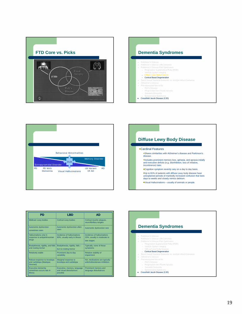
FTD Core vs. Picks Dementia Syndromes
Parkinson’s DiseaseParkinson’s DiseaseParkinson’s DiseaseParkinson’s Disease with DementiaParkinson’s Disease with DementiaParkinson’s Disease with DementiaParkinson’s Disease Plus SyndromesParkinson’s Disease Plus SyndromesParkinson’s Disease Plus Syndromes
––– Progressive Supranuclear Palsy (PSP)Progressive Supranuclear Palsy (PSP)Progressive Supranuclear Palsy (PSP)––– Multiple System AtrophyMultiple System AtrophyMultiple System Atrophy– Diffuse Lewy Body Disease–– Cortical Basal DegenerationCortical Basal Degeneration
Small Vessel Vascular Dementia vs. Multiple Infarct DementiaSmall Vessel Vascular Dementia vs. Multiple Infarct DementiaSmall Vessel Vascular Dementia vs. Multiple Infarct DementiaAlzheimer’s DiseaseAlzheimer’s DiseaseAlzheimer’s DiseaseFrontotemporal DementiaFrontotemporal DementiaFrontotemporal Dementia
Creutzfeld Jacob Disease (CJD)Creutzfeld Jacob Disease (CJD)
DLB
Visual Hallucinations
Behavioral Abnormalities
PD PD WithDementia
LB VariantOf AD
AD
Extrapyramidal Disorder
Memory Disorder
Diffuse Lewy Body Disease
Cardinal FeaturesShares similarities with Alzheimer's disease and Parkinson's
disease.
Includes prominent memory loss, aphasia, and apraxia initially and executive deficits (e.g, disinhibition, loss of initiative, incontinence) later.
Cognitive symptom severity vary on a day to day basis.
Up to 81% of patients with diffuse Lewy body disease have unexplained periods of markedly increased confusion that lasts days to weeks and closely mimics delirium.
Visual Hallucinations – usually of animals or people.
NA; medications are typically anticholinesterase inhibitors.
Marginal response to levodopa and carbidopa
Robust response to levodopa and carbidopa (Madopar, Sinemet)
Typically, none of these symptoms.
Bradykinesia, rigidity, falls –
but no resting tremor
Bradykinesia, rigidity, and falls and resting tremor
Prominent memory and language disturbances
Executive, memory, language and visual disturbances possible
Executive dementia sometimes occurs late in illness
Relative stability of impairment
Prominent day-to-day variability
Relatively stable
Incidence of hallucinations 20%, usually in moderate to
late stages.
Incidence of hallucinations 80%, usually early in illness
Hallucinations only in response to antiparkinsonian drugs
Autonomic dysfunction rareAutonomic dysfunction often seen
Parkinson’s DiseaseParkinson’s DiseaseParkinson’s DiseaseParkinson’s Disease with DementiaParkinson’s Disease with DementiaParkinson’s Disease with DementiaParkinson’s Disease Plus SyndromesParkinson’s Disease Plus SyndromesParkinson’s Disease Plus Syndromes
––– Progressive Supranuclear Palsy (PSP)Progressive Supranuclear Palsy (PSP)Progressive Supranuclear Palsy (PSP)––– Multiple System AtrophyMultiple System AtrophyMultiple System Atrophy––– Diffuse Lewy Body DiseaseDiffuse Lewy Body DiseaseDiffuse Lewy Body Disease–– Cortical Basal DegenerationCortical Basal Degeneration
Small Vessel Vascular Dementia vs. Multiple Infarct DementiaSmall Vessel Vascular Dementia vs. Multiple Infarct DementiaSmall Vessel Vascular Dementia vs. Multiple Infarct DementiaAlzheimer’s DiseaseAlzheimer’s DiseaseAlzheimer’s DiseaseFrontotemporal DementiaFrontotemporal DementiaFrontotemporal Dementia
Unusual presentations, for example, primary progressive aphasia and progressive buccofacial apraxia
Usually no hallucinations; if present – may be LBD.
Hisopathologies in the white matter, subcortical and cortical structures
Creutzfeldt-Jakob Disease
Cardinal Features– Progression = extremely rapid– Myoclonus (muscle contractions in the form of "jerks" or
twitches) is the most constant physical sign– visual abnormalities or cerebellar dysfunction including muscle
incoordination and gait and speech abnormalities. – abnormal reflexes, spasticity, tremors and rigidity– behavioral changes with agitation, depression or confusion. – akinetic mutism during the terminal stages of the illness.
Creutzfeldt-Jakob Disease
Rare disorder - affecting only one person per million population.
Cases have been recorded in patients as young as 17 years and asold as 83 years
There are three major categories of CJD:
sporadic CJD, hereditary CJD, and acquired (variant) CJD.
Variant CJD has been linked to consuming beef products contaminated with central nervous system tissue from cattle infected with Bovine Spongiform Encephalopathy (BSE, often called mad cow disease).
Creutzfeldt-Jakob Disease
CJD is characterized as a prion disease because it is caused by an infectious protein particle known as a prion that binds with cells, altering their composition.
Prions are the only known pathogens that are devoid of nucleic acid (prions contain no DNA or RNA). Unlike Alzheimer’s disease, which is not transmissible, CJD can be transmitted through exposure to the pathogenic form of the prion protein molecule that causes it.