5. In intact cells, the incorporation of label into material comigrating with phosphatidylcholine is often great enough to overwhelm the signal from palmitoyl-CoA. If this is the case, aliquots of lipid extract can be enriched for palmitoyl-CoA by phase partitioning. Redissolve the dried lipid extracts in 1.1 ml chloroform/methanol/10 mM Tris, pH 7.4 (5 : 5 : 1, v/v/v), then add 0.5 ml chloroform and 0.275 ml water and split the phases as described above for the synthesis of [3H]palmitoyl-CoA. Transfer the phases and dry separately in a Speed-Vac concentrator. The upper aqueous phase will be substantially enriched in palmitoyl-CoA (see Fig. 2).
Measurement of Protein Synthesis and Glycosylation
1. Cells are prepared and incubated under the same conditions as for the relevant protein palmitoylation experiment, but instead of [3H]palmitate, [35S]cysteine (Amersham, Arlington Heights, IL, 50-500 /.~Ci/ml) or [3H]mannose (DuPont NEN, 50/xCi/ml) are used as the radiolabel.
2. After the appropriate labeling period, incubations are quenched by the addition of an equal volume of 20% (w/v) ice-cold trichloroacetic acid (TCA), and the samples are placed on ice for 60 min.
3. Cells grown on monolayers are then scraped and transferred to poly- propylene vials.
4. Insoluble material is pelleted (14,000 g for 15 min at 4 °) and the supernatant discarded.
5. The pellet is washed with the same volume of 1% (w/v) TCA, and then twice with 1/10 volume ether (presaturated with water), with centrifu- gation as above in between each wash.
6. Redissolve the pellet in a minimum volume of 100 mM Tris-buffered 1% SDS (pH 7.4) with heating.
7. Determine incorporation of radioactivity by counting in a scintillation counter after addition of an appropriate water-tolerant scintillant.
[24] D y n a m i c P a l m i t o y l a t i o n o f G - P r o t e i n - C o u p l e d R e c e p t o r s i n E u k a r y o t i c C e l l s
By MICHEL BOUVIER, PETER CHIDIAC, TERENCE E. HEBERT, THOMAS P. LOISEL, SERGE MOFFETT, and BERNARD MOUILLAC
Introduction
Fatty acylation of proteins in eukaryotic cells has been found to be a more frequent modification than originally anticipated. Three general
[24] PALMITOYLATION OF G-PROTEIN-COUPLED RECEPTORS 301
classes of modification via acylation can be distinguished: N-terminal myris- toylation, C-terminal prenylation, and palmitoylation of cysteine residues. It has been proposed that these modifications may contribute to the tar- geting and anchoring of modified proteins to distinct biological membranes.l The observation that integral membrane proteins can also be fatty-acylated, even though they are anchored in the membrane through one or more transmembrane domains, has led to speculation that such acylation may also serve other purposes. Evidence has been found to support the involve- ment of fatty acylation in protein-protein interaction, 2 protein trafficking, 3'4 and protein phosphorylation. 5
Observations that many proteins involved in signal transduction are fatty-acylated have raised the intriguing possibility that the modifica- tions may play a regulatory role in the process. In particular, proteins in- volved in G-protein-mediated signaling appear to be subject to the entire spectrum of the posttranslational modifications. At least six G-protein- coupled receptors have been shown to be palmitoylated. These include the visual pigment rhodopsin, 6 the/32-adrenergic receptor (/92-AR), 7 the a2A-
adrenergic receptor, 8 the serotonergic 5-HTlb receptor, 9 the luteinizing hormone receptor, 9a and the dopamine D1 receptorJ ° In each of the recep- tors, a cysteine residue located in the carboxyl tail approximately 12 resi- dues from the seventh transmembrane domain has been shown to be thio- esterified by a palmitic acid. Such palmitoylation is likely to be a general phenomenon, since cysteine residues located in a similar position are found in the vast majority of the G-protein-coupled receptors sequenced to date (Table I). Although y subunits of the heterotrimeric G proteins have been found to be isoprenylated by the addition of geranylgeranyl residues, H
1 G. James and E. N. Olson, Biochemistry 29, 2623 (1990). 2 C. J. Marshall, Science 259, 1865 (1993). 3 E. Alvarez, N. Girones, and R. J. Davis, 3. Biol. Chem. 265, 16644 (1990). 4 j. A. Thissen and P. J. Casey, J. Biol. Chem. 268, 13780 (1993). 5 S. Moffett, B. Mouillac, H. Bonin, and M. Bouvier, E M B O J. 12, 349 (1993). 6 y. A. Ovchinnikov, N. G. Abdulaev, and A. S. Bogachuk, FEBS Lett. 230, 1 (1988). 7 B. F. O'Dowd, M. Hnatowich, M. G. Caron, R. J. Lefkowitz, and M. Bouvier, J. Biol.
Chem. 264, 7564 (1989). 8 M. E. Kennedy and L. E. Limbird, J. Biol. Chem. 268, 8003 (1993). 9 G. Y. Ng, S. R. George, R. L. Zastawny, M. Caron, M. Bouvier, M. Dennis and B. F.
O'Dowd, Biochemistry 32, 11727 (1993). 9a N. Kawate and K. M. J. Menon, J. Biol. Chem. 269, 30651 (1994).
i0 G. Y. Ng, B. Mouillac, S. George, M. Caron, M. Dennis, M. Bouvier, and B. O'Dowd, Eur. J. Pharmacol. 267, 7 (1994).
11 S. M. Mumby, P. J. Casey, A. G. Gilman, S. Gutowski, and P. C. Sternweis, Proc. Natl. Acad. Sci. U.S.A. 87, 5873 (1990).
302 PALMITOYLATION [24]
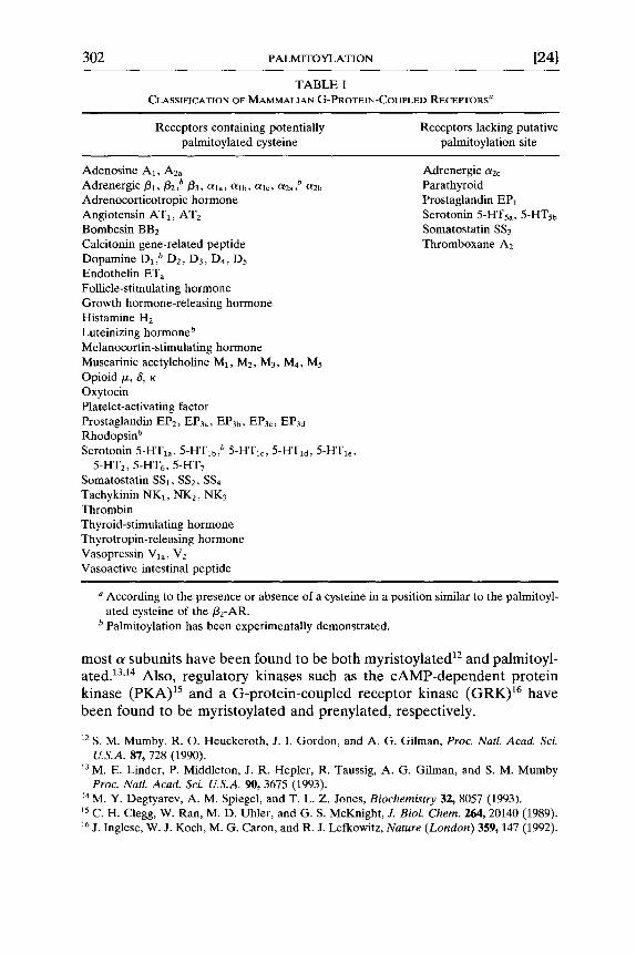
TABLE I CLASSIFICATION OF MAMMALIAN G-PROTEIN-COUPLED RECEPTORS a
Receptors containing potentially Receptors lacking putative palmitoylated cysteine palmitoylation site
"According to the presence or absence of a cysteine in a position similar to the palmitoyl- ated cysteine of the/32-AR.
b Palmitoylation has been experimentally demonstrated.
most a subuni t s have b e e n found to be bo th myr is toyla ted a2 and palmitoyl- a t e d ) 3A4 Also, regula tory kinases such as the c A M P - d e p e n d e n t p ro te in kinase ( P K A ) 15 and a G-p ro te in -coup led receptor k inase ( G R K ) 16 have
b e e n found to be myr is toyla ted and prenyla ted , respectively.
12 S. M. Mumby, R. O. Heuckeroth, J. I. Gordon, and A. G. Gilman, Proc. Natl. Aead. Sci. U.S.A. 87, 728 (1990).
13 M. E. Linder, P. Middleton, J. R. Hepler, R. Taussig, A. G. Gilman, and S. M. Mumby Proc. Natl. Acad. Sci. U.S.A. 90, 3675 (1993).
14 M. Y. Degtyarev, A. M. Spiegel, and T. L. Z. Jones, Biochemistry 32, 8057 (1993). 15 C. H. Clegg, W. Ran, M. D. Uhler, and G. S. McKnight, J. BioL Chem. 264, 20140 (1989). 16 j. Inglese, W. J. Koch, M. G. Caron, and R. J. Lefkowitz, Nature (London) 359, 147 (1992).
[24] PALMITOYLATION OF G-PROTEIN-COUPLED RECEPTORS 303
The specific roles played by the modifications in signaling remain the subject of intense investigation, but the following concepts are emerging. Acylation of the G-protein subunits and regulatory kinases is believed to play an important role in the targeting to proper membrane locations a7 and may influence the ability to form functional multimeric complexes. TM For the receptors, which are integral membrane proteins composed of seven transmembrane domains, palmitoylation has been proposed to promote the formation of an additional cytoplasmic loop. 7 This in turn has been suggested to affect the coupling, 7 phosphorylation, 5 and even internaliza- tion 19 of some receptors.
Palmitoylation, unlike other lipid modifications, is truly posttransla- tional rather than cotranslational, 2° and moreover it has been shown to be reversible. 2a This suggests the possibility of regulatory cycles of palmitoyla- tion-depalmitoylation that may be involved in signal transduction. In this respect, studies have confirmed that palmitoylation of receptors 9'1°'22 and of G-protein c~ subunits 23,24,24a'24b can be regulated on biological activation. Although the nature of the enzymes which are specifically involved in regulating the palmitoylation sate of the proteins is still unknown, a palmi- toyl-protein thioesterase that cleaves palmitate from H-ras has been purified and cloned. 25'25a
Studying the dynamics of receptor palmitoylation is essential for eluci- dating the role of the modification in regulating signaling. However, this presents a number of technical problems which are linked to the low abun- dance of the receptors in native systems, the nature of the modification, and the lack of knowledge of the enzymatic processes involved. We have developed a number of tools and approaches to deal with some of these problems. These are discussed in the following sections of this chapter.
17 j. A. Pitcher, J. Inglese, J. B. Higgins, J. L. Arriza, P. J. Casey, C. Kim, J. Benovic, M. M. Kwatra, M. Caron, and R. J. Lefkowitz, Science 257, 1264 (1992).
a8 j. A. Iniguez-Lluhi, M. I. Simon, J. D. Robishaw, and A. G. Gilman, J. Biol. Chem. 267, 23409 (1992).
19 D. R. Nussenzveig, M. Heinflink, and M. C. Gershengorn, J. Biol. Chem. 268, 2389 (1993). 20 S. Bonatti, M. Giovanni, and K. Simons, J. BioL Chem. 264, 12590 (1989). 21 M. B. Omary and I. S. Trowbridge, J. Biol. Chem. 256, 4715 (1981). 22 B. Mouillac, M. Caron, H. Bonin, M. Dennis, and M. Bouvier, J. Biol. Chem. 267, 21733
(1992). 23 M. Y. Degtyarev, A. M. Spigel, and T. L. Z. Jones, .L Biol. Chem. 268, 23769 (1993). 24 p. B. Wedegaertner, D. A. Chu, P. T. Wilson, M. J. Levis, and H. R. Bourne, J. Biol. Chem.
268, 25001 (1993). 24a S. M. Mumby, C. Kleus, and A. G. Gilman, Proc. Natl. Acad. Sci. U.S.A. 91, 2800 (1994). 24b p. B. Wedegaertner and H. R. Bourne, Cell 77, 1063 (1994). 25 L. A. Camp and S. L. Hofmann, J. Biol. Chem. 268, 22566 (1993). 25a L. A. Camp, L. A. Verkruyse, S. J. Afendis, A. A. Slaughter, and S. L. Hofmann, J. Biol.
Chem. 269, 23212 (1994).
304 PALMITOYLATION [24l
Palmitoylation of G-Protein-Coupled Receptors in Native and Heterologous Systems
The abundance of rhodopsin in rod outer segments makes it feasible to study the palmitoylation of the G-protein-coupled receptor in cells where it occurs endogenously. 6 However, posttranslational modifications are dif- ficult to observe in tissues expressing other members of the receptor family owing to the relatively low levels of native expression associated with such proteins. This is particularly true for palmitoylation because of the rela- tively low specific activity of [3H]- or [14C]palmitate in comparison with the [32p]Pi used to detect phosphorylation, for example. One way to circum- vent the problem is to study fatty acylation in heterologous systems express- ing higher levels of receptor. Palmitoylation of a G-protein-coupled hor- mone receptor was first demonstrated in Chinese hamster fibroblasts (CHW cells) transfected with the human fl2-AR cDNA and stably express- ing 2 pmol of the receptor per milligram of membrane protein] Even at that level of expression, purification of the 3H-palmitoylated receptor from large quantities of cells (-108), followed by several weeks of exposure, is necessary for the autoradiographic detection of the labeled receptor. Although such a protocol can be used to detect major differences among experimental conditions, quantitation of the relatively subtle dynamic changes associated with palmitate incorporation and turnover requires a more refined system.
Spodoptera frugiperda (Sf9) cells infected with recombinant Auto- graphica californica nuclear polyhedrosis viruses (baculoviruses) have been used successfully in many cases to express large quantities of proteins. Proteins expressed using the system (unlike those produced using bacterial expression systems) have largely been found to maintain normal character- istics, including specific posttranslational modifications such as palmitoyla- tion and phosphorylation. Sf9 cells can transiently express up to 50 pmol/32- AR per milligram of protein after infection with a recombinant baculovirus encoding the receptor, t2,26,27 The detection of relatively small differences in/32-AR palmitoylation is more feasible with the insect system than with mammalian cells, since more receptors can be purified from a given volume of cells and thus time-dependent changes can be detected on autoradio- grams after relatively short durations of exposure.
Construction of Recombinant Baculovirus. To prepare the recombinant baculovirus encoding the human flz-AR, the plasmid vector pTZ18R (Phar- macia LKB Biotechnology Inc., Piscataway, N J) containing the entire hu-
26 E. M. Parker, K. Kameyama, T. Higashijima, and E. M. Ross, J. BioL Chem. 266, 519 (1991). 27 S. T. George, M. A. Arbabian, A. E. Ruoho, J. Kiely, and C. C. Malbon, Biochem. Biophys.
Res. Commun. 163, 1265 (1989).
[ 2 4 ] PALMITOYLATION OF G-PROTEIN-COUPLED RECEPTORS 305
man/32-AR sequence is digested with EcoRI-HindlI128 and filled in by Klenow DNA polymerase. The/32-AR cDNA fragment is then placed into the baculovirus recombination vector pJVETLZ at the NheI site by blunt- end ligation to yield the pJV/32-AR construct. Transfer of the/32-AR and /3-galactosidase coding sequences from pJV/32-AR to the baculovirus ge- nome is achieved by homologous recombination in Sf9 cells. The recombi- nant baculovirus is purified by successive plaque assays using/3-galactosi- dase detection. 22
Culture and Infection of Sf9 Cells. Sf9 cells are maintained at 27 °- 28 ° either in serum-supplemented or serum-free media. The experiments described here were carried out on cells grown in Grace's insect medium (GIBCO, Grand Island, NY) supplemented with fetal bovine serum (FBS; 10%, v/v). As outlined below, metabolic labeling with [3H]palmitate should be carried out at minimal levels of serum, and thus the contents of the medium may need to be modified before the experiment is started. Sf9 cells can be grown either as monolayers (in plastic petri dishes or T flasks) or in suspension [in spinner bottles (Bellco, Vineland, NJ) or in Erlenmeyer flasks placed in an orbital shaker] in the presence of an agent which inhibits tearing of cell membranes caused by agitation (e.g., pluronic acid).
Cells should be infected with baculovirus when they are in a logarithmic phase of growth. With cells cultured in serum-supplemented Grace's me- dium, optimal conditions for infection are found at a density of 1 x 10⁶ to 2 x 10⁶ cells/ml for cells in suspension and near confluence for attached cells (2 x 10⁶ to 4 x 10⁶ cells/ml). At a multiplicity of infection (MOI) of approximately 5 recombinant baculovirus molecules per Sf9 cell, expression of/3-adrenergic receptor should be evident 24 hr after exposing the cells to the virus. The presence of the receptor in membranes from infected cells can be detected by the specific binding of a radiolabeled/32-AR antagonist (e.g., [125I]iodocyanopindolol, [125I]CYP), and receptor function can be as- sessed with appropriate agonists (e.g., isoproterenol-stimulated adenylate cyclase activity). After 48 hr of infection, the level of receptor expression is found to be maximal or near-maximal, typically around 25 pmol/mg protein for cells in suspension and about half that for attached cells. This difference may be due to the decreased accessibility of the attached cells to the virus. To obtain comparable receptor levels, cells grown as mono- layers thus tend to require a higher MOI than do cells in suspension. The proportion of viable cells after 48 hr, as assessed by the ability of cells to exclude trypan blue, is greater than 90%. Further incubation, however, leads to substantial cell morbidity without substantial gain in the level of
28 B. K. Kobilka, R. A. F. Dixon, T. Frielle, H. G. Dohlman, M. A. Bolanowski, I. S. Sigal, T. Z. Yang-Feng, V. Francke, M. G. Caron, and R. J. Lefkowitz, Proc. Natl. Acad. Sci.
U.S.A. 84, 46 (1987).
306 PALMITOYLATION [241
expression. Therefore, we and others have chosen 48 hr as an optimal time of infection. Additional details about baculovirus and Sf9 cells are found in O'Reilly et al. 29
Charcacterization o f Human/3e-Adrenergic Receptor Expressed in Sf9 Cells. When using a heterologous expression system to study a given recep- tor, an appropriate assessment of the properties of the receptor is required. Although characterization of the/32-AR in Sf9 cells has been carried out, similar studies would be needed before such a system could be used to study the palmitoylation of other receptors. The binding affinities of/3- adrenergic agonists and antagonists determined using the baculovirus ex- pression system are consistent with those reported in mammalian sys- tems. 22,26,3° The agonist isoproterenol stimulates adenylate cyclase activity in a dose-dependent manner in membranes derived from Sf9 cells infected with baculovirus encoding either human/32-AR 22 (Fig. 1) or avian/3-AR. 26 Whereas isoproterenol dose-response curves are similar in appearance in membranes from Sf9 and mammalian cells, maximal stimulation of cAMP production occurs at lower agonist concentrations in Sf9 cells, presumably owing to the relatively large number of/32-AR typically observed in such preparations.
Similar to observations in mammalian cells, t reatment of/32-AR-ex- pressing Sf9 cells with isoproterenol leads to an increase in the intracellular level of cAMP. 3° Furthermore, short-term (5-30 min) agonist t reatment is associated with a t ime-dependent decrease in maximal agonist-induced stimulation in membranes (Fig. 1A). Similar changes in maximal activity in mammalian systems have been attributed to receptor desensitization. 31 These agonist-induced decreases are accompanied by apparent decreases in agonist potency and by increased receptor phosphorylation (Fig. 1B), supporting the notion that/32-AR desensitization in Sf9 cells is similar to that in mammalian cells. Moreover, desensitization in Sf9 cells appears to involve phosphorylation of the receptor by at least two protein kinases. As in mammalian cells, partial desensitization and phosphorylation are observed after t reatment of/32-AR-expressing Sf9 cells with the P K A activa- tor dibutyryl-cAMP, 31a suggesting that a second enzyme, possibly a G- protein-coupled receptor kinase (GRK), is also involved. Thus, both the activation of second messenger systems and the desensitization of G-pro-
29 D. R. O'Reilly, L. K. Miller, and V. A. Luckow, "Baculovirus Expression Vectors: A Laboratory Manual." Freeman, New York, 1992.
30 p. Chidiac, T. E. Hebert, M. Valiquette, M. Dennis, and M. Bouvier, Mol. PharmacoL 45, 490 (1994).
31 j. L. Benovic, M. Bouvier, M. G. Caron, and R. J. Lefkowitz, Annu. Rev. Cell Biol. 4, 405 (1988).
31a S. St. Onge, B. Mouillac, P. Chidiac, and M. Bouvier, manuscript in preparation.
[ 2 4 ] PALMITOYLATION OF G-PROTEIN-COUPLED RECEPTORS 307
A
9 0
e- ~ 7 5 -
~ 6 0 -
E 45
,~ 30 o
Q .
0 - i " 1 1 i i i i i
-10 -9 -8 -7 -6 -5
log [ISOPROTERENOL]
B Affinity-purified c-myc 82-AR
1 2 !~!!!iiii!iiiiii ii i ~iii i i
97- }
4 3 - -
~ii!!ii~iiiii~!ii ~!i ~ ii ii ~i~ii~!!i!~i! ~
3o- iiiiiiiill iiiiii!
FIG. 1. Effect of isoproterenol treatment of/32-AR-expressing Sf9 cells on agonist-stimu- lated adenylate cyclase activity and receptor phosphorylation. (A) Sf9 cells infected for 48 hr with recombinant baculovirus encoding the human/3e-AR were treated with vehicle (0.1 mM ascorbate) for 30 min ((>) or with i / zM isoproterenol for 15 (11) or 30 min ((3). Membrane adenylate cyclase activity was assayed and data were analyzed as described. 3° (B) Sf9 cells infected for 48 hr with/32-AR-encoding baculovirus were labeled for 120 min with [32p]p i and subsequently exposed to 1 /zM isoproterenol for 15 min. /32-AR were purified by affinity chromatography. Lane 1, Untreated cells; lane 2, isoproterenol-treated cells. Molecular weight markers are indicated to the left of Lane 1.
tein-coupled receptors in /32-AR-expressing Sf9 cells are comparable to those in mammalian systems.
In addition to the/32-AR, several other G-protein-coupled receptors have been expressed in the baculovirus/Sf9 system. Pharmacological as well as biochemical properties similar to those found in mammalian systems were reported. 9"1°'32 In spite of the functional similarities, however, it should be noted that the glycosylation of integral membrane proteins expressed using the baculovirus/Sf9 system differs from that found in mammalian expression systems owing to the incomplete processing of N-linked oligosac- charides in the insect cell line. 33 Although this leads to decreases in the apparent molecular weights of G-protein-linked receptors in Sf9 cells com- pared to mammalian cells (see Mouillac e t al. 22 and references cited therein), flz-AR function, which is relatively unaffected by deglycosylation, appears to be normal. However, in a receptor where function or ligand binding is affected by changes in the attached sugar residues, some functional differ-
32 S. K.-F. Wong, E. M. Parker, and E. M. Ross, J. Biol. Chem. 265, 6219 (1990). 33 V. A. Luckow and M. D. Summers, Bio/Technology 6, 47 (1988).
308 PALMITOYLATION [241
ences between receptors expressed in mammalian and Sf9 cells might be an- ticipated.
Dynamic Palmitoyla t ion of/~2-Adrenergic Receptor
Incorporation of [3H]palmitate has been demonstrated for a number of proteins in mammalian cells. These include several proteins involved in signal transduction such as a 64-KDa growth factor-sensitive protein in the BC3H1 muscle cell line, 34 the transferrin receptor, 21 the insulin receptor, 35 p21N-RAS, 36 GAP-43, 37 and several G-protein-coupled receptors. 6-1° How- ever, as outlined above, the study of the dynamic regulation of palmitate incorporation in mammalian cells has been difficult. Because palmitoylation of proteins has been shown to occur normally in baculovirus-infected Sf9 cells, this system offers an alternative to study the dynamics of protein palmitoylation.
Metabolic Labeling
Optimization of Labeling of [3e-Adrenergic Receptor for Dynamic Stud- ies. Before one can characterize the regulation of receptor acylation, an analysis of the labeling kinetics must be undertaken. This is especially true when labeling cells with a compound such as [3H]palmitate. Indeed, the large cellular pool of palmitate and rapid turnover of the fatty acid prevents isotopic equilibrium of the cellular palmitate and palmitoylated proteins.
To study the kinetics of flz-AR palmitoylation, 100 ml of Sf9 cells are grown in spinner flasks and infected with the/32-AR-encoding baculovirus as described above. At 30 hr postinfection, cells are transferred to serum- free Grace's medium. At 47 hr cells are counted and brought up to 1% (v/v) FBS at a density of approximately 50 million cells/20 ml. At 48 hr, [9,10-3H]palmitic acid (60 Ci/mmol) dissolved in 50/~1 of dimethyl sulfoxide (DMSO) is added to the culture at a final concentration of 0.2 mCi/ml. After various times, the labeling is terminated by centrifugation at 500 g for 5 min at 4 ° followed by two washes with ice-cold PBS. The cells are then disrupted by sonication in 10 ml of ice-cold buffer containing 5 mM Tris-HCl, pH 7.4, 2 mM EDTA, plus a protease inhibitor cocktail (5 mg/ml leupeptin, 10 mg/ml benzamidine, and 5 mg/ml soybean trypsin inhibitor). Lysates are centrifuged at 500 g for 5 min at 4 °, the pellets are sonicated as before and spun again, and the supernatants are pooled. The pooled
34 G. James and E. N. Olson, J. Biol. Chem. 264, 20998 (1989). 35 j. Hedo, E. Collier, and A. Watkinson, J. Biol. Chem. 262, 954 (1987). 36 A. I. Magee, L. Gutierrez, I. A. McKay, C. J. Mafshall, and A. Hall, EMBOJ. 6, 3353 (1987). 37 j. p. H. Skane and I. Virhg, J. Cell. Biol. 108, 613 (1989).
[24] PALMITOYLATION OF G-PROTEIN-COUPLED RECEPTORS 309
t -
a
o
(1)
0 EX
0 0 t-"
. D
t~ .--I
0.12
0.10
0.08
0.06
0.04 002t 0 . 0 0
0 . 5
Time 30min lhr MW
68.
2hr 3hr
~1 fJ2-AR
3 0 . . . . . . . .
Z////_z r / l / l /> . / / / / / ,
/////. ,4
V / / / / / / / / / / /~ / / / 1 " / / ,
1 2 3
Time of laboring (hr)
FIG. 2. Tritiated palmitate incorporated into fl2-AR purified by affinity chromatography on alprenolol-Sepharose. Sf9 cells were metabolically labeled for the times indicated with tritiated palmitic acid as described in the text. Bars represent a densitometric scan of the fluorogram shown in the inset. Molecular weight markers are indicated to the left of Lane 1 of the inset.
supernatant is then centrifuged at 45,000 g for 20 min, and the pellets are washed twice in the same buffer. The receptor is solubilized in digitonin and purified as described below.
Following purification, sodium dodecyl sulfate-polyacrylamide gel elec- trophoresis (SDS-PAGE) is carried out under nonreducing conditions (the thioester bond of cysteine to palmitate is somewhat sensitive to reducing agents such as dithiothreitol and 2-mercaptoethanol) on 10% slab gels. 38 Equal amounts of receptor, as determined by a soluble radioligand binding assay using [125I]CYP as the radioligand, are loaded into each lane. To allow fluorographic detection of the incorporated palmitate, fixed gels are incubated in Enlightening (DuPont, Mississauga, Canada) for 30 min, dried, and exposed to Kodak (Rochester, NY) XAR-5 film at - 7 0 ° for several days. Using this protocol, incorporation of tritiated palmitate can be de- tected in purified proteins (in the case of the/32-AR, - 5 pmol of receptor) after 1 to 2 weeks of exposure.
Figure 2 shows the results of a densitometric scan of an autoradiogram representing different times of labeling. As seen in Fig. 2, label incorpora- tion peaks at 1 hr and declines sharply thereafter. This indicates clearly that turnover of the incorporated palmitate is rapid. The observed decrease
38 U. K. Laemmli, Nature (London) 227, 680 (1970).
310 PALMITOYLATION [241
in labeling is most likely due to a decrease in specific activity of the palmitate resulting from exhaustion of the labeled palmitate in poorly defined meta- bolic pathways. Thus, a 1-hr labeling period is selected for optimal labeling. One caveat to be considered is that the specific activity of the donor pool of palmitate is not at equilibrium. This must be taken into account when the dynamics of palmitate incorporation are studied.
Characterization of Incorporated RadiolabeL The most direct way to identify the nature of the incorporated fatty acid would certainly be analysis by mass spectrometry. 39 However, a simpler approach requiring consider- ably less purified protein can satisfactorily confirm the identity of the [3H]palmitate incorporated. Unlike the N-terminal amide linkage or the C- terminal thioether linkage of myristoylation and prenylation, respectively, thioesterification of cysteine by palmitic acid is sensitive to neutral hydroxyl- amine. Sensitivity to hydroxylamine can be determined by treating either the soluble purified protein or, alternatively, the polyacrylamide gel follow- ing electrophoresis. Solubilized receptors (as described below) can be treated with 1 M hydroxylamine, pH 7.0, for 12 hr at 22 °. Alternatively, fixed SDS-polyacrylamide gels can be incubated with 1 M hydroxylamine, pH 7.0, for 16 hr, washed extensively, dried, and exposed to Kodak XAR film at - 70 ° . This technique also has the advantage of distinguishing be- tween covalently attached [3H]palmitate and nonspecifically adsorbed triti- ated lipid.
Covalent attachment of the palmitate can also be confirmed by assessing the resistance of the labeling to organic extraction. Noncovalently bound lipids can be extracted from the purified receptor in a mixture of chloroform/ methanol (2:1, v/v). The extract is mixed vigorously, and after a 30-min incubation at room temperature the protein is pelleted by centrifugation at 4500 g for 15 min at room temperature. This extraction is repeated twice and then twice with chloroform/methanol/water (1 : 1 : 0.3, v/v/v) and finally with methanol alone. After each extraction step, the mixture is incubated for 10 min at room temperature. The pellets can then be solubilized in nonreducing SDS-PAGE sample buffer.
Analysis of the tritiated lipids incorporated into the/32-AR can also be performed using ascending chromatography. The receptor band is excised from polyacrylamide gels, homogenized in 0.1 M (NHa)HCO3, pH 7.7, and digested with trypsin (0.3 mg/ml) for 15 min at 37 °. The digest is acidified and extracted with hexane. The extract is lyophilized and treated with 1 M KOH for 12 hr at 37 ° to cleave the attached lipid. Finally, the extract is dried under nitrogen and applied to a silica gel thin-layer chromatography
39 D. I. Papac, K. R. Thornburg, E. E. Bullesbach, R. K. Crouch, and D. R. Knapp, J. BioL Chem. 267, 16889 (1992).
[24] PALMITOYLATION OF G-PROTEIN-COUPLED RECEPTORS 311
(TLC) plate along with tritiated lipid standards in parallel lanes. The chro- matogram is developed with hexane/ethyl acetate/acetic acid (80:20:1, v/v/v).
Receptor Purification
To characterize the incorporation of labeled palmitate into the/32-AR, it is necessary to purify the receptor. Following metabolic labeling carried out as described above,/32-AR is solublized from membranes in 12 ml of a buffer containing 100 mM NaC1, 10 mM Tris-HCl, 5 mM EDTA, pH 7.4, 0.3% (w/v) digitonin, and a protease inhibitor cocktail with mild agitation at 4 ° for 90 min. Nonsolubilized material is removed by centrifugation at 45,000 g for 20 min at 4 °. At this point, either of two procedures can be used to purify the metabolically labeled/32-AR.
Affinity Purification of fl2-Adrenergic Receptor. Solubilized fl2-AR can be purified by affinity chromatography using a Sepharose matrix coupled to the/3-adrenergic antagonist alprenolol. 4° Alprenolol-Sepharose columns (6 ml of gel per column) are equilibrated with 5 bed volumes of buffer A (100 mM NaC1, 10 mM Tris-HCl, 2 mM EDTA, pH 7.4, 0.05% (w/v) digitonin, and protease inhibitors). Solubilized receptor is loaded on the column and shaken gently for 2 hr at room temperature to allow binding of the receptor to the matrix. The supernatant is then allowed to flow through, and the columns are placed at 4 ° and washed with 25 ml of a buffer containing 500 mM NaC1, 50 mM Tris-HC1, 2 mM EDTA, pH 7.4, 0.05% (w/v) digitonin, and protease inhibitors. The original ionic strength is restored by washing with 50 ml of buffer A. After the columns are returned to room temperature, receptors are eluted with buffer A con- taining 60/xM alprenolol. The eluate is concentrated with Centriprep and Centricon cartridges (Amicon, Danvers, MA) down to 50/.d. Recovery of /32-AR after affinity purification is measured by soluble binding after de- salting on a Sephadex G-50 gel filtration column to remove alprenolol. The purified sample can then be prepared for electrophoretic analysis.
Until relatively recently, Sepharose-alprenolol stationary phase affinity chromatography of digitonin-solubilized receptor was the only available method to purify the /32-AR. Although this method is commonly used, yields rarely exceed 30%.
Immunopurification of c-myc-Tagged Be-Adrenergic Receptor. Several attempts to raise antibodies against native G-protein-coupled receptors have failed to produce high-affinity antibodies which could be used in immunoaffinity purification procedures. To circumvent this problem, an
40 j. L. Benovic, R. G. L. Shorr, M. G. Caron, and R. J. Lefkowitz, Biochemistry 23, 4510 (1984).
312 PALMITOYLATION [241
epitope-tagged receptor is constructed. Eleven amino acids from the c-myc protein are inserted immediately before the initial methionine at the NH2 terminus of the/32-AR. The site of epitope insertion is chosen because that domain of the/32-AR does not appear to be involved in ligand recognition or signal transduction. The recombinant baculovirus encoding the epitope- tagged/32-AR is constructed as described above with the following modifi- cations. Two complementary oligodeoxynucleotides encoding the c-myc epitope (5' A A T T C ^ A T G G A G C A A A A G C T C A T T T C T G A A G A G - GACTTGAAT^GC 3' and 3' G^TACCTCGTTTTCGAGTAAAGACT - TCTCCTGAACTFAC ^ GGTAC 5') are inserted between EcoRI and NcoI sites in the pTZ/32-AR. The pharmacological and biochemical properties of the epitope-tagged/32-AR are found to be indistinguishable from those of the wild-type receptor when expressed in Sf9 cells. = The presence of the epitope allows visualization of the receptor by immunoblotting and quantitative immunoprecipitation.
Following metabolic labeling of Sf9 cells expressing the c-myc/32-AR and solubilization of the receptor as described above, the tagged receptor can be immunoprecipitated using the mouse anti-c-myc monoclonal anti- body (9E10). A crucial factor for optimal immunoprecipitation is to reduce the concentration of the detergent digitonin to the minimum required to maintain the receptor in solution. Removal of digitonin and concentration of the solublized receptor can be accomplished by dialysis using Centriprep cartridges (Amicon). A final volume of 700 /xl of an ice-cold solution containing 100 mM NaC1, 10 mM Tris-HC1, pH 7.4, 2 mM EDTA, and a protease inhibitor cocktail (buffer B), which does not exceed 0.05% digito- nin, is used to perform the immunoprecipitation. Then, purified anti-c-myc 9El0 antibody (at a 7 : 1 antibody to receptor molar ratio) is added to the concentrate and agitated for 2 hr at 4 °. Eighty microliters of buffer B and 250/zl of anti-mouse immunoglobulin G (IgG) agarose (Sigma, St. Louis, MO; at an 11 : 1 secondary to primary antibody molar ratio) are then added. The reaction proceeds for 10 to 15 hr at 4 ° under gentle agitation. The immunoprecipitate is centrifuged at 12,000 rpm in a microcentrifuge for 10 min. The pellet is rinsed three times with 1 ml of buffer B. Finally, the pellet is resuspended in 200/xl of nonreducing SDS-PAGE loading buffer and incubated at room temperature for 30 min, after which the receptors are released from the matrix by sonication. Yields can be estimated by comparing the number of receptors determined in soluble binding assays performed before and after immunoprecipitation.
With the immunoaffinity method, yields are dramatically higher than those generally obtained by affinity chromatography, and up to 85% of the solubilized receptor can be recovered. Use of this purification technique considerably shortens the time of exposure required to detect the palmitoyl-
[241 PALMITOYLATION OF G-PROTEIN-COUPLED RECEPTORS 313
ated receptor. In the best cases, less than 1 week of exposure is sufficient to visualize the labeled receptor. A similar epitope-tagging approach fol- lowed by immunoprecipitation was successfully used to study the palmitoyl- ation of the serotonin 5-HYlb 9 and the dopamine D11° receptors. Theoreti- cally, this approach could be used to study the posttranslational modifica- tions of any cloned receptor for which no other purification technique is available.
Dynamic Regulat ion of Palmita te Incorpora t ion into ]32-Adrenergic Receptor
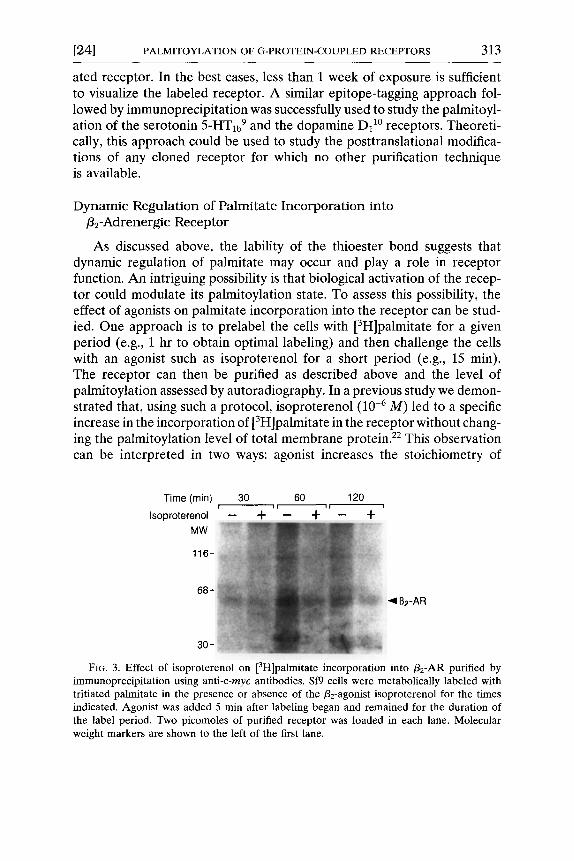
As discussed above, the lability of the thioester bond suggests that dynamic regulation of palmitate may occur and play a role in receptor function. An intriguing possibility is that biological activation of the recep- tor could modulate its palmitoylation state. To assess this possibility, the effect of agonists on palmitate incorporation into the receptor can be stud- ied. One approach is to prelabel the ceils with [3H]palmitate for a given period (e.g., 1 hr to obtain optimal labeling) and then challenge the cells with an agonist such as isoproterenol for a short period (e.g., 15 rain). The receptor can then be purified as described above and the level of palmitoylation assessed by autoradiography. In a previous study we demon- strated that, using such a protocol, isoproterenol (10 -6 M) led to a specific increase in the incorporation of [3H]palmitate in the receptor without chang- ing the palmitoylation level of total membrane protein. 2e This observation can be interpreted in two ways: agonist increases the stoichiometry of
Time (min) 30 [ i i - -
Isoproterenol -- q-
MW
116-
68-
30-
6O 120 _ + " _ +
~1B2-AR
FIG. 3. Effect of isoproterenol on [3H]palmitate incorporation into /32-AR purified by immunoprecipitation using anti-c-myc antibodies. Sf9 cells were metabolically labeled with tritiated palmitate in the presence or absence of the/32-agonist isoproterenol for the times indicated. Agonist was added 5 rain after labeling began and remained for the duration of the label period. Two picomoles of purified receptor was loaded in each lane. Molecular weight markers are shown to the left of the first lane.
314 PALMITOYLATION [2 51
palmitoylation or, alternatively, agonist treatment increases the turnover rate of palmitate linked to the receptor. Modulation of receptor palmitoyla- tion by agonist can also be observed in mammalian cells expressing the human/3e-AR, 5 but kinetic studies are considerably more difficult using those cells. In a similar approach, three other groups observed comparable increases in the apparent incorporation of palmitate into the a subunit of Gs following its stimulation. 23,24,24a'24b
A different approach to study the influence of receptor activation on palmitoylation is to treat the cells with the agonist over the entire course of the labeling period. For these studies, labeling of Sf9 cells expressing the c -myc flz-AR is performed as described above. After 5 min of labeling, agonist (10 -6 M) is added to the cells and incubation continued for various lengths of time. As shown in Fig. 3, a treatment of 30 min with isoproterenol reduces the incorporation of tritiated palmitate into the/32-AR by approxi- mately 35% relative to the control condition. After 1 hr the reduction reaches above 65%. The results are more consistent with an effect of the agonist on the turnover rate of palmitate linked to receptor than a change in the stoichiometry of receptor palmitoylation. It should be remembered that the experiments cannot be carried out at isotopic equilibrium of the palmitate donor pools. Therefore, although it is clear that activation of the /32-AR modulates its palmitoylation, caution should be exercised when making kinetic interpretations of changes in tritiated palmitate incorpo- ration.
[25] P a l m i t o y l a t i o n o f G - P r o t e i n a S u b u n i t s
B y MAURINE E. LINDER, CHRISTIANE KLEUSS, and SUSANNE M. MUMBY
Introduction
A family of guanine nucleotide-binding regulatory proteins (G proteins) serve in signal transduction systems to link receptors exposed at the cell surface to intracellular effectors such as enzymes and ion channels, a On activation by receptor, a heterotrimeric G protein dissociates into a GTP- bound ot subunit and a/3y complex which are then able to modulate effector activity. Many ot subunits are acylated at (or near) their amino termini by myristate and/or palmitate. G-protein y polypeptide chains are prenylated
1 j. R. Hepler and A. G. Gilman, Trends Biochem. Sci. 17, 383 (1992).


![Inactivation of the KcsA potassium channel explored with ... · PDF filegating of KcsA at low pH is affected by the applied po-tential (Cuello et al., ... toyl-2-oleoyl-sn-glycero-3-[phospho-rac-(1-glycerol)];](https://static.documents.pub/doc/80x56/5a83b0f17f8b9a682c8ef255/inactivation-of-the-kcsa-potassium-channel-explored-with-of-kcsa-at-low-ph-is.jpg)


![I i pase-cat al y zed hydrolysis phosphatidylcholine of ... · PDF filephosphatidylcholine of guinea pig very low density ... [ ''C]palm~toyl phosphatidylcholine and purified bovine](https://static.documents.pub/doc/80x56/5a83b0f17f8b9a682c8ef22c/i-i-pase-cat-al-y-zed-hydrolysis-phosphatidylcholine-of-of-guinea-pig-very-low.jpg)


