25
Measures to Support Safety in Dental Radiography Pamela Alston, DDS, MPP Lead Oral Health Specialist
| Date post: | 29-Dec-2015 |
| Category: |
Documents |
| Upload: | denis-ross-stewart |
| View: | 215 times |
| Download: | 0 times |
Measures to Support Safety in Dental Radiography
Pamela Alston, DDS, MPPLead Oral Health Specialist
Participants will be able to:
• Describe dental radiation risks• Describe the proper safety
measures for dental x-ray equipment operators
• Explain the safety measures necessary for the protection of the students when dental x-rays are taken

Radiation Physics• Energy—ability to do
work• Matter—occupies
space & has form• Matter—composed of
atoms• Molecule—smallest
particle
X-rays
• Form of energy penetrating matter
• Classified as electromagnetic radiation traveling through space with a wavelike motion
• Shorter the wavelength, greater the energy
Radiation Effects
• All ionizing radiation is harmful
• Produces biologic changes in living tissues
• Entire x-ray area is a radiation hazard area
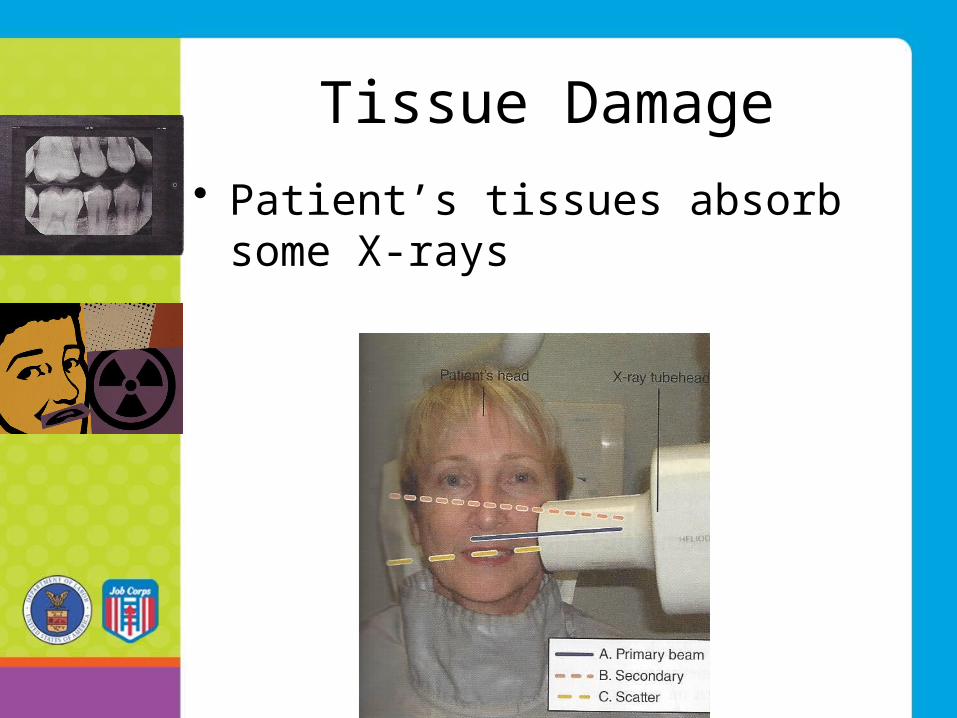
Tissue Damage
• Patient’s tissues absorb some X-rays

Biologic Effects
• Affects body chemicals, cells, tissues & organs
• Effects may not become evident for years
• Time lag is called latent period
Cumulative Effects
• Repeated exposure damages tissue
• Some repair occurs
• Tissues do not return to original state
Acute & Chronic Radiation Exposure
• Acute—large dose absorbed in short time
• Chronic—small doses absorbed repeatedly over long period

Genetic & Somatic Effects
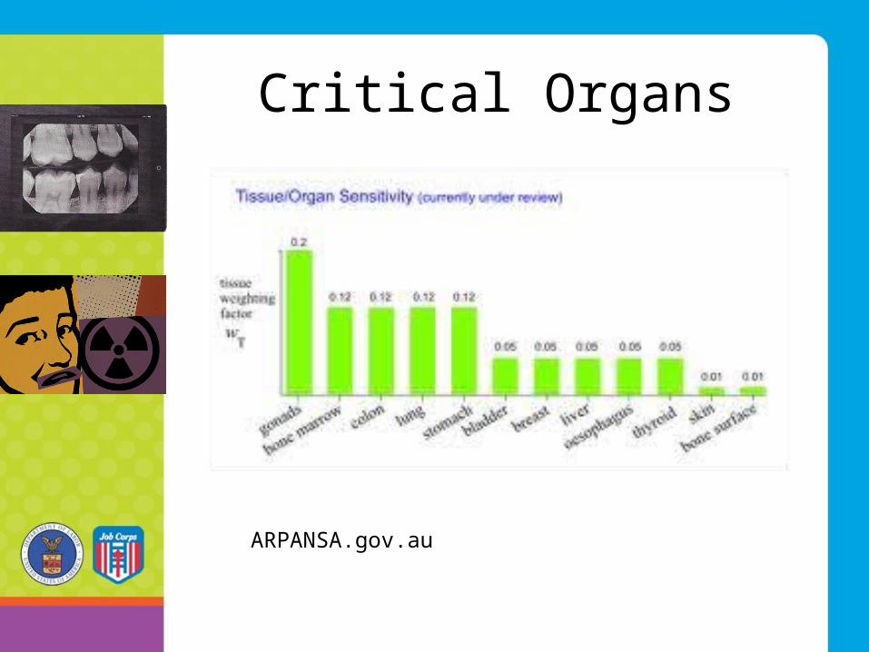
Critical Organs
ARPANSA.gov.au
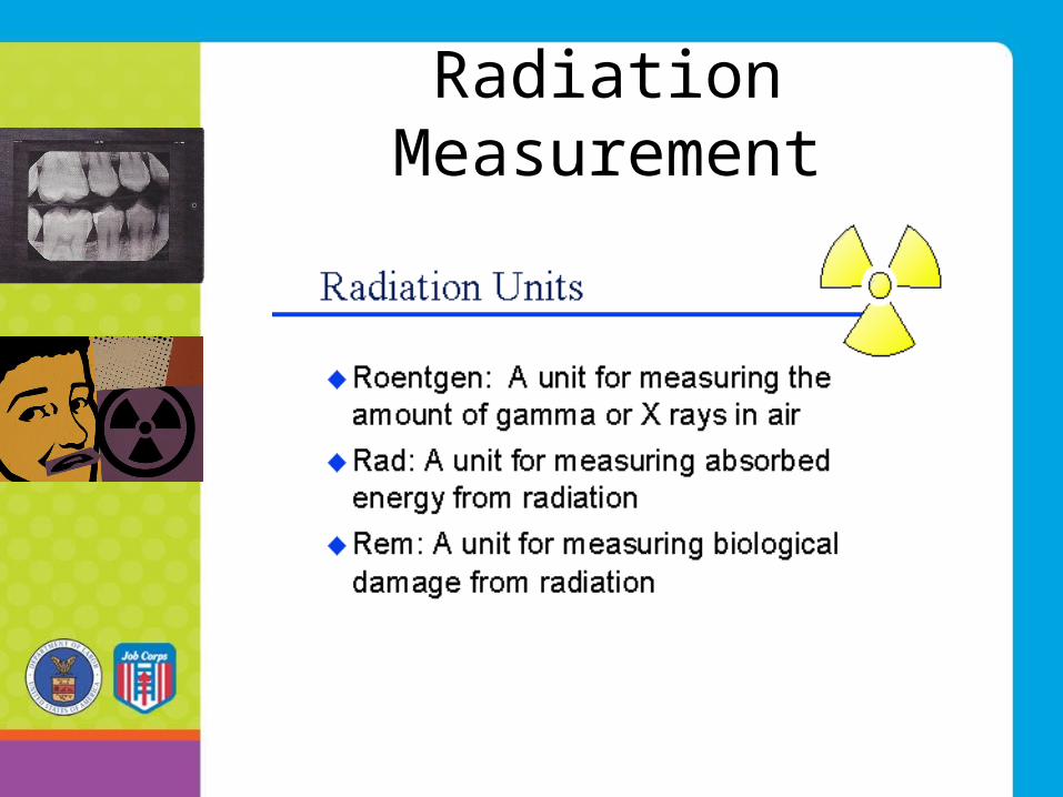
Radiation Measurement
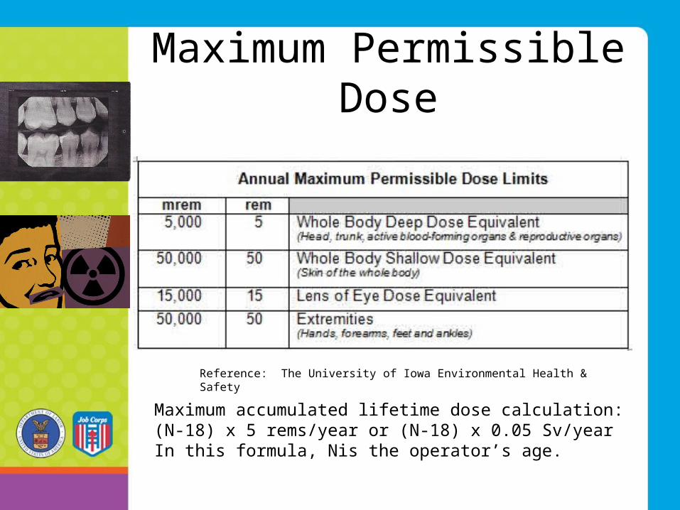
Maximum Permissible Dose
Reference: The University of Iowa Environmental Health & Safety
Maximum accumulated lifetime dose calculation:(N-18) x 5 rems/year or (N-18) x 0.05 Sv/yearIn this formula, Nis the operator’s age.
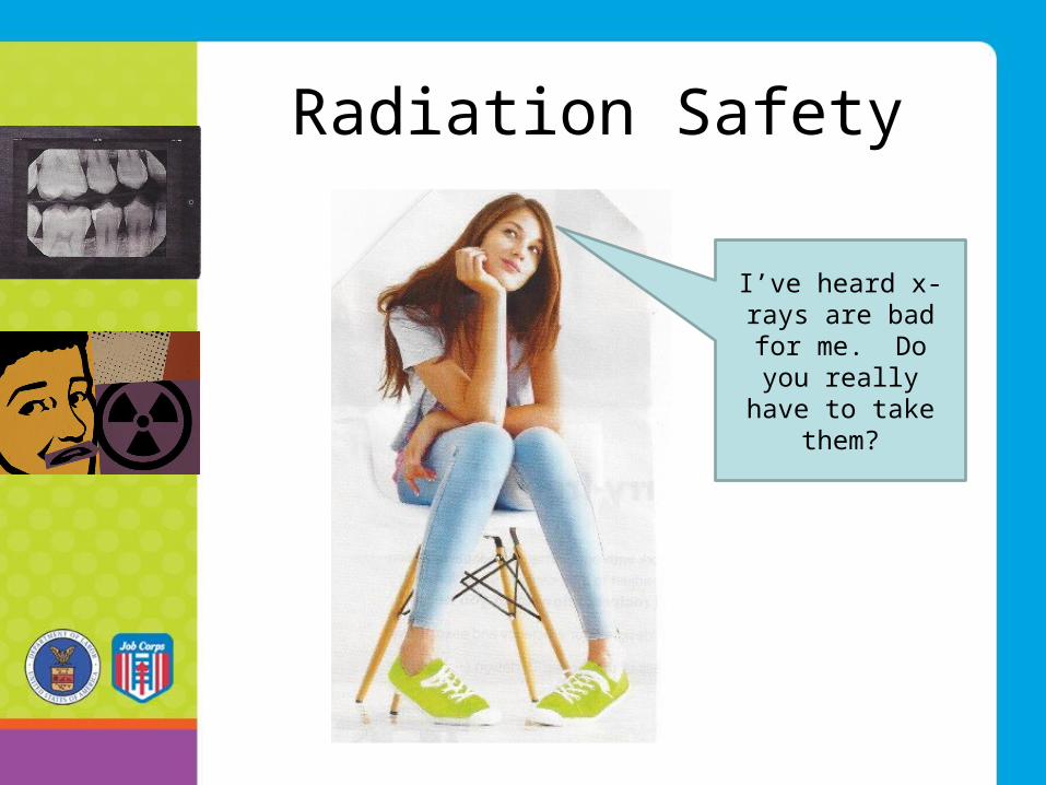
Radiation Safety
I’ve heard x-rays are bad for me. Do you really have to
take them?
Patient Education
• “Dental manufacturers are continually striving to develop products.”
• “Digital radiography requires much less radiation than conventional radiography.”
• “With less radiation, the absorbed dose to the pt is significantly lower.”
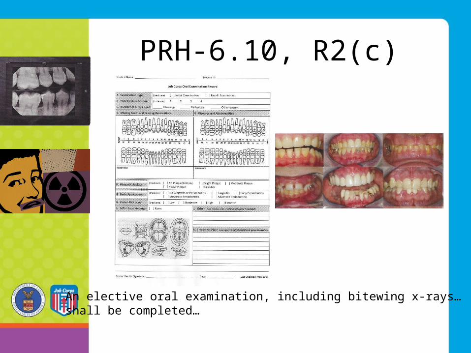
PRH-6.10, R2(c)
An elective oral examination, including bitewing x-rays…shall be completed…
The Dentist has Responsibilities for Dental Imaging
• Prescribe only images that are required for dx purposes
• Ensure radiographic equipment is safe• Ensure appropriate shielding• Require that all personnel are properly trained
& supervised• Use techniques that will produce diagnostic-
quality images• Follow the state’s radiographic licensing
requirements• Participate in obtaining informed consent
Protective Devices
• Aluminum filtration
• Collimator
• Position indicator device
• Lead apron and thyroid collar
Protective Devices (continued)
• Fast-speed film
• Image receptor-holding devices
• Exposure factor
• Proper technique
Pregnancy
• No need to alter radiographic procedures
Monitoring
• Operator protection & monitoring
• Radiation monitoring
• Equipment monitoring
ALARA Concept
• All exposure to radiation must be kept to a minimum or “as low as reasonably achievable.”
Infection Control
• Potential for cross-contamination• Wear personal protective
equipment• Use heat tolerant intraoral devices• Transport and handle exposed film
aseptically• Consult manufacturer for
disinfection/sterilization advice
References
Major References: Bird DL, Ronbinson DS. Modern Dental Assisting, El Sevier Saunders Publisher (2012)
Safety in Dental Radiography, GSC Home Study Course (2008)


