Measuring Hypnosis, Analgesia, and EEG Burst Suppression Pattern During Intravenous Anaesthesia Effective Doses, Catecholamine and Cardiovascular Responses A c t a U n i v e r s i t a t i s T a m p e r e n s i s 1004 ACADEMIC DISSERTATION To be presented, with the permission of the Faculty of Medicine of the University of Tampere, for public discussion in the main auditorium of Building K, Medical School of the University of Tampere, Teiskontie 35, Tampere, on May 7th, 2004, at 12 oclock. SEPPO MUSTOLA
Transcript
Measuring Hypnosis, Analgesia, and EEGBurst Suppression Pattern During Intravenous
Electronic dissertationActa Electronica Universitatis Tamperensis 337ISBN 951-44-5951-2ISSN 1456-954Xhttp://acta.uta.fi
ACADEMIC DISSERTATIONUniversity of Tampere, Medical SchoolTampere University Hospital, Departments of Anaesthesia & ChemistrySouth Karelia Central Hospital, Department of AnaesthesiaFinland
Supervised byDocent Gerhard BaerUniversity of Tampere
Reviewed byDocent Riku AantaaUniversity of TurkuDocent Olli ErkolaUniversity of Helsinki
MEASURING HYPNOSIS, ANALGESIA, and EEG BURST SUPPRESSION PATTERN
DURING INTRAVENOUS ANAESTHESIA:
Effective Doses, Catecholamine and Cardiovascular Responses.
CONTENTS: 3
LIST OF ORIGINAL PUBLICATIONS 5
ABBREVIATIONS 6
A. INTRODUCTION 7
B. REVIEW OF LITERATURE 10
B.1. Intravenous anaesthesia 10
B.1.1. Intravenous anaesthetics 12
B.1.1.1. Propofol 14
B.1.1.2. Thiopental 16
B 1.1.3. Ketamine 18
B.1.2. Intravenous opioids 20
B.1.2.1. Alfentanil 21
B.1.2.2. Remifentanil 22
B.2. Responses to laryngoscopy and intubation 23
B.2.1. Regulation of cardiovascular system 23
B.2.2. Cardiovascular and catecholamine response 24
B.2.3. Attenuation of the cardiovascular and catecholamine responses 25
B.3. Clinical measures of adequate anaesthesia 29
B.3.1. Physiology and neuroanatomy of the clinical measuring points 29
B.3.2. Clinical measuring points in anaesthesia monitoring 34
B.3.2.1. Hypnosis 34
B.3.2.2. Analgesia 35
B.3.2.3. Electroencephalography (EEG) 37
B.3.2.3.1. Burst suppression pattern (BSP) 38
C. PURPOSE OF THE STUDY 40
D. MATERIAL AND METHODS 41
D.1. Patients and experimental animals 41
D.1.1. Experimental animals 41
D.1.1.1. Rabbit anaesthesia 41
3
D.1.2. Surgical patients 43
D.1.2.1. Anaesthesia methods 43
D.2. Trial designs 45
D.2.1. Objectives of the studies 46
D.2.2. Clinical measuring points 47
D.2.2.1. Rabbits 47
D.2.2.2. Surgical patients 48
D.2.3. Anaesthesia monitoring 48
D.2.4. EEG monitoring 49
D.2.5. Cardiovascular measurements 49
D.2.6. Catecholamine measurements and analysis 50
D.2.7. Analysis of plasma concentrations of intravenous anaesthetics 50
D.3. Statistical methods 51
E. RESULTS 53
E.1. Responses at different measuring points 53
E.1.1. Cardiovascular responses 53
E.1.2. Plasma catecholamines 55
E.2. Effective doses 56
E.3. Effective concentrations 58
E.4. EEG and BIS 60
E.5. Other results 61
F. DISCUSSION 63
F.1. General aspects 63
F.2. Cardiovascular and catecholamine responses 66
F.3. Anaesthetic efficacy of IV anaesthetics 68
F.4. EEG and BIS 70
F.5. Clinical implications 72
G. SUMMARY AND CONCLUSIONS 74
REFERENCES 76
ACKNOWLEDGEMENTS 97
ORIGINAL COMMUNICATIONS 99
4
LIST OF ORIGINAL PUBLICATIONS
This thesis is based on the following original communications and referred to in the text by their
roman numerals.
I. Mustola S, Rorarius M, Baer G, Rosenberg P, Seppälä T, Harmoinen A (2000).
Potency of propofol, thiopentone and ketamine at various endpoints in New Zealand
white rabbits. Lab Anim 34: 36-45.
II. Mustola S, Baer G, Metsä-Ketelä T, Laippala P (1995). Haemodynamic and plasma
catecholamine responses during total intravenous anaesthesia for laryngomicroscopy.
Anaesthesia 50: 108-113.
III. Mustola S, Baer G, Toivonen J, Salomäki A, Scheinin M, Huhtala H, Laippala P,
Jäntti V (2003). Electroencephalographic burst suppression versus loss of reflexes
anesthesia with propofol or thiopental: Differences of variance in the catecholamine
and cardiovascular response to tracheal intubation. Anesth Analg 97: 1040-1045.
IV. Mustola S, Baer G, Neuvonen P, Toivonen J. Requirements of propofol at different
endpoints without adjuvant and during two different steady infusion of remifentanil
(submitted).
V. Särkelä M, Mustola S, Seppänen T, Koskinen M, Lepola P, Suominen K, Juvonen
T, Tolvanen-Laakso H, Jäntti V (2002). Automatic analysis and monitoring of burst
suppression in anesthesia. J Clin Monit Comput 17: 125-134.
The publishers of the original communications have kindly granted permission to reproduce the
articles in this thesis.
5
ABBREVIATIONS
ADR adrenaline
ANS autonomic nervous system
ASA anaesthetic risk groups according to the American Society of
Anaesthesiologists
BIS bispectral index
BSP burst suppression pattern of the EEG
CA catecholamines
CNS central nervous system
CL95 95% confidence limits
DAP diastolic arterial pressure
EC50 effective concentration when 50% of patients have lost their
reaction to a stimulus
EC95 effective concentration 95% (see above)
ED50 effective dose when 50% of patients have lost their reaction to
a stimulus
ED95 effective dose 95% (see above)
EEG electroencephalography
HPLC high-pressure liquid chromatography
HR heart rate
IV intravenous
LOC loss of consciousness
MAC minimal alveolar concentration
NOR noradrenaline
OAA/S observer's assessment of alertness/sedation scale
PNS parasympathetic nervous system
SAP systolic arterial pressure
SNS sympathetic nervous system
TIVA total intravenous anaesthesia
6
A. INTRODUCTION
Awareness during anaesthesia has an incidence of 0.2-2% (Ranta et al 1998, Sandin et al 2000,
Ranta 2002). Despite its rarity, it is a serious problem causing persistent psychological disturbances
resulting in individual suffering and inability to work (Ranta et al 1998). For the clinician,
unconsciousness is only one aspect of anaesthesia. For the patient, unconsciousness is an all-or-none
response: he either knows what has happened, or he does not. For the surgeon, anaesthesia is a state
that enables him to perform the procedure; thus, a conscious immobile patient might be sufficient.
However, anaesthesiologist and surgeon are interested in intra and postoperative prevention of pain,
intraoperative depression of reflexes, and a level of anaesthesia that would not cause postoperative
morbidity. Replacement of “level of anaesthesia” by “level of intraoperative medication” would help
to clarify the problem.
Different terms are used for similar facts; “awareness” and “consciousness” obviously have
the same meaning. “Adequate hypnosis”, “anaesthesia”, and “unconsciousness” are also used as
synonyms. There are two levels of consciousness during anaesthesia (Ghoneim 2000). First, a
patient actually remember some events during anaesthesia; i.e. explicit memory. Second, a patient
can have subconscious perception of events during anaesthesia which may be elicited by specific
tests; i.e. implicit memory. The possibility of implicit memories has been questioned, because mood
changes are seen after anaesthesia even without adverse intraoperative events (Bailey and Jones
1997). Patients also may recall disturbing dreams (Ghoneim 2000). An additional confusing
phenomenon is that patients may obey orders during anaesthesia without any recall afterwards
(Cormack 1993). It has been proposed that drugs causing amnesia, like benzodiazepines, might be
used routinely (Cormack 1993) or when consciousness is suspected during anaesthesia (Ghoneim
2000). Others, however, argue this practise to be ethically unsound (Sandin et al 2000). Adequate
anaesthesia has been proposed as a continuum (Griffiths and Jones 1990, Ghoneim 2000); adequate
anaesthesia results in complete unconsciousness, lighter anaesthesia results in dream recall or
implicit memory, and still lighter anaesthesia results in explicit memory.
The EEG has been used to measure depth of anaesthesia for over 50 years. However, good
correlations were found only under laboratory conditions with one drug a time. Technical progress
has now enabled the calculation of scores from the raw data almost in real time; such were, for
instance, aperiodic analysis and power spectrum analysis (Levy 1984, Levy 1986). Again, these
tools worked well with one drug and no surgical disturbance. Recently, several new scores have
been developed: bispectral index (BIS) (Sigl and Chamoun 1994), ENTROPY (Sleigh and Donovan
7
1999), and auditory evoked potential index (AEPindex) (Gajraj et al 1998). These scores measure the
level of the effect of medication on the cerebral cortex. Below certain scores, the patient’s mental
function is disturbed (sedation) and at deeper levels mental function is inhibited (unconsciousness).
However, patients medicated down to the level of EEG burst suppression may display exact
protective movements suggesting preserved orientation and observation without memory (Baer G,
pers. comm.). Patients whose EEG score indicates “unconscious” may have some implicit
memories. Finally, at the same level of EEG depression, some patients may have explicit memories
(Ghoneim 2000).
The concept of MAC (minimum alveolar concentration) for volatile anaesthetics provided a
useful measure to evaluate the depth of anaesthesia (Eger et al 1965). One MAC prevents 50% of
patients from reacting to skin incision and is about twice the concentration needed to produce
unconsciousness (Newton et al 1990). Later, other noxious stimuli have been employed in place of
and laser stimulation (heat stimulus). With IV anaesthetics effective dose 50% (ED50) and effective
concentration 50% (EC50) have been used as an equivalent to 1 MAC of volatile anaesthetics
(Davidson et al 1993). Monitoring of end tidal volatile concentrations and infusion automats
providing certain drug concentrations in the bloodstream gave a sense of security. However, MAC
measures the blockage of noxious stimuli in the spinal cord Rampil et al 1993; fortunately, with
most drugs unconsciousness is a definite side effect of this level of intraoperative medication.
Employing one drug only, the level of anaesthesia (medication) has been deepened by increasing
drug delivery to the patient until surgery was possible to perform. With ether, due to severe side
effects, the level of “surgical anaesthesia” was near to the level of dangerous anaesthesia. The level
of anaesthesia was and is still evaluated using clinical signs such as protective movements and the
eyelash and pupillary light reflexes (Guedel 1936).
The most distressing feature of consciousness during anaesthesia is pain perception during
surgery. The patient can be conscious but cannot move (Ghoneim 2000). Therefore, less use of
neuromuscular blockers and deeper levels of medication have been advised (Cormack 1993).
Movement as a reaction to painful stimuli is mainly due to spinal reflexes, which appear also in
unconscious patients (Cormack 1993). On the other hand, apparent lack of reflex activity does not
prove unconsciousness and complete areflexia requires a very deep level of medication. The
correlation between different autonomic and somatic reflexes is variable and there is no consensus
on which of them correlate best with consciousness (Cormack 1993). Measurement of
unconsciousness during anaesthesia is very difficult, and there is no reliable method of detecting
8
consciousness during anaesthesia (Drummond 2000), whch is why surrogate measures of adequate
anaesthesia are still widely used.
In the present study we compared propofol, thiopental, and ketamine (rabbits) during
different surrogate measures of the depth of anaesthesia (later end-points). In human studies we used
loss of eyelash reflex, loss of pupillary light reflex, loss of counting, and loss of obeying verbal
command as hypnotic end-points. As analgesic end-points we used electrical tetanic stimulation,
laryngoscopy, intubation, and laryngomicroscopy (a longer lasting noxious stimulus). In
experimental animals we used loss of righting reflex and tail clamping as hypnotic and analgesic
end-points, respectively. Furthermore, in rabbits we used a more centrally mediated end-point, loss
of purposeful movement to intranostril instillation of ammonia vapour. In humans we used the onset
of electroencephalographic (EEG) burst suppression pattern (BSP) as a measure of deep level of
medication, i.e. EEG end-point.
9
B. REVIEW OF LITERATURE
B.1. Intravenous anaesthesia
IV anaesthesia is a smooth, reliable and fast anaesthetic technique. Pharmacokinetically there are
short-acting IV anaesthetics, which do not cause gas pollution, are inexpensive, easy to titrate, and
easy to administer. Some IV anaesthetics have analgesic properties (ketamine) while others do not
(propofol, thiopental). Furthermore, IV anaesthetics do not cause airway irritation during induction
as is seen with most volatile anaesthetics; recovery can be faster with volatile anaesthetic than with
IV anaesthetic (Van Hemelrijck et al 1991). After IV anaesthesia recovery is smooth.
Postoperatively, propofol possesses significant antiemetic properties, and ketamine analgesic
activity (McCollum et al 1988, Gan et al 1996, Reves et al 2000).
The partial pressure of inhaled anaesthetic in the exhaled air can be measured non-invasively
and equals the arterial value. IV anaesthetics are generally delivered on a dose per kilogram of body
weight rule, real-time measurement of blood drug concentration is not possible, and the excretion of
overdoses cannot be hastened. For this reason, manually (Roberts et al 1988, Maitre and Shafer
1990) and computer controlled (Shafer et al 1988a, Tackley et al 1989, White and Kenny 1990)
infusion pumps have been developed. They enable the administration of IV anaesthetics in
accordance with their pharmacokinetic profile. Accuracy of the prediction of blood concentration of
propofol by a computer-controlled infusion device has been tested (Vuyk et al 1995a). The
performance of the computer-controlled infusion device was similar with the pharmacokinetic
parameters sets described by Cockshott et al (1987), Gepts et al (1987), Shafer et al (1988b), and
Tacley et al (1989). The measured concentrations of propofol exceeded the predicted concentrations
by approximately 25% (Vuyk et al 1995a). The parameter set of Kirckpatrik et al. (1989) produced
the most unreliable results. There are considerable interindividual variations, the therapeutic
concentration is highly dependent on the surgical stimulus, and predicted concentrations are not
accurate for all patients (Vuyk et al 1995a). Therefore, despite the use of manually or computer
controlled infusion pumps, the dosage and rate of IV anaesthetics administration must be adjusted to
meet individual patient needs. The Observer's Assessment of Alertness/Sedation (OAA/S) scale was
developed to measure the level of alertness in subjects who are sedated. The OAA/S scale has
proved to be reliable and valid to measure the depth of sedation (from light to heavy) as compared to
a Visual Analogue Scale and two performances tests (Digit Symbol Substitution Test and Serial
Sevens Subtraction) (Chernik et al 1990).
10
Pharmacokinetic characterization of IV anaesthetic drugs are mostly described using a
multicompartment mammillary model that yields two distribution half-lives and a terminal
“elimination” half-life; i.e. a three-compartment model. The model consists of unit disposition
functions of the form C(t) = Ae-αt + Be-βt + Ce-γt where C(t) is the central compartment drug
concentration profile resulting from a bolus input as a function of time (t), and α, β, and γ are
exponential rate constants of the relationship, α>β>γ. Elimination half-life is calculated as ln2/γ, and
distribution half-life ln2/α (central) and ln2/β (rapid peripheral). A, B, and C are constants (y-axis
intercepts). Principal pharmacokinetic parameters are clearance (CL), volume of distribution (V),
and half-lives (T½). Also, intercompartmental rate constants are usually calculated.
The equation is in form C(T+t) = A(1 - e-αΤ)e-αt + B(1 - e-βΤ)e-βt + C(1 - e-γT)e-γt during IV
infusions where T is duration of infusion and t is post infusion time. When a drug’s pharmacokinetic
behaviour can be presented by a one-compartment model, the elimination half-life can be used to
describe the time required for the concentration to decrease by a certain percentage when the
infusion is stopped. In multicompartment pharmacokinetic models, distribution of the drug between
central and peripheral compartments is a significant contributor to central compartment drug
disposition. Elimination half-life may be of little value in describing multicompartment models
(Shafer and Varvel 1991). The reason for the inadequacy is that the terminal elimination half-life
does not take into account the distribution of the drug to tissues after discontinuation of the drug
infusion. To find better ways to measure the offset of drug action, the “context-sensitive half-time”
was introduced to clinical practise (Hughes et al 1992). The “half-time” is the time required for the
drug concentration in the central compartment to decrease by 50% after discontinuation of drug
administration. The term “context” relates to the duration of drug infusion before its discontinuation.
By incorporating the effect compartment, the context-sensitive half-time of the pharmacodynamic
effect can be modelled. This concept of “context-sensitive half-time” has clinically relevant
implications for the administration of IV anaesthetics and would allow a more accurate prediction of
recovery from IV infusion anaesthesia after the termination of surgery (Hughes et al 1992).
When predicting the effect-site concentration (and effect) of an IV anaesthetic agent after bolus
or during infusion, the equilibrium half-life between blood and effect-site has important clinical
implications (Jacobs and Reves 1993). The effect-site is the hypothetical compartment that relates
the time course of plasma drug concentration to the time course of drug effect where k e0 is the rate
constant of drug elimination from the effect-site and t½ k e0 is ln2/ke0. After a bolus dose or start of
anaesthetic infusion, the effect-site concentration starts from zero and increases over time until it
11
equals the plasma concentration. When the k e0 is available for an IV anaesthetic, theoretical effect
compartment concentrations can be simulated. When dealing with anaesthetic drug infusions, it
should be taken into account that the 90% equilibrium between blood and brain (effect-site) will
occur within about three to four equilibrium half-lives (Glass et al 2000).
B.1.1. Intravenous anaesthetics
An ideal IV anaesthetic should produce a rapid, smooth, and safe induction of and emergence from
anaesthesia. It should have a large distribution volume, rapid degradation to inactive and non-toxic
metabolites, no accumulation, and dosing of the drug should not be influenced by hepatic or renal
insufficiency. It should have minimal effects on cardiovascular and respiratory functions or
functions of other organs, and preferably have analgesic properties at sub-anaesthetic doses. It
should be non-irritating to tissues or veins and have low potential to hypersensitivity reactions. It
should not raise intracranial pressure. It should be water-soluble, inexpensive, stable in solution, and
have a long shelf life. None of the presently available IV anaesthetics matches all these
specifications (Reves et al 2000). Pharmacokinetic parameters of IV anaesthetics are presented in
Table 1.
12
Table 1. Pharmacokinetic parameters of intravenous anaesthetics and opioids.
CLss = clearance at steady-state, Vdss = volume of distribution at steady-state, t½ = half-life, t½ k e0 = equilibrium half-life between plasma and effect-site
Drug
CLss (ml/kg/min)
Vdss (l/kg)
t½ (h) (elimination)
Time to peak effect (min)
T½ k e0 (min-1)
Context-sensitive t½ (min)
References
Propofol
22-30
1.5-4
3-10
1-2.5
1-3.5
20-50
Gepts et al 1987, Kirkpatrick et al 1988, Shafer et al 1988b, Hughes et al 1992, Shafer 1993, Smith et al 1994, Schnider et al 1999
Thiopental
1.5-4
1.5-4
5-15
1-2
1-2.5
100-200
Ghoneim and Korttila 1977, Morgan et al 1981, Homer and Stanski 1985, Stanski and Maitre 1990, Hughes et al 1992, Shanks et al 1993, Cordato et al 1999
Ketamine
12-18
2.5-3.5
2-3
1-2
-
25-50
Idvall et al 1979, Clements and Nimmo 1981, Domino et al 1982, Reves et al 2000
Fentanyl
10-20
3-5
3-4
3-5
4-6
100-300
Camu et al 1982, Scott et al 1985 & 1991, Maitre et al 1987, Hughes et al 1992, Egan et al 1996
Alfentanil
4-8
0.5-1.0
1-2
1-2
1-1.5
40-50
Camu et al 1982, Scott et al 1985, Maitre et al 1987, Hughes et al 1992, Egan et al 1996
Sufentanil
10-15
2.5-3.0
2-3
4-6
5-6
20-50
Scott et al 1991, Shafer and Varvel 1991, Hughes et al 1992, Bailey et al 2000
Remifentanil
40-60
0.3-0.5
0.1-0.2
1-2
1-2
3-5
Egan et al 1993, Westmoreland et al 1993, Egan 1995, Kapila et al 1995, Minto et al 1997
13
B.1.1.1. Propofol
Propofol is a substituted phenol (2,6-diisopropylphenol) that was first introduced in the early 1980s
(Rogers et al 1980, Major et al 1981). Recovery from either a single injection or IV infusion was
and is very rapid (Major et al 1981, Prys-Roberts et al 1983, Grant and Mackenzie 1985). The
potency of propofol compared to thiopental varies from 1:1.3 to 1:2.9 (Grounds et al 1986, Naguib
et al 1992). It is more suitable for use in infusion anaesthesia than thiopental (Kashtan et al 1990).
Propofol is insoluble in aqueous solutions but is highly lipophilic. The initial distribution
clearance of propofol (3-4 l/kg/min) is similar to that of thiopental (Shafer 1993), but subsequently it
is much more rapid because of its high metabolic clearance rate. Total body clearance of propofol at
steady state is 22-30 ml/kg/min, the distribution volume at steady state is 1.5-3 l/kg, and the
elimination half-life 3.5–10 h after infusion (Gepts et al 1987, Kirkpatrick et al 1988, Shafer et al
1988b), but recovery from its clinical effects is rapid. Propofol has a context-sensitive half-time of
less than 25 min after infusions as long as 3 hours, which increases to only 50 min after more
prolonged infusions. This is due to the fact that the long elimination half-life is related to slow
elimination from highly lipophilic tissue compartments and is largely irrelevant in clinical situations
(Shafer and Stanski 1992). Propofol has a large steady-state volume of distribution, indicating
extensive redistribution of the drug into muscle, fat, and other poorly perfused tissues (Kirkpatrick
et al 1988, Shafer et al 1988b). The concentration in the central compartment decreases both because
of metabolism and continuing redistribution after termination of infusion of propofol. Because the
capacity of the peripheral compartments is large, redistribution from the central compartment can
still occur even after prolonged drug administration. The complete elimination of propofol from the
body may take many hours or even days but have little effect on clinical recovery.
For successful induction of anaesthesia, i.e. an effective dose when 95% of patients (ED95) have
lost consciousness, 2.0 to 2.5 mg/kg of propofol is needed and ED50 of propofol is 1-1.5 mg/kg
(Major et al 1981, Rolly and Versichelen 1985, McCollum and Dundee 1986). The duration of
hypnosis is 5-10 min after single anaesthetic dose (Major et al 1981) and is slightly longer than that
of thiopental (Flaishon et al 1997). ED95 of infants needs an induction dose of 3.0-3.5 mg/kg
(Westrin 1991) and ED95 of elderly patients is 0.5-1.5 mg/kg (Dundee et al 1986). Infusion rates of 4
to 18 mg/kg/h are required during TIVA with propofol depending on the surgical procedure and
concomitant anaesthesia drugs (Fragen et al 1983, Turtle et al 1987, Sear et al 1988, Reyneke et al
1989, Milligan et al 1990). The need for reduced dose requirements of propofol in the elderly
(Scheepstra et al 1989) is related to pharmacokinetic rather than pharmacodynamic factors
14
(Kirkpatrick et al 1988). Children require a larger induction dose (Valtonen et al 1989) and an
increased maintenance rate of infusion than adults because the volume of central compartment, and
thus the volume of distribution is larger than that of adults (Jones et al 1990, Kataria et al 1994).
Gender does not seem to significantly influence propofol pharmacokinetics (Kay et al 1986), but
there are studies that report faster emergence from propofol anaesthesia in women than in men (Gan
et al 1999, Høymork et al 2001). Considerable variation in the blood concentration of propofol and
other drugs has been observed during infusion schemes designed to achieve a predetermined blood
concentration. Variations occur between groups of patients given an identical infusion scheme,
despite attempts to normalize for variables such as weight, age, and gender (Coetzee et al 1995,
Vuyk et al 1995a).
ED50 values of propofol for the onset of BSP have been about 5.5-6.5 mg/kg with an initial bolus
and infusion thereafter (Illievich et al 1993) or with infusion only (Cheng et al 1996). Effective
concentrations of propofol when 50% of patients (EC50) have lost their consciousness are for loss of
eyelash reflex 2-2.5 µg/ml and for loss of response to verbal command 3-4.5 µg/ml, respectively
(Smith et al 1994, Vuyk et al 1996, Kazama et al 1997). EC50 for loss of purposeful movement to
noxious stimulus is for tetanic stimulation or laryngoscopy 8-11 µg/ml, for skin incision 10-15
µg/ml, and for laryngoscopy followed by intubation 15-20 µg/ml (Smith et al 1994, Kazama et al
1997). EC50 to induce BSP has been 7-9 µg/ml (Illievich et al 1993). The reduction of EC50 for loss
of response to verbal command was minimal with fentanyl whereas the reduction of EC50 for
noxious stimuli varied from 30% to 50% when increasing fentanyl concentration from 0.6 ng/ml to
3 ng/ml was used (Smith et al 1994, Kazama et al 1997). Age reduces the requirements of many
anaesthetic drugs by approximately 20% for each decade of years above 20 years of age (Smith et al
1994). EC50 of propofol for skin incision with temazepam premedication was 8.1 µg/ml (Davidson
et al 1993). Midazolam reduced the dose of propofol necessary to prevent a response to a tetanic
stimulus by 50% (Short et al 1992). In conjunction with 70% nitrous oxide, the concentrations of
propofol during superficial surgery are in the range of 2.0-5.5 µg/ml (Turtle et al 1987, Shafer et al
1988b, Davidson et al 1993). It has been suggested that the lowest concentration of propofol that
produces LOC is 1.2 µg/ml regardless of opioid doses (Vuyk et al 1996). EC50 to awakening is 0.7-
1.2 µg/ml (Shafer et al 1988b). It has been recommended that during TIVA with propofol, the
plasma concentration of propofol should be maintained above 3.3 µg/ml, which is equal to a
propofol infusion of approximately 5 mg/kg/h (Smith et al 1994).
15
Propofol affects the respiratory system similarly to the action of thiopental, i.e. it causes central
respiratory depression, but laryngeal reflexes are more depressed compared to thiopental at
equivalent doses (Barker et al 1992). Cardiovascular changes after IV induction have demonstrated
that in comparison to thiopental propofol has more of a depressive effect on systolic and diastolic
arterial pressures, and reduces peripheral vascular resistance more severely than thiopental (Price et
al 1992). Propofol also significantly decreases myocardial contractility, cardiac output and stroke
volume, and has venodilating properties without significantly affecting heart rate (HR) (Coetzee et
al 1989, Mulier et al 1991, Illievich et al 1993).
Propofol does not release histamine, but can induce an anaphylactic reaction. Propofol possesses
significant antiemetic activity (McCollum et al 1988). Pain at the injection site is a usual complaint
(Hynynen et al 1985b). During longer infusions of propofol urine colour can change to green
(Blakey et al 2000).
B.1.1.2. Thiopental
Thiopental is a thiobarbiturate. It was first used in surgical patients at the Mayo clinic in 1934 and
thus has been in clinical practise already for 70 years. It has remained the standard IV anaesthetic
for evaluation of new IV anaesthetics. Despite its long elimination half-life (12h), it is still widely
used as an induction agent all over the world because of its rapid onset of action and being a
standard when compared to the faster degrading competition, propofol.
Sodium thiopental is a water-soluble and highly lipophilic molecule. Total body clearance of
thiopental ranges from 1.5 to 4 ml/kg/min, distribution volume at steady state from 1.5 to 3 L/kg,
and elimination half-life from approximately 5 to12 hours (Ghoneim and Korttila 1977, Morgan et
al 1981). During longer infusion, thiopental shows nonlinear kinetics, which is probably due to
saturation of the thiopental-oxidizing hepatic enzyme system resulting in accumulation of thiopental
because its elimination half-life can exceed 30-80 hours (Stanski et al 1980, Turcant et al 1985).
Thiopental has a context-sensitive half-time of 100 min after infusions as long as 3 hours, which
increases to 200 min after more prolonged infusions. Gender does not significantly influence
thiopental pharmacokinetics but advancing age does increase the terminal half-life and decrease
distribution volume (Christensen et al 1981, Homer and Stanski 1985, Stanski and Maitre 1990).
Also, children have higher clearance rate (Sorbo et al 1984).
In healthy adults the ED95 of thiopental for induction of anaesthesia is 4-6 mg/kg and ED50 is 2-4
mg/kg. ED95 of children and infants is 5-7 mg/kg for induction of anaesthesia (Jonmarker et al 1987)
16
and for elderly people 2-3 mg/kg (Homer and Stanski 1985, Steib et al 1988). The duration of
hypnosis is 4-8 min after a single anaesthetic dose. The elderly and women need less than the young
and men as an induction dose (Avram et al 1993). Decreased requirements of thiopental in the
elderly might be due to decreased initial volume of distribution and decreased cardiac output
(Stanski and Maitre 1990, Avram et al 1993, Shanks et al 1993). Higher requirements of thiopental
in children and infants might be due to greater cardiac output in relation to body weight or faster
central distribution kinetics (Jonmarker et al 1987). Thiopental administered by a continuous
infusion for maintenance of anaesthesia is rarely used because of its prolonged recovery time. A
successful thiopental infusion achieves a blood concentration of 15-20 µg/ml on induction, 10-
20 µg/ml during maintenance of anaesthesia, with a mean plasma concentration on awakening of 5-
7 µg/ml (Fragen and Avram 2000). Premedication with an opioid, benzodiazepine, or alpha-2-
agonist decreases the induction dose of thiopental by approximately 30% (Fragen and Avram 2000).
In young and healthy 20-30 years old patients, ED50 of thiopental to produce BSP is
approximately 10-12 mg/kg, and decreases to 6-7 mg/kg in patients 60-70 years of age (Homer and
Stanski 1985). EC50 of thiopental to induce BSP is 30-40 µg/ml (Bührer et al 1992, Hung et al 1992,
Shanks et al 1993). EC50 of thiopental for syringe dropping, loss of verbal command or loss of
eyelid reflex is 11-20 µg/ml. EC50 of thiopental for loss of corneal reflex, for loss of purposeful
movement such as reaction to tetanic stimulation, trapezius squeeze, or laryngoscopy are 30-50
µg/ml, and to laryngoscopy followed by intubation approximately 80 µg/ml (Becker 1978, Becker
and Tonnesen 1978, Hung et al 1992).
Thiopental when compared to propofol is less depressant to the cardiovascular system when
arterial blood pressure, total peripheral resistance, or cardiac output are measured (Grounds et al
1985, Stowe et al 1992), but more depressant as seen in the isolated guinea pig myocardial
preparation and in human atrial strips (Azari and Cork 1993, Gelissen et al 1996). Thiopental
depresses myocardial function more than propofol in the acute ischemic heart than in the normal
heart (De Hert et al 1990) and may evoke reductions in coronary flow that are profoundly
exaggerated under conditions of coronary endothelial dysfunction (Moore et al 1994). Thiopental
also increases HR, which results in increased myocardial oxygen consumption.
Thiopental decreases plasma cortisol levels but does not prevent adrenocortical stimulation from
the stress of surgery. Thiopental injection causes release of histamine and occasionally urticarial
rash, but anaphylactic reactions are rare. Pain on injection site is rare but extravasated thiopental
produces local pain, oedema, erythema, and sometimes tissue necrosis (Fragen and Avram 2000).
17
B 1.1.3. Ketamine
Ketamine is an arylcyclohexylamine and is structurally related to phencyclidine. Racemic
ketamine was introduced in 1965 and approved for clinical use in 1970. Ketamine has been used
widely in veterinary medicine (Green et al 1981, Borkowski et al 1990). Ketamine produces a
clinical anaesthetic state in which the eyes remain open showing a slow nystagmus; corneal and
pupillary light reflexes remain intact, as do laryngeal protective reflexes (Carson et al 1973).
Varying degrees of hypertonus and myoclonic movements are seen, and spontaneous respiration is
preserved. The preserved reflexes and spontaneous movements make it difficult to judge adequacy
of anaesthesia.
Ketamine is water-soluble, painless and non-irritating following parenteral injection, and has
high lipid solubility (White et al 1982). The clearance of ketamine at steady state is 12-18
ml/kg/min, the volume of distribution at steady state 2.5-3.5 L/kg, and the elimination half-life 2-3
hours after a single bolus (Clements and Nimmo 1981, Domino et al 1982). Ketamine has been used
as an infusion anaesthetic (1-3.5 mg/kg/h, mean 2.5) with rapid recovery, cardiovascular stability,
and an elimination half-life of 80 min (Idvall et al 1979). Neither gender nor age seems to
significantly influence ketamine pharmacokinetics. The context sensitivity half-time of ketamine is
about the same as with propofol, 20-50 min (Reves et al 2000).
ED95 of ketamine to induce anaesthesia after an intravenous bolus dose is 1.5-3.0 mg/kg with
consciousness returning after 10-15 minutes. Children need 2.5-3.0 mg/kg (Westrin 1991), and
elderly patients 0.5-2.0 mg/kg as an induction dose (ED95) (Reves et al 2000). ED50 for loss of
verbal command is 0.4-0.9 mg/kg and ED50 for loss of reaction to tetanic stimulation 0.6-1.3 mg/kg
(Hong et al 1993). ED95 for loss of response to tetanic stimulation is 1-2.5 mg/kg (Hong et al 1993).
EC50 of ketamine that caused one-half of the maximal median frequency decrease in the EEG was
2.0 µg/ml for racemic ketamine and 0.8 µg/ml for S-(+)-ketamine (Schüttler et al 1987). During
successful IV anaesthesia, plasma concentrations of ketamine have been 0.6-2.0 µg/ml in adults, and
children require plasma concentrations of 0.8-4.0 µg/ml (Reves et al 2000). Infusion rates of 1 to 5
mg/kg/h are required during TIVA with ketamine depending on concomitant anaesthetic drugs and
types of surgery (White et al 1982).
Ketamine is an effective analgesic even at sub-anaesthetic doses in humans, but not in all
animals (Green et al 1981). Ketamine has been used as an analgesic during TIVA with propofol
18
(Guit et al 1991). Analgesia occurs at lower blood concentrations than loss of consciousness; the
plasma concentration of ketamine at elevated pain threshold is 0.1 µg/ml or higher. The analgesic
action is postulated to be due to N-Methyl-D-aspartate receptor interaction and inhibition of dorsal
horn wide dynamic range neuronal activity. Ketamine has excitatory effects on the CNS and can
induce generalized high voltage synchronized theta and delta wave activity and petit mal like
activity in the hippocampus (Kochs et al 1988, Reves et al 2000). Even at higher doses, ketamine
does not induce BSP (Rosen and Hagerdal 1976).
Ketamine has minimal effect on the central respiratory drive. It is an effective bronchodilatator
but sometimes considerably increases salivation (Reves et al 2000). Ketamine increases the rate-
pressure product and the cardiac index, but does not significantly change the stroke index (Idvall et
al 1979). The cardiovascular effects are thought to be produced by direct stimulation of the CNS.
Ketamine is a direct vasodilator but in postganglionic adrenergic neurons, ketamine inhibits the re-
uptake of catecholamines resulting in no alteration of peripheral vascular resistance. On the other
hand, it increases pulmonary vascular resistance. The centrally mediated sympathetic stimulation
usually overrides the direct myocardial depressant effect of ketamine. With doses used to induce
anaesthesia, ketamine depresses cardiac function less than thiopental or propofol (Stowe et al 1992).
During ketamine anaesthesia, patients experience psychic reactions like vivid dreams or
hallucinations, which may be fantastic, interesting, disturbing or nightmarish quality for the patient.
The postoperative alterations in mood state and body image, floating sensations, illusions and
delirium (Perel and Davidson 1976, White et al 1980) may be a nightmare for the patient and
personnel. The quality of the hallucinations depends on the patient’s psychic state of mind before
anaesthesia induction, which might be positively influenced during the pre-anaesthetic round; on the
other hand, the disturbed short-time memory makes it difficult to co-operate rationally afterwards
with the strange events (Baer and Parkas 1981). Thus, ketamine can be the ideal anaesthetic during
the harsh circumstances of war or catastrophes (anaesthesia and analgesia with one drug), but totally
unsuitable, for example, in gynaecologic surgery of ambulant patients (the patient fearing cancer,
infertility, pregnancy) (Dundee1971, Coppel et al 1973). Benzodiazepines efficiently attenuate these
psychic disturbances, but also depress respiration and protective laryngeal reflexes (Coppel et al
1973, Korttila and Levänen 1978).
Clinical trials have shown that the anaesthetic potency of the ketamine isomer S-(+)-ketamine is
twice that of the racemic mixture. A bolus dose of 2 mg/kg of racemic ketamine equals a dose of 1
mg/kg of S-(+)-ketamine, producing comparative effects in endocrine and cardiovascular parameters
19
(Adams et al 1992). Recovery is significantly faster and cognitive performance better after S-(+)-
ketamine than after racemic ketamine (Doenicke et al 1992).
B.1.2. Intravenous opioids
Opioids act via the central and peripheral µ-opioid receptors. Opioid receptor distribution within the
neuraxis coupled with the widespread association between opioid receptors and cardiovascular and
autonomic regulatory areas within the CNS define much of the pharmacophysiological basis for
opioid-induced hemodynamic effects (Bailey et al 2000). The ventrolateral periaqueductal gray
region, a key central site mediating analgesia, also affects hemodynamic control, and different
opiate receptor subtypes occupy this site (Keay et al 1997). Activation of µ-opioid receptors
suppresses somatosympathetic reflexes at the level of the spinal cord and modulates them at the
brain stem. These actions contribute to the anaesthetic capabilities of opioids. Opioids can also
modulate the stress response through receptor-mediated actions on the hypothalamic-pituitary-
adrenal axis. Most opioids reduce sympathetic and enhance vagal tone (Sato et al 1995). The
predominant and usual effect of opioids on the HR is to produce bradycardia resulting from
stimulation of the central vagal nucleus but blockade of sympathetic chronotropic actions may also
play a role in opioid-induced bradycardia (Bailey et al 2000).
Opioids do not reliably produce unconsciousness (Lang et al 1996, Jhaveri et al 1997, Veselis et
al 1997), but they do reduce MAC of inhaled anaesthetics (Brunner et al 1994, Tammisto et al 1995,
Lang et al 1996) and requirements of IV anaesthetics (Short et al 1992, Wessen et al 1993, Wang et
al 1996, Kazama et al 1998, O’Hare et al 2001) during anaesthesia. Opioids are widely used as
analgesics to supplement general anaesthesia for various surgical procedures.
Equipotent dose ratios (mg/kg) of alfentanil, remifentanil, fentanyl, and sufentanil measured by
EEG quantitation are approximately 100:10:10:1 (Scott et al 1985, Scott et al 1991, Bailey et al
2000, Thomson et al 2000). This ratio of equipotency was also confirmed in a later clinical study
(Ahonen et al 2000a). Equipotent plasma concentration ratios (ng/ml) that produce maximal EEG
effect in 50% of patients (EC50) of alfentanil, remifentanil, fentanyl, and sufentanil are
approximately 800:20:15:1 (Scott et al 1985, Scott et al 1991, Shafer and Varvel 1991, Egan et al
1996). Pharmacokinetic parameters of intravenous opioids are presented in Table 1.
20
B.1.2.1. Alfentanil
Alfentanil is a tetrazole derivative of fentanyl and is a potent µ-agonist. It came to use in clinical
practise in the early 1980s. It has adverse effects similar to the other opioids: ventilatory depression,
nausea, vomiting, muscular rigidity, and bradycardia. Alfentanil does not release histamine upon
injection. When compared with fentanyl, alfentanil has a faster onset (1-2 min) and one-third the
duration of action, one fourth to one tenth of the potency, and less lipid solubility (Camu et al 1982,
Hynynen et al 1985a). In the base of equipotent plasma concentrations the potency of fentanyl is 30
times greater than that of alfentanil (Langevin et al 1999).
When compared to fentanyl alfentanil has a lower total body clearance (4-8 vs. 10-20
ml/kg/min), smaller volume of distribution at steady state (0.5-1.0 vs. 3-5 L/kg), and shorter
elimination half-life (1-2 vs. 3-4 h) (Camu et al 1982, Maitre et al 1987, Egan et al 1996). The
elderly need less alfentanil when compared to young people (Helmers et al 1984, Scott and Stanski
1987). After infusion of 3 h, the context-sensitive half-time and terminal elimination half-life of
alfentanil is about 50 and 80 min (vs. fentanyl about 200 and 240 min), respectively (Shafer and
Varvel 1991, Westmoreland et al 1993, Kapila et al 1995). Because of its pharmacokinetic profile,
alfentanil is more suitable for infusion anaesthesia than fentanyl. The clearance of alfentanil is
reduced in patients with hepatic impairment (Ferrier et al 1985) whereas fentanyl kinetics is
unchanged (Haberer et al 1982). The free fraction of alfentanil and the volume of distribution at
steady-state are increased in chronic renal failure (Chauvin et al 1987). TIVA with propofol and
alfentanil for renal transplantation resulted in slower recovery than in previous studies of ASA 1-2
patients (Kirvelä et al 1994). Propofol inhibits enzymatic degradation, decreases greatly the fast
distribution clearance, and decreases minimally the elimination clearance of alfentanil (Janicki et al
1992, Mertens et al 2001).
When compared to fentanyl, clinical studies during surgical procedures show that alfentanil
produces an earlier peak analgesic effect, faster recovery of consciousness, and a more pronounced
narcotic effect without increased adverse effects (Fragen et al 1983, De Grood et al 1985, Jenstrup et
al 1990, Raftery and Sherry 1992). Alfentanil is useful for supplementation of analgesia for
outpatient surgical procedures and as an infusion for maintenance of analgesia during surgery
(Raftery and Sherry 1992, Langevin 1999). EC50 of alfentanil to induce maximal EEG effect is 500-
600 ng/ml (Scott et al 1985). Alfentanil has been successfully combined with propofol for induction
of anaesthesia followed by tracheal or nasotracheal intubation without muscle relaxant; given before
21
propofol it reduces injection pain of propofol (Saarnivaara and Klemola 1991, Coghlan et al 1993,
Fletcher et al 1994).
B.1.2.2. Remifentanil
Remifentanil is a synthetic 4-anilidopiperidine opioid and was brought into clinical practice in the
early 1990s. Pharmacodynamically, remifentanil equals the other fentanyl congeners, for example
alfentanil. Remifentanil produces physiological changes consistent of potent µ-agonist activity;
including analgesia, sedation, ventilatory depression, nausea, vomiting, muscular rigidity,
bradycardia and pruritus. It does not release histamine upon injection. The onset of action of
remifentanil is similar to that of alfentanil (1-2 min), but its offset is considerably more rapid. The
reason for this is that remifentanil has an ester side chain, which is rapidly hydrolysed by non-
specific plasma and tissue esterases. The rapid hydrolysis of remifentanil results in brevity of action,
precise and rapidly adjustable effects, non-cumulative opioid effects, and rapid recovery after
cessation of administration (Rosow 1993).
Remifentanil is lipophilic and widely distributed in body tissues with an extremely rapid
clearance of 40-60 ml/kg/min, the volume of distribution at steady state is 0.3-0.5 L/kg, and the
terminal half-life is only 10-20 min (Egan et al 1993, Westmoreland et al 1993). Because of its
unique metabolic pathway and rapid clearance, remifentanil represents a new pharmacokinetic class
of opioids. Unlike the other fentanyl congeners, termination of the therapeutic effect of remifentanil
mostly depends on metabolic clearance rather than on redistribution (Egan 1995). The context-
sensitive half-time of remifentanil is only 3-5 minutes, and is independent of infusion duration
(Westmoreland et al 1993, Kapila et al 1995). Age has an effect on the pharmacokinetics and
pharmacodynamics of remifentanil, but gender has not (Minto et al 1997). Pharmacokinetics and
pharmacodynamics of remifentanil are not altered in patients with renal or liver diseases (Dershwitz
et al 1996, Hoke et al 1997), or are altered clinically slightly (Dahaba et al 2002), i.e. the central
clearance is reduced and the elimination half-life is prolonged. Co-administration of propofol
decreases the central volume of distribution, the distributional clearance, and elimination clearance
of remifentanil, but remifentanil does not influence propofol kinetics (Boullion et al 2002).
Remifentanil’s brevity of action ensures not only a rapid resolution of adverse effects but also a
rapid offset of its analgesic effect. Therefore, appropriate postoperative analgesia should be
established before discontinuation of remifentanil infusion. The unique pharmacokinetic profile of
remifentanil facilitates real-time management of intraoperative stress, as well as provision of
22
optimal intraoperative analgesia without compromising recovery for a variety of surgical procedures
(Hogue et al 1996).
TIVA with remifentanil include fewer cardiovascular side effects, a low incidence of
postoperative nausea and vomiting and an attenuated neurohumoral stress response to surgery. The
therapeutic potency of remifentanil is approximately 20 times greater than that of alfentanil with an
EC50 to induce maximal EEG effect of 10-20 ng/ml (Egan et al 1996). Remifentanil is about 40-
times more potent than alfentanil when whole blood concentrations of remifentanil and alfentanil are
compared at equipotent doses to depress the minute ventilatory response (Glass et al 2000). On the
other hand, alfentanil concentrations are usually measured in plasma and remifentanil in whole
blood; when these are compared, remifentanil is 70 times more potent than alfentanil (Glass et al
2000).
B.2. Responses to laryngoscopy and intubation
B.2.1. Regulation of cardiovascular system
The hypothalamus is the highest level of integration of the autonomic nervous system (ANS). It is
under the influence of the cortex and the limbic system. The hypothalamus controls ANS via the
pituitary (and its dependent endocrine glands) and via direct descending nervous pathways such as
those used in cardiovascular regulation. The afferent fibres of the ANS go with peripheral nerves
towards the central nervous system (CNS). The peripheral efferent pathways of the ANS consist of
neurons outside the CNS. Primary parasympathetic neurons affecting cardiovascular control are
located in the brainstem, hence are subject directly to the cortical, hypothalamic, and afferent
impulses delivered to the brainstem (Moss and Renz 2000). The sympathetic neurons operate via
nervous relays that descend from neurons located in the lateral portions of the formatio reticularis in
the brainstem (vasomotor centre) through the bulbospinal tract in the intermediolateral column of
the spinal cord to the primary neurons of the sympathetic nervous system (SNS). The sympathetic
preganglionic neuron bodies are located in the anterolateral grey matter of the thoracic and lumbar
spinal cord from where their axons travel inside the anterior nerve roots to the sympathetic ganglia
(Moss and Renz 2000). Most of the sympathetic ganglia lie in the paravertebral chains, which are
interconnected and contain rami to the spinal nerves. The postganglionic fibres for the head, neck,
trunk, and extremities travel mostly with the somatic nerves. All smooth muscles of blood vessels
receive their motor innervation from the fibres of SNS, which control the tonus of blood vessels.
23
The sympathetic celiac ganglion gives branches to the adrenal medulla. The parasympathetic
nervous system (PNS) is cholinergic, whereas the SNS is adrenergic (NOR) except for the adrenal
medulla and the sweat glands, which are cholinergic (Moss and Renz 2000).
The baroreceptors of the aorta and carotid arteries sense pressure; when the blood pressure
increases, baroreceptors mediate information to the vasomotor centre in the brainstem. This causes
deactivation of sympathetic vasoconstrictor fibres and activation of vagal nerve fibres eliciting
vasodilatation and decrease in heart rate, cardiac output, and blood pressure. Acute decrease in
blood pressure causes activation of efferent SNS via baroreceptors (Blanck and Lee 2000). The
nucleus solitarius is the primary central synapse for baroreceptor-mediated reflexes and is an
important relay station for peripheral information destined for hypothalamic sympathetic control
centres. The nucleus solitarius also projects directly to the intermediolateral nucleus in the spinal
cord, the common pathway for preganglionic sympathetic outflow (Blanck and Lee 2000).
Rapid physiological changes in blood pressure occur through the activity of the SNS. Regulatory
mechanisms for rapid pressure control consist mainly of very rapidly acting nervous and hormonal
mechanisms. The noradrenaline (NOR)-adrenaline (ADR) vasoconstrictor system is a part of the
sympathetic mechanism for arterial pressure control. Stimulation of the SNS causes a release of
ADR and NOR from the adrenal medulla into the circulating blood, and direct nervous excitation of
the blood vessels and the heart. ADR and NOR have throughout the body almost the same effect as
direct sympathetic stimulation; their effect lasts up to one or two minutes after cessation of
stimulation. NOR stimulates alpha-receptors resulting in increased total peripheral resistance and
thereby elevates the arterial pressure. ADR mainly stimulates the beta-receptors and thus increases
cardiac contractility, heart rate, and cardiac output more than NOR (Moss and Renz 2000). NOR is
also a neurotransmitter in the sympathetic nerve endings. Sympathetic stimulation causes release of
the NOR from the sympathetic nerve endings, but the amount is very small and its clinical
importance in relation to cardiovascular stimulation is probably slight (Derbyshire and Smith 1984).
B.2.2. Cardiovascular and catecholamine responses
Laryngoscopy and intubation cause a strong cardiovascular and catecholamine (CA) response,
which subsequently increases the risk of morbidity and mortality especially for patients with
coronary artery disease, hypertension, pre-eclampsia, and cerebrovascular disorders (Prys-Roberts
1984, Thomson 1989, Kovac 1996). Increases in HR and acute hypertension may deleteriously
affect myocardial oxygen supply and consumption (Kovac 1996, Roizen 2000). The response is
24
mediated via the SNS. Plasma CAs show similar changes whether samples are obtained from central
venous, peripheral venous or arterial lines (Derbyshire et al 1983). The major stimulus to
cardiovascular response during and after laryngoscopy followed by intubation is claimed to be the
force exerted by the laryngoscope blade on the base of the tongue or by lifting the epiglottis. When
laryngoscopy with intubation was compared to laryngoscopy without intubation, the former did not
cause an additional increase in cardiovascular or CA parameters; only HR was further increased
(Shribman et al 1987, Cros et al 1991). A reduced stimulus to the oropharynx triggered by
laryngoscopy has decreased the cardiovascular response (Kautto 1983, Hawkyard et al 1992,
McCoy et al 1995, Kitamura et al 2001). Direct laryngoscopy causes a longer lasting stimulus to the
pharynx and more potent changes in cardiovascular and CA response than conventional
laryngoscopy and intubation (Cros et al 1991). On the other hand, in effective doses finding studies,
laryngoscopy without intubation has clearly been a slighter stimulus than laryngoscopy with
intubation (Zbinden et al 1994a, Kazama et al 1997). It appears that the maximal increase in blood
pressure occurs with laryngoscopy, but the maximal increase in HR occurs with laryngoscopy
followed by endotracheal intubation. The number of techniques of hemodynamic modification is
many, but the method may be less important than the final result (Thomson 1989).
B.2.3. Attenuation of the cardiovascular and catecholamine responses
The cardiovascular response may be attenuated by lidocaine, by alpha-2-adrenergic agonists, by
vaso-active drugs (vasodilators, calcium channel blocker, and beta-adrenergic blockers), or by
increasing the depth of anaesthesia (volatile or IV anaesthetics). However, the most frequently used
agents to attenuate cardiovascular and CA response to laryngoscopy and intubation are opioids
(Kovac 1996).
Lidocaine has been administered in aerosol form, by laryngotracheal spray, or intravenously in
an attempt to attenuate cardiovascular response to laryngoscopy and intubation. Inhaled lidocaine
was found to be effective (Sklar et al 1992) and ineffective (Laurito et al 1988) in attenuating the
cardiovascular response to laryngoscopy and intubation. In another study combination of
laryngotracheal lidocaine spray and IV lidocaine 1.5 mg/kg was found effective in attenuating the
cardiovascular response to laryngoscopy and intubation (Stoelting 1977). Lidocaine 1 mg/kg IV
appeared to improve intubation conditions with alfentanil (Stevens et al 1997) but did not attenuate
(2 mg/kg) the cardiovascular response to laryngoscopy and intubation without an opioid (Pathak et
al 1990). There are studies showing lidocaine 1.5 mg/kg IV to be ineffective (Miller and Warren
25
1990) and effective (Splinter and Cervenko 1989) depending on the time of administration before
tracheal intubation. It can be said that the effectiveness of lidocaine to attenuate cardiovascular
response to laryngoscopy and intubation is controversial.
Alpha-2-agonists are sympatholytic and stimulate the presynaptic alpha-2-adrenoreceptors,
thereby inhibiting the release of noradrenaline and renin. They also inhibit central neural
transmission in the dorsal horn (Moss and Renz 2000). Clonidine has been shown to reduce
anaesthetic requirements, to improve hemodynamics, and to reduce plasma CAs during anaesthesia
(Flacke et al 1987, Howie et al 1996). Dexmedetomidine, a selective alpha-2-adrenoreceptor
agonist, also reduces anaesthetic requirements during anaesthesia and attenuates response to
laryngoscopy and intubation (Aantaa et al 1990, Aho et al 1992, Jaakola et al 1992).
A direct-acting vasodilator, sodium nitroprusside, decreases blood pressure by reducing
peripheral vascular resistance and venous return, while nitro-glycerine causes mainly venodilation.
Vasodilators attenuate the rise of blood pressure but not of HR induced by laryngoscopy and
intubation (Kovac 1996). Like direct vasodilators, calcium channel blockers attenuate the rise of
blood pressure but not of HR induced by laryngoscopy and intubation (Kovac 1996). Furthermore,
diltiazem slows elimination of midazolam and alfentanil and may delay tracheal extubation after
large doses of these anaesthetic adjuncts (Ahonen et al 1996). Beta-adrenergic receptor blockers
have been shown to prevent cardiovascular responses to laryngoscopy and intubation (Prys-Roberts
et al 1973). Labetalol, a non-selective alpha- and beta-blocker, has been shown to attenuate
cardiovascular response to laryngoscopy and intubation (Leslie et al 1989), especially in patients
with pre-eclampsia (Ramanathan et al 1988). Esmolol, a short-acting beta-2-adrenergic receptor-
blocking agent, attenuates cardiovascular response as well as alfentanil, but not the CA response to
laryngoscopy and intubation (Miller et al 1991, Johansen et al 1998, Maguire et al 2001b). The HR
response to the superimposed effect of the operating laryngoscope during laryngomicroscopy was
attenuated by esmolol but the blood pressure response was not (Korpinen et al 1997).
The adrenergic response to noxious stimulus can also be blocked by volatile anaesthetics; for
this purpose the concept of MACBAR (minimum alveolar concentration to block the adrenergic
response) of volatile anaesthetics has been proposed (Roizen et al 1981). MACBAR for laryngoscopy
and intubation is twice or more higher than MAC for skin incision. However, volatile agents as sole
drugs unreliably suppress the cardiovascular response to laryngoscopy and intubation (Yasuda et al
1991, Zbinden et al 1994b); only halothane may be better than other volatile agents in blunting the
cardiovascular response to laryngoscopy and intubation (Kautto and Saarnivaara 1983).
26
Ketamine causes central sympathetic stimulation and increases HR and blood pressure after
administration. Cardiovascular responses further increases after tracheal intubation and a significant
CA response follow (Gutzke et al 1989, Raza et al 1989, Katz et al 1998). These cardiovascular
responses of ketamine during induction of anaesthesia and tracheal intubation can be abolished by
opioids (Raza et al 1989, Katz et al 1998).
Propofol and thiopental
Propofol attenuates more efficiently than thiopental the cardiovascular response to tracheal
intubation and the subsequent increase of CAs whether using bolus doses (Harris et al 1988, Lingren
et al 1993) or infusion regimens (Kashtan et al 1990). On the other hand, the CA response to
laryngoscopy and intubation was better attenuated with propofol than with thiopental but there were
no difference in the cardiovascular response (Brossy et al 1994). In one study propofol attenuated
more efficiently than thiopental heart rate and NOR response to laryngoscopy and intubation but not
the arterial pressure response (Coley et al 1989); the reason for this unusual reaction may be that
they gave fentanyl before induction of anaesthesia. Increasing plasma concentrations of propofol
reduce intraoperative analgesic requirements (Smith et al 1994, Vuyk et al 1995b). This can be
related to an assumed analgesic action of propofol at the spinal level (Briggs et al 1982, Uchida et al
1995) and direct central sympatholytic or vagotonic actions (Cullen et al 1987). On the other hand,
both propofol and thiopental have been shown to be neutral or hypo-algesic, and to inhibit spinal
nociceptive transmission (Jewett et al 1992, Wilder-Smith et al 1995). Propofol attenuates reflex
sympathetic responses to hypotension at high doses but reflex responses are preserved with low
doses; reflex HR (vagal) responses to hypertensive stimulus are well preserved (Ebert and Muzi
1994). In contrast, thiopental reduces tonic sympathetic nerve activity and nearly abolishes the
reflex increase in sympathetic activity that occurs during systemic hypotension. Despite of these
effects, laryngoscopy and tracheal intubation during thiopental anaesthesia results in profound
increases of sympathetic neural outflow (Ebert et al 1990). The better attenuation of the responses
seen after propofol compared to thiopental may also be explained by greater depression of laryngeal
reflexes as documented in good laryngeal mask insertion or intubation conditions after propofol
alone (Keaveny and Knell 1988, Brown et al 1991), a greater relaxation of vocal cords (Barker et al
1992), or a longer lasting hypotensive effect (Mulier et al 1991). Furthermore, it has been shown
that the lack of motor response to noxious stimulus is not an accurate predictor of the ability of an
agent to depress hemodynamic reaction (Zbinden et al 1994b, Kazama et al 1997).
27
To summarize, the above-cited studies show that doses of propofol or thiopental sufficient to
induce anaesthesia (losses of eyelash reflex or obeying verbal command) alone do not adequately
block the cardiovascular or CA response to laryngoscopy followed by intubation.
Opioids
Opioids have been shown to attenuate cardiovascular and CA responses to laryngoscopy and
intubation effectively, but in some studies they have failed to do so (Cros et al 1991). Fentanyl has
been effective with increasing doses (Kautto 1982, Giesecke et al 1988, Kazama et al 1997).
Alfentanil has been effective in attenuating cardiovascular and CA responses to laryngoscopy and
intubation (Crawford et al 1987, Scheinin et al 1989, Saarnivaara and Klemola 1991, Miller et al
1993). Alfentanil in dose of 10 µg/kg efficiently attenuated the cardiovascular response to tracheal
intubation in elderly patients (Kirby et al 1988). Fentanyl or alfentanil in a dose ratio of 1:13
(Hynynen et al 1986) and fentanyl or sufentanil in a dose ratio of 7:1 (Thomson et al 1987) have
produced similar hemodynamic profiles and clinical courses in patients undergoing coronary artery
surgery. TIVA with propofol combined with alfentanil, fentanyl, or sufentanil in a dose ratio of
100:10:1 produced similar cardiovascular stability, but in patients receiving fentanyl the trachea was
extubated on an average of 2-3 h later than in those receiving sufentanil or alfentanil (Ahonen et al
2000a). Recovery of patients undergoing minimally invasive coronary artery surgery is significantly
shorter and more predictable after TIVA with remifentanil-propofol than with alfentanil-propofol
(Ahonen et al 2000b). Alfentanil has been found to attenuate similarly to remifentanil the
cardiovascular response to laryngoscopy and intubation (Klemola et al 2000, Maguire et al 2001a,
Habib et al 2002), but in another study remifentanil was better than alfentanil (Wiel et al 2003).
Sufentanil has been similar to fentanyl in attenuating hemodynamic and CA responses to
surgical stimulation (Kietzmann et al 1991). Sufentanil and remifentanil after midazolam
premedication resulted in a similar and clinically acceptable effectiveness in blunting the
cardiovascular changes during anaesthesia (Thomson et al 2000, Casati et al 2001). During major
abdominal surgery, remifentanil appears to offer superior intra-operative hemodynamic stability
during stressful surgical events compared with alfentanil, but the incidence of intra-operative
hypotension and bradycardia was higher in the remifentanil group (Schüttler et al 1997).
Remifentanil can be very useful for attenuation of brief but noxious stimuli because it has a rapid
onset and offset of action. Remifentanil has been used with higher infusion rate or bolus dose before
and during laryngoscopy and intubation and reduced infusion thereafter (Philip et al 1997, Hall et al
2000). Remifentanil offers excellent hemodynamic control for brief, intense outpatient procedures
28
performed in high-risk patients (Mackey et al 2000). Remifentanil infusion also produced better
cardiovascular stability than fentanyl boluses during rigid bronchoscopy (Prakash et al 2001).
Compared to other opioids, remifentanil can more reliably suppress autonomic, hemodynamic, and
somatic responses to noxious stimulation and allows the most predictable and rapid trouble-free
emergence from anaesthesia (Patel and Spencer 1996).
Remifentanil attenuated better than alfentanil the cardiovascular response to the longer lasting
stimulus of the base of the tongue and the pharynx caused by laryngeal microscopic procedures
(Pandazi et al 2003). In children, fentanyl seemed to provide a more stable hemodynamic profile
prior to laryngoscopy and tracheal intubation when compared to remifentanil (Abdallah et al 2002).
In obese patients, fentanyl, alfentanil, and remifentanil at equipotent doses attenuated cardiovascular
response to tracheal intubation similarly (Salihoglu et al 2002). In another study, sufentanil and
remifentanil were similar in controlling hemodynamics during carotid surgery but remifentanil was
better during tracheal intubation (Mouren et al 2001).
The most commonly used dose of remifentanil at induction of anaesthesia that has produced best
results in attenuating cardiovascular and CA response to intubation and surgery is 0.5-1.0 µg/kg
bolus followed by 0.5 µg/kg/min infusion before tracheal intubation and thereafter about 0.25
µg/kg/min adjusted to clinical signs (Hogue et al 1996, Thompson et al 1998, Hall et al 2000). For
the same purposes, alfentanil has been used at a bolus of 20 µg/kg followed by 2 µg/kg/min
infusion, which is then reduced to 1 µg/kg/min, respectively (Philip et al 1997).
B.3. Clinical measures of adequate anaesthesia
B.3.1. Physiology and neuroanatomy of the clinical measuring points
Measures of hypnosis
Pupillary light reflex: The irido-constrictor fibres originate in the nucleus of third cranial nerve in
the midbrain. Entering the third nerve, they run to the ciliary ganglion and the circular muscle of the
iris. If one eye is exposed to light, a constriction of both pupils normally occurs, i.e. a direct and
consensual reaction. It is not an all-or-none reaction. An impairment of the reaction becomes evident
as a reduction of its normal amplitude. Loss of the reaction of the pupil to light is a result of a lesion
involving the reflex pathway at some point. Paralysis of the irido-constrictor fibres either in the
nucleus of the third nerve or in the course of the nerve itself causes dilatation of the pupil that fails
29
to react to light and accommodation (Mason and Kandel 1991). On the afferent side, the reflex runs
via the visual pathways to the superior colliculus. From there, pathways run to the irido-constrictor
part of the third nucleus on both sides, thereby enabling the light stimulus entering one eye to evoke
a contraction of both pupils. For this reason, a lesion that impairs conduction in one optic nerve will
diminish or abolish the reaction of both pupils to light. A lesion in the neighbourhood of the cerebral
aqueduct of Sylvius may have the same effect, usually bilaterally, while, as stated above, a lesion
involving the third nerve at any point in its course may interrupt the efferent pathway for the reflex
to the ipsilateral eye (Mason and Kandel 1991). The pupillary light reflex is tested by allocating a
flashlight to one eye and observing both pupils for constriction. More precisely, the pupillary light
reflex can be quantified by using an infrared pupillometer (Belani et al 1993). The pupillary reflex
dilatation, as a response to noxious stimulus, is primarily a sympathetic reflex (Yang et al 2003).
Corneal reflex: The cornea has a dense sensory innervation. The nerve supply to the cornea is
derived from the first branch of the fifth cranial nerve (trigeminal nerve). Normally the sensations
that can be evoked by corneal stimulation are pain, touch and pressure. The afferents of the corneal
reflex are Aδ-fibres. The reflex circuit descends through the spinal trigeminal tract and enters a
polysynaptic chain of interneurons in the lateral reticular formation and finally projects to the facial
motoneurons bilaterally (Ongerboer de Wisser 1983). Differences exist between corneal and blink
reflexes (Berardelli et al 1985); they do not share the same interneurons. The corneal reflex is
relayed through fewer synapses and there are indications that the circuit of corneal reflex is
specifically nociceptive (Cruccu et al 1991). The corneal reflex habituates less than the blink reflex,
which may be due to fewer synapses and less modulation by cortical activity. It is one of the
principal reflexes mediated by the trigeminal nerve and is tested by applying a wisp of cotton wool
to the cornea. The corneal reflex can also be evoked by electrical stimulation using special
electrodes (Mourisse et al 2003). A stimulus applied to one eye normally causes blinking of both
eyes. Loss of the reaction of the cornea to touch is the result of a lesion involving the reflex pathway
at some point. If the corneal sensibility is diminished, this response will be reduced, or may be
absent. Weakness of the orbicularis oculi muscle will cause diminution or loss of the corneal reflex
on the side stimulated even though corneal sensibility is normal, but normal blinking will occur on
the opposite side. Disappearance of the corneal reflex represents the same level of sedation as the R2
component of the blink reflex (see below) as measured by the OAA/S scale (Mourisse et al 2003).
Eyelash reflex (R2 reflex): The human blink reflex has three components (R1, R2, and R3). The
R1 component has short latency, is one sided, and is not clinically visible. The circuit is intrapontine
and consists of 2-3 interneurons. The R2 component is the clinically visible reflex, is more
30
prolonged, bilateral, and causes actual contraction of the orbicularis oculi muscle. The R3
component has the longest latency, is bilateral, and occurs only after strong stimulation. The R3
circuit may be nociceptive (Cruccu et al 1991). The blink reflex (R2) is mediated by Aβ-fibres. The
reflex circuit descends through the spinal trigeminal tract and enters a polysynaptic chain of
interneurons in the lateral reticular formation and finally projects to the facial motoneurons
bilaterally. The suppression of the blink reflex is related to its polysynaptic circuit through the
reticular formation, where GABA and benzodiazepine receptors are coupled. Corticoreticular
projections are also believed to play a role (Ongerboer de Wisser 1983). The eyelash reflex is tested
by brushing the eyelashes with a moving object (e.g. a finger). The blink reflex can also be evoked
by electrical stimulation of the supraorbital nerve (Mourisse et al 2003). The stimulus applied to one
eye normally causes blinking of both eyes. Loss of the reaction of the eyelash to touch is the result
of a lesion involving the reflex pathway at some point. If eyelid sensibility is diminished this
response will be reduced, or may be absent. Weakness of the orbicularis oculi muscle will cause
diminution or loss of the eyelash reflex on the side stimulated even though eyelid sensibility is
normal, but normal blinking will occur on the opposite side. A normal blink reflex is dependent on
normal neuromuscular transmission; i.e. muscle relaxants abolish the blink reflex. The components
of the blink reflex have different sensitivity to electrical stimulus during different levels of sedation
as measured by OAA/S scale (Mourisse et al 2003).
Loss of counting involves higher central areas and is not related to any spinal reflexes such as
eyelash, corneal, and pupillary light reflexes. The patient is asked to count slowly (a verbal task) as
long as he can from the start of injection/infusion of the hypnotic drug; he is not repeatedly asked to
count and will not hear anything. Loss of counting has been shown represent a shallower level of
anaesthesia than loss of eyelash reflex or loss of obeying verbal command (de Grood et al 1985,
Dunnet et al 1994).
Syringe dropping, as counting, affects more central functions compared to eye reflexes. Patient
is asked to hold a syringe from its hub (mostly 20-ml syringes) between the thumb and forefinger for
as long as he is able (a motor task). Again, nobody repeatedly asks him to do so. Syringe dropping
has been shown to represent a shallower level of anaesthesia than loss of eyelash reflex (Leslie et al
1996).
Loss of obeying verbal command affects also central functions. The patient is asked to “squeeze
my hand” or “open your eyes” at 10-20 s intervals from the start of injection/infusion of the
hypnotic drug. The patient must first hear the request (the eighth cranial nerve), understand the
request (the cortex), the cortex must give a command for the hand to squeeze, and finally, the hand
31
carries out the request. Because of the above-mentioned long chain of activity, the obeying of verbal
command is a more complicated task than holding the syringe or counting. It has been shown that
loss of response to verbal command and loss of eyelash reflex represent a similar level of
anaesthesia (Newton et al 1990).
Measures of analgesia
Pain and avoidance reaction: The sensory afferent fibres in the peripheral nerves are the axons of
the ganglion cells of the spinal dorsal root ganglia or the corresponding ganglia of the sensory
cranial nerves. Pain fibres in the peripheral nerves are A-δ- and C-fibres. They conduct mechanical,
thermal, and chemical pain impulses. They enter the posterior horn of the spinal cord through the
dorsal root of the spinal nerve (Bevan 1999). There they communicate (synapse) with dorsal root
cells, projection neurons, and excitatory or inhibitory interneurons. Projection neurons transfer pain
information to the higher parts of the CNS. Excitatory interneurons transfer nociception to the
projection neurons, to other interneurones, or to the motor neurons that supply spinal reflexes. The
most important parts of the dorsal horn in pain transmission are lamina I and V. From the dorsal
horn the pain fibres cross the midline, enter the anterolateral column of the spinal cord, and turn
upward constituting the spinothalamic tract ending in several different nuclei of the medulla and
thalamus. Spinoreticular fibres are also important in pain transmission and they rise with
spinothalamic fibres in the anterolateral part of the spinal cord to the formatio reticularis and further
to the medial nuclei of the thalamus. From the thalamus, pain fibres rise to the cerebral cortex where
pain is perceived and affect is added (Craig and Dostrovsky 1999). The movement reaction to a
noxious stimulus is mainly a spinal reflex (Rampil et al 1993), but in addition to action at the spinal
level, anaesthetics can diminish the transmission of noxious input to the brain (Antognini et al
2000).
An electrical tetanic stimulation may be the most commonly used pain stimulus. It is applied via
self-adhesive or needle electrodes to the lower or upper limb. It is considered as abolished when
purposeful somatic movement has ceased as reaction to stimulation. The advantage is that it can be
standardized and repeated, and the disadvantage is that it also stimulates sensory fibres and is not
purely noxious (Tran et al 2002). The electrical tetanic stimulation is a useful alternative to skin
incision as it can be repeated and motor responses to both the tetanic stimulation as well as to the
skin incision are abolished with similar plasma concentration of propofol and end tidal isoflurane
concentration (Zbinden et al 1994a, Kazama et al 1997).
32
A pain stimulus to the nasal cavity is mediated by the first division of the trigeminal nerve;
branches of the nasociliary nerve supply the septum and the lateral wall of the nasal cavity. The
sensory fibres from the trigeminal nerve involved in the appreciation of mechanical or thermal pain
or touch enter the spinal tract and nucleus of trigeminal nerve. From the nucleus of trigeminal nerve
relay fibres cross the midline to the opposite side of the medulla forming the trigeminothalamic tract
that is associated with the spinothalamic tract in the pons. All these sensory pathways pass upwards
to the posterior part of the ventral nucleus of the thalamus. From there they run to the cerebral cortex
of the postcentral gyrus, and partly to the precentral gyrus (Craig and Dostrovsky 1999).
Laryngoscopy and intubation: Laryngoscopy causes a strong pressure to the base of the tongue
and stretches pharyngeal structures. Fibres for sensibility of the posterior part of the tongue, the
tonsils, and the pharynx are supplied by the ninth cranial nerve (glossopharyngeal). A portion of the
afferent fibres from the pharynx is carried by the tenth cranial nerve (vagus). The sensory and motor
fibres of the trachea are carried by the vagus nerve. The motor fibres arise from nucleus ambiguous,
which is an elongation of the grey matter situated deep in the medulla. The motor fibres are
distributed through the glossopharyngeal, vagus, and accessory nerves to the muscles of the palate,
pharynx, and larynx. The terminal branches of the vagus nerve innervate all the muscles of the
larynx except for the cricothyroid muscle (external laryngeal branch of the vagus). The internal
laryngeal branch is the principal sensory nerve of the larynx (Role and Kelly 1991). Thus, the
glossopharyngeal nerve mediates the sensory stimulus of the laryngoscopy and the vagus nerve
mediates that of the intubation. They mediate the sensory information to the pons and from there the
reflexive avoidance reaction results.
In summary, the avoidance reaction to pain is mainly spinal (brain stem) reflex where afferent
pain pathways communicate with motor neurons at the spinal or medullar level causing movement
of the head, trunk, hand, or foot. Modulation of pain happens in the thalamus, formatio reticularis
and cortex.
Other measures
The sensory fibres that perceive impulses concerned with the appreciation of vibration, posture and
passive movements are thick A-α- and A-β-fibres. They enter the spinal cord with pain fibres
through the dorsal root. There they pass upwards in the posterior column of the same side, moving
gradually towards the midline constituting the fasciculus gracilis (lower limb) and fasciculus
cuneatus (upper limb). They terminate in the medulla in the nuclei gracilis and cuneatus. From these
nuclei the second fibres cross to the opposite side in the sensory decussation, after which they
33
continue as the medial lemniscus and pass upwards to the thalamus. The medial lemniscus is joined
in the pons by the fibres from the principal sensory nucleus of the trigeminal nerve. From the
thalamus, fibres run to the cerebral cortex of the postcentral gyrus, and partly to the precentral gyrus
(Martin and Jessell 1991). Vibration stimulus induces EEG bursts during BSP level of isoflurane
anaesthesia in humans (Yli-Hankala et al 1993). Vibrotactile stimulation also elicits a significant
increase of the regional cerebral blood flow in the thalamus and in several cortical regions; propofol
reduces the increase of the cerebral blood flow induced by vibrotactile stimulation in a dose
dependent manner first at the cortex level and later at the thalamus level (Bonhomme et al 2001).
Sense of smell: The filaments of the olfactory nerve (the first cranial nerve) carry impulses from
smell receptors in the nasal mucosa to the olfactory bulb, from which the olfactory tract conveys
them along the floor of the anterior fossa of the skull to the olfactory area of the cerebral cortex in
the neighbourhood of the uncinate gyrus (Dodd and Castellucci 1991). As a result, when smell is
used as a measure of unconsciousness, the chain of action starts with instillation of the substance to
a nostril so the patient/animal may perceive the smell in the olfactory cortex, then the cortex gives
the command for an evasive movement, and finally, the patient obeys the command. Odorized air
blown into a nostril elicits a gamma wave response bilaterally in the dentate gyrus and shows that
the hippocampus plays a primary role in an olfacto-motor mechanism (Vanderwolf 2001). Therefore
the avoidance reaction to an unpleasant smell is more a cortical function than for example that for
tail clamping. Part of the evasive movement to smell can be conducted via irritation of sensory
fibres of the trigeminal nerve branches in the nasal mucosa.
B.3.2. Clinical measuring points in anaesthesia monitoring
Hypnotic and analgesic measuring points determine the doses and concentrations of anaesthetic
agent at which patients are both unconscious and pain free.
B.3.2.1. Hypnosis
Dropping a syringe and loss of the eyelash reflex have been widely used as simple indicators of
LOC (Jacobs and Reves 1993). With increasing levels of anaesthetic medication, loss of reaction to
verbal command (LVC) appeared simultaneously with or a bit later than loss of the eyelash reflex,
both followed at a deeper level by loss of the corneal reflex (Schwilden et al 1985). Using EEG
median power frequency (MPF) and propofol infusion ED50 and ED95 values of propofol for the
34
suppression of response to verbal command were similar to the values for the suppression of the
eyelash reflex (Forrest et al 1994). However, ED95 of propofol and thiopental for loss of the eyelash
reflex was somewhat higher than ED95 for LVC (Naguib et al 1992). EC50 and EC95 values of
propofol for LVC were higher than those for loss of the eyelash reflex (Vuyk et al 1992). Additional
different EC50 values of propofol for LVC have been reported (Vuyk et al 1992; higher, Forrest et al
1994; lower). ED50 and ED95 of propofol for LVC and eyelash reflex did not differ significantly and
loss of the corneal reflex was not achieved even at doses that abolished reaction to supraorbital pain
(Dunnet et al 1994). On the other hand, with midazolam the corneal reflex disappears before the
patient is unconscious (Mourisse et al 2003). It has been reported that loss of the eyelash reflex and
pupillary light reflex correspond to a deeper level of anaesthetic medication than syringe dropping,
and to a similar or deeper level than LVC (Forrest et al 1994, Leslie et al 1996). Ablation of reflex
responses has been shown to occur below the level of the cortex unrelated to the state of
consciousness (Rampil et al 1993, Rampil and King 1996). Monitoring of blink and corneal reflexes
generally could probably measure the suppression of reflex activity (Mourisse et al 2003). Recently,
LVC has been used mostly to test LOC (Zbinden et al 1994a, Kazama et al 1997, Iselin-Chaves
2000).
It might be concluded from the above-cited studies that the loss of response to verbal command,
loss of pupillary light reflex, and loss of the eyelash reflex are equal for estimation of LOC during
induction of anaesthesia. Syringe drop and loss of counting obviously represents levels of sedation.
More studies are needed to assign loss of the corneal reflex in its appropriate place. As ablation of
reflex responses occurs below the level of the cortex, measures of brain stem or spinal cord function
might perform better in predicting movement response to noxious stimuli than EEG parameters
(BIS, SEF95) (Leslie et al 1996).
In animals loss of the righting reflex has been used as a measure of LOC; i.e. loss of ability to
keep the sternal recumbent position when the operating table is tilted 20o laterally.
B.3.2.2. Analgesia
It has been shown that laryngoscopy without intubation is as strong a stimulus as tetanic stimulation
(10 s, 50Hz, 50mA) or skin incision (slightly stronger) and is a stronger stimulus than trapezius
muscle squeeze, but the strongest stimulus is laryngoscopy with intubation (Zbinden et al 1994a). In
another study EC50 of propofol was the same at loss of response to tetanic stimulation (10 s, 50Hz,
80 mA), laryngoscopy, or skin incision; again, laryngoscopy with intubation was the strongest
35
stimulus (Kazama et al 1997). ED50 and ED95 of propofol for loss of the reaction to supraorbital pain
did not produce loss of corneal reflex (Dunnet et al 1994). The pupillary reflex dilatation has
revealed a more dramatic response to painful electrical stimulation than did hemodynamic indicators
(Larson et al 1993, Leslie et al 1996), but there were no significant differences in performance
between effect-site concentration and Bispectral Index, 95% spectral edge frequency, pupillary
reflex amplitude, or systolic arterial blood pressure (Leslie et al 1996). The pupillary reflex
dilatation as a response to noxious stimulus is primarily a sympathetic reflex but it requires a
supraspinal component for completion (Yang et al 2003). Recently, it was shown that the decrease
in pupil response to a noxious stimulus was a better measurement of the progressive increase of the
target effect-site concentration of remifentanil than hemodynamic measurements (Barvais et al
2003).
It has been reported that loss of corneal reflex, loss of reaction to trapezius muscle squeeze, and
loss of response to skin incision all defined the same depth of thiopental anaesthesia (Becker 1978,
Becker and Tonnesen 1978). In another study the EC50 of thiopental for tetanic stimulation (10s,
50Hz, 50mA) was slightly lower than EC50 for trapezius muscle squeeze, the EC50 for laryngoscopy
was higher, and the highest EC50 was for laryngoscopy followed by intubation (Hung et al 1992).
Vuyk et al (1993) also found that laryngoscopy with intubation was the strongest stimulus, but
opening of the peritoneum was nearly as strong; these responses were better attenuated with
alfentanil-propofol than with alfentanil-nitrous oxide anaesthesia.
A conclusion that may be drawn is that it seems that loss of response to trapezius muscle
squeeze, electrical tetanic stimulation, and skin incision are equal in estimating loss of movement
reaction to noxious stimulus. Therefore, tetanic stimulation can be used as an alternative to skin
incision when studying differences of anaesthetic drug action on pain stimulus. Furthermore, tetanic
stimulation can be standardized and repeated whereas standardizing skin incision or laryngoscopy
with intubation is more complicated. Also, laser stimulation (heat) has been used as a noxious
stimulus (Petersen-Felix et al 1996) and can also be standardized. However, special and expensive
equipment is needed whereas equipment for tetanic stimulation is inexpensive (muscle relaxant
monitor). One advantage of laser stimulation is that it enables the selective stimulation of pain fibres
(Tran et al 2002), whereas electrical tetanic stimulation irritates both pain (Aδ, C) and sensory (Aβ)
fibres.
In animals, the most frequently used analgesic measuring point is tail clamping where an
alligator clip or haemostat is applied to the tail and moved back and forth (Drummond 1985,
Rorarius et al 1993). Tail clamping has been represented as the most noxious stimulation that is
36
clinically reproducible and not excessively traumatic in animals (Eger et al 1965). Electrical
stimulation has been proposed as a substitute for tail clamping; stimulation of 15 V with needle
electrodes in the tail and tail clamping produced comparable results (Laster et al 1993). Also, laser
stimulation can be used in animals (Danneman et al 1994).
B.3.2.3. Electroencephalography (EEG)
EEG is a method of recording changes of electrical potentials in the brain. The recordings are
usually made using electrodes applied to the scalp. The spontaneous electrical activity of the brain
consists of potential variations, which are called waveforms. These waveforms are registered as
potential differences between two EEG electrodes. The frequencies of the EEG waves are highly
variable. The frequencies are traditionally divided into different bands: delta (< 4 Hz), theta (4-8
Hz), alpha (8-13 Hz), and beta (> 13 Hz) (Black et al 2000). The field from which waves arise must
be close to the electrode and large enough to be seen in the EEG. All EEG waves arise in the cortex
of the brain. Functions of the deeper parts of the brain can be estimated only indirectly from their
synchronous effects to the EEG. However, all brain potentials do not become evident in the surface
of the head. The cortex of the brain is heavily folded (gyri) causing a large part of the cortex to be
too far away from the surface of the head resulting in difficulties in its registration (Black et al
2000).
Although the EEG is not, in and of itself, a clinically useful measure of adequate anaesthetic
medication, it offers several experimental advantages over clinical measures of drug effect. The
EEG is a nearly instantaneous and continuous measuring device that provides an objective and
reproducible measure of drug effect. The benefits of using the EEG are specifically that it tells us of
the time course of clinically important anaesthetic drug effects and about the relative potency of the
drugs. The EEG predicts a more rapid and evanescent effect from a bolus of remifentanil and
alfentanil compared with fentanyl or sufentanil. The EEG changes correlate with the observed time
course of effect of these drugs in the operating room for such effects as ventilatory depression,
sedation, and analgesia (Egan et al 1996).
IV anaesthetics generally have biphasic effects on the EEG, as manifested first by an increase of
alpha and beta activity followed by an increase of delta activity and decrease of alpha and beta
activity. Propofol and thiopental, when given as a bolus or infusion first produce increased alpha and
beta activity of the EEG, then theta and delta activity is increased (Hung et al 1992, Veselis et al
37
1992, Kuizenga et al 2001); at higher doses BSP is achieved, and finally total suppression of EEG is
seen using both drugs (Hung et al 1992, Illievich et al 1993).
Alfentanil and remifentanil are not reliable in producing unconsciousness but they do produce
changes in the raw EEG. The changes are identical to all potent µ-agonists and consist of decreasing
frequency and increasing amplitude of the raw EEG waves, culminating eventually in pronounced
delta-wave activity at maximal drug effect but opioids do not induce BSP (Scott et al 1985, Scott et
al 1991, Egan et al 1996). Alfentanil and remifentanil produce similar rapid onset of EEG peak
effect when administered in sufficient doses indicating fast equilibrium between plasma and effect-
site concentrations (Egan et al 1996). Fentanyl exhibits slower equilibrium time (Scott et al 1985).
B.3.2.3.1. Burst suppression pattern (BSP)
BSP represents a very abnormal cortical activity, where 95% of cortical cells are silent during
suppression (Steriade et al 1994). Obviously, at least cerebral cortex is incapable of participating in
information processing such as remembering or other cognitive processing in this situation, i.e. the
patient is deeply unconscious. Different anaesthetics produce morphologically very different BSPs
(Akrawi et al 1996, Watts et al 1999, Jäntti et al 2002) indicative of their different mechanisms of
action. In healthy brain, BSP is induced only by high concentrations of anaesthetics (Hung et al
1992, Illievich et al 1993, Hoffman and Edelman 1995). Inhaled anaesthetics induce BSP at
approximately 1.5 MAC (Hoffman and Edelman 1995, Watts et al 1999). Thiopental and propofol
induce BSP at concentration of approximately 30-40 µg/ml (Hung et al 1992) and 8-9 µg/ml
(lllievich et al 1993), respectively. Thiopental and propofol induce BSP near the EC50 of suppression
of a noxious stimulus (Hung et al 1992, Shanks et al 1993, Kazama et al 1997). A higher EC50 of
propofol without adjuvant drugs for suppression of reaction to skin incision has been reported
(Smith et al 1994). The EC50 of suppression of a noxious stimulus for IV anaesthetics has been used
as an equivalent to 1 MAC of inhaled anaesthetics. If this is granted, the above-mentioned studies
show that at BSP level, patients are at different levels of anaesthetic medication with inhaled than
with IV anaesthetics but at the same level with propofol and thiopental. During BSP induced by
inhaled anaesthetics, vibration, auditory, visual, or somatosensory stimuli can evoke bursts (Yli-
Hankala et al 1993, Hartikainen et al 1995), but this does not happen during IV anaesthesia.
Tracheal intubation performed during BSP induced by etomidate produced minimal changes in
cardiovascular reactions and in intracranial pressure (Modica and Tempelhoff 1992). Propofol
38
infused at a rate sufficient to suppress the EEG does not depress the heart or excessively prolong
emergence from anaesthesia after cardiopulmonary bypass and deep hypothermic circulatory arrest
(Stone et al 1996).
39
C. PURPOSE OF THE STUDY
1. To evaluate propofol, thiopental, and ketamine infusion anaesthesia in rabbits and to find ED50
and EC50 at hypnotic and analgesic end-points in rabbits (I).
2. To compare the cardiovascular and catecholamine responses to tracheal intubation with propofol
or thiopental after bolus and infusion doses (II, III), and the superimposed noxious stimulus of
prolonged laryngoscopy during laryngomicroscopy (II).
3. To test the hypothesis, that using an objective measure of depth of anaesthesia like BSP instead of
clinical signs would help to decide, whether there is a difference between thiopental and propofol in
their ability to suppress the cardiovascular and catecholamine response to laryngoscopy with
intubation (III).
4. To determine the effective doses and concentrations of propofol and thiopental with and without
remifentanil (IV) at hypnotic, analgesic, and EEG-end-points (III, IV).
5. To determine the concentrations of thiopental and propofol at BSP (III, IV), to describe the
differences of BSP with thiopental and propofol IV anaesthesia, and to develop an algorithm for
automatic detection of BSP (V).
40
D. MATERIAL AND METHODS
D.1. Patients and experimental animals
The designs of our studies were approved either by the Ethics Committee of South Karelia Central
Hospital, the Tampere University Hospital, or by the Ethics Committee for studies on laboratory
animals of the University of Tampere Medical School. Informed written consent was obtained from
all patients.
One hundred and forty-three healthy (ASA I-II) patients (54 male, 89 female, mean age 42 yr,
range 18-64 yr) participated in the studies. Seven patients with incomplete data were excluded from
the final analysis. The experimental animal study (I) included 9 New Zealand White rabbits. Study I
was carried out at the Department of Biomedical Sciences of the University of Tampere Medical
School. Study II was performed in Tampere University Hospital, and all other studies at the
Department of Anaesthesia, South Karelia Central Hospital. The signal processing of study V was
carried out at the Information Processing Laboratory of Oulu University. Mathematical and signal
processing methods of study V are not included in this thesis. All studies were randomized (Table
1).
D.1.1. Experimental animals
Nine New Zealand White male rabbits were used in study I. All rabbits were in good health and
examined by a veterinarian before the start of the study. They were housed individually in stainless
steel cages and had free access to standard rabbit pellets and tap water until the induction of
anaesthesia.
D.1.1.1. Rabbit anaesthesia
No premedication was used. Each rabbit served as its own control and was given propofol,
thiopental or ketamine in a randomized fashion with an interval of at least 7 days. Reactions to
ammonia vapour instillation and tail clamping were tested in different sessions. During ammonia
vapour sessions as well as in tail clamping session’s loss and return of the righting reflex was
recorded. Thus each rabbit was anaesthetized six times. After local anaesthesia to the basis of the
ear, the central auricular artery and a marginal ear vein were cannulated with 22-gauge needles that
41
were fixed to the ear with adhesive tapes. Anaesthesia was induced with an infusion of the study
drug at 30 ml/h using a syringe pump (Braun Infusomat, Medical Braun, Melsungen, Germany). The
infusion rate had been chosen to provide a slowly increasing plasma level of the anaesthetic drugs.
The concentrations of the drugs were 25 mg/ml for thiopental, 10 mg/ml for propofol, and 20 mg/ml
for ketamine. The rabbits breathed spontaneously during the study. Supramaximal stimuli were
applied to the rabbits at 20 s intervals after they had lost the righting reflex. The infusion was
stopped at loss of reaction to the supramaximal stimulus. After the recovery of the righting reflex the
animal was decannulated and brought back to its cage.
Table 1. Study designs, subjects, and tested end-points in different studies.
Study Study design Number of subjects Subjects Tested end-points
In rabbits, EC50 and EC95 of a steady infusion of propofol, thiopental, and ketamine for the loss of
the righting reflex, loss of reaction to tail clamping, and loss of reaction to intranostril insufflations
of ammonia vapour were defined (I). In humans, EC50 and EC95 of a steady infusion of propofol and
thiopental for loss of consciousness (IV), loss of response to noxious stimulus (IV), and onset of
BSP (III, IV) were defined with two different infusion doses of remifentanil (IV), and without
remifentanil (III, IV).
In the rabbits EC50 and EC95 at loss of righting reflex, reaction to ammonia vapour, and reaction
to tail clamp with propofol, thiopental, and ketamine are presented in Table 6.
Table 6. EC50 and EC95 (CL95) values (µg/ml) of propofol, thiopental, and ketamine at various end-
points in rabbits during the infusion of the study drugs (I).
End-points Propofol Thiopental Ketamine
Loss of righting reflex
EC50
EC95
3.86 (3.31-4.41)
4.79 (4.24-5.34)
51.1 (43.8-58.4)
81.3 (74.0-88.6)
14.2 (12.4-16.0)
17.4 (15.6-19.2)
Loss of reaction to ammonia
vapour
EC50
EC95
4.91 (3.68-6.14)
7.68 (6.45-8.91)
69.0 (56.8-81.2)
87.5 (75.0-100.0)
23.9 (18.5-29.3)
34.8 (29.4-40.2)
Loss of reaction to tail clamping
EC50
EC95
8.32 (7.14-9.50)
9.77 (8.59-10.96)
98.0 (81.8-114.2)
130.9 (114.7-147.1)
21.4 (14.2-28.6)
39.8 (32.6-47.0)
Venous EC50 and EC95 values for propofol after infusion of 30 mg/kg/h with diazepam 0.1
mg/kg premedication and with or without remifentanil infusion at LOC, loss of pain reaction, and
onset of BSP are presented in Table 7. Remifentanil reduced EC50 for LOC and loss of pain reaction
about 50-70%, but EC50 for BSP was unchanged. Arterial EC50 and EC95 for infusions of propofol
(30 mg/kg/h) and thiopental (75 mg/kg/h) at BSP without premedication are presented in Table 7.
Arterial and venous EC50 of propofol differed significantly at the onset of BSP but EC95 did not
(Table 7).
58
Table 7. EC50 and EC95 (CL95) values (µg/ml) after infusion of propofol and thiopental at various
end-points in diazepam 0.1 mg/kg premedicated (P) and non-premedicated (N) patients (III, IV).
End-points
Propofol
(arterial) (N)
Saline+
Propofol 30
mg/kg/h (P)
Remifentanil
7.5 µg/kg/h +
Propofol 30
mg/kg/h (P)
Remifentanil
30 µg/kg/h +
Propofol 30
mg/kg/h (P)
Thiopental
(arterial) (N)
Loss of consciousness
EC50
EC95
3.33
(2.54-4.12)
5.70
(4.91-6.49)
1.61*
(0.51-2.71)
6.52
(5.42-7.52)
1.04 *
(0.37-1.73)
3.91 *
(3.24-4.58)
Loss of reaction to
tetanic stimulation
EC50
EC95
6.15
(4.87-7.43)
10.79
(9.51-12.07)
3.31 *
(2.19-4.43)
7.08 *
(5.96-8.20)
2.90 *
(1.83-3.97)
6.85 *
(5.78-7.92)
Onset of BSP
EC50
EC95
9.65
(9.03-10.27)
10.82
(10.20-11.44)
7.55 #
(6.34-8.76)
10.93
(9.71-12.14)
7.47 #
(6.12-8.82)
11.10
(10.75-12.45)
6.59 #
(5.98-7.97)
10.77
(9.39-12.15)
31.60
(27.46-35.74)
42.78
(38.64-46.92)
Steady state BSP
EC50
EC95
7.13
(6.49-7.77)
9.48
(8.84-10.12)
6.78
(5.97-7.60)
10.17
(9.35-10.99)
7.70
(6.65-8.75)
10.67
(9.62-11.72)
8.00
(6.94-9.06)
10.56
(9.50-11.62)
27.85
(23.96-31.74)
38.12
(34.23-42.01)
BSP = burst suppression pattern; * = p<0.05 compared to the saline group; # = p<0.05 compared to the
propofol (arterial) group
59
E.4. EEG and BIS
A typical evolution of EEG patterns is seen both with propofol and thiopental. This consists of an
increase in fast activity, then an increase in slow activity and finally burst suppression (III, IV, V).
In study IV, BIS values between remifentanil groups did not differ significantly at any end-
point. BIS values did not change significantly in the remifentanil groups from baseline to loss of
counting as they did in the saline group. BIS values changed significantly in all groups from loss of
counting to loss of obeying verbal command (LVC), and further to loss of reaction to tetanic
stimulation. BIS values did not change significantly from loss of pain reaction to BSP in the saline
group but they did in the remifentanil groups. Differences between groups appeared in loss of
response to tetanic stimulation and BSP end-points, which were achieved with higher BIS values in
the remifentanil groups than in the saline group (Table 8).
Table 8. BIS values and percentage changes (compared to saline group) at different end-points.
Time-points
Saline +
Propofol 30 mg/kg/h
Remifentanil 7.5 µg/kg/h
+ Propofol 30 mg/kg/h
Remifentanil 30 µg/kg/h
+ Propofol 30 mg/kg/h
Baseline
95 (93-97)
97 (93-100) +2%
94 (93-96) -1%
Loss of counting
87 (83-91)
89 (87-91) +2%
90 (84-96) +3%
Loss of verbal command
73 (64-81)
80 (75-85) +10%
77 (72-82) +5%
Loss of pain reaction
35 (31-39)
55 (48-63) * +57%
60 (53-67) * +71%
Onset of BSP
32 (28-35)
41 (37-45) * +28%
38 (33-43) * +19%
Values are median (=BIS50) (CL95), BSP= Burst suppression pattern of the EEG.
* = p < 0.05 compared with saline group.
60
Times to reach BSP end-points did not differ significantly between propofol and thiopental with
the infusion regimen (III). BSP could be maintained in all patients (III, IV, V) before tracheal
intubation, but some patients (III, V) did not preserve BSP at the end of propofol infusion when the
infusion was decreased to 15 mg/kg/h 5 min before the end of the study. In the thiopental group with
the infusion regimen, all patients preserved BSP.
In study V, the propofol anaesthesia data for a total of 80 minutes of BSP was comprised of
bursts (41.7%), suppressions (22.3%), and artefacts (35.9%). In the thiopental anaesthesia data for a
total of 80 minutes of BSP, the percentages were: 34.9% bursts, 22.8% suppressions, and 42.3%
artefacts. The data included a large number of artefacts because of the mask ventilation before the
insertion of the tracheal tube, the insertion of the tracheal tube itself, and the connection to a
ventilator after insertion of the tracheal tube. All these actions lead to head movements and resulted
in artefacts. Thus, during anaesthesia there are long periods of artefacts making detection of
artefacts very important during EEG monitoring.
During BSP, the bursts consisted of parietal negative slow wave or a series of slow waves (V).
On top of this, 10 Hz mixed frequency activity is seen. In addition, 13 – 15 Hz spindles resembling
the sleep spindles are seen with propofol, and these are either on the bursts or during suppression.
Furthermore, negative sharp waves are seen sometimes at the onset of bursts. These typical patterns
are presented in figure 1. Notice that the bursts do not fulfil the requirement of stationarity or
Gaussian distribution, and therefore calculation of power spectrum and parameters such as spectral
edge during burst suppression are not mathematically correct (Miller et al BJA 2004). The
segmentation and classification method presented in study V worked well and showed that BSP
detection could be automated. It also accurately detected artefacts.
E.5. Other results
Alfentanil bolus of 17.5 µg/kg (II) and high dose remifentanil infusion 0.5 µg/kg/min (IV) totally
abolished the injection pain of propofol at the infusion site. The low dose remifentanil infusion
0.125 µg/kg/min was ineffective in abolishing the infusion pain of propofol (IV). Recovery was
prolonged significantly with thiopental compared to propofol, but there was no significant difference
in blood gas values between groups (II). Eight patients in the propofol bolus group and 7 patients in
the thiopental bolus group needed one extra bolus to abolish reflexes; additional boluses were not
needed (III). Tracheal intubation times did not differ between groups (III).
61
10
20
30
10
20
30
1s 1s
Figure 1. Propofol (right) and thiopental (left) burst suppression patterns during anaesthesia (V). On
the bottom of the picture two corresponding spectrograms for each column are seen. The upper one
represents the amplitude spectra versus time, and the lower one the amplitudes are divided by 1/f for
illustrative purposes to enhance the high frequency contents.
62
F. DISCUSSION
F.1. General aspects
Movement reaction to noxious stimuli, classically skin incision, is used as a measurement of the
depth of anaesthesia. Tail clamping was used in rabbits and electrical stimulation instead of skin
incision was used as noxious stimulus in human patients. However, tail clamping, like skin incision,
is not a pure pain stimulus; tactile receptors are also activated and the latter information is
transmitted through sensory Aβ−fibres. Nor is electrical stimulation, on the other hand, a purely
noxious stimulus as it also causes vibrotactile stimulation. The use of laser stimulation is the only
way to achieve a rather pure pain stimulus (Tran et al 2002). Anaesthetics affect not only cortical
function but also spinal reflexes to a different degree; for example, isoflurane affects the spinal cord
and spinal reflexes more than propofol or thiopental (Zhou et al 1997, Kertz et al 2001, Antognini et
al 2002). Therefore hypnosis, analgesia, function of spinal reflexes, and muscle relaxation must be
considered specifically instead of speaking generally about the "depth of anaesthesia".
A constant supramaximal stimulus is essential for the determination of the analgesic potency of
an anaesthetic drug (Quasha et al 1980). It has been proposed that tetanic stimulation of the
magnitude of 65-70 mA, 100-Hz, and lasting 10 s, is supramaximal (Hornbein et al 1982). This
study used 60 mA, 50 Hz, and 3 s stimulation (IV); the intensity of the stimulus was nearly the
same, but the duration was shorter. Higher (80 mA and 50 Hz, Kazama et al 1997) and lower (50
mA and 50 Hz, Zbinden et al 1994a, Zbinden et al 1994b) intensity of electrical stimuli have been
used. We think that this study’s and others use of non-supramaximal electrical stimulation may still
be used as a reliable substitute for skin incision.
Pre-treatment with barbiturate or with ketamine increases the metabolism of ketamine
(Livingstone and Waterman 1978, Woolf and Adams 1987). Pre-treatment with barbiturate or
ketamine might increase the metabolism of propofol as well, because propofol is mainly
metabolized in the liver (Simons et al 1991). The different sessions in our study were carried out at
intervals of at least 7 days to minimize a possible influence on the results of the induction of liver
enzymes (I). If induction of liver enzymes were persistent, this would be balanced out by the
randomization of the administration of the study drugs.
In study I, plasma concentrations of propofol and thiopental increased steeply during the whole
infusion period, but those of ketamine remained almost unchanged from the loss of reaction to
ammonia vapour to loss of reaction to tail clamping (Table 6). Thus, accumulation of ketamine
63
happened, but plasma concentrations increased very slowly compared to propofol or thiopental. The
reason for this might be that with the infusion regimen the steady-state concentration of ketamine
had already been achieved when the first sample was taken, but not that of propofol or thiopental. It
has been shown with propofol in rats, that end-points can be achieved with smaller dosages when
slower infusion rates are used, but with too slow rates of infusion, higher doses again become
necessary (Larsson et al 1994).
The duration to loss of reaction to tail clamping, loss of reaction to ammonia vapour, and
awakening times were significantly longer with ketamine than with propofol or thiopental (I). Of
these end-points, tail clamping measures the function of the spinal reflex arch, whereas smelling and
awakening depend on the function of the CNS. In healthy adults, alteration of qualitative
consciousness starts at a bolus dose of 0.2 mg/kg of ketamine, pain is abolished after 0.5 mg/kg, but
surgical anaesthesia, i.e. sufficient depression of quantitative consciousness, requires at least 2
mg/kg; at the latter stage, spinal arch reflexes still function. Thus, in the rabbits the same order of
events was seen as in the humans: depression of spinal arch reflexes with ketamine appears at
concentrations equal to or higher than those needed for total depression of cortical function. There
is the possibility that using a pure pain stimulus instead of tail clamping would have provided a
different picture of the analgesic potency of ketamine and its metabolites in rabbits.
In study II, a rather small number of patients (8 + 8) were studied and no power analysis was
used before the study; thus the data does not permit us to draw definite conclusions. The small
number of patients was one reason why we used powerful statistical procedures; i.e. baseline
covariates and Huynh-Feldt approximation, but when the results of study III were added, the results
were more reliable when cardiovascular and catecholamine responses to tracheal intubation were
compared with propofol and thiopental.
In study IV, loss of response to tetanic stimulation in the remifentanil groups would have been
achieved earlier, and at lower propofol doses and concentrations (and higher BIS values) when
taking into account that reaction to the first tetanic stimulation was seen in 8 patients in the low
remifentanil group and in 3 patients from the high dose remifentanil groups, compared to all patients
in the saline group. This was one weakness of study IV. Another weakness was that steady-state
concentrations of propofol were not used; venous concentrations from the study were not
comparable to other studies where steady-state concentrations were used. We designed our study
protocol to resemble clinical practice. The only nearly steady-state concentration of propofol in
study IV was the time-point when the infusion was stopped (infusion of 18 mg/kg/h was continued
about 10 min). The BSP steady-state concentration of propofol in study IV was slightly lower than
64
the concentrations at BSP in the study of Illievich et al (1993) but the same as in study III. The
reason for this may be that we used venous instead of arterial concentrations. Ultimately, groups in
study IV were comparable to each other because they were treated similarly and percentage changes
in plasma concentrations of propofol might be put into clinical practice.
If opioids have synergic action on hypnotic drugs but no influence on BIS values, co-
administration of a hypnotic drug and an opioid would result in faster achieving of end-points,
especially analgesic, with lower doses and plasma concentrations of hypnotics and with higher BIS
values than with hypnotics alone. However, the results of research done on the influence of opioids
on propofol doses, concentrations, and BIS values at hypnotic and analgesic end-points are
conflicting. Iselin-Chaves et al (1998) reported that without painful stimulation, LOC and BIS
revealed no interaction between propofol and alfentanil; Guignard et al (2000) reported similar
results with propofol and remifentanil. On the other hand, during a steady target-controlled infusion
of propofol, increasing infusion rates of remifentanil reduced BIS (Strachan and Edwards 2000). Mi
et al (1999) found that LOC and inhibition of noxious stimulus were present at higher BIS values
and lower propofol doses with the combination of propofol and fentanyl than with propofol alone.
Recently, LOC was observed at a mean BIS value of 46, and a mean effect-site propofol
concentration of 3.5 µg/ml with a target controlled infusion of propofol. After LOC, remifentanil
was infused progressively to change the concentration from 1 ng/ml to 5 ng/ml; BIS remained
unchanged (Barvais et al 2003).
In study IV, the patients of the remifentanil groups lost response to tetanic stimulation and
achieved BSP end-point with higher BIS values than those of saline group; however, there was no
difference at the LOC end-point (Table 8). BIS is a univariate index that includes data from several
different anaesthetics and tries to use information from several different processes to produce a
monotonous function of the change from continuous EEG to suppression. During the induction of
anaesthesia, BIS values and plasma concentrations of propofol change very quickly. The BIS
version we used in study IV is 30 s delayed (Høymork et al 2001) compared to the state of the
patient. Thus, timing of BIS-registration and blood sampling are very critical and hard to
standardise. Høymork et al (2001) were also unable to correlate BIS with the measured levels of
anaesthetic drugs during stable level of anaesthetic medication. Furthermore, BIS values in the range
of 30-50 are not a sensitive indicator of different effect levels, and BIS also has limitations at BSP
level of anaesthesia (Bruhn et al 2001). Therefore, the conflicting results of BIS values in study IV
and the above-mentioned studies might have been due to methodological limitations.
65
In order to study the behaviour of different commercial indices we should have the original
wide-band EEG, process the individual components separately, and analyze their contribution to the
final index. Simultaneous study of the topography of the EEG as well as evoked potentials and event
related potentials should yield important insight into cerebral function. Integration with other
research methods and basic neurophysiology such as sleep research and epileptology should finally
help us to understand what we really are measuring with these indices – and what is not measured.
F.2. Cardiovascular and catecholamine responses
Measurement methods
The reliability of the result of catecholamine measurements (II, III) depends not only on the
determination technique but also on blood sampling and processing. For example, the duration of
sample storage and temperature are important. That is why we collected samples into prechilled
EDTA tubes and maintained them at 0 oC in an ice bath before separation of plasma. The storage
temperature was -20 oC in study II and – 70 oC in study III until analyzed by HPLC methods. The
sampling site might have an effect on results because NOR is metabolized in the lungs and the
biological half-life of plasma CAs is about 2 min. On the other hand, there have not been very large
differences in CA concentrations and responses in the studies where different sampling sites have
been used (Gin et al 1993, Lindgren et al 1993, Brossy et al 1994). We used an arterial line for blood
sampling. In study II we used HPLC method of Goldstein et al (1981); its detection limit is about
0.2 nM for CAs. In study III we used the HPLC method of Scheinin et al (1991); its detection limit
is 0.05nM for CAs. However, in study II the plasma concentrations of ADR and NOR were mostly
in the limits of 0.2-4.0 nM: only ADR concentrations after induction of anaesthesia before insertion
of the operating laryngoscope were below 0.2 nM in some patients. To conclude, our sampling and
storage were equal and appropriate but the method of CA determination was more accurate in study
III than in study II.
The reliability of the result of propofol and thiopental measurements (I, III, IV) depends not so
critically on the blood sampling and processing as the CA results. Measurements of propofol and
thiopental plasma concentrations were accurate and are described in former studies (Parviainen et al
1984, Plummer 1998); their quantitation limits were 3 ng/ml and 0.3 µg/ml with interassay
coefficients of variations 5% and 4%, respectively. Gas chromatographic analysis of ketamine,
66
norketamine, and dehydronorketamine (I) has not been used before and its intra-day variations were
small 3.7, 7.4, and 6.1%, respectively. Thus, it seems to be an accurate method.
Responses
In study III, the results did not support the hypothesis that a well-defined BSP would cause smaller
variances of mean values in cardiovascular or plasma CA responses to tracheal intubation than the
loss-of-reflexes anaesthesia; they do not explain why attenuation of catecholamine and
hemodynamic responses to tracheal intubation is better in both BSP groups compared with the loss
of reflexes groups. The differences of the hemodynamic responses to tracheal intubation between
bolus and BSP groups may be explained by two different mechanisms (III). First, with bolus doses
there is a relatively high contribution of direct depression of the cardiovascular system because of
high peak concentrations. Second, during drug infusion, such high peak concentrations are not
achieved and depression of the CNS becomes more prominent. Some of the differences between
bolus doses and an infusion regimen may be related to the half-life of equilibrium between blood
and effect site.
Study II showed that cardiovascular response to tracheal intubation is attenuated well with
alfentanil pre-treatment and this is in accordance with previous studies (Crawford et al 1987,
Saarnivaara and Klemola 1991). The cardiovascular and CA response after insertion of the operating
laryngoscope reflects different alfentanil concentrations, as we did not gave further doses of
alfentanil. Neither propofol nor thiopental could attenuate well the hemodynamic or CA response to
insertion of the operating laryngoscope, but propofol did attenuate them slightly better than
thiopental. The cardiovascular and CA responses possibly correlate with the higher forces applied to
the base of the tongue by the straight operating laryngoscope as the forces applied during
laryngoscopy have been implicated as factors involved in the cardiovascular response to tracheal
intubation (Schribman et al 1987, Hassan et al 1991, McCoy et al 1995, Kitamura et al 2001). It has
been shown that opioids attenuate well the hemodynamic or CA response to insertion of the
operating laryngoscope (Pandazi et al 2003). The better attenuation of the NOR response with
propofol than with thiopental in study III was not seen in study II. The reason may be found in
different levels of anaesthetic medication, as we did not monitor EEG changes in study II. The
reason for no differences in HR reaction between propofol and thiopental may be partly due to
glycopyrrolate that was given to both groups in study II before the induction of anaesthesia.
In bolus groups (III), propofol attenuated more efficiently plasma ADR response to tracheal
intubation than thiopental, but neither of them totally blocked plasma NOR response to tracheal
67
intubation, which is in accordance with previous studies (Gin et al 1993, Lindgren et al 1993, Brossy
et al 1994). During BSP propofol almost totally blocked the NOR response to tracheal intubation
whereas thiopental did not. In our BSP group (III), cardiovascular response was seen after tracheal
intubation with propofol, which was insignificant except for HR. The better attenuation of the
hemodynamic response to intubation seen after propofol than thiopental may be partly due to a
stronger effect on pharyngeal and laryngeal mechanoreceptors as documented in good intubation
conditions after propofol alone (Keaveny and Knell 1988, Barker et al 1992), a specific peripheral
effect (Yamazaki et al 2002), or a longer lasting hypotensive effect (Mulier et al 1991). Propofol
also decreases sympathetic activity but only in the unstimulated state (Ebert and Muzi 1994).
Sympathetic outflow does not necessarily increase uniformly to all tissues. Stimuli producing
increased sympathetic neural traffic to muscle and skin may not be associated with increased HR or
MAP (Sellgren et al 1990); this may confound attempts to correlate changes in plasma CA
concentrations and changes in HR or MAP in response to noxious stimuli.
Opioids attenuate the responses to noxious stimuli, but even large doses of opioids used for
patients undergoing coronary artery bypass grafting surgery, including fentanyl at doses up to 100
µg/kg, did not reliably prevent an increase of heart rate or blood pressure in response to tracheal
intubation or sternotomy (Wynands et al 1983, Hynynen et al 1986, Philbin et al 1990). Much lower
doses of opioids are needed to attenuate the responses to noxious stimuli when a hypnotic agent is
added (Parsons et al 1994). It seems that both hypnotic and analgesic agents are needed to attenuate
responses to tracheal intubation.
Different sampling sites have been used as a possible explanation for different results regarding
the CA response to tracheal intubation. However, the explanation seems to be improbable as our
results are in accordance with previous studies where sampling was from a central venous line
(Lindgren et al 1993), a peripheral line (Gin et al 1993), and an arterial line (Brossy et al 1994). In
study III, we confirmed that serum levels of norepinephrine’s metabolite DHPG do not change when
NOR is changing (Scheinin et al 1991). Thus, it seems useless to monitor DHPG levels in future
similar studies.
F.3. Anaesthetic efficacy of IV anaesthetics
One weakness of our (III, IV) studies was that we did not use steady-state concentrations and
another that we measured venous plasma concentrations of propofol (IV); venous concentrations of
propofol differ significantly from those of arterial concentrations (Wang et al 1994, Zheng et al
68
2000). Premedication with the benzodiazepine midazolam reduced propofol requirements at
hypnotic and analgesic end-point (Wilder-Smith et al 2001). With drug infusion, end-points are
attained at smaller doses than with bolus doses (Stokes and Hutton 1991, Peacock et al 1992, Avram
et al 1993). The smaller doses at LOC in study IV compared with study III are obviously result of
infusion induction of anaesthesia and premedication with diazepam, but premedication and infusion
do not explain similar plasma concentrations at BSP in studies III and IV (see below).
Propofol doses (ED50 and ED95) at the BSP end-point in study IV were significantly different
from those of study III, but of the drug concentrations, only EC50 was significantly smaller in study
IV than in study III. Differences in plasma concentration of propofol can be explained by venous
(IV) and arterial (III) sampling and by not having been at steady state during sampling. The propofol
concentrations (III, IV) did not differ significantly between each other10 min after attainment of
BSP with same infusion rate (20 mg/kg/h). Plasma concentrations of propofol during BSP seem to
be similar regardless of anaesthetic adjuvant used; Stone et al (1996) induced anaesthesia with
midazolam, thiopental and fentanyl and administered isoflurane in nitrous oxide and oxygen
throughout the procedure, but plasma concentration of propofol at BSP was the same as in our
studies (III, IV). Cheng et al (1996) induced BSP with propofol without premedication at a mean
dose of 5.7 mg/kg, which is in accordance with our study III. After premedication with midazolam,
Illievich et al (1993) gave an initial bolus of propofol 1 mg/kg followed by an infusion of 20
mg/kg/h for 30 minutes. They achieved BSP with a mean dose of 6.2 mg/kg (bolus + infusion) vs.
our 3.9 mg/kg in study IV (diazepam premedication + infusion only). The difference in this study’s
results may be due to bolus and infusion (Illievich et al. 1993) vs. infusion (IV), and to a different
definition of BSP; they regarded BSP to be present after 4 s of suppression whereas we regarded
BSP to be present after 2-4 s of suppression between bursts. Furthermore, we used higher infusion
rate (30 mg/kg/h). Again, plasma propofol concentrations were the same at the attainment of BSP.
The addition of remifentanil in two different doses caused significantly faster attainment of end-
points, and at lower doses of propofol compared to the saline group (IV).
The infusion dose, plasma concentrations, and effects of propofol, thiopental, and ketamine have
not been studied systematically in rabbits previously (I). All our rabbits survived the infusion
anaesthesia with different anaesthetics. In a previous study (Peeters et al 1988) where surgical
anaesthesia was achieved with nearly the same doses of thiopental as in study I, five of the six
rabbits underwent sustained respiratory arrest. Peeters et al (1988) used bolus doses, whereas we
used infusion of thiopental. It seems that infusion anaesthesia is better tolerated in rabbits than
anaesthesia with repeated bolus doses, which has also been shown in humans (Stokes and Hutton
69
1991). We showed that norketamine and dehydronorketamine are important metabolites of ketamine
in rabbits, as they are in humans (I). Interestingly, plasma concentrations of propofol at loss of
reaction to tail clamping and at reappearance of righting reflex were nearly same as during sufficient
surgical anaesthesia (measured by purposeful movement to painful stimulus) and awakening
concentrations in humans, respectively (Davidson et al 1993, Wessen et al 1993)
F.4. EEG and BIS
The effects of anaesthetic drugs on the EEG have been known for over 50 years. Several monitors
which graphically present these changes in compressed, easy to red form have been on the market,
but only recently when Aspect in their BIS monitor reduced the information to one single number,
the BIS-index, did a breakthrough occur in monitoring the effects of anaesthetics with the EEG
(Sebel et al 1997). The BIS index utilizes multivariate analysis and a selection of spectral, bispectral
and time domain measures. The actual contribution of bispectral information has been questioned
(Miller et al 2004). Another approach was the Entropy module introduced by DATEX (Viertiö-Oja
et al 2004). It measures spectral entropy by applying Shannon’s function to the power spectrum
derived with Fourier transform. It therefore measures how much the slow activity resembles pure
sinusoidal waveform, i.e. contrary to what is claimed, it does not evaluate the general predictability
or regularity of the signal. Furthermore, it measures EMG of frontalis muscle as did its predecessor,
the ABM-monitor (Edmonds and Paloheimo 1985), and burst suppression. Other commercial
equipment such as Narcotrend (Kreuer et al 2003) or SNAP (Willmann et al 2002) also mainly rely
on spectral power estimates; phase as well as pattern information is not used except for burst
suppression detection.
The above-mentioned indices work with different anaesthetics and certain index values can be
roughly used as estimates of the level of anaesthetic medication as the indices change monotonically
with increasing drug concentrations (Sebel et al 1997, Bruhn et al 2000b). The indices are only
intended to estimate the hypnotic component of anaesthesia, not muscle relaxation or reflexes. Being
derived from the brain’s cortex, the indices are not good at predicting movement of the patient in
response to pain. BSP is a sign of intermittent suppression of majority of cortical cells (Steriade et al
1994), indicating that the cortex is not capable of functioning normally. There is not a single report
suggesting memory of events at this level of anaesthesia. While this level is unnecessary deep for
most routine surgery, short suppressions are frequently seen during induction of anaesthesia with
bolus doses. The pattern is easy to detect with little practice, and therefore could be a very useful
70
indicator of the appropriate point for, for instance, intubation with minimal risk of catecholamine
activation. Therefore, we chose the BSP of the EEG, the level of which is easily defined as
suppression durations between bursts (III, IV). Automatic detection of BSP (V) might increase its
value for research, but the results of study III shows that cardiovascular reflexes are still highly
variable apart from loss of CA response with propofol at BSP level.
The pharmacokinetic profile of thiopental is not ideal for infusion anaesthesia. Additionally,
thiopental has been claimed to be worse than propofol in attenuating the cardiovascular and CA
responses to intubation. These conclusions have been made from studies where the depth of
anaesthesia was controlled by analysis of clinical signs. We hypothesized that more precise
information might be obtained about differences in reaction to tracheal intubation between propofol
and thiopental when unconsciousness is controlled by aid of the BSP. Thiopental is an old and
standard anaesthetic, and it is still widely used. Our study III was not intended to provide new
margins for a cardiovascular safety of intubation. We excluded patients at risk from our study
population. We hoped to find a better tool for studies comparing induction agents at tracheal
intubation. We found that at BSP level, thiopental attenuated the hemodynamic response to
intubation nearly as well as propofol, but propofol better attenuated the CA response.
The biphasic effect of propofol and thiopental on the EEG is one reason for the imperfect
prediction of the EEG parameters. Initially an increase of fast activity occurs, with an increase in
power in the beta range (propofol blood concentration: 1-2 µg/ml) (Seifert et al 1993); increase of
slow activity, and finally burst suppression follow. There is a problem, therefore, interpreting the
meaning of a particular value of an EEG indicator (Bührer et al 1992), because one indicator value
may result from two different plasma concentrations.
It has been reported that during isoflurane anaesthesia, median frequency and spectral edge
frequency based indices may produce illogical values during the BSP (Thomsen and Prior 1996).
That phenomenon can be solved by reliable BSP detection. The BIS is a univariate index that
combines several disparate descriptors of the EEG into a single value and it is not without problems
in detecting BSP or unconsciousness; at the beginning of BSP, BIS does not indicate an increased
anaesthetic drug effect (Bruhn et al 2000a, Bruhn et al 2001); and with rapidly changing drug
concentrations during the induction of anaesthesia, BIS could not be correlated to the moment of
loss of consciousness (Kuizenga et al 2001). Different anaesthetics produce very different burst
suppression patterns (Akrawi et al 1996, Watts et al 1999, Jäntti et al 2002), indicating their
different mechanisms of action. It is obvious that differences in EEG and BSP with different
anaesthetics have an effect on BIS monitoring. If one would produce a separate algorithm for each
71
anaesthetic, the BIS would be more accurate. Our measure of depth of anaesthesia (III, IV) was a
certain level of BSP (2-5 s suppressions between bursts) or loss of reflexes (III). When one
anaesthetic drug is employed, BSP starts at a BIS level around 40 (Bruhn et al 2000a, Ludbrook et al
2002); at that level the patient is deeply unconscious. Approximate entropy, a measure of signal
complexity and regularity, quantifies EEG changes during anaesthesia and classifies correctly the
occurrence of BSP as increasing anaesthetic drug effect, but EEG median frequency or spectral edge
frequency 95 without burst compensation do not (Bruhn et al 2000c, Bruhn et al 2001).
We conclude that these EEG-based indices are useful in research only when changes can be
explained with changes in the original EEG; for instance, increase of fast or slow activity or onset of
burst suppression. Particularly, we must then carefully confirm that when univariate indices are
used, whether BIS, spectral edge, median frequency, or spectral entropy etc, their calculation is
sound; i.e. the EEG used for spectral estimate is stationary, for example. Calculation of power
spectrum during burst suppression, for instance, is not mathematically correct, as can be seen from
Fig 1, which demonstrates the non-stationarity of the signal. Studies of the topography of EEG, as
well as evoked and event-related potentials, should yield important insight in the cerebral function.
F.5. Clinical implications
In rabbits we showed that propofol, thiopental, and ketamine are poor analgesics when used alone in
cases where tail clamping is used as the pain stimulus. During non-noxious diagnostic procedures,
propofol seemed to be the best choice because recovery was rapid and breathing pattern was well
preserved during infusion. During painful surgical procedures, additional analgesics are obviously
necessary and study I provides guidelines for effective doses and concentrations for hypnotic and
CNS end-points.
In studies II and III it was clearly shown that propofol attenuates better the CA response to
tracheal intubation than thiopental, but neither of them totally blocked cardiovascular response. We
hypothesized that more precise information might be obtained on the reaction to tracheal intubation
when unconsciousness was controlled by aid of the BSP, but cardiovascular reflexes were still
highly variable at BSP level apart from loss of catecholamine response with propofol. However, we
found that at BSP level, thiopental attenuated the hemodynamic response to intubation nearly as
well as propofol. We think that when unconsciousness is measured more objectively than by loss of
reflexes, e.g. using BSP, more accurate information may be received about different responses and
actions of anaesthetic drugs.
72
Our results demonstrate that the BIS value at BSP onset is variable and depends on the presence
of opioid, among other factors. BIS depends on a variety of processes; i.e. oscillations in the EEG.
BSP is another very different nonlinear oscillation, which is partly independent of the other
processes. Our results demonstrate that alfentanil and remifentanil at higher doses are effective in
reducing injection pain at the propofol infusion site. Loss of verbal command was better than loss of
counting to measure loss of consciousness during anaesthesia induction, because all except one
patient in high dose remifentanil group reacted to verbal command after loss of counting (IV).
Remifentanil given before steady infusion of propofol significantly reduces propofol doses and
shortens the times to reach hypnotic, analgesic, and EEG end-points. Effects of the combination of
remifentanil and propofol on BIS values at analgesic and EEG end-points were logical, but were
illogical at hypnotic end-point (IV).
We analyzed BSP differences between propofol and thiopental and showed that BSP can be
detected automatically with our method (V). This automatic detection is useful when comparing
different anaesthetics at certain EEG effect level. It is also useful when BSP level of anaesthesia is
needed.
73
SUMMARY AND CONCLUSIONS
In this study, differences, effective doses, and concentrations of propofol and thiopental in
hypnotic, analgesic, and EEG end-point were evaluated. Nine New Zealand White rabbits, and 143
patients scheduled for elective surgical procedures were studied. Infusions of propofol and
thiopental were used in all studies (I-V); bolus doses were used as an induction of anaesthesia in
study II before the infusion of the anaesthetic agent started. Furthermore, in study III there was two
bolus groups (loss of reflexes) as controls. In addition to propofol and thiopental, ketamine was used
in study I.
1. A comparison of ketamine, propofol, and thiopental in rabbits revealed propofol to be the best
choice during non-noxious diagnostic procedures because recovery was rapid and breathing patterns
were well preserved during infusion. Recovery was prolonged after thiopental and ketamine.
Effective doses and concentrations for hypnotic and analgesic end-points were defined. The main
metabolites of ketamine in rabbits were norketamine and dihydronorketamine.
2. Alfentanil 17.5 µg/kg given 2 min before tracheal intubation with propofol 2.0 mg/kg or
thiopental 5 mg/kg, followed by infusion attenuated arterial pressure response but not heart rate
response (II). Neither infusion of propofol (12 mg/kg/h) nor infusion of thiopental (18 mg/kg/h)
could attenuate the cardiovascular or catecholamine response to the insertion of the operating
laryngoscope 10 min after alfentanil bolus (II). The response of SAP and ADR was more
pronounced with thiopental (II).
According to the present study, propofol better attenuates the catecholamine response to tracheal
intubation than thiopental (II, III), especially the NOR response (III). This partially explains the
differences in cardiovascular responses to tracheal intubation between propofol and thiopental. In
fact, CA response to tracheal intubation was almost totally blocked during burst suppression pattern
(BSP) of EEG induced by propofol. It is obvious that neither propofol nor thiopental alone can
totally block the cardiovascular response to tracheal intubation. This shows that mechanical stimuli
from the larynx and the base of the tongue are important in mediating the cardiovascular responses
to tracheal intubation.
74
3. Our hypothesis that more precise information might be obtained on the reaction to tracheal
intubation when unconsciousness is controlled by aid of the BSP instead of clinical signs was not
supported. Levene’s test showed insignificant differences of variance between BSP level anaesthesia
compared with loss of reflexes level anaesthesia (III). Cardiovascular reflexes were still highly
variable at BSP level except for the loss of catecholamine response with propofol. However, we did
find that at BSP level, thiopental attenuated the hemodynamic response to intubation nearly as well
as propofol (III).
4. The effective doses of thiopental at hypnotic and EEG end-points were determined (III).
Remifentanil infusion started before the infusion of propofol significantly reduced times and doses
of propofol at the attainment of hypnotic, analgesic and BSP end-points (IV). Remifentanil also
affects the BIS value at BSP onset level. However, when remifentanil was present, plasma
concentrations of propofol were not reduced at BSP end-point as they were in hypnotic and
analgesic end-points (IV). These results suggest that remifentanil is kinetically useful for loss of
consciousness because it accelerates the hypnotic onset of propofol.
5. The arterial concentrations of thiopental and propofol at the onset of BSP were determined (III,
IV). Propofol and thiopental have certain similarities in burst suppression pattern of EEG, but
differences exist especially in the terminal parts of the bursts (V). During BSP induced by propofol
13-15 Hz spindle oscillations are seen both during bursts and suppressions. These oscillations are
also present at a continuous suppression level. Different anaesthetics produce different BSPs
indicating their different mechanisms of action. An algorithm for automatic detection of BSP was
developed (V). Automatic detection is useful when comparing different anaesthetics at certain EEG
effect level and when BSP level of anaesthesia is needed.
75
REFERENCES
Aantaa R, Kanto J, Scheinin M, Kallio A, Scheinin H (1990). Dexmedetomidine, an alpha-2-adrenoceptor agonist, reduces anesthetic requirements for patients undergoing minor gynecologic surgery. Anesthesiology 73: 230-235.
Abdallah C, Karsli C, Bissonnette B (2002). Fentanyl is more effective than remifentanil at preventing increases in cerebral blood flow velocity during intubation in children. Can J Anaesth 49: 1070-1075.
Adams HA, Thiel A, Jung A, Fengler G, Hempelmann G (1992). Effects of S-(+)-ketamine on endocrine and cardiovascular parameters. Recovery and psychomimetic reaction in volunteers. Anaesthetist 41: 588-596.
Aho M, Erkola O, Kallio A, Scheinin H, Korttila K (1992). Dexmedetomidine infusion for maintenance of anesthesia in patients undergoing abdominal hysterectomy. Anesth Analg 75: 940-946.
Ahonen J, Olkkola KT, Hynynen M, Seppälä T, Ikavalko H, Remmerie B (2000a). Comparison of alfentanil, fentanyl and sufentanil for total intravenous anaesthesia with propofol in patients undergoing coronary artery bypass surgery. Br J Anaesth 85: 533-540.
Ahonen J, Olkkola KT, Salmenpera M, Hynynen M, Neuvonen PJ (1996). Effect of diltiazem on midazolam and alfentanil disposition in patients undergoing coronary artery bypass grafting. Anesthesiology 85: 1246-52.
Ahonen J, Olkkola KT, Verkkala K, Heikkinen L, Järvinen A, Salmenperä M (2000b). A comparison of remifentanil and alfentanil for use with propofol in patients undergoing minimally invasive coronary artery bypass surgery. Anesth Analg 90: 1269-1274.
Akrawi WP, Drummond JC, Kalkman CJ, Patel PM (1996). A comparison of the electrophysiologic characteristics of EEG burst-suppression as produced by isoflurane, thiopental, etomidate, and propofol. J Neurosur Anesth 8: 40-46.
Antognini JF, Carstens E, Sudo M, Sudo S (2000). Isoflurane depresses electroencephalographic and medial thalamic responses to noxious stimulation via an indirect spinal action. Anesth Analg 91: 1282-1288.
Antognini JF, Carstens E, Atherley R (2002). Does the immobilizing effect of thiopental in brain exceed that of halothane? Anesthesiology 96: 980-986.
Avram MJ, Sanghvi R, Henthorn TK, Krejcie TC, Shanks CA, Fragen RJ, Howard KA, Kaczynski DA (1993). Determinants of thiopental induction dose requirements. Anesth Analg 76: 10-17.
Azari DM, Cork RC (1993). Comparative myocardial depressive effects of propofol and thiopental. Anesth Analg 77: 324-329.
Baer G, Parkas P (1981). Ketamine-induced psychopathological changes in normal volunteers during conditions used for experimental psychoses. Anaesthesist 30: 251-256.
Bailey AR, Jones JG (1997). Patients' memories of events during general anaesthesia. Anaesthesia. 52: 460-476.
Bailey PL, Egan TD, Stanley TH (2000). Intravenous opioid anesthetics. In: Anesthesia, pp. 273-376, Ed. R Miller, Churchill Livingstone, Philadelphia.
76
Barker P, Langton JA, Wilson IG, Smith G (1992). Movements of the vocal cords on induction of anaesthesia with thiopentone or propofol. Br J Anaesth 69: 23-25.
Barvais L, Engelman E, Eba JM, Coussaert E, Cantraine F, Kenny GN (2003). Effect site concentration of remifentanil and pupil response to noxious stimulation. Br J Anaesth 91: 347-352.
Becker KE (1978). Plasma Levels of Thiopental Necessary for Anesthesia. Anesthesiology 49: 192-196.
Becker KE, Tonnesen AS (1978). Cardiovascular Effects of Plasma Levels of Thiopental Necessary for Anesthesia. Anesthesiology 49: 197-200.
Belani K, Sessler D, Larson M, Lopez M, Washington D, Ozaki M, McGuire J, Merrifield B, Schroeder M (1993). The pupillary light reflex: Effects of anesthetics and hyperthermia. Anesthesiology 79: 23-27.
Berardelli A, Cruccu G, Manfredi M, Rothwell JC, Day BL, Marsden CD (1985). The corneal reflex and the R2 component of the blink reflex. Neurology 35: 797-801.
Bevan S (1999). Nociceptive periferal neurons. In: Textbook of pain, pp. 85-103, Ed. P. Wall and R. Melzack, Harcourt, London.
Black S, Mahla ME, Cucchiara RF (2000). Neurologic monitoring. In: Anesthesia, pp. 1324-1350, Ed. R Miller, Churchill Livingstone, Philadelphia.
Blakey SA, Hixson-Wallace JA (2000). Clinical significance of rare and benign side effects: propofol and green urine. [Case Reports] Pharmacotherapy 20: 1120-1122.
Blanck TJJ, Lee DL (2000). Cardiac physiology. In: Anesthesia, pp. 641-642, Ed. R Miller, Churchill Livingstone, Philadelphia.
Bonhomme V, Fiset P, Meuret P, Backman S, Ploudre G, Paus T, Bushnell MC, Evans AC (2001). Propofol anesthesia and cerebral blood flow changes elicted by vibrotactile stimulation: a positron emission tomography study. J Neurophysiol 85: 1299-1308.
Borkowski GL, Danneman PJ, Russel GB, Lang M (1990). An evaluation of three intravenous anesthetic regimens in New Zealand rabbits. Lab Anim Sci 40: 270-276.
Boullion T, Bruhn J, Radu-Radulescu L, Bertaccini E, Park S, Shafer S (2002). Non-steady state analysis of the pharmacokinetic interaction between propofol and remifentanil. Anesthesiology 97: 1350-1362.
Briggs LP, Dundee JW, Bahar M, Clarke RSJ (1982). Comparison of the effect of diisopropyl phenol (ICI 35 868) and thiopentone on response to somatic pain. Br J Anaesth 54: 307-311.
Brossy MJ, James MFM, Janicki PK (1994). Haemodynamic and catecholamine changes after induction of anaesthesia with either thiopentone or propofol with suxamethonium. Br J Anaesth 72: 596-598.
Brown HS, Patel N, Ellis FR (1991). Comparison of propofol and thiopentone for laryngeal mask insertion. Anaesthesia 46: 771-772.
Bruhn J, Bouillon TW, Shafer SL (2000a). Bispectral index (BIS) and burst suppression: revealing a part of the BIS algorithm. J Clin Monit Comput 16: 593-596.
Bruhn J, Bouillon TW, Shafer SL (2001). Onset of propofol-induced burst suppression may be correctly detected as deepening of anaesthesia by approximate entropy but not by bispectral index. Br J Anaesth 87: 505-507.
77
Bruhn J, Röpcke H, Hoeft A (2000b). Approximate entropy as an electroencephalographic measure of anesthetic drug effect during desflurane anesthesia. Anesthesiology 92: 715-726.
Bruhn J, Röpcke H, Rehberg B, Bouillon T, Hoeft A (2000c). Electroencephalogram approximate entropy correctly classifies the occurrence of burst suppression pattern as increasing anesthetic drug effect. Anesthesiology 93: 981-985.
Brunner MD, Braithwaite P, Jhaveri R, McEvan AI, Goodman DK, Smith LR, Glass PSA (1994). MAC reduction of isoflurane by sufentanil. Br J Anaesth 72: 42-46.
Bryant E, Gillings D (1985). Statistical analysis of longitudinal repeated measures design. In: Biostatistics: statistics in biomedical, public health, and environmental sciences, pp. 251-282. Elsevier, Amsterdam.
Bührer M, Maitre PO, Hung OR, Ebling WF, Shafer SL, Stanski DR (1992). Thiopental pharmacodynamics. I. Defining the pseudo-steady-state serum concentration-EEG effect relationship. Anesthesiology 77: 226-236.
Camu F, Gepts E, Rucquoi M, Heykants J (1982). Pharmacokinetics of alfentanil in man. Anesth Analg 61: 657-661.
Carson IW, Moore J, Balmer JP, Dundee JW, McNabb TG (1973). Laryngeal competence with ketamine and other drugs. Anesthesiology 38: 128-133.
Casati A, Fanelli G, Albertin A, Deni F, Danelli G, Grifoni F, Torri G (2001). Small doses of remifentanil or sufentanil for blunting cardiovascular changes induced by tracheal intubation: a double-blind comparison. Eur J Anaesth 18: 108-112.
Chauvin M, Lebrault C, Levron JC, Duvaldestin P (1987). Pharmacokinetics of alfentanil in chronic renal failure. Anesth Analg 66: 53-56.
Chernik DA, Gillings D, Laine H, Hendler J, Silver JM, Davidson AB, Schwam EM, Siegel JL (1990). Validity and reliability of the observer's assessment of alertness/sedation scale: study with intravenous midazolam. J Clin Psychopharmacol 10: 244-251.
Christensen JH, Andreasen F, Jansen JA (1981). Influence of age and sex on the pharmacokinetics of thiopentone. Br J Anaesth 53: 1189-1194.
Clements JA, Nimmo WS (1981). Pharmacokinetics an analgesic effect of ketamine in man. Br J Anaesth 53: 27-30.
Cockshott ID, Briggs LP, Douglas EJ, White M (1987). Pharmacokinetics of propofol in female patients. Studies using single bolus injections. Br J Anaesth 59: 1103-1110.
Coetzee A, Fourie P, Coetzee J, Badenhorst E, Rebel A, Bolliger C, Uebel R, Wium C, Lombard C (1989). Effect of various propofol plasma concentrations on regional myocardial contractility and left ventricular afterload. Anesth Analg 69: 473-483.
Coetzee JF, Glen JB, Wium CA, Boshoff L (1995). Pharmacokinetic model selection for target controlled infusions of propofol: Assessment of three parameter sets. Anesthesiology 82: 1328-1345.
78
Coghlan SFE, McDonald PF, Csepregi G (1993). Use of alfentanil with propofol for nasotracheal intubation without neuromuscular block. Br J Anaesth 70: 89-91.
Coley S, Mobley KA, Bone ME, Fell D (1989). Haemodynamic changes after induction of anaesthesia and tracheal intubation following propofol or thiopentone in patients of ASA grade I and III. Br J Anaesth 63: 423-428.
Coppel DL, Bovill JG, Dundee JW (1973). The taming of ketamine. Anaesthesia 28: 293-296.
Cordato DJ, Mather LE, Gross AS, Herkes GK (1999). Pharmacokinetics of thiopental enantiomers during and following prolonged high-dose therapy. Anesthesiology 91: 1693-1702.
Craig AD, Dostrovsky JO (1999). Medulla to thalamus. In: Textbook of pain, pp. 183-214, Ed. P. Wall and R. Melzack, Harcourt, London.
Crawford DC, Fell D, Achola KJ, Smith G (1987). Effects of alfentanil on the pressor and catecholamine responses to tracheal intubation. Br J Anaesth 59: 707-712.
Cros AM, Janvier G, Boudey C, Bernard S, Simonnet G (1991). Haemodynamic and catecholamine responses to direct laryngoscopy. Eur J Anaesth 8: 418-419.
Cruccu G, Ferracuti S, Leardi MG, Fabbri A, Manfredi M (1991). Nociceptive quality of the orbicularis oculi reflexes as evaluated by distinct opiate- and benzodiazepine-induced changes in man. Brain Res 556: 209-217.
Cullen PM, Turtle M, Prys-Roberts C, Way WL, Dye J (1987). Effect of propofol anesthesia on baroreflex activity in humans. Anesth Analg 66: 1115-1120.
Dahaba AA, Oettl K, von Klobucar F, Reibnegger G, List WF (2002). End-stage renal failure reduces central clearance and prolongs the elimination half life of remifentanil. Can J Anaesth 49: 369-374.
Danneman PJ, Kiritsy-Roy J, Morrow TJ, Casey KL (1994). Central delay of the laser-activated rat tail-flick reflex. Pain 58: 39-44.
Davidson JAH, MacLeod AD, Howie JC, White M, Kenny GNC (1993). Effective concentration 50 for propofol with and without 67% nitrous oxide. Acta Anaesthesiol Scand 37: 458-464.
De Grood PMRM, Ruys AHC, van Egmond J, Booij LHDJ, Crul JF (1985). Propofol emulsion for total intravenous anaesthesia. Postgr Med J 61(suppl.): 65-69.
De Hert SG, Vermeyen KM, Adriaensen HF (1990). Influence of thiopental, etomidate, and propofol on regional myocardial function in the normal and acute ischemic heart segment in dogs. Anesth Analg 70: 600-607.
Derbyshire DR, Chmielewski A, Fell D, Vater M, Achola K, Smith G (1983). Plasma catecholamine responses to tracheal intubation. Br J Anaesth 55: 855-860.
Derbyshire DR, Smith G (1984). Sympathoadrenal responses to anaesthesia and surgery (review). Br J Anaesth 56: 725-739.
79
Dershwitz M, Hoke JF, Rosow CE, Michalowski P, Connors PM, Muir KT, Dienstag JL (1996). Pharmacokinetics and pharmacodynamics of remifentanil in volunteer subjects with severe liver disease. Anesthesiology 84: 812-820.
Dodd J, Castellucci VF (1991). Smell and taste: The chemical senses. In: Principles of neural science, pp. 512-529. Ed. Kandel ER, Schwartz JH, Jessell TM. Elsevier, New York.
Doenicke A, Kugler J, Mayer M, Angster R, Hoffmann P (1992). Influence of racemic ketamine and S-(+)-ketamine on vigilance, performance and wellbeing. Anaesthetist 41: 610-618.
Domino EF, Zsigmond EK, Domino LE, Domino KE, Kothary SP, Domino SE (1982). Plasma levels of ketamine and two of its metabolites in surgical patients using a gas chromatographic mass fragmentographic assay. Anesth Analg 61: 87-92.
Drummond JC (1985). MAC for halothane, enflurane, and isoflurane in the New Zealand White rabbits: And a test for the validity of MAC determinations. Anesthesiology 62: 336-338.
Drummond JC (2000). Monitoring depth of anesthesia. With emphasis on the application of the bispectral index and the middle latency auditory evoked response to the prevention of recall. Anesthesiology 93: 876-882.
Dundee JW (1971). Ketamine. Proceedings of the Royal Society of Medicine 64: 1159-1160.
Dundee JW, Robinson FP, McCollum JSC, Patterson CC (1986). Sensitivity to propofol in the elderly. Anaesthesia 41: 482-485.
Dunnet JM, Prys-Roberts C, Holland DE, Browne BL (1994). Propofol infusion and the suppression of consciousness: dose requirements to induce loss of consciousness and to suppress response to noxious and non-noxious stimuli. Br J Anaesth 72: 29-34.
Ebert TJ, Kanitz DD, Kampine JP (1990). Inhibition of sympathetic neural outflow during thiopental anesthesia in humans. Anesth Analg 71: 319-326.
Ebert TJ, Muzi M (1994). Propofol and autonomic reflex function in humans. Anesth Analg 78: 369-367.
Edmonds HL, Paloheimo M (1985). Computerized monitoring of the EMG and EEG during anesthesia. An evaluation of the anesthesia and brain activity monitor (ABM). Int J Clin Monit Comput 1: 201-210.
Egan TD (1995). Remifentanil pharmacokinetics and pharmacodynamics. A preliminary appraisal. Clin Pharmacokin 29: 80-94.
Egan TD, Lemmens HJM, Fiset P, Hermann DJ, Muir KT, Stanski DR, Shafer SL (1993). The pharmacokinetics of the new short-acting opioid remifentanil (GI87084B) in healthy adult male volunteers. Anesthesiology 79: 881-892.
Egan TD, Minto CF, Hermann DJ, Barr J, Muir KT, Shafer SL (1996). Remifentanil versus alfentanil: comparative pharmacokinetics and pharmacodynamics in healthy adult male volunteers. Anesthesiology 84: 821-833.
Eger EI, Saidman LJ, Brandstater B (1965). Minimum alveolar anesthetic concentration: A standard of anesthetic potency. Anesthesiology 26: 756-763.
Ferrier C, Marty J, Bouffard Y, Haberer JP, Levron JC, Duvaldestin P (1985). Alfentanil pharmacokinetics in patients with cirrhosis. Anesthesiology 62: 480-484.
80
Flacke JW, Bloor BC, Flacke WE, Wong D, Dazza S, Stead SW, Laks H (1987). Reduced narcotic requirement by clonidine with improved hemodynamic and adrenergic stability in patients undergoing coronary bypass surgery. Anesthesiology 67: 11-19.
Flaishon R, Windsor A, Sigl J, Sebel PS (1997). Recovery of consciousness after thiopental or propofol. Bispectral index and isolated forearm technique. Anesthesiology 86: 613-619.
Fletcher JE, Seawell CR, Bowen DJ (1994). Pretreatment with alfentanil reduces pain caused by propofol. Br J Anaesth 72: 342-344.
Forrest FC, Tooley MA, Saunders PR, Prys-Roberts C (1994). Propofol infusion and the suppression of consciousness: the EEG and dose requirements. Br J Anaesth 72: 35-41.
Fragen RJ, Avram MJ (2000). Barbiturates. In: Anesthesia, pp. 209-227, Ed. R Miller, Churchill Livingstone, Philadelphia.
Gajraj RJ, Doi M, Mantzaridis H, Kenny GN (1998). Analysis of the EEG bispectrum, auditory evoked potentials and the EEG power spectrum during repeated transitions from consciousness to unconsciousness. Br J Anaesth 80: 46-52.
Gan TJ, Ginsberg B, Grant AP, Glass PSA (1996). A double-blind randomized comparison of ondansetron and intraoperative propofol to prevent postoperative nausea and vomiting. Anesthesiology 85: 1036-1042.
Gan TJ, Glass PS, Sigl J, Sebel P, Payne F, Rosow C, Embree P (1999). Women emerge general anesthesia with propofol/alfentanil/nitrous oxide faster than men. Anesthesiology 90: 1283-1287.
Gelissen HP, Epema AH, Henning RH, Krijnen HJ, Hennis PJ, den Hertog A (1996). Inotropic effects of propofol, thiopental, midazolam, etomidate, and ketamine on isolated human atrial muscle. Anesthesiology 84: 397-403.
Gepts E, Camu F, Cockshott ID, Douglas EJ (1987). Disposition of propofol administered as constant rate intravenous infusion in humans. Anesth Analg 66: 1256-1263.
Ghoneim MM (2000). Awareness during anesthesia. Anesthesiology 92: 597-602.
Ghoneim MM, Korttila K (1977). Pharmacokinetics of intravenous anaesthesia for clinical use. Clin Pharmacokin 2: 344-372.
Giesecke K, Hamberger B, Järnberg P-O, Klingstedt C, Persson B (1988). High- and low-dose fentanyl anaesthesia: Hormonal and metabolic responses during cholecystectomy. Br J Anaesth 61: 575-582.
Gin T, O'Meara ME, Kan AF, Leung RK, Tan P, Yau G (1993). Plasma catecholamines and neonatal condition after induction of anaesthesia with propofol or thiopentone at caesarean section. Br J Anaesth 70: 311-316.
Glass PSA, Shafer SL, Reves JG (2000). Intravenous drug delivery systems. In: Anesthesia, pp. 377-411. Ed. R Miller, Churchill Livingstone, Philadelphia.
81
Goldstein DS, Feuerstein G, Izzo JL, Kopin IJ, Keiser HR (1981). Validity and reliability of liquid chromatography with electrochemical detection of measuring plasma levels of norepinephrine and epinephrine in man. Life Sci 28: 467-475.
Grant IS, Mackenzie N (1985). Recovery following propofol (‘Diprivan’) anaesthesia - a review of three different anaesthetic techniques. Postgrad Med J 61(suppl): 133-137.
Green JC, Knight J, Precious S, Simpkins S (1981). Ketamine alone and combined with diazepam or xylazine in laboratory animals: A 10 year experience. Lab Anim 15: 163-170.
Griffiths D, Jones JG (1990). Awareness and memory in anaesthetized patients. Br J Anaesth 65: 603-606.
Grounds RM, Moore M, Morgan M (1986). The relative potencies of thiopentone and propofol. Eur J Anaesth 3: 11-17.
Grounds RM, Twigley AJ, Carli F, Whitwam JG, Morgan M (1985). The haemodynamic effects of intravenous induction. Comparison of the effects of thiopentone and propofol. Anaesthesia 40: 735-740.
Guedel AE (1937). Inhalation anesthesia: A fundamental guide. New York, Macmillan, pp 14-60.
Guignard B, Menigaux C, Dupont X, Fletcher D, Chauvin M (2000). The effect of remifentanil on the bispectral index change and hemodynamic responses after orotracheal intubation. Anesth Analg 90: 161-167.
Guit JB, Koning HM, Coster ML, Niemeijer RP, Mackie DP (1991). Ketamine as analgesic for total intravenous anaesthesia with propofol. Anaesthesia 46: 24-27.
Gutzke GE, Shah KB, Glisson SN, Griesemer RW, Kleinman BS, Healy PM, Pifarre R, Rao TL (1989). Cardiac transplantation: a prospective comparison of ketamine and sufentanil for anesthetic induction. J Cardioth Anesth 3: 389-395.
Haberer JP, Schoeffler P, Couderc E, Duvaldestin P (1982). Fentanyl pharmacokinetics in anaesthetized patients with cirrhosis. Br J Anaesth 54: 1267-1270.
Habib AS, Parker JL, Maguire AM, Rowbotham DJ, Thompson JP (2002). Effects of remifentanil and alfentanil on the cardiovascular responses to induction of anaesthesia and tracheal intubation in the elderly. Br J Anaesth 88: 430-433.
Hair JF, Anderson RE, Tatham RL, Black WC (1998). Multivariate data analysis. Upper Saddle River, New Jersey 07458: Prentice Hall.
Hall AP, Thompson JP, Leslie NAP, Fox AJ, Kumar N, Rowbotham DJ (2000). Comparison of different doses of remifentanil on the cardiovascular response to tracheal intubation. Br J Anaesth 84: 100-102.
Harris CE, Murray AM, Anderson JM, Grounds RM, Morgan M (1988). Effects of thiopental, etomidate and propofol on the haemodynamic response to tracheal intubation. Anaesthesia 43(suppl): 32-36.
Hartikainen KM, Rorarius M, Peräkylä JJ, Laippala PJ, Jäntti V (1995). Cortical reactivity during isoflurane burst-suppression anesthesia. Anesth Analg 81: 1223-1228.
82
Hassan HG, El-Sharkawy TY, Renck H, Mansour G, Fouda A (1991). Hemodynamic and catecholamine responses to laryngoscopy with vs. without endotracheal intubation. Acta Anaesthesiol Scand 35: 442-447.
Hawkyard SJ, Morrison A, Doyle LA, Croton RS, Wake PN (1992). Attenuating the hypertensive response to laryngoscopy and endotracheal intubation using awake fiberoptic intubation. Acta Anaesthesiol Scand 36: 1-4.
Helmers H, van Peer A, Woestenborghs R, Noorduin H, Heykants J (1984). Alfentanil kinetics in the elderly. Clin Pharmacol Ther 36: 239-243.
Hoffman WE, Edelman G (1995). Comparison of isoflurane and desflurane anesthetic depth using burst suppression of the electroencephalogram in neurosurgical patients. Anesth Analg 81: 811-816.
Hogue CW, Bowdle TA, O’Leary C, Duncalf D, Miguel R, Pitts M, Streisand J, Kirvassilis G, Jamerson B, McNeal S, Batenhorst R (1996). A multicenter evaluation of total intravenous anesthesia with remifentanil and propofol for elective inpatient surgery. Anesth Analg 83: 279-285.
Hoke JF, Shlugman D, Dershwitz M, Michalowski P, Malthouse-Dufore S, Connors PM, Martel D, Rosow CE, Muir KT, Rubin N, Glass PSA (1997). Pharmacokinetics and pharmacodynamics of remifentanil in persons with renal failure compared with healthy volunteers. Anesthesiology 87: 533-541.
Homer TD, Stanski DR (1985). The effects of increasing age on thiopental disposition and anaesthetic requirements. Anesthesiology 62: 714-724.
Hong W, Short TG, Hui TW (1993). Hypnotic and anesthetic interactions between ketamine and midazolam in female patients. Anesthesiology 79: 1227-1232.
Hornbein T, Eger E, Winter P, Smith G, Welstone D, Smith K (1982). The minimum alveolar concentration of nitrous oxide in man. Anesth Analg 61:553-556.
Howie MB, Hiestand DC, Jopling MW, Romanelli VA, Kelly WB, McSweeney TD (1996). Effect of oral clonidine premedication on anesthetic requirement, hormonal response, hemodynamics, and recovery in coronary artery bypass graft surgery patients. J Clin Anesth 8: 263-272.
Hughes MA, Glass PSA, Jacobs JR (1992). Context-sensitive half-time in multicompartment pharmacokinetic models for intravenous anesthetic drugs. Anesthesiology 76: 334-341.
Hung OR, Varvel JR, Shafer SL, Stanski DR (1992). Thiopental pharmacodynamics. II. Quantitation of clinical and electroencephalographic depth of anesthesia. Anesthesiology 77: 237-244.
Hynynen M, Korttila K, Tammisto T (1985b). Pain on i.v. injection of propofol (ICI 35 868) in emulsion formulation. Acta Anaesthesiol Scand 29: 651-652.
Hynynen M, Korttila K, Wirtavuori K, Lehtinen AM (1985a). Comparison of alfentanil and fentanyl as supplements to induction of anaesthesia with thiopentone. Acta Anaesthesiol Scand 29: 168-174.
Hynynen M, Takkunen O, Salmenperä M, Haataja H, Heinonen J (1986). Continuous infusion of fentanyl or alfentanil for coronary artery surgery. Plasma opiate concentrations, haemodynamics and postoperative course. Br J Anaesth 58: 1252-1259.
Høymork SC, Ræder J, Grimsmo B, Steen PA (2001). Bispectral index, predicted and measured drug levels of target-controlled infusions of remifentanil and propofol during laparoscopic cholecystectomy and emergence. Acta Anaesthesiol Scand 44: 1138-1144.
83
Idvall J, Ahlgren I, Aronsen KF, Stenberg P (1979). Ketamine infusions: Pharmacokinetics and clinical effects. Br J Anaesth 51: 1167-1173.
Illievich UM, Petricek W, Schramm W, Weindlmayrgoettel M, Czech T, Spiss CK (1993). Electroencephalographic Burst Suppression by Propofol Infusion in Humans - Hemodynamic Consequences. Anesth Analg 77: 155-160.
Iselin-Chaves IA, El Moalem HE, Gan TJ, Ginsberg B, Glass PSA (2000). Changes in the auditory evoked potentials and the bispectral index following propofol or propofol and alfentanil. Anesthesiology 92: 1300-1310.
Iselin-Chaves IA, Flaishon R, Sebel P, Howell S, Gan TJ, Sigl J, Ginsberg B, Glass PSA (1998). The effect of the interaction of propofol and alfentanil on recall, loss of consciousness, and the bispectral index. Anesth Analg 87: 949-955.
Jaakola ML, Ali-Melkkilä T, Kanto J, Kallio A, Scheinin H, Scheinin B (1992). Dexmedetomidine reduces intraocular pressure, intubation responses and anaesthetic requirements in patients undergoing ophthalmic surgery. Br J Anaesth 68: 570-575.
Jacobs J, Reves J (1993). Effect site equilibrium time is a determinant of induction dose requirement. Anesth Analg 76: 1-6.
Janicki PK, James MFM, Erskine WAR (1992). Propofol inhibits enzymatic degradation of alfentanil by isolated liver microsomes in vitro. Br J Anaesth 68: 311-312.
Jenstrup M, Nielsen J, Fruergard K, Moller AM, Wiberg-Jorgensen F (1990). Total i.v. anaesthesia with propofol-alfentanil or propofol-fentanyl. Br J Anaesth 64: 717-722.
Jewett BA, Gibbs LM, Tarasiuk A, Kendic JJ (1992). Propofol and barbiturate depression of spinal nociceptive neurotransmission. Anesthesiology 77: 1148-1154.
Jhaveri R, Joshi P, Batenhorst R, Baughman V, Glass PS (1997). Dose comparison of remifentanil and alfentanil for loss of consciousness. Anesthesiology 87: 253-259.
Jones RDM, Chan K, Andrew LJ (1990). Pharmacokinetics of propofol in children. Br J Anaesth 65: 661-667.
Jonmarker C, Westrin P, Larsson S, Werner O (1987). Thiopental requirements for induction of anesthesia in children. Anesthesiology 67: 104-107.
Jäntti V, Mustola S, Huotari AM, Koskinen M (2002). The importance of looking at the EEG when presenting univariate variables to describe it. Br J Anaesth 88: 739.
Kapila A, Glass PSA, Jacobs JR, Muir KT, Hermann D, Shiraishi M, Howell S, Smith RL (1995). Measured context-sensitive half-times of remifentanil and alfentanil. Anesthesiology 83: 968-975.
Kashtan H, Edelist G, Mallon J, Kapala D (1990). Comparative evaluation of propofol and thiopentone for total intravenous anaesthesia. Can J Anaesth 37: 170-176.
84
Kataria BK, Ved SA, Nicodemus HF, Hoy GR, Lea D, Dubois MY, Mandema JW, Shafer SL (1994). The pharmacokinetics of propofol in children using three different data analysis approaches. Anesthesiology 80: 104-122.
Katz RI, Levy A, Slepian B, Sobel B, Lagasse RS (1998). Haemodynamic stability and ketamine-alfentanil anaesthetic induction. Br J Anaesth 81: 702-706.
Kautto U-M (1982). Attenuation of the circulatory response to laryngoscopy and intubation by fentanyl. Acta Anaesthesiol Scand 26: 217-221.
Kautto U-M (1983). Effect of combinations of topical anaesthesia, fentanyl, halothane or N2O on circulatory intubation responses in normo- and hypertensive patients. Acta Anaesthesiol Scand 27: 245-251.
Kautto U-M, Saarnivaara L (1983). Attenuation of the cardiovascular intubation response with N2O, halothane or enflurane. Acta Anaethesiol Scand 27: 289-293.
Kay NH, Sear JW, Uppington J, Cockshott ID, Douglas EJ (1986). Disposition of propofol in patients undergoing surgery. A comparison in men and women. Br J Anaesth 58: 1075-1079.
Kazama T, Ikeda K, Morita K (1997). Reduction by fentanyl of the Cp50 values of propofol and hemodynamic responses to various noxious stimuli. Anesthesiology 87: 213-227.
Kazama T, Ikeda K, Morita K, Katoh T, Kikura M (1998). Propofol concentrations required for endotracheal intubation with a laryngoscope or fiberoscope and its interaction with fentanyl. Anesth Analg 86: 872-879.
Keaveny JP, Knell PJ (1988). Intubation under induction doses of propofol. Anaesthesia 43 (suppl): 80-81.
Keay KA, Crowfoot LJ, Floyd NS, Henderson LA, Christie MJ, Bandler R (1997). Cardiovascular effects of microinjections of opioid agonists into the “depressor region” of the ventrolateral periaqueductal grey region. Brain Res 762: 61-71.
Kertz T, Hennes T-J, Fève A, Decq P, Filipetti P, Duvaldestin P (2001). Effects of propofol on H-reflex in humans. Anesthesiology 94: 32-37.
Kietzmann D, Larsen R, Rathgeber J, Bolte M, Kettler D (1991). Comparison of sufentanil-nitrous oxide anaesthesia with fentanyl-nitrous oxide anaesthesia in geriatric patients undergoing major abdominal surgery. Br J Anaesth 67: 269-276.
Kirby IJ, Northwood D, Dodson ME (1988). Modification by alfentanil of the haemodynamic response to tracheal intubation in elderly patients. A dose-response study. Br J Anaesth 60: 384-387.
Kirkpatrick T, Cockshott ID, Douglas EJ, Nimmo WS (1988). Pharmacokinetics of propofol (diprivan) in elderly patients. Br J Anaesth 60: 146-150.
Kirvelä M, Yli-Hankala A, Lindgren L (1994). Comparison of propofol/alfentanil anaesthesia with isoflurane/N2O/fentanyl anaesthesia for renal transplantation. Acta Anaesthesiol Scand 38: 662-666.
Kitamura T, Yamada Y, Chinzei M, Du HL Hanaoka K (2001). Attenuation of haemodynamic responses to tracheal intubation by the styletscope. Br J Anaesth 86: 275-277.
Klemola UM, Mennander S, Saarnivaara L (2000). Tracheal intubation without the use of muscle relaxants: remifentanil or alfentanil in combination with propofol. Acta Anaesthesiol Scand 44: 465-469.
85
Kochs E, Blanc I, Werner Ch, Schulte am Esch J (1988). Electroencephalogram and somatosensory evoked potentials following administration of low-dose-ketamine. Anaesthesist 37: 625-630.
Korpinen R, Simola M, Saarnivaara L (1997). Effect of esmolol on the heart rate, arterial pressure and electrocardiographic changes during laryngomicroscopy. Acta Anaesthesiol Scand 41: 371-375.
Korttila K, Levänen J (1978). Untoward effects of ketamine combined with diazepam for supplementing conduction anaesthesia in young and middle-aged adults. Acta Anaesthesiol Scand 22: 640-648.
Kovac AL (1996). Controlling the hemodynamic response to laryngoscopy and endotracheal intubation. J Clin Anesth 8: 63-79.
Kreuer S, Biedler A, Larsen R, Altmann S, Wilhelm W (2003). Narcotrend monitoring allows faster emergence and a reduction of drug consumption in propofol-remifentanil anesthesia. Anesthesiology 99: 34-41.
Kuizenga K, Wierda JM, Kalkman CJ (2001). Biphasic EEG changes in relation to loss of consciousness during induction with thiopental, propofol, etomidate, midazolam or sevoflurane. Br J Anaesth 86: 354-360.
Lang E, Kapila A, Shlugman D, Hoke JF, Sebel PS, Glass PSA (1996). Reduction of isoflurane minimal alveolar concentration by remifentanil. Anesthesiology 85: 721-728.
Langevin S, Lessard MR, Trépanier CA, Baribault J-P (1999). Alfentanil causes less postoperative nausea and vomiting than equipotent doses of fentanyl or sufentanil in outpatients. Anesthesiology 91: 1666-1673.
Larson M, Sessler D, Washington D, Merrifield B, Hynson J, McGuire J (1993). Pupillary response to noxious stimulation during isoflurane and propofol anesthesia. Anesth Analg 76: 1072-1078.
Larsson JE, Wahlström G (1994). Optimum rate of administration of propofol for induction of anaesthesia in rats. Br J Anaesth 73: 692-694.
Laster M, Liu J, Eger EI, Taheri S (1993). Electrical stimulation as a substitute for the tail clamp in the determination of minimum alveolar concentration. Anesth Analg 76: 1310-1312.
Laurito CE, Baughman VL, Becker GL, Polek WV, Riegler FX, VadeBoncouer TR (1988). Effects of aerosolized and/or intravenous lidocaine on hemodynamic responses to laryngoscopy and intubation in outpatients. Anesth Analg 67: 389-392.
Leslie JB, Kalayjian RW, Mcloughlin TM, Plachetka JR (1989). Attenuation of the hemodynamic responses to endotracheal intubation with preinduction intravenous labetalol. J Clin Anesth 1: 194-200.
Leslie K, Sessler DI, Smith WD, Larson MD, Ozaki M, Blanchard D, Crankshaw DP (1996). Prediction of movement during propofol/nitrous oxide anesthesia. Performance of concentration, electroencephalographic, pupillary, and hemodynamic indicators. Anesthesiology 84: 52-63.
Levy WJ (1984). Intraoperative EEG patterns: Implications for EEG monitoring. Anesthesiology 60: 430-434.
Levy WJ (1986). Power spectrum correlates of changes in consciousness during anesthetic induction with enflurane. Anesthesiology 64: 688-693.
86
Lindgren L, Yli-Hankala A, Randell T, Kirvelä M, Scheinin M, Neuvonen PJ (1993). Haemodynamic and catecholamine responses to induction of anaesthesia and tracheal intubation: comparison between propofol and thiopentone. Br J Anaesth 70: 306-310.
Livingston A, Waterman AE (1978). The development of tolerance to ketamine in rats and the significance of hepatic metabolism. Br J Pharmacol 64: 63-69.
Ludbrook GL, Visco E, Lam AM (2002). Propofol: relation between brain concentrations, electroencephalogram, middle cerebral artery blood flow velocity, and cerebral oxygen extraction during induction of anesthesia. Anesthesiology 97: 1363-1370.
Mackey JJ, Parker SD, Nass CM, Snyder DS, Curreri S, Kazim D, Zuckerman RL, Fleisher LA (2000). Effectiveness of remifentanil versus traditional fentanyl-based anesthetic in high-risk outpatient surgery. J Clin Anesth 12: 427-432.
Maguire AM, Kumar N, Parker JL, Rowbotham DJ, Thompson JP (2001a). Comparison of effects of remifentanil and alfentanil on cardiovascular response to tracheal intubation in hypertensive patients. Br J Anaesth 86: 90-93.
Maguire AM, Thompson JP, Guest C, Sadler PJ, Strupish JW, West KJ (2001b). Comparison of the effects of intravenous alfentanil and esmolol on the cardiovascular response to double-lumen endobronchial intubation. Anaesthesia 56: 319-325.
Maitre PO, Shafer SL (1990). A simple pocket calculator approach to predict anesthetic drug concentrations from pharmacokinetic data. Anesthesiology 73: 332-336.
Maitre PO, Vozeh S, Heykants J, Thomson DA, Stanski DR (1987). Population pharmacokinetics of alfentanil: The average dose-plasma concentration relationship and interindividual variability in patients. Anesthesiology 66: 3-12.
Major E, Verniquet AJW, Waddell TK, Savage TM, Hoffler DE, Aveling W (1981). A study of three doses of ICI 35868 for induction and maintenance of anaesthesia. Br J Anaetsh 53: 267-272.
Martin JH, Jessell TM (1991). Anatomy of the somatic sensory system. In: Principles of neural science, pp. 353-366. Ed. Kandel ER, Schwartz JH, Jessell TM. Elsevier, New York.
Mason C, Kandel ER (1991). Central visual pathways In: Principles of neural science, pp. 420-439. Ed. Kandel ER, Schwartz JH, Jessell TM. Elsevier, New York.
McCollum JSC, Dundee JW (1986). Comparison of induction characteristics of four intravenous anaesthetic agents. Anaesthesia 41: 995-1000.
McCollum JSC, Milligan KR, Dundee JW (1988). The antiemetic action of propofol. Anaesthesia 43: 239-240.
McCoy EP, Mirakhur RK, McCloskey BV (1995). A comparison of the stress response to laryngoscopy: The Macintosh versus McCoy blade. Anaesthesia 50: 943-946.
Mertens MJ, Vuyk J, Olofsen E, Bovill JG, Burm AGL (2001). Propofol alters the pharmacokinetics of alfentanil in healthy male volunteers. Anesthesiology 94: 949-957.
Mi WD, Sakai T, Singh H, Kudo T, Matsuki A (1999). Hypnotic endpoints vs. the bispectral index, 95% spectral edge frequency and median frequency during propofol infusion with or without fentanyl. Eur J Anaesth 16: 47-52.
87
Miller A, Sleigh JW, Barnard J, Steyn-Ross DA (2004). Does bispectral analysis of the electroencephalogram add anything but complexity? Br J Anaesth 92: 8-13.
Miller CD, Warren SJ (1990). IV lignocaine fails to attenuate the cardiovascular response to laryngoscopy and tracheal intubation. Br J Anaesth 65: 216-219.
Miller DR, Martineau RJ, O'Brien H, Hull KA, Oliveras L, Hindmarsh T, Greenway D (1993). Effects of alfentanil on the hemodynamic and catecholamine response to tracheal intubation. Anesth Analg 76: 1040-1046.
Miller DR, Martineau RJ, Wynands JE, Hill J (1991). Bolus administration of esmolol for controlling the haemodynamic response to tracheal intubation: the Canadian Multicentre Trial. Can J Anaesth 38: 849-858.
Milligan KR, Coppel DL, Johnston JR, Cosgrove J (1990). Propofol anesthesia for major thoracic surgery. J Cardiothorac Anesth 4: 323-325.
Minto CF, Schnider TW, Shafer SL (1997). Pharmacokinetics and pharmacodynamics of remifentanil. II. Model application. Anesthesiology 86: 24-33.
Modica PA, Tempelhoff R (1992). Intracaranial pressure during induction of anaesthesia and tracheal intubation with etomidate-induced EEG burst suppression. Can J Anaesth 39: 236-241.
Moore PG, Kien ND, Boldy RM, Reitan JA (1994). Comparative effects of nitric oxide inhibition on the coronary vasomotor responses to etomidate, propofol and thiopental in anesthetized dogs. Anesth Analg 79: 439-446.
Morgan DJ, Blackman GL, Paull JD, Wolf LJ (1981). Pharmacokinetics and plasma binding of thiopental. I: Studies in surgical patients. Anesthesiology 54: 468-473.
Moss J, Renz CL (2000). The autonomic nervous system. In: Anesthesia, pp. 523-577, Ed. R Miller, Churchill Livingstone, Philadelphia.
Mouren S, De Winter G, Guerrero SP, Baillard C, Bertrand M, Coriat P (2001). The continuous recording of blood pressure in patients undergoing carotid surgery under remifentanil versus sufentanil analgesia. Anesth Analg 93: 1402-1409.
Mourisse J, Gerrits W, Lerou J, Van Egmond J, Zwarts MJ, Booij L (2003). Electromyographic assessment of blink and corneal reflexes during midazolam administration: useful methods for assessing depth of anaesthesia? Acta Anaesthesiol Scand 47: 593-600.
Mulier JP, Wouters PF, Van Aken H, Vermaut G, Vandermeersch E (1991). Cardiodynamic effects of propofol in comparison with thiopental: assessment with a transesophageal echocardiographic approach. Anesth Analg 72: 28-35.
Naguib M, Sari-Kouzel A, Seraj M, El-Gammal M, Gomma M (1992). Induction dose-response studies with propofol and thiopentone. Br J Anaesth 68: 308-310.
Newton DEF, Thornton C, Konieczko K, Frith CD, Doré CJ, Webster NR, Luff NP (1990). Levels of consciousness in volunteers breathing sub-MAC concentrations of isoflurane. Br J Anaesth 65: 609-615.
O’Hare RA, Mirakhur RK, Reid JE, Breslin DS, Hayes A (2001). Recovery from propofol anaesthesia supplemented with remifentanil. B J Anaesth 86: 361-365.
88
Ongerboer de Visser BW (1983). Anatomical and functional organization of reflexes involving the trigeminal system in man: jaw reflex, blink reflex, corneal reflex, and exteroceptive suppression. Adv Neurol 39: 727-738.
Pandazi AK, Louizos AA, Davilis DJ, Stivaktakis JM, Georgiou LG (2003). Inhalational anesthetic technique in microlaryngeal surgery: a comparison between sevoflurane-remifentanil and sevoflurane-alfentanil anesthesia. Ann Otol Rhinol Laryngol 112: 373-378.
Parsons RS, Jones RM, Wrigley SR, MacLeod KG, Platt MW (1994). Comparison of desflurane and fentanyl-based anaesthetic techniques for coronary artery bypass surgery. Br J Anaesth 72: 430-438.
Parviainen MT, Harmoinen A, Baer G, Jokela H (1984). Assay of serum thiopental concentrations by high-performance liquid chromatography. J Liq Chromatogr 7: 2229-2235.
Patel SS, Spencer CM (1996). Remifentanil. Drugs 52: 417-427.
Pathak D, Slater RM, Ping SS, From RP (1990). Effects of alfentanil and lidocaine on the hemodynamic responses to laryngoscopy and tracheal intubation. J Clin Anesth 2: 81-85.
Peacock JE, Spiers SP, McLauchlan GA, Edmondson WC, Berthoud M, Reilly CS (1992). Infusion of propofol to identify smallest effective doses for induction of anaesthesia in young and elderly patients. Br J Anaesth 69: 363-367.
Peeters ME, Gil D, Teske E, Eyzenbach V, Brom v.d.WE, Lumeij JT, de Vries HW (1988). Four methods for general anaesthesia in the rabbit: a comparative study. Lab Anim 22: 355-360.
Perel A, Davidson JT (1976). Recurrent hallucinations following ketamine. Anaesthesia 31: 1081-1083.
Petersen-Felix S, Arendt-Nielsen L, Bak P, Fischer M, Zbinden AM (1996). Psychophysical and electrophysiological responses to experimental pain may be influenced by sedation: comparison of the effects of a hypnotic (propofol) and an analgesic (alfentanil). Br J Anaesth 77: 165-171.
Philbin DM, Rosow CE, Schneider RC, Koski G, D'Ambra MN (1990). Fentanyl and sufentanil anesthesia revisited: How much is enough? Anesthesiology 73: 5-11.
Philip BK, Scuderi PE, Chung F, Conahan TJ, Maurer W, Angel JJ, Kallar SK, Skinner EP, Jamerson BD (1997). Remifentanil compared with alfentanil for ambulatory surgery using total intravenous anesthesia. Anesth Analg 84: 515-521.
Plummer GF (1998). Improved method for the demonstration of propofol in blood by high-performance liquid chromatography with fluorescence detection. J Chromatogr 421: 171-176.
Prakash N, McLeod T, Smith FG (2001). The effects of remifentanil on haemodynamic stability during rigid bronchoscopy. B J Anaesth 56: 568-584.
Price ML, Millar B, Grounds M, Cashman J (1992). Changes in cardiac index and estimated systemic vascular resistance during induction of anaesthesia with thiopentone, methohexitone, propofol and etomidate. Br J Anaesth 69: 172-176.
Prys-Roberts C (1984). Anaesthesia and hypertension. Br J Anaesth 56: 711-724.
Prys-Roberts C, Davies JR, Calverley RK, Goodman-NW (1983). Haemodynamic effects of infusions of diisopropyl phenol (ICI 35 868) during nitrous oxide anaesthesia in man. Br J Anaesth 55: 105-111.
89
Prys-Roberts C, Foex P, Biro GP, Roberts JG (1973). Studies of anaesthesia in relation to hypertension. V. Adrenergic beta-receptor blockade. Br J Anaesth 45: 671-681.
Quasha AL, Eger EI, Tinker JH (1980). Determination and applications of MAC. Anesthesiology 53: 315-334.
Raftery S, Sherry E (1992). Total intravenous anaesthesia with propofol and alfentanil protects against postoperative nausea and vomiting. Can J Anaesth 39: 37-40.
Ramanathan J, Sibai BM, Mabie WC, Chauhan D, Ruiz AG (1988). The use of labetalol for attenuation of the hypertensive response to endotracheal intubation in preeclampsia. Am J Obstet Gynecol 159: 650-654.
Rampil IJ, King BS (1996). Volatile anesthetics depress spinal motor neurons. Anesthesiology 85: 129-134.
Rampil I, Mason P, Singh H (1993). Anesthetic potency (MAC) is independent of forebrain structures in the rat. Anesthesiology 78: 707-712.
Ranta S (2002). Awareness with recall during general anaethesia. Academic dissertation. The medical faculty of the University of Helsinki, Finland.
Ranta SOV, Laurila L, Saario J, Ali-Melkkilä T, Hynynen M (1998). Awareness with recall during general anesthesia: incidence and risk factors. Anesth Analg 86: 1084-1089.
Raza SM, Masters RW, Zsigmond EK (1989). Haemodynamic stability with midazolam-ketamine-sufentanil analgesia in cardiac surgical patients. Can J Anaesth 36: 617-623.
Reves JG, Glass PSA, Lubarsky DA (2000). Nonbarbiturate intravenous anesthetics. In: Anesthesia, pp. 240-272, Ed. R Miller, Churchill Livingstone, Philadelphia.
Reyneke CJ, James MF, Johnson R (1989). Alfentanil and propofol infusions for surgery in the burned patient. Br J Anaesth 63: 418-422.
Richards MJ, Skues MA, Jarvis AP, Prys-Roberts C (1990). Total i.v. anaesthesia with propofol and alfentanil: dose requirements for propofol and the effect of premedication with clonidine. Br J Anaesth 65: 157-163.
Roberts FL, Dixon J, Lewis GTR, Tackley RM, Prys-Roberts C (1988). Induction and maintenance of propofol anaesthesia: A manual infusion scheme. Anaesthesia 43(suppl): 14-17.
Rogers KM, Dewar KMS, McCubbin TD, Spence AA (1980). Preliminary experience with ICI 35868 as an iv induction agent: Comparison with Althesin. Br J Anaesth 52: 807-809.
Roizen MF (2000). Anesthetic implications of concurrent diseases. In: Anesthesia, pp. 935-962, Ed. R Miller, Churchill Livingstone, Philadelphia.
Roizen MF, Horrigan RW, Frazer BM (1981). Anesthetic doses blocking adrenergic (stress) and cardiovascular responses to incision - MAC BAR. Anesthesiology 54: 390-398.
Role LW, Kelly JP (1991). The brain stem: Cranial nerve nuclei and the monoaminergic systems. In: Principles of neural science, pp. 683-699. Ed. Kandel ER, Schwartz JH, Jessell TM. Elsevier, New York.
90
Rolly G, Versichelen L (1985). Comparison of propofol and thiopentone for induction of anaesthesia in premedicated patients. Anaesthesia 40: 945-948.
Rorarius MGF, Baer GA, Metsä-Ketelä T (1993). The effect of diclofenac and ketoprofen on halothane MAC in rabbit. Agents & Actions 38: 286-289.
Rosen I, Hagerdal M (1976). Electroencephalographic study of children during ketamine anesthesia. Acta Anaesthesiol Scand 20: 32-39.
Rosow C (1993). Remifentanil: A unique analgesic. Anesthesiology 79: 875-876.
Saarnivaara L, Klemola UM (1991). Injection pain, intubating conditions and cardiovascular changes following induction of anaesthesia with propofol alone or in combination with alfentanil. Acta Anaesthesiol Scand 35: 19-23.
Salihoglu Z, Demiroluk S, Demirkiran, Kose Y (2002). Comparison of effects of remifentanil, alfentanil and fentanyl on cardiovascular responses to tracheal intubation in morbidly obese patients. Eur J Anaesth 19: 125-128.
Sandin RH, Enlund G, Samuelsson P, Lennmarken C (2000). Awareness during anaesthesia: a prospective study. Lancet 355: 707-711.
Sato A, Sato Y, Schmidt RF (1995). Modulation of somatocardiac sympathetic reflexes mediated by opioid receptors at the spinal and brain-stem level. Exp Brain Res 105: 1-6.
Scheepstra GL, Booij LH, Rutten CL, Coenen LG (1989). Propofol for induction and maintenance of anaesthesia: comparison between younger and older patients. Br J Anaesth 62: 54-60.
Scheinin M, Karhuvaara S, Ojala-Karlsson P, Kallio A, Koulu M (1991). Plasma 3, 4-dihydroxyphenylglycol (DHPG) and 3-methoxy-4-hydroxyphenylglycol (MHPG) are insensitive indicators of alpha 2-adrenoceptor mediated regulation of norepinephrine release in healthy human volunteers. Life Sci 49: 75-84.
Scheinin B, Scheinin M, Vuorinen J, Lindgren L (1989). Alfentanil obtunds the cardiovascular and sympathoadrenal responses to suxamethonium-facilitated laryngoscopy and intubation. Br J Anaesth 62: 385-392.
Schnider TW, Minto CF, Shafer SL, Gambus PL, Andresen C, Goodale DB, Youngs EJ (1999). The influence of age on propofol pharmacodynamics. Anesthesiology 90: 1502-1516.
Schwilden H, Schüttler J, Stoeckel H (1985). Quantitation of the EEG and pharmacodynamic modelling of hypnotic drugs: etomidate as an example. Eur J Anaesth 2: 121-131.
Schüttler J, Albrecht S, Breivik H, Osnes S, Prys-Roberts C, Holder K, Chauvin M, Viby-Mogensen J, Mogensen T, Gustafson I, Lof L, Noronha D, Kirkham AJ (1997). A comparison of remifentanil and alfentanil in patients undergoing major abdominal surgery. Anaesthesia 52: 307-317.
Schüttler J, Stanski DR, White PF, Trevor AJ, Horai Y, Verotta D. Sheiner LB (1987). Pharmacodynamic modeling of the EEG effects of ketamine and its enantiomers in man. J Pharmacokin Biopharm 15: 241-253.
Scott JC, Cooke JE, Stanski DR (1991). Electroencephalographic quantitation of opioid effect: Comparative pharmacodynamics of fentanyl and sufentanil. Anesthesiology 74: 34-42.
91
Scott JC, Ponganis KV, Stanski DR (1985). EEG quantitation of narcotic effect: the comparative pharmacodynamics of fentanyl and alfentanil. Anesthesiology 62: 234-241.
Scott JC, Stanski DR (1987). Decreased fentanyl and alfentanil dose requirements with age: A simultaneous pharmacokinetic and pharmacodynamic evaluation. J Pharmacol Exp Ther 240: 159-161.
Sear JW, Shaw I, Wolf A, Kay NH (1988). Infusions of propofol to supplement nitrous oxide-oxygen for the maintenance of anaesthesia. A comparison with halothane. Anaesthesia 43(suppl): 18-22.
Sebel PS, Lang E, Rampil IJ, White PF, Cork R, Jopling M, Smith NT, Glass PS, Manberg P (1997). A multicenter study of bispectral electroencephalogram analysis for monitoring anesthetic effect. Anesth Analg 84: 891-899.
Seifert H, Blouin R, Conard P, Gross J (1993). Sedative doses of propofol increase beta activity of the processed electroencephalogram. Anesth Analg; 76: 976-978.
Sellgren J, Ponten J, Wallin BG (1990). Percutaneous recording of muscle nerve sympathetic activity during propofol, nitrous oxide, and isoflurane anesthesia in humans. Anesthesiology 73: 20-27.
Servin F, Desmonts JM, Haberer JP, Cockshott ID, Plummer GF, Farinotti R (1988). Pharmacokinetics and protein binding of propofol in patients with cirrhosis. Anesthesiology 69: 887-891.
Shafer A, Van Doze A, Shafer SL, White PF (1988b). Pharmacokinetics and pharmacodynamics of propofol infusions during general anesthesia. Anesthesiology 69: 348-356.
Shafer SL (1993). Advances in propofol pharmacokinetics and pharmacodynamics. J Clin Anesth 11(suppl): 12-13.
Shafer SL, Siegel LC, Cooke JE, Scott JC (1988a). Testing computer controlled infusion pumps by simulation. Anesthesiology 68: 261-266.
Shafer SL, Stanski DR (1992). Improving the clinical utility of anesthetic drug pharmacokinetics (editorial). Anesthesiology 76: 327-330.
Shafer SL, Varvel JR (1991). Pharmacokinetis, pharmacodynamics, and rational opioids selection. Anesthesiology 74: 53-63.
Shanks CA, Avram MJ, Krejcie TC, Henthorn TK, Gentry WB (1993). A pharmacokinetic-pharmacodynamic model for quantal responses with thiopental. J Pharmacokin Biopharm 21:309-321.
Short TG, Plummer JL, Chui PT (1992). Hypnotic and anaesthetic interactions between midazolam, propofol and alfentanil. Br J Anaesth 69: 162-167.
Shribman AJ, Smith G, Achola KJ (1987). Cardiovascular and catecholamine responses to laryngoscopy with and without tracheal intubation. Br J Anaesth 59: 295-299.
Sigl JC, Chamoun NG (1994). An introduction to bispectral analysis for the electroencephalogram. J Clin Monit 10: 392-404.
Simons PJ, Cockshott ID, Douglas EJ, Gordon EA, Knott S, Ruane RJ (1991). Species differences in blood profiles, metabolism and excretion of 14C-propofol after intravenous dosing to rat, dog and rabbit. Xenobiotica 21: 1243-1256.
92
Sklar BZ, Lurie S, Erzi T, Krichelli D, Savir I, Soroker D (1992). Lidocaine inhalation attenuates the circulatory response to laryngoscopy and endotracheal intubation. J Clin Anesth 4: 382-385.
Sleigh JW, Donovan J (1999). Comparison of bispectral index, 95% spectral edge frequency and approximate entropy of the EEG, with changes in heart rate variability during induction of general anaesthesia. Br J Anaesth 82: 666-671.
Smith C, McEwan AI, Jhaveri R, Wilkinson M, Goodman D, Smith R, Canada AT, Glass PSA (1994). The interaction of fentanyl on Cp50 of propofol for loss of consciousness and skin incision. Anesthesiology 81: 820-828.
Sorbo S, Hudson RJ, Loomis JC (1984). The pharmacokinetics of thiopental in pediatric surgical patients. Anesthesiology 61: 666-670.
Splinter WM, Cervenko F (1989). Haemodynamic responses to laryngoscopy and tracheal intubation in geriatric patients: effects of fentanyl, lidocaine and thiopentone. Can J Anaesth 36: 370-376.
Stanski DR, Maitre PO (1990). Population pharmacokinetics and pharmacodynamics of thiopental: The effect of age revisited. Anesthesiology 72: 412-422.
Stanski DR, Mihm FG, Rosenthal MH, Kalman SM (1980). Pharmacokinetics of high-dose thiopental used in cerebral resuscitation. Anesthesiology 53: 169-171.
Steib A, Freys G, Beller JP, Curzola U, Otteni C (1988). Propofol in elderly high risk patients. A comparison of haemodynamic effects with thiopentone during induction of anaesthesia. Anaesthesia 43(suppl): 111-114.
Steriade M, Amzica F, Contreras D (1994). Cortical and thalamic cellular correlates of electroencephalographic burst-suppression. Electroencephalogr Clin Neurophysiol 90: 1-16.
Stevens JB, Vescovo MV, Harris KC, Walker SC, Hickey R (1997). Tracheal intubation using alfentanil and no muscle relaxant: is the choice of hypnotic important? Anesth Analg 84: 1222-1226.
Stoelting RK (1977). Circulatory changes during direct laryngoscopy and tracheal intubation: influence of duration of laryngoscopy with and without prior lidocaine. Anesthesiology 47: 381-384.
Stokes DN, Hutton P (1991). Rate-dependent induction phenomena with propofol: implications for the relative potency of intravenous anesthetics. Anesth Analg 72: 578-583.
Stone JG, Young WL, Marans ZS, Solomon RA, Smith CR, Jamdar SC, Ostapkovich N, Diaz J (1996). Consequences of electroencephalographic-suppressive doses of propofol in conjunction with deep hypothermic circulatory arrest. Anesthesiology 85: 497-501.
Stowe DF, Bosnjak ZJ, Kampine JP (1992). Comparison of etomidate, ketamine, midazolam, propofol and thiopental on function and metabolism of isolated hearts. Anesth Analg 74: 547-558.
Strachan AN, Edwards ND (2000). Randomized placebo-controlled trial to assess the effect of remifentanil and propofol on bispectral index and sedation. Br J Anaesth 84: 489-490.
Tackley RM, Lewis GTR, Prys-Roberts C, Boaden RW, Dixon J, Harvey JT (1989). Computer controlled infusion of propofol. Br J Anaesth 62: 46-53.
Tammisto T, Olkkola KT, Tigerstedt I (1995). Quantitation of the enflurane-sparing effect of alfentanil during general surgery. Eur J Anaesth 12: 301-308.
93
Thompson JP, Hall AP, Russell J, Cagney B, Rowbotham DJ (1998). Effect of remifentanil on the haemodynamic response to orotracheal intubation. Br J Anaesth 80: 467-469.
Thomsen CE, Prior PF (1996). Quantitative EEG in assessment of anaesthetic depth: comparative study of methodology. Br J Anaesth; 77: 172-178.
Thomson IR (1989). The haemodynamic response to intubation: a perspective. Can J Anaesth 36: 367-369.
Thomson IR, Harding G, Hudson RJ (2000). A comparison of fentanyl and sufentanil in patients undergoing coronary artery bypass graft surgery. J Cardioth Vasc Anesth 14: 652-656.
Thomson IR, Hudson RJ, Rosenbloom M, Meatherall RC (1987). A randomized double-blind comparison of fentanyl and sufentanil anaesthesia for coronary artery surgery. Can J Anaesth 34: 227-232.
Tran TD, Inui K, Hoshiyama M, Lam K, Kakigi R (2002). Conduction velocity of the spinothalamic tract following CO2 laser stimulation of C-fibres in humans. Pain 95: 125-131.
Turcant A, Delhumeau A, Premel-Cabic A, Granry J-C, Cottineau C, Six P, Allain P (1985). Thiopental pharmacokinetics under conditions of long-term infusion. Anesthesiology 63: 50-54.
Turtle MJ, Cullen P, Prys-Roberts C, Coates D, Monk CR, Faroqui MH (1987). Dose requirements of propofol by infusion during nitrous oxide anaesthesia in man. II: Patients premedicated with lorazepam. Br J Anaesth 59: 283-287.
Uchida H, Kishikawa K, Collins JG (1995). Effect of propofol on spinal dorsal horn neurons. Comparison with lack of ketamine effects. Anesthesiology 83: 1312-1322.
Valtonen M, Iisalo E, Kanto J, Rosenberg P (1989). Propofol as an induction agent in children: Pain on injection and pharmacokinetics. Acta Anaesthesiol Scand 33: 152-155.
Vanderwolf CH (2001). The hippocampus as an olfacto-motor mechanism: were the classical anatomists right after all? Behav Brain Res 127: 25-47.
Van Hemelrijck J, Smith I, White PF (1991). Use of desflurane for outpatients anesthesia. A comparison with propofol and nitrous oxide. Anesthesiology 75: 197-203.
Veselis RA, Reinsel RA, Feshenko VA, Wronski M (1997).The comparative amnesic affects of midazolam, propofol, thiopental, and fentanyl at equisedative concentrations. Anesthesiology 87: 749-764.
Veselis RA, Reinsel RA, Wronski M, Marino P, Tong WP, Bedford RF (1992). EEG and memory effects of low-dose infusions of propofol. Br J Anaesth 69: 246-254.
Viertiö-Oja H, Maja V, Särkelä M, Talja P, Tenkanen N, Tolvanen-Laakso H, Paloheimo M, Vakkuri A, Yli-Hankala A, Meriläinen P (2004). Description of the EntropyTM algorithm as applied in the Datex-Ohmeda S/5TM entropy module. Acta Anaesthesiol Scand 48: 154-161.
Vuyk J, Engbers FHM, Burm AGL, Vletter AA, Bovill JG (1995a). Performance of computer-controlled infusion of propofol: An evaluation of five pharmacokinetic parameter sets. Anesth Analg 81: 1275-1282.
Vuyk J, Engbers FHM, Burm AGL, Vletter AA, Griever GER, Olofsen E, Bovill JG (1996). The pharmacodynamic interaction between propofol and alfentanil when given for induction of anesthesia. Anesthesiology 84: 288-299.
Vuyk J, Lim T, Engbers FHM, Burm AGL, Vletter AA, Bovill JG (1993). Pharmacodynamics of alfentanil as a supplement to propofol or nitrous oxide for lower abdominal surgery in female patients. Anesthesiology 78: 1036-1045.
Vuyk J, Lim T, Engbers FHM, Burm AGL, Vletter AA, Bovill JG (1995b). The pharmacodynamic interaction of propofol and alfentanil during lower abdominal surgery in women. Anesthesiology 83:8-22.
Wang Y-P, Cheng Y-J, Fan S-Z, Liu C-C (1994). Arteriovenous concentration differences of propofol during and after a stepdown infusion. Anesth Analg 79: 1148-1150.
Wang LP, Herman C, Westrin P (1996). Thiopentone requirements in adults after varying doses of fentanyl. Anaesthesia 51: 831-835.
Watts AD, Herrick IA, McLachlan RS, Craen RA, Gelb AW (1999). The effect of sevoflurane and isoflurane anesthesia on interictal spike activity among patients with refractory epilepsy. Anesth Analg 89: 1275-1281.
Wessen A, Persson PM, Nilsson A, Hartvig P (1993). Concentration-effect relationships of propofol after total intravenous anesthesia. Anesth Analg 77: 1000-1007.
Westmoreland CL, Hoke JF, Sebel PS, Hug CC, Muir KT (1993). Pharmacokinetics of remifentanil (GI87084B) and its major metabolite (GI90291) in patients undergoing elective inpatient surgery. Anesthesiology 79: 893-903.
Westrin P (1991). The induction dose of propofol in infants 1-6 months of age and in children 10-16 years of age. Anesthesiology 74: 455-458.
White M, Kenny GNC (1990). Intravenous propofol anaesthesia using a computerised infusion system. Anaesthesia 45: 204-209.
White PF, Ham J, Way WL, Trevor AJ (1980). Pharmacology of ketamine isomers in surgical patients. Anesthesiology 52: 231-239.
White PF, Way WL, Trevor AJ (1982). Ketamine: Its pharmacology and therapeutic uses. Anesthesiology 56: 119-136.
Wiel E, Davette M, Carpentier L, Fayoux P, Erb C, Chevalier D, Vallet B (2003). Comparison of remifentanil and alfentanil during anaesthesia for patients undergoing direct laryngoscopy without intubation. Br J Anaesth 91: 421-423.
Wilder-Smith OHG, Kolletzki M, Wilder-Smith CH (1995). Sedation with intravenous infusions of propofol or thiopentone. Effects on pain perception. Anaesthesia 50: 218-222.
Wilder-Smith OHG, Ravussin PA, Decosterd LA, Despland PA, Bissonnette B (2001). Midazolam premedication reduces propofol dose requirements for multiple anaesthetic endpoints. Can J Anaesth 48: 439-445.
Willmann K, Springman S, Rusy D, Daily E (2002). A preliminary evaluation of a new derived EEG index monitor in anesthetized patients. J Clin Monit Comp 17: 345-350.
95
Woolf TF, Adams JD (1987). Biotransformation of ketamine, (Z)-6-hydroxyketamine, and (E)-6-hydroxyketamine by rat, rabbit, and human liver microsomal preparations. Xenobiotica 17: 839-847.
Wynands JE, Townsend GE, Wong P, Whalley DG, Srikant CB, Patel YC (1983). Blood pressure response and plasma fentanyl concentrations during high- and very high-dose fentanyl anesthesia for coronary artery surgery. Anesth Analg 62: 661-665.
Yamazaki M, Nagakawa T, Hatakeyama N, Shibuya N, Stekiel A (2002). The effects of propofol on neural and endothelial control of in situ rat mesenteric vascular smooth muscle transmembrane potentials. Anesth Analg 94: 892-897.
Yang LL, Nieman CU, Larson MD (2003). Mechanism of pupillary reflex dilatation in awake volunteer and in organ donors. Anesthesiology 99: 1281-1286.
Yasuda N, Weiskopf RB, Cahalan MK, Ionescu P, Caldwell JE, Eger EI 2nd, Rampil IJ, Lockhart SH (1991). Does desflurane modify circulatory responses to stimulation in humans? Anesth Analg 73: 175-179.
Yli-Hankala A, Jäntti V, Pyykkö I, Lindgren L (1993). Vibration stimulus induced EEG bursts in isoflurane anaesthesia. Electroenceph Clin Neurophysiol 87: 215-220.
Zbinden AM, Maggiorini M, Petersen-Felix S, Lauber R, Thomson DA, Minder CE (1994a). Anesthetic depth defined using multiple noxious stimuli during isoflurane/oxygen anesthesia. I. Motor reactions. Anesthesiology 80: 253-260.
Zbinden AM, Petersen-Felix S, Thomson DA (1994b). Anesthetic depth defined using multiple noxious stimuli during isoflurane/oxygen anesthesia. II Hemodynamic responses. Anesthesiology 80: 261-267.
Zheng D, Upton RN, Martinez A (2000). Skeletal muscle kinetics of propofol in anaesthetized sheep: effect of altered muscle blood flow. Xenobiotica 30: 1079-1090.
Zhou HH, Mehta M, Leiss AA (1997). Spinal cord motoneuron excitability during isoflurane and nitrous oxide anesthesia. Anesthesiology 86: 302-307.
96
ACKNOWLEDGEMENTS
This study was carried out at the animal laboratory of the University of Tampere Medical School,
and at the Departments of Anaesthesia, Tampere University Hospital and South Karelia Central
Hospital.
I am deeply grateful to my supervisor Docent Gerhard Baer MD, who suggested this subject
to me. This work has had some difficulties, and there were times I thought that it might never be
completed. One reason was that I work in a small Central Hospital, and there was no one to say that
I must work harder.
I extend my gratitude to Dr. Matti Vähämurto MD, former chief of the Department of
Anaesthesia, South Karelia Central Hospital. He arranged time for me to investigate surgical
patients and allowed me to use the facilities and drugs needed in this study. I warmly thank Dr.
Juhani Toivonen MD, who was not only a co-author but with whom I had many discussions
regarding study protocols and statistics.
I’m also grateful to Docent Riku Aantaa MD and Docent Olli Erkola MD for their
constructive and valuable criticism during the final preparation of the manuscript.
I thank my co-author Docent Ville Jäntti MD, who inspired me with EEG protocols and
registrations. He was always ready to help with EEG problems. For the now deceased Professor
Pekka Laippala, who was also a co-author, I express my respect not only for his extensive
knowledge of statistics but also how statistics can be used and applied in clinical investigations. I
warmly thank the deceased Professor and co-author Timo Metsä-Ketelä MD. In addition, I want to
thank my co-authors Professor Per Rosenberg MD, Professor Mika Scheinin MD, Docent Michael
Rorarius MD, Mika Särkelä, MSc, Miika Koskinen, MSc, and all other co-authors not mentioned
here for their contribution to the present study.
I likewise warmly thank my colleagues and the nursing staff at the Department of
Anaesthesia and Surgery, South Karelia Central Hospital for their help, patience, and support during
this study. I also wish to thank all other persons not mentioned here, but who have given me
enormous support and empathy during my work.
Ms. Lola Lucke, RN, BA, thank you for carefully reviewing the English language of this
thesis.
Finally, I wish to express my personal gratitude to my wife Leena and my daughters Henna,
Anu, and Kaisa. They have grandly empathized with me all during my work.
97
This study was financially supported by grants from South Karelia Medical Association of
“Duodecim”, the Tuberculosis Foundation of Vyborg, the Medical Research Fund of Tampere
University Hospital, and the Institutional Fund (EVO) of the South Karelia Central Hospital.

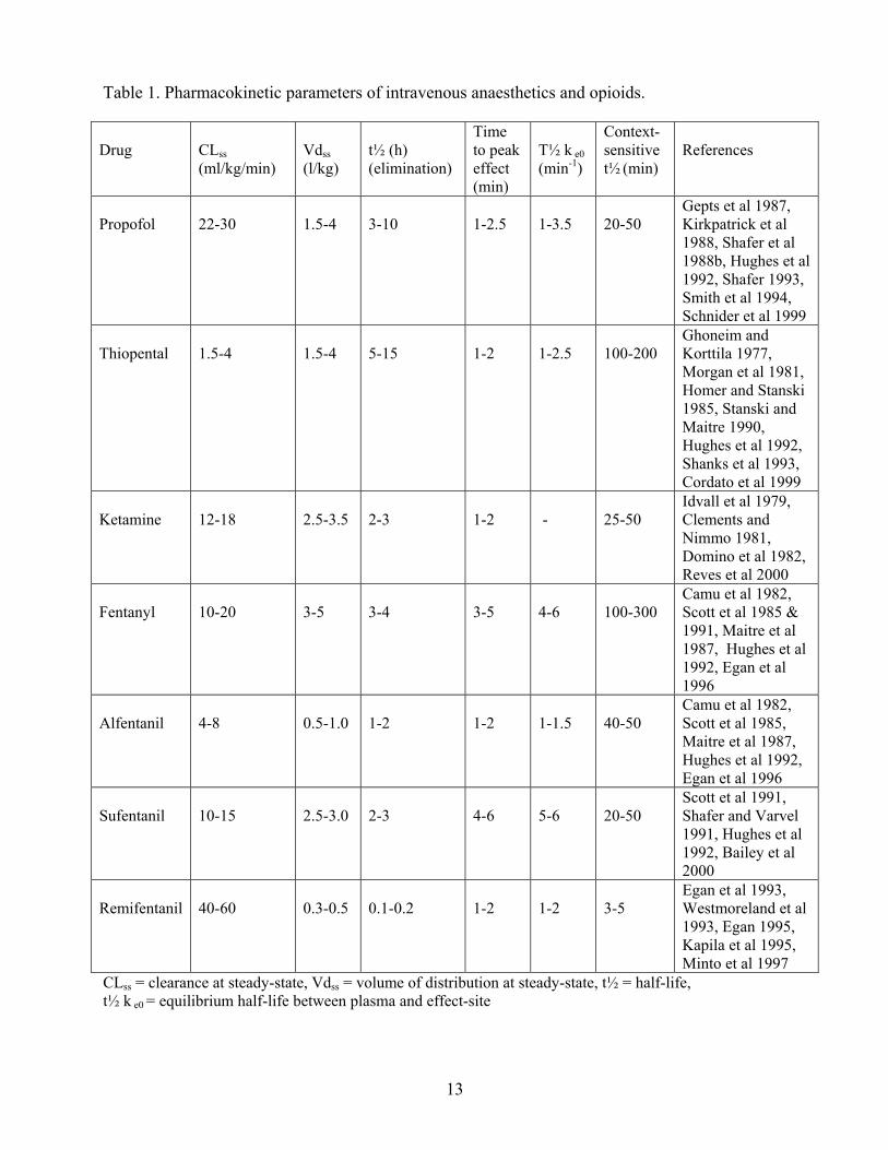


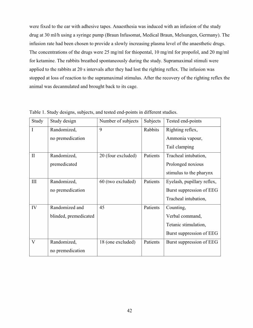
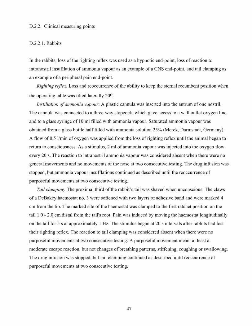

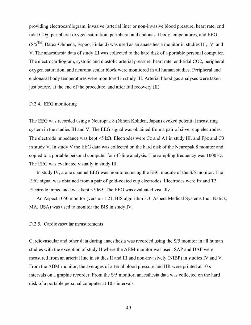
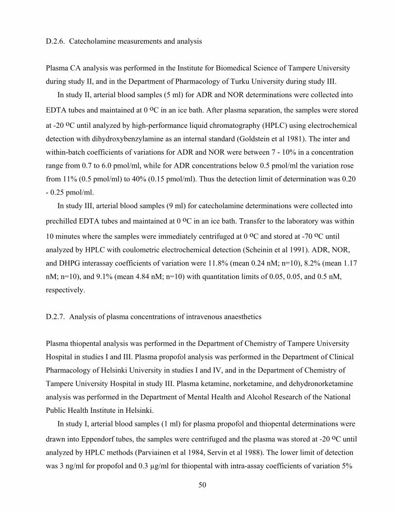
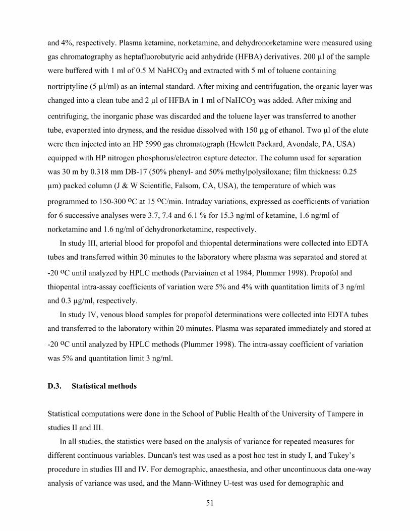
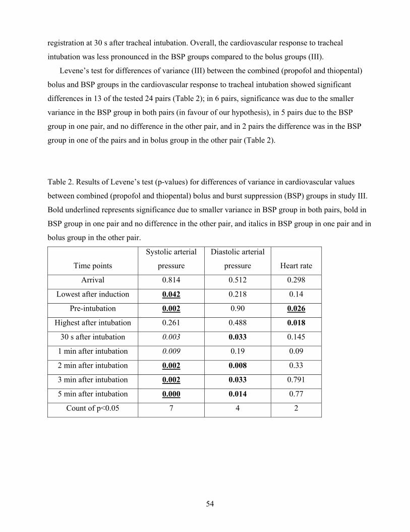
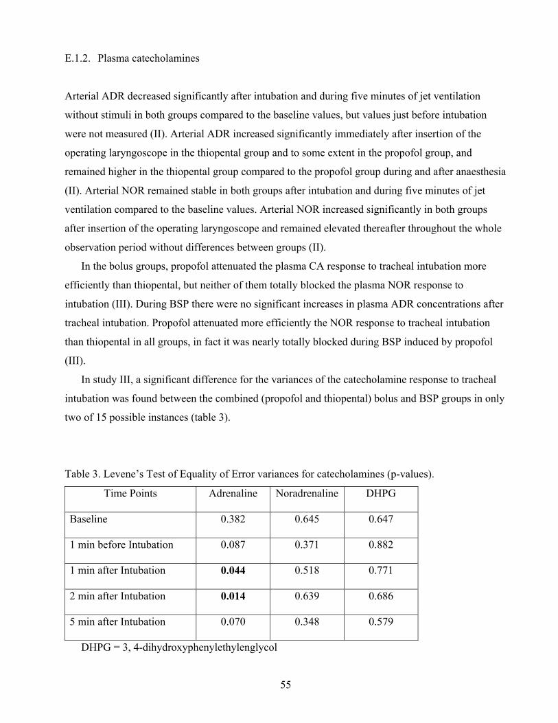

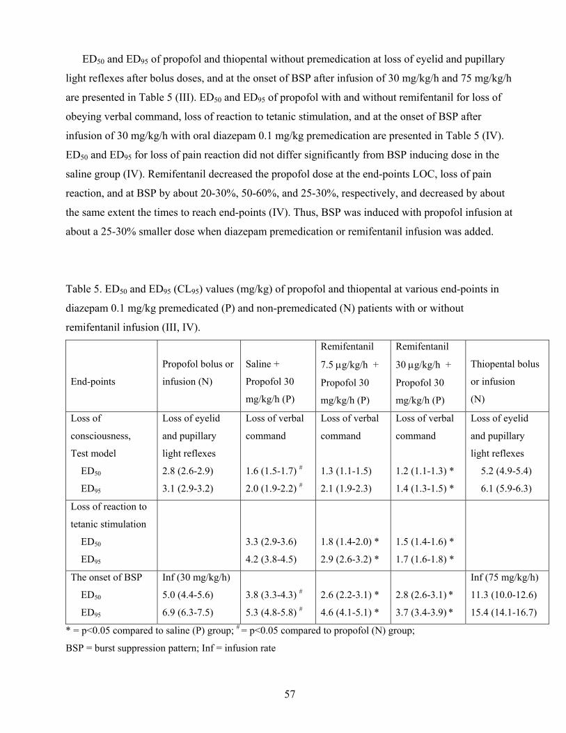
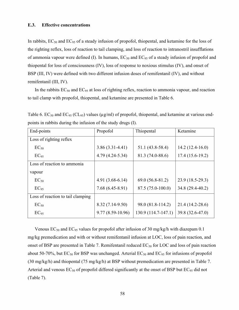
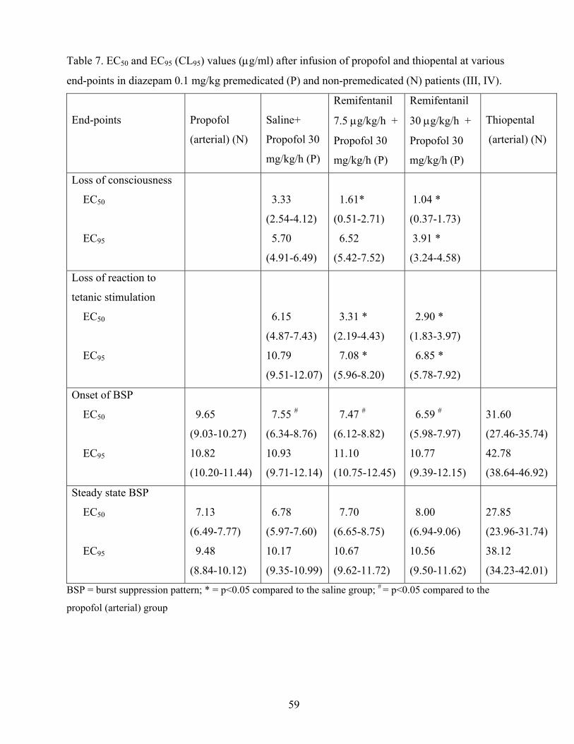
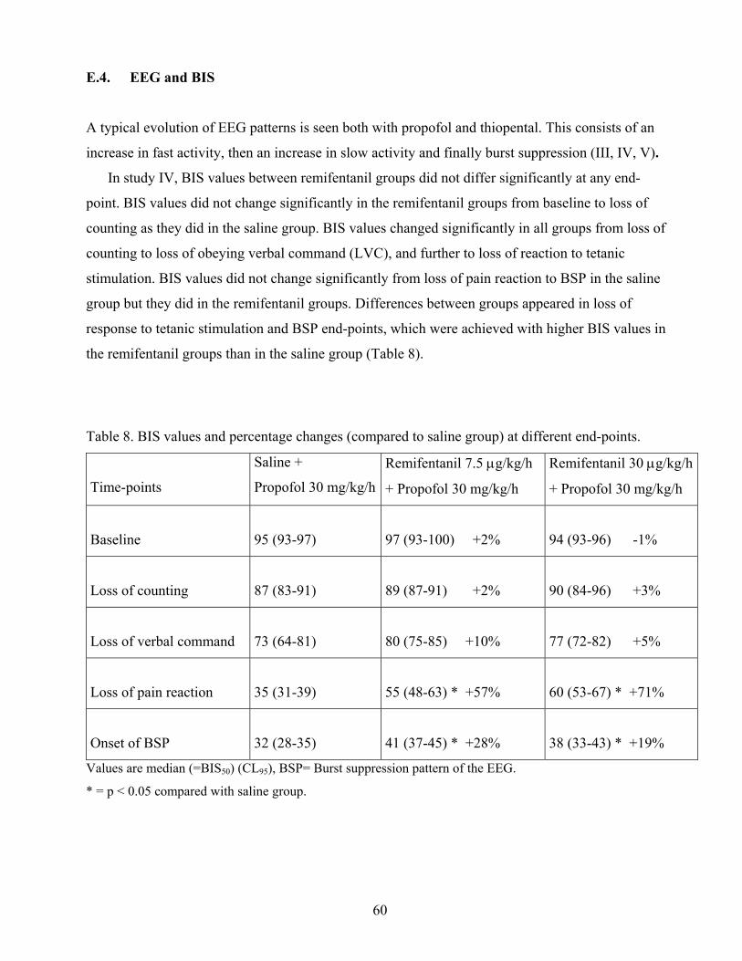
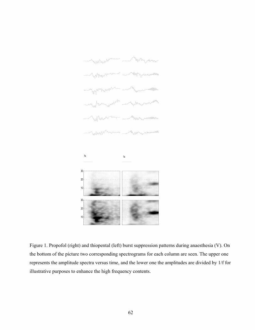


![A bispectral index guided study on the effect of ...€¦ · sedation, hypnosis and analgesia [1]. Drugs with such effects already established in the literature include benzodiazepines](https://static.documents.pub/doc/80x56/5f95b3bdb9b77d7dd15f4af9/a-bispectral-index-guided-study-on-the-effect-of-sedation-hypnosis-and-analgesia.jpg)


